Article

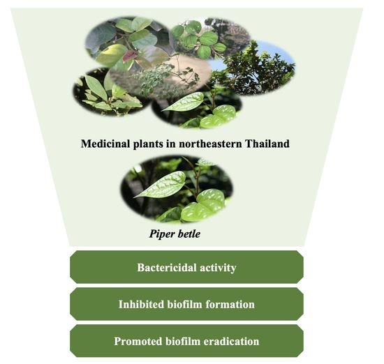
Efficacy of Thai Plant Extracts for Antibacterial and Anti-Biofilm Activities against Pathogenic Bacteria

1
Faculty of Medical Technology, Prince of Songkla University, Hat Yai, Songkhla 90110, Thailand
2
Biology Program, Faculty of Science and Technology, Nakhon Ratchasima Rajabhat University, Nakhon Ratchasima 30000, Thailand
*
Author to whom correspondence should be addressed.
Antibiotics 2021, 10(12), 1470; https://doi.org/10.3390/antibiotics10121470
Submission received: 31 October 2021 / Revised: 24 November 2021 / Accepted: 28 November 2021 / Published: 30 November 2021
(This article belongs to the Special Issue Antimicrobial Activity of Extracts from Plant)

Abstract

:
The emergence of drug-resistant bacteria has impacted the outcome of current therapeutics as a threat to global healthcare; novel medicines are urgently needed. Thirteen medicinal plants were collected in Northeastern Thailand, and their crude ethanolic extracts were evaluated for antibacterial activities against Staphylococcus aureus ATCC25923 and Escherichia coli ATCC25922 using the broth micro-dilution method. Piper betle leaf ethanolic extract showed optimal activity against both representative bacterial strains. Activity was also observed against clinical isolates of methicillin-resistant S. aureus (MRSA) and E. coli, with minimal inhibitory concentration (MIC) ranging from 0.31 mg/mL to 2.5 mg/mL and minimal bactericidal concentration (MBC) ranging from 0.62 mg/mL to 2.5 mg/mL. A time-kill study revealed that the extract activity was time- and dose-dependent, and also bactericidal on the tested bacteria. P. betle extract inhibited biofilm formation and promoted biofilm eradication in both S. aureus and E. coli. 4-Allyl-1,2-diacetoxybenzene and eugenol were identified as the most abundant compounds in the extract and may play major roles in the anti-bacterial and anti-biofilm activity. Results suggest that ethanolic P. betle leaf extract shows promise as an alternative method for the prevention of bacterial diseases.

Graphical Abstract

1. Introduction

Drug-resistant bacteria are now a serious global healthcare problem, leading to poor outcomes of current therapeutics. Biofilm composed of polysaccharides, proteins, nucleic acids, lipids and other organic components is considered an essential bacterial virulence factor. Microorganisms generate biofilms and form a community on the host surface to protect themselves from the environment. Biofilms are mostly found in medical, industrial, food processing and water distribution systems [1,2]. Biofilm-embedded bacteria are able to increase their defenses against antibiotics at up to a thousand-fold, compared to planktonic cells [3]. Most current antibiotics are effective against unattached bacteria, but the treatment of biofilm infections requires antibiotics at high concentrations, usually above peak serum levels. Therefore, therapeutics for latent and recurrent infections are less effective [4]. The elimination of bacteria within the biofilm requires degradation of the biofilm matrix or the discovery of antibiotics that can enter through the biofilm.
Currently, natural products have attracted widespread interest in the search for alternative medicines [5,6,7,8,9]. Plants are excellent sources of antimicrobials that have minimal treatment side effects [5]. Thai herbal medicines have been used successfully to treat many bacterial infections [6,7,8]. The antibacterial activity of Thai plants is well documented but research concerning their anti-biofilm activity is sparse. Previous studies screened the antibacterial and anti-biofilm activity of Thai medicinal plant extracts against oral pathogens [9]. A source of antibacterial and anti-biofilm agents was determined to be 4-chromanol in an ethanolic extract of Piper betle leaves [9], illustrating the potential application of natural products in the therapeutic prevention of diseases associated with oral biofilms. However, investigation of the efficacy of Thai herbs in combatting foodborne and nosocomial pathogens, such as Staphylococcus aureus and Escherichia coli, has not been widely conducted.
Here, the antibacterial and anti-biofilm activities of selected indigenous Thai plants, commonly used for bacterial infection against S. aureus and E. coli, were evaluated.

2. Results

2.1. Antibacterial Activity of Plant Extracts

Thirteen ethanolic extracts from selected plants were tested for antibacterial activity against Gram-positive and Gram-negative bacteria by the broth microdilution method. The primary results showed that P. betle leaf extract inhibited the reference bacteria (Table 1), and this leaf extract was selected as a representative agent to evaluate antibacterial activity against 91 clinical bacterial isolates. The results in Table 2 show the MICs of P. betle extract against Gram-positive and Gram-negative bacteria. The MIC and MBC values of the extract for hospital isolates of MRSA ranged from 0.31 mg/mL to 2.5 mg/mL and from 0.31 mg/mL to 5.0 mg/mL, respectively, while the MIC and MBC values of the extract against clinical isolates of E. coli were 1.25 mg/mL to 2.5 mg/mL and 2.5 mg/mL to 5.0 mg/mL, respectively.

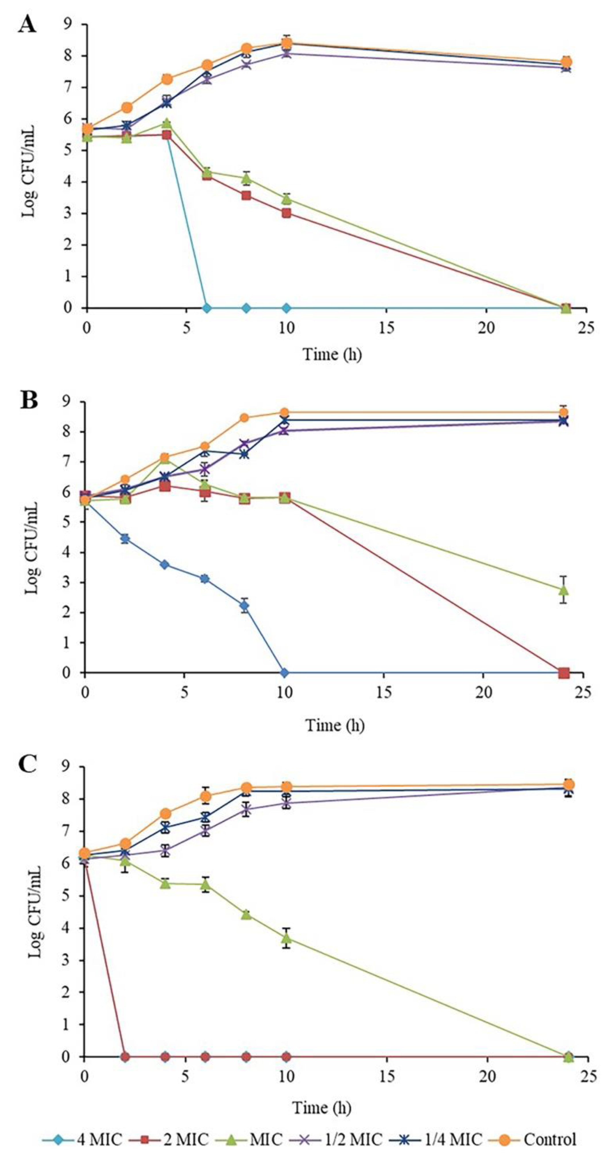
2.2. Time-Kill Kinetic Assay of Piper betle Extract against the Bacteria

A time-kill kinetic assay of P. betle extract against the reference strains, S. aureus ATCC29213, MRSA NPRC001R and E. coli ATCC25922, was investigated, with the results demonstrated in Figure 1A–C, respectively. Patterns of cell survival and cell decrease after treatment with different concentrations of the extract were similar among different bacteria. A time-kill study revealed that the extract activity was time- and dose-dependent, while the extract also exhibited a bactericidal effect on the tested bacteria. At 2MIC and 4MIC, the extract reduced bacterial cells at the starting log CFU/mL by more than 3 logs within 24 h, with E. coli showing a decrease within 2 h. At MIC, the extract reduced bacterial cells within 24 h, except for MRSA, which decreased by more than 2 logs. These results were consistent with antibacterial activity.

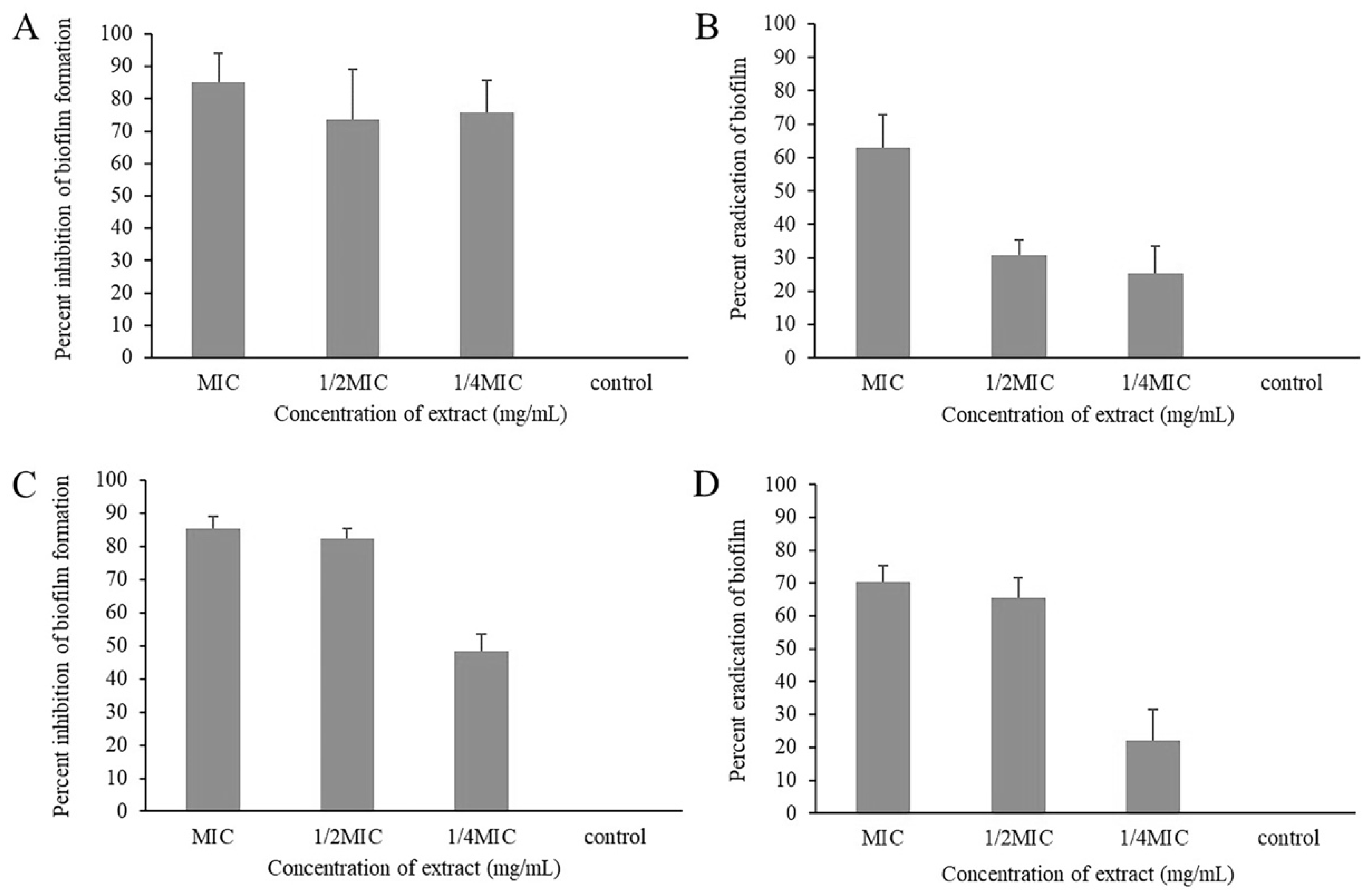
2.3. Biofilm Inhibitory and Eradicating Activity of Piper betle Extract

The effects of P. betle extract on the inhibition and eradication of biofilm formation against S. aureus ATCC29213 and E. coli ATCC25922 are demonstrated in Figure 2. Sub-inhibitory concentration (1/4MIC and 1/2MIC) and MIC of P. betle showed significant biofilm inhibiting activity against tested strains (p < 0.05) (Figure 2A,C). The inhibition ranged 70–85% with S. aureus and 50–85% with E. coli. The biofilm eradicating activity of the ethanolic extract is shown in Figure 2B,D. The biofilm removal capacity of the extract at 1/4MIC, 1/2MIC and MIC was 25%, 30%, and 60% for S. aureus and 20%, 65%, and 70% for E. coli.

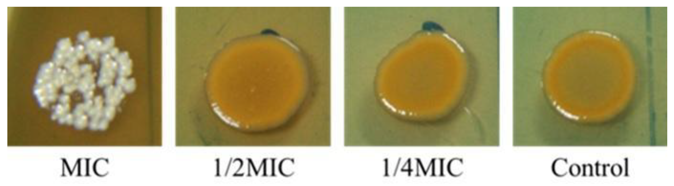
2.4. Inhibition of Staphyloxanthin Biosynthesis in Staphylococcus aureus by Piper betle

The effect of P. betle extract on staphyloxanthin biosynthesis in colonies of S. aureus was evaluated. The extract exhibited complete inhibition of staphyloxanthin biosynthesis of the bacterial colonies at MIC value (Figure 3).

2.5. Phytochemical Analysis of Piper betle Leaf Extract

Thirty phytochemical components were identified from the ethanolic extract of P. betle leaves by GC and GC-MS analyses. Major constituents calculated from peak areas showed that 4-allyl-1,2-diacetoxybenzene (70.32%) and eugenol (18.80%) were recorded as the highest two amounts (Table 3).

3. Discussion

The emergence of drug-resistant bacteria has impacted the outcome of current therapeutics as a threat to global healthcare.Therefore, searching for an alternative treatment to solve this problem has attracted increasing interest. Over thousands of years, 80% of the global population has used traditional herbal medicines to treat various diseases [10,11]. Plants are a valuable source of medicines. Many demonstrate antibacterial efficacy for the treatment of bacterial infections. In this study, the antimicrobial and anti-biofilm activities of 13 plant extracts were tested against bacteria. All plants were obtained from Nakhon Ratchasima Province in Northeastern Thailand. Ethanol was used for plant extraction to obtain a high number of active agents with no toxicity [12]. S. aureus, MRSA and E. coli were selected as representatives of Gram-positive bacteria, antibiotic-resistant bacteria and Gram-negative bacteria, respectively.
Differences in the MIC and MBC values of the crude ethanolic extracts demonstrated selective antibacterial activity. Four of the thirteen extracts, including Boesenbergia rotunda, Caseria grewiifolia, P. betle and Rhodomyrtus tomentosa, showed antibacterial activity against S. aureus and MRSA, with MIC values less than 1 mg/mL. These were considered active crude extracts [13]. The MIC index (MIC/MBC) for all extracts was less than four, indicating bactericidal effects on S. aureus and MRSA [14]. Moreover, the lowest MIC value was recorded on a standard strain of E. coli, while Gram-negative bacteria were found in P. betle. The antimicrobial effects were confirmed in more isolates with MIC ranges of less than 1 mg/mL. P. betle presented bactericidal potential with a broad spectrum. Time-kill determination demonstrated that the antibacterial efficacy of the extract on Gram-negative bacteria was faster than for Gram-positive bacteria. Furthermore, P. betle possessed strong anti-biofilm activity with dual actions of preventing and eradicating the biofilm of S. aureus and E. coli. In addition, P. betle also inhibited staphyloxanthin, the golden pigment of S. aureus. Although the biofilm cells produced staphyloxanthin at higher levels than the planktonic cells [15], the extract obviously inhibited pigment production of biofilms, possibly resulting from the loss of the biofilm-forming ability in the bacteria.
An earlier study recorded that the ethanolic leaf extract of P. betle mainly contained 4-chromanol with antibacterial and anti-biofilm activity against oral pathogens [9]. By contrast, here, 4-allyl-1,2-diacetoxybenzene was identified as the most dominant compound. Despite using the same solvent to extract bioactive compounds from the plant, the varying extraction methods and conditions provided different chemical compounds [16]. 4-Allyl-1,2-diacetoxybenzene might play a major role in significant antibacterial activity. This idea concurred with previous reports indicating that essential oils of Melaleuca species rich in 4-allyl-1,2-diacetoxybenzene exhibited bacteriostatic and bactericidal effects against foodborne pathogens [17,18]. In this study, eugenol was the second most abundant compound in P. betle leaf extract. Eugenol was detected as a major component in essential oils from Etlingera sayapensis, which was characterized as being highly effective against a variety of Gram-positive and Gram-negative bacteria [19]. Correspondingly, the remarkable antibacterial activity of clove and cinnamon essential oil were attributed to the eugenol content [20,21]. Moreover, typical biofilm and cell membrane structures were shown to be disturbed by eugenol [22].

4. Materials and Methods

4.1. Plant Materials and Extract Preparation

Thirteen plant species were sourced from Nakhon Ratchasima Province, Thailand with details shown in Table 1. Each plant material was dried at 60 °C for 3–5 days, powdered and extracted by soaking in 95% ethanol for one week and then filtrated through Whatman No. 4 filter paper. Each filtrate was evaporated under reduced pressure in a rotary evaporator until complete dryness and kept at 4 °C until required for antibacterial testing.

4.2. Bacterial Strains and Growth Conditions

S. aureus ATCC29213, MRSA NPRC001R and E. coli ATCC25922 were obtained from the Division of Biological Science and Natural Products Research Center, Faculty of Science, Prince of Songkla University. In total, 48 isolates of MRSA were obtained from the Songklanagarind Hospital, and 43 isolates of E. coli were received from Maharat Nakhon Ratchasima Hospital as clinical specimens. The bacteria were maintained on tryptic soy agar (TSA) and strains were grown under aerobic condition (37 °C, 16–18 h).

4.3. Evaluation of Antibacterial Activity

4.3.1. Determination of Minimum Inhibitory Concentrations (MICs)

The broth microdilution method was carried out according to the CLSI guideline [23]. The extracts were serially diluted two-fold in Mueller Hinton broth (MHB) in 96-well microtiter plates to obtain final concentrations ranging from 0.15 mg/mL to 10 mg/mL. Then, an equal volume of 100 µL of log-phase bacterial culture, approximately 106 CFU/mL, was added to each well. After incubation at 37 °C for 16–18 h, bacterial growth was observed by the turbidity of the medium. The MIC was considered the lowest concentration of the extracts or antibiotics required to inhibit bacterial growth without turbidity of the medium compared with the negative control. All assays were performed in triplicate.

4.3.2. Determination of Minimum Bactericidal Concentrations (MBCs)

All wells showing bacteria inhibited in the broth microdilution method were plated on Mueller Hinton agar (MHA) with 10 µL aliquots of the contents and grown at 37 °C for 16–18 h. The MBC was recorded as the lowest concentration of the extracts or antibiotics that killed 99.9% of bacteria, showing no growth on MHA. All assays were performed in triplicate.

4.3.3. Time-Kill Assay

The primary results of the plant extracts revealed that P. betle leaf extract showed the strongest antibacterial activity on S. aureus and E. coli. The antibacterial activities of P. betle leaf extract on S. aureus, MRSA and E. coli were studied using a time-kill assay. A bacterial culture (5·105 CFU/mL) was added to MHB containing the extract at 4MIC, 2MIC, MIC 1/2MIC and 1/4MIC, and untreated cultures were incubated at 37 °C. The samples were collected at 0, 2, 4, 6, 8, 10, 12 and 24 h by culturing on TSA plates. A control incubation was performed with 1% DMSO. Surviving colony bacteria were counted, and log10 CFU/mL was calculated. A time-kill curve was analyzed by plotting log CFU/mL against time.

4.4. Evaluation of Anti-Biofilm Activity

4.4.1. Biofilm Formation

The effect of P. betle extract on the biofilm formation of each representative strain of S. aureus and E. coli was examined, using the modified microdilution method [24]. Briefly, each bacterial strain was grown in tryptic soy broth (TSB) containing 1% glucose overnight. The culture was diluted to 106 CFU/mL and transferred to a 96-well microtiter plate containing two-fold serial dilutions of P. betle extract at 4MIC, 2MIC, MIC, 1/2MIC and 1/4MIC. After incubation at 37 °C for 24 h, each well of microtiter plate was washed twice with phosphate buffer saline (PBS), fixed by absolute methanol for 20 min, and dried overnight. After that, the wells were stained with 200 µL of 2% crystal violet solution for 15 min. Subsequently, the plate was washed with water and air-dried. Stained biofilms were dissolved in 200 µL of 33% (v/v) glacial acetic acid, and the plate was measured at 570 nm using a microplate reader (BioTek, Winooski, VT, USA). The relative percentage of biofilm formation was defined as (mean 570 nm of treated well/mean 570 nm of control well) × 100.

4.4.2. Biofilm Eradication

Established biofilms were cultured as defined by Saising et al. [25]. Two hundred microliters of growing culture (106 CFU/mL) were transferred to a 96-well microtiter plate and incubated at 37 °C for 24 h. Then, planktonic cells were removed and 100 µL of TSB was added. After that, P. betle extract at different concentrations was added. After incubation at 37 °C for 24 h, each well of the microtiter plate was washed with PBS twice and air-dried. The plate was then used for biofilm formation.

4.4.3. Production of Staphyloxanthin

The ability of P. betle extract to reduce the production of the golden yellow pigment staphyloxanthin was investigated. An overnight culture of the tested strains in TSB was prepared. The bacterial suspensions were diluted 1:100 in a TSA plate containing MIC, 1/2MIC and 1/4MIC of P. betle extract. A control plate without the extract was prepared in the same way. The cultural plates were incubated for 24 h at 37 °C. The pigment colonies were photographed, and the golden yellow pigment was compared in treated and untreated samples.

4.5. Bioactive Compound Assay

Gas chromatography-mass spectrometry (GC-MS) analysis of ethanolic extracts of P. betle leaves was conducted at the Center for Scientific and Technological Equipment, Suranaree University of Technology, Thailand. GC-MS was performed by a Bruker series 3XO model with an electron ionization detector (Karlsruhe, Germany) operated through a data system. One microliter of extract (50 mg/mL) was added to the injection port of the GC column. The identification of individual compounds was made by mass spectra library matching against the database of National Institute Standard and Technology (NIST MS 14.0). The matching result for a compound was represented by percent of probability values (%Prob). Relative percentages of the chemical compositions were calculated by considering the summation of all GC peak areas in the total ion chromatogram of one sample as 100%. Each peak percentage area (%Area) was obtained by dividing its area by the total area of all compounds.

4.6. Statistical Analysis

Data were expressed as the mean and standard deviation (SD) by computational analysis from the three experiments, with duplicate or triplicate independent experiments.

5. Conclusions

P. betle has high antibacterial and anti-biofilm potential as a promising anti-infective phytotherapeutical. This natural agent presents positive options which are appropriate alternative approaches to control pathogenic bacteria. The ability of P. betle extract to prevent S. aureus and E. coli, especially in the case of biofilms, may be beneficial for the food industry and medical devices where these pathogens are generally found.

Author Contributions

Conceptualization, D.S. and M.V.; methodology, D.S. and M.V.; validation, D.S. and M.V.; formal analysis, D.S. and M.V.; investigation, D.S. and M.V.; resources, D.S. and M.V.; data curation, D.S. and M.V.; writing—original draft preparation, D.S. and M.V.; writing—review and editing, D.S. and M.V.; visualization, D.S. and M.V.; supervision, D.S. and M.V.; project administration, D.S. and M.V.; funding acquisition, D.S. and M.V. All authors have read and agreed to the published version of the manuscript.

Funding

This research study was supported by National Science, Research and Innovation Fund (NSRF), fiscal year 2022 (Project No. 2493736), Nakhon Ratchasima Rajabhat University and Faculty of Medical Technology, Prince of Songkla University.

Institutional Review Board Statement

Not applicable.

Informed Consent Statement

Not applicable.

Data Availability Statement

Not applicable.

Conflicts of Interest

The authors declare no conflict of interest.

References

  1. Abebe, G.M. The Role of Bacterial Biofilm in Antibiotic Resistance and Food Contamination. Int. J. Microbiol. 2020, 2020, 1705814. [Google Scholar] [CrossRef]
  2. Reuben, R.; Roy, P.; Sarkar, S.; Ha, S.-D.; Jahid, I.K. Multispecies interactions in biofilms and implications to safety of drinking water distribution system. Microbiol. Biotechnol. Lett. 2019, 47, 473–486. [Google Scholar] [CrossRef]
  3. Olsen, I. Biofilm-specific antibiotic tolerance and resistance. Eur. J. Clin. Microbiol. Infect. Dis. 2015, 34, 877–886. [Google Scholar] [CrossRef]
  4. Kim, N.-H.; Kang, Y.M.; Han, W.D.; Park, K.U.; Park, K.-H.; Yoo, J.I.; Lee, D.-G.; Park, C.; Song, K.-H.; Kim, E.S.; et al. Small-colony variants in persistent and recurrent Staphylococcus aureus bacteremia. Microb. Drug Resist. 2016, 22, 538–544. [Google Scholar] [CrossRef]
  5. Barbieri, R.; Coppo, E.; Marchese, A.; Daglia, M.; Sobarzo-Sánchez, E.; Nabavi, S.F.; Nabavi, S.M. Phytochemicals for human disease: An update on plant-derived compounds antibacterial activity. Microbiol. Res. 2017, 196, 44–68. [Google Scholar] [CrossRef] [PubMed]
  6. Jarriyawattanachaikul, W.; Chaveerach, P.; Chokesajjawatee, N. Antimicrobial activity of Thai-herbal plants against food-borne pathogens E. coli, S. aureus and C. jejuni. Agric. Agric. Sci. Procedia 2016, 11, 20–24. [Google Scholar] [CrossRef]
  7. Siriwatanametanon, N.; Dodgson, W.; Dodgson, J. Investigation of antimicrobial activity of 13 Thai medicinal plants against bacteria and fungi. J. Pure Appl. Microbiol. 2017, 11, 1351–1356. [Google Scholar] [CrossRef]
  8. Parichat, P.; Kanit, V.; Jaehong, H.; Sukanda, V. High antioxidant and phenolic contents related to antibacterial activity against gastrointestinal pathogenic bacteria of some Thai medicinal plants. Pharmacogn. J. 2018, 10, 341–348. [Google Scholar]
  9. Teanpaisan, R.; Kawsud, P.; Pahumunto, N.; Puripattanavong, J. Screening for antibacterial and antibiofilm activity in Thai medicinal plant extracts against oral microorganisms. J. Tradit. Complement. Med. 2017, 7, 172–177. [Google Scholar] [CrossRef] [PubMed] [Green Version]
  10. Beloborodova, N.V.; Bairamov, I.T.; Olenin, A.Y. Microbial biofilms produce higher concentrations of sepsis-associated exometabolites compared with the planktonic phenotype. Glob. J. Pathol. Microbiol. 2017, 5, 15–25. [Google Scholar]
  11. Woo, C.S.J.; Lau, J.S.H.; El-Nezami, H. Chapter 10-herbal medicine: Toxicity and recent trends in assessing their potential toxic effects. Adv. Bot. Res. 2012, 62, 365–384. [Google Scholar]
  12. Huie, C.W. A review of modern sample-preparation techniques for the extraction and analysis of medicinal plants. Anal. Bioanal. Chem. 2002, 373, 23–30. [Google Scholar] [CrossRef] [PubMed]
  13. Ríos, J.L.; Recio, M.C. Medicinal plants and antimicrobial activity. J. Ethnopharmacol. 2005, 100, 80–84. [Google Scholar] [CrossRef]
  14. Radhakrishnan, N.; Gnanamani, A.; Mandal, A.B. A potential antibacterial agent Embelin, a natural benzoquinone extracted from Embelia ribes. Biol. Med. 2011, 3, 1–7. [Google Scholar]
  15. Resch, A.; Rosenstein, R.; Nerz, C.; Götz, F. Differential gene expression profiling of Staphylococcus aureus cultivated under biofilm and planktonic conditions. Appl. Environ. Microbiol. 2005, 71, 2663–2676. [Google Scholar] [CrossRef] [Green Version]
  16. Altemimi, A.; Lakhssassi, N.; Baharlouei, A.; Watson, D.G.; Lightfoot, D.A. Phytochemicals: Extraction, isolation, and identifica-tion of bioactive compounds from plant extracts. Plants 2017, 6, 42. [Google Scholar] [CrossRef] [PubMed]
  17. Siddique, S.; Parveen, Z.; Firdaus, E.B.; Mazhar, S. Chemical composition, antibacterial and antioxidant activities of essential oils from leaves of three Melaleuca species of Pakistani flora. Arab. J. Chem. 2020, 13, 67–74. [Google Scholar] [CrossRef]
  18. Siddique, S.; Parveen, Z.; Firdaus, E.B.; Chaudhary, M.N.; Mazhar, S.; Nawaz, S. The essential oil of Melaleuca armillaris (Sol. ex Gaertn.) Sm. leaves from Pakistan: A potential source of eugenol methyl ether. Ind. Crop. Prod. 2017, 109, 912–917. [Google Scholar] [CrossRef]
  19. Mahdavi, B.; Yaacob, W.A.; Din, L.B. Chemical composition, antioxidant, and antibacterial activity of essential oils from Etlingera sayapensis A.D. Poulsen & Ibrahim. Asian Pac. J. Trop. Med. 2017, 10, 819–826. [Google Scholar] [CrossRef]
  20. Xu, J.-G.; Liu, T.; Hu, Q.-P.; Cao, X.-M. Chemical composition, antibacterial properties and mechanism of action of essential oil from clove buds against Staphylococcus aureus. Molecules 2016, 21, 1194. [Google Scholar] [CrossRef]
  21. Božik, M.; Nový, P.; Klouček, P. Susceptibility of postharvest pathogens to esential oils. Sci. Agric. Bohem. 2017, 48, 103–111. [Google Scholar] [CrossRef] [Green Version]
  22. Olszewska, M.A.; Gędas, A.; Simões, M. The effects of eugenol, trans-cinnamaldehyde, citronellol, and terpineol on Escherichia coli biofilm control as assessed by culture-dependent and -independent methods. Molecules 2020, 25, 2641. [Google Scholar] [CrossRef]
  23. CLSI. M100 Performance Standards for Susceptibility Testing; Clinical and Laboratory Standards Institute: Wayne, PA, USA, 2018. [Google Scholar]
  24. Jantorn, P.; Heemmamad, H.; Soimala, T.; Indoung, S.; Saising, J.; Chokpaisarn, J.; Wanna, W.; Tipmanee, V.; Saeloh, D. Antibiotic resistance profile and biofilm production of Staphylococcus pseudintermedius isolated from dogs in Thailand. Pharmaceuticals 2021, 14, 592. [Google Scholar] [CrossRef] [PubMed]
  25. Saising, J.; Ongsakul, M.; Voravuthikunchai, S.P. Rhodomyrtus tomentosa (Aiton) Hassk. ethanol extract and rhodomyrtone: A potential strategy for the treatment of biofilm-forming staphylococci. J. Med. Microbiol. 2011, 60, 1793–1800. [Google Scholar] [CrossRef] [PubMed] [Green Version]
Figure 1. Time-kill determination of (A) Staphylococcus aureus ATCC29213, (B) methicillin-resistant S. aureus NPRC001R and (C) Escherichia coli ATCC25922 after treatment with Piper betle leaf ethanolic extract.
Antibiotics 10 01470 g001
Figure 2. Inhibition of biofilm formation and biofilm eradication potential of Piper betle leaf ethanolic extract at various concentrations against (A,B) Staphylococcus aureus ATCC29213 and (C,D) Escherichia coli ATCC25922.
Antibiotics 10 01470 g002
Figure 3. Pigmentation of Staphylococcus aureus ATCC29213 grown on TSA with or without Piper betle leaf ethanolic extract supplemented with various concentrations at 37 °C for 24 h.
Antibiotics 10 01470 g003
Table 1. Minimum inhibitory concentration (MIC) and minimum bactericidal concentration (MBC) of selected plant extracts against pathogenic bacteria.
Medicinal PlantCommon NameLocal NamePlant PartSusceptibility Testing (mg/mL)
S. aureus ATCC29213MRSA NPRC001RE. coli ATCC25922
Artocarpus lakoocha Roxb.Green TampangKa noon paLeaf10/1010/1010/>10
Boesenbergia rotunda (Roxb.) Schltr.FingerrootKrachaiRhizome0.15/0.150.15/0.15>10/>10
Casearia grewiifolia Vent.-Kruai PaFruit0.62/0.621.25/2.5>10/>10
Chromolaena odorata (L.) R.M.King & H. Rob.Christmas bushSap suealeaf5/>1010/>10>10/>10
Limnophila aromatica (Lam.) MerrRice paddy herbPhak kha YeangWhole plant2.5/52.5/5>10/>10
Millingtonia hortensis Linn.Cork treeHianLeaf2.5/>2.52.5/2.55/>10
Oroxylum indicum (L.) Kurz.Broken bones plantPhekaLeaf2.5/>2.52.5/>2.5>10/>10
Piper betle Linn.BetelPlueLeaf0.62/0.620.62/0.622.5/2.5
Rhodomyrtus tomentosa (Aiton) Hassk.Rose myrtlePhruatLeaf0.31/0.620.62/0.62>10/>10
Syzygium cumini Linn.Black plumWaaLeaf2.5/52.5/510/>10
Xanthostemon chrysanthus (F.Muell.) Benth.Golden myrtleRak raek popLeaf1.25/2.501.25/2.50>10/>10
Zingiber officinale Roscoe.GingerKhing daengRhizome1.25/>10>10/>1010/>10
Ziziphus mauritiana Lam.JujubePhutsaLeaf2.5/105/>10>10/>10
Table 2. MIC and MBC values of Piper betle ethanolic extract against other isolates of methicillin-resistant Staphylococcus aureus (MRSA) and Escherichia coli.
Number of IsolatesAntibacterial Activities of Piper betle Ethanol Extract
MIC Range (mg/mL)MBC Range (mg/mL)
MRSA (n = 48)0.31–2.50.31–5.0
E. coli (n = 43)1.25–2.51.25–5.0
Table 3. Chemical constituents of Piper betle leaf ethanolic extract.
No.RT (min)CAS RNExtract Constituent%Area%Prob
110.841501-92-84-Allylphenol0.3371.0
215.35897-53-0Eugenol18.8027.4
316.89187-44-5Caryophyllene0.9031.7
418.1626753-98-6Alpha-caryophyllene0.2549.1
519.0861460-97-5Gamma-cadinene1.2129.1
620.37113620-82-14-Allyl-1,2-diacetoxybenzene70.3242.2
720.938483-76-1Delta-cadinene0.7833.6
823.1296750-60-3(-)-Spathulenol0.0629.7
923.3021139-30-6Caryophyllene oxide0.0727.4
1036.594628-97-7Hexadecanoic acid, ethyl ester0.1545.1
1139.109150-86-7Phytol0.7683.5
1239.992544-35-4Linoleic acid ethyl ester0.1520.8
1340.114112-63-0Linoleic acid methyl ester0.2313.9
1441.01210236-16-5Phytol acetate0.7651.4
1542.0573033-62-3Bis(2-(dimethylamino)ethyl) ether0.1639.2
1643.470122-79-2Acetic acid, 3-(adamant-2-ylidene-methoxymethyl)-,phenyl ester0.1057.8
1744.0001778-02-56,16-Dimethyl-20-oxo pregn-5-en-3-yl acetate0.1323.2
1844.403604-09-114-Hydroxypregn-4-ene-3,20-dione0.1722.0
1945.42823470-00-0Glycerol beta-palmitate0.6965.0
2045.575542-44-9Glycerol alpha-palmitate0.0741.0
2146.78555268-70-7Hexadecanoic acid, 2,3-bis(acetyloxy)propyl ester0.2459.8
2247.7853443-82-1Beta-monolinolein0.4227.7
2347.87056797-43-4Cis,cis,cis-7,10,13-hexadecatrienal0.3129.4
2448.97655401-63-39-Octadecenoic acid (Z)-, 2-(acetyloxy)-1-[(acetyloxy)methyl] ethyl ester0.2243.5
2553.7597695-91-2DL-alpha-tocopherol0.2153.7
2655.666474-62-4Campesterol0.3030.2
2756.36483-48-7Stigmasterol0.3543.4
2857.80983-47-6Gamma-sitosterol1.1575.0
2959.6551259-10-5Cycloartenol acetate0.207.3
3061.46579897-80-6Stigmastan-3,5-diene0.5166.0
Publisher’s Note: MDPI stays neutral with regard to jurisdictional claims in published maps and institutional affiliations.

