Article

Genomic Characterization of a Proteus sp. Strain of Animal Origin Co-Carrying blaNDM-1 and lnu(G)

1
School of Basic Medical Sciences, Southwest Medical University, Luzhou 646000, China
2
Immunological Technology Platform, Southwest Medical University, Luzhou 646000, China
3
Pathogen Biology Platform, Southwest Medical University, Luzhou 646000, China
*
Authors to whom correspondence should be addressed.
Antibiotics 2021, 10(11), 1411; https://doi.org/10.3390/antibiotics10111411
Submission received: 14 October 2021 / Revised: 10 November 2021 / Accepted: 14 November 2021 / Published: 18 November 2021
(This article belongs to the Special Issue Bacterial Drug Resistance and Transmission Mechanism)

Abstract

:
The emergence of carbapenem-resistant Proteus represents a serious threat to global public health due to limited antibiotic treatment options. Here, we characterize a Proteus isolate NMG38-2 of swine origin that exhibits extensive drug resistance, including carbapenems. Whole-genome sequencing based on Illumina and MinION platforms showed that NMG38-2 contains 24 acquired antibiotic resistance genes and three plasmids, among which, pNDM_NMG38-2, a pPvSC3-like plasmid, is transferable and co-carries blaNDM-1 and lnu(G). Sequence analysis of pPvSC3-like plasmids showed that they share a conserved backbone but have a diverse accessory module with complex chimera structures bearing abundant resistance genes, which are facilitated by transposons and/or homologous recombination. The acquisition of blaNDM-1 in pNDM_NMG38-2 was due to the ISCR1-mediated integration event. Comprehensive analysis of the lnu(G)-bearing cassettes carried by bacterial plasmids or chromosomes revealed a diversification of its genetic contexts, with Tn6260 and ISPst2 elements being the leading contributors to the dissemination of lnu(G) in Enterococcus and Enterobacteriaceae, respectively. In conclusion, this study provides a better understanding of the genetic features of pPvSC3-like plasmids, which represent a novel plasmid group as a vehicle mediating the dissemination of blaNDM-1 among bacteria species. Moreover, our results highlight the central roles of Tn6260 and ISPst2 in the spread of lnu(G).

1. Introduction

Proteus is a member of Enterobacteriaceae and ranks third in regard to the cause of hospital-acquired infections [1]. Proteus spp. are important opportunistic pathogens that are commonly found in the environment and in the normal gut biota of humans and animals [1]. Proteus species are intrinsically resistant to polymyxin, tigecycline, and nitrofurantoin [2], and the acquisition of additional carbapenem resistance is of particular concern. In particular, strains producing the New Delhi metallo-β-lactamase (NDM), which is able to hydrolyze almost all of the β-lactams (including carbapenems), except monobactam, severely limit therapeutic options for treating infections caused by them [3]. The blaNDM gene, since its initial identification in India in 2009 [4], has spread worldwide. The mobilization of blaNDM is primarily associated with an ISAba125-bounded composite transposon Tn125 [3]. However, a variety of complex genetic contexts of blaNDM have been identified, and mobile genetic elements, such as IS26 [5], IS903 [6], MITE [7], and ISCR1 [8], were found to mobilize blaNDM. The blaNDM gene is largely carried on plasmids with a variety of replicon types [3], which play a vital role in the wide dissemination of blaNDM. In Proteus spp., blaNDM was either integrated into an SXT/R391 integrative and conjugative element [9,10] on bacterial chromosomes, or carried on plasmids with unknown replicon types, such as the recently reported pPrY2001-like plasmids [11,12]. In fact, the genetic characters of blaNDM-harboring plasmids in Proteus remain largely uncharacterized.
Lincosamide antibiotics, including lincomycin, clindamycin and pirlimycin, are potently active against Gram-positive cocci (Staphylococcus, Streptococcus, and Enterococcus) and anaerobic bacteria [13]. Lincomycin is commonly used in veterinary practice, alone or in combination with spectinomycin, and clindamycin is approved to treat infections in human medicine [14]. Lincosamides prevent protein synthesis by inhibiting the peptidyl transferase, by mainly binding to the key nucleotide A2058 of 23S rRNA in the 50S subunit of the bacterial ribosome [15]. Two major mechanisms are responsible for resistance to lincosamides: (i) methylation of 23S rRNA, a common mode of action for resistance to macrolides, lincosamides, and streptogramin B (MLS phenotype) [16]; (ii) enzymatic inactivation of lincosamides due to nucleotidylation by O-nucleotidyltransferase, conferring resistance to lincosamides only (L phenotype) [17]. Since the first report of lincosamide nucleotidyltransferase encoded by lnu(A) (formerly linA) [17], several lnu genes, including lnu(B), lnu(C), lnu(D), lnu(E), lnu(F), lnu(G), and lnu(P), were subsequently identified in animal and human isolates [14,18,19,20,21]. Among them, lnu(G) was first identified in a swine Enterococcus faecalis isolate WT E531 in 2016 in China, and it was embedded in an active Tn6260 [14]. In silico analysis using the GenBank database indicated that lnu(G) was presented in a number of isolates recovered worldwide. To reveal the dissemination patterns of lnu(G), a systematic analysis of genetic contexts of lnu(G) in different plasmids and chromosomes is required.
The present study was conducted to characterize a carbapenem-resistant Proteus sp. isolate of animal origin. This strain harbors a blaNDM-1 gene that is located on a pPvSC3-like plasmid with an unknown replicon type [22]. We elucidate the complete sequence of this blaNDM-1-bearing plasmid and compare it with two other similar plasmids to obtain insights into the mechanisms mediating the mobilization of blaNDM-1 and the diversification of these plasmids. In addition, we analyze the genetic features of lnu(G) systematically to gain a deeper understanding of the complex and diversified dissemination patterns of this important resistance gene.

2. Results and Discussion

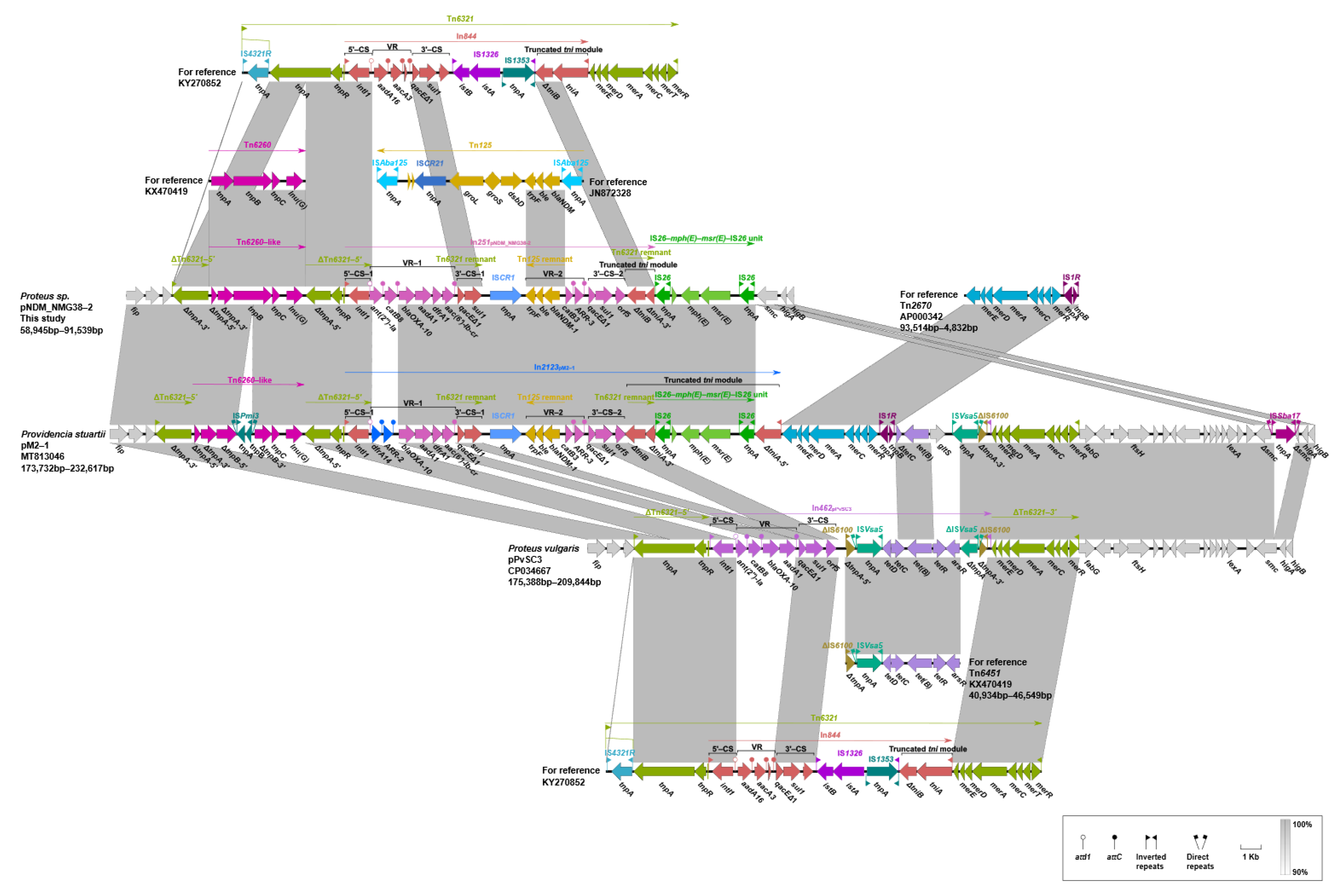
2.1. Genetic Characterization of the blaNDM-1-Harboring Proteus sp. NMG38-2

A carbapenem-resistant strain, named NMG38-2, was recovered from one of the 40 fecal samples of swine. Whole-genome analysis revealed that NMG38-2 contains a single circular chromosome of 4,433,346 bp, with an average GC content of 37.62%. It also carries three circular plasmids, designated as pNDM_NMG38-2, p1_NMG38-2, and p2_NMG38-2. The species identification was performed by genome-to-genome sequence comparison of NMG38-2 and the reference strains of known Proteus species. As a result, the average nucleotide identity (ANI) values between NMG38-2 and known Proteus species were all ≤91.23%, which is below the recommended 95% cut-off for species circumscription. Consistent with ANI data, the digital DNA-DNA hybridization (dDDH) identities between the genome of NMG38-2 and those of reference Proteus species were ≤44.7%, below the suggested 70% cut-off to define a species. Both ANI and dDDH values indicated that the NMG38-2 belongs to a novel species that is distinct from all hitherto known Proteus species. ANI and dDDH analysis also revealed that NMG38-2 showed a higher identity (ANI value: 91.23%, dDDH value: 44.7%) with the Proteus vulgaris strain ATCC 49132 (GenBank accession no. NZ_KN150745.1) than with the other known Proteus species.
While being intrinsically resistant to polymyxin and tigecycline, Proteus sp. NMG38-2 was extensively drug resistant [23], exhibiting resistance to all tested antimicrobials except ciprofloxacin (Table 1). Consistent with its multidrug resistance profile, NMG38-2 contained 24 intact acquired antibiotic resistance genes (ARGs), conferring resistance to aminoglycosides (ant(2″)-Ia, aph(6)-Id, aac(6′)-Ib3, aadA1, aph(3″)-Ib, and aac(6′)-Ib-cr), rifampicin (ARR-3), sulfonamides (sul1 and sul2), trimethoprim (dfrA1), quinolones (aac(6′)-Ib-cr and qnrD1), phenicol (catB8, catB3, catB2, and floR), β-lactams (blaOXA-10 and blaNDM-1), macrolide (mph(E) and msr(E)), and lincosamide (lnu(G)) (Table 2). These ARGs were mainly located on the plasmid pNDM_NMG38-2, except that the chromosome harbored an aadA1, dfrA1, and catB2, and the qnrD1 was carried by the small plasmid p2_NMG38-2. We proposed that the extensive use of drugs might contribute to the emergence of the extensively drug resistant strain, for the reason that multiple antibiotics, including amoxicillin, ceftiofur, sulfamethazine sodium, gentamycin sulfate, and so on, were used within one month prior to sample collection. The abusive use of antimicrobials in animal husbandry, swine industry in this case, is believed to be an important driving force for the emergence and transmission of ARGs in bacteria of animal origin [24], which may further act as the potential reservoir of ARGs for clinical pathogens resistant to multiple antibiotics, including carbapenems.
The blaNDM-1-bearing plasmid pNDM_NMG38-2 is 262,556 bp in size with an average G+C content of 46.65%, and encodes 369 open reading frames (ORFs, Table 2). This plasmid could not be assigned to any known incompatibility group. BLAST analysis of pNDM_NMG38-2 against the GenBank database revealed that it showed the highest 98% query coverage and 99.99% identity to pM2-1 (GenBank accession no. MT813046), a blaNDM-1-harboring plasmid in a Providencia stuartii strain isolated from mouse feces from a commercial goose farm in China [22] (Table S1). It also showed 99.98% identity with 92% query coverage to a blaNDM-1-negative plasmid pPvSC3 (accession no. CP034667) in a P. vulgaris isolate from a chicken in China [25] (Table S1). No other similar plasmid was found in the GenBank database. Using the nucleotide sequence of the replication gene repA of pNDM_NMG38-2 as a query, matches with 100% query coverage and at least 99% identity were pM2-1 and pPvSC3. Thus, the three plasmids, pM2-1, pPvSC3, and pNDM_NMG38-2 were assigned into the same unknown incompatibility group. pPvSC3 is the earliest reported plasmid and considered to be the reference plasmid, so these three plasmids are called pPvSC3-like plasmids. Notably, the pPvSC3-like plasmids were all recovered from isolates of animal origins from different regions of China, highlighting that this novel plasmid group may serve as an epidemic vehicle in mediating the dissemination of blaNDM-1 in animals in China. Continuous surveillance on the spread of pPvSC3-like plasmids in humans and the environment in China is urgently required.
p1_NMG38-2 is a 5500 bp plasmid with a mean G+C content of 34.11% and contains 10 predicted ORFs (Table 2). It did not belong to any known incompatibility group. BLAST analysis revealed that p1_NMG38-2 was partial homology to four plasmids, pZF2-7kb (CP047342, Proteus terrae, swine, China), pZF2-7kb (CP045010, P. terrae, swine, China), pLC-693_2 (CP063316, P. vulgaris, patient, Switzerland), and pOXA181-15-1091(CP012904, Providencia rettgeri, patient, Canada), with only 49–67% coverage and 90.67–92.53% nucleotide identity, indicating that p1_NMG38-2 was a novel plasmid. This plasmid encodes a replication protein RepB belonging to the Rep_3 type family, a DNA-binding protein and a putative toxin-antitoxin system RelE/RelB for plasmid maintenance, as well as six hypothetical proteins. p2_NMG38-2 is 2683 bp in length with a 41.78 GC content (Table 2). It harbors two predicted ORFs, including a resistance gene qnrD1, and belongs to the Col3M incompatibility group. p2_ NMG38-2 shared high similarity (100% coverage, >99.9% identity) to a number of plasmids in the GenBank database, such as pMP63B in Morganella morganii (CP048808, wastewater, China), plasmid unnamed2 in Proteus mirabilis (CP049755, patient, Brazil), pRHB14-C12_2 in Citrobacter freundii (CP057825, swine, United Kingdom), and pAB213 in P. rettgeri (MH085193, vegetable, Brazil), suggesting a wide spread of p2_NMG38-2-like plasmids in various niches worldwide.

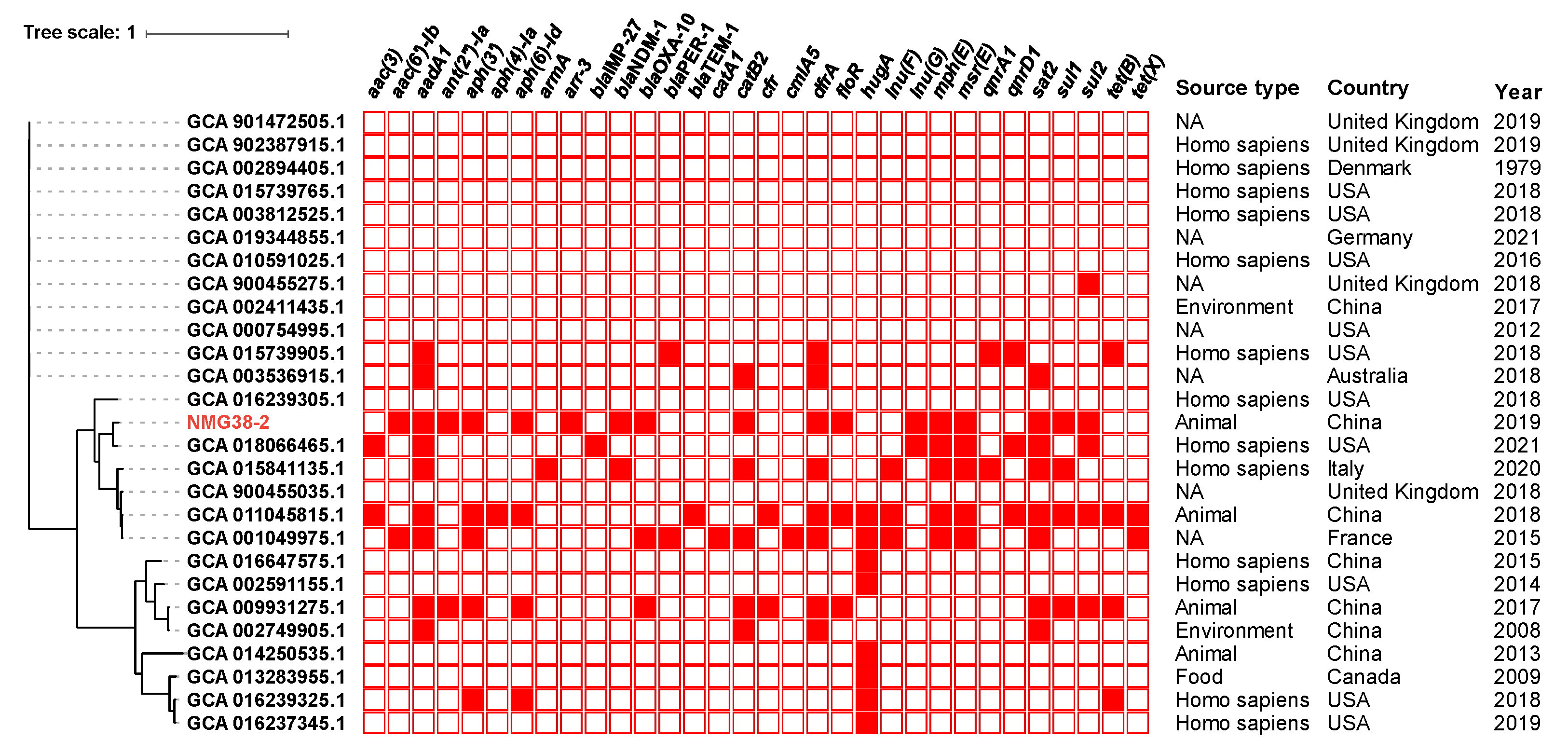
2.2. Phylogenetic Analysis of Proteus sp. NMG38-2

To determine a possible clinical relevance of NMG38-2 and explore the evolutionary relationships between NMG38-2 and P. vulgaris strains, phylogenetic analysis based on core genome alignment of NMG38-2 and all 26 available P. vulgaris strains from GenBank (on 2 September 2021), was performed. In total, 1323 core genes with 232,421 single nucleotide polymorphisms (SNPs) were identified from these genome sequences. These P. vulgaris isolates were mainly recovered from clinical specimens around the world, and found in the animals and environment. The phylogenetic tree (Figure 1) depicted a partitioning into three groups, and NMG38-2 was clustered with isolates of human and animal origins across various countries (including USA, Italy, France, United Kingdom, and China). NMG38-2 had the most closely genetic relationship with a clinical strain (GenBank assembly accession number GCA_018066465.1) from a patient with leg wound in USA in 2021, with 15,188 SNPs difference. This result indicated a close clinical relevance of NMG38-2. Resistance genes profiles showed that NMG38-2 and other strains in the same branch carried significantly more ARGs than the other two groups. A sporadic acquisition of carbapenemase genes was identified in these Proteus strains, with NMG38-2 and a clinical isolate from Italy in 2020 representing the only two strains harboring blaNDM-1.

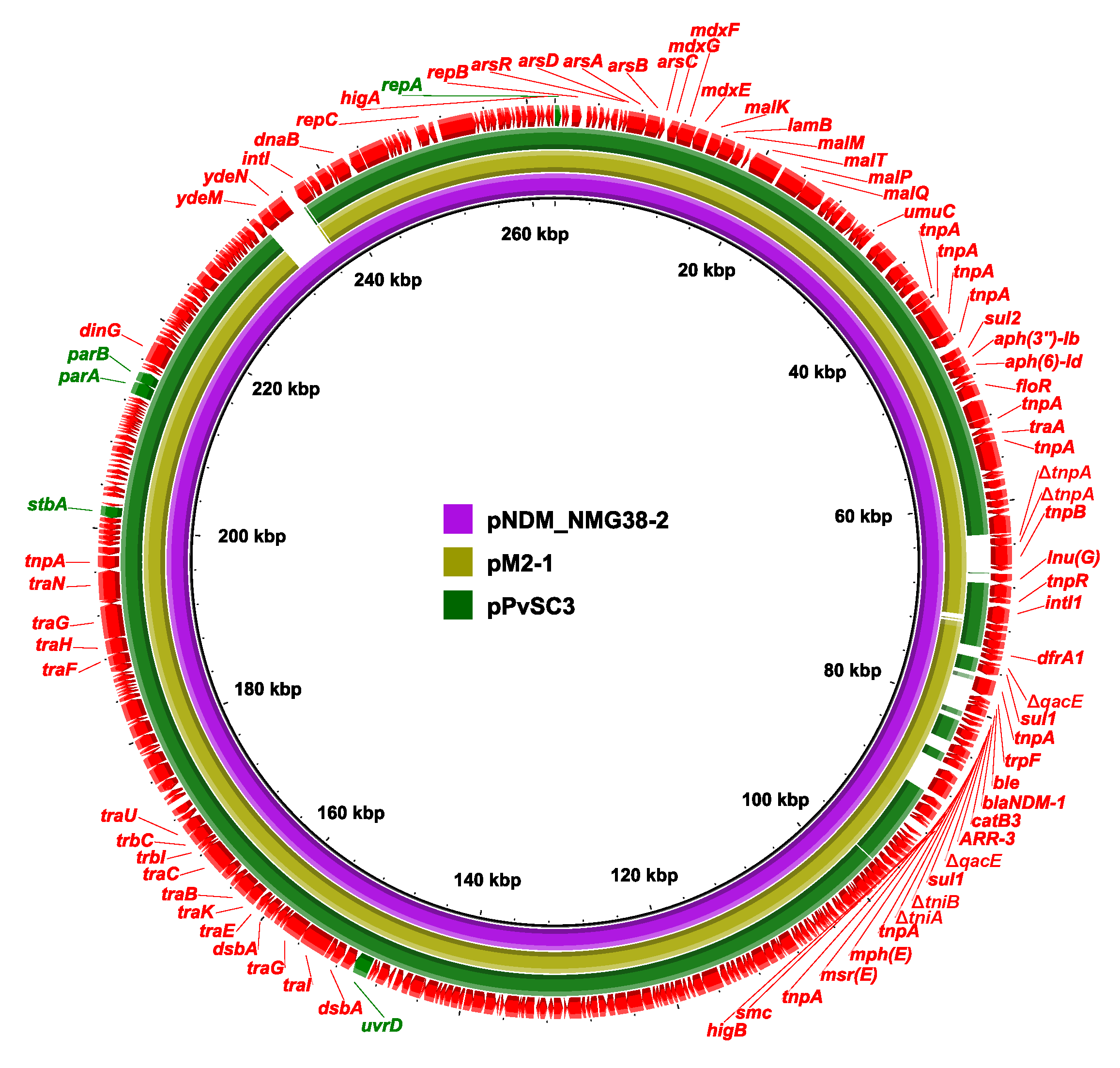
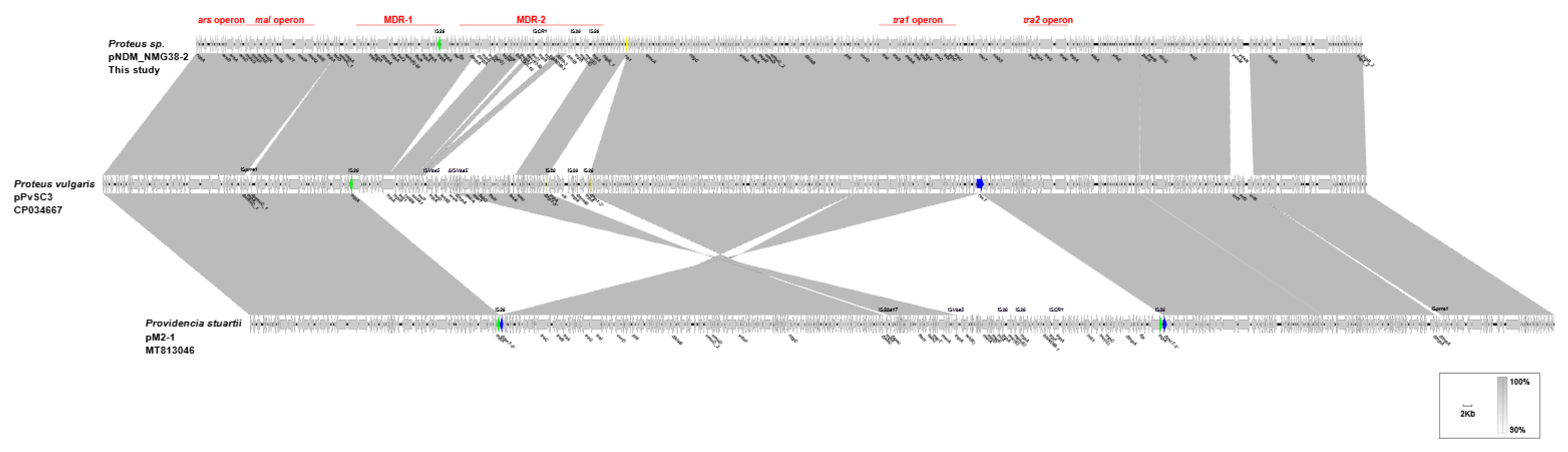
2.3. Genetic Structure of pPvSC3-like Plasmids

Pairwise sequence comparison using BLASTN showed that these three plasmids had almost identical backbone (>99.98% nucleotide identity across >96% sequences, nucleotide (nt) 1 to 37,670 bp and 97,255 to 262,556 bp of pNDM_NMG38-2, Table S2, Figure 2 and Figure 3). The major backbone genes included repA and its iterons for replication, parAB and stbA for plasmid partition, umuCD and uvrD for plasmid maintenance, tra1 and tra2 operon for conjugal transfer (Figure 2 and Figure 3). These three plasmids also harbored the mal operon for maltose metabolism and ars operon for arsenic resistance in the backbone region (Figure 2 and Figure 3). There were minor differences among their backbones: (i) the umuC_1 gene in pPvSC3 and pM2-1 is disrupted into ΔumuC_1-5′ and ΔumuC_1-3′ by an insertion of ISprre1; (ii) the orf1-orf3-orf6 segment (nt 84,819–89,762 bp) in pPvSC3 was replaced by an ydeM-ydeN structure (encoding sulfatase and sulfatase-maturating enzyme) in pNDM_NMG38-2; and (iii) an ISprre1 was inserted at a site downstream of the orf1-orf3-orf6 segment in pM2-1.
Two resistance accessory modules, designated MDR-1 and MDR-2, were identified in pPvSC3-like plasmids. The MDR-1 region was highly similar in these three plasmids with 100% nucleotide identity across >98.97% sequences. It had a chimera structure comprising of segments of Tn6535, Tn5075, and Tn6451 (Figure 4). This region contained four ARGs, including aph(3″)-Ib, aph(6)-Id, sul2, and floR, and an abundance of mobile genetic elements, which were responsible for the formation of this mosaic MDR region. The MDR-2 regions found in these three plasmids were identified as derivatives of Tn6321, which was a unit transposon belonging to the Tn21 subgroup of Tn3 family, and was generated by an insertion of a class 1 integron In844 at a site downstream of the tnpA-tnpR-res module in the primary backbone structure (Figure 5). Various types of insertion or homologous recombination events occurred within these MDR-2 regions, leading to gene truncations as well as the integration of foreign resistance markers.
Differences in three major modulars in the MDR-2 region, namely the tnpA-tnpR-res, integrons and mer, were identified (Figure 5). First, a core lnu(G) platform, Tn6260-like structure, was inserted into the transposase gene of Tn6321, truncating and breaking tnpA into separate ΔtnpA-5′ and ΔtnpA-3′ in both pNDM_NMG38-2 and pM2-1. The Tn6260-like structure was different from Tn6260 by an internal termination of tnpA gene caused by a base mutation (C to T at the 280th base). The tnpB gene of Tn6260 was further disrupted by an insertion of ISPmi3 in pM2-1. Second, the integron-related region showed distinct profiles: (i) compared to the In462 in pPvSC3, a “complex” class 1 integron harboring blaNDM-1 was found in pM2-1 and pNDM_NMG38-2. (ii) A novel integron, designated In2123, was identified in pM2-1, which consists of one 5′-conserved segment (5′-CS: intI1-attI1), two 3′-conserved segments (3′-CSs: qacED1-sul1-orf5-orf6), one ISCR1, two variable regions (VRs, dfrA14-ARR-2-blaOXA-10-aadA1-dfrA1-aac(6′)-Ib-cr in VR1 and trpF-ble-blaNDM-1-catB3-ARR-3 in VR2), an IS26-mph(E)-msr(E)-IS26 unit and a tni region truncated by the IS26 unit. (iii) In pNDM_NMG38-2, an In251 derived “complex” class 1 integron was found, which was similar to the In2123 in pM2-1, except that the gene cassettes dfrA14-ARR-2 was replaced by ant(2″)-Ia-catB8, and the tniA-5′ as well as the IRt (IR at the tni end) was deleted in pNDM_NMG38-2. Third, the mer region varies greatly in three plasmids: (i) a Tn6451 remnant containing tet gene clusters (tetDCBR) related to tetracycline resistance was inserted at a site within the IS6100 upstream of the mer region mediated by ISVsa5 in pPvSC3. (ii) A Tn2670 remnant comprising of mer gene clusters (merEDACPTR) followed by a mobile element IS1R and a fragment containing an ΔtetC-tetB-gltS-ISVsa5-ΔIS6100 module were successively integrated upstream of the mer region of Tn6321 in pM2-1, with two mer gene clusters showing 99% nucleotide coverage and 81% identity. (iii) Compared to pM2-1, a 24,455-bp region covering the tniA-5′, the complete mer region, as well as the surrounding backbone region was deleted in pNDM_NMG38-2, which was most likely due to the IS26-mediated recombination.
It is notable that an IS26-bounded structure (IS26-cfr-IS26-ΔermB-IS26), bearing the multiresistance gene cfr, was inserted within a hypothetic gene hp1 at 15,730 bp downstream of the mer region, truncating and splitting it into two parts Δhp1-5 and Δhp1-3′ in pPvSC3 (Figure 3). Additionally, an IS26 was inserted at a site within the recT gene in pM2-1, breaking it into two segments ΔrecT-5 and ΔrecT-3′. The inserted IS26 within recT, and the one at the right-hand end of MDR-1 region, were most likely to have accounted for the subsequent inversion of the 144,246-bp fragment covering MDR-2 and its surrounding backbone regions, by complex homologous recombination events (Figure 3). In conclusion, pPvSC3-like plasmids share a conserved backbone, but possess highly genetic plasticity in the MDR-2 region, which harbors a large number of resistance genes that are associated with insertion sequences, integrons, and transposons. The ISCR element, which is known to mobilize associated resistance genes via a rolling-circle mechanism [26], plays a key role in the acquisition of blaNDM-1 in pNDM_NMG38-2 and pM2-1.

2.4. Transfer Ability of pNDM_NMG38-2 and Fitness Cost

Conjugation assays showed that pNDM_NMG38-2 was able to conjugate into E. coli J53 at a frequency of ~10−6. Antimicrobial susceptibility results showed that the acquisition of pNDM_NMG38-2 greatly increased meropenem and imipenem resistance in J53 by at least 1024-fold. These results indicate that plasmid pNDM_NMG38-2 was self-transmissible, confirming that pPvSC3-like plasmids are transferable [22,25]. Growth curves showed that no significant differences in the growth rates were observed in both lag and logarithmic phases among the recipient strain J53 and transconjugants (Figure S1), suggesting that the carriage of pNDM_NMG38-2 does not significantly exert the fitness cost in its recipient host.

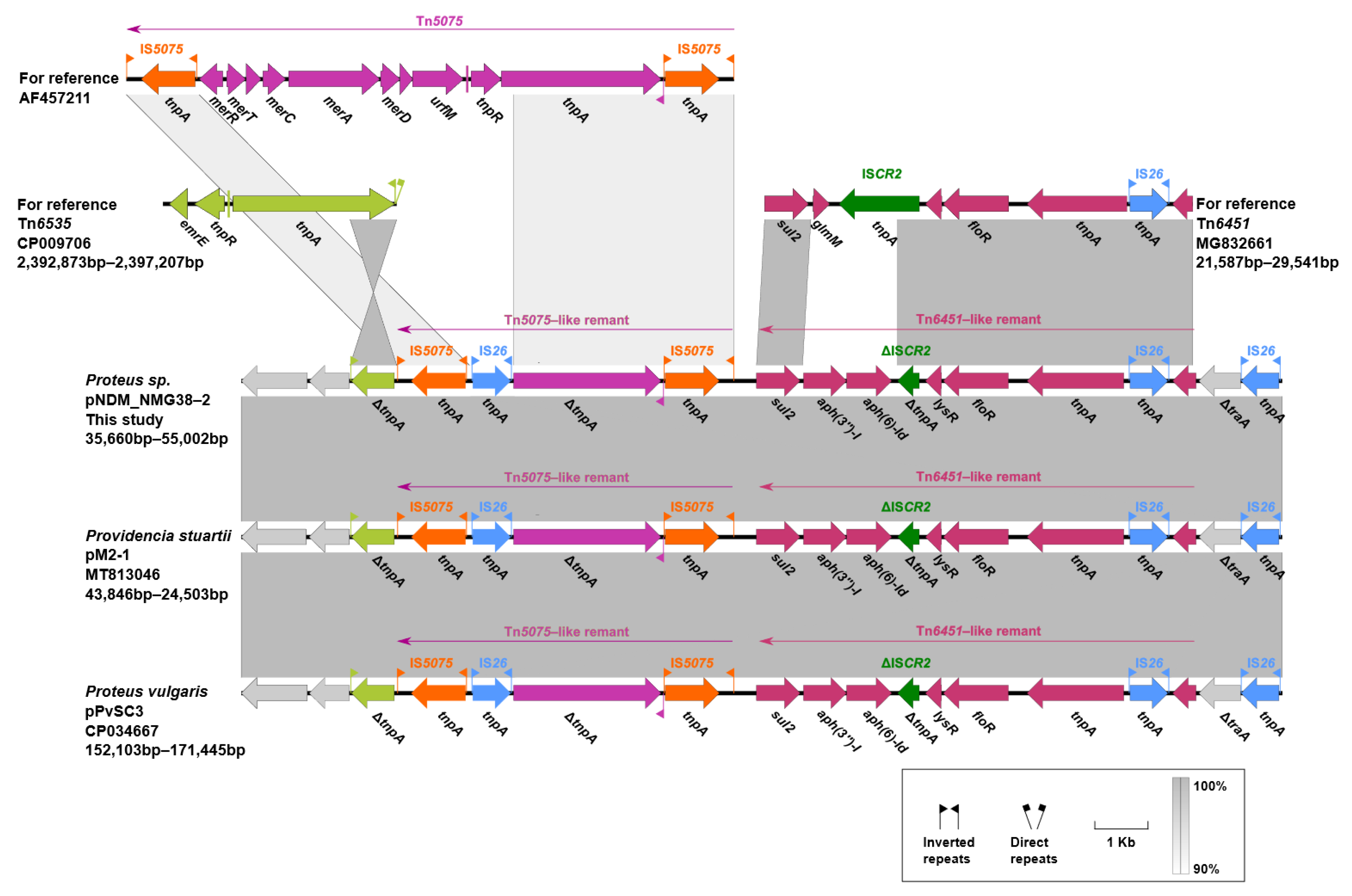
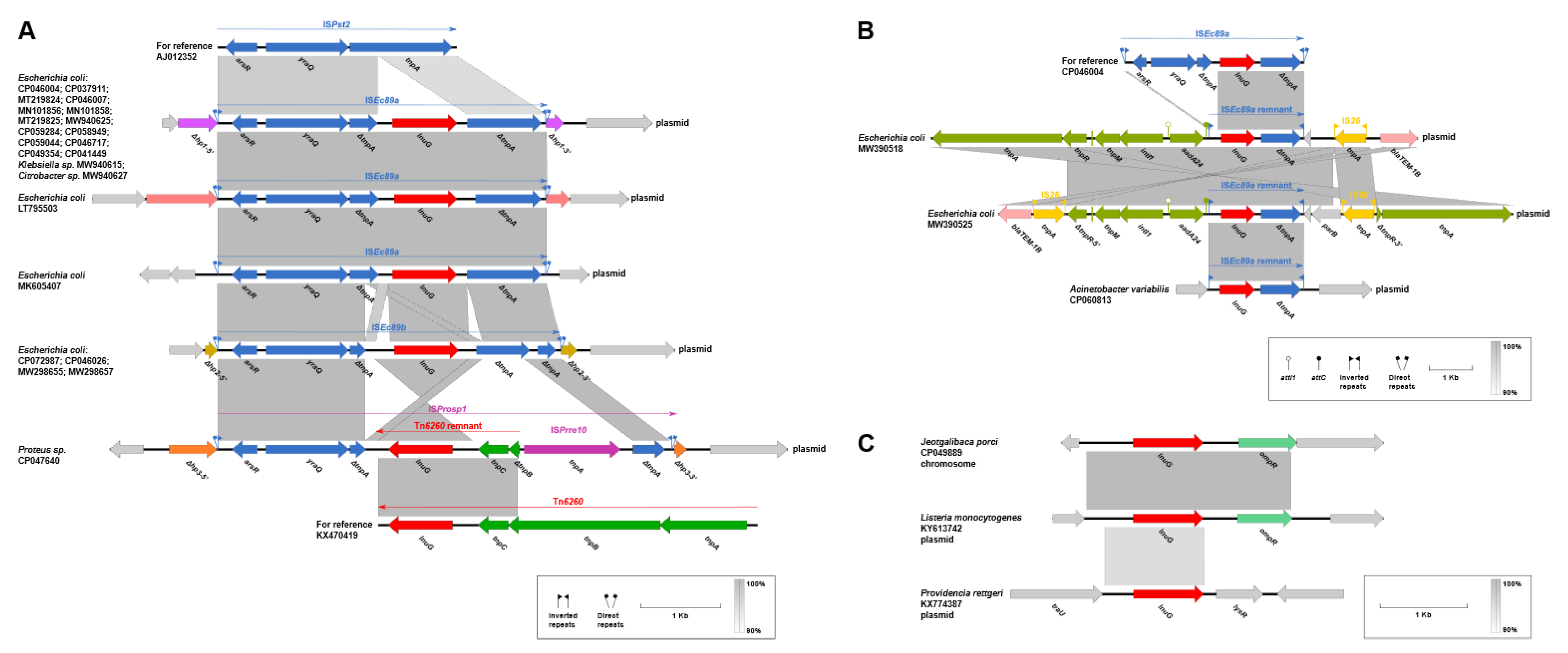
2.5. Genetic Contexts of lnu(G)

A total of 53 lnu(G)-bearing plasmids and chromosomes were retrieved from the GenBank database (accessed on 18 August 2021, Table S3). lnu(G) has a wide host spectrum of different origins with a worldwide distribution. It is largely located on the chromosomes of Enterococcus spp., and sporadically found on those of Providencia sp., M. morganii, P. mirabilis etc. Besides, lnu(G) is carried on plasmids that are widespread in Enterobacteriaceae, with Escherichia coli being the most common species, followed by Proteus spp. It is also identified on plasmids in bacterial species including Acinetobacter variabilis, Listeria monocytogenes, P. rettgeri, and E. faecalis. Plasmids with a variety of replicon types are associated with the dissemination of lnu(G) (Table S3). Among them, hybrid plasmids containing replicons of IncFIA(HI1), IncHI1A, and IncHI1B(R27), or in combination with other types of replicons, such as IncFII or IncX4, are most commonly found to carry lnu(G) in Enterobacteriaceae.
The genetic features of lnu(G) showed diversification and could be categorized into five different types, designated A (n = 22), B (n = 1), C (n = 2), D (n = 26), and E (n = 3) (Figure 6A–C and Figure 7A–C).
Type A: Tn6260, first identified on the chromosome of a swine E. faecalis (accession no. KX470419) [14], is a major vehicle in mediating the dissemination of lnu(G). An intact Tn6260 was particularly common in Enterococcus and was identified in other bacterial species (Figure 6A).
Tn6260-like structures found in P. mirabilis strains (accession no. MH491967 and MG516911), Proteus sp. NMG38-2, M. morganii (accession no. CP064826), and P. stuartii (accession no. MT813046) were different from the wild type transposon by an internal termination of tnpA gene or/and an interruption of tnpB gene by the insertion of ISPmi3 (Figure 6A).
In a P. mirabilis (accession no. CP042907), the complete tnpA and partial tnpB (tnpB-5′) of Tn6260 was lost, and a truncated ISCR2 took their places. Genetic events by ISCR2 might have contributed to the disruption of Tn6260. On this basis, the tnpB remnant (tnpB-3′) together with the downstream tnpC was further deleted in a plasmid from an uncultured Eubacterium (accession no. AJ293027) (Figure 6A).
Type B: on the chromosome of an E. faecalis (accession no. LR962846), a novel lnu(G)-bearing transposon, designated Tn7366, was inserted into the radC gene, splitting it into two separate parts (Figure 6B). It exhibits 98% nucleotide coverage and 90.37% identity to Tn6260, with 92.36% (100% coverage), 91.16% (99% coverage), and 81.79% (100% coverage) identities to tnpA, tnpB, and tnpC, respectively.
Type C: a novel lnu(G)-bearing composite transposon bounded by two copies of ISEnfa1, designated Tn7367, was detected in a plasmid from an E. faecalis isolate (accession no. MK140641), where it was inserted into a hypothetical gene and leaved a paired terminal 6-bp direct repeats (DRs, target site duplication signals for transposition, TTTTTA, Figure 6C). In Tn7367, a Tn6260 remnant containing a 1011-bp tnpB (tnpB-3′), a 123-bp tnpC (tnpC-5′) and the intact lnu(G) was integrated into a site downstream of IS1216V near the ISEnfa1 at the left-hand. An identical Tn6260 remnant was also found to be carried by a plasmid from another E. faecalis strain (accession no. MT723965); however, in this isolate, it was located in a region bracketed by two IS1216V in the opposite orientation (Figure 6C). This IS1216V bounded region was most likely to assemble from a structure like Tn7367 by multiple homologous recombination events of IS1216V.
Type D1: another common mechanism mediating the wide spread of lnu(G) was associated with ISPst2, a member of the ISL3 family, which was widely identified in Pseudomonas stutzeri strain OX1 [27]. The ISPst2 is composed of three genes, tnpA, yraQ, and arsR, encoding a transposase, putative permease, and an ArsR family transcriptional regulator, respectively. lnu(G), together with its flanking sequences, was integrated into a site within the tnpA gene by an unknown genetic event (Figure 7A). The resulting novel lnu(G)-bearing ISL3-like transposon, designated ISEc89a or ISEc89b, was primarily identified in Enterobacteriaceae strains, where it was inserted into various sites of plasmids, with the presence of paired terminal 8-bp DRs. The different integrating position of lnu(G) within tnpA represents the main modular difference between ISEc89a and ISEc89b. Another lnu(G)-bearing transferable element, designated ISProsp1, was detected in a plasmid from a Proteus sp. (accession no. CP047640), where it splits a hypothetic gene into two separate parts and meanwhile leaves 8-bp DR (TTTTTTTG). ISProsp1 was generated from integration of an ISprre10 of the IS91 family and its linked Tn6260 remnant covering a 144-bp tnpB residue (tnpB-3′), the intact tnpC and lnu(G) into the tnpA gene of ISPst2.
Type D2: an ISEc89a remnant covering the inverted repeat left (IRL), the intact lnu(G), a 924-bp tnpA fragment (tnpA-3′), and inverted repeat right (IRR) was found to be integrated into the cassette region, downstream of aadA24, of a class 1 integron carried by plasmids from two E. coli strains (accession no. MW390518 and MW390525, (Figure 7B). The identical ISEc89a remnant was also detected in a plasmid from an Acinetobacter variabilis strain (accession no. CP060813), where it was inserted at a site between two genes encoding for a putative integrase and a recombinase, respectively. The detection of the same ISEc89a remnant in different plasmids from different bacterial species indicates a possible transposition event of this lnu(G)-bearing element.
Type E: both on a plasmid from a L. monocytogenes strain (accession no. KY613742) and the chromosome of a Jeotgalibaca porci strain (accession no. CP049889), lnu(G) was linked to an ompR gene (encoding an ABC transporter-like sensor linked response regulator), whereas there was no mobile genetic element found adjacent to the lnu(G)-ompR structure (Figure 7C). Similarly, a lnu(G) was detected in a plasmid from a P. rettgeri isolate (accession no. KX774387), where it was located between traU (encoding a conjugal transfer pilus assembly protein) and lysR (encoding a transcriptional regulator) in the plasmid backbone region. The mechanisms involved in the mobilization of lnu(G) in these cases remain unknown.
Taken together, Tn6260 serves as a major vehicle in mediating the dissemination of lnu(G) in Enterococcus, while ISPst2 element is the leading contributor to the mobilization of lnu(G) in Gram-negative bacteria, especially Enterobacteriaceae.

3. Materials and Methods

3.1. Bacterial Strain and In Vitro Susceptibility Testing

Proteus sp. NMG38-2 was isolated from an intensive pig farm in Inner Mongolia, China, in December 2018. This strain was recovered from a fecal sample of swine according to the method described previously, with minor modifications [28]. A total of 40 non-duplicate fecal samples from healthy pigs were collected and feces suspension was directly plated on MacConkey agar plates containing 2 μg/mL meropenem using sterile swabs. Colonies recovered were further purified in LB agar plates and broth (Sangon Biotech, Shanghai, China) containing 2 μg/mL meropenem and initial species identification was performed by PCR amplifying of 16S rRNA gene and Sanger sequencing [29]. The minimum inhibitory concentrations (MICs) of the following antimicrobials against Proteus sp. NMG38-2 were determined using the microdilution broth method, according to the Clinical and Laboratory Standards Institute (CLSI) guidelines [30]: amikacin, gentamicin, meropenem, imipenem, cefoxitin, aztreonam, ciprofloxacin, cefotaxime, clindamycin, colistin, and chloramphenicol. E. coli ATCC 25922 served as a quality control for MIC determination. All antibiotics used in this study were obtained from Yuanye Bio-Technology Co. (Shanghai, China).

3.2. Conjugation Assay

Conjugation experiments were performed using broth-based methods as described previously with minor modification [31]. The azide-resistant E. coli strain J53 was used as the recipient. Both the NMG38-2 and J53 were grown to exponential stage (the optical density at 600 nm reaches ~0.5) and then mixed at a donor/recipient ratio of 1:1. After incubation at 37 °C for 24 h, transconjugants were selected on LB agar plates containing 2 μg/mL meropenem plus 150 μg/mL sodium azide. The presence of blaNDM-1 in transconjugants was confirmed by PCR using the primers blaNDM-F 5′-ATTTACTAGGCCTCGCATTTGC-3′ and blaNDM-R 5′-GCCTCTGTCACATCGAAATCG-3′.

3.3. In Vitro Growth Assays

Three independent cultures of J53 and transconjugants harboring pNDM_NMG38-2 were grown overnight and diluted to 1:100 in LB broth. Bacteria cultures were incubated while shaking (200 rpm) at 37 °C. In a total period of 12 h, the value of optical density (OD) at 600 nm (OD600) was consistently recorded at an interval of 1 h using the iMark microplate Reader (Bio-Rad, Hercules, CA, USA).

3.4. Genome Sequencing and Data Analyses

Genomic DNA of Proteus sp. NMG38-2 was extracted using the QIAamp DNA Mini Kit (Qiagen) and purified DNA was subjected to whole genome sequencing using the long-read MinION Sequencer (Nanopore, Oxford, UK), as well as a paired-end library with an insert size of 150 bp on a HiSeq 2000 sequencer (Illumina, San Diego, CA, USA). The de novo hybrid assembly of both long MinION reads and short Illumina reads was performed using Unicycler under the conservative mode [32]. Pilon was employed to correct the assembled contigs with Illumina reads for several rounds until no change was detected [33]. Annotations of the genome and plasmid sequences were conducted using the RAST tools [34] combined with BLASTp/BLASTn searches against the UniProtKB/Swiss-Prot database [35]. The ANI value between genome sequences of NMG38-2 and those of the reference strain of the known Proteus species was calculated with JSpeciesWS (http://jspecies.ribohost.com/jspeciesws/#analyse, accessed on 20 October 2021). dDDH values were calculated using GGDC 3.0 server (http://ggdc.dsmz.de/distcalc2.php, accessed on 20 October 2021) by means of genome-to-genome sequence comparison. The annotation of resistance genes, mobile elements, and other features was carried out using the online databases ResFinder [36], ISfinder [37], and INTEGRALL [38]. Alignments with homologous plasmid sequences of pNDM_NMG38-2 available in NCBI were performed by using the BRIG tool [39] and Easyfig v 2.2.3 (University of Queensland, Brisbane QLD 4072, Australia) [40] (accessed on 20 September 2021 to 7 October 2021). Gene organization diagrams were visualized with Inkscape 0.92.4 (https://inkscape.org/en/, accessed on 7 October 2021 to 17 November 2021).

3.5. Phylogenetic Analysis

Genomes were annotated using Prokka and annotated GFF3 files were piped into Roary to create a core genome alignment. SNPs were extracted using SNP-sites v2.3.2 (accessed on 12 September 2021) [41]. A maximum-likelihood phylogenetic tree based on the SNPs was constructed using FastTree version 2.1.10 (accessed on 12 September 2021) [42]. The antimicrobial resistance gene profiles were determined using ABRicate (https://github.com/tseemann/abricate, accessed on 16 September 2021). Carriage of resistance genes and detail information (source type, country, and isolation year) of isolates were annotated on the trees using iTOL (accessed on 17 September 2021) [43].

3.6. Comparative Analysis of lnu(G)-Harboring Genome Sequences

To obtain lnu(G)-harboring genome sequences, a nucleotide BLAST with standard options was performed in the NCBI GenBank database with the nucleotide base sequence of lnu(G) as a query. Models and uncultured environmental samples were excluded. Complete plasmids and chromosomes containing at least one contig with a full-length hit to lnu(G), with ≥99% query coverage and at least 97% identity, were selected and exported. Sequence alignments among lnu(G)-carrying plasmids or chromosomes were performed with Easyfig v 2.2.3 (accessed on 9 October 2021 to 14 October 2021) [40].

3.7. Nucleotide Sequence Accession Numbers

The complete genome sequences of Proteus sp. NMG38-2 and its plasmids have been deposited in GenBank under BioProject no. PRJNA769966.

4. Conclusions

In the present study, we characterized a drug resistant Proteus sp. from swine, which harbored blaNDM-1 carried on a conjugative plasmid of the untypable pPvSC3-like plasmid group. Our data significantly extended our understanding of genetic features of the pPvSC3-like plasmids, which could be a possible contributor to the spread of blaNDM-1 between Proteus and other species, in addition to the previously reported pPrY2001-like plasmids. More whole-genome sequencing-based epidemiological studies are warranted to achieve better insights into the mechanisms of the spread of carbapenemase genes in Proteus. Moreover, this work also allowed us to obtain a clear picture of the genetic contexts of lnu(G), highlighting the central roles of Tn6260 and ISPst2 in the spread of lnu(G) among diverse bacterial species.

Supplementary Materials

The following are available online at https://www.mdpi.com/article/10.3390/antibiotics10111411/s1, Table S1: Pairwise comparison of complete sequences of pPvSC3-like plasmids using BLASTN. Table S2: Pairwise comparison of plasmid backbone sequences of pPvSC3-like plasmids using BLASTN. Table S3: Detailed information of lnu(G)-carrying plasmids or chromosomes (last accessed 18 August 2021). Figure S1: Growth curves for the transconjugants (JM-1, JM-2, and JM-3) and the recipient strain J53.

Author Contributions

Conceptualization, Y.L. and L.Z.; formal analysis, Y.L. and X.D.; methodology, Y.Q. and J.S.; resources, L.Z.; software, X.W.; writing—original draft, Y.L.; writing—review and editing, X.D. and L.Z. All authors have read and agreed to the published version of the manuscript.

Funding

This research was funded by the National Natural Science Foundation of China (31900125), the scientific and technological project in Sichuan province (22MZGC0069), and the Joint Funds of the Luzhou and Southwest Medical University Natural Science Foundation (2019LZXNYDJ47 and 2020LZXNYDJ34).

Institutional Review Board Statement

Not applicable.

Informed Consent Statement

Not applicable.

Data Availability Statement

The complete genome sequences of Proteus sp. NMG38-2 and its plasmids can be found in public NCBI Genbank databases under BioProject no. PRJNA769966.

Conflicts of Interest

The funders had no role in the design of the study; in the collection, analyses, or interpretation of data; in the writing of the manuscript, or in the decision to publish the results.

References

  1. Kumar Trivedi, M. Phenotyping and genotyping characterization of Proteus vulgaris after biofield treatment. Int. J. Genet. Genom. 2015, 3, 66. [Google Scholar] [CrossRef]
  2. O’Hara, C.M.; Brenner, F.W.; Miller, J.M. Classification, identification, and clinical significance of Proteus, Providencia, and Morganella. Clin. Microbiol. Rev. 2000, 13, 534–546. [Google Scholar] [CrossRef] [PubMed]
  3. Wu, W.; Feng, Y.; Tang, G.; Qiao, F.; McNally, A.; Zong, Z. NDM metallo-β-lactamases and their bacterial producers in health care settings. Clin. Microbiol. Rev. 2019, 32, e00115–e00118. [Google Scholar] [CrossRef] [PubMed] [Green Version]
  4. Yong, D.; Toleman, M.A.; Giske, C.G.; Cho, H.S.; Sundman, K.; Lee, K.; Walsh, T.R. Characterization of a new metallo-beta-lactamase gene, bla(NDM-1), and a novel erythromycin esterase gene carried on a unique genetic structure in Klebsiella pneumoniae sequence type 14 from India. Antimicrob. Agents Chemother. 2009, 53, 5046–5054. [Google Scholar] [CrossRef] [Green Version]
  5. Feng, Y.; Liu, L.; McNally, A.; Zong, Z. Coexistence of two blaNDM-5 genes on an IncF plasmid as revealed by Nanopore sequencing. Antimicrob. Agents Chemother. 2018, 62, e00110-18. [Google Scholar] [CrossRef] [Green Version]
  6. Ahmed, N.; Sekizuka, T.; Matsui, M.; Yamane, K.; Takeuchi, F.; Ohnishi, M.; Hishinuma, A.; Arakawa, Y.; Kuroda, M. Complete sequencing of the blaNDM-1-positive IncA/C plasmid from Escherichia coli ST38 isolate suggests a possible origin from plant pathogens. PLoS ONE 2011, 6, e25334. [Google Scholar] [CrossRef]
  7. Wailan, A.M.; Sidjabat, H.E.; Yam, W.K.; Alikhan, N.F.; Petty, N.K.; Sartor, A.L.; Williamson, D.A.; Forde, B.M.; Schembri, M.A.; Beatson, S.A.; et al. Mechanisms involved in acquisition of blaNDM genes by IncA/C2 and IncFIIY plasmids. Antimicrob. Agents Chemother. 2016, 60, 4082–4088. [Google Scholar] [CrossRef] [Green Version]
  8. Le-Vo, H.N.; Tran, P.T.; Le, L.; Matsumoto, Y.; Motooka, D.; Nakamura, S.; Jones, J.W.; Iida, T.; Cao, V. Complex class 1 integron in a clinical Escherichia coli strain from Vietnam carrying both mcr-1 and blaNDM-1. Front. Microbiol. 2019, 10, 2472. [Google Scholar] [CrossRef]
  9. Kong, L.-H.; Xiang, R.; Wang, Y.-L.; Wu, S.-K.; Lei, C.-W.; Kang, Z.-Z.; Chen, Y.-P.; Ye, X.-L.; Lai, Y.; Wang, H.-N. Integration of the blaNDM-1 carbapenemase gene into a novel SXT/R391 integrative and conjugative element in Proteus vulgaris. J. Antimicrob. Chemother. 2020, 75, 1439–1442. [Google Scholar] [CrossRef]
  10. He, J.; Sun, L.; Zhang, L.; Leptihn, S.; Yu, Y.; Hua, X. A novel SXT/R391 integrative and conjugative element carries two copies of the blaNDM-1 gene in Proteus mirabilis. mSphere 2021, 6, e0058821. [Google Scholar] [CrossRef]
  11. Dong, D.; Li, M.; Liu, Z.; Feng, J.; Jia, N.; Zhao, H.; Zhao, B.; Zhou, T.; Zhang, X.; Tong, Y.; et al. Characterization of a NDM-1- encoding plasmid pHFK418-NDM from a clinical Proteus mirabilis isolate harboring two novel transposons, Tn6624 and Tn6625. Front. Microbiol. 2019, 10, 2030. [Google Scholar] [CrossRef] [Green Version]
  12. Li, Y.; Wang, Q.; Peng, K.; Liu, Y.; Li, R.; Wang, Z. Emergence of carbapenem- and tigecycline-resistant Proteus cibarius of animal origin. Front. Microbiol. 2020, 11, 1940. [Google Scholar] [CrossRef] [PubMed]
  13. Spizek, J.; Rezanka, T. Lincosamides: Chemical structure, biosynthesis, mechanism of action, resistance, and applications. Biochem. Pharmacol. 2017, 133, 20–28. [Google Scholar] [CrossRef]
  14. Zhu, X.-Q.; Wang, X.-M.; Li, H.; Shang, Y.-H.; Pan, Y.-S.; Wu, C.-M.; Wang, Y.; Du, X.-D.; Shen, J.-Z. Novel lnu(G) gene conferring resistance to lincomycin by nucleotidylation, located on Tn6260 from Enterococcus faecalis E531. J. Antimicrob. Chemother. 2017, 72, 993–997. [Google Scholar] [CrossRef] [Green Version]
  15. Spizek, J.; Novotna, J.; Rezanka, T. Lincosamides: Chemical structure, biosynthesis, mechanism of action, resistance, and applications. Adv. Appl. Microbiol. 2004, 56, 121–154. [Google Scholar] [CrossRef] [PubMed]
  16. Leclercq, R. Mechanisms of resistance to macrolides and lincosamides: Nature of the resistance elements and their clinical implications. Clin. Infect. Dis. 2002, 34, 482–492. [Google Scholar] [CrossRef] [PubMed] [Green Version]
  17. Brisson-Noel, A.; Delrieu, P.; Samain, D.; Courvalin, P. Inactivation of lincosaminide antibiotics in Staphylococcus. Identification of lincosaminide O-nucleotidyltransferases and comparison of the corresponding resistance genes. J. Biol. Chem. 1988, 263, 15880–15887. [Google Scholar] [CrossRef]
  18. Bozdogan, B.; Berrezouga, L.; Kuo, M.S.; Yurek, D.A.; Farley, K.A.; Stockman, B.J.; Leclercq, R. A new resistance gene, linB, conferring resistance to lincosamides by nucleotidylation in Enterococcus faecium HM1025. Antimicrob. Agents Chemother. 1999, 43, 925–929. [Google Scholar] [CrossRef] [PubMed] [Green Version]
  19. Achard, A.; Villers, C.; Pichereau, V.; Leclercq, R. New lnu(C) gene conferring resistance to lincomycin by nucleotidylation in Streptococcus agalactiae UCN36. Antimicrob. Agents Chemother. 2005, 49, 2716–2719. [Google Scholar] [CrossRef] [Green Version]
  20. Petinaki, E.; Guerin-Faublee, V.; Pichereau, V.; Villers, C.; Achard, A.; Malbruny, B.; Leclercq, R. Lincomycin resistance gene lnu(D) in Streptococcus uberis. Antimicrob. Agents Chemother. 2008, 52, 626–630. [Google Scholar] [CrossRef] [Green Version]
  21. Zhao, Q.; Wendlandt, S.; Li, H.; Li, J.; Wu, C.; Shen, J.; Schwarz, S.; Wang, Y. Identification of the novel lincosamide resistance gene lnu(E) truncated by ISEnfa5-cfr-ISEnfa5 insertion in Streptococcus suis: De novo synthesis and confirmation of functional activity in Staphylococcus aureus. Antimicrob. Agents Chemother. 2014, 58, 1785–1788. [Google Scholar] [CrossRef] [PubMed] [Green Version]
  22. Cen, D.-J.; Sun, R.-Y.; Mai, J.-L.; Jiang, Y.-W.; Wang, D.; Guo, W.-Y.; Jiang, Q.; Zhang, H.; Zhang, J.-F.; Zhang, R.-M.; et al. Occurrence and transmission of blaNDM -carrying Enterobacteriaceae from geese and the surrounding environment on a commercial goose farm. Appl. Environ. Microb. 2021, 87, e00087-21. [Google Scholar] [CrossRef] [PubMed]
  23. Falagas, M.E.; Karageorgopoulos, D.E. Pandrug resistance (PDR), extensive drug resistance (XDR), and multidrug resistance (MDR) among Gram-negative bacilli: Need for international harmonization in terminology. Clin. Infect. Dis. 2008, 46, 1121–1122, author reply 1122. [Google Scholar] [CrossRef] [PubMed] [Green Version]
  24. Sun, J.; Zhang, H.; Liu, Y.H.; Feng, Y. Towards understanding MCR-like colistin resistance. Trends Microbiol. 2018, 26, 794–808. [Google Scholar] [CrossRef]
  25. Zhang, Y.; Lei, C.W.; Wang, H.N. Identification of a novel conjugative plasmid carrying the multiresistance gene cfr in Proteus vulgaris isolated from swine origin in China. Plasmid 2019, 105, 102440. [Google Scholar] [CrossRef]
  26. Toleman, M.A.; Bennett, P.M.; Walsh, T.R. ISCR elements: Novel gene-capturing systems of the 21st century? Microbiol. Mol. Biol. Rev. 2006, 70, 296–316. [Google Scholar] [CrossRef] [Green Version]
  27. Cameron, A.; Klima, C.L.; Ha, R.; Gruninger, R.J.; Zaheer, R.; McAllister, T.A. A novel aadA aminoglycoside resistance gene in bovine and porcine pathogens. mSphere 2018, 3, e00568-17. [Google Scholar] [CrossRef] [Green Version]
  28. Li, Y.; Ma, X.; Li, C.; Dai, X.; Zhang, L. Occurrence and genomic characterization of ESBL-producing Escherichia coli ST29 strains from swine with abundant virulence genes. Microb. Pathog. 2020, 148, 104483. [Google Scholar] [CrossRef]
  29. Yuan, Y.; Li, Y.; Wang, G.; Li, C.; Chang, Y.F.; Chen, W.; Nian, S.; Mao, Y.; Zhang, J.; Zhong, F.; et al. blaNDM-5 carried by a hypervirulent Klebsiella pneumoniae with sequence type 29. Antimicrob. Resist. Infect. Control 2019, 8, 140. [Google Scholar] [CrossRef]
  30. CLSI. Performance Standards for Antimicrobial Susceptibility Testing, 28th ed.; CLSI supplement M100; Clinical and Laboratory Standards Institute: Wayne, PA, USA, 2018. [Google Scholar]
  31. Li, Y.; Luo, L.; Xiao, Z.; Wang, G.; Li, C.; Zhang, Z.; Zhou, Y.; Zhang, L. Characterization of a carbapenem-resistant Kluyvera Cryocrescens isolate carrying blaNDM-1 from hospital sewage. Antibiotics 2019, 8, 149. [Google Scholar] [CrossRef] [Green Version]
  32. Wick, R.R.; Judd, L.M.; Gorrie, C.L.; Holt, K.E. Unicycler: Resolving bacterial genome assemblies from short and long sequencing reads. PLoS Comput. Biol. 2017, 13, e1005595. [Google Scholar] [CrossRef] [Green Version]
  33. Walker, B.J.; Abeel, T.; Shea, T.; Priest, M.; Abouelliel, A.; Sakthikumar, S.; Cuomo, C.A.; Zeng, Q.; Wortman, J.; Young, S.K.; et al. Pilon: An integrated tool for comprehensive microbial variant detection and genome assembly improvement. PLoS ONE 2014, 9, e112963. [Google Scholar] [CrossRef]
  34. Aziz, R.K.; Bartels, D.; Best, A.A.; DeJongh, M.; Disz, T.; Edwards, R.A.; Formsma, K.; Gerdes, S.; Glass, E.M.; Kubal, M.; et al. The RAST Server: Rapid annotations using subsystems technology. BMC Genom. 2008, 9, 75. [Google Scholar] [CrossRef] [Green Version]
  35. Boutet, E.; Lieberherr, D.; Tognolli, M.; Schneider, M.; Bansal, P.; Bridge, A.J.; Poux, S.; Bougueleret, L.; Xenarios, I. UniProtKB/Swiss-Prot, the manually annotated section of the UniProt KnowledgeBase: How to use the entry view. Methods Mol. Biol. 2016, 1374, 23–54. [Google Scholar] [CrossRef]
  36. Zankari, E.; Hasman, H.; Cosentino, S.; Vestergaard, M.; Rasmussen, S.; Lund, O.; Aarestrup, F.M.; Larsen, M.V. Identification of acquired antimicrobial resistance genes. J. Antimicrob. Chemother. 2012, 67, 2640–2644. [Google Scholar] [CrossRef] [PubMed]
  37. Siguier, P.; Perochon, J.; Lestrade, L.; Mahillon, J.; Chandler, M. ISfinder: The reference centre for bacterial insertion sequences. Nucleic Acids Res. 2006, 34, D32–D36. [Google Scholar] [CrossRef] [PubMed] [Green Version]
  38. Moura, A.; Soares, M.; Pereira, C.; Leitao, N.; Henriques, I.; Correia, A. INTEGRALL: A database and search engine for integrons, integrases and gene cassettes. Bioinformatics 2009, 25, 1096–1098. [Google Scholar] [CrossRef] [PubMed] [Green Version]
  39. Alikhan, N.F.; Petty, N.K.; Ben Zakour, N.L.; Beatson, S.A. BLAST Ring Image Generator (BRIG): Simple prokaryote genome comparisons. BMC Genom. 2011, 12, 402. [Google Scholar] [CrossRef] [Green Version]
  40. Sullivan, M.J.; Petty, N.K.; Beatson, S.A. Easyfig: A genome comparison visualizer. Bioinformatics 2011, 27, 1009–1010. [Google Scholar] [CrossRef]
  41. Page, A.J.; Taylor, B.; Delaney, A.J.; Soares, J.; Seemann, T.; Keane, J.A.; Harris, S.R. SNP-sites: Rapid efficient extraction of SNPs from multi-FASTA alignments. Microb. Genom. 2016, 2, e000056. [Google Scholar] [CrossRef] [Green Version]
  42. Price, M.N.; Dehal, P.S.; Arkin, A.P. FastTree 2—Approximately maximum-likelihood trees for large alignments. PLoS ONE 2010, 5, e9490. [Google Scholar] [CrossRef] [PubMed]
  43. Letunic, I.; Bork, P. Interactive Tree Of Life (iTOL) v4: Recent updates and new developments. Nucleic Acids Res. 2019, 47, W256–W259. [Google Scholar] [CrossRef] [PubMed] [Green Version]
Figure 1. Phylogenetic tree of the Proteus sp. NMG38-2 with 26 P. vulgaris genomes available from GenBank. The tree is based on 1323 core genes with 232,421 SNPs and the tree scale indicates substitutions per site. NMG38-2 is indicated in red. Resistance gene profiles are visualized in compliance to the tree. The annotation denotes (from left to right) isolation sources, locations and years of strains. NA, not available.
Antibiotics 10 01411 g001
Figure 2. Circular comparisons of pNDM_NMG38-2 with pPvSC3 and pM2-1. pNDM_NMG38-2 was used as a reference to compare with other plasmids. The outer circle with red arrows denotes the annotation of reference plasmid, with arrows indicating deduced ORFs and their orientations. Gaps indicate regions that were missing in the respective plasmid compared to the reference plasmid. Key backbone genes are indicated in green.
Antibiotics 10 01411 g002
Figure 3. Linear comparisons of pNDM_NMG38-2, pPvSC3, and pM2-1. The tra1, tra2 operon, mal operon, ars operon, MDR-1, and MDR-2 regions are pointed out. Arrows indicate deduced ORFs and their orientations. Gray shading denotes regions of shared homology ranging from 90% to 100%. Δ represents truncated genes or mobile genetic elements.
Antibiotics 10 01411 g003
Figure 4. Organization of the MDR-1 of pPvSC3-like plasmids and comparison to related regions. Genes are denoted by arrows. Genes, mobile elements, and other features are colored based on their functional classification. Gray shading denotes regions of shared homology ranging from 90% to 100%. Δ represents truncated genes or mobile genetic elements.
Antibiotics 10 01411 g004
Figure 5. Organization of the MDR-2 of pPvSC3-like plasmids and comparison to related regions. Genes are denoted by arrows. Genes, mobile elements, and other features are colored based on their functional classification. Gray shading denotes regions of shared homology ranging from 90% to 100%. Δ represents truncated genes or mobile genetic elements.
Antibiotics 10 01411 g005
Figure 6. Tn6260-associated genetic contexts of lnu(G) from different plasmids and chromosomes. The lnu(G) was indicated in red. Genes are colored based on their functional classification. Gray shading denotes genetic regions that exhibit sequence homology among different segments. Δ represents truncated genes or mobile genetic elements.
Antibiotics 10 01411 g006
Figure 7. The genetic contexts of lnu(G) from other plasmids and chromosomes. ISPst2-associated genetic contexts of lnu(G), type D1 and type D2, are shown in (A) and (B), respectively. The lnu(G) genetic contexts of type E is shown in (C). The lnu(G) was indicated in red. Genes are colored based on their functional classification. Gray shading denotes genetic regions that exhibit sequence homology among different segments. Δ represents truncated genes or mobile genetic elements.
Antibiotics 10 01411 g007
Table 1. MICs for NMG38-2, its transformants, and the recipient strain J53.
Strains aSpecies’ NameMIC (μg/mL) b
AMKGENCSTMEMIMPCFTAZTCIPCTXCLNCHL
NMG38-2Proteus sp.128256512512>512512256≤0.5256>512256
JM-1E. coli6432256512>5125122561256>512256
JM-2E. coli6432512512>5125122561512512256
JM-3E. coli6464256512>5125121282256512256
J53E. coli162≤0.5≤0.5≤0.5168≤0.5≤0.52568
ATCC25922E. coli44≤0.5≤0.5≤0.582≤0.5≤0.525616
a JM-1, JM-2 and JM-3 are transconjugants harboring pNDM_NMG38-2. b AMK, amikacin; GEN, gentamicin; CST, colistin; MEM, meropenem; IMP, imipenem; CFT, cefoxitin; AZT, aztreonam; CIP, ciprofloxacin; CTX, cefotaxime; CLN: clindamycin; CHL, chloramphenicol. Resistant MICs are highlighted in bold.
Table 2. Summary of the genetic features of Proteus sp. NMG38-2.
Chromosome
/Plasmid
Length (bp) GC%No. of Predicted Open Reading FramesInc TypeDrug Resistance Gene
Chromosome4,433,34637.624231-aadA1, dfrA1, catB2
pNDM_NMG38-2262,55646.65369UTaac(6′)-Ib-cr, ant(2)-Ia, aph(6)-Id, aac(6′)-Ib3, aph(3)-Ib, aac(6′)-Ib-cr, aadA1, ARR-3, sul1 a, dfrA1, sul2, catB8, floR, catB3, blaOXA-10, blaNDM-1, mph(E), msr(E), lnu(G)
P1_SCLZS62550034.1110UT
P2_SCLZS62268341.782Col3MqnrD1
a Multiple copies on the plasmid. -, not available; UT, unknown type.
Publisher’s Note: MDPI stays neutral with regard to jurisdictional claims in published maps and institutional affiliations.

