Carbon Nanotube-Based Field-Effect Transistor Biosensors for Biomedical Applications: Decadal Developments and Advancements (2016–2025)


Abstract
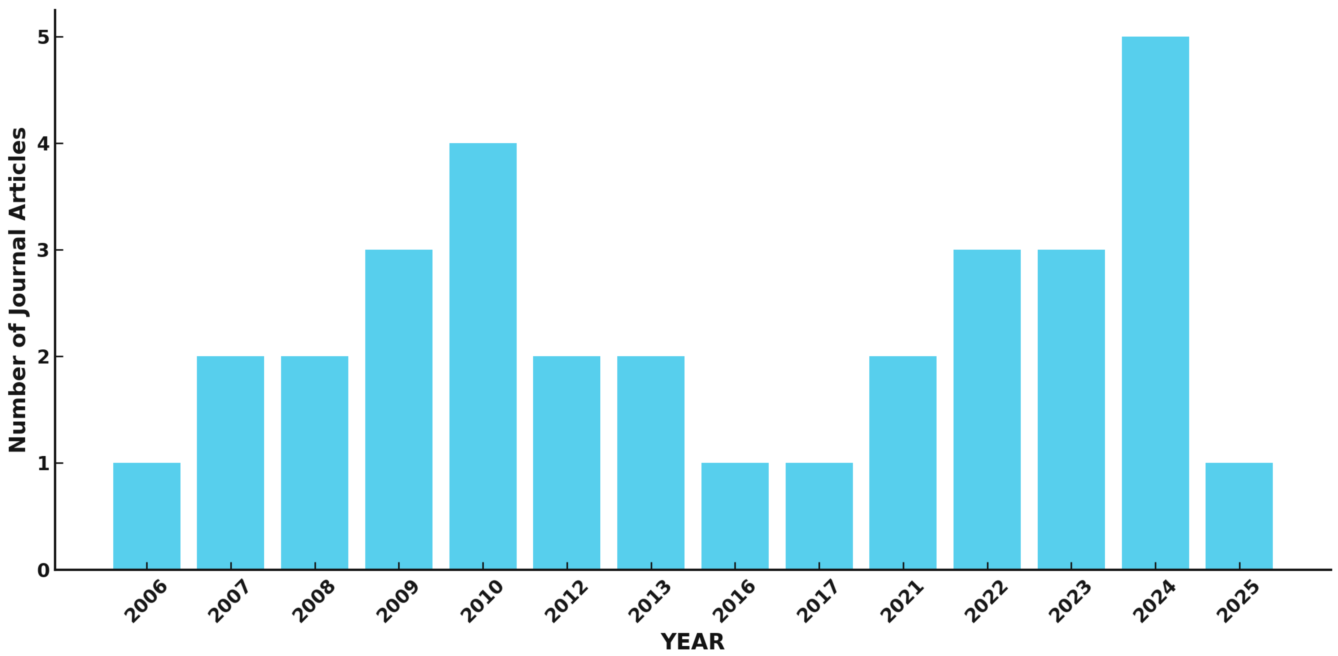
1. Introduction
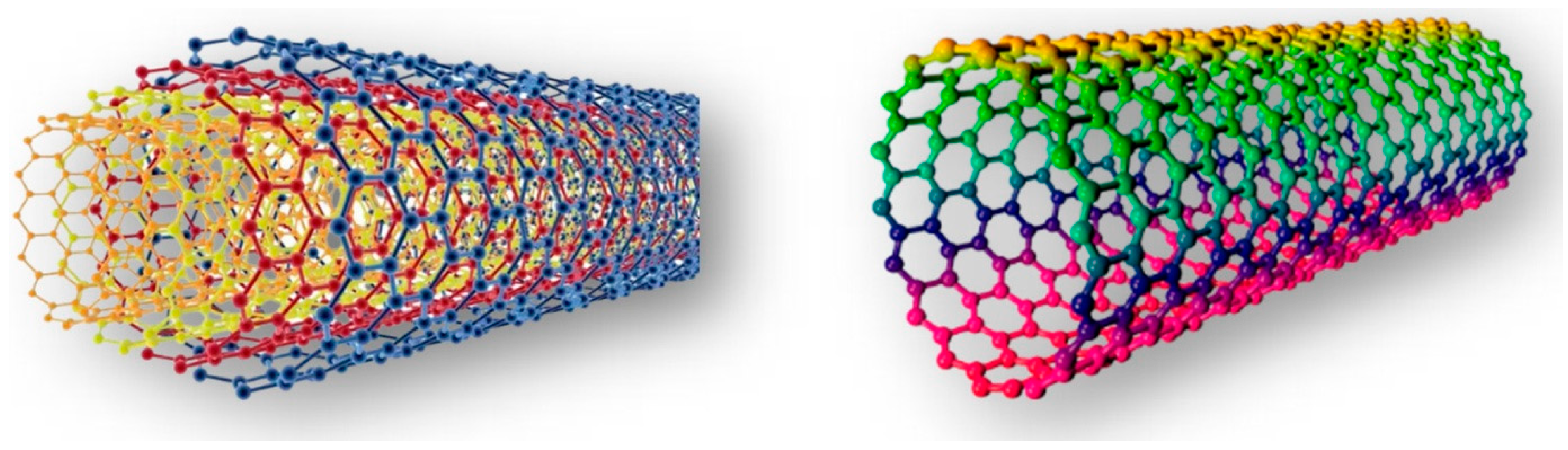
2. Carbon Nanotubes: Types, Synthesis, and Properties
3. CNT-FET Biosensors
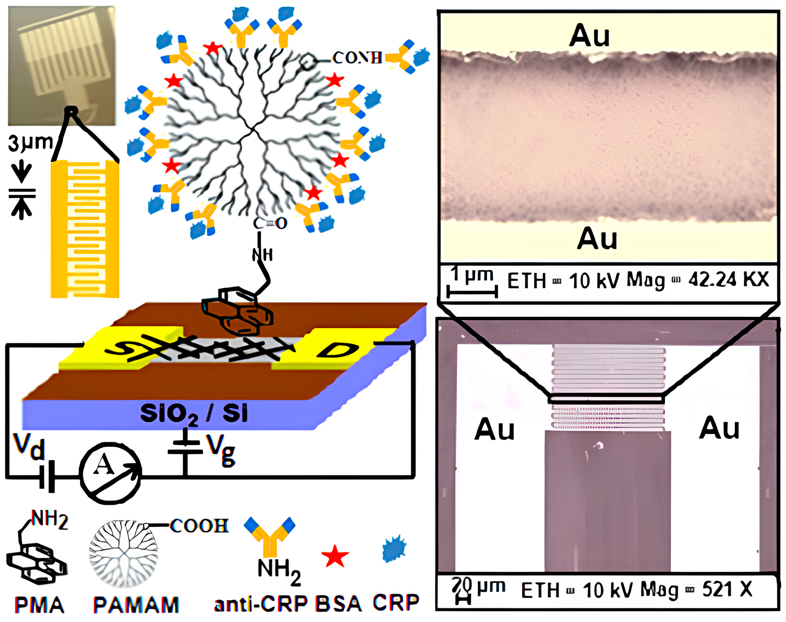
3.1. Basic Configuration and Advanced Architectures
3.2. Functionalization Strategies for Enhanced Biosensing
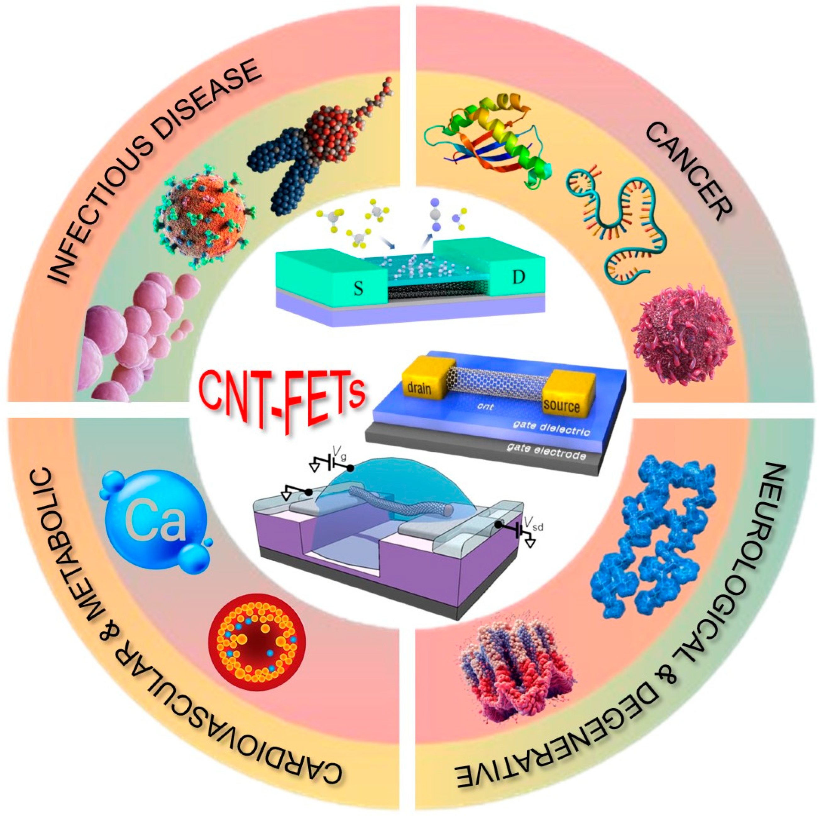
4. Applications of CNT-FET Biosensors in Disease Diagnostics
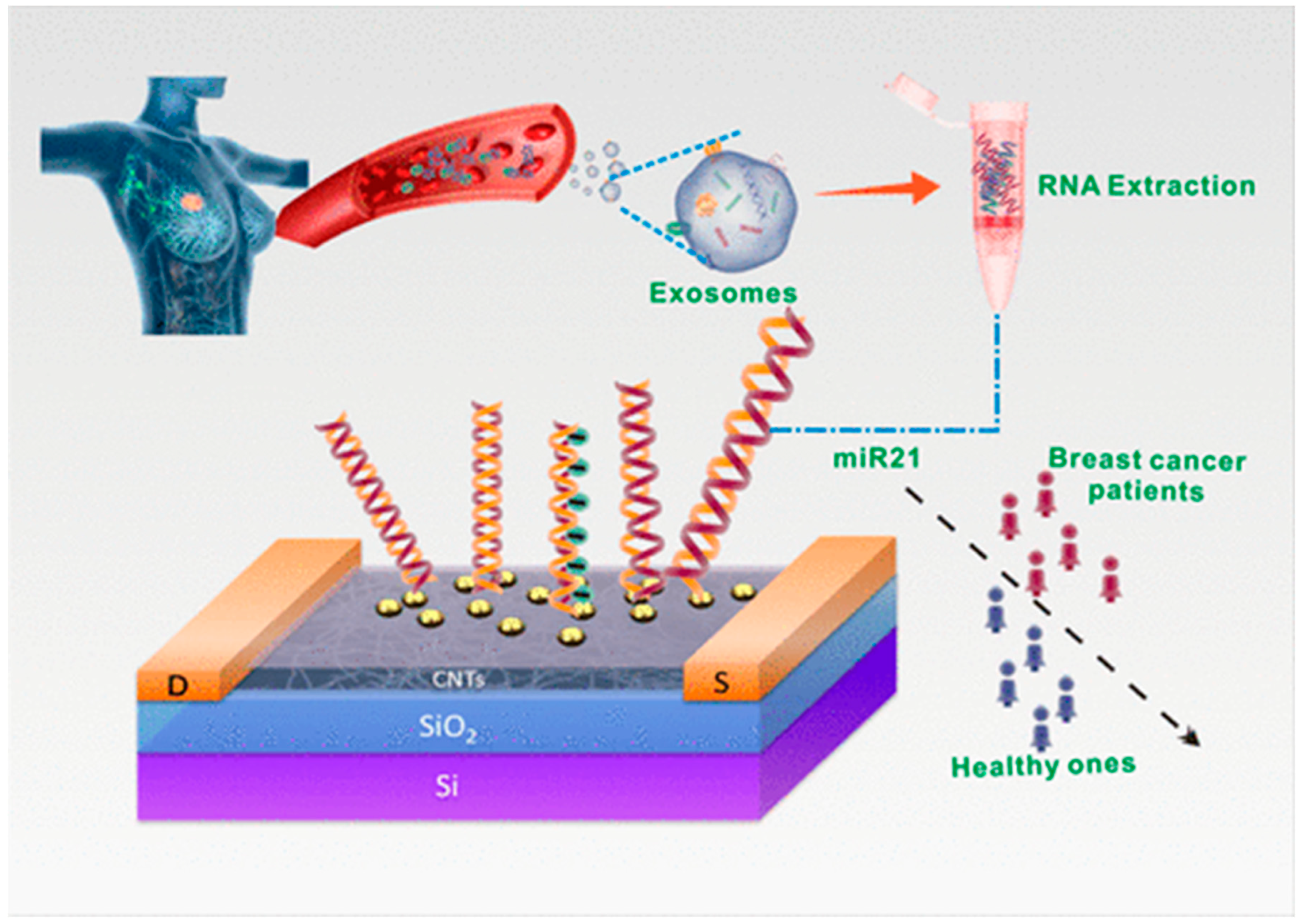
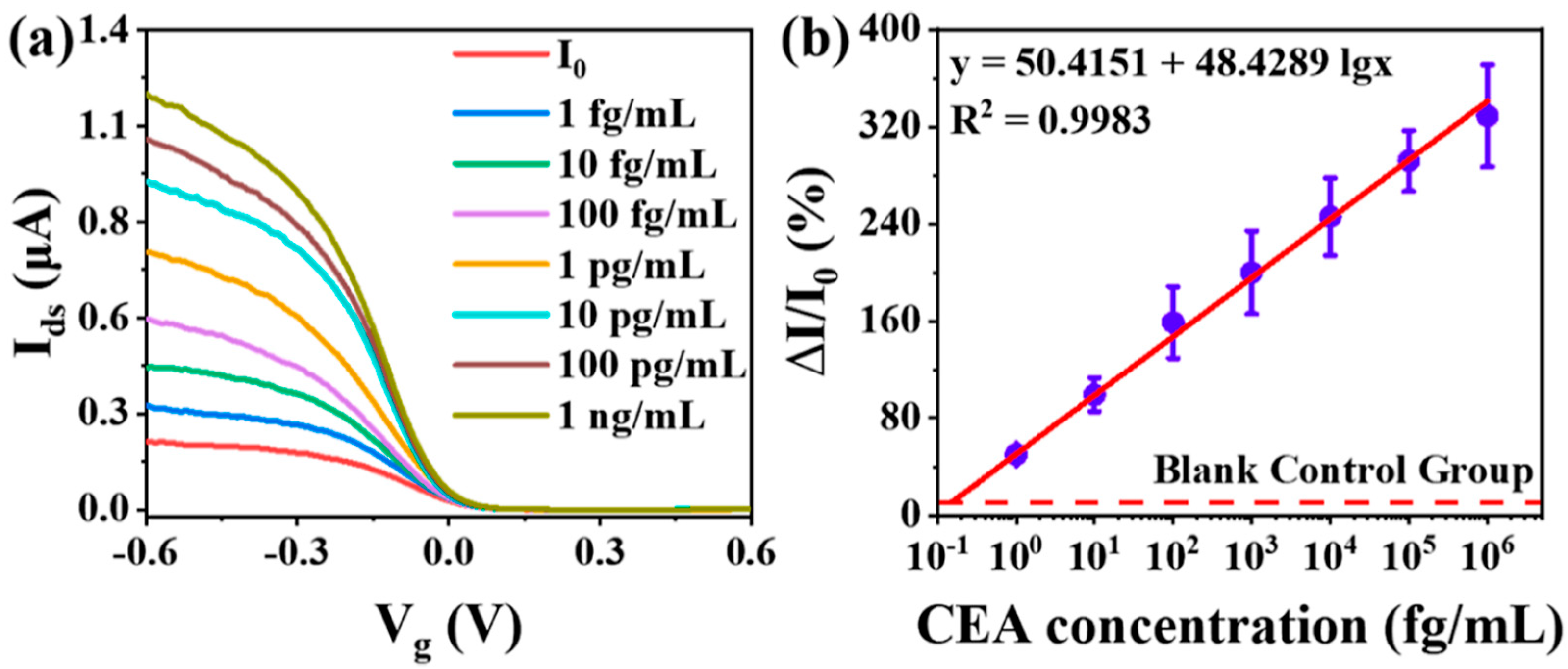
4.1. Cancer Detection
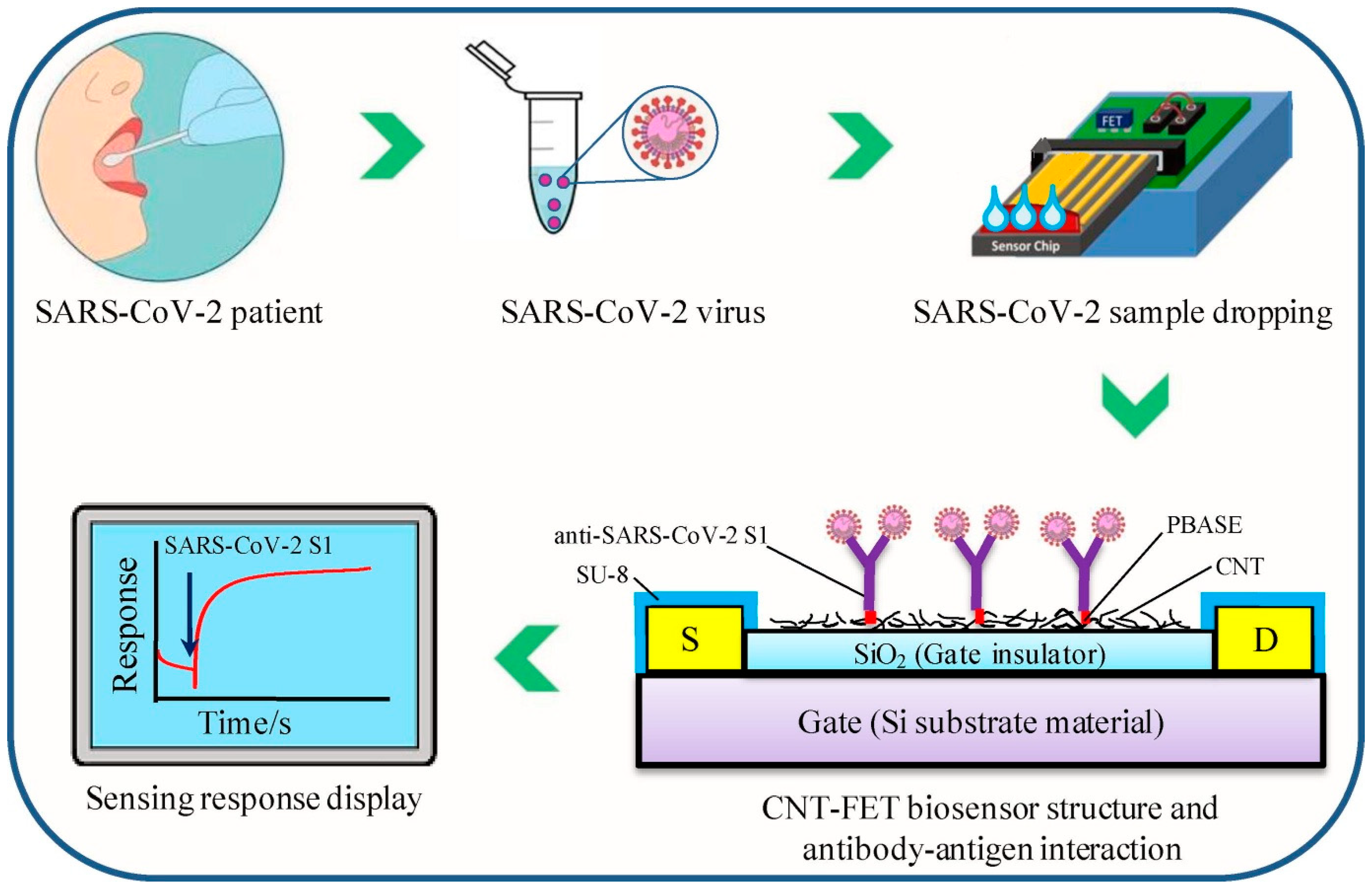
4.2. Infectious Disease Diagnosis
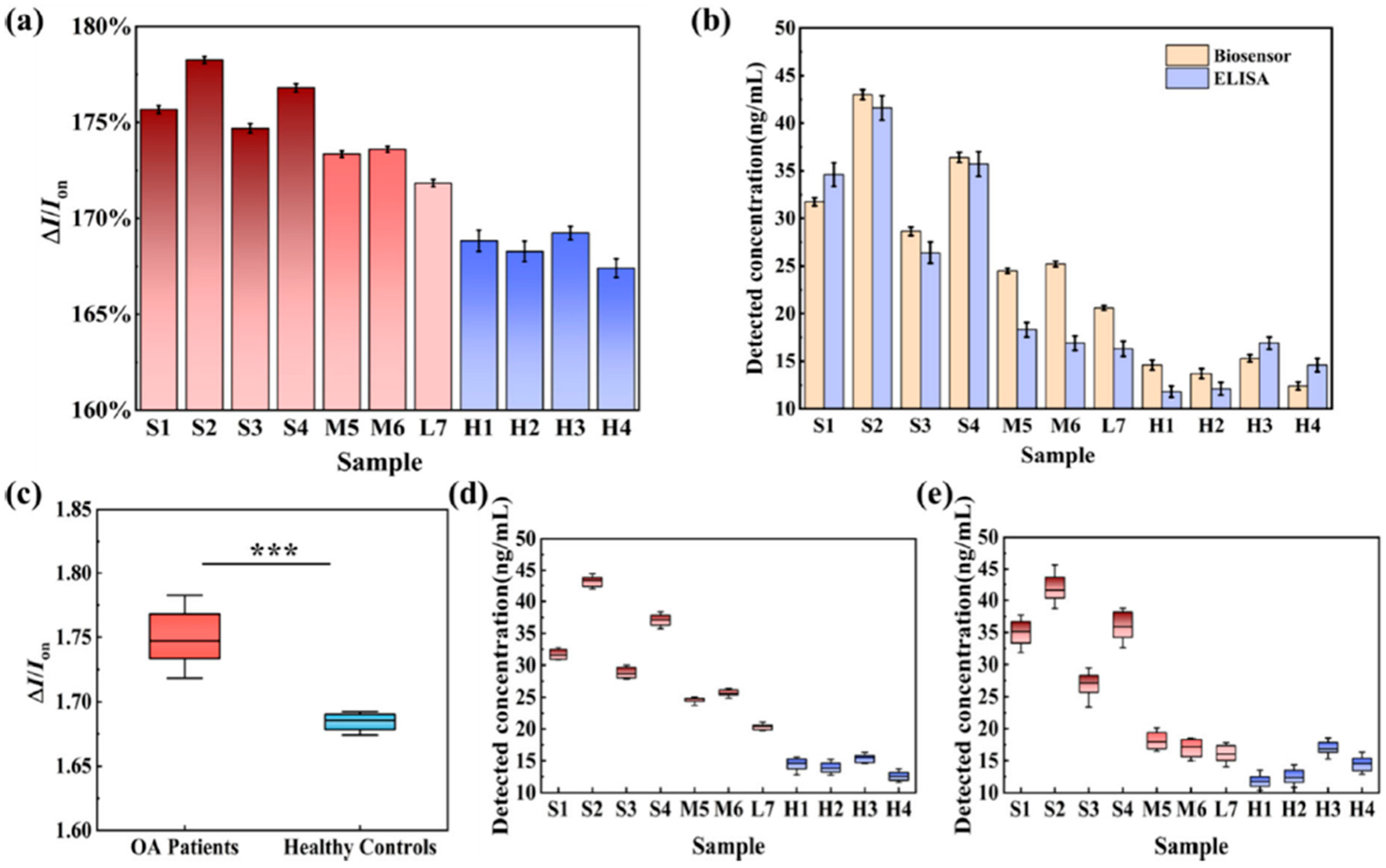
4.3. Neurological and Degenerative Disease Monitoring
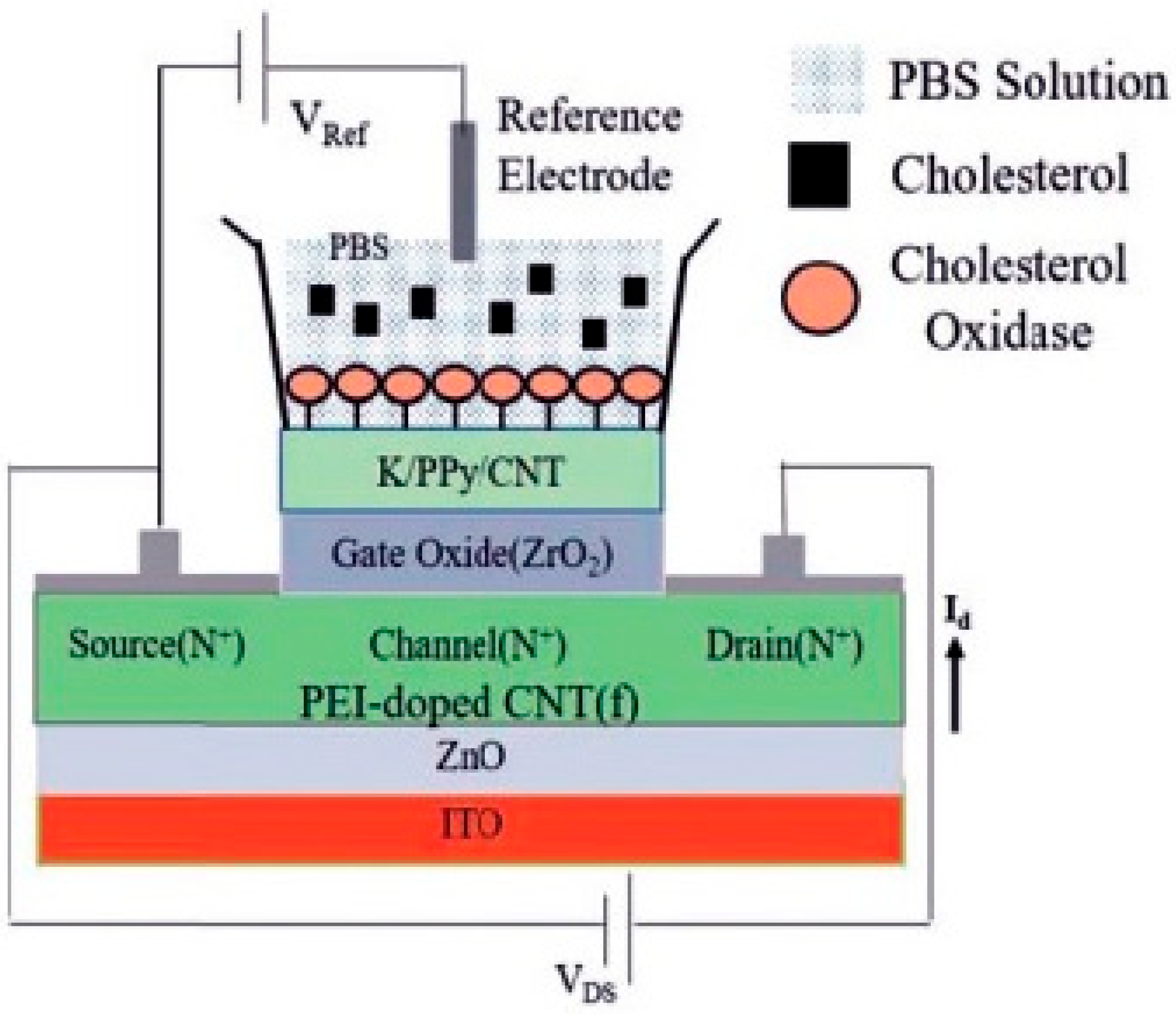
4.4. Cardiovascular and Metabolic Disorder Detection
5. Challenges and Future Perspectives
6. Conclusions
Author Contributions
Funding
Data Availability Statement
Acknowledgments
Conflicts of Interest
References
- Bhatia, D.; Paul, S.; Acharjee, T.; Ramachairy, S.S. Biosensors and Their Widespread Impact on Human Health. Sens. Int. 2024, 5, 100257. [Google Scholar] [CrossRef]
- Gwyther, R.E.A.; Côté, S.; Lee, C.-S.; Miao, H.; Ramakrishnan, K.; Palma, M.; Dafydd Jones, D. Optimising CNT-FET Biosensor Design through Modelling of Biomolecular Electrostatic Gating and Its Application to β-Lactamase Detection. Nat. Commun. 2024, 15, 7482. [Google Scholar] [CrossRef] [PubMed]
- Zhang, Z.; Hu, J.-J.; Lin, S.; Wu, J.; Xia, F.; Lou, X. Field Effect Transistor Biosensors for Healthcare Monitoring. Interdiscip. Med. 2024, 2, e20240032. [Google Scholar] [CrossRef]
- Ferrier, D.C.; Honeychurch, K.C. Carbon Nanotube (CNT)-Based Biosensors. Biosensors 2021, 11, 486. [Google Scholar] [CrossRef] [PubMed]
- Li, L.; Liu, X.; Wei, T.; Wang, K.; Zhao, Z.; Cao, J.; Liu, Y.; Zhang, Z. Carbon Nanotube Field-Effect Transistor Biosensor with an Enlarged Gate Area for Ultra-Sensitive Detection of a Lung Cancer Biomarker. ACS Appl. Mater. Interfaces 2023, 15, 27299–27306. [Google Scholar] [CrossRef] [PubMed]
- Wang, H.; Chen, R.; Zhang, F.; Yu, Z.; Wang, Y.; Tang, Z.; Yang, L.; Tang, X.; Xiong, B. Superhydrophobic Paper-Based Microfluidic Field-Effect Transistor Biosensor Functionalized with Semiconducting Single-Walled Carbon Nanotube and DNAzyme for Hypocalcemia Diagnosis. Int. J. Mol. Sci. 2022, 23, 7799. [Google Scholar] [CrossRef]
- Cui, Q.; Li, J.; Li, Y.; Tang, L.; Li, K.; Li, T.; Chen, X.; Zhang, Z.; Zhang, G.-J. Sensitive and Rapid Detection of Bacterial Endotoxin with a Functional Carbon Nanotube Field-Effect Transistor Biosensor. Talanta 2024, 266, 125035. [Google Scholar] [CrossRef]
- Li, Z.; Xiao, M.; Jin, C.; Zhang, Z. Toward the Commercialization of Carbon Nanotube Field Effect Transistor Biosensors. Biosensors 2023, 13, 326. [Google Scholar] [CrossRef]
- Deshmukh, M.A.; Jeon, J.-Y.; Ha, T.-J. Carbon Nanotubes: An Effective Platform for Biomedical Electronics. Biosens. Bioelectron. 2020, 150, 111919. [Google Scholar] [CrossRef]
- Sengupta, J. Key Aspects of Biosensing for Instant Screening Tests. Biosens. Bioelectron. X 2024, 20, 100529. [Google Scholar] [CrossRef]
- Syduzzaman, M.; Islam Saad, M.S.; Piam, M.F.; Talukdar, T.A.; Shobdo, T.T.; Pritha, N.M. Carbon Nanotubes: Structure, Properties and Applications in the Aerospace Industry. Results Mater. 2025, 25, 100654. [Google Scholar] [CrossRef]
- Sánchez-Romate, X.F.; Jiménez-Suárez, A.; Ureña, A. Electrical Properties of Carbon Nanotubes. In Handbook of Carbon Nanotubes; Abraham, J., Thomas, S., Kalarikkal, N., Eds.; Springer International Publishing: Cham, Switzerland, 2020; pp. 1–35. ISBN 9783319706146. [Google Scholar]
- Fuad, M.H.; Noor, S.S.; Nayan, M.F.; Mahmud, R.R. Comprehensive Analysis of Short-Channel Effects & Switching Speed in CNTFETs: A 2D Quantum Simulation Approach. Results Eng. 2025, 26, 104513. [Google Scholar] [CrossRef]
- Rahman, M.M.; Sarkar, B.; Rahman, M.T.; Jin, G.J.; Uddin, M.J.; Bhuiyan, N.H.; Shim, J.S. Development of a Highly Sensitive CNT-Metal Graphene Hybrid Nano-IDA Electrochemical Biosensor for the Diagnosis of Alzheimer’s Disease. Biomater. Sci. 2024, 12, 5203–5214. [Google Scholar] [CrossRef] [PubMed]
- Wang, J.; Xu, L. Continuous Blood Pressure Monitoring Based on Flexible CNT/Ecoflex Porous Composite Materials. Sens. Actuators A Phys. 2025, 386, 116370. [Google Scholar] [CrossRef]
- UrRehman, T.; Khan, S.A.; Shah, L.A.; Fu, J. Gum Arabic-CNT Reinforced Hydrogels: Dual-Function Materials for Strain Sensing and Energy Storage in next-Generation Supercapacitors. Mater. Adv. 2025, 6, 1288–1299. [Google Scholar] [CrossRef]
- Sengupta, J.; Hussain, C.M. Decadal Journey of CNT-Based Analytical Biosensing Platforms in the Detection of Human Viruses. Nanomaterials 2022, 12, 4132. [Google Scholar] [CrossRef]
- Lee, J.; Bak, M.; Yoo, P.J.; Kim, W.-J. Correlation between CNT Characteristics and the Conductivity of CNT Electrode Films: Optimization of Fabrication Conditions for Enhancing Electrical Properties. Appl. Surf. Sci. 2025, 688, 162440. [Google Scholar] [CrossRef]
- Al Baroot, A.; Elsayed, K.A.; Khan, F.A.; Haladu, S.A.; Ercan, F.; Çevik, E.; Drmosh, Q.A.; Almessiere, M.A. Anticancer Activity of Au/CNT Nanocomposite Fabricated by Nanosecond Pulsed Laser Ablation Method on Colon and Cervical Cancer. Micromachines 2023, 14, 1455. [Google Scholar] [CrossRef]
- Sengupta, J. Chapter 9—Carbon Nanotube Fabrication at Industrial Scale: Opportunities and Challenges. In Handbook of Nanomaterials for Industrial Applications; Mustansar Hussain, C., Ed.; Micro and Nano Technologies; Elsevier: Amsterdam, The Netherlands, 2018; pp. 172–194. ISBN 9780128133514. [Google Scholar]
- Krishna, A.; Aravinda, L.S.; Kumar, S.; Reddy, N.; Balashanmugam, N.; Mamilla, R.S. Synthesis and Thermal Simulations of Novel Encapsulated CNT Multifunctional Thin-Film Based Nanomaterial of SiO2-CNT and TiN-CNT by PVD and PECVD Techniques for Thermal Applications. Diam. Relat. Mater. 2020, 109, 108029. [Google Scholar] [CrossRef]
- Yuan, G.; Liu, Z.; Cao, Z.; Xie, J.; Li, H.; Li, L.; Sun, Y.; Tian, Y. Direct Growth of Vertically Well-Aligned Carbon Nanotube Arrays on Atomic Layer Deposition of ZnO Films. Chem. Phys. Lett. 2021, 773, 138602. [Google Scholar] [CrossRef]
- Karlsson, T.; Hallander, P.; Liu, F.; Poot, T.; Åkermo, M. Sensing Abilities of Embedded Vertically Aligned Carbon Nanotube Forests in Structural Composites: From Nanoscale Properties to Mesoscale Functionalities. Compos. Part B Eng. 2023, 255, 110587. [Google Scholar] [CrossRef]
- Bandaru, P.R. Electronic Transport and Electrical Properties of Carbon Nanotubes. In Handbook of Carbon Nanotubes; Abraham, J., Thomas, S., Kalarikkal, N., Eds.; Springer International Publishing: Cham, Switzerland, 2020; pp. 1–39. ISBN 9783319706146. [Google Scholar]
- Mirnezhad, M.; Ansari, R.; Falahatgar, S.R.; Aghdasi, P. Analyzing Fine Scaling Quantum Effects on the Buckling of Axially-Loaded Carbon Nanotubes Based on the Density Functional Theory and Molecular Mechanics Method. Sci. Rep. 2024, 14, 7435. [Google Scholar] [CrossRef]
- Charoenpakdee, J.; Suntijitrungruang, O.; Boonchui, S. Chirality Effects on an Electron Transport in Single-Walled Carbon Nanotube. Sci. Rep. 2020, 10, 18949. [Google Scholar] [CrossRef] [PubMed]
- Zhu, Y.; Chen, C.; Wu, S.; Cheng, R.; Cheng, L.; Zhou, W.-L. Edge-Dependent Ballistic Transport through Copper-Decorated Carbon-Nanotube–Graphene Covalent Junction with Low Schottky Barrier. J. Appl. Phys. 2020, 128, 064302. [Google Scholar] [CrossRef]
- Nurazzi, N.M.; Sabaruddin, F.A.; Harussani, M.M.; Kamarudin, S.H.; Rayung, M.; Asyraf, M.R.M.; Aisyah, H.A.; Norrrahim, M.N.F.; Ilyas, R.A.; Abdullah, N.; et al. Mechanical Performance and Applications of CNTs Reinforced Polymer Composites—A Review. Nanomaterials 2021, 11, 2186. [Google Scholar] [CrossRef] [PubMed]
- Kumanek, B.; Janas, D. Thermal Conductivity of Carbon Nanotube Networks: A Review. J. Mater. Sci. 2019, 54, 7397–7427. [Google Scholar] [CrossRef]
- Huang, Y.; Zhu, L.; Huang, Q.; He, Z. The Light Absorption Enhancement of Nanostructured Carbon-Based Coatings Fabricated by High-Voltage Electrostatic Spraying Technique. Opt. Mater. 2022, 133, 112902. [Google Scholar] [CrossRef]
- Meskher, H.; Chaudhery Mustansar, H.; Kumar Thakur, A.; Sathyamurthy, R.; Lynch, I.; Singh, P.; Kim Han, T.; Saidur, R. Recent Trends in Carbon Nanotube (CNT)-Based Biosensors for the Fast and Sensitive Detection of Human Viruses: A Critical Review. Nanoscale Adv. 2023, 5, 992–1010. [Google Scholar] [CrossRef]
- Demidenko, N.A.; Kuksin, A.V.; Molodykh, V.V.; Pyankov, E.S.; Ichkitidze, L.P.; Zaborova, V.A.; Tsymbal, A.A.; Tkachenko, S.A.; Shafaei, H.; Diachkova, E.; et al. Flexible Strain-Sensitive Silicone-CNT Sensor for Human Motion Detection. Bioengineering 2022, 9, 36. [Google Scholar] [CrossRef]
- Shkodra, B.; Petrelli, M.; Costa Angeli, M.A.; Garoli, D.; Nakatsuka, N.; Lugli, P.; Petti, L. Electrolyte-Gated Carbon Nanotube Field-Effect Transistor-Based Biosensors: Principles and Applications. Appl. Phys. Rev. 2021, 8, 041325. [Google Scholar] [CrossRef]
- Salah, L.S.; Ouslimani, N.; Bousba, D.; Huynen, I.; Danlée, Y.; Aksas, H. Carbon Nanotubes (CNTs) from Synthesis to Functionalized (CNTs) Using Conventional and New Chemical Approaches. J. Nanomater. 2021, 2021, 4972770. [Google Scholar] [CrossRef]
- Yao, X.; Zhang, Y.; Jin, W.; Hu, Y.; Cui, Y. Carbon Nanotube Field-Effect Transistor-Based Chemical and Biological Sensors. Sensors 2021, 21, 995. [Google Scholar] [CrossRef] [PubMed]
- Zahoor, F.; Hussin, F.A.; Khanday, F.A.; Ahmad, M.R.; Mohd Nawi, I. Ternary Arithmetic Logic Unit Design Utilizing Carbon Nanotube Field Effect Transistor (CNTFET) and Resistive Random Access Memory (RRAM). Micromachines 2021, 12, 1288. [Google Scholar] [CrossRef]
- Liu, X.; Sun, B.; Li, X.; Zhang, Z.; Wang, W.; Zhang, X.; Huang, Z.; Liu, H.; Chang, H.; Jia, R.; et al. Floating Gate Carbon Nanotube Dual-Gate Field-Effect Transistor for Reconfigurable AND/OR Logic Gates. ACS Appl. Electron. Mater. 2022, 4, 1684–1691. [Google Scholar] [CrossRef]
- Thanihaichelvan, M.; Browning, L.A.; Dierkes, M.P.; Reyes, R.M.; Kralicek, A.V.; Carraher, C.; Marlow, C.A.; Plank, N.O.V. Data on Liquid Gated CNT Network FETs on Flexible Substrates. Data Brief 2018, 21, 276–283. [Google Scholar] [CrossRef]
- Lv, J.; Shen, Z.; Meng, D.; Peng, L.-M.; Qiu, C. High-Performance Dual-Gate Transistors Based on Aligned Carbon Nanotubes. ACS Appl. Mater. Interfaces 2024, 16, 58864–58871. [Google Scholar] [CrossRef] [PubMed]
- Peng, B.; Annamalai, M.; Mothes, S.; Schröter, M. Device Design and Optimization of CNTFETs for High-Frequency Applications. J. Comput. Electron. 2021, 20, 2492–2500. [Google Scholar] [CrossRef]
- Bhookya, T.; Malyala, L.; Karingula, S.; Gobi, K.V. NanoAg Impregnated rGO-Wedged CNT Composite with Nafion Polymer for Facile Electrochemical Detection of Etilefrine. Microchem. J. 2025, 212, 113269. [Google Scholar] [CrossRef]
- Dai, B.; Zhou, R.; Ping, J.; Ying, Y.; Xie, L. Recent Advances in Carbon Nanotube-Based Biosensors for Biomolecular Detection. TrAC Trends Anal. Chem. 2022, 154, 116658. [Google Scholar] [CrossRef]
- Nguyen, T.T.-H.; Nguyen, C.M.; Huynh, M.A.; Vu, H.H.; Nguyen, T.-K.; Nguyen, N.-T. Field Effect Transistor Based Wearable Biosensors for Healthcare Monitoring. J. Nanobiotechnol. 2023, 21, 411. [Google Scholar] [CrossRef]
- Liu, D.; Shi, L.; Dai, Q.; Lin, X.; Mehmood, R.; Gu, Z.; Dai, L. Functionalization of Carbon Nanotubes for Multifunctional Applications. Trends Chem. 2024, 6, 186–210. [Google Scholar] [CrossRef]
- Kumar, S.; Sidhu, H.K.; Paul, A.K.; Bhardwaj, N.; Thakur, N.S.; Deep, A. Bioengineered Multi-Walled Carbon Nanotube (MWCNT) Based Biosensors and Applications Thereof. Sens. Diagn. 2023, 2, 1390–1413. [Google Scholar] [CrossRef]
- Feng, X.; Li, P.; Li, T.; Cao, X.; Liu, D.; Xiao, M.; Wang, L. Ultra-Sensitive and Rapid Detection of Salmonella enterica and Staphylococcus aureus to Single-Cell Level by Aptamer-Functionalized Carbon Nanotube Field-Effect Transistor Biosensors. Biosens. Bioelectron. 2024, 257, 116333. [Google Scholar] [CrossRef]
- Zamzami, M.A.; Rabbani, G.; Ahmad, A.; Basalah, A.A.; Al-Sabban, W.H.; Nate Ahn, S.; Choudhry, H. Carbon Nanotube Field-Effect Transistor (CNT-FET)-Based Biosensor for Rapid Detection of SARS-CoV-2 (COVID-19) Surface Spike Protein S1. Bioelectrochemistry 2022, 143, 107982. [Google Scholar] [CrossRef]
- Li, T.; Liang, Y.; Li, J.; Yu, Y.; Xiao, M.-M.; Ni, W.; Zhang, Z.; Zhang, G.-J. Carbon Nanotube Field-Effect Transistor Biosensor for Ultrasensitive and Label-Free Detection of Breast Cancer Exosomal miRNA21. Anal. Chem. 2021, 93, 15501–15507. [Google Scholar] [CrossRef]
- Keshwani, G.; Dutta, J.C. CNT Based High-κ Dielectric Ion Sensitive Field Effect Transistor Based Cholesterol Biosensor. Curr. Trends Biotechnol. Pharm. 2021, 15, 182–188. [Google Scholar] [CrossRef]
- Inoue, T.; Mizoguchi, K.; Tokita, M.; Shibuta, M.; Nakaya, M.; Eguchi, T.; Nakajima, A. Localized Surface Plasmon Resonances of Size-Selected Large Silver Nanoclusters (n = 70–100) Soft-Landed on a C60 Organic Substrate. Phys. Chem. Chem. Phys. 2024, 26, 16597–16602. [Google Scholar] [CrossRef]
- Li, T.; Tang, L.; Li, K.; Liu, B.; Xiao, M.-M.; Liu, N.; Ni, W.; Li, Y.; Zhang, Z.; Zhang, G.-J. Functionalized Carbon Nanotube Field-Effect Transistor Biosensor for Highly Sensitive Detection of Exosomal Protein. Anal. Chim. Acta 2023, 1273, 341511. [Google Scholar] [CrossRef] [PubMed]
- Liu, P.; Liu, Y.; Cai, B.; Li, J.; Zhang, G.-J. Probe-Screened Carbon Nanotube Field-Effect Transistor Biosensor to Enhance Breast Cancer-Related Gene Assay. Green Anal. Chem. 2025, 13, 100267. [Google Scholar] [CrossRef]
- Marques, I.; Pinto da Costa, J.; Justino, C.; Santos, P.; Duarte, K.; Freitas, A.; Cardoso, S.; Duarte, A.; Rocha-Santos, T. Carbon Nanotube Field Effect Transistor Biosensor for the Detection of Toxins in Seawater. Int. J. Environ. Anal. Chem. 2017, 97, 597–605. [Google Scholar] [CrossRef]
- Chen, H.; Xiao, M.; He, J.; Zhang, Y.; Liang, Y.; Liu, H.; Zhang, Z. Aptamer-Functionalized Carbon Nanotube Field-Effect Transistor Biosensors for Alzheimer’s Disease Serum Biomarker Detection. ACS Sens. 2022, 7, 2075–2083. [Google Scholar] [CrossRef] [PubMed]
- Lv, T.; Liu, J.; Li, F.; Ma, S.; Wei, X.; Li, X.; Han, C.; Wang, X. Label-Free and Ultrasensitive Detection of Cartilage Acidic Protein 1 in Osteoarthritis Using a Single-Walled Carbon Nanotube Field-Effect Transistor Biosensor. ACS Appl. Mater. Interfaces 2024, 16, 36804–36810. [Google Scholar] [CrossRef]
- Rajesh; Sharma, V.; Puri, N.K.; Mulchandani, A.; Kotnala, R.K. High Performance Dendrimer Functionalized Single-Walled Carbon Nanotubes Field Effect Transistor Biosensor for Protein Detection. Appl. Phys. Lett. 2016, 109, 243504. [Google Scholar] [CrossRef]
- Xu, J.; Xiao, Z.; Jia, C.; Wei, Y.; Sun, Y.; Kang, L.; Cui, N.; Li, P.; Lei, Y.; Ma, X. Progress in the Fabrication of High-Purity Semiconducting Carbon Nanotube Arrays. J. Mater. Chem. C 2025, 13, 4304–4326. [Google Scholar] [CrossRef]
- Kaushal, A.; Alexander, R.; Prakash, J.; Dasgupta, K. Engineering Challenges and Innovations in Controlled Synthesis of CNT Fiber and Fabrics in Floating Catalyst Chemical Vapor Deposition (FC-CVD) Process. Diam. Relat. Mater. 2024, 148, 111474. [Google Scholar] [CrossRef]
- Tomada, J.; Dienel, T.; Hampel, F.; Fasel, R.; Amsharov, K. Combinatorial Design of Molecular Seeds for Chirality-Controlled Synthesis of Single-Walled Carbon Nanotubes. Nat. Commun. 2019, 10, 3278. [Google Scholar] [CrossRef]
- Moyer-Vanderburgh, K.; Park, S.J.; Fornasiero, F. Growth of Carbon Nanotube Forests on Flexible Metal Substrates: Advances, Challenges, and Applications. Carbon 2023, 206, 402–421. [Google Scholar] [CrossRef]
- Yadav, M.D.; Patwardhan, A.W.; Joshi, J.B.; Dasgupta, K. Selective Synthesis of Metallic and Semi-Conducting Single-Walled Carbon Nanotube by Floating Catalyst Chemical Vapour Deposition. Diam. Relat. Mater. 2019, 97, 107432. [Google Scholar] [CrossRef]
- Wang, J.; Lei, T. Separation of Semiconducting Carbon Nanotubes Using Conjugated Polymer Wrapping. Polymers 2020, 12, 1548. [Google Scholar] [CrossRef]
- Ciria, J.C.; Ansón-Casaos, A.; Benito, A.M.; Maser, W.K. Rational Description and Modelling of the Separation of Nanotubes from Solid Nanoparticles in Centrifugation Processes. Carbon Trends 2021, 5, 100084. [Google Scholar] [CrossRef]
- Khamidullin, T.; Galyaltdinov, S.; Valimukhametova, A.; Brusko, V.; Khannanov, A.; Maat, S.; Kalinina, I.; Dimiev, A.M. Simple, Cost-Efficient and High Throughput Method for Separating Single-Wall Carbon Nanotubes with Modified Cotton. Carbon 2021, 178, 157–163. [Google Scholar] [CrossRef]
- Elgrabli, D.; Dachraoui, W.; de Marmier, H.; Ménard-Moyon, C.; Bégin, D.; Bégin-Colin, S.; Bianco, A.; Alloyeau, D.; Gazeau, F. Intracellular Degradation of Functionalized Carbon Nanotube/Iron Oxide Hybrids Is Modulated by Iron via Nrf2 Pathway. Sci Rep 2017, 7, 40997. [Google Scholar] [CrossRef]
- Xu, J.; Lee, H. Anti-Biofouling Strategies for Long-Term Continuous Use of Implantable Biosensors. Chemosensors 2020, 8, 66. [Google Scholar] [CrossRef]
- DeBrosse, M.C.; Suresh, S.; Karajic, A.; Kim, S.; Hussain, S.; Heikenfeld, J. A Novel Conductive Membrane Sensor Protection Technique to Mitigate Redox-Active Interferences. Biosens. Bioelectron. X 2023, 14, 100366. [Google Scholar] [CrossRef]
- Mondal, J.; An, J.M.; Surwase, S.S.; Chakraborty, K.; Sutradhar, S.C.; Hwang, J.; Lee, J.; Lee, Y.-K. Carbon Nanotube and Its Derived Nanomaterials Based High Performance Biosensing Platform. Biosensors 2022, 12, 731. [Google Scholar] [CrossRef]
- Meunier, J.-L.; Ouellet, J.; Basu, K.; Aufoujal, A.; Boudreault, R.; Tavares, J.R. Continuous Reactive-Roll-to-Roll Growth of Carbon Nanotubes for Fog Water Harvesting Applications. C 2024, 10, 9. [Google Scholar] [CrossRef]
- Adhikari, A.; Sengupta, J. Toxicity of Carbon Nanomaterials. In Environmental Applications of Carbon Nanomaterials-Based Devices; John Wiley & Sons, Ltd.: Hoboken, NJ, USA, 2021; pp. 147–171. ISBN 9783527830978. [Google Scholar]
- Karimi, F.; Alizadeh, S.; Alizadeh, H. Immunogenicity of multi-walled carbon nanotubes functionalized with recombinant protective antigen domain 4 toward development of a nanovaccine against anthrax. J. Drug Deliv. Sci. Technol. 2018, 47, 322–329. [Google Scholar] [CrossRef]
- Fraser, K.; Kodali, V.; Yanamala, N.; Birch, M.E.; Cena, L.; Casuccio, G.; Bunker, K.; Lersch, T.L.; Evans, D.E.; Stefaniak, A.; et al. Physicochemical Characterization and Genotoxicity of the Broad Class of Carbon Nanotubes and Nanofibers Used or Produced in U.S. Facilities. Part. Fibre Toxicol. 2020, 17, 62. [Google Scholar] [CrossRef]
- Rifa’i, A.F.; Kaavessina, M.; Distantina, S. Mechanical, Morphological, and Electrical Properties of Polylactic Acid-Based Conductive Polymer Composites through Polyethylene Glycol and Carbon Nanotubes Integration. Period. Polytech. Chem. Eng. 2025, 69, 149–162. [Google Scholar] [CrossRef]
- Soyalp, S.; Eksin, E.; Erdem, A. Chitosan and Carbon Nanotube Modified DNA Biosensor for Determination of Drug-DNA Interaction. J. Pharm. Biomed. Anal. Open 2025, 5, 100050. [Google Scholar] [CrossRef]
- Castro, V.O.; Livi, S.; Sperling, L.E.; dos Santos, M.G.; Merlini, C. Biodegradable Electrospun Conduit with Aligned Fibers Based on Poly(Lactic-Co-Glycolic Acid) (PLGA)/Carbon Nanotubes and Choline Bitartrate Ionic Liquid. ACS Appl. Bio Mater. 2024, 7, 1536–1546. [Google Scholar] [CrossRef] [PubMed]
- Shen, C.; Zou, G.; Guo, W.; Gao, H. Lipid Coating and End Functionalization Govern the Formation and Stability of Transmembrane Carbon Nanotube Porins. Carbon 2020, 164, 391–397. [Google Scholar] [CrossRef]
- Law, S.S.Y.; Kuzumoto, M.; Fujita, S.; Fujigaya, T.; Numata, K. Carbon Nanotubes Functionalized with α-Aminoisobutyric Acid-Containing Peptide Increase Gene Delivery Efficiency in Plant Mitochondria. Polym. J. 2024, 56, 915–924. [Google Scholar] [CrossRef]
- Peng, W.; Li, S.; Gao, H.; Su, M.; Zhou, Y.; Ding, Z.; Jiang, Q.; Yu, C. Non-Invasive Alcohol Biosensor Based on Gold Nanoparticles and Carbon Nanotubes Network for Dynamic Monitoring of Sweat Alcohol. Bioelectrochemistry 2025, 164, 108943. [Google Scholar] [CrossRef]
- Yang, M.; Zhang, M. Biodegradation of Carbon Nanotubes by Macrophages. Front. Mater. 2019, 6, 225. [Google Scholar] [CrossRef]
- Awasthi, S.; Srivastava, A.; Kumar, D.; Pandey, S.K.; Mubarak, N.M.; Dehghani, M.H.; Ansari, K. An Insight into the Toxicological Impacts of Carbon Nanotubes (CNTs) on Human Health: A Review. Environ. Adv. 2024, 18, 100601. [Google Scholar] [CrossRef]
- Daneshvar, F.; Chen, H.; Noh, K.; Sue, H.-J. Critical Challenges and Advances in the Carbon Nanotube–Metal Interface for next-Generation Electronics. Nanoscale Adv. 2021, 3, 942–962. [Google Scholar] [CrossRef]
- Shrestha, S.; Parajuli, S.; Park, J.; Yang, H.; Cho, T.-Y.; Eom, J.-H.; Cho, S.-K.; Lim, J.; Cho, G.; Jung, Y. Improving Stability of Roll-to-Roll (R2R) Gravure-Printed Carbon Nanotube-Based Thin Film Transistors via R2R Plasma-Enhanced Chemical Vapor-Deposited Silicon Nitride. Nanomaterials 2023, 13, 559. [Google Scholar] [CrossRef]
- Zhang, W.; Shrestha, S.; Parajuli, S.; Maskey, B.B.; Park, J.; Yang, H.; Jung, Y.; Cho, G. Tuning the Charge Carrier Polarity of Roll-to-Roll Gravure Printed Carbon Nanotube-Based Thin Film Transistors by an Atomic Layer Deposited Alumina Nanolayer. Nanoscale Adv. 2023, 5, 3879–3886. [Google Scholar] [CrossRef]
- Li, J.; Li, M.; Chen, Z.; Shao, S.; Gu, W.; Gu, Y.; Fang, Y.; Zhao, J. Large Area Roll-to-Roll Printed Semiconducting Carbon Nanotube Thin Films for Flexible Carbon-Based Electronics. Nanoscale 2023, 15, 5317–5326. [Google Scholar] [CrossRef]
- Zhang, Q.; Ren, Y.; Wang, Z.; Chen, X.; Portilla, L.; Sun, L.; Zhang, D.; Zhao, J. Preparation of Large-Area, High-Performance Single-Walled Carbon Nanotube (SWCNT)-Based Heater Films by Roll-to-Roll Gravure Printing. Flex. Print. Electron. 2022, 7, 015007. [Google Scholar] [CrossRef]
- Li, C.; Guo, X.; Zhou, Y.; Zhou, F.; Li, Y.; Wu, S.; Jerrams, S.; Chen, S.; Jiang, L. Biodegradable Poly(Lactic Acid) Blocked Polyurethane/Carbon Nanotubes Coated Cotton Fabric Prepared by Ultrasonic-Assisted Inkjet Printing for High Performance Strain Sensors. Int. J. Biol. Macromol. 2024, 274, 133269. [Google Scholar] [CrossRef]
- Jiang, R.; Liu, J.; Yang, K.; Zhao, J.; Zeng, B. Enhanced Field Emission of Single-Wall Carbon Nanotube Cathode Prepared by Screen Printing with a Silver Paste Buffer Layer. Nanomaterials 2022, 12, 165. [Google Scholar] [CrossRef]
- Hu, Y.; Peng, L.-M.; Xiang, L.; Zhang, H. Flexible Integrated Circuits Based on Carbon Nanotubes. Acc. Mater. Res. 2020, 1, 88–99. [Google Scholar] [CrossRef]
- Zhang, S.; Pang, J.; Li, Y.; Yang, F.; Gemming, T.; Wang, K.; Wang, X.; Peng, S.; Liu, X.; Chang, B.; et al. Emerging Internet of Things Driven Carbon Nanotubes-Based Devices. Nano Res. 2022, 15, 4613–4637. [Google Scholar] [CrossRef]
- Cho, M.-Y.; Kim, S.H.; Kim, J.S. Real-Time Position Detecting of Large-Area CNT-Based Tactile Sensors Based on Artificial Intelligence. Korean J. Met. Mater. 2022, 60, 793–799. [Google Scholar] [CrossRef]
- Geethukrishnan; Apte, O.; Tadi, K.K. Electrochemical Sensor for Dopamine Detection Based on Multiwalled Carbon Nanotube/Molybdenum Disulfide Quantum Dots with Machine Learning Integration and Anti-Interference Capability. Tungsten 2025, 7, 137–151. [Google Scholar] [CrossRef]
- Emami, H.; Mahinnezhad, S.; Al Shboul, A.; Ketabi, M.; Shih, A.; Izquierdo, R. Flexible Chemiresistive pH Sensor Based on Polyaniline/Carbon Nanotube Nanocomposite for IoT Applications. In Proceedings of the 2021 IEEE Sensors, Sydney, Australia, 31 October–3 November 2021; pp. 1–4. [Google Scholar]
- Ashraf, G.; Ahmed, K.; Aziz, A.; Asif, M.; Kong, J.; Fang, X. Microneedle Wearables in Advanced Microsystems: Unlocking next-Generation Biosensing with AI. TrAC Trends Anal. Chem. 2025, 187, 118208. [Google Scholar] [CrossRef]
- Jeon, D.-Y.; Park, J.; Lee, C.; Kim, S.M. Carbon Nanotube Fiber-Based High-Performance Force and Position Sensors for Smart Fabrics and the Internet of Things. APL Mater. 2023, 11, 041129. [Google Scholar] [CrossRef]
- Hosain, M.N.; Kwak, Y.-S.; Lee, J.; Choi, H.; Park, J.; Kim, J. IoT-Enabled Biosensors for Real-Time Monitoring and Early Detection of Chronic Diseases. Phys. Act. Nutr. 2024, 28, 060–069. [Google Scholar] [CrossRef]
- Gil Rosa, B.; Akingbade, O.E.; Guo, X.; Gonzalez-Macia, L.; Crone, M.A.; Cameron, L.P.; Freemont, P.; Choy, K.-L.; Güder, F.; Yeatman, E.; et al. Multiplexed Immunosensors for Point-of-Care Diagnostic Applications. Biosens. Bioelectron. 2022, 203, 114050. [Google Scholar] [CrossRef] [PubMed]
- Khan, N.I.; Song, E. Lab-on-a-Chip Systems for Aptamer-Based Biosensing. Micromachines 2020, 11, 220. [Google Scholar] [CrossRef] [PubMed]
- Dai, H.; Thostenson, E.T. Scalable and Multifunctional Carbon Nanotube-Based Textile as Distributed Sensors for Flow and Cure Monitoring. Carbon 2020, 164, 28–41. [Google Scholar] [CrossRef]
- Arun Kumar, D.S.; Chauhan, S.S.; Bharathi, K.D.; Ravikumar, A.; Rahman, M.R. Flexible and Cost Effective CNT Coated Cotton Fabric for CO Gas Sensing Application. Sens. Actuators A Phys. 2023, 362, 114640. [Google Scholar] [CrossRef]
- Nedelcu, S.; Thodkar, K.; Hierold, C. A Customizable, Low-Power, Wireless, Embedded Sensing Platform for Resistive Nanoscale Sensors. Microsyst. Nanoeng. 2022, 8, 10. [Google Scholar] [CrossRef]
- Luo, Y.; Abidian, M.R.; Ahn, J.-H.; Akinwande, D.; Andrews, A.M.; Antonietti, M.; Bao, Z.; Berggren, M.; Berkey, C.A.; Bettinger, C.J.; et al. Technology Roadmap for Flexible Sensors. ACS Nano 2023, 17, 5211–5295. [Google Scholar] [CrossRef]


| Substrate | Source/Drain Material | Gate Type | Active Layer/ Transducer | Receptor | Target | Sample/Sample Mixed in | LOD | Sensitivity | Detection Range | Response Time | Identified Diseases | Ref |
|---|---|---|---|---|---|---|---|---|---|---|---|---|
| SiO2/Si | Ti/Pd/Au | Floating gate | CNT | DNA probe | miRNA21 | Plasma | 0.87 aM | NA | 10 aM to 100 nM | NA | Breast cancer | [48] |
| SiO2/Si | Au | Solution gate | Poly-lysine functionalised CNT | Sulfhydryl aptamer probe | exosomal protein (MUC1) | Plasma | 0.34 fg/mL | NA | 1 fg/mL to 100 pg/mL | <1 h | Breast cancer | [51] |
| SiO2/Si | Ti/Pd/Au | Liquid gate | CNT | DNA, PMO, PNA probes | BRCA1 | Serum | 1.38 aM | NA | 1 aM to 100 pM | 30 min | Breast cancer | [52] |
| SiO2/Si | Ti/Pd/Au | Floating gate | CNT | Monoclonal carcinoembryonic antibody | Carcinoembryonic antigen (CEA) | Serum | 72 ag/mL | NA | 1 fg/mL to 1 ng/mL | 1 h | Lung cancer | [5] |
| SiO2/Si | Ti/Au | Back gated | CNT | SARS-CoV-2 S1 antibody | SARS-CoV-2 S1 antigen | Saliva | 4.12 fg/mL | NA | 0.1 fg/mL to 5.0 pg/mL | 2 to 3 min | COVID-19 | [47] |
| SiO2/Si | Au | Liquid gate | cGQD coupled CNT | Polymyxin B | Bacterial endotoxin | Serum | 30.3 fg/mL | NA | 100 fg/mL to 1 ng/mL | 3 min | Fatal fever | [7] |
| SiO2/Si | Ti/Pd/Au | Liquid gate | CNT | Nucleic acid aptamers | Salmonella enterica and Staphylococcus aureus | Pork, Chicken, Beef, Milk, Fish, and Eggs | 1 CFU (S. enterica) and 1.2 CFUs (S. aureus) | NA | 102 to 1.28 × 10⁴ CFU/mL | 200 s | Foodborne diseases | [46] |
| SiO2/Si | Ti/Au | Back-gate | CNT | Antibodies specific to domoic acid | Domoic acid (Neurotoxin) | Seawater | 10 ng/L | NA | 10 to 500 ng/L | 15 min | Amnesic shellfish poisoning | [53] |
| SiO2/Si | Ti/Pd/Au | Liquid gate | CNT | Oligonucleotide aptamers | AD core blood biomarkers of β-amyloid (Aβ) | Serum | 50 aM | NA | 1 fM to 10 pM | 30 min | Alzheimer’s disease | [54] |
| SiO2/Si | Ti/Pd/Au | Bottom-gated | SWCNT | CRTAC1 antibody | Cartilage Acidic Protein 1 (CRTAC1) | Serum | 0.2 fg/mL | NA | 1 fg/mL to 100 ng/mL | 30 min | Osteoarthritis | [55] |
| ITO | Al | Solution-gated | Polyethylenimine (PEI) doped CNT | Potassium doped CNT with polypyrrole | Cholesterol | PBS | 0.23 mM | 60 mV/decade | 0.5 to 25 mM | ~1 s | Heart disease | [49] |
| Cellulose paper | Ag | Back gated | SWCNT | Dual-MFB | Ca2+ | Cow blood | 10.7 μM | NA | 25 μM to 5 mM | 7 min | Hypocalcemia | [6] |
| SiO2/Si | Ti/Au | Back gated | SWCNT | anti-CRP | CRP | CRP in Phosphate Buffer Saline | 85 pM | 20% change in current per decade | 0.01 to 10 µg/mL | 10 min | Inflammatory diseases | [56] |
Disclaimer/Publisher’s Note: The statements, opinions and data contained in all publications are solely those of the individual author(s) and contributor(s) and not of MDPI and/or the editor(s). MDPI and/or the editor(s) disclaim responsibility for any injury to people or property resulting from any ideas, methods, instructions or products referred to in the content. |
© 2025 by the authors. Licensee MDPI, Basel, Switzerland. This article is an open access article distributed under the terms and conditions of the Creative Commons Attribution (CC BY) license (https://creativecommons.org/licenses/by/4.0/).
Share and Cite
Sengupta, J.; Hussain, C.M. Carbon Nanotube-Based Field-Effect Transistor Biosensors for Biomedical Applications: Decadal Developments and Advancements (2016–2025). Biosensors 2025, 15, 296. https://doi.org/10.3390/bios15050296
Sengupta J, Hussain CM. Carbon Nanotube-Based Field-Effect Transistor Biosensors for Biomedical Applications: Decadal Developments and Advancements (2016–2025). Biosensors. 2025; 15(5):296. https://doi.org/10.3390/bios15050296
Chicago/Turabian StyleSengupta, Joydip, and Chaudhery Mustansar Hussain. 2025. "Carbon Nanotube-Based Field-Effect Transistor Biosensors for Biomedical Applications: Decadal Developments and Advancements (2016–2025)" Biosensors 15, no. 5: 296. https://doi.org/10.3390/bios15050296
APA StyleSengupta, J., & Hussain, C. M. (2025). Carbon Nanotube-Based Field-Effect Transistor Biosensors for Biomedical Applications: Decadal Developments and Advancements (2016–2025). Biosensors, 15(5), 296. https://doi.org/10.3390/bios15050296