Review

Doping Detection Based on the Nanoscale: Biosensing Mechanisms and Applications of Two-Dimensional Materials

Shanghai Institute of Doping Analyses, Shanghai University of Sport, Shanghai 200438, China
*
Author to whom correspondence should be addressed.
These authors contributed equally to this work.
Biosensors 2025, 15(4), 227; https://doi.org/10.3390/bios15040227
Submission received: 19 February 2025 / Revised: 30 March 2025 / Accepted: 1 April 2025 / Published: 3 April 2025

Abstract

:
Doping undermines fairness in sports and threatens athlete health, while conventional detection methods like LC-MS and GC-MS face challenges such as complex procedures, matrix interferences, and lengthy processing times, limiting on-site applications. Two-dimensional (2D) materials, including graphene, MoS2, and metal–organic frameworks (MOFs), offer promising solutions due to their large surface areas, tunable electronic structures, and special interactions with doping agents, such as hydrogen bonding, π-π stacking, and electrostatic forces. These materials enable signal transduction through changes in conductivity or fluorescence quenching. This review highlights the use of 2D materials in doping detection. For example, reduced graphene oxide–MOF composites show high sensitivity for detecting anabolic steroids like testosterone, while NiO/NGO nanocomposites exhibit strong selectivity for stimulants like ephedrine. However, challenges such as environmental instability and high production costs hinder their widespread application. Future efforts should focus on improving material stability through chemical modifications, reducing production costs, and integrating these materials into advanced systems like machine learning. Such advancements could revolutionize doping detection, ensuring fairness in sports and protecting athlete health.

1. Introduction

In the context of the highly competitive sports environment, the use of performance-enhancing substances has become a significant issue that threatens both the fairness of sports and the health of athletes [1]. The use of doping not only undermines the integrity of competitions but may also have long-term negative impacts on athletes’ physical health [2]. Although the World Anti-Doping Agency (WADA) has implemented various policies and measures to regulate athletes, reports of doping remain prevalent, with many athletes resorting to such substances in pursuit of improved performance or enhanced stimulation [3,4,5]. Therefore, the effective detection and control of doping substances has become an urgent task that requires the concerted efforts of both the sports and scientific communities.
Traditional methods for detecting doping, such as liquid chromatography–mass spectrometry (LC-MS) [6] and gas chromatography–mass spectrometry (GC-MS) [7], while offering high sensitivity, require complex sample preparation [8] and are easily interfered with by the sample matrix. Furthermore, these methods typically involve lengthy processing times, making on-site, real-time testing during competitions or athletes’ training sessions difficult [9,10]. This increases the risk of unethical individuals attempting to cheat during the testing period. Moreover, with new doping strategies constantly emerging, traditional detection methods are struggling to keep up and failing to meet the ever-increasing demands of testing. Accurate and timely detection of doping is essential, as it not only helps identify violations quickly but also serves as a strong deterrent against potential doping violations, reinforcing the fairness, integrity, and purity of sports competitions. Therefore, the development and application of nanoscale detection technology offer great potential for advancing regulatory management and strengthening the integrity of the sports sector.
Two-dimensional (2D) materials are a class of layered materials composed of one or more layers of inorganic or organic molecules or polymers [11], usually with a thickness of a few nanometers in the vertical direction [12]. The interaction between layers is mainly driven by van der Waals forces, so the layers can be effectively peeled and stacked during the manufacturing process, and the thickness is expressed in angstroms when materials reach the atomic level [13]. As shown in Figure 1, 2D materials can be divided into six categories: single-element 2D materials (Xenes), transition metal sulfides (TMDCs), hexagonal boron nitride (h-BN), MXenes, 2D organic materials, and other 2D materials [14]. Typical single-element materials include graphene, black sintering, silicene, etc., which are composed of a single element to form a layered structure. The chemical formula of Xenes is MX2, where M is a transition metal, commonly Mo and W, and X is a chalcogen element, such as S, Se, and Te. Two-dimensional organic materials are further divided into metal–organic frameworks (MOFs) and covalent organic frameworks (COFs), the first coordinated by metal ions and the second connected by covalent bonds. The development of two-dimensional materials is changing with each passing day. Common ones include two-dimensional oxides, two-dimensional perovskites, and two-dimensional heterojunctions. Their physical and chemical properties in two-dimensional structures are different from those in three-dimensional structures. Researchers have explored their excellent performance in the field of optoelectronics, and they have been widely used in devices such as thin-film transistors, solar cells, organic light-emitting diodes (OLEDs), and sensors [15,16,17,18,19,20,21,22,23]. Two-dimensional materials have great application potential in flexible electronic devices, photoelectric conversion, and information storage [24,25,26,27]. This article focuses on the application of two-dimensional materials and detection, especially doping detection, and attempts to improve detection efficiency using two-dimensional materials to enrich small molecules or macromolecules and to improve the signals of common instruments. The development of sensors in biomedicine is a fast-growing area, where two-dimensional materials are used to design wearable devices or sensors, converting the test object into photoelectric signals, improving the response speed, and focusing on real-time detection [28]. In the field of doping detection, most of the electrochemistry and two-dimensional materials are combined to improve sensitivity [28,29,30,31].
This review focuses on the detection of doping at the nanoscale, with a particular emphasis on the biosensing mechanisms and applications of 2D materials. It explores their binding mechanisms and selective recognition processes, as well as the intrinsic interactions between 2D materials and doping molecules. Additionally, the review discusses the development prospects of these materials for doping detection.

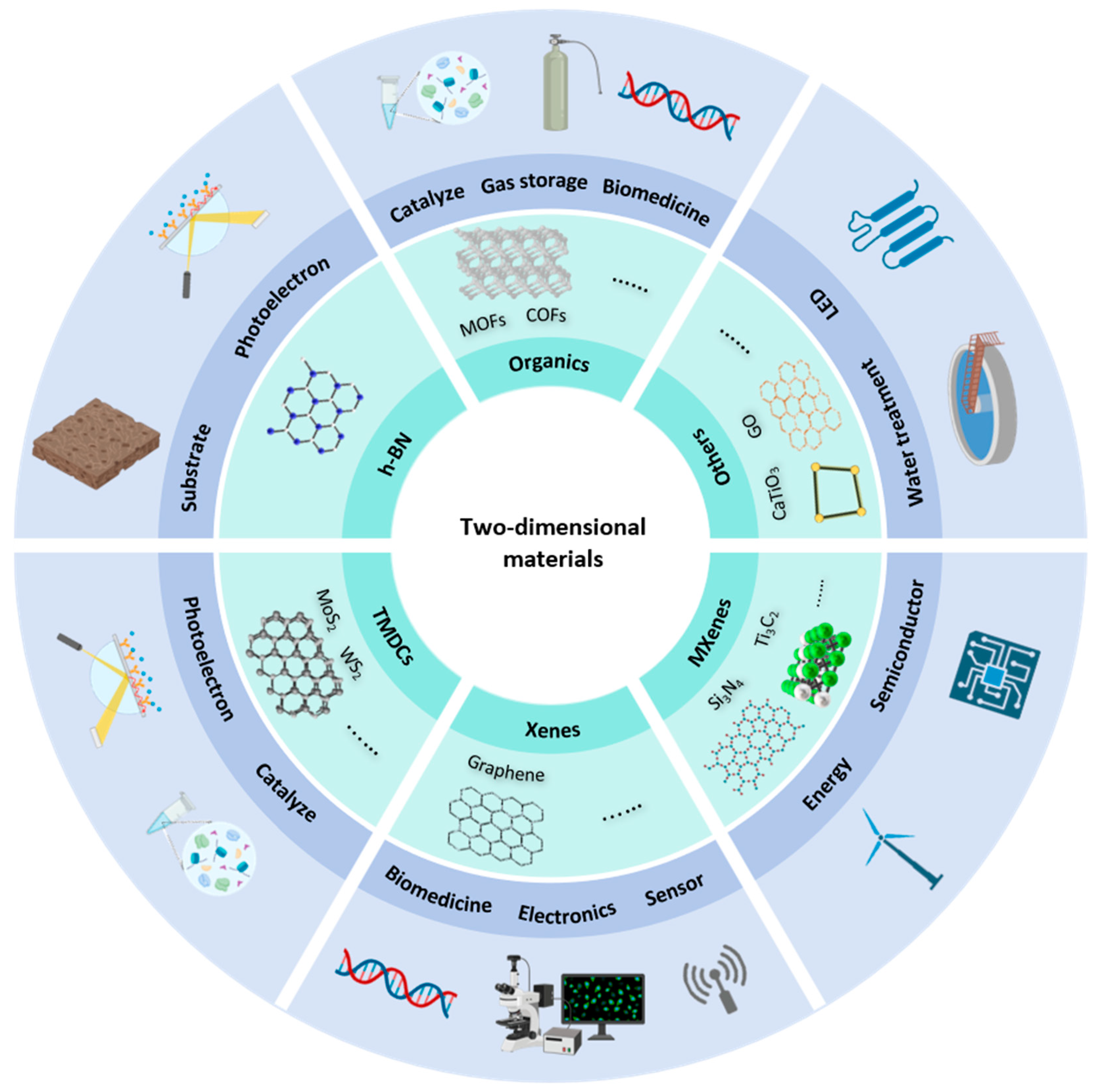
2. Characteristics of 2D Materials

2.1. Structure and Properties

Detection of doping at the nanoscale is gradually becoming an important research direction in the field of biomedical science [32,33]. In particular, 2D materials exhibit great potential due to their unique physical and chemical properties. With atomic-level thickness and a 2D planar structure, they offer exceptional surface sensitivity and selectivity, making them ideal platforms for biosensing applications [34,35,36,37,38].
Graphene is a prominent representative of 2D materials, renowned for its unique structure and remarkable properties. As shown in Figure 2a, it consists of carbon atoms arranged in a hexagonal honeycomb lattice, with each carbon atom bonded to three neighboring carbon atoms through sp2 hybridized orbitals, forming σ-bonds with equal bond lengths of approximately 0.142 nm and bond angles of 120°. This highly symmetric arrangement results in a regular hexagonal lattice that extends across the entire plane [39,40,41]. The strong covalent bonds between the carbon atoms impart significant structural stability to the material [42]. Additionally, each carbon atom retains one unhybridized p-electron, which is orthogonal to the graphene plane. These p-electrons form off-plane π-bonds, which are delocalized over the entire structure [43]. The presence of these off-plane π-bonds not only plays a crucial role in the electrical and optical properties of graphene but also facilitates interactions with other molecules via π-π stacking interactions [44].
MoS2, a transition metal sulphone, has a unique crystal structure. As shown in Figure 2b, it consists of three atomic layers arranged in a “sandwich” configuration, with molybdenum (Mo) atoms residing in the central layer, while sulfur (S) atoms form the upper and lower layers [47,48]. These atoms are firmly bonded by covalent interactions within each layer, forming a stable and orderly structure [45]. The interlayer interactions, however, are governed by weaker van der Waals forces, allowing for the easy exfoliation of MoS2 into monolayers or few-layer structures. This layered structure endows MoS2 with remarkable surface characteristics, where the exposed Mo and S atoms act as active sites. These sites are highly reactive and can engage in specific interactions with biomolecules, functioning like “keys” that selectively bind to corresponding “locks” on biomolecules [49,50]. Such interactions occur through ligand binding or electrostatic forces, facilitating the attachment of proteins, nucleic acids, and other biomolecules to the MoS2 surface. This selective binding capability enhances the sensitivity and specificity of MoS2-based biosensors, providing a reliable foundation for biosensing applications [51,52]. In addition to its surface reactivity, MoS2 also exhibits excellent chemical stability. Under appropriate conditions, MoS2 maintains its structural integrity and performance across a variety of complex biological environments, which is essential for the sustained function of biosensors [53]. This stability ensures that MoS2-based sensors can operate reliably over extended periods, thereby expanding their applicability in practical detection scenarios, including doping detection, where accuracy and long-term stability are paramount. Consequently, MoS2 holds significant promise as a material for advancing biosensor technologies, offering both high performance and versatility in real-world applications [54,55].
Metal–organic frameworks (MOFs) represent a class of advanced materials in the field of materials science distinguished by their porous, periodic network structures formed through the self-assembly of metal ions or metal clusters with organic ligands [46]. The intricate structural characteristics of MOFs confer exceptional physicochemical properties, which make them highly suitable for a wide range of applications, particularly in the realm of biosensing. One of the most notable features of MOFs is their extremely high porosity and specific surface area. The internal pore structure of MOFs is both complex and highly developed, with specific surface areas that can reach several thousand square meters per gram [56]. This high surface area renders MOFs highly effective as “molecular adsorption platforms”, enabling them to adsorb a large variety of molecules, including biomolecules. The double-walled aluminum-based MOF shown in Figure 2c has a micropore specific surface area of 2235 m2/g, and its excellent chemical stability enables it to effectively adsorb trace amounts of benzene [46]. The abundance of available binding sites on the MOF surface significantly enhances the sensitivity of biosensors, allowing for the precise capture and detection of even trace amounts of biomolecules. This feature is particularly advantageous in applications where high detection sensitivity is required, such as in the detection of low-abundance analytes [57,58]. In addition to their high porosity, MOFs offer remarkable design flexibility [59]. The structure of MOFs can be tailored at the molecular level by selecting different metal ions and organic ligands, which confers a high degree of customization in terms of both structure and function. This structural tunability allows for the design of MOFs with specific properties suited to particular applications [60]. For instance, the incorporation of organic ligands with biomolecular recognition capabilities enables MOFs to act as “molecular-specific probes”, capable of selectively binding to target biomolecules. This selectivity improves the specificity of biosensors, effectively minimizing interference from non-relevant molecules and ensuring the accuracy and reliability of biosensing assays [61,62,63]. Furthermore, MOFs exhibit a range of physicochemical properties that can be leveraged for biosensing applications. Depending on the choice of metal ions and organic ligands, MOFs can display fluorescence, magnetism, and other properties that enhance their functionality [64,65]. These characteristics provide multiple detection modalities, such as fluorescence-based real-time monitoring of biomolecular dynamics and the use of magnetic properties for the rapid separation and enrichment of biomolecules. These capabilities not only improve the sensitivity and efficiency of biosensing but also facilitate the development of more sophisticated detection strategies for real-time, high-throughput applications [66,67]. Therefore, the combination of high porosity, structural design flexibility, and multifunctional physicochemical properties makes MOFs an ideal material for advancing biosensor technologies.
This review primarily focuses on the application of two-dimensional materials in the field of stimulant detection. The properties of other two-dimensional materials that have not been applied in stimulant detection will not be discussed further in this review.

2.2. Advantages of Applications in Biosensing

As biosensor technologies continue to evolve, 2D materials are poised to play a central role in the next generation of diagnostic devices, providing innovative solutions for a wide array of applications in medicine, environmental monitoring, and beyond. The combination of advanced materials and nanotechnology is expected to drive significant advancements in the speed, accuracy, and scalability of biosensing platforms, positioning them as essential tools for addressing global health challenges.
From the perspective of high sensitivity, the unique atomic-scale thickness and 2D planar structure of 2D materials confer exceptional properties, including an ultra-high specific surface area and a wealth of active sites [25,35]. These characteristics significantly enhance their ability to interact with and detect biomolecules, making them ideal candidates for sensitive biosensing applications. For instance, graphene, with its theoretical specific surface area exceeding 2600 m2/g [68], provides an expansive surface for the adsorption of biomolecules. This extensive adsorption capacity allows for the enrichment of biomolecules at the surface, thereby amplifying the detection signal and enhancing the sensitivity of biosensors [69,70,71]. Similarly, the active sites on the surface of MoS2 offer highly specific interactions with biomolecules, functioning as precise molecular “grasping hands” that can selectively capture target molecules [72]. These interactions are particularly advantageous for detecting low-concentration biomarkers or drug components, even in trace amounts. Molybdenum disulfide-based sensors exhibit high sensitivity, with a detection range capable of reaching femtomolar levels (as shown in Figure 3), accurately detecting trace amounts of biomolecules, which is crucial for applications that require the detection of trace substances in biological samples or drug testing [73]. Such capabilities are particularly relevant for early disease diagnosis and pharmacokinetic studies, where the ability to detect small amounts of biomarkers or pharmaceutical compounds can provide valuable insights into disease progression and drug development [72,73,74,75]. These materials, such as graphene [76] and molybdenum disulfide (MoS2) [77], with their ultra-high surface area and specific molecular interactions, are thus poised to play a pivotal role in advancing the field of biosensing, providing robust technical support for early disease diagnosis, personalized medicine, and the monitoring of drug efficacy in the context of pharmaceutical research and development. By refining sensor architecture and experimental protocols, detection limits for these materials have reached the picomolar and even femtomolar levels, surpassing the performance of traditional detection methodologies [78,79,80]. For instance, a research team developed an electrochemical sensing platform integrating molybdenum disulfide with carbon nanodots, achieving ultra-sensitive detection of salicylic acid, catechol, and resorcinol at trace levels [81]. This platform demonstrated exceptional sensitivity and the ability to detect multiple substances simultaneously, underscoring the value of 2D materials in complex analytical contexts.
In terms of high specificity, highly selective recognition of target biomolecules or drug molecules can be achieved through the strategic design of the structure and ligands of MOFs, as well as through meticulous surface modification of materials such as zinc oxide (ZnO) [82,83]. By incorporating ligands with tailored recognition capabilities, MOFs can be engineered to selectively capture target molecules, such as proteins, nucleic acids, or metabolites, functioning as customized “molecular locks” that facilitate highly specific molecular interactions. The cavities in the molecularly imprinted polymers shown in Figure 4 can specifically recognize the target, blocking electron transfer and “turning off” the response signal, thereby achieving selective detection of tetracycline (TC). This design approach significantly reduces the likelihood of non-specific binding, thereby minimizing interference from structurally similar molecules and enhancing the precision of the detection process [84]. This highly selective binding is particularly valuable when detecting specific targets in complex biological matrices, where the presence of numerous other biomolecules may otherwise complicate detection efforts. The high specificity of these materials ensures the accuracy and reliability of biosensing assays, which is critical in reducing false-positive and false-negative results—issues that can have significant consequences in clinical diagnostics, forensic analysis, and personalized medicine [85,86,87]. In applications such as clinical diagnostics, where accurate identification of biomarkers is essential for disease detection and monitoring, or in forensic identification, where the precise analysis of biological samples is required, the ability to achieve highly specific recognition of target molecules is crucial. Moreover, in the field of personalized medicine, where treatment strategies are tailored to the individual patient’s molecular profile, the use of highly specific biosensors can enable more accurate drug monitoring and therapeutic interventions. Therefore, the high specificity of MOF-based and surface-modified materials provides a critical advantage in these applications, ensuring the reliability and accuracy of biosensing technologies in real-world scenarios.
The stability of 2D materials is another critical advantage, particularly in the context of biosensing applications. The chemical stability of MoS2, for instance, allows it to maintain its structural integrity and functional properties across a range of complex chemical environments. These include biological samples with varying pH levels (acidic or alkaline) and the presence of diverse biological enzymes and electrolytes [47]. Such stability ensures the reliable performance of MoS2-based biosensors in biological settings, where the material is often exposed to dynamic and challenging conditions. Similarly, the biocompatibility of ZnO is a key attribute that enables its use in biosensing applications. ZnO exhibits minimal interaction with living organisms, thus avoiding adverse immune responses or biotoxicity. This biocompatibility ensures that ZnO-based biosensors can operate stably over extended periods without inducing harm to biological systems, making them suitable for long-term monitoring applications [88]. The inherent stability of these materials not only minimizes potential detection errors arising from changes in material properties but also contributes to the extended lifespan of biosensors [89,90]. Such durability enhances the cost-effectiveness of biosensor technologies by reducing the need for frequent material replacement or recalibration.
Two-dimensional materials are also distinguished by their remarkable versatility. A prime example of this versatility is MOFs, which exhibit a wide range of physicochemical properties, including fluorescence, magnetism, and electrochemical activity. MOFs with inherent fluorescence properties offer a powerful tool for real-time tracking of biomolecule binding events. By monitoring changes in fluorescence intensity and wavelength, MOFs can provide an intuitive and dynamic visualization of biomolecular interactions. This capability allows for the continuous observation of the binding process, which is invaluable for applications that require precise and time-resolved detection of biomolecules [91]. Furthermore, the structural and compositional flexibility of MOFs allows for the customization of their properties, enabling the specific detection of a wide variety of biomolecules or drug molecules. By fine-tuning the metal ions, ligands, or pore structures, MOFs can be designed to selectively bind with target molecules, catering to a broad range of detection requirements. This adaptability makes MOFs highly suitable for diverse applications in biosensing, ranging from disease biomarker detection to drug monitoring and environmental analysis [92,93]. In addition to their inherent versatility, 2D materials can be integrated with other advanced technologies, such as microfluidic chip technology [94] and nanotechnology [95], to further extend their capabilities. The combination of 2D materials with microfluidics, for example, allows for the miniaturization and automation of biosensing platforms, enabling more efficient, cost-effective, and high-throughput detection. Similarly, the integration of nanotechnologies, such as nanoparticles and nanostructures, can enhance the sensitivity, specificity, and functionality of biosensors, enabling them to detect biomolecules at extremely low concentrations. These synergies open up exciting possibilities for the development of advanced biosensing platforms that are not only highly efficient and accurate but also more user-friendly and adaptable to a wide range of biological and drug detection scenarios.
In addition, recovery rates are critical for evaluating the efficacy of these sensors in complex biological matrices [96]. Efficient extraction and recovery of target analytes from biological samples, such as urine and serum, have been achieved through optimized functionalization of 2D material-based sensors [97] In one study, molybdenum disulfide was employed in a fluorescence detection platform to accurately quantify pathological microRNAs, achieving recovery rates exceeding 95% in complex sample matrices [98]. Such high recovery rates highlight the reliability and practical utility of these sensors in real-world applications [99]. Reproducibility is another crucial parameter, as it ensures the reliability and consistency of sensor performance across multiple tests [100]. The intrinsic structural stability of 2D materials enables reproducible signal responses [101]. For example, a photoelectrochemical sensor utilizing a CdS/MoS2 heterostructure demonstrated a coefficient of variation of less than 5% across repeated measurements, even under different experimental conditions or operators, showcasing excellent reproducibility [102,103]. The utility of 2D materials extends beyond conventional sensor designs to advanced detection technologies. Surface-enhanced Raman spectroscopy (SERS), for instance, has been significantly enhanced through the integration of nickel–cobalt hydroxide and silver nanoparticles, enabling ultra-sensitive detection of specific analytes [104]. The synergistic interaction between 2D materials and nanostructures in such systems illustrates their potential for analyzing complex biological and environmental samples with unprecedented precision.
Two-dimensional materials not only overcome existing challenges but also set new benchmarks in detection limits, recovery efficiency, and reproducibility. With continued advancements, their integration into emerging detection technologies will expand their applications from laboratory-based research to practical, on-site scenarios. This convergence of theoretical innovation and practical implementation marks a transformative step forward in the evolution of biosensor technology, laying a robust foundation for addressing the challenges of modern doping detection.

3. Application of 2D Materials in Doping Detection

In the field of doping detection, 2D materials also show great application potential with their unique physical and chemical properties. Doping encompasses a wide variety of substances, including anabolic steroids, peptide hormones, stimulants, masking agents, and other performance-enhancing compounds [105]. Each class of doping possesses unique physiological functions and structural characteristics, necessitating the development of sensors with both high sensitivity and selectivity to achieve accurate and reliable detection [106,107,108]. Moreover, the detection of doping substances presents substantial challenges, including the intricate composition of biological samples [109], the ultra-low concentrations of target analytes [110], and the presence of various interfering background substances [111]. Recent advancements in biosensing technologies based on 2D materials have led to notable progress in the detection of doping substances. These materials offer an ideal platform for the development of sensors capable of specifically identifying trace amounts of doping substances in biological samples. The high surface-to-volume ratio and the abundance of active sites on the surface of 2D materials, such as MoS2, graphene, and MOFs, enable them to capture and interact with target molecules efficiently, enhancing the sensitivity and specificity of doping detection.
The detection mechanisms for different types of doping substances often rely on the precise molecular interactions between the doping substances and the surface of the 2D material. For example, in the case of anabolic steroids or peptide hormones, sensors can be designed to selectively bind these molecules through specific recognition sites, such as antibodies or functionalized ligands, which are integrated into the material structure. This selectivity minimizes interference from other biomolecules and improves the accuracy of the detection process. Furthermore, the integration of additional detection modalities, such as fluorescence, electrochemical signals, or SERS, can amplify the detection signal, facilitating the identification of even trace amounts of doping agents.
The application advantages of 2D materials in doping detection are substantial. These materials not only provide high sensitivity and specificity but also exhibit excellent stability in complex biological environments, which is crucial for ensuring reliable and consistent performance over time. Moreover, the tunable properties of 2D materials allow for the customization of sensors to target specific doping agents, making them versatile tools for detecting a broad range of substances in various biological matrices. The flexibility in sensor design also facilitates the development of portable, cost-effective, and high-throughput detection platforms, which are essential for large-scale doping control in sports and clinical settings.
In summary, 2D materials present a promising avenue for the development of advanced doping detection technologies. Their unique physical and chemical properties, combined with recent advancements in biosensing techniques, position them as powerful tools for the accurate, selective, and efficient detection of doping substances, ultimately contributing to the integrity of sports and public health initiatives.

3.1. Detection of Anabolic Steroids

Anabolic steroids, such as testosterone, nandrolone, and trenbolone, are commonly used performance-enhancing drugs that promote muscle growth by stimulating protein synthesis [112,113,114,115]. As a result, the detection of anabolic steroids, particularly synthetic testosterone, has become a critical focus in doping control. A novel composite material combining reduced graphene oxide (rGO) with MOFs has been developed for the detection of synthetic testosterone [116]. This material was synthesized through a tailored process that ensures effective integration of rGO with MOF structures. The rGO component provides excellent electrical conductivity, while the MOF contributes unique structural properties, enhancing the overall performance of the sensor. As shown in Table 1, the resulting rGO-MOF composite sensor exhibits remarkable sensitivity and selectivity, making it an effective tool for detecting anabolic testosterone in athletes’ biological samples. As shown in Figure 5, in a separate study, a biocarbon-derived porous reduced graphene oxide (BC-rGO) material was developed as a sensing platform for testosterone detection [117]. The BC-rGO material is a two-dimensional nanostructure characterized by a transparent layered structure and a rich folding dispersed throughout the entire substrate plane. Its synthesis from agricultural waste demonstrates an environmentally friendly scientific concept. When used to modify electrodes in a photoelectrochemical sensor, the BC-rGO material demonstrated exceptional sensitivity and selectivity for testosterone detection. The high sensitivity can be attributed to its unique porous structure, abundant oxygen-containing functional groups, and superior electrical conductivity, which facilitate efficient interaction with the target molecules and enhance the sensor’s response. This innovative sensor offers not only an effective and efficient method for detecting testosterone but also provides an environmentally sustainable alternative, which has significant potential in the field of biomedical detection.

3.2. Detection of Stimulants

The detection of stimulants, such as benzedrine and ephedrine, presents distinct challenges due to their unique structural characteristics and modes of action [118,119,140]. These substances require specialized detection strategies to achieve accurate and selective measurements. A nickel oxide–nitrogen-doped graphene oxide (NiO/NGO) nanocomposite was successfully synthesized through a hydrothermal method [119]. This material was used to modify a glassy carbon electrode (GCE), creating a novel sensor for the detection of ephedrine. The electrochemical catalytic activity of NiO, combined with the enhanced performance provided by nitrogen doping in the graphene oxide, resulted in a sensor with high sensitivity and excellent selectivity for ephedrine. The synergistic effect between NiO and nitrogen-doped graphene oxide significantly improved the electrochemical properties of the sensor, making it an effective platform for detecting ephedrine with high accuracy. Another advancement, shown in Figure 6, involves the development of ion-conducting metal–organic frameworks (IC-MOFs) [140], in which active metal anions are incorporated into layered MOFs with porous structures, allowing these anions to function as charge carriers. The resulting IC-MOFs demonstrated effective adsorption and binding capabilities, particularly for N-methylphenethylamine (MPEA). The sensor constructed from this material exhibited remarkable sensitivity for MPEA detection at room temperature, with an ultra-low theoretical detection limit of 20 ppt. The sensor also displayed rapid response times (approximately 5 s), excellent selectivity, and long-term stability.
These studies highlight the potential of advanced materials, such as IC-MOFs and NiO/NGO composites, in developing high-performance sensors for the detection of stimulants. The ability to achieve rapid, sensitive, and selective detection of substances like MPEA and ephedrine is critical for a variety of applications, including drug testing, forensic analysis, and public health monitoring. Moreover, the versatility and tunability of these materials make them suitable for further optimization, potentially enabling the detection of a broader range of stimulants and other performance-enhancing drugs.

3.3. Detection of Peptide Hormones

Peptides hormones, as class S2 substances on the prohibited list, particularly erythropoietin (EPO) and human growth hormone (hGH), are among the most commonly abused substances in sports due to their performance-enhancing effects. EPO stimulates the bone marrow to produce more red blood cells, thereby increasing the oxygen-carrying capacity of the blood, which enhances endurance performance in athletes [141]. hGH, on the other hand, promotes muscle growth, improves strength, and accelerates recovery, making it particularly attractive in sports that require explosive power or high-intensity exertion [142]. Both EPO and hGH are banned substances due to their potential to undermine the fairness and integrity of competitive sports. Recent advancements in biosensing technologies have led to the development of innovative detection methods for these peptides. A notable example is the development of an electrochemical immunosensor based on nitrogen-doped reduced graphene oxide (N-rGO) and copper oxide (CuO) nanocomposites [143]. This sensor leverages the high electrical conductivity and large specific surface area of nitrogen-doped reduced graphene oxide, combined with the electrocatalytic properties of copper oxide, to enable highly specific detection of EPO. The sensor operates through antibody modification, which allows for the selective binding of EPO, showing a wide linear detection range and a low detection limit (as shown in Table 1) and facilitating its detection in complex biological samples. Figure 7 shows a biosensor based on a bimodal waveguide (BiMW) for the highly sensitive detection of hGH [139]. The sensor uses a waveguide structure consisting of a silicon nitride (Si3N4) waveguide core layer and a silicon dioxide (SiO2) cladding, fabricated with the aid of microelectronics, to provide high surface sensitivity and mode-specific optical behavior, along with the advantage of cost-effectiveness. The sensors utilize the evanescent field detection principle to achieve highly sensitive detection through changes in the refractive index triggered by the biometric process, leading to changes in the speed of light propagation in the waveguide. The bioreceptor layer is modified by anti-hGH antibodies and exhibits high specificity for target proteins, with no significant response to non-target hormones (e.g., hTSH). In the urine assay, the sensor exhibits low non-specific adsorption and good linear response for efficient detection of low concentrations of hGH, making it suitable for complex biological samples. In a dual-mode waveguide, artificially modified quasi-two-dimensional materials such as silicon dioxide may achieve higher sensitivity.
The studies listed in Table 1 highlight the great potential of 2D materials in the field of doping detection. Most of these materials are applied to graphene or graphene oxide electrodes for electrochemistry and to some MOFs. Although the application of 2D materials in this field is limited, the few reported properties show that they can achieve highly selective detection in complex biological samples. These properties can accurately identify trace target analytes in biological fluids, making them an ideal platform for the development of next-generation biosensors.

4. Biosensing Mechanisms for Doping Detection

4.1. Principles of Molecular Recognition

In the design of modern molecular recognition and drug delivery systems, the specific interactions between 2D materials and target molecules are fundamental to their functionality. As shown in Figure 8a, among these interactions, hydrogen bonding and π-π stacking interactions play particularly crucial roles in enhancing the precision and efficacy of molecular recognition [144]. Covalent organic frameworks (COFs), for example, possess surface-rich functional groups that are capable of forming hydrogen bonds with target molecules [145]. This interaction significantly improves the stability of molecule adsorption, especially for biomolecules containing hydroxyl or amino groups, thereby increasing the material’s affinity for these targets. These hydrogen bonding interactions exhibit a high affinity for molecules with aromatic structures, thereby enhancing both surface adsorption and the enrichment of target molecules [146,147,148], which is key to the effectiveness of molecular recognition systems. In drug delivery systems, π-π stacking interactions have emerged as a promising mechanism to enhance the stability and functionality of innovative materials, as well as to optimize the interactions between drug carriers and biomolecules [149]. These non-covalent interactions are particularly advantageous for improving the solubility, bioavailability, and metabolic stability of drugs. For instance, a study demonstrated that leveraging π-π stacking interactions between therapeutic molecules and carrier materials can significantly enhance the solubility and pharmacokinetic profiles of drugs [150]. Moreover, this approach enables precise modulation of drug release kinetics, facilitating controlled and sustained drug delivery, which is critical for achieving therapeutic efficacy and minimizing side effects [151]. In the context of 2D COFs, π-π stacking interactions play a pivotal role in improving both the structural and functional properties of these materials. The presence of π-conjugated systems within COFs enhances their electrical conductivity and biocompatibility. One study successfully synthesized COFs with highly ordered π-conjugated architectures, resulting in superior electron transport properties [152]. Additionally, the self-assembly characteristics and chemical stability conferred by π-π interactions enable COFs to perform reliably in challenging environments, such as those encountered in physiological and pathological conditions. These properties render COFs particularly promising for the development of smart drug release systems, where precise control over release rates and targeting is essential. Consequently, the integration of π-π stacking interactions within drug delivery frameworks provides a robust foundation for the development of next-generation materials with enhanced stability, functionality, and versatility. A recent study on π-stacking isomers provided valuable insights into the variations in π-π gaps and stacking orientations among different isomers, revealing the significant impact of these structural differences on the design of advanced drug delivery materials [153]. This phenomenon is particularly relevant for engineering multifunctional carriers, as it enables precise tuning of their chemical and physical properties by adjusting molecular stacking distances and angles. Such fine control facilitates enhanced drug loading capacities and greater adaptability of the carriers in diverse biological environments, paving the way for more effective and versatile therapeutic solutions.
Additionally, the synergistic effects of electrostatic interactions and van der Waals forces further enhance the specificity of detection (Figure 8b), ensuring that only the target molecules are bound, while minimizing non-specific adsorption [154]. Beyond their ability to precisely capture target molecules, these materials also maintain excellent biocompatibility, which is crucial for repeated use in biological systems without causing adverse effects. By integrating multiple 2D layers through van der Waals interactions, researchers have achieved novel physical properties, including improved mechanical strength and thermal stability. These multilayered structures offer an ideal framework for embedding and stabilizing drug molecules, while simultaneously enabling finely tuned, controllable release profiles [155]. Furthermore, some studies have focused on quantifying the interfacial adhesion forces of 2D materials and their van der Waals heterostructures under physiological conditions. This line of research aims to develop more practical and efficient drug delivery systems capable of targeted delivery and sustained release of therapeutic agents in complex in vivo environments [156]. Leveraging these enhanced interfacial adhesion characteristics, drug carriers can ensure precise targeting and minimize off-target effects, thereby improving therapeutic outcomes and reducing adverse reactions.
In terms of functionalization (Figure 8b), the surfaces of 2D materials are highly amenable to modification that can enhance their selective adsorption and sensing performance. A recent study highlighted that such surface functionalization not only enhances the material’s affinity for particular molecules but also facilitates the regulation of these interactions, allowing for more controlled and efficient detection processes [157]. By incorporating specific functional groups, it is possible to optimize the material’s performance for a broad range of applications, including the detection of biomolecules, environmental monitoring, and medical diagnostics. Two-dimensional materials hold immense potential for advancing the development of highly sensitive, selective, and versatile biosensors that can meet the diverse needs of modern scientific and medical applications [158].
Another innovative approach involves the use of light-driven self-assembly technologies, which incorporate light-responsive groups into 2D materials. This strategy enables dynamic modulation of drug carriers through light-induced processes, allowing on-demand activation of drug release and real-time adjustments to carrier morphology [159]. Such light-controlled mechanisms significantly enhance the flexibility and efficiency of drug transport, providing a robust platform for the development of intelligent drug delivery systems.
In conclusion, advancements in understanding and applying π-stacking interactions, van der Waals forces, and light-responsive self-assembly have opened new avenues for the development of next-generation drug delivery systems. These innovations hold immense potential for enhancing therapeutic precision, optimizing drug delivery efficiency, and tailoring treatments to individual patient needs, while also facilitating the development of doping testing.

4.2. Signal Transduction Mechanisms

In the field of biosensors employing 2D materials, particularly for doping detection, 2D materials demonstrate exceptional potential due to their unique physicochemical properties. Electrochemical and optical sensing technologies based on these materials offer substantial advantages in sensitivity, selectivity, and portability, presenting innovative solutions for detecting trace substances in complex biological and chemical environments. This review examines the specific performance characteristics and sensing mechanisms of these materials, highlighting findings from various research efforts in Figure 9.
From the perspective of electrical signal transduction, the interaction of doping molecules with 2D materials, such as graphene, induces alterations in the material’s charge distribution. This interaction results in measurable changes in electrical parameters, including resistance and capacitance [160]. Graphene, renowned for its ultra-high conductivity and large specific surface area, has been shown to exhibit extraordinary sensitivity to electrical perturbations caused by molecular adsorption [161]. By monitoring variations in conductivity, it is possible to quantitatively analyze the number of molecules adsorbed onto the surface, allowing for rapid and direct detection of doping concentrations through resistance fluctuations [162].
Beyond electrical detection, molybdenum disulfide (MoS2) has been extensively studied for its fluorescence quenching properties [163]. When MoS2 nanosheets interact with specific target molecules, the fluorescence intensity decreases proportionally to the concentration of the molecules, enabling highly sensitive quantification [164]. This fluorescence quenching mechanism offers an effective optical signal transduction pathway particularly suited for detecting doping in complex biological fluids and environmental matrices.
Surface plasmon resonance (SPR) sensing technology represents another advanced optical technique with significant applicability to doping detection [165,166]. By measuring changes in the refractive index at a metal film’s surface, SPR sensors enable real-time, label-free monitoring of molecular interactions. Studies have demonstrated that modifying metal surfaces with 2D materials can markedly enhance the sensitivity and selectivity of SPR-based sensors [167]. This enhancement facilitates the detection of small-molecule doping and provides novel methodologies for studying their interactions with biomacromolecules [168].
Raman spectroscopy also plays a pivotal role in characterizing and employing 2D materials for sensing applications. When 2D nanomaterials interact with target molecules, the resulting changes in Raman scattering signals allow both quantitative and qualitative analysis of the molecules [169]. Optimization of the interfaces of graphene and MoS2 has been shown to significantly enhance Raman signal intensities, improving detection sensitivity [97]. This capability positions Raman spectroscopy as a powerful tool for rapid and precise detection of illicit drugs and doping, offering extensive potential for future applications.
The diverse sensing mechanisms enabled by 2D materials, ranging from electrical conductivity changes to optical signal transduction, have substantially improved the efficiency and accuracy of doping detection. Advances in material synthesis and sensor design have further expanded their applicability, allowing for the development of versatile and high-performance sensing platforms. These innovations not only enhance the establishment of highly sensitive detection systems but also provide robust tools for trace detection in complex environments.

4.3. Factors Affecting Sensitivity and Selectivity

Various factors critically influence the sensitivity and selectivity of doping detection systems based on 2D materials, including their crystal structure, defect density, surface functionalization, and environmental parameters [170]. Extensive studies have been conducted to elucidate and optimize these aspects, significantly advancing the application potential of such materials in sensing technologies.
The crystal structure of 2D materials has been identified as a key determinant of their electronic and sensing performance. For example, high-crystalline-quality graphene exhibits superior electronic conductivity due to the formation of uninterrupted conductive pathways, which enhances both the intensity and stability of sensing signals [171]. This characteristic is essential for achieving rapid and reliable doping detection. In contrast, the deliberate introduction of controlled defects into 2D frameworks has been shown to create additional active sites, thereby improving the adsorption and molecular recognition capabilities of the materials [172]. Similar mechanisms have been reported in MOF studies [173], where moderate defect incorporation enhances functionality. However, excessive defects can compromise the mechanical integrity and conductivity of the material, particularly under high-stress conditions, underscoring the need for precise defect engineering [174].
Surface modification represents another critical factor in tailoring the selectivity of sensors [175]. By introducing specific functional groups, 2D materials can achieve precise recognition and effective binding of target molecules. For instance, functionalization of 2D materials with tailored moieties has been shown to significantly enhance selectivity [176]. Similarly, the modification of zinc oxide nanowires with organ silane molecules enabled the selective detection of acetone while suppressing interference from other gases [177]. These findings highlight the potential of surface engineering to fine-tune material interactions with specific analytes, thereby advancing sensor performance in complex detection environments.
Physicochemical conditions such as pH, temperature, and ionic strength play a pivotal role in modulating the performance of 2D material-based sensors. A study on material surface charge dynamics revealed that variations in pH directly influence molecular adsorption capacity, with optimized pH conditions significantly improving sensor sensitivity [178]. Similarly, adjustments in ionic strength have been shown to enhance the adsorption properties of 2D materials [179], thereby further optimizing detection performance. Regarding temperature effects [180], investigations into SPR sensors indicate that temperature fluctuations can substantially impact signal intensity and detection limits, emphasizing the necessity for rigorous temperature control during sensor operation.
Beyond material-specific factors, the optimization of detection devices and application environments has proven critical for real-world implementation. For instance, research on the on-site detection of fentanyl demonstrated that tailoring device conditions to specific environmental parameters significantly improved detection accuracy and response time [181]. Such advancements bridge the gap between laboratory research and practical applications, facilitating the development of portable, efficient detection systems for real-world scenarios.
In summary, the integration of optimized crystal structures, defect engineering, surface functionalization, and environmental adjustments has markedly enhanced the sensitivity and selectivity of doping detection systems based on 2D materials. Future research directions include the holistic integration of these factors to develop robust and efficient sensing platforms. Furthermore, translating these advancements into practical applications, such as drug testing, environmental monitoring, and health diagnostics, will have far-reaching implications for public safety and healthcare. With ongoing advancements in materials science and the evolving demands of detection technologies, this field is poised for transformative breakthroughs, enabling the development of next-generation sensing systems with unprecedented precision and reliability.

5. Challenges and Prospects

Although nanomaterials have shown great potential in pharmaceutical and electrochemical applications, their accuracy and stability in actual application scenarios still face considerable challenges. Especially in the application of two-dimensional materials, there is still controversy over whether the specific force by which they bind to the target substance is stable enough and whether they can effectively identify and detect the target substance. Therefore, for quantitative detection methods based on two-dimensional materials, further efforts are still needed to achieve breakthroughs in stability and sensitivity.
There are also many areas that can be optimized for materials of various forms. One of the foremost issues is the environmental stability of 2D materials. Susceptibility to oxidative degradation and hydrolysis under ambient conditions can compromise their structural integrity and sensing performance, thereby limiting their reliability and longevity in real-world applications [182]. Moreover, interfering substances present in complex biological matrices, such as proteins and metabolites [183], can compete with target doping molecules for active binding sites on the sensor surface. Such interference not only diminishes the selectivity of the sensors but also amplifies background noise, leading to distorted detection outcomes. Additionally, the high production costs associated with the synthesis of high-quality 2D materials, coupled with the financial burden of integrating and maintaining sensor systems, pose significant economic challenges that hinder large-scale deployment.
To address these limitations, several research directions hold promise. Enhancing the environmental stability of 2D materials through chemical modification or composite engineering can mitigate degradation [184]. For instance, introducing robust functional groups or incorporating metal ion complexes can fortify the materials against oxidative and hydrolytic effects [185]. Multimodal detection technologies represent another critical avenue for improving detection reliability [186]. By integrating electrical, optical, and magnetic signal modalities, multimodal platforms can achieve heightened resistance to interference while enhancing sensitivity to a broader range of analytes [187]. This integrated approach offers a robust framework for comprehensive data validation and analysis. Furthermore, reducing production and application costs is essential for scalability. Innovations in material synthesis, such as the adoption of cost-effective, sustainable processes, and leveraging economies of scale can significantly lower the economic barriers to widespread implementation.
Two-dimensional material-based detection methods exhibit distinct advantages, including reduced analysis time, simplified sample preparation, and decreased procedural complexity [188]. These characteristics make them particularly suitable for rapid detection and preliminary screening in high-throughput or time-critical settings [189]. The portability of such sensors further facilitates their application in field settings, such as in doping tests for athletes, where on-site detection is paramount [190]. However, conventional chromatography–mass spectrometry techniques remain the gold standard for qualitative and quantitative analyses, offering unparalleled accuracy, high resolution, and versatility in handling complex sample matrices [191]. These attributes are critical for detailed component analysis, particularly in scenarios involving matrix effects and interference from complex biological backgrounds [192,193]. Given the complementary strengths of nanotechnology-based sensors and traditional MS methods, their integration represents a promising research direction. Hybrid approaches that combine the rapid screening capabilities of 2D material-based sensors with the high-resolution analysis of MS can harness the advantages of both techniques. For example, recent studies have demonstrated that preliminary screening with 2D material-modified sensors, followed by detailed analysis with MS, significantly improves the overall sensitivity, specificity, and efficiency of stimulant detection [194,195]. Such integrative platforms provide a balanced solution, ensuring rapid initial screening while maintaining the precision required for confirmatory testing.
The integration of 2D materials with advanced technologies such as deep machine learning may further enhance the capabilities of biosensing platforms, particularly in the context of doping detection [196]. Machine learning algorithms have the potential to improve the efficiency and accuracy of detection by enabling real-time data analysis and decision-making. With ongoing technological progress and the expanding range of application scenarios, biosensors based on 2D materials are poised to move beyond laboratory settings, with potential applications in on-site detection [197], biomedical monitoring [198], and other critical areas of public health and sports safety.
In summary, there is still great potential for the development of doping detection technologies using two-dimensional materials at the nanoscale. Future researchers are required to fully understand the biosensing mechanism and combine it with precise optimization of material properties and innovative detection methods. At the same time, the inherent complexity and variability of the detection environment also need to be taken into account. This requires promoting interdisciplinary collaboration and building bridges between materials science, analytical chemistry, bioengineering, and computational modeling. Such collaboration can drive technological innovation, thereby developing next-generation detection platforms with better performance and a wider range of applications. By integrating these materials into advanced detection frameworks, the scientific community can contribute to maintaining the fairness of competitive sports, ensuring fair competition for athletes, and promoting public health and well-being. These emerging materials are not only a technological milestone but also demonstrate the transformative potential of nanotechnology in solving major social challenges.

Funding

This research was funded by Shanghai’s Construction Project of Four Types of Peak Disciplines and Collaborative Innovation Capacity Construction Project, Shanghai University of Sport (No. A1-N3017-2024-0008, A1-N3017-2025-0001).

Conflicts of Interest

The authors declare no conflicts of interest.

Abbreviations

The following abbreviations are used in this manuscript:
LC-MSLiquid chromatography–mass spectrometry
GC-MSGas chromatography–mass spectrometry
2DTwo-dimensional
MOFsMetal–organic frameworks
WADAWorld Anti-Doping Agency
OLEDsOrganic light-emitting diodes
MoMolybdenum
SSulfur
ZnOZinc oxide
SERSSurface-enhanced Raman spectroscopy
rGoReduced graphene oxide
BC-rGOBiocarbon-derived porous reduced graphene oxide
NiO/NGONickel oxide–nitrogen-doped graphene oxide
GCEGlassy carbon electrode
IC-MOFsIon-conducting metal–organic frameworks
MPEAN-methylphenethylamine
EPOErythropoietin
HghHuman growth hormone
N-rGONitrogen-doped reduced graphene oxide
CuOCopper oxide
Si3N4Silicon nitride
SiO2Silicon dioxide
COFsCovalent organic frameworks
SPRSurface plasmon resonance

References

  1. Palmi, I.; Berretta, P.; Tini, A.; Ricci, G.; Marinelli, S. The unethicality of doping in sports. Clin. Ter. 2019, 170, e100–e101. [Google Scholar] [CrossRef] [PubMed]
  2. Heuberger, J.; Henning, A.; Cohen, A.F.; Kayser, B. Dealing with doping. A plea for better science, governance and education. Br. J. Clin. Pharmacol. 2022, 88, 566–578. [Google Scholar] [CrossRef] [PubMed]
  3. Davoren, A.K.; Rulison, K.; Milroy, J.; Grist, P.; Fedoruk, M.; Lewis, L.; Wyrick, D. Doping Prevalence among U.S. Elite Athletes Subject to Drug Testing under the World Anti-Doping Code. Sports Med. Open 2024, 10, 57. [Google Scholar] [CrossRef]
  4. Gjelstad, A.; Herlofsen, T.M.; Bjerke, A.L.; Lauritzen, F.; Björnsdottir, I. Use of pharmaceuticals amongst athletes tested by Anti-Doping Norway in a five-year period. Front. Sports Act. Living 2023, 5, 1260806. [Google Scholar] [CrossRef]
  5. Christiansen, A.V.; Frenger, M.; Chirico, A.; Pitsch, W. Recreational Athletes’ Use of Performance-Enhancing Substances: Results from the First European Randomized Response Technique Survey. Sports Med. Open 2023, 9, 1. [Google Scholar] [CrossRef]
  6. Colpaert, T.; Risseeuw, M.; Deventer, K.; Van Eenoo, P. Investigating the detection of the novel doping-relevant peptide kisspeptin-10 in urine using liquid chromatography high-resolution mass spectrometry. Biomed. Chromatogr. BMC 2024, 38, e5946. [Google Scholar] [CrossRef]
  7. Narciso, J.; Luz, S.; Bettencourt da Silva, R. Assessment of the Quality of Doping Substances Identification in Urine by GC/MS/MS. Anal. Chem. 2019, 91, 6638–6644. [Google Scholar] [CrossRef]
  8. Klöppner, L.; Harps, L.C.; Parr, M.K. Sample Preparation Techniques for Growth-Promoting Agents in Various Mammalian Specimen Preceding MS-Analytics. Molecules 2024, 29, 330. [Google Scholar] [CrossRef]
  9. Zhao, X.; Yuan, Y.; Wei, H.; Fei, Q.; Luan, Z.; Wang, X.; Xu, Y.; Lu, J. Identification and characterization of higenamine metabolites in human urine by quadrupole-orbitrap LC-MS/MS for doping control. J. Pharm. Biomed. Anal. 2022, 214, 114732. [Google Scholar] [CrossRef]
  10. You, Y.; Proctor, R.M.; Haughan, J.; Missanelli, J.R.; Robinson, M.A. Use of Liquid Chromatography--Tandem Mass Spectrometry to Quantify and Confirm the Fentanyl Metabolite N-[1-(2-Phenethy-4-Piperidinyl)] Maloanilinic Acid in Equine Urine for Doping Control. J. Anal. Toxicol. 2023, 47, 393–402. [Google Scholar] [CrossRef]
  11. Geim, A.K.; Grigorieva, I.V. Van der Waals heterostructures. Nature 2013, 499, 419–425. [Google Scholar] [CrossRef] [PubMed]
  12. Govindaraju, T.; Avinash, M.B. Two-dimensional nanoarchitectonics: Organic and hybrid materials. Nanoscale 2012, 4, 6102–6117. [Google Scholar] [CrossRef] [PubMed]
  13. Yu, X.; Wang, X.; Zhou, F.; Qu, J.; Song, J. 2D van der Waals Heterojunction Nanophotonic Devices: From Fabrication to Performance. Advenced Funct. Mater. 2021, 31, 2104260. [Google Scholar] [CrossRef]
  14. Chang, C.; Chen, W.; Chen, Y.; Chen, Y.; Chen, Y.; Ding, F.; Fan, C.; Fan, H.J.; Fan, Z.; Gong, C.; et al. Recent Progress on Two-Dimensional Materials. Acta Phys. Chim. Sin. 2021, 37, 2108017. [Google Scholar] [CrossRef]
  15. Park, H.; Oh, D.S.; Lee, K.J.; Jung, D.Y.; Lee, S.; Yoo, S.; Choi, S.Y. Flexible and Transparent Thin-Film Transistors Based on Two-Dimensional Materials for Active-Matrix Display. ACS Appl. Mater. Interfaces 2020, 12, 4749–4754. [Google Scholar] [CrossRef]
  16. Nirosha, R.; Agarwal, R. Gate dielectric based steady state & transient analysis of channel characteristics for organic thin-film transistors. J. Mater. Sci. Mater. Electron. 2023, 34, 2120. [Google Scholar] [CrossRef]
  17. Wang, P.; Chen, B.; Li, R.; Wang, S.; Li, Y.; Du, X.; Zhao, Y.; Zhang, X. 2D perovskite or organic material matter? Targeted growth for efficient perovskite solar cells with efficiency exceeding 24%. Nano Energy 2022, 94, 106914. [Google Scholar] [CrossRef]
  18. Yang, Y.; Yao, C.; Li, L.; Bo, M.; He, M.; Wang, J. Isomerization of two-dimensional non-fullerene electron acceptor materials for developing high-performance organic solar cells. J. Mater. Chem. C 2022, 10, 11286–11295. [Google Scholar] [CrossRef]
  19. Bao, Z.; Yu, S.; Guo, X.; Wang, Y.; Lv, Y.; Zou, D.; Song, L.; Liu, X. Regulating the continuous and concentrated distribution of Quasi-2D perovskite phases to achieve Sky-Blue Light-Emitting diodes with efficiency approaching 17%. Chem. Eng. J. 2024, 482, 148875. [Google Scholar] [CrossRef]
  20. Maßmeyer, O.; Günkel, R.; Glowatzki, J.; Klement, P.; Ojaghi Dogahe, B.; Kachel, S.R.; Gruber, F.; Müller, M.; Fey, M.; Schörmann, J.; et al. Synthesis of 2D Gallium Sulfide with Ultraviolet Emission by MOCVD. Small 2024, 20, 2402155. [Google Scholar] [CrossRef]
  21. Hu, M.; Lyu, J.; Murrietta, N.; Fernández, S.; Michaels, W.; Zhou, Q.; Narayanan, P.; Congreve, D.N. 2D mixed halide perovskites for ultraviolet light-emitting diodes. Device 2024, 2, 100511. [Google Scholar] [CrossRef]
  22. Liu, Y.; Liu, M.; Shang, S.; Gao, W.; Wang, X.; Hong, J.; Hua, C.; You, Z.; Liu, Y.; Chen, J. Recrystallization of 2D C-MOF Films for High-Performance Electrochemical Sensors. ACS Appl. Mater. Interfaces 2023, 15, 16991–16998. [Google Scholar] [CrossRef]
  23. Wang, S.; Xue, Y.; Yu, Z.; Huang, F.; Jin, Y. Layered 2D MOF nanosheets grown on CNTs substrates for efficient nitrite sensing. Mater. Today Chem. 2023, 30, 101490. [Google Scholar] [CrossRef]
  24. Chen, K.; Pan, J.; Yin, W.; Ma, C.; Wang, L. Flexible electronics based on one-dimensional inorganic semiconductor nanowires and two-dimensional transition metal dichalcogenides. Chin. Chem. Lett. 2023, 34, 108226. [Google Scholar] [CrossRef]
  25. Hoang, A.T.; Hu, L.; Katiyar, A.K.; Ahn, J.-H. Two-dimensional layered materials and heterostructures for flexible electronics. Matter 2022, 5, 4116–4132. [Google Scholar] [CrossRef]
  26. Liao, K.; Lei, P.; Tu, M.; Luo, S.; Jiang, T.; Jie, W.; Hao, J. Memristor Based on Inorganic and Organic Two-Dimensional Materials: Mechanisms, Performance, and Synaptic Applications. ACS Appl. Mater. Interfaces 2021, 13, 32606–32623. [Google Scholar] [CrossRef]
  27. Parreiras, S.O.; Martín-Fuentes, C.; Moreno, D.; Mathialagan, S.K.; Biswas, K.; Muñiz-Cano, B.; Lauwaet, K.; Valvidares, M.; Valbuena, M.A.; Urgel, J.I.; et al. 2D Co-Directed Metal-Organic Networks Featuring Strong Antiferromagnetism and Perpendicular Anisotropy. Small 2024, 20, e2309555. [Google Scholar] [CrossRef]
  28. An, J.D.; Wang, T.T.; Shi, Y.F.; Wu, X.X.; Liu, Y.Y.; Huo, J.Z.; Ding, B. A multi-responsive regenerable water-stable two-dimensional cadmium (II) fluorescent probe for highly selective, sensitive and real-time sensing of nitrofurazone and cupric ion. J. Mol. Struct. 2020, 1216, 128328. [Google Scholar] [CrossRef]
  29. Wang, F.; Wu, J.; Zhang, Y.; Yang, S.; Zhang, N.; Li, H.; Zhai, T. High-sensitivity shortwave infrared photodetectors of metal-organic frameworks integrated on 2D layered materials. Sci. China Mater. 2022, 65, 451–459. [Google Scholar] [CrossRef]
  30. Su, F.; Zhang, S.; Ji, H.; Zhao, H.; Tian, J.Y.; Liu, C.S.; Zhang, Z.; Fang, S.; Zhu, X.; Du, M. Two-Dimensional Zirconium-Based Metal-Organic Framework Nanosheet Composites Embedded with Au Nanoclusters: A Highly Sensitive Electrochemical Aptasensor toward Detecting Cocaine. ACS Sens. 2017, 2, 998–1005. [Google Scholar] [CrossRef]
  31. Chen, S.; Zhou, D.; Yu, J.; Huang, Z.; Wang, L. Porous carbon nanosheets derived from two-dimensional Fe-MOF for simultaneous voltammetric sensing of dopamine and uric acid. Nanotechnology 2023, 34, 495102. [Google Scholar] [CrossRef]
  32. Sun, Y.; Liu, D. Progress in electrochemical analysis of sports doping substances with two-dimensional materials. Int. J. Electrochem. Sci. 2024, 19, 100465. [Google Scholar] [CrossRef]
  33. Mari, E.; Duraisamy, M.; Eswaran, M.; Sellappan, S.; Won, K.; Chandra, P.; Tsai, P.-C.; Huang, P.-C.; Chen, Y.-H.; Lin, Y.-C.; et al. Highly electrochemically active Ti3C2Tx MXene/MWCNT nanocomposite for the simultaneous sensing of paracetamol, theophylline, and caffeine in human blood samples. Microchim. Acta 2024, 191, 212. [Google Scholar] [CrossRef]
  34. Lobo-Checa, J.; Hernández-López, L.; Otrokov, M.M.; Piquero-Zulaica, I.; Candia, A.E.; Gargiani, P.; Serrate, D.; Delgado, F.; Valvidares, M.; Cerdá, J.; et al. Ferromagnetism on an atom-thick & extended 2D metal-organic coordination network. Nat. Commun. 2024, 15, 1858. [Google Scholar] [CrossRef]
  35. Slathia, S.; Tripathi, M.; Tromer, R.; Gowda, C.C.; Pandey, P.; Galvao, D.S.; Dalton, A.; Tiwary, C.S. Thickness dependent nanoscale magnetism in two-dimensional manganese telluride (MnTe). Mater. Today Chem. 2024, 38, 102134. [Google Scholar] [CrossRef]
  36. Seki, S.; Paitandi, R.P.; Choi, W.; Ghosh, S.; Tanaka, T. Electron Transport over 2D Molecular Materials and Assemblies. Acc. Chem. Res. 2024, 57, 2665–2677. [Google Scholar] [CrossRef]
  37. Li, Z.; Tsuneyuki, T.; Paitandi, R.; Nakazato, T.; Odawara, M.; Tsutsui, Y.; Tanaka, T.; Miyake, Y.; Shinokubo, H.; Takagi, M.; et al. Ultrafine Spatial Modulation of Diazapyrene-Based Two-Dimensional Conjugated Covalent Organic Frameworks. J. Am. Chem. Soc. 2024, 146, 23497–23507. [Google Scholar] [CrossRef]
  38. Wang, M.; Zhang, Z.; Zhong, H.; Huang, X.; Li, W.; Hambsch, M.; Zhang, P.; Wang, Z.; St Petkov, P.; Heine, T.; et al. Surface-Modified Phthalocyanine-Based Two-Dimensional Conjugated Metal-Organic Framework Films for Polarity-Selective Chemiresistive Sensing. Angew. Chem. 2021, 60, 18666–18672. [Google Scholar] [CrossRef]
  39. Xiong, H.; Liu, H.; Feng, X.; Sun, Y.; Huang, Q.; Xiao, C. A review of two-dimensional porous graphene with in-plane pores: Pore construction and membrane applications. Carbon 2024, 229, 119547. [Google Scholar] [CrossRef]
  40. Li, X.; Li, B.-h.; He, Y.-b.; Kang, F.-y. A review of graphynes: Properties, applications and synthesis. New Carbon Mater. 2020, 35, 619–629. [Google Scholar] [CrossRef]
  41. Majidi, R. A review of structural and electronic properties of graphyne-based nanotubes. J. Comput. Electron. 2024, 23, 759–781. [Google Scholar] [CrossRef]
  42. Li, M.; Zhao, Y.; Gao, Z.; Yuan, K.; Zhao, X. Theoretically modelling graphene-like carbon matryoshka with strong stability and particular three-center two-electron π bonds. Phys. Chem. Chem. Phys. 2021, 23, 11907–11916. [Google Scholar] [CrossRef] [PubMed]
  43. Jhaa, G.; Pancharatna, P.D.; Balakrishnarajan, M.M. Topological Impact of Delocalization on the Stability and Band Gap of Partially Oxidized Graphene. ACS Omega 2023, 8, 5124–5135. [Google Scholar] [CrossRef] [PubMed]
  44. Biswas, M.C.; Lubna, M.M.; Mohammed, Z.; Ul Iqbal, M.H.; Hoque, M.E. Graphene and Carbon Nanotube-Based Hybrid Nanocomposites: Preparation to Applications. In Graphene and Nanoparticles Hybrid Nanocomposites: From Preparation to Applications; Qaiss, A.e.K., Bouhfid, R., Jawaid, M., Eds.; Springer: Singapore, 2021; pp. 71–112. [Google Scholar]
  45. Lu, X.; Cui, M.; Pan, X.; Wang, P.; Sun, L. Investigation of the structural and electronic properties of pristine and Au-embedded MoS2/C60 and WSe2/C60 van der Waals heterostructures: A first-principles study. Appl. Surf. Sci. 2020, 503, 144328. [Google Scholar] [CrossRef]
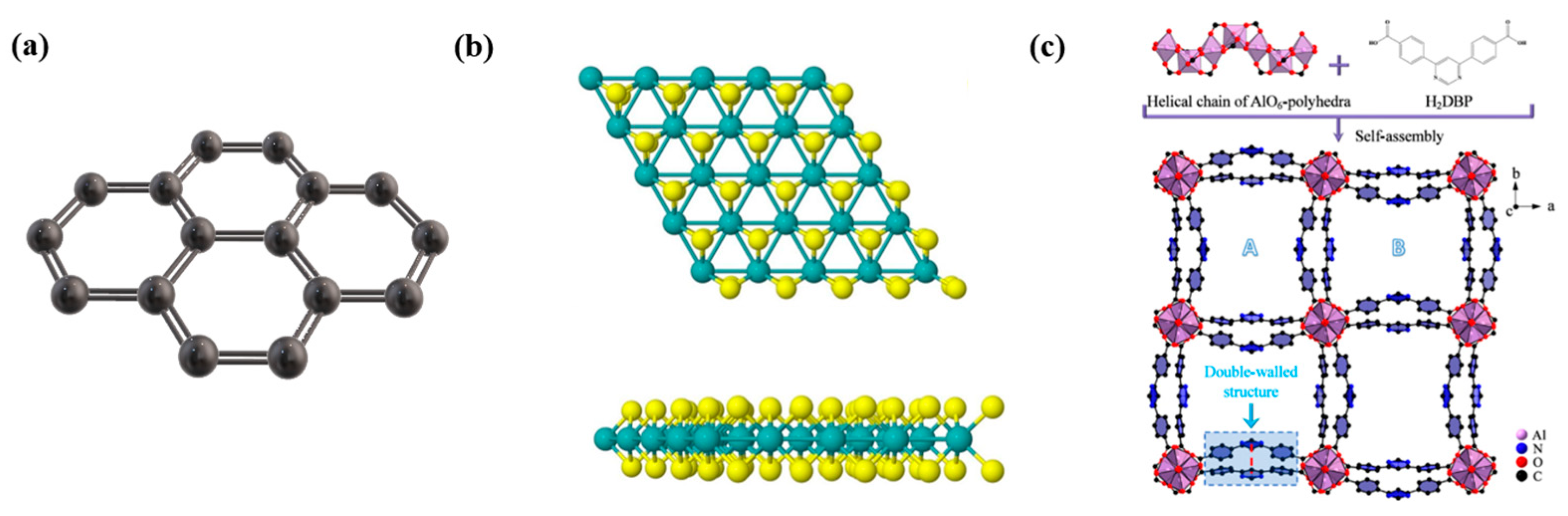
  46. Hu, L.; Wu, W.; Hu, M.; Jiang, L.; Lin, D.; Wu, J.; Yang, K. Double-walled Al-based MOF with large microporous specific surface area for trace benzene adsorption. Nat. Commun. 2024, 15, 3204. [Google Scholar] [CrossRef] [PubMed]
  47. Harms, H.; Stollenwerk, A.J.; Cunningham, C.; Sadler, C.; O’Leary, E.; Kidd, T.E.; Lukashev, P.V. Adsorption of Ag, Au, Cu, and Ni on MoS2: Theory and experiment. J. Phys. Condens. Matter An. Inst. Phys. J. 2024, 37, 015001. [Google Scholar] [CrossRef]
  48. Singh, E.; Singh, P.; Kim, K.S.; Yeom, G.Y.; Nalwa, H.S. Flexible Molybdenum Disulfide (MoS2) Atomic Layers for Wearable Electronics and Optoelectronics. ACS Appl. Mater. Interfaces 2019, 11, 11061–11105. [Google Scholar] [CrossRef]
  49. Su, H.-Y.; Calle-Vallejo, F.; Sun, K. A structure-sensitive descriptor for the design of active sites on MoS2 catalysts. Catal. Sci. Technol. 2023, 13, 5290–5300. [Google Scholar] [CrossRef]
  50. Su, H.-Y.; Liao, W.; Sun, K. First-principles and microkinetic simulation studies of CO2 hydrogenation mechanism and active site on MoS2 catalyst. Appl. Surf. Sci. 2023, 635, 157721. [Google Scholar] [CrossRef]
  51. Mehdizadeh, A.; Zeynali, M.; Karimi, M. Engineering of MoS2 nanoribbons as high-performance materials for biosensing applications. Appl. Surf. Sci. 2021, 540, 148349. [Google Scholar] [CrossRef]
  52. Gao, Y.; Wang, S.; Wang, B.; Jiang, Z.; Fang, T. Recent Progress in Phase Regulation, Functionalization, and Biosensing Applications of Polyphase MoS2. Nano Micro Small 2022, 18, 2202956. [Google Scholar] [CrossRef]
  53. Pariari, D.; Varma, R.M.; Nair, M.N.; Zeller, P.; Amati, M.; Gregoratti, L.; Nanda, K.K.; Sarma, D.D. On the origin of metallicity and stability of the metastable phase in chemically exfoliated MoS2. Appl. Mater. Today 2020, 19, 100544. [Google Scholar] [CrossRef]
  54. Chatterjee, S.; Singh, H.; Hudda, D.; Sweety; Kumar, D. A Novel Acetylcholinesterase-Based Electrochemical Biosensor Using g-C3N4@MoS2 Nanohybrid for the Detection of Trichlorfon. Appl. Organomet. Chem. 2024, 38, e7721. [Google Scholar] [CrossRef]
  55. Li, P.; Ye, Y.; Li, Y.; Xie, Z.; Ye, L.; Huang, J. A MoS2 nanosheet-based CRISPR/Cas12a biosensor for efficient miRNA quantification for acute myocardial infarction. Biosens. Bioelectron. 2024, 251, 116129. [Google Scholar] [CrossRef]
  56. Pham, H.T.B.; Choi, J.Y.; Fang, X.; Claman, A.; Huang, S.; Coates, S.; Wayment, L.; Zhang, W.; Park, J. Macrocyclic ligand-driven ion selectivity and high surface area in a 2D conductive MOF. Chem 2024, 10, 199–210. [Google Scholar] [CrossRef]
  57. Wen, Y.; Xu, W.; Hu, L.; Xu, M.; Gu, W.; Sun, H.; Zhu, C. Tuning atomic-scale sites in metal–organic framework-based nanozymes for sensitive biosensing. Sens. Diagn. 2023, 2, 1376–1389. [Google Scholar] [CrossRef]
  58. Xu, D.; Yang, F.; Zheng, D.; Gao, L.; Zhao, G.; Muhammad, P.; Wu, Q. MOF-derived yolk-shell CoN/Co-NC@SiO2 nanozyme with oxidase mimetic activities for colorimetric detection of glutathione. Microchem. J. 2024, 201, 110671. [Google Scholar] [CrossRef]
  59. Wang, R.; Liu, C.; Wei, Y.; Ran, Z.; Jiang, T.; Liu, C.; Shi, C.; Ren, Z.; Wang, X.; Liu, Z.; et al. Fiber SPR biosensor sensitized by MOFs for MUC1 protein detection. Talanta 2023, 258, 124467. [Google Scholar] [CrossRef]
  60. Elgazar, A.; Sabouni, R.; Ghommem, M.; Majdalawieh, A.F. Sustainable synthesis of MOF for COVID-19 detection using a 23 factorial design. Colloids Surf. A Physicochem. Eng. Asp. 2025, 705, 135616. [Google Scholar] [CrossRef]
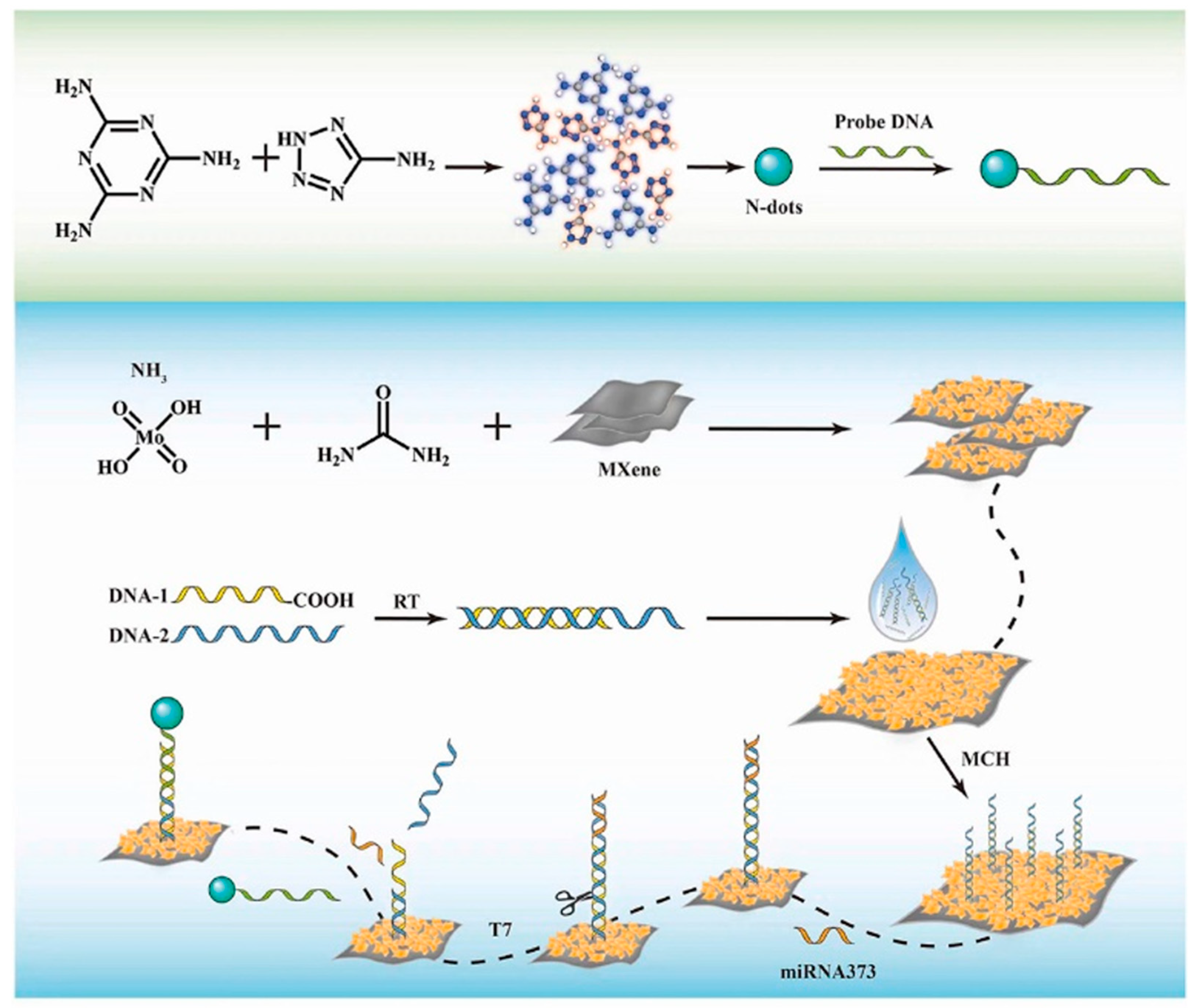
  61. Xu, J.; Cui, X.; Wang, L.; Chen, G.; Ji, S.; Zhao, S.; Wang, H.; Luo, Z.; Zeng, A.; Fu, Q. DNA-functionalized MOF fluorescent probes for the enzyme-free and pretreatment-free detection of MicroRNA in serum. Talanta 2024, 275, 126083. [Google Scholar] [CrossRef]
  62. Zhang, R.; Yang, J.; Cao, Y.; Zhang, Q.; Xie, C.; Xiong, W.; Luo, X.; He, Y. Efficient 2D MOFs nanozyme combining with magnetic SERS substrate for ultrasensitive detection of Hg2+. Spectrochim. Acta. Part A Mol. Biomol. Spectrosc. 2024, 312, 124062. [Google Scholar] [CrossRef] [PubMed]
  63. Wu, Q.; Liang, J.; Wang, D.; Wang, R.; Janiak, C. Host molecules inside metal–organic frameworks: Host@MOF and guest@host@MOF (Matrjoschka) materials. Chem. Soc. Rev. 2025, 54, 601–622. [Google Scholar] [CrossRef] [PubMed]
  64. Piguillem, S.V.; Gomez, G.E.; Tortella, G.R.; Seabra, A.B.; Regiart, M.D.; Messina, G.A.; Fernández-Baldo, M.A. Paper-Based Analytical Devices Based on Amino-MOFs (MIL-125, UiO-66, and MIL-101) as Platforms towards Fluorescence Biodetection Applications. Chemosensors 2024, 12, 208. [Google Scholar] [CrossRef]
  65. Pandey, S.; Demaske, B.; Ejegbavwo, O.A.; Berseneva, A.A.; Setyawan, W.; Shustova, N.; Phillpot, S.R. Electronic structures and magnetism of Zr-, Th-, and U-based metal-organic frameworks (MOFs) by density functional theory. Comput. Mater. Sci. 2020, 184, 109903. [Google Scholar] [CrossRef]
  66. Pal, T.K. Metal–organic framework (MOF)-based fluorescence “turn-on” sensors. Mater. Chem. Front. 2023, 7, 405–441. [Google Scholar] [CrossRef]
  67. Liao, S.; Gui, L.; Yang, Y.; Liu, Y.; Hu, X. Fluorescence/visual aptasensor based on Au/MOF nanocomposite for accurate and convenient aflatoxin B1 detection. Microchim. Acta 2024, 191, 497. [Google Scholar] [CrossRef] [PubMed]
  68. Zhang, S.; Wang, H.; Liu, J.; Bao, C. Measuring the specific surface area of monolayer graphene oxide in water. Mater. Lett. 2020, 261, 127098. [Google Scholar] [CrossRef]
  69. Singh, A.; Shishodia, M.S. Graphene vs. silica coated refractory nitrides based core-shell nanoparticles for nanoplasmonic sensing. Phys. E Low Dimens. Syst. Nanostructures 2020, 124, 114288. [Google Scholar] [CrossRef]
  70. Ribeiro, D.S.; Santos, J.C.C.; Grieger, S.; Campos, J.L.E.; Machado, L.R.P.; Pacheco, F.G.; Fernandes, T.F.D.; Haase, C.C.; Silva, D.L.; Guterres, M.; et al. Measuring the Surface Area Concentration and Specific Surface Area of Mass-Produced Graphene Nanoflakes via Fluorescence Quenching. ACS Appl. Nano Mater. 2023, 6, 11198–11210. [Google Scholar] [CrossRef]
  71. Xiao, Y.; Cui, D.; Zhong, Y.; Li, Z.; Zhang, J.; Yu, J. Theoretical Design of Near-Infrared Tunable Surface Plasmon Resonance Biosensors Based on Gate-Controlled Graphene Plasmons. Coatings 2024, 14, 56. [Google Scholar] [CrossRef]
  72. Narayanan, T.N.; Vusa, C.S.; Alwarappan, S. Selective and efficient electrochemical biosensing of ultrathin molybdenum disulfide sheets. Nanotechnology 2014, 25, 335702. [Google Scholar] [CrossRef]
  73. Sun, B.; Wang, P.; Liang, Z.; Li, Z.; Ma, Q. MoS(2)/MXene Van der Waals heterojunction-based electrochemiluminescence sensor for triple negative breast cancer detection. Talanta 2024, 277, 126343. [Google Scholar] [CrossRef]
  74. Khoddam, Z.; Pourmadadi, M.; Abdouss, M.; Mazinani, S.; Behzadmehr, R.; Rahdar, A.; Ali Aboudzadeh, M. Hybrid nanocarriers based on polyacrylic acid, polyvinyl pyrrolidone, and molybdenum disulfide for enhanced 5-fluorouracil delivery in lung cancer therapy. Inorg. Chem. Commun. 2024, 159, 111749. [Google Scholar] [CrossRef]
  75. Wu, J.; Chang, X.; Guo, Y.; Xia, N. Synthesis of a hybrid nanocomposite: Incorporating MoS2 within a ZIF-8 for fluorescence-based detection and adsorption of tetracyclines antibiotics. Dye. Pigment. 2024, 226, 112137. [Google Scholar] [CrossRef]
  76. Song, R.; Zhang, H.; Lv, J. NiO Nanoparticle-decorated graphene oxide nanosheets modified glassy carbon electrode for sensitive electrochemical detection of pethidine. Int. J. Electrochem. Sci. 2023, 18, 100221. [Google Scholar] [CrossRef]
  77. Xu, T.; Wang, R.; Gu, C.; Jiang, T. Recyclable detection of gefitinib in clinical sample mediated by multifunctional Ag-anchored g-C3N4/MoS2 composite substrate. Spectrochim. Acta Part A Mol. Biomol. Spectrosc. 2023, 299, 122801. [Google Scholar] [CrossRef]
  78. Elfiky, M.; Gaber, M.; Mousa, M.; Salahuddin, N. Synthesis and validation of ultrasensitive stripping voltammetric sensor based on polypyrrole@ZnO/Fe3O4 core–shell nanostructure for picomolar detection of artesunate and dopamine drugs. Anal. Methods 2022, 14, 3739–3750. [Google Scholar] [CrossRef]
  79. Alanazi, S.; Rhouati, A.; Chrouda, A.; Cialla-May, D.; Popp, J.; Muthana, S.; Dasouki, M.; Zourob, M. Design of an innovative aptasensor for the detection of chemotherapeutic drug Fludarabine phosphate. Sci. Rep. 2024, 14, 26300. [Google Scholar] [CrossRef]
  80. Kokab, T.; Shah, A.; Khan, M.A.; Nisar, J.; Ashiq, M.N. Electrochemical sensing platform for the simultaneous femtomolar detection of amlodipine and atorvastatin drugs. RSC Adv. 2021, 11, 27135–27151. [Google Scholar] [CrossRef]
  81. Nair, A.J.S.; Saisree, S.; Sandhya, K.Y. Picomolar level electrochemical detection of hydroquinone, catechol and resorcinol simultaneously using a MoS2 nano-flower decorated graphene. Analyst 2022, 147, 2966–2979. [Google Scholar] [CrossRef]
  82. Wiwasuku, T.; Chuaephon, A.; Habarakada, U.; Boonmak, J.; Puangmali, T.; Kielar, F.; Harding, D.; Youngme, S. A Water-Stable Lanthanide-Based MOF as a Highly Sensitive Sensor for the Selective Detection of Paraquat in Agricultural Products. ACS Sustain. Chem. Eng. 2022, 10, 2761–2771. [Google Scholar] [CrossRef]
  83. Zhang, T.; Cao, R.; Tang, H.; Feng, W.; Zhang, Z. Ratiometric fluorescence turn-off probe for highly selective recognition of 2-pentanone based on a Eu3+- functionalized MOF. Inorganica Chim. Acta 2024, 565, 122003. [Google Scholar] [CrossRef]
  84. Wang, T.; Zhang, M.; Lu, Y.; Liu, Q.; Niu, Q.; You, T. Metal-organic-framework-confined quantum dots enhance photocurrent signals: A molecularly imprinted photoelectrochemical cathodic sensor for rapid and sensitive tetracycline detection. Anal. Chim. Acta 2024, 1293, 342269. [Google Scholar] [CrossRef]
  85. Zuo, C.; Guo, Y.; Li, J.; Peng, Z.; Bai, S.; Yang, S.; Wang, D.; Chen, H.; Xie, G. A nanoprobe for fluorescent monitoring of microRNA and targeted delivery of drugs. RSC Adv. 2021, 11, 8871–8878. [Google Scholar] [CrossRef]
  86. Kulandaivel, S.; Lu, Y.-K.; Lin, C.-H.; Yeh, Y.-C. Dual-functional PCN-242 (Fe2Co) MOF for sensitive bacterial endotoxin detection. J. Mater. Chem. B 2025, 13, 151–159. [Google Scholar] [CrossRef]
  87. Khafaga, D.S.R.; El-Morsy, M.T.; Faried, H.; Diab, A.H.; Shehab, S.; Saleh, A.M.; Ali, G.A.M. Metal–organic frameworks in drug delivery: Engineering versatile platforms for therapeutic applications. RSC Adv. 2024, 14, 30201–30229. [Google Scholar] [CrossRef]
  88. Nikshitha, M.; Sudhakara, S.M.; Shetty, M.S. Physics. Organic-inorganic hybrids: A comprehensive review on synthesis and their potential applications. Mater. Chem. Phys. 2025, 331, 130181. [Google Scholar] [CrossRef]
  89. Gayathri, K.; Arumugam Madan, K.; Chinmai Sriamulya, Y.; Nagabhishek Sirpu, N.; Rajender, B.; Ramyakrishna, P. Combination of ZnO Nanoparticle with Marine Sponge Derived Dipeptide for Enhanced Anticancer Efficacy in Liver Cancer Cells and their Toxicity Evaluation on Embryonic Zebrafish. Curr. Anal. Chem. 2021, 17, 677–688. [Google Scholar] [CrossRef]
  90. Ouyang, Y.; Zhou, M.; Liu, Y.; Zhang, L.; Zhong, C.; Yang, Q.; Liu, M. Mg-doped ZnO nanoparticle as an effective nanocarrier in delivery of 5-Fluorouracil anti-gastric cancer drug. J. Mol. Struct. 2024, 1314, 138706. [Google Scholar] [CrossRef]
  91. Li, Y.; Chai, B.-L.; Xu, H.; Zheng, T.-F.; Chen, J.-L.; Liu, S.-J.; Wen, H.-R. Temperature- and solvent-induced reversible single-crystal-to-single-crystal transformations of TbIII-based MOFs with excellent stabilities and fluorescence sensing properties toward drug molecules. Inorg. Chem. Front. 2022, 9, 1504–1513. [Google Scholar] [CrossRef]
  92. Chen, X.-X.; Hou, M.-J.; Mao, G.-J.; Wang, W.-X.; Xu, F.; Li, Y.; Li, C.-Y. ATP-responsive near-infrared fluorescence MOF nanoprobe for the controlled release of anticancer drug. Microchim. Acta 2021, 188, 287. [Google Scholar] [CrossRef] [PubMed]
  93. Zhao, B.; Yang, Q.; Wang, J.-S.; Xie, F.-Y.; Yu, H.-Y.; Li, Y.; Ma, Y.-X.; Ruan, W.-J. An anionic-ligand installed pyrene-based MOF for the fluorescence detection of paraquat. New J. Chem. 2021, 45, 4401–4407. [Google Scholar] [CrossRef]
  94. Yan, X.; Tan, D.; Yu, L.; Li, D.; Huang, W.; Huang, W.; Wu, H. A High-Throughput and Logarithm-Serial-Dilution Microfluidic Chip for Combinational Drug Screening on Tumor Organoids. ACS Pharmacol. Transl. Sci. 2024, 7, 4135–4143. [Google Scholar] [CrossRef]
  95. Hamimed, S.; Jabberi, M.; Chatti, A. Nanotechnology in drug and gene delivery. Naunyn-Schmiedeberg’s Arch. Pharmacol. 2022, 395, 769–787. [Google Scholar] [CrossRef] [PubMed]
  96. Merone, G.M.; Tartaglia, A.; Rossi, S.; Santavenere, F.; Bassotti, E.; D’Ovidio, C.; Bonelli, M.; Rosato, E.; de Grazia, U.; Locatelli, M.; et al. Fast Quantitative LC-MS/MS Determination of Illicit Substances in Solid and Liquid Unknown Seized Samples. Anal. Chem. 2021, 93, 16308–16313. [Google Scholar] [CrossRef]
  97. Xiao, J.; Ding, J.; Sun, C.; Liu, D.; Gao, H.; Liu, Y.; Lu, Y.; Gao, X. Simultaneous Detection of Clenbuterol and Higenamine in Urine Samples Using Interference-Free SERS Tags Combined with Magnetic Separation. ACS Sens. 2024, 9, 5394–5404. [Google Scholar] [CrossRef]
  98. Su, S.; Sun, Q.; Ma, J.; Zhu, D.; Wang, F.; Chao, J.; Fan, C.; Li, Q.; Wang, L. Ultrasensitive analysis of microRNAs with gold nanoparticle-decorated molybdenum disulfide nanohybrid-based multilayer nanoprobes. Chem. Commun. 2020, 56, 9012–9015. [Google Scholar] [CrossRef]
  99. Thevis, M.; Görgens, C.; Guddat, S.; Thomas, A.; Geyer, H. Mass spectrometry in sports drug testing-Analytical approaches and the athletes’ exposome. Scand. J. Med. Sci. Sports 2024, 34, e14228. [Google Scholar] [CrossRef]
  100. Thomas, A.; Thevis, M. Recent advances in mass spectrometry for the detection of doping. Expert Rev. Proteom. 2024, 21, 27–39. [Google Scholar] [CrossRef]
  101. Göktaş, E.F.; Kabil, E.; Arıöz, F. Quantification and validation of nine nonsteroidal anti-inflammatory drugs in equine urine using gas chromatography-mass spectrometry for doping control. Drug Test. Anal. 2020, 12, 1065–1077. [Google Scholar] [CrossRef]
  102. Duan, M.; Che, X.; Ma, S.; Xu, H.; Zhen, Q.; Zhang, C.; Pang, H. Efficient luteolin detection sensor achieved by CdS-MoS2 heterostructures. Inorg. Chem. Commun. 2024, 163, 112374. [Google Scholar] [CrossRef]
  103. Wubie, S.T. Analytical Detection Methods of Performance Enhancing Drugs in Athletes, Review Study. Glob. Acad. J. Pharm. Drug Res. 2021, 3, 29–43. [Google Scholar]
  104. Wen, X.; Cheng, H.; Zhang, W.; You, L.; Li, J. Multifunctional Ni(OH)2/Ag composites for ultrasensitive SERS detection and efficient photocatalytic degradation of ciprofloxacin and methylene blue. Talanta 2024, 266, 125140. [Google Scholar] [CrossRef] [PubMed]
  105. WADA. Prohibited List. 2025. Available online: https://www.wada-ama.org/en/resources/world-anti-doping-code-and-international-standards/prohibited-list (accessed on 1 January 2025).
  106. Hagedorn, H.-W.; Zankl, H.; Grund, C.; Schulz, R. Excretion of the anabolic steroid boldenone by racing pigeons. Am. J. Vet. Res. 1997, 58, 224–227. [Google Scholar] [CrossRef]
  107. Li, C.; Han, D.; Liang, Z.; Han, F.; Fu, W.; Wang, W.; Han, D.; Wang, Y.; Niu, L. Novel electrochemical-surface plasmon resonance (EC-SPR) sensor for amphetamine-type stimulants detection based on molecularly imprinted strategy. Sens. Actuators B Chem. 2022, 369, 132258. [Google Scholar] [CrossRef]
  108. Jung, J.; Jeong, Y.; Xu, Y.; Yi, J.; Kim, M.; Jeong, H.-J.; Shin, S.H.; Yang, Y.-H.; Son, J.; Sung, C. Production and engineering of nanobody-based quenchbody sensors for detecting recombinant human growth hormone and its isoforms. Drug Test. Anal. 2023, 15, 1439–1448. [Google Scholar] [CrossRef]
  109. Evans, R.L.; Bryant, D.J.; Voliotis, A.; Hu, D.; Wu, H.; Syafira, S.A.; Oghama, O.E.; McFiggans, G.; Hamilton, J.F.; Rickard, A.R. A Semi-Quantitative Approach to Nontarget Compositional Analysis of Complex Samples. Anal. Chem. 2024, 96, 18349–18358. [Google Scholar] [CrossRef]
  110. Ishii, H.; Shibuya, M.; Kusano, K.; Sone, Y.; Kamiya, T.; Wakuno, A.; Ito, H.; Miyata, K.; Sato, F.; Kuroda, T.; et al. Generic approach for the discovery of drug metabolites in horses based on data-dependent acquisition by liquid chromatography high-resolution mass spectrometry and its applications to pharmacokinetic study of daprodustat. Anal. Bioanal. Chem. 2022, 414, 8125–8142. [Google Scholar] [CrossRef]
  111. Ye, M.; Tang, S. Construction of SERS chip based on silver nanoparticles and detection of sports doping β-agonists. Alex. Eng. J. 2023, 83, 134–139. [Google Scholar] [CrossRef]
  112. Skoupá, K.; Šťastný, K.; Sládek, Z. Anabolic Steroids in Fattening Food-Producing Animals-A Review. Anim. Open Access J. 2022, 12, 2115. [Google Scholar] [CrossRef]
  113. Peng, C.; Wang, C.; Li, Z.; Wang, Z. A novel approach to detecting doping agents in food using electrochemical sensor based on zinc oxide/graphene oxide nanocomposites. J. Food Meas. Charact. 2024, 18, 6770–6781. [Google Scholar] [CrossRef]
  114. Wang, B.; Zhao, L.; Li, Y.; Liu, X.; Fan, D.; Wu, D.; Wei, Q. Porphyrin-based metal-organic frameworks enhanced electrochemiluminescence (ECL) by overcoming aggregation-caused quenching: A new ECL emitter for the detection of trenbolone. Anal. Chim. Acta 2023, 1276, 341616. [Google Scholar] [CrossRef] [PubMed]
  115. Oleksak, P.; Nepovimova, E.; Valko, M.; Alwasel, S.; Alomar, S.; Kuca, K. Comprehensive analysis of prohibited substances and methods in sports: Unveiling trends, pharmacokinetics, and WADA evolution. Environ. Toxicol. Pharmacol. 2024, 108, 104447. [Google Scholar] [CrossRef]
  116. Huang, S.; Feng, R. A new electrochemical biosensor based on graphene oxide for rapid detection of synthetic testosterone as performance-enhancing drugs in athletes. Alex. Eng. J. 2024, 98, 281–289. [Google Scholar] [CrossRef]
  117. Ram, G.D.; Kumar, S.P. Bio-carbon-derived porous reduced graphene oxide photo- and electrochemical sensor for ultra-sensitive detection of testosterone hormone. Chem. Pap. 2024, 78, 9543–9557. [Google Scholar] [CrossRef]
  118. Sun, J.-Y.; Huang, K.-J.; Wei, S.-Y.; Wu, Z.-W.; Ren, F.-P. A graphene-based electrochemical sensor for sensitive determination of caffeine. Colloids Surf. B Biointerfaces 2011, 84, 421–426. [Google Scholar] [CrossRef]
  119. Li, Y.; Liu, W. Electrochemical detection of blood doping in sports: A novel biosensor based on nickel oxide/nitrogen-doped graphene oxide nanocomposite. Alex. Eng. J. 2024, 96, 176–184. [Google Scholar] [CrossRef]
  120. Zhang, Y. A novel approach to detect salbutamol as a doping agent in athletes: A nanoparticle-modified electrode. Alex. Eng. J. 2024, 96, 185–194. [Google Scholar] [CrossRef]
  121. Zhao, Z.; Mu, S.; Zheng, J.; Gu, L.; Shen, G.; Shen, Y. A new modification method of a Cetyl Trimethyl Ammonium Bromide/Nano-ZnO and Multi-walled Carbon Nanotubes Electrode for Determination of Anti Doping in Urine. IOP Conf. Ser. Mater. Sci. Eng. 2017, 220, 012009. [Google Scholar] [CrossRef]
  122. Liu, X.; Jade, N.; Rouhi, O. Reduced graphene oxide-based sensor for triamcinolone acetonide detection: Advancements in doping agent surveillance for athletes. Inorg. Chem. Commun. 2024, 162, 112285. [Google Scholar] [CrossRef]
  123. Lu, C.; Xu, M.; Lu, Y.; Zhang, Z.; Han, W.; Mahdi, A.B. Manufacturing of a Novel Sensor Based CuO@Graphene Catalyst for Voltammetric Detection of Prednisolone as an Important Doping Agent in Sport. In Topics in Catalysis; Springer: Singapore, 2024. [Google Scholar] [CrossRef]
  124. Hudari, F.F.; Zanoni, M.V.B. A glassy carbon electrode modified with reduced graphene oxide for sensitive determination of bumetanide in urine at levels required for doping analysis. Microchim. Acta 2017, 184, 4117–4124. [Google Scholar] [CrossRef]
  125. Adegoke, O.; Zolotovskaya, S.; Abdolvand, A.; Daeid, N.N. Biomimetic graphene oxide-cationic multi-shaped gold nanoparticle-hemin hybrid nanozyme: Tuning enhanced catalytic activity for the rapid colorimetric apta-biosensing of amphetamine-type stimulants. Talanta 2020, 216, 120990. [Google Scholar] [CrossRef]
  126. Tian, Y.; Cao, S.; Xi, C.; Su, H.; Chen, Z. Designing polydopamine nanohybrid based on template-mediated for effectively remove amphetamine-type stimulants in sewage: Performance and mechanism. J. Environ. Chem. Eng. 2021, 9, 105870. [Google Scholar] [CrossRef]
  127. Gupta, P.; Goyal, R.N. Amino Functionalized Graphene Oxide and Polymer Nanocomposite Based Electrochemical Platform for Sensitive Assay of Anti-Doping Drug Atenolol in Biological Fluids. J. Electrochem. Soc. 2016, 163, B601. [Google Scholar] [CrossRef]
  128. Anvari, L.; Ghoreishi, S.M.; Faridbod, F.; Ganjali, M.R. Electrochemical Determination of Methamphetamine in Human Plasma on a Nanoceria Nanoparticle Decorated Reduced Graphene Oxide (rGO) Glassy Carbon Electrode (GCE). Anal. Lett. 2021, 54, 2509–2522. [Google Scholar] [CrossRef]
  129. Zhao, L.; Song, X.; Li, Y.; Jia, H.; Zhang, N.; Wei, Q.; Wu, D.; Ju, H. Europium-based metal-organic framework with acid-base buffer structure as electrochemiluminescence luminophore for hyperstatic trenbolone trace monitoring under wide pH range. Biosens. Bioelectron. 2023, 221, 114925. [Google Scholar] [CrossRef]
  130. Zhao, L.; Wang, M.; Song, X.; Liu, X.; Ju, H.; Ai, H.; Wei, Q.; Wu, D. Annihilation luminescent Eu-MOF as a near-infrared electrochemiluminescence probe for trace detection of trenbolone. Chem. Eng. J. 2022, 434, 134691. [Google Scholar] [CrossRef]
  131. Jalalvand, A.R.; Rashidi, Z.; Khajenoori, M. Sensitive and selective simultaneous biosensing of nandrolone and testosterone as two anabolic steroids by a novel biosensor assisted by second-order calibration. Steroids 2022, 189, 109138. [Google Scholar] [CrossRef]
  132. Shan, L. Electrochemical Determination of Methandrostenolone Using a Molecularly Imprinted Sensor. Int. J. Electrochem. Sci. 2020, 15, 12587–12598. [Google Scholar] [CrossRef]
  133. Xu, X.; Bai, G.; Song, L.; Zheng, Q.; Yao, Y.; Liu, S.; Yao, C. Fast steroid hormone metabolism assays with electrochemical liver microsomal bioreactor based on polydopamine encapsulated gold-graphene nanocomposite. Electrochim. Acta 2017, 258, 1365–1374. [Google Scholar] [CrossRef]
  134. Alimohammadi, S.; Kiani, M.A.; Imani, M.; Rafii-Tabar, H.; Sasanpour, P. Electrochemical Determination of Dexamethasone by Graphene Modified Electrode: Experimental and Theoretical Investigations. Sci. Rep. 2019, 9, 11775. [Google Scholar] [CrossRef]
  135. Khoshhesab, Z.M. Simultaneous electrochemical determination of acetaminophen, caffeine and ascorbic acid using a new electrochemical sensor based on CuO–graphene nanocomposite. RSC Adv. 2015, 5, 95140–95148. [Google Scholar] [CrossRef]
  136. Rocha, D.P.; Dornellas, R.M.; Nossol, E.; Richter, E.M.; Silva, S.G.; Santana, M.H.P.; Munoz, R.A.A. Electrochemically Reduced Graphene Oxide for Forensic Electrochemistry: Detection of Cocaine and its Adulterants Paracetamol, Caffeine and Levamisole. Electroanalysis 2017, 29, 2418–2422. [Google Scholar] [CrossRef]
  137. Maleki, F.; Rashidi, M.-R.; Razmi, H.; Ghorbani, M. Label-free electrochemical immunosensor for detection of insulin-like growth factor-1 (IGF-1) using a specific monoclonal receptor on electrospun Zein-based nanofibers/rGO-modified electrode. Talanta 2023, 265, 124844. [Google Scholar] [CrossRef] [PubMed]
  138. Liu, Z.; Chen, J.; Huang, S.; Yan, Y.; Niu, Y.; Zeng, J.; Huang, Q.; Jin, M.; Shui, L. Label-free biosensor for trace insulin-like growth factor-I assay based on rGO-SnS2 heterostructure nanocomposite. Sens. Actuators B Chem. 2022, 370, 132415. [Google Scholar] [CrossRef]
  139. González-Guerrero, A.B.; Maldonado, J.; Dante, S.; Grajales, D.; Lechuga, L.M. Direct and label-free detection of the human growth hormone in urine by an ultrasensitive bimodal waveguide biosensor. J. Biophotonics 2017, 10, 61–67. [Google Scholar] [CrossRef]
  140. Xiong, C.; Li, L.; Yang, M.; Chen, Y.; Wang, L.; Xiong, L.; Huang, J.; Zou, Y. Rationally Designed Ionically Conductive Metal-Organic Frameworks for Ultrasensitive Methamphetamine Analogs Sensor. Adv. Funct. Mater. 2024, 34, 2316511. [Google Scholar] [CrossRef]
  141. Tiwari, N.K.; Sathyanesan, M.; Kumar, V.; Newton, S.S. A Comparative Analysis of Erythropoietin and Carbamoylated Erythropoietin Proteome Profiles. Life 2021, 11, 359. [Google Scholar] [CrossRef]
  142. Bohlooli, S.; Kia, S.; Bohlooli, S.; Sariri, R. Development of molecularly imprinted polymer on ferric oxide nanoparticles modified electrode as electrochemical sensor for detection of human growth hormone. Monatshefte Für Chem. Chem. Mon. 2022, 153, 39–48. [Google Scholar] [CrossRef]
  143. Zhang, Z.; Li, S. A bioanalytical tool for monitoring athlete health and detecting performance-enhancing drugs: An electrochemical biosensor based on graphene oxide. Alex. Eng. J. 2024, 102, 58–65. [Google Scholar] [CrossRef]
  144. Zhang, L.; Zhu, Q.-H.; Zhou, Y.-R.; Wang, S.-L.; Fu, J.; Liu, J.-Y.; Zhang, G.-H.; Ma, L.; Tao, G.; Tao, G.-H.; et al. Hydrogen-bonding and π-π interaction promoted solution-processable covalent organic frameworks. Nat. Commun. 2023, 14, 8181. [Google Scholar] [CrossRef]
  145. Patnaik, A. Hydrogen Bond Directed 2D Materials at Modulated Interfaces. In Advanced Supramolecular Nanoarchitectonics; Ariga, K., Aono, M., Eds.; William Andrew Publishing: Norwich, NY, USA, 2019; Chapter 1.3; pp. 31–87. [Google Scholar]
  146. Lin, X.; Lu, J.C.; Shao, Y.; Zhang, Y.Y.; Wu, X.; Pan, J.B.; Gao, L.; Zhu, S.Y.; Qian, K.; Zhang, Y.F.; et al. Intrinsically patterned two-dimensional materials for selective adsorption of molecules and nanoclusters. Nat. Mater. 2017, 16, 717–721. [Google Scholar] [CrossRef] [PubMed]
  147. Igarashi, M.; Nozawa, T.; Matsumoto, T.; Yagihashi, F.; Kikuchi, T.; Sato, K. Parallel-stacked aromatic molecules in hydrogen-bonded inorganic frameworks. Nat. Commun. 2021, 12, 7025. [Google Scholar] [CrossRef] [PubMed]
  148. Chen, T.; Li, M.; Liu, J. π–π Stacking Interaction: A Nondestructive and Facile Means in Material Engineering for Bioapplications. Cryst. Growth Des. 2018, 18, 2765–2783. [Google Scholar] [CrossRef]
  149. Lin, S.-P. (Invited) Development of Cell-Based Biosensor Platform for Monitoring the Effect of Functional Nanocarriers As Drug Delivery System on Breast Cancer Cells. ECS Meet. Abstr. 2023, 243, 1928. [Google Scholar] [CrossRef]
  150. Altalbawy, F.M.A.; Altameemi, K.K.A.; Ballal, S.; Monsi, M.; Walia, C.; Prasad, G.V.S.; Al-saray, M.J.; Alsaadi, S.B.; Al-Mashhadani, Z.I.; Alsayah, A.M. A comparative DFT study of drug delivery system based on Pt-doped and Au-modified MoS2 nanosheets for β-lapachone drug. In Structural Chemistry; Springer: Singapore, 2024. [Google Scholar] [CrossRef]
  151. Wei, W.; Zhou, S.; Ma, D.-D.; Li, Q.; Ran, M.; Li, X.; Wu, X.-T.; Zhu, Q.-L. Ultrathin Conductive Bithiazole-Based Covalent Organic Framework Nanosheets for Highly Efficient Electrochemical Biosensing. Adv. Funct. Mater. 2023, 33, 2302917. [Google Scholar] [CrossRef]
  152. Arshad, M.; Arshad, S.; Majeed, M.K.; Frueh, J.; Chang, C.; Bilal, I.; Niaz, S.I.; Khan, M.S.; Tariq, M.A.; Yasir Mehboob, M. Transition Metal-Decorated Mg12O12 Nanoclusters as Biosensors and Efficient Drug Carriers for the Metformin Anticancer Drug. ACS Omega 2023, 8, 11318–11325. [Google Scholar] [CrossRef]
  153. Wong, K.-Y.; Liu, Y.; Phan, C.-M.; Jones, L.; Wong, M.-S.; Liu, J. Selection of DNA aptamers for sensing drugs treating eye disease: Atropine and timolol maleate. Sens. Diagn. 2024, 3, 1679–1688. [Google Scholar] [CrossRef]
  154. Chen, S.; Ju, Y.; Yang, Y.; Xiang, F.; Yao, Z.; Zhang, H.; Li, Y.; Zhang, Y.; Xiang, S.; Chen, B.; et al. Multistate structures in a hydrogen-bonded polycatenation non-covalent organic framework with diverse resistive switching behaviors. Nat. Commun. 2024, 15, 298. [Google Scholar] [CrossRef]
  155. Piao, H.; Choi, G.; Sanoj Rejinold, N.; Yu, J.; Choi, S.-J.; Choy, J.-H. Pemetrexed-layered double hydroxide with Eudragit® S100; 2D van der Waals nanohybrid with drug delivery function. J. Ind. Eng. Chem. 2024, 130, 572–579. [Google Scholar] [CrossRef]
  156. Sayed, Z.S.; Hieba, E.M.; Batakoushy, H.A.; Rashdan, H.R.M.; Ismail, E.; Elkatlawy, S.M.; Elzwawy, A. Cancer treatment approaches within the frame of hyperthermia, drug delivery systems, and biosensors: Concepts and future potentials. RSC Adv. 2024, 14, 39297–39324. [Google Scholar] [CrossRef] [PubMed]
  157. Dinda, S.; Mahato, B.; Maiti, A.; Ghoshal, D. Selective Detection of Primary Aromatic Amines through Enhanced Luminescence of a 2D + 2D Inclined Polycatenated Microporous Nitro-Functionalized Metal–Organic Framework. Inorg. Chem. 2024, 63, 5996–6004. [Google Scholar] [CrossRef]
  158. Wang, Y.; Niu, Z.; Xu, C.; Zhan, M.; Koh, K.; Niu, J.; Chen, H. 2D MOF-enhanced SPR sensing platform: Facile and ultrasensitive detection of Sulfamethazine via supramolecular probe. J. Hazard. Mater. 2023, 456, 131642. [Google Scholar] [CrossRef]
  159. Das, G.; Prakasam, T.; Alkhatib, N.; AbdulHalim, R.G.; Chandra, F.; Sharma, S.K.; Garai, B.; Varghese, S.; Addicoat, M.A.; Ravaux, F.; et al. Light-driven self-assembly of spiropyran-functionalized covalent organic framework. Nat. Commun. 2023, 14, 3765. [Google Scholar] [CrossRef]
  160. Chintala, K. 5-Graphene based materials for electrochemical sensing. In 2D Materials-Based Electrochemical Sensors; Rout, C.S., Ed.; Elsevier: Amsterdam, The Netherlands, 2023; pp. 159–198. [Google Scholar]
  161. Uçar, A.; Aydoğdu Tığ, G.; Er, E. Recent advances in two dimensional nanomaterial-based electrochemical (bio)sensing platforms for trace-level detection of amino acids and pharmaceuticals. TrAC Trends Anal. Chem. 2023, 162, 117027. [Google Scholar] [CrossRef]
  162. Yang, T.; Jiang, X.; Huang, Y.; Tian, Q.; Zhang, L.; Dai, Z.; Zhu, H. Mechanical sensors based on two-dimensional materials: Sensing mechanisms, structural designs and wearable applications. iScience 2022, 25, 103728. [Google Scholar] [CrossRef]
  163. Chen, J.; Zhou, D.; Chen, Z.; Liu, X.; Xu, Y.; Guo, L. Enhanced Polymeric Carbon Nitride Nanosheet-Based Fluorescence for Biosensing Applications. ACS Appl. Nano Mater. 2023, 6, 1441–1447. [Google Scholar] [CrossRef]
  164. Zhong, Y.; Guo, L.; Zou, Y.; Ge, J.; Chen, Y.; Lu, Z.; Wang, D. Cys-Functionalized MoS2 Quantum Dots and Calcium Ion for Ratiometric Fluorescence Probes for Doxycycline. ACS Appl. Nano Mater. 2023, 6, 22355–22362. [Google Scholar] [CrossRef]
  165. Zhang, L.; Liu, R.; Liu, L.; Xing, X.; Cai, H.; Fu, Y.; Sun, J.; Ruan, W.; Chen, J.; Qiu, X.; et al. Study of cell and drug interactions based on dual-mode detection using SPR and fluorescence imaging. Spectrochim. Acta Part A Mol. Biomol. Spectrosc. 2024, 314, 124170. [Google Scholar] [CrossRef]
  166. Yang, H.; Xu, X.; Shu, B. Surface plasmon resonance (EC-SPR) sensor based on molecularly imprinted strategy for the determination of stimulant modafinil in saliva samples. Int. J. Electrochem. Sci. 2024, 19, 100739. [Google Scholar] [CrossRef]
  167. Basova, T. Phthalocyanine and Porphyrin Derivatives and Their Hybrid Materials in Optical Sensors Based on the Phenomenon of Surface Plasmon Resonance. Chemosensors 2024, 12, 56. [Google Scholar] [CrossRef]
  168. Yesudasu, V.; Srivastava, R.; Pal, S.; Verma, A.; Prajapati, Y.K. Performance Enhancement of SPR Sensor for Dengue Virus Detection: Influence of Aluminum Nitride and 2D Materials. In Plasmonics; Springer: Singapore, 2024. [Google Scholar] [CrossRef]
  169. Feng, D.; Xu, S.-S.; Wen, B.-Y.; Murugavel, K.; Zhang, Y.-J.; Wang, A.; Zhang, F.-L.; Jin, S.; Li, J.-F. Ultrasensitive and rapid detection for multiple dopings in saliva and urine using surface-enhanced Raman spectroscopy. Spectrosc. Lett. 2023, 56, 249–262. [Google Scholar] [CrossRef]
  170. Piras, A.; Ehlert, C.; Gryn’ova, G. Sensing and sensitivity: Computational chemistry of graphene-based sensors. WIREs Comput. Mol. Sci. 2021, 11, e1526. [Google Scholar] [CrossRef]
  171. Li, H.; Brédas, J.-L. Impact of Structural Defects on the Elastic Properties of Two-Dimensional Covalent Organic Frameworks (2D COFs) under Tensile Stress. Chem. Mater. 2021, 33, 4529–4540. [Google Scholar] [CrossRef]
  172. Choi, P.G.; Fuchigami, T.; Kakimoto, K.-i.; Masuda, Y. Effect of Crystal Defect on Gas Sensing Properties of Co3O4 Nanoparticles. ACS Sens. 2020, 5, 1665–1673. [Google Scholar] [CrossRef] [PubMed]
  173. Jamdade, S.; Yu, Z.; Boulfelfel, S.E.; Cai, X.; Thyagarajan, R.; Fang, H.; Sholl, D.S. Probing Structural Defects in MOFs Using Water Stability. J. Phys. Chem. C 2024, 128, 3975–3984. [Google Scholar] [CrossRef]
  174. He, M.; Zhang, W.; Zhang, H.; Lian, J.; Gao, Y.; Wang, J.; Lv, Y.; Wang, X.; Miao, R.; Wang, L. Controlling amine monomers via UiO-66-NH2 defect sites to enhance forward osmosis membrane performance for lithium recovery. Chem. Eng. J. 2024, 493, 152321. [Google Scholar] [CrossRef]
  175. Sen, D.; Lazenby, R.A. Selective Aptamer Modification of Au Surfaces in a Microelectrode Sensor Array for Simultaneous Detection of Multiple Analytes. Anal. Chem. 2023, 95, 6828–6835. [Google Scholar] [CrossRef]
  176. Rashed, M.A.; Ahmed, J.; Faisal, M.; Alsareii, S.A.; Jalalah, M.; Tirth, V.; Harraz, F.A. Surface modification of CuO nanoparticles with conducting polythiophene as a non-enzymatic amperometric sensor for sensitive and selective determination of hydrogen peroxide. Surf. Interfaces 2022, 31, 101998. [Google Scholar] [CrossRef]
  177. Singh, M.; Kaur, N.; Casotto, A.; Sangaletti, L.; Comini, E. Tailoring the surface chemistry of ZnO nanowires via mixed self-assembly of organosilanes for selective acetone detection. Sens. Actuators B Chem. 2023, 384, 133653. [Google Scholar] [CrossRef]
  178. Scott, A.J.; Duever, T.A.; Penlidis, A. The role of pH, ionic strength and monomer concentration on the terpolymerization of 2-acrylamido-2-methylpropane sulfonic acid, acrylamide and acrylic acid. Polymer 2019, 177, 214–230. [Google Scholar] [CrossRef]
  179. Malakhovsky, P.; Reznikov, I.; Murausky, D.; Artemyev, M. Role of Particle Shape, Substrate Streaming Potential, and Ionic Strength in Kinetics and Spatial Distribution of Electrostatically Deposited Silver Nanoparticles. J. Phys. Chem. C 2023, 127, 20398–20410. [Google Scholar] [CrossRef]
  180. Luo, W.; Meng, J.; Li, X.; Xie, Q.; Yi, D.; Wang, Y.; Hong, X. Temperature effects on surface plasmon resonance sensor based on side-polished D-shaped photonic crystal fiber. Measurement 2021, 181, 109504. [Google Scholar] [CrossRef]
  181. Shao, W.; Zeng, Z.; Star, A. An Ultrasensitive Norfentanyl Sensor Based on a Carbon Nanotube-Based Field-Effect Transistor for the Detection of Fentanyl Exposure. ACS Appl. Mater. Interfaces 2023, 15, 37784–37793. [Google Scholar] [CrossRef] [PubMed]
  182. Guselnikova, O.; Lim, H.; Kim, H.-J.; Kim, S.H.; Gorbunova, A.; Eguchi, M.; Postnikov, P.; Nakanishi, T.; Asahi, T.; Na, J.; et al. New Trends in Nanoarchitectured SERS Substrates: Nanospaces, 2D Materials, and Organic Heterostructures. Small 2022, 18, 2107182. [Google Scholar] [CrossRef]
  183. Cannaert, A.; Vandeputte, M.; Wille, S.M.R.; Stove, C.P. Activity-based reporter assays for the screening of abused substances in biological matrices. Crit. Rev. Toxicol. 2019, 49, 95–109. [Google Scholar] [CrossRef]
  184. Garrido, M.; Naranjo, A.; Pérez, E.M. Characterization of emerging 2D materials after chemical functionalization. Chem. Sci. 2024, 15, 3428–3445. [Google Scholar] [CrossRef]
  185. Du, Z.; Qiao, F.; Tong, L.; Zhang, W.; Mou, X.; Zhao, X.; Maitz, M.F.; Wang, H.; Huang, N.; Yang, Z. Mimicking Mytilus edulis foot protein: A versatile strategy for robust biomedical coatings. Innovation 2024, 5, 100671. [Google Scholar] [CrossRef]
  186. Ohnuma, A.; Tozaki, T.; Kikuchi, M.; Ishige, T.; Kakoi, H.; Hirota, K.-i.; Takahashi, Y.; Nagata, S.-i. Multiplex Detection of Transgenes Using πCode Technology for Gene Doping Control. Anal. Chem. 2023, 95, 10149–10154. [Google Scholar] [CrossRef]
  187. Argyriou, A.V.; Tektonidis, N.; Alevizos, E.; Ferentinos, K.P.; Kourgialas, N.N.; Mathioudakis, M.M. Precision Farming Multimodal Technologies Using Optical Sensors for the Detection of Citrus Tristeza Virus Endemics. Sustainability 2024, 16, 5748. [Google Scholar] [CrossRef]
  188. Liu, Q.; You, J.; Xiong, Y.; Liu, W.; Song, M.; Ren, J.; Xue, Q.; Tian, J.; Zhang, H.; Wang, X. Synergistic effect of interstitial phosphorus doping and MoS2 modification over Zn0.3Cd0.7S for efficient photocatalytic H2 production. J. Colloid Interface Sci. 2024, 675, 772–782. [Google Scholar] [CrossRef] [PubMed]
  189. Ramachandran, R.; Chen, T.-W.; Chen, S.-M.; Baskar, T.; Kannan, R.; Elumalai, P.; Raja, P.; Jeyapragasam, T.; Dinakaran, K.; Gnana kumar, G.p. A review of the advanced developments of electrochemical sensors for the detection of toxic and bioactive molecules. Inorg. Chem. Front. 2019, 6, 3418–3439. [Google Scholar] [CrossRef]
  190. Ahmed, S.R.; Chand, R.; Kumar, S.; Mittal, N.; Srinivasan, S.; Rajabzadeh, A.R. Recent biosensing advances in the rapid detection of illicit drugs. TrAC Trends Anal. Chem. 2020, 131, 116006. [Google Scholar] [CrossRef]
  191. Jayawardena, H.S.N.; Liyanage, S.H.; Rathnayake, K.; Patel, U.; Yan, M. Analytical Methods for Characterization of Nanomaterial Surfaces. Anal. Chem. 2021, 93, 1889–1911. [Google Scholar] [CrossRef]
  192. Bahru, T.B.; Ajebe, E.G. A Review on Nanotechnology: Analytical Techniques Use and Applications. Int. Res. J. Pure Appl. Chem. 2019, 19, 45545. [Google Scholar] [CrossRef]
  193. Lu, Y.; Yan, J.; Ou, G.; Fu, L. A Review of Recent Progress in Drug Doping and Gene Doping Control Analysis. Molecules 2023, 28, 5483. [Google Scholar] [CrossRef]
  194. Radhakrishnan, S.; Mathew, M.; Rout, C.S. Microfluidic sensors based on two-dimensional materials for chemical and biological assessments. Mater. Adv. 2022, 3, 1874–1904. [Google Scholar] [CrossRef]
  195. Nguyen, H.A.; Phuong, N.T.T.; Trinh, T.N.D.; Tran, N.H.T.; Trinh, K.T.L. Synthesis and applications of MXene-based materials in biosensors for screening infectious pathogens. Sens. Actuators A Phys. 2024, 378, 115784. [Google Scholar] [CrossRef]
  196. Yang, Q.; Xu, W.; Sun, X.; Chen, Q.; Niu, B. The Application of Machine Learning in Doping Detection. J. Chem. Inf. Model. 2024, 64, 8673–8683. [Google Scholar] [CrossRef]
  197. Wang, J.; Duan, Z.; Luo, D. Fiber Optic SPR POCT: A Novel Nucleic Acid Detection Biosensor for Environmental Viruses. Research 2024, 7, 0296. [Google Scholar] [CrossRef]
  198. Suan Ng, S.; Ling Lee, H.; Bothi Raja, P.; Doong, R.-a. Recent Advances in Nanomaterial-based Optical Biosensors as Potential Point-of-Care Testing (PoCT) Probes in Carcinoembryonic Antigen Detection. Chem. Asian J. 2022, 17, e202200287. [Google Scholar] [CrossRef] [PubMed]
Figure 1. Categories, properties, and applications of two-dimensional materials.
Biosensors 15 00227 g001
Figure 2. Schematic diagram of (a) graphene, (b) MoS2 [45], and (c) double-walled Al-based MOFs [46]. Reprinted with permission from references [45,46].
Biosensors 15 00227 g002
Figure 3. Van der Waals heterojunction-based biosensors can effectively enhance the ECL signal at the N-point to detect miRNA-373 in the range of 1 fM~1 μM, improving sensitivity. Reprinted with permission from reference [73].
Biosensors 15 00227 g003
Figure 4. A novel MOF pore domain-confined quantum dot photosensitive material was designed and synthesized to construct a novel MIP-PEC cathode sensor for fast and sensitive TC detection. Reprinted with permission from reference [84].
Biosensors 15 00227 g004
Figure 5. Electrochemical flow chart for the detection of testosterone based on BC-rGO. Reprinted with permission from reference [117].
Biosensors 15 00227 g005
Figure 6. (a) Synthesis of Zn3(HHTP)2-MOFs and (b) flowchart of IC-MOF sensor detection. Reprinted with permission from reference [140].
Biosensors 15 00227 g006
Figure 7. Schematic diagram of the sensing principle of a BiMW device. Reprinted with permission from reference [139].
Biosensors 15 00227 g007
Figure 8. Functional schematic diagram of molecular recognition. (a) Hydrogen bonding and π-π stacking interactions play crucial roles in enhancing the precision and efficacy of molecular recognition, which is particularly advantageous for improving the solubility, bioavailability, and metabolic stability of drugs. (b) The synergy of electrostatic interactions and van der Waals forces further enhances the specificity of detection, while the ease of surface functionalization through van der Waals forces ensures that only target molecules bind, minimizing non-specific adsorption.
Biosensors 15 00227 g008
Figure 9. Schematic diagrams of signal transduction modes. (a) The figure shows an electrical signal, which is observed by changes in voltage or other parameters, such as resistance and capacitance. (b) The figure shows the intensity of fluorescence signals, which varies with the concentration of the analyte. (c) The figure is a schematic diagram of the change in the refractive index of a metal film surface used to detect the refractive index of a sample on the metal substrate. (d) The figure shows the detection of the concentration of a sample through the intensity of Raman signals.
Biosensors 15 00227 g009
Table 1. Application of 2D materials for detecting different types of doping.
SensorDoping Doping CategoryLODLinear RangeRSDRecovery RateReference
2D AuNCs@521-MOFCocaineS61.29 pM0.001–1.0 ng·mL0.02%101.2–112.7%[30]
NiO–GO/GCEPethidine (PTD)S73.8 ng/mL0–500 μg/mL5.41%90.00–99.66%[76]
ZnO/GO/MOFNandrolone (NDL)S10.09 µM0.5 µM–138 µM4.18%94.00–99.00%[113]
Pt-NPs@MOFs/Au-ZnOTrenbolone (TB)S13.61f g/mL10 fg/mL–100 ng/mL2.3–4.5%96–107%[114]
rGO/GCETestosteroneS10.1 nM2.0–210.0 nM1.97%98.1–104.2%[116]
Nafion–Gr/GCECaffeine *---0.12 µM0.4–40 μM5.20%98.6–102.0%[118]
NiO/NGOEphedrine (EPD)S60.09 μM10–2580 μM3.591–4.21%98.80–99.40%[119]
Tb4O7/RGOSalbutamol (SBM)S321 nM1 μM–710 μM4.17%96.50%[120]
CTAB/ZnOCarteolol Hydrochloride (CH)P10.2 μg/mL1 × 10−3–2 × 10⁻¹ mg/mL1.97–4.29%96.5–110.5%[121]
Ag-rGOPETriamcinolone Acetonide (TAA)S90.005 μM10–300 μM4.37%96.00%[122]
Gr-CuOPrednisoloneS90.008 µM0.01–25 µM3.40%97.1–103.5%[123]
rGO/GCEBumetanide (BMT)S575 nM0.255–50.0 μM3.40%99.3–101%[124]
GO-CTAB-AuNPMethamphetamine (MAMP)S628.6 ng/mL0.5–100 μM4.70%74.6–84.0%[125]
magGO/PDAAmphetamine-type stimulants (ASTs)S60.272 μg/L2–100 μg/L0.9–6.0%85.7–121.9%[126]
GO/p-(AHNSA)Atenolol (ATN)P120 nM0.1–300 μM2.70%97%[127]
CeO2/rGOMethamphetamine (MAMP)S68.7 µM25.0–166.6 µM4.70%——[128]
Eu(H4BDPO)-MOFTrenbolone (TB)S12.36 fg/mL10 fg/mL–100 ng/mL3.20%96.7–103%[129]
Eu-MOF (Eu2[Ru(dcbpy)3]3)Trenbolone (TB)S14.83 fg/mL5 fg/mL–100 ng/mL2.70%98.3–103.3%[130]
C60/MWCNTs-Gr-IL/ITONandrolone Decanoate (ND)
Testosterone Decanoate (TS)
S1——2.12–11.38 Μm
1.03–7.75 μM
3.08%
3.33%
97–106%
96.3–103%
[131]
PtNPs/AuNWs/IL/CrGOMetandienoneS10.4 nM2 nM–9.6 μM7.70%95.70%[132]
HLM/Au@RGO-CS/GCETestosteroneS14.2 mM25–200 mM3.13%——[133]
GNP/GCEDexamethasone (DEX)S915 nM0.1–50 μM0.40%——[134]
CuO-Gr/CPECaffeine (CA)---0.01 μM0.025–5.3 μM3.70%96.5–98%[135]
ERGOCaffeine (CAF)---0.055 μM50–650 μM5.00%103.00%[136]
PAN/Zein-rGOIGF-1S255.72 fg/mL1 pg/mL–10 ng/mL0.03%95.81–101.54%[137]
AuNPs@rGO-SnSAgIGF-IS20.12 pg/mL0.25–750.0 pg/mL3.12%96.86–123.55%[138]
BiMW/Si3N4/SiO2 hGHS20.30 pg/mL3–10 pg/mL————[139]
* Caffeine has not been on the Prohibited List since 2004, but its use is still monitored.
Disclaimer/Publisher’s Note: The statements, opinions and data contained in all publications are solely those of the individual author(s) and contributor(s) and not of MDPI and/or the editor(s). MDPI and/or the editor(s) disclaim responsibility for any injury to people or property resulting from any ideas, methods, instructions or products referred to in the content.

