Recent Advances in Electrochemical Biosensors for Neurodegenerative Disease Biomarkers


Abstract
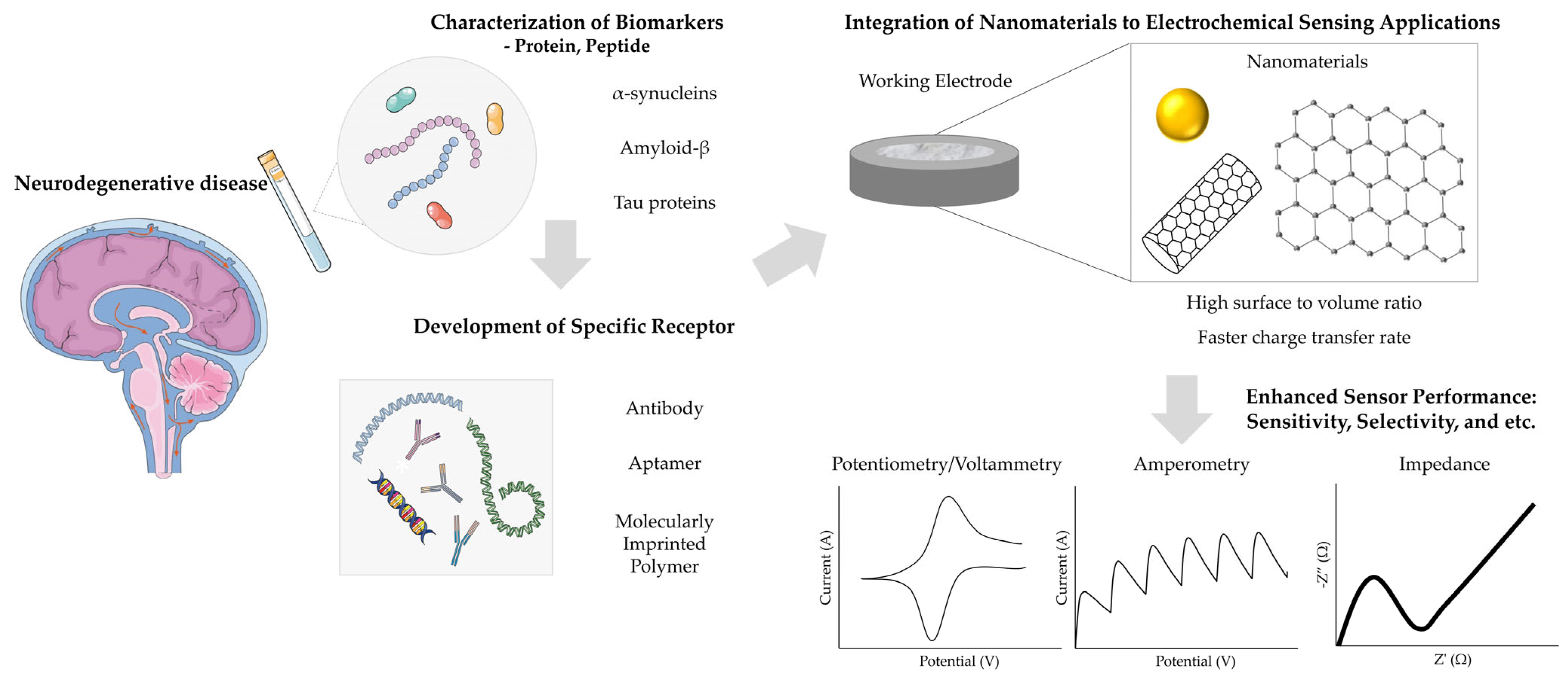
1. Introduction
2. Electrochemical Biosensor for α-Synucleins
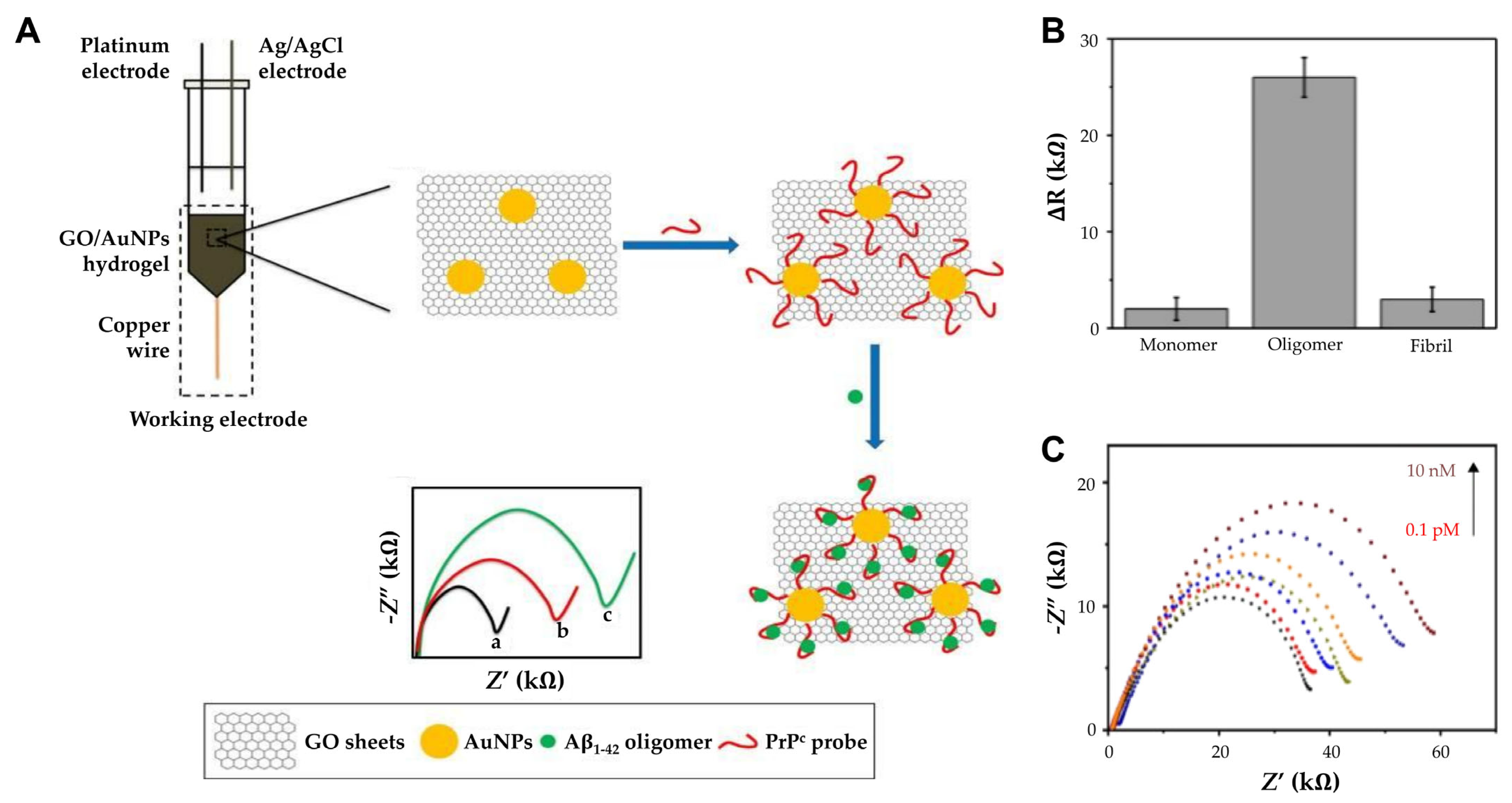
3. Electrochemical Biosensor for Amyloid-β
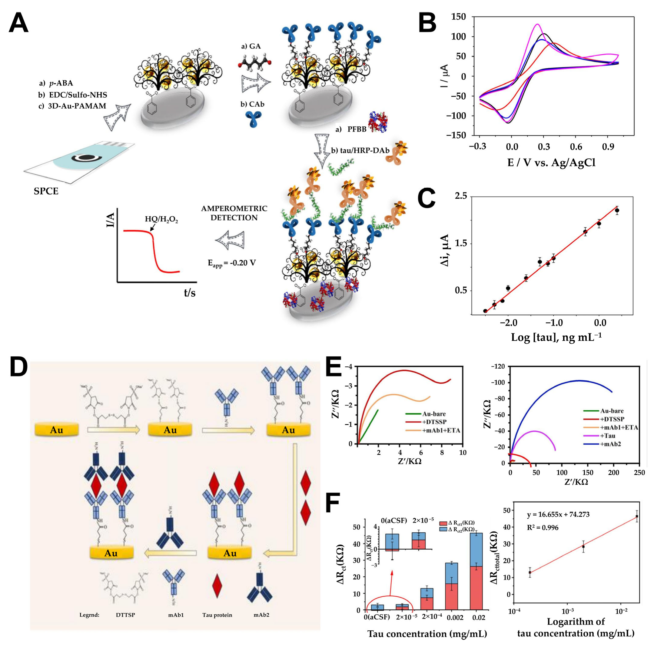
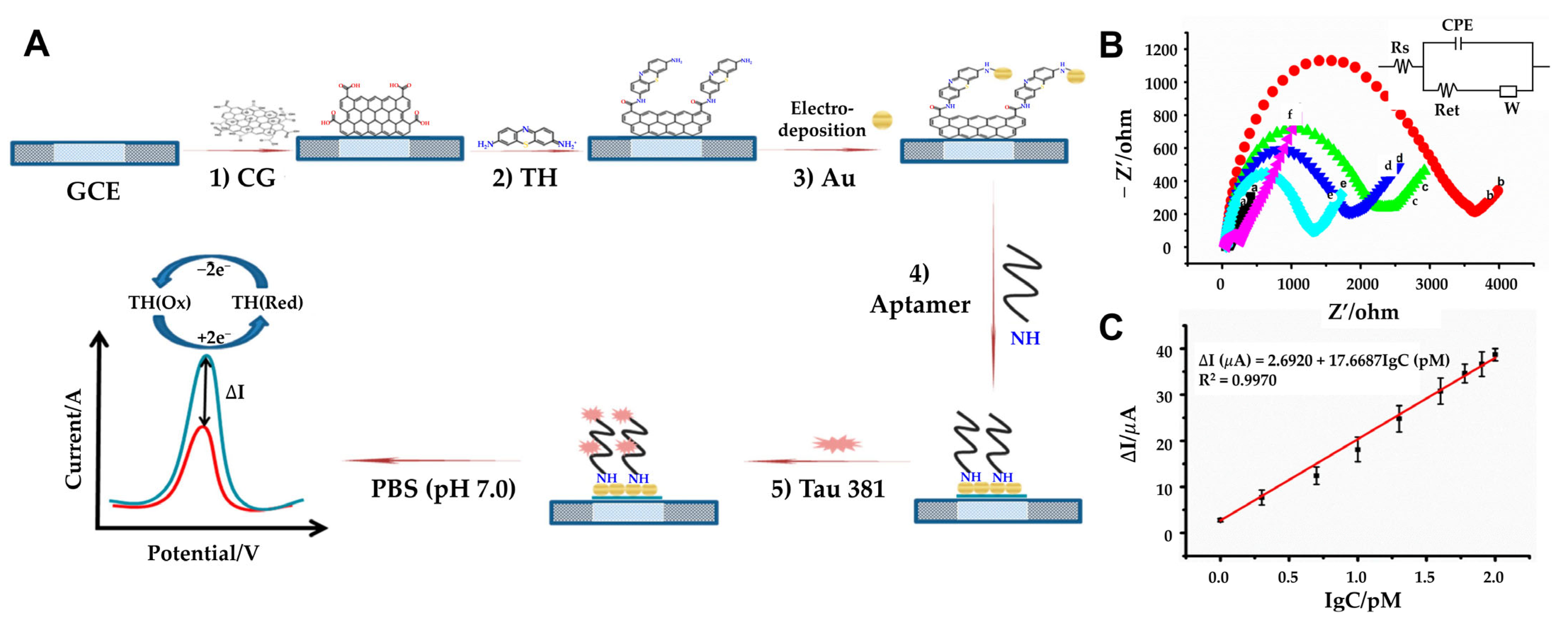
4. Electrochemical Biosensor for Tau Proteins
5. Conclusions and Future Perspectives
Author Contributions
Funding
Conflicts of Interest
References
- Hansson, O. Biomarkers for neurodegenerative diseases. Nat. Med. 2021, 27, 954–963. [Google Scholar] [CrossRef] [PubMed]
- Stephenson, J.; Nutma, E.; van der Valk, P.; Amor, S. Inflammation in CNS neurodegenerative diseases. Immunology 2018, 154, 204–219. [Google Scholar] [CrossRef] [PubMed]
- Ball, N.; Teo, W.P.; Chandra, S.; Chapman, J. Parkinson’s Disease and the Environment. Front. Neurol. 2019, 10, 218. [Google Scholar] [CrossRef]
- Gitler, A.D.; Dhillon, P.; Shorter, J. Neurodegenerative disease: Models, mechanisms, and a new hope. Dis. Model. Mech. 2017, 10, 499–502. [Google Scholar] [CrossRef]
- Emamzadeh, F.N. Role of Apolipoproteins and alpha-Synuclein in Parkinson’s Disease. J. Mol. Neurosci. 2017, 62, 344–355. [Google Scholar] [CrossRef]
- Kwon, E.H.; Tennagels, S.; Gold, R.; Gerwert, K.; Beyer, L.; Tonges, L. Update on CSF Biomarkers in Parkinson’s Disease. Biomolecules 2022, 12, 329. [Google Scholar] [CrossRef]
- Blennow, K.; Zetterberg, H. Biomarkers for Alzheimer’s disease: Current status and prospects for the future. J. Intern. Med. 2018, 284, 643–663. [Google Scholar] [CrossRef]
- Jack, C.R., Jr.; Bennett, D.A.; Blennow, K.; Carrillo, M.C.; Dunn, B.; Haeberlein, S.B.; Holtzman, D.M.; Jagust, W.; Jessen, F.; Karlawish, J.; et al. NIA-AA Research Framework: Toward a biological definition of Alzheimer’s disease. Alzheimers Dement. 2018, 14, 535–562. [Google Scholar] [CrossRef]
- Ding, M.; Ding, S.; Du, D.; Wang, X.; Hu, X.; Guan, P.; Lyu, Z.; Lin, Y. Recent advances in electrochemical biosensors for the detection of Aβ42, a biomarker for Alzheimer disease diagnosis. TrAC Trends Anal. Chem. 2023, 164, 117087. [Google Scholar] [CrossRef]
- Sharma, A.; Angnes, L.; Sattarahmady, N.; Negahdary, M.; Heli, H. Electrochemical Immunosensors Developed for Amyloid-Beta and Tau Proteins, Leading Biomarkers of Alzheimer’s Disease. Biosensors 2023, 13, 742. [Google Scholar] [CrossRef]
- Karki, H.P.; Jang, Y.; Jung, J.; Oh, J. Advances in the development paradigm of biosample-based biosensors for early ultrasensitive detection of alzheimer’s disease. J. Nanobiotechnology 2021, 19, 72. [Google Scholar] [CrossRef]
- Shin, M.-K.; Schuck, A.; Kang, M.; Kim, Y.-S. Electrochemical Analysis of Amyloid Plaques and ApoE4 with Chitosan-Coated Gold Nanostars for Alzheimer’s Detection. Biosensors 2024, 14, 510. [Google Scholar] [CrossRef] [PubMed]
- Schirinzi, T.; Zenuni, H.; Grillo, P.; Bovenzi, R.; Guerrera, G.; Gargano, F.; Pieri, M.; Bernardini, S.; Biagio Mercuri, N.; Battistini, L.; et al. Tau and Amyloid-beta Peptides in Serum of Patients with Parkinson’s Disease: Correlations with CSF Levels and Clinical Parameters. Front. Neurol. 2022, 13, 748599. [Google Scholar] [CrossRef] [PubMed]
- Aminabad, E.D.; Mobed, A.; Hasanzadeh, M.; Hosseinpour Feizi, M.A.; Safaralizadeh, R.; Seidi, F. Sensitive immunosensing of alpha-synuclein protein in human plasma samples using gold nanoparticles conjugated with graphene: An innovative immuno-platform towards early stage identification of Parkinson’s disease using point of care (POC) analysis. RSC Adv. 2022, 12, 4346–4357. [Google Scholar] [CrossRef]
- Carneiro, P.; Loureiro, J.A.; Delerue-Matos, C.; Morais, S.; Pereira, M.D.C. Nanostructured label-free electrochemical immunosensor for detection of a Parkinson’s disease biomarker. Talanta 2023, 252, 123838. [Google Scholar] [CrossRef]
- Gupta, S.; Murthy, C.N.; Prabha, C.R. Recent advances in carbon nanotube based electrochemical biosensors. Int. J. Biol. Macromol. 2018, 108, 687–703. [Google Scholar] [CrossRef]
- Kaya, H.O.; Cetin, A.E.; Azimzadeh, M.; Topkaya, S.N. Pathogen detection with electrochemical biosensors: Advantages, challenges and future perspectives. J. Electroanal Chem. 2021, 882, 114989. [Google Scholar] [CrossRef]
- Bryan, T.; Luo, X.L.; Forsgren, L.; Morozova-Roche, L.A.; Davis, J.J. The robust electrochemical detection of a Parkinson’s disease marker in whole blood sera. Chem. Sci. 2012, 3, 3468–3473. [Google Scholar] [CrossRef]
- Fredj, Z.; Singh, B.; Bahri, M.; Qin, P.W.; Sawan, M. Enzymatic Electrochemical Biosensors for Neurotransmitters Detection: Recent Achievements and Trends. Chemosensors 2023, 11, 388. [Google Scholar] [CrossRef]
- Guo, L.; Zhao, Y.; Huang, Q.; Huang, J.; Tao, Y.; Chen, J.; Li, H.-Y.; Liu, H. Electrochemical protein biosensors for disease marker detection: Progress and opportunities. Microsyst. Nanoeng. 2024, 10, 65. [Google Scholar] [CrossRef]
- Andriukonis, E.; Celiesiute-Germaniene, R.; Ramanavicius, S.; Viter, R.; Ramanavicius, A. From Microorganism-Based Amperometric Biosensors Towards Microbial Fuel Cells. Sensors 2021, 21, 2442. [Google Scholar] [CrossRef] [PubMed]
- Nur Ashakirin Binti Mohd Nashruddin, S.; Hani Mohamed Salleh, F.; Ayunni Mohd Raub, A. Early detection of kidney problems through voltammetry, potentiometry, amperometry, and impedance electrochemical techniques: A comprehensive review. Measurement 2024, 230, 114475. [Google Scholar] [CrossRef]
- Romero, M.R.; Ahumada, F.; Garay, F.; Baruzzi, A.M. Amperometric Biosensor for Direct Blood Lactate Detection. Anal. Chem. 2010, 82, 5568–5572. [Google Scholar] [CrossRef] [PubMed]
- Sumitha, M.S.; Xavier, T.S. Recent advances in electrochemical biosensors—A brief review. Hybrid. Adv. 2023, 2, 100023. [Google Scholar] [CrossRef]
- Karimi-Maleh, H.; Orooji, Y.; Karimi, F.; Alizadeh, M.; Baghayeri, M.; Rouhi, J.; Tajik, S.; Beitollahi, H.; Agarwal, S.; Gupta, V.K.; et al. A critical review on the use of potentiometric based biosensors for biomarkers detection. Biosens. Bioelectron. 2021, 184, 113252. [Google Scholar] [CrossRef]
- Özbek, O.; Isildak, Ö.; Isildak, I. A potentiometric biosensor for the determination of valproic acid: Human blood–based study of an anti–epileptic drug. Biochem. Eng. J. 2021, 176, 108181. [Google Scholar] [CrossRef]
- Fahmy Taha, M.H.; Ashraf, H.; Caesarendra, W. A Brief Description of Cyclic Voltammetry Transducer-Based Non-Enzymatic Glucose Biosensor Using Synthesized Graphene Electrodes. Appl. Syst. Innov. 2020, 3, 32. [Google Scholar] [CrossRef]
- Lopez-Tellez, J.; Ramirez-Montes, S.; Ferreira, T.A.; Santos, E.M.; Rodriguez, J.A. Application of Voltammetric Sensors for Pathogen Bacteria Detection: A Review. Chemosensors 2022, 10, 424. [Google Scholar] [CrossRef]
- Štukovnik, Z.; Bren, U. Recent Developments in Electrochemical-Impedimetric Biosensors for Virus Detection. Int. J. Mol. Sci. 2022, 23, 15922. [Google Scholar] [CrossRef]
- Štukovnik, Z.; Fuchs-Godec, R.; Bren, U. Nanomaterials and Their Recent Applications in Impedimetric Biosensing. Biosensors 2023, 13, 899. [Google Scholar] [CrossRef]
- Qian, L.; Durairaj, S.; Prins, S.; Chen, A. Nanomaterial-based electrochemical sensors and biosensors for the detection of pharmaceutical compounds. Biosens. Bioelectron. 2021, 175, 112836. [Google Scholar] [CrossRef] [PubMed]
- Liu, X.; Huang, L.; Qian, K. Nanomaterial-Based Electrochemical Sensors: Mechanism, Preparation, and Application in Biomedicine. Adv. Nanobiomed Res. 2021, 1, 2000104. [Google Scholar] [CrossRef]
- Manikandan, V.S.; Adhikari, B.; Chen, A. Nanomaterial based electrochemical sensors for the safety and quality control of food and beverages. Analyst 2018, 143, 4537–4554. [Google Scholar] [CrossRef]
- Damiati, S.; Schuster, B. Electrochemical Biosensors Based on S-Layer Proteins. Sensors 2020, 20, 1721. [Google Scholar] [CrossRef]
- Theyagarajan, K.; Kim, Y.J. Recent Developments in the Design and Fabrication of Electrochemical Biosensors Using Functional Materials and Molecules. Biosensors 2023, 13, 424. [Google Scholar] [CrossRef]
- Cho, I.-H.; Kim, D.H.; Park, S. Electrochemical biosensors: Perspective on functional nanomaterials for on-site analysis. Biomater. Res. 2020, 24, 6. [Google Scholar] [CrossRef]
- Reta, N.; Saint, C.P.; Michelmore, A.; Prieto-Simon, B.; Voelcker, N.H. Nanostructured Electrochemical Biosensors for Label-Free Detection of Water- and Food-Borne Pathogens. ACS Appl. Mater. Interfaces 2018, 10, 6055–6072. [Google Scholar] [CrossRef]
- Keles, G.; Sifa Ataman, E.; Taskin, S.B.; Polatoglu, İ.; Kurbanoglu, S. Nanostructured Metal Oxide-Based Electrochemical Biosensors in Medical Diagnosis. Biosensors 2024, 14, 238. [Google Scholar] [CrossRef]
- Naresh, V.; Lee, N. A Review on Biosensors and Recent Development of Nanostructured Materials-Enabled Biosensors. Sensors 2021, 21, 1109. [Google Scholar] [CrossRef]
- Grieshaber, D.; MacKenzie, R.; Vörös, J.; Reimhult, E. Electrochemical Biosensors—Sensor Principles and Architectures. Sensors 2008, 8, 1400–1458. [Google Scholar] [CrossRef]
- Ramesh, M.; Janani, R.; Deepa, C.; Rajeshkumar, L. Nanotechnology-Enabled Biosensors: A Review of Fundamentals, Design Principles, Materials, and Applications. Biosensors 2023, 13, 40. [Google Scholar] [CrossRef] [PubMed]
- Zhu, C.; Yang, G.; Li, H.; Du, D.; Lin, Y. Electrochemical Sensors and Biosensors Based on Nanomaterials and Nanostructures. Anal. Chem. 2015, 87, 230–249. [Google Scholar] [CrossRef] [PubMed]
- Doroszkiewicz, J.; Groblewska, M.; Mroczko, B. Molecular Biomarkers and Their Implications for the Early Diagnosis of Selected Neurodegenerative Diseases. Int. J. Mol. Sci. 2022, 23, 4610. [Google Scholar] [CrossRef]
- Twohig, D.; Nielsen, H.M. alpha-synuclein in the pathophysiology of Alzheimer’s disease. Mol. Neurodegener. 2019, 14, 23. [Google Scholar] [CrossRef]
- Goedert, M. Alpha-synuclein and neurodegenerative diseases. Nat. Rev. Neurosci. 2001, 2, 492–501. [Google Scholar] [CrossRef]
- Bridi, J.C.; Hirth, F. Mechanisms of alpha-Synuclein Induced Synaptopathy in Parkinson’s Disease. Front. Neurosci. 2018, 12, 80. [Google Scholar] [CrossRef]
- Jin, M.; Matsumoto, S.; Ayaki, T.; Yamakado, H.; Taguchi, T.; Togawa, N.; Konno, A.; Hirai, H.; Nakajima, H.; Komai, S.; et al. DOPAnization of tyrosine in α-synuclein by tyrosine hydroxylase leads to the formation of oligomers. Nat. Commun. 2022, 13, 6880. [Google Scholar] [CrossRef]
- Ishimoto, T.; Oono, M.; Kaji, S.; Ayaki, T.; Nishida, K.; Funakawa, I.; Maki, T.; Matsuzawa, S.-i.; Takahashi, R.; Yamakado, H. A novel mouse model for investigating α-synuclein aggregates in oligodendrocytes: Implications for the glial cytoplasmic inclusions in multiple system atrophy. Mol. Brain 2024, 17, 28. [Google Scholar] [CrossRef]
- Yoon, Y.S.; Ahn, W.J.; Ricarte, D.; Ortiz, D.; Shin, C.Y.; Lee, S.J.; Lee, H.J. Alpha-Synuclein Inclusion Formation in Human Oligodendrocytes. Biomol. Ther. 2021, 29, 83–89. [Google Scholar] [CrossRef]
- Calabresi, P.; Mechelli, A.; Natale, G.; Volpicelli-Daley, L.; Di Lazzaro, G.; Ghiglieri, V. Alpha-synuclein in Parkinson’s disease and other synucleinopathies: From overt neurodegeneration back to early synaptic dysfunction. Cell Death Dis. 2023, 14, 176. [Google Scholar] [CrossRef]
- Massey, R.S.; McConnell, E.M.; Chan, D.; Holahan, M.R.; DeRosa, M.C.; Prakash, R. Non-invasive Monitoring of α-Synuclein in Saliva for Parkinson’s Disease Using Organic Electrolyte-Gated FET Aptasensor. ACS Sens. 2023, 8, 3116–3126. [Google Scholar] [CrossRef] [PubMed]
- Sonuc Karaboga, M.N.; Sezginturk, M.K. Cerebrospinal fluid levels of alpha-synuclein measured using a poly-glutamic acid-modified gold nanoparticle-doped disposable neuro-biosensor system. Analyst 2019, 144, 611–621. [Google Scholar] [CrossRef] [PubMed]
- Aminabad, E.D.; Hasanzadeh, M.; Ahmadalipour, A.; Mahmoudi, T.; Feizi, M.A.H.; Safaralizadeh, R.; Mobed, A. Sensitive electrochemical recognition of alpha-synuclein protein in human plasma samples using bioconjugated gold nanoparticles: An innovative immuno-platform to assist in the early stage identification of Parkinson’s disease by biosensor technology. J. Mol. Recognit. 2023, 36, e2952. [Google Scholar] [CrossRef] [PubMed]
- Guo, X.; Wang, J.; Bu, J.; Zhang, H.; Arshad, M.; Kanwal, A.; Majeed, M.K.; Chen, W.-X.; Saxena, K.K.; Liu, X. Designing Nanocomposite-Based Electrochemical Biosensors for Diabetes Mellitus Detection: A Review. ACS Omega 2024, 9, 30071–30086. [Google Scholar] [CrossRef]
- Zeng, H.; Xie, Y.; Liu, T.; Chu, Z.; Dempsey, E.; Jin, W. Conductive polymer nanocomposites: Recent advances in the construction of electrochemical biosensors. Sens. Diagn. 2024, 3, 165–180. [Google Scholar] [CrossRef]
- Medrano-Lopez, J.A.; Villalpando, I.; Salazar, M.I.; Torres-Torres, C. Hierarchical Nanobiosensors at the End of the SARS-CoV-2 Pandemic. Biosensors 2024, 14, 108. [Google Scholar] [CrossRef]
- Zhang, Y.; Li, J.; Wang, Z.; Ma, H.; Wu, D.; Cheng, Q.; Wei, Q. Label-free electrochemical immunosensor based on enhanced signal amplification between Au@Pd and CoFe2O4/graphene nanohybrid. Sci. Rep. 2016, 6, 23391. [Google Scholar] [CrossRef]
- Soto, D.; Orozco, J. Hybrid Nanobioengineered Nanomaterial-Based Electrochemical Biosensors. Molecules 2022, 27, 3841. [Google Scholar] [CrossRef]
- Liu, Y.; Li, H.; Gong, S.; Chen, Y.; Xie, R.; Wu, Q.; Tao, J.; Meng, F.; Zhao, P. A novel non-enzymatic electrochemical biosensor based on the nanohybrid of bimetallic PdCu nanoparticles/carbon black for highly sensitive detection of H2O2 released from living cells. Sens. Actuators B Chem. 2019, 290, 249–257. [Google Scholar] [CrossRef]
- Su, S.; Lu, Z.; Li, J.; Hao, Q.; Liu, W.; Zhu, C.; Shen, X.; Shi, J.; Wang, L. MoS2–Au@Pt nanohybrids as a sensing platform for electrochemical nonenzymatic glucose detection. New J. Chem. 2018, 42, 6750–6755. [Google Scholar] [CrossRef]
- Yoon, J.; Lim, J.; Shin, M.; Lee, S.-N.; Choi, J.-W. Graphene/MoS2 Nanohybrid for Biosensors. Materials 2021, 14, 518. [Google Scholar] [CrossRef] [PubMed]
- Ao, Y.; Ao, J.; Zhao, L.; Hu, L.; Qu, F.; Guo, B.; Liu, X. Hierarchical Structures Composed of Cu(OH)2 Nanograss within Directional Microporous Cu for Glucose Sensing. Langmuir 2022, 38, 13659–13667. [Google Scholar] [CrossRef] [PubMed]
- Di Mari, G.M.; Scuderi, M.; Lanza, G.; Salluzzo, M.G.; Salemi, M.; Caraci, F.; Bruno, E.; Strano, V.; Mirabella, S.; Scandurra, A. Pain-Free Alpha-Synuclein Detection by Low-Cost Hierarchical Nanowire Based Electrode. Nanomaterials 2024, 14, 170. [Google Scholar] [CrossRef] [PubMed]
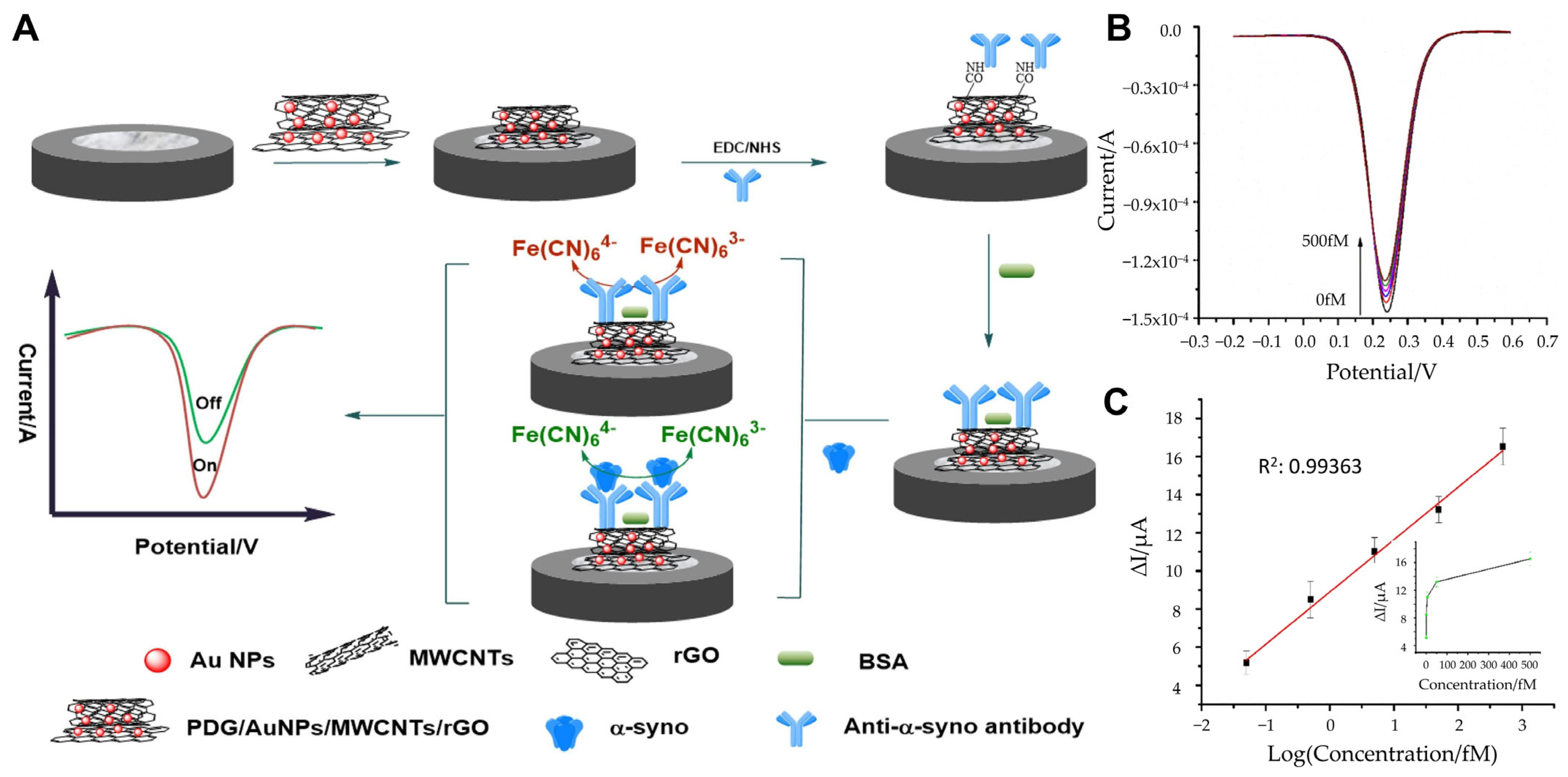
- Tao, D.; Gu, Y.Y.; Song, S.Z.; Nguyen, E.P.; Cheng, J.; Yuan, Q.; Pan, H.Z.; Jaffrezic-Renault, N.; Guo, Z.Z. Ultrasensitive detection of alpha-synuclein oligomer using a PolyD-glucosamine/gold nanoparticle/carbon-based nanomaterials modified electrochemical immunosensor in human plasma. Microchem. J. 2020, 158, 105195. [Google Scholar] [CrossRef]
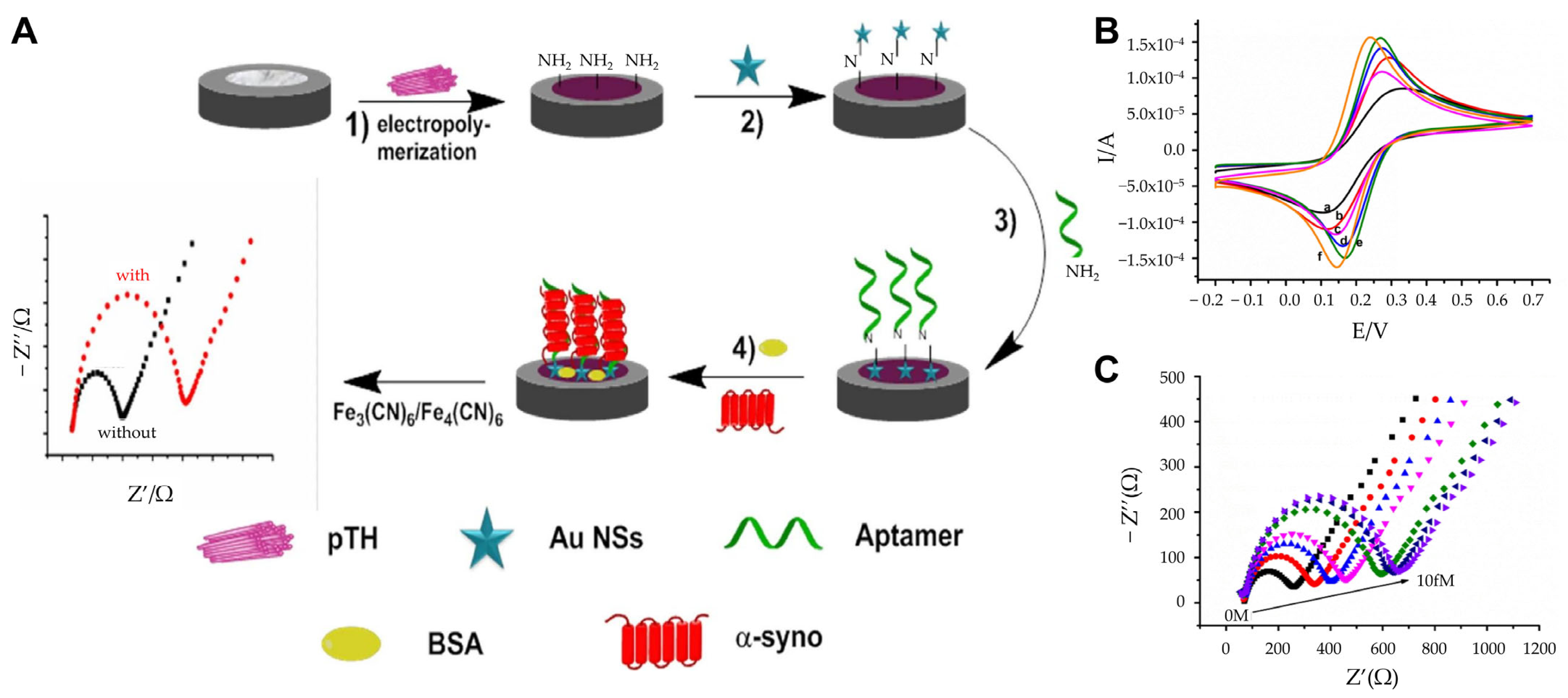
- Tao, D.; Wang, J.; Song, S.Z.; Cai, K.; Jiang, M.D.; Cheng, J.; Hu, L.; Jaffrezic-Renault, N.; Guo, Z.Z.; Pan, H.Z. Polythionine and gold nanostar-based impedimetric aptasensor for label-free detection of α-synuclein oligomers. J. Appl. Electrochem. 2021, 51, 1523–1533. [Google Scholar] [CrossRef]
- Yao, T.; Wang, R.; Meng, Y.; Hun, X. Photoelectrochemical Sensing of alpha-Synuclein Based on a AuNPs/Graphdiyne-Modified Electrode Coupled with a Nanoprobe. ACS Appl. Mater. Interfaces 2021, 13, 26515–26521. [Google Scholar] [CrossRef]
- Zhang, Z.H.; Hu, J.; Chen, Q.; Chen, J.; Hu, X.; Koh, K.; Chen, H.; Xu, X.H. The magnetic-nanoparticle-assisted sensitive detection of nitrated alpha-syn in blood based on a sensitizing electrochemical layer. Nanoscale 2021, 13, 8107–8117. [Google Scholar] [CrossRef]
- Guo, C.P.; Hu, M.Y.; Li, Z.Z.; Duan, F.H.; He, L.H.; Zhang, Z.H.; Marchetti, F.; Du, M. Structural hybridization of bimetallic zeolitic imidazolate framework (ZIF) nanosheets and carbon nanofibers for efficiently sensing α-synuclein oligomers. Sens. Actuat B Chem. 2020, 309, 127821. [Google Scholar] [CrossRef]
- Lee, M.H.; Liu, K.T.; Thomas, J.L.; Su, Z.L.; O’Hare, D.; van Wuellen, T.; Chamarro, J.M.; Bolognin, S.; Luo, S.C.; Schwamborn, J.C.; et al. Peptide-Imprinted Poly(hydroxymethyl 3,4-ethylenedioxythiophene) Nanotubes for Detection of α Synuclein in Human Brain Organoids. ACS Appl. Nano Mater. 2020, 3, 8027–8036. [Google Scholar] [CrossRef]
- Sadigh-Eteghad, S.; Sabermarouf, B.; Majdi, A.; Talebi, M.; Farhoudi, M.; Mahmoudi, J. Amyloid-beta: A crucial factor in Alzheimer’s disease. Med. Princ. Pract. 2015, 24, 1–10. [Google Scholar] [CrossRef]
- Esparza, T.J.; Zhao, H.; Cirrito, J.R.; Cairns, N.J.; Bateman, R.J.; Holtzman, D.M.; Brody, D.L. Amyloid-beta oligomerization in Alzheimer dementia versus high-pathology controls. Ann. Neurol. 2013, 73, 104–119. [Google Scholar] [CrossRef] [PubMed]
- Ittner, L.M.; Ke, Y.D.; Delerue, F.; Bi, M.; Gladbach, A.; van Eersel, J.; Wolfing, H.; Chieng, B.C.; Christie, M.J.; Napier, I.A.; et al. Dendritic function of tau mediates amyloid-beta toxicity in Alzheimer’s disease mouse models. Cell 2010, 142, 387–397. [Google Scholar] [CrossRef] [PubMed]
- Arbel-Ornath, M.; Hudry, E.; Boivin, J.R.; Hashimoto, T.; Takeda, S.; Kuchibhotla, K.V.; Hou, S.; Lattarulo, C.R.; Belcher, A.M.; Shakerdge, N.; et al. Soluble oligomeric amyloid-β induces calcium dyshomeostasis that precedes synapse loss in the living mouse brain. Mol. Neurodegener. 2017, 12, 27. [Google Scholar] [CrossRef] [PubMed]
- Kurucu, H.; Colom-Cadena, M.; Davies, C.; Wilkins, L.; King, D.; Rose, J.; Tzioras, M.; Tulloch, J.H.; Smith, C.; Spires-Jones, T.L. Inhibitory synapse loss and accumulation of amyloid beta in inhibitory presynaptic terminals in Alzheimer’s disease. Eur. J. Neurol. 2022, 29, 1311–1323. [Google Scholar] [CrossRef]
- Zhang, Y.; Chen, H.; Li, R.; Sterling, K.; Song, W. Amyloid β-based therapy for Alzheimer’s disease: Challenges, successes and future. Signal Transduct. Target. Ther. 2023, 8, 248. [Google Scholar] [CrossRef]
- Qin, J.L.; Kim, S.; Cho, M.; Lee, Y. Hierarchical and ultra-sensitive amyloid beta oligomer sensor for practical applications. Chem. Eng. J. 2020, 401, 126055. [Google Scholar] [CrossRef]
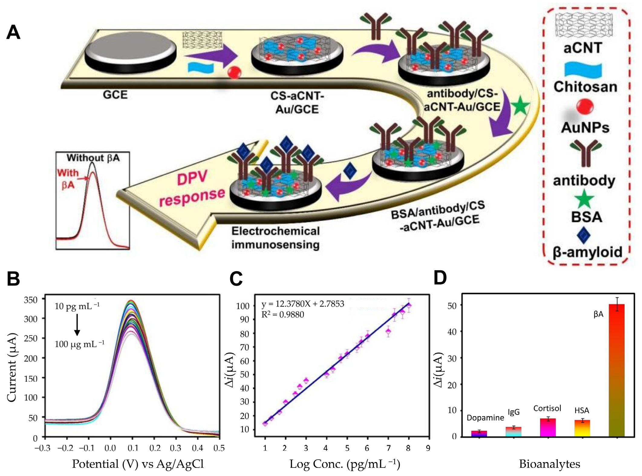
- Ranjan, P.; Khan, R. Electrochemical Immunosensor for Early Detection of beta-Amyloid Alzheimer’s Disease Biomarker Based on Aligned Carbon Nanotubes Gold Nanocomposites. Biosensors 2022, 12, 1059. [Google Scholar] [CrossRef]
- Sethi, J.; Van Bulck, M.; Suhail, A.; Safarzadeh, M.; Perez-Castillo, A.; Pan, G. A label-free biosensor based on graphene and reduced graphene oxide dual-layer for electrochemical determination of beta-amyloid biomarkers. Mikrochim. Acta 2020, 187, 288. [Google Scholar] [CrossRef]
- Sun, L.; Zhong, Y.; Gui, J.; Wang, X.; Zhuang, X.; Weng, J. A hydrogel biosensor for high selective and sensitive detection of amyloid-beta oligomers. Int. J. Nanomed. 2018, 13, 843–856. [Google Scholar] [CrossRef]
- Palla, G.; Malecka, K.; Dehaen, W.; Radecki, J.; Radecka, H. Immunosensor incorporating half-antibody fragment for electrochemical monitoring of amyloid-beta fibrils in artificial blood plasma. Bioelectrochemistry 2021, 137, 107643. [Google Scholar] [CrossRef]
- Negahdary, M.; Heli, H. An ultrasensitive electrochemical aptasensor for early diagnosis of Alzheimer’s disease, using a fern leaves-like gold nanostructure. Talanta 2019, 198, 510–517. [Google Scholar] [CrossRef] [PubMed]
- Zhang, Y.T.; Figueroa-Miranda, G.; Lyu, Z.Z.; Zafiu, C.; Willbold, D.; Offenhäusser, A.; Mayer, D. Monitoring amyloid-β proteins aggregation based on label-free aptasensor. Sens. Actuat B Chem. 2019, 288, 535–542. [Google Scholar] [CrossRef]
- You, M.; Yang, S.; An, Y.; Zhang, F.; He, P.G. A novel electrochemical biosensor with molecularly imprinted polymers and aptamer-based sandwich assay for determining amyloid-β oligomer. J. Electroanal. Chem. 2020, 862, 114017. [Google Scholar] [CrossRef]
- Özcan, N.; Medetalibeyoglu, H.; Akyıldırım, O.; Atar, N.; Yola, M.L. Electrochemical detection of amyloid-β protein by delaminated titanium carbide MXene/multi-walled carbon nanotubes composite with molecularly imprinted polymer. Mater. Today Commun. 2020, 23, 101097. [Google Scholar] [CrossRef]
- Dani, M.; Brooks, D.J.; Edison, P. Tau imaging in neurodegenerative diseases. Eur. J. Nucl. Med. Mol. Imaging 2016, 43, 1139–1150. [Google Scholar] [CrossRef]
- Kolarova, M.; Garcia-Sierra, F.; Bartos, A.; Ricny, J.; Ripova, D. Structure and pathology of tau protein in Alzheimer disease. Int. J. Alzheimers Dis. 2012, 2012, 731526. [Google Scholar] [CrossRef]
- Brandt, R.; Trushina, N.I.; Bakota, L. Much More Than a Cytoskeletal Protein: Physiological and Pathological Functions of the Non-microtubule Binding Region of Tau. Front. Neurol. 2020, 11, 590059. [Google Scholar] [CrossRef]
- Gao, Y.L.; Wang, N.; Sun, F.R.; Cao, X.P.; Zhang, W.; Yu, J.T. Tau in neurodegenerative disease. Ann. Transl. Med. 2018, 6, 175. [Google Scholar] [CrossRef]
- Meng, J.X.; Zhang, Y.; Saman, D.; Haider, A.M.; De, S.; Sang, J.C.; Brown, K.; Jiang, K.; Humphrey, J.; Julian, L.; et al. Hyperphosphorylated tau self-assembles into amorphous aggregates eliciting TLR4-dependent responses. Nat. Commun. 2022, 13, 2692. [Google Scholar] [CrossRef]
- Hallinan, G.I.; Vargas-Caballero, M.; West, J.; Deinhardt, K. Tau Misfolding Efficiently Propagates between Individual Intact Hippocampal Neurons. J. Neurosci. 2019, 39, 9623–9632. [Google Scholar] [CrossRef]
- Wang, Z.; Wu, L.; Gerasimenko, M.; Gilliland, T.; Shah, Z.S.A.; Lomax, E.; Yang, Y.; Gunzler, S.A.; Donadio, V.; Liguori, R.; et al. Seeding activity of skin misfolded tau as a biomarker for tauopathies. Mol. Neurodegener. 2024, 19, 92. [Google Scholar] [CrossRef] [PubMed]
- Guo, T.; Noble, W.; Hanger, D.P. Roles of tau protein in health and disease. Acta Neuropathol. 2017, 133, 665–704. [Google Scholar] [CrossRef] [PubMed]
- Zhang, H.; Cao, Y.; Ma, L.; Wei, Y.; Li, H. Possible Mechanisms of Tau Spread and Toxicity in Alzheimer’s Disease. Front. Cell Dev. Biol. 2021, 9, 707268. [Google Scholar] [CrossRef]
- Sonuc Karaboga, M.N.; Sezginturk, M.K. Analysis of Tau-441 protein in clinical samples using rGO/AuNP nanocomposite-supported disposable impedimetric neuro-biosensing platform: Towards Alzheimer’s disease detection. Talanta 2020, 219, 121257. [Google Scholar] [CrossRef]
- Razzino, C.A.; Serafin, V.; Gamella, M.; Pedrero, M.; Montero-Calle, A.; Barderas, R.; Calero, M.; Lobo, A.O.; Yanez-Sedeno, P.; Campuzano, S.; et al. An electrochemical immunosensor using gold nanoparticles-PAMAM-nanostructured screen-printed carbon electrodes for tau protein determination in plasma and brain tissues from Alzheimer patients. Biosens. Bioelectron. 2020, 163, 112238. [Google Scholar] [CrossRef]
- Yang, M.; Chen, Y.; Sun, H.; Li, D.; Li, Y. A Simple Sandwich Electrochemical Immunosensor for Rapid Detection of the Alzheimer’s Disease Biomarker Tau Protein. Biosensors 2024, 14, 279. [Google Scholar] [CrossRef]
- Yola, B.B.; Karaman, C.; Özcan, N.; Atar, N.; Polat, I.; Yola, M.L. Electrochemical Tau Protein Immunosensor Based on MnS/GO/PANI and Magnetite-incorporated Gold Nanoparticles. Electroanalysis 2022, 34, 1519–1528. [Google Scholar] [CrossRef]
- Shui, B.; Tao, D.; Cheng, J.; Mei, Y.; Jaffrezic-Renault, N.; Guo, Z. A novel electrochemical aptamer-antibody sandwich assay for the detection of tau-381 in human serum. Analyst 2018, 143, 3549–3554. [Google Scholar] [CrossRef]
- Tao, D.; Shui, B.; Gu, Y.; Cheng, J.; Zhang, W.; Jaffrezic-Renault, N.; Song, S.; Guo, Z. Development of a Label-Free Electrochemical Aptasensor for the Detection of Tau381 and its Preliminary Application in AD and Non-AD Patients’ Sera. Biosensors 2019, 9, 84. [Google Scholar] [CrossRef]
- Carlin, N.; Martic-Milne, S. Anti-Tau Antibodies Based Electrochemical Sensor for Detection of Tau Protein Biomarkers. J. Electrochem. Soc. 2018, 165, G3018–G3025. [Google Scholar] [CrossRef]

| Sensor Platform | Sensor Type | Detection Technique | Linear Range | LOD | Reference |
|---|---|---|---|---|---|
| AuNPs, ZnO NSs | Immunosensor | CV, EIS | 0.5–10 pg mL−1 | 0.08 pg mL−1 | [63] |
| AuNPs, polyglutaminic acid | SWV | 4–2000 pg mL−1 | 0.135 pg mL | [52] | |
| AuNPs, PDG, MWCNTs, rGO | SWV | 0.05 fM–500 fM | 0.03 fM | [64] | |
| AuNPs, MNP | EIS | 1–1000 ng/mL−1 | 310 pg mL−1 | [67] | |
| AuNPs | DPV (Differential pulse voltammetry), SWV, ChA (chronoamperometry) | 4–64 ng mL−1 | 4 ng mL−1 | [53] | |
| AuNPs, graphene | ChA, EIS | 4–128 ng mL−1 | 4 ng mL−1 | [14] | |
| AuNSs, pTH, ssDNA | aptasensor | EIS | 0.10 aM–10.00 fM | 0.07 aM | [65] |
| AuNPs, GDY, DA/MBA/WSe2 | CV, EIS | 10 aM–1.0 nM | 3.3 aM | [66] | |
| carbon nanofibers, zeolitic imidazolate framework nanosheets | EIS | 1 fg mL−1–0.2 ng mL−1 (52.6 fM–0.1 nM) | 0.87 fg mL−1 (45.7 fM) | [68] | |
| Peptide-imprinted polymers | Molecularly imprinted polymer | CV | 0.065 pM to 0.65 nM | 4.0 pM | [69] |
| Sensor Platform | Sensor Type | Detection Technique | Linear Range | LOD | Reference |
|---|---|---|---|---|---|
| uNP, TBBT | immunosensor | EIS | 0.5 pM–4.0 pM | 0.64 pM | [80] |
| dual layer graphene, rGO | DPV | 11 pM–55 nM | 2.398 pM | [78] | |
| AuNP, chitosan, CNT | DPV | 10.0 pg mL–100.0 ug mL | 0.87 pg mL | [77] | |
| AuNP, RNA | aptasensor | DPV | 0.002–1.28 ng mL−1 | 0.4 pg mL−1 (88.6 amol L−1) | [81] |
| AuNP, GO, Thiolated PrP | EIS | 0.1 pM–10 nM | 0.1 pM | [79] | |
| AuD, ssDNA | EIS | 0.1–500 nM | 0.03 nM | [82] | |
| poly(pyrrole-3-carboxylic acid), PrP | EIS | 10−9–10 nM | 1 aM | [76] | |
| AgNP, SiO2 | MIP | DPV | 5 pg mL−1–10 ng mL−1 | 1.22 pg mL−1 | [83] |
| titanium carbide Mxene, multi-walled carbon nanotubes | DPV | 1.0 fg mL−1–100.0 fg mL−1 | 0.3 fg mL−1 | [84] |
| Sensor Platform | Sensor Type | Detection Technique | Linear Range | LOD | Reference |
|---|---|---|---|---|---|
| MnS/GO/PANI, AuNP@Fe3O4 | immunosensor | DPV | 0.1 pM–1.0 μM | 0.01 pM | [97] |
| AuNP, PAMAM dendrimers | CV, EIS, CA | 6–5000 pg mL (0.11–91 pM) | 0.031 pM (1.7 pg mL) | [95] | |
| rGO, AuNPs, 11-MUA | CV, EIS, SFI (Single Frequency Impedance) | 1–500 pg/mL | 0.091 pg/mL | [94] | |
| DTSSP | CV, EIS | 1 × 10⁻⁴ mg mL−1 to 0.01 mg mL−1 | 1 × 10⁻⁴ mg mL−1 | [96] | |
| lipoic acid, n-butylamine, hexanethiol | EIS | - | - | [100] | |
| AuNPs | aptasensor | DPV | 0.5 pM–100 pM | 0.42 pM | [98] |
| AuNPs, carboxyl graphene, thionin | DPV | 1.0 pM–100 pM | 0.70 pM | [99] |
Disclaimer/Publisher’s Note: The statements, opinions and data contained in all publications are solely those of the individual author(s) and contributor(s) and not of MDPI and/or the editor(s). MDPI and/or the editor(s) disclaim responsibility for any injury to people or property resulting from any ideas, methods, instructions or products referred to in the content. |
© 2025 by the authors. Licensee MDPI, Basel, Switzerland. This article is an open access article distributed under the terms and conditions of the Creative Commons Attribution (CC BY) license (https://creativecommons.org/licenses/by/4.0/).
Share and Cite
Bae, M.; Kim, N.; Cho, E.; Lee, T.; Lee, J.-H. Recent Advances in Electrochemical Biosensors for Neurodegenerative Disease Biomarkers. Biosensors 2025, 15, 151. https://doi.org/10.3390/bios15030151
Bae M, Kim N, Cho E, Lee T, Lee J-H. Recent Advances in Electrochemical Biosensors for Neurodegenerative Disease Biomarkers. Biosensors. 2025; 15(3):151. https://doi.org/10.3390/bios15030151
Chicago/Turabian StyleBae, Mingyu, Nayoung Kim, Euni Cho, Taek Lee, and Jin-Ho Lee. 2025. "Recent Advances in Electrochemical Biosensors for Neurodegenerative Disease Biomarkers" Biosensors 15, no. 3: 151. https://doi.org/10.3390/bios15030151
APA StyleBae, M., Kim, N., Cho, E., Lee, T., & Lee, J.-H. (2025). Recent Advances in Electrochemical Biosensors for Neurodegenerative Disease Biomarkers. Biosensors, 15(3), 151. https://doi.org/10.3390/bios15030151