Perspective

Bioluminescent Microbial Bioreporters: A Personal Perspective

Institute of Life Sciences, The Hebrew University of Jerusalem, Jerusalem 9190401, Israel
Biosensors 2025, 15(2), 111; https://doi.org/10.3390/bios15020111
Submission received: 7 January 2025 / Revised: 4 February 2025 / Accepted: 7 February 2025 / Published: 14 February 2025

Abstract

:
This review attempts to summarize my three decades-long involvement in, and contribution to, the design, construction and testing of bioluminescent microbial sensor strains (bioreporters). With the understanding that such a document cannot be completely free of bias, the review focuses on studies from my own lab only, with almost no coverage of the parallel progress made by others in similar fields. This admittedly subjective approach by no way detracts from the achievements of countless excellent researchers who are not mentioned here, and whose contributions to the field are at least as important as that of my own. The review covers basic aspects of microbial sensor design, and then progresses to describe approaches to performance improvement of sensor strains aimed at the detection of either specific chemicals, groups of chemicals sharing similar characteristics, or global effects, such as toxicity and genotoxicity. The need for integration of live sensor cells into a compatible hardware platform is highlighted, as is the importance of long-term maintenance of the cells’ viability and activity. The use of multi-member sensors’ panels is presented as a means for enhancing the detection spectrum and sample “fingerprinting”, along with a list of different purposes to which such sensors have been put to use.

1. Introduction

Trying to summarize one’s academic career in writing is awkward; it is even more so, as is the case here, when your career has not really ended. I still maintain a dynamic research group, and plan to continue to be active in years to come. The suggestion that I review my long-term involvement in the study of bioluminescent bioreporters, while highly flattering, has thus caught me by surprise. Nevertheless, I have willingly agreed to do so, hoping that it will allow me to show my appreciation and gratitude to the students, technicians, researchers and collaborators from Israel and abroad who played an essential role in this trip, and without whom none of the research achievements described below could have been possible. The current effort is thus not an unbiased comprehensive review of bioluminescent microbial sensors, but rather of the progress made in this field by my coworkers and myself, presented from my own personal viewpoint.
My first encounter with the world of bioluminescent bacteria was in the late Professor Moshe Shilo’s Hebrew University Marine Microbiology course, held in the 1970s at the Interuniversity Institute for Marine Sciences in Eilat. I carried this fascination with me when years later I was part of the teaching team of the very same course, and for almost 20 years have always included at least one microbial bioluminescence-related project to challenge students with.
It was Prof. Eugene Rosenberg (Tel Aviv University), also a teacher on that course who, over an evening beer in Eilat, mentioned the option of employing bioluminescent bacteria for toxicity assessment, as a solution for my difficulty in assaying the toxicity of the industrial wastewaters of the Ramat Hovav chemical industrial park in southern Israel. He suggested that I discuss this with the most prominent Israeli figure in the field, Prof. Shimon (Moni) Ulitzur (Technion, Israel Institute of Technology). My collaboration with Moni was indeed highly constructive, but the path I ultimately took was different from his: while he advocated assaying the decrease in light emission of a natural luminescent bacterium as a measure of sample toxicity (a “lights off” assay, versions of which were later standardized and commercialized [1]), I ended up deeply involved in the development of “lights on” assays.
That crucial turning point took place during my 1993–1994 Sabbatical with Dr. Bob LaRossa’s group at the DuPont Central Research and Development campus in Wilmington, Delaware. Under Bob’s guidance, I was not only initiated in the art of molecular engineering of bioluminescent sensor strains, but I also came to realize that the rather simplistic tool I was mastering could be useful in many different ways, from elucidating the fate of wastewater bacteria in the marine environment to the construction of sensor systems for either specific chemicals, classes of compounds, or general environmental stress conditions.
This review is focused on the research path that stemmed from that encounter and which I follow till this day; it does not touch upon my decades-long involvement with other significant aspects of my academic interests, mostly revolving around diverse facets of environmental microbiology and microbial ecology.

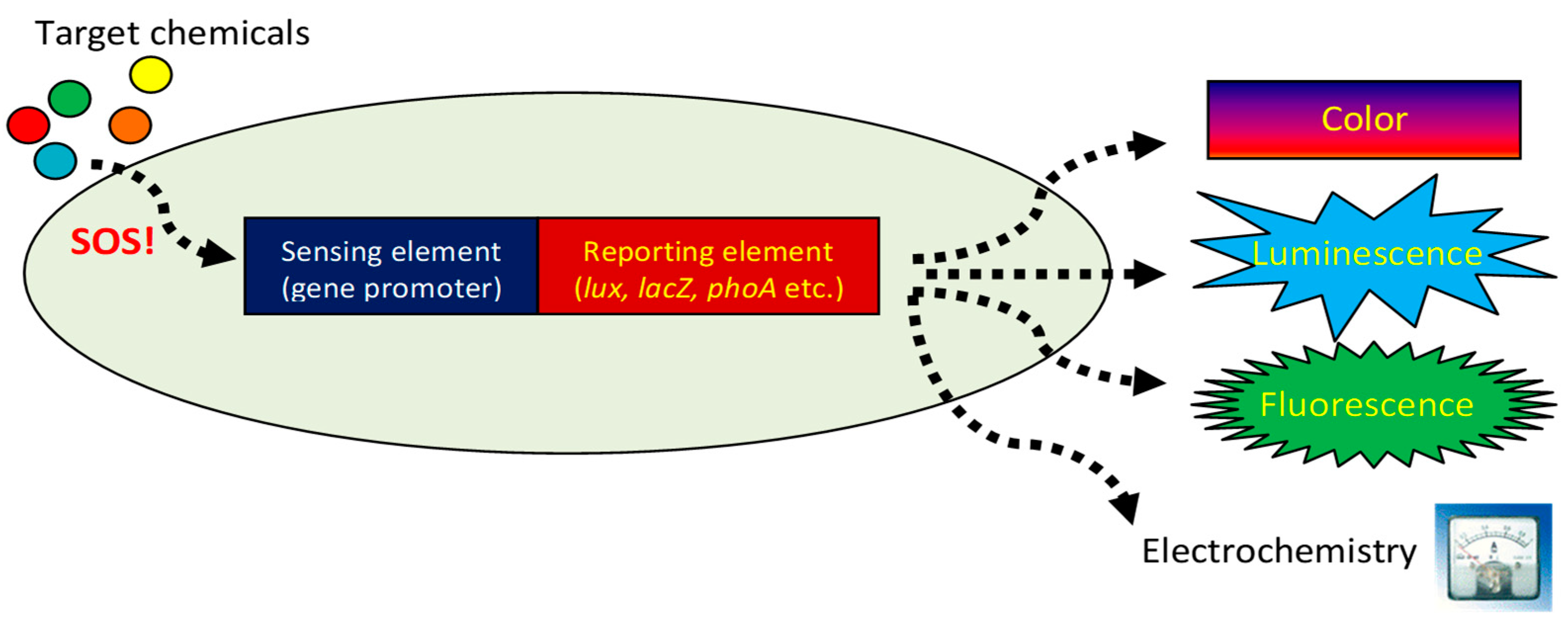
2. The Basic Story: Selecting the Appropriate Sensing Element

In our hands, the basis for the construction of a microbial sensor strain always involved the a priori selection of three elements (Figure 1): (a) a sensing element (in most cases, a gene promoter induced by the target compound(s) or stress condition); (b) a reporter element—a gene or a group of genes, the activity or the presence of the products of which can be monitored quantitatively; (c) the host microorganism, selected either for convenience or for its relevance to the envisaged application [2,3,4]. Our organism of choice for most applications was the conveniently manipulated Escherichia coli. Out of a rather limited list of reporter options—primarily fluorescence, color generation, electric current or bioluminescence—we usually worked with the latter. In addition to the detection sensitivity afforded by the bacterial luxCDABE(G) gene cassette, it has the added advantage of intracellular production of the long-chain aldehyde luciferase substrate, alleviating the need for adding it externally, as is the case of other enzymatic reporters. Thus, the main challenge when designing a new sensor strain was the selection of the most suitable sensing element, and—when relevant—identification of its associated regulatory components.
An obvious cornerstone of every scientific progress and innovation is preexisting knowledge. The continuously expanding microbial biochemical/molecular/physiological knowledge base allows the selection of gene promoters that will respond to the presence of many target compounds. This was how, in the pioneering work of Sayler and his colleagues [5], the nahG gene promoter was selected as the sensing element in their Pseudomonas fluorescens naphthalene sensor, and how shortly afterwards the merR system served as an excellent component of the first E. coli mercury bioreporter [6]. Similarly, the series of papers that came out of the LaRossa group a few years later were all based on the same principle: the promoter of the catalase katG gene, for example, served for the construction of a hydrogen peroxide sensor [7], or the promoter of the recA gene for a DNA-damage sensor [8].
However, how do you select a sensing element if there is no straightforward information as to a cellular response to a compound, or to a biochemical reaction involved in its metabolism? The first DNA chips introduced in the early 1990s provided a high-throughput unbiased answer: the promoters of genes, shown on-chip to be induced by the target chemical or condition, may be suitable candidates for sensor construction. In our hands, regrettably, this promising technology did not yield the desirable results; a different approach was much more successful: employing a transcriptional GFP fusion library constructed in the Alon’s lab at the Weizmann institute [9] has identified several E. coli gene promotors responsive to 2,4-dinitrotoluene (DNT) [10], the signature chemical accompanying 2,4,6-trinitrotoluene (TNT), the main explosive component of landmines. The most prominent among those was yqjF, the gene promoter that served as the sensing element in a subsequent series of DNT/TNT sensor strains. Another relevant DNT-inducible gene promoter, azoR, was similarly identified by the use of RNA-Seq analysis [11], another methodology that allows for an unbiased screening for genes responsive to specific environmental conditions.

3. How to Improve Your Sensor’s Performance

The basic promoter–reporter fusion, the core concept in microbial bioreporter design, is as effective as it is simple [3]. Often, however, even if the desired specificity is achieved, other performance parameters, most importantly signal intensity, detection sensitivity and response time, are insufficient for the envisaged application. Over the years we have adopted and developed several strategies for performance enhancement. These spanned genetic manipulations of the sensing, reporting and regulatory elements of the original construct, as well as of the host cell.
Host cell modifications mostly included mutations which either affected cellular permeability [12,13] or enhanced sensor performance for reasons that were not always clear. Worth mentioning in the latter category is B. Shemer’s monumental screening effort of the entire E. coli mutation spectrum for mutations beneficial to yqjF activation by DNT [14]. Two of the mutations identified in that study, in the ygdD and eutE genes, are still a part of the bioreporter in use today for explosives’ detection.
Beneficial adjustments to the bioluminescence reporter element included either switching the entire lux gene cassette, achieving variations in temperature dependence as well as in signal intensity [15], or splitting the lux gene cassette into two independently regulated components, luxAB and luxCDE [16]. The latter strategy, by providing an increased substrate supply to luciferase due to a constitutive expression of luxCDE, removed a potential bottleneck in the enzyme’s activity and boosted its light production.
Significant enhancements in DNT detection performance were also achieved by modifying YhaJ, the LysR-type transcriptional regulator of yqjF [17]. Two different experimental approaches were adopted for this purpose: “Directed evolution” of the YhaJ protein by random mutagenesis [18], and screening a focused saturation library of over 6 × 107 mutated YhaJ variants [19]. Directed evolution by several consecutive rounds of random mutagenesis was also successfully employed for significant enhancement of the yqjF gene promotor [10,20], as well as for “gene shuffling” performed on a fusion of the yqjF and azoR gene promotors [21]. Further improvements were achieved by the introduction of quorum sensing elements [22] into the induction chain, as well as the modification of specific ribosomal binding sites. Several other manipulations we and others have successfully integrated into microbial bioreporter designs are described elsewhere [23].

4. Just Having a Good Sensor Strain May Not Be Enough

The term “Biosensor” is defined by the International Union of Pure and Applied Chemistry [24] as a “device that uses specific biochemical reactions mediated by isolated enzymes, immuno-systems, tissues, organelles or whole cells”. In other words, in order to be considered a “biosensor”, the bioreporter cells need to be integrated into a hardware platform that will allow, for example, field or point-of-care use. Numerous approaches for the integration of live cells into inanimate devices have been described in the scientific literature [25,26]. Our contributions to these efforts were manifested in several directions. Melamed et al. have demonstrated that nanodrops of functional live sensor cells can be spotted onto a glass surface [27] and be successfully revived after storage. For online water monitoring, Sharon Yagur-Kroll and Tal Elad et al. have demonstrated the integration of bioreporter cells in miniature porous aluminum oxide-based [28] and microfluidic polydimethylsiloxane (PDMS)-based flow cells [29], respectively. The latter device uninterruptedly monitored untreated tap water for two weeks. A different approach was adopted by the group of A. J. Agranat, who successfully incorporated alginate-immobilized bioreporters into optoelectronic biosensor modules designed for detection of trace explosives in the soil under the device’s footprint [30]. In the group of Ji-Yen Chen in Academica Sinica, Taiwan, the cells were immobilized in agar-agar and embedded in several biosensor devices, including a smartphone-based variant, which allowed sensitive detection of simulated pollutants in water and antibiotics in foodstuff [31,32,33]. Other original approaches for the integration of live bacterial sensor cells with a signal transducer were demonstrated by the group of R. S. Marks, who successfully immobilized genotoxicity bioreporters onto the tips of optic fibers [34,35], of U. Levy, who immobilized and visualized single reporter cells in nano-plasmonic waveguides [36], and of T. Scheper, who sensitively measures gaseous DNT by placing alginate-immobilized sensor cells in front of a photodiode housed in a 3D-printed environmentally controlled chamber [37].
An essential prerequisite for live cell integration into any kind of hardware platform is ensuring the long-term maintenance of their viability and activity. In most reported cases, this ultimately involves encapsulation in a polymeric matrix; out of several available hydrogel options, we have mostly adhered to either agar-agar [29] or alginate [15]; the Hebrew University group of S. Magdassi and L. Larush has amended the latter with several additives that efficiently slowed dehydration rates [19]. A completely different tactic was proposed and demonstrated by O. Lev (Hebrew University), who immobilized fluorescent sensor cells in bio-compatible sol-gel biofilms [38,39,40,41]. Though possibly less significant for integration into biosensor devices, cell viability preservation by lyophilization (freeze drying) is also highly relevant for conservation of bioreporter performance. When carried out properly, removal of water by sublimation from frozen cells can be highly beneficial for long-term maintenance of cell viability [41,42,43].

5. Toxicity and Genotoxicity Assessment, the Panel Approach and Group Classification

Whole-cell sensor cells may be broadly divided into two groups: those designed to detect specific chemicals or groups of related compounds, and those aimed at the detection of global effects, such as toxicity or genotoxicity, for monitoring non-specific toxic threats in media such as water, air or food. The question addressed by sensors from the latter class is not “what are the specific toxic components of the sample?”, as in most chemical analytical techniques, but rather a general “how toxic is the sample?” Following an initial attempt at the use of “lights off” assays [44,45,46] for this purpose, I was attracted by the approach of Robert LaRossa and his group in Dupont, who championed the “lights on” induction of specific E. coli gene promoters as an indication of sample toxicity. For the detection of overall toxicity, the gene promoter acting as the sensing element needs to be as non-specific as possible [47,48,49,50]: recA for the detection of DNA damage [8], micF [51,52] and katG [7] for the detection of oxidative stress, fabA for membrane damage [53], grpE for heat-shock inducing chemicals [51,54], etc. It soon became clear, however, that, regardless of the breadth of its response spectrum, no single reporter strain can detect all potential threats. For that reason, bioreporter panels were proposed, which cover a much more extensive range of toxic chemicals. The advantages of such panels lie not only in their broad coverage; the combined responses to the same sample of different sensors with different specificities not only provide information as to the presence of toxic compounds but also generate a “fingerprint” that may indicate the chemical or biological nature of the sample components. This has been demonstrated using sensor panels composed of seven [55] and five [56] bioreporter strains, a 12-member panel for antibiotics detection [57,58], and much larger panels of fluorescent sensors for chemical [59] and pharmaceutical [60] classification. A printed solid-phase array for heavy metal detection was described by Chien et al. [61], and for antibiotics detection and characterization by Huang et al. [62]. The array concept has also been covered in several reviews [63,64].
Another sub-class of microbial sensors are those for which the specificity is directed not at single chemicals but to groups of chemicals sharing similar chemical structures or biological activities. Examples include heavy metals [65,66], halo-organic acids [67], chloro-catechols (in combination with heavy metals) [68], as well as the yeast-based sensors of endocrine-disrupting chemicals described in Section 7 below.

6. Detection of Specific Compounds and Buried Explosives

As noted above, microbial sensors may be genetically engineered and employed to detect and respond to specific compounds. Such was the detection of p-chloro-benzoic acid described by Y. Rozen and S. Kohler [69,70], of 1,3,5-trinitro-1,3,5-triazinane (RDX) by Lifshitz et al. [71], and of methyl (1R,2R,3S,5S)-3- (benzoyloxy)-8-methyl-8-azabicyclo [3.2.1] octane-2-carboxylate (cocaine) by Grimm et al. [72].
Probably the longest running single research effort in our group involves the design, construction and testing of microbial sensor strains for the remote detection of buried explosives. While the concept had already been proposed over 25 years ago and has been awarded at least two patents [73,74], there was no evident follow up, and no scientific article on the topic has been published. We have first addressed this challenge in 2014, when we first described E. coli strains capable of sensing trace amounts of 2,4,6 trinitrotoluene (TNT) and 2,4 dinitro-toluene (DNT) [10]. Since then, we have reported progressively improved explosive detection performance by newer bioreporter generations [14,15,18,19,20,21,22,75,76]. Figure 2A displays a night image of alginate beads harboring bioluminescent DNT-sensing bacteria dispersed on an uneven field, and Figure 2B of similar beads over a buried anti-personnel landmine.
Hopefully the coming years will see an actual application of this emerging technology, playing a role in the endless war against the humanitarian tragedy of unexploded landmines worldwide.

7. Not Only Prokaryotes

Traditionally, most whole-cell biosensors harbor bacteria as their sensing entities, mostly due to their facility for growth and maintenance and their easy amenability to molecular manipulations. The field, however, is not exclusively prokaryotic; numerous eukaryotic cell types, from fungi to mammalian, have also been employed in varied sensing applications, from environmental monitoring to medical diagnostics and imaging. A prominent group of eukaryotic microorganisms belonging to this category are yeasts, most often the baker’s yeast Saccharomyces cerevisiae. Notable among the genetically engineered yeast sensors are those designed for the detection of traces of endocrine disrupting compounds (EDCs), both estrogenic and androgenic in nature, in environmental samples. Under the leadership of Liat Moscovici, and with close collaboration with Georg Reifferscheid and Sebastian Buchinger from the German Federal Institute of Hydrology, several sensitive fluorescent S. cerevisiae-based EDC sensor strains have been developed [13,77]. These were tested not only on model chemicals in liquid samples but also on complex wastewater samples separated on high-precision thin layer chromatographic plates [78]. The latter effect-based approach, which was also successfully demonstrated for the detection and separation of genotoxic agents in wastewater [79], allows to single out the individual components responsible for the sample’s deleterious activity.

8. Not Only Pollutants and Explosives

Some of our research efforts involving bioluminescent sensor strains had nothing to do with environmental monitoring. Yael Rozen’s Ph.D. thesis, which revolved around the fate of enteric bacteria released into the marine environment as a result of uncontrolled wastewater discharges, employed luminescent E. coli sensor strains to evaluate the nature of the physiological stress exerted by the entry of an intestinal bacterium into the sea [80,81,82]. Michael (Miki) Ionescu has used the same approach while studying the basis of E. coli’s reaction to osmotic stress [83,84,85]. Osnat Gillor, in her thesis work, developed bioreporters that assayed the bioavailability of two major essential elements, nitrogen and phosphorus. In the latter case the host microorganism was a cyanobacterium (Synechococcus PCC 7942), and the sensor elements were the promoters of the glnA [86] and phoA [87,88] genes. The reporters were the Vibrio harveyi luxAB genes only, necessitating the external addition of decanal as a substrate. These strains were then successfully employed [89] to assess nutrients’ bioavailability to cyanobacterial primary producers in the Sea of Galilee (Lake Kinneret).
Bioluminescent reporter strains were also involved in the study of the antibacterial effects and mode of action of either CdSe quantum dots [90], rice seedlings exudates [91] or UV radiation [92,93], as well as that of particulate air samples [94]. The role of such sensors was also paramount in unravelling the DNA-damaging effects of lyophilization [95], a common method for long-term preservation of microbial cultures, and in the study of the bactericidal action of hypochlorous acid [96].

9. Summary

The present review highlights some of the roles my coworkers and I have played, and the contributions we have made, along the road opened in the early 1990s, when the first bioluminescent microbial sensor strains were described [5,6]. That groundbreaking period paved the way for numerous reports describing whole-cell sensor systems of different origins and diverse applications. Now, over 30 years later, the scientific literature abounds with descriptions of cell-based sensors, mostly microbial, with potential applications in diverse fields, including environmental, industrial, security, or medical diagnostics [97,98,99,100]. An obvious question arises from reading these articles: if such sensor systems are so good, why have so few of them been adopted by regulatory and other relevant authorities as part of approved toolkits for relevant applications?
One obvious answer, and the simplest, is that, in many cases, cell-based sensors are simply inferior to chemical analysis. Modern analytical equipment can often provide information regarding a sample’s composition at levels of sensitivity and accuracy that no live bioreporter is capable of. Many of the aforesaid publications, therefore, remain as elegant exercises in molecular biology without the potential for maturing into a real application. Another inhibitory factor may be the complexity and time consumption involved in the successful integration of a new method into an existing set of regulations, or as an integral part of standard analytical or diagnostic practices. The use of naturally luminescent bacteria in toxicity bioassays serves as an excellent example of this point: the time between the first scientific publications in the 1970s and their regulatory adoption in the 1990s [1] was roughly 15–25 years. More specific potential problems are associated with the fact that the functionality of live cells is by nature restricted to a rather limited set of environmental conditions, as well as with the limitations of long-term preservation of cellular viability and activity. In many cases, these cannot compete with the shelf life of chemicals and cell-free reagents. In addition, all of these important concerns are overshadowed by issues associated with the use and environmental release of genetically modified microorganisms. These have important regulatory and public perception aspects which need to be addressed, as recently pointed out by Chemla et al. [101].
These constraints notwithstanding, the advantages of using live sensor cells as analytical tools should nevertheless be highlighted. As indicated in Section 5 above, no chemical analysis or molecule-based biosensor can answer questions such as “how toxic is the sample” or “how mutagenic is a chemical”. Monitoring the responses of a live organism is essential for this purpose, and microbial systems are already replacing toxicity and genotoxicity assays based on higher organisms [102]. In a similar vein, only a live cell can report on the bioavailability of a pollutant, as compared to the chemical’s total concentration in a sample. This information may be important, for example, in assessing the risk in contaminated soils, where the bioavailable components may pose a much higher risk than the fraction tightly adsorbed to soil particles. Another inherent advantage in the use of live bioreporters is the technical possibility of using them in schemes in which the sensing agent (i.e., the live bioreporter) is separated in space from the signal capturing hardware. An example of this type of application is the standoff detection of buried explosives [75], in which bioluminescent DNT/TNT-sensing bacteria are spread on the ground and the imaging apparatus is positioned remotely.
Will these advantages be better utilized in the future? with the increasing introduction of engineering principles into molecular circuits’ design [103,104], along with the envisaged advent of artificial intelligence-based tools, one can hope that the field will continue to evolve and expand, potentially impacting different aspects of our lives.

Funding

The preparation of this review was not supported by external funding. The organizations who supported the research described in this review are listed in the next section.

Acknowledgments

First and foremost, my eternal gratitude to the long list of undergraduate and graduate students I was fortunate to mentor; to my loyal lab managers Rachel Rosen and Liat Moscovici; to the invaluable researchers in our group, Sharon Yagur-Kroll, Etai Shpigel, Rami Pedahzur and Haim Azulay; and to numerous academic collaborators in Israel and overseas. Each and every one of you played an important role in the research efforts summarized in this review. I would also like to convey my great appreciation also to two Roberts: to Bob LaRossa, who opened my eyes to a completely different research path, and to Robert Marks, who took it upon himself to organize and edit this issue and who prompted me to write this review. Thank you both for your friendship. I am deeply indebted to the many research funding organizations who have supported our research over the years. This long list includes (in alphabetical order) Academia Sinica—Hebrew University Center for Nanoscience and Nanotechnology Program; EU programs Aquasense, Biomonar, Braavoo, intoDBP, Re-sense, Sensei, Toxichip; German Israeli Foundation (GIF); German Federal Ministry of Education and Research (BMBF); German state of Niedersachsen; Hebrew University R&D Authority; Israel Innovation Authority; Israel Ministry of Defense; Israel Ministry of Science and Technology; Israel Science Foundation (ISF); Minerva Center for Bio-hybrid Complex Systems; Minerva Stiftung; Ministry of Science and Technology, Taiwan; NATO Science for Peace program; Republic of Korea Global Research Network Program (GRN); US-Israel Binational Science Foundation (BSF); US-Israel Binational Agricultural Research and Development Fund (BARD); US Defense Advanced Research Projects Agency (DARPA); Yissum, the Hebrew University’s Technology Transfer Company.

Conflicts of Interest

The author declares no conflicts of interest.

References

  1. ISO 11348; Water Quality—Determination of the Inhibitory Effect of Water Samples on the Light Emission of Vibrio fischeri (Luminescent Bacteria Test). International Organization for Standardization: Geneva, Switzerland, 1998.
  2. Belkin, S. Microbial whole-cell sensing systems of environmental pollutants. Curr. Opin. Microbiol. 2003, 6, 206–212. [Google Scholar] [CrossRef]
  3. Köhler, S.; Belkin, S.; Schmid, R.D. Reporter gene bioassays in environmental analysis. Fresenius’ J. Anal. Chem. 2000, 366, 769–779. [Google Scholar] [CrossRef] [PubMed]
  4. van der Meer, J.R.; Belkin, S. Where microbiology meets microengineering: Design and applications of reporter bacteria. Nat. Rev. Microbiol. 2010, 8, 511–522. [Google Scholar] [CrossRef]
  5. King, J.M.H.; Digrazia, P.M.; Applegate, B.; Burlage, R.; Sanseverino, J.; Dunbar, P.; Larimer, F.; Sayler, G.S. Rapid, sensitive bioluminescent reporter technology for naphthalene exposure and biodegradation. Science 1990, 249, 778–781. [Google Scholar] [CrossRef]
  6. Selifonova, O.; Burlage, R.; Barkay, T. Bioluminescent sensors for detection of bioavailable Hg(II) in the environment. Appl. Environ. Microbiol. 1993, 59, 3083–3090. [Google Scholar] [CrossRef] [PubMed]
  7. Belkin, S.; Smulski, D.R.; Vollmer, A.C.; Van Dyk, T.K.; LaRossa, R.A. Oxidative stress detection with Escherichia coli harboring a katG’::lux fusion. Appl. Environ. Microbiol. 1996, 62, 2252–2256. [Google Scholar] [CrossRef]
  8. Vollmer, A.C.; Belkin, S.; Smulski, D.R.; Van Dyk, T.K.; LaRossa, R.A. Detection of DNA damage by use of Escherichia coli carrying recA’::lux, uvrA’::lux or alkA’::lux reporter plasmids. Appl. Environ. Microbiol. 1997, 63, 2566–2571. [Google Scholar] [CrossRef] [PubMed]
  9. Zaslaver, A.; Bren, A.; Ronen, M.; Itzkovitz, S.; Kikoin, I.; Shavit, S.; Liebermeister, W.; Surette, M.G.; Alon, U. A comprehensive library of fluorescent transcriptional reporters for Escherichia coli. Nat. Methods 2006, 3, 623–628. [Google Scholar] [CrossRef] [PubMed]
  10. Yagur-Kroll, S.; Lalush, C.; Rosen, R.; Bachar, N.; Moskovitz, Y.; Belkin, S. Escherichia coli bioreporters for the detection of 2,4-dinitrotoluene and 2,4,6-trinitrotoluene. Appl. Microbiol. Biotechnol. 2014, 98, 885–895. [Google Scholar] [CrossRef] [PubMed]
  11. Henshke, Y.; Shemer, B.; Belkin, S. The Escherichia coli azoR gene promoter: A new sensing element for microbial biodetection of trace explosives. Curr. Res. Biotechnol. 2021, 3, 21–28. [Google Scholar] [CrossRef]
  12. Davidov, Y.; Rozen, R.; Smulski, D.R.; Van Dyk, T.K.; Vollmer, A.C.; Elsemore, D.A.; LaRossa, R.A.; Belkin, S. Improved bacterial SOS promoter::lux fusions for genotoxicity detection. Mutat. Res. Genet. Toxicol. Environ. Mutagen. 2000, 466, 97–107. [Google Scholar] [CrossRef] [PubMed]
  13. Abu-Rmailah, N.; Moscovici, L.; Riegraf, C.; Atias, H.; Buchinger, S.; Reifferscheid, G.; Belkin, S. Enhanced detection of estrogen-like compounds by genetically engineered yeast sensor strains. Biosensors 2024, 14, 193. [Google Scholar] [CrossRef] [PubMed]
  14. Shemer, B.; Shpigel, E.; Glozman, A.; Yagur-Kroll, S.; Kabessa, Y.; Agranat, A.J.; Belkin, S. Genome-wide gene-deletion screening identifies mutations that significantly enhance explosives’ vapor detection by a microbial sensor. New Biotechnol. 2020, 59, 65–73. [Google Scholar] [CrossRef] [PubMed]
  15. Shemer, B.; Shpigel, E.; Hazan, C.; Kabessa, Y.; Agranat, A.J.; Belkin, S. Detection of buried explosives with immobilized bacterial bioreporters. Microb. Biotechnol. 2020, 14, 251–261. [Google Scholar] [CrossRef] [PubMed]
  16. Yagur-Kroll, S.; Belkin, S. Upgrading bioluminescent bacterial bioreporter performance by splitting the lux operon. Anal. Bioanal. Chem. 2011, 400, 1071–1082. [Google Scholar] [CrossRef]
  17. Palevsky, N.; Shemer, B.; Connolly, J.P.R.; Belkin, S. The highly conserved Escherichia coli transcription factor YhaJ regulates aromatic compound degradation. Front. Microbiol. 2016, 7, 1490. [Google Scholar] [CrossRef] [PubMed]
  18. Elad, T.; Shemer, B.; Simanowitz, S.; Kabessa, Y.; Mizrachi, Y.; Gold, A.; Shpigel, E.; Agranat, A.J.; Belkin, S. Enhancing DNT detection by a bacterial bioreporter: Directed evolution of the transcriptional activator YhaJ. Front. Bioeng. Biotechnol. 2022, 10, 821835. [Google Scholar] [CrossRef]
  19. David, L.; Shpigel, E.; Levin, I.; Moshe, S.; Zimmerman, L.; Dadon-Simanowitz, S.; Shemer, B.; Levkovich, S.A.; Larush, L.; Magdassi, S.; et al. Performance upgrade of a microbial explosives’ sensor strain by screening a high throughput saturation library of a transcriptional regulator. Comput. Struct. Biotechnol. J. 2023, 21, 4252–4260. [Google Scholar] [CrossRef]
  20. Yagur-Kroll, S.; Amiel, E.; Rosen, R.; Belkin, S. Detection of 2,4-Dinitrotoluene and 2,4,6-Trinitrotoluene by an Escherichia coli bioreporter: Performance enhancement by directed evolution. Appl. Microbiol. Biotechnol. 2015, 99, 7177–7188. [Google Scholar] [CrossRef]
  21. Shpigel, E.; Shemer, B.; Elad, T.; Glozman, A.; Belkin, S. Bacterial bioreporters for the detection of trace explosives: Performance enhancement by DNA shuffling and random mutagenesis. Appl. Microbiol. Biotechnol. 2021, 105, 4329–4337. [Google Scholar] [CrossRef] [PubMed]
  22. Shpigel, E.; Nathansohn, S.; Glozman, A.; Rosen, R.; Shemer, B.; Yagur-Kroll, S.; Elad, T.; Belkin, S. Introduction of quorum sensing elements into bacterial bioreporter circuits enhances explosives’ detection capabilities. Eng. Life Sci. 2022, 22, 308–318. [Google Scholar] [CrossRef] [PubMed]
  23. Yagur-Kroll, S.; Bilic, B.; Belkin, S. Strategies for enhancing bioluminescent bacterial sensor performance by promoter region manipulation. Microb. Biotechnol. 2010, 3, 300–310. [Google Scholar] [CrossRef] [PubMed]
  24. International Union of Pure and Applied Chemistry (IUPAC). Glossary for Chemists of Terms Used in Biotechnology. 1992. Available online: https://goldbook.iupac.org/terms/view/B00663 (accessed on 6 February 2025).
  25. Ben-Yoav, H.; Melamed, S.; Freeman, A.; Shacham-Diamand, Y.; Belkin, S. Whole-cell biochips for bio-sensing: Integration of live cells and inanimate surfaces. Crit. Rev. Biotechnol. 2011, 31, 337–353. [Google Scholar] [CrossRef] [PubMed]
  26. Cheng, J.-Y.; Belkin, S. Miniaturized bioluminescent whole-cell sensor systems. Curr. Opin. Biotechnol. 2023, 82, 102952. [Google Scholar] [CrossRef]
  27. Melamed, S.; Ceriotti, L.; Weigel, W.; Rossi, F.; Colpo, P.; Belkin, S. A printed nanolitre-scale bacterial sensor array. Lab Chip 2011, 11, 139–146. [Google Scholar] [CrossRef]
  28. Yagur-Kroll, S.; Schreuder, E.; Ingham, C.J.; Heideman, R.; Rosen, R.; Belkin, S. A miniature porous aluminum oxide-based flow-cell for online water quality monitoring using bacterial sensor cells. Biosens. Bioelectron. 2015, 64, 625–632. [Google Scholar] [CrossRef] [PubMed]
  29. Elad, T.; Almog, R.; Yagur-Kroll, S.; Levkov, K.; Melamed, S.; Shacham-Diamand, Y.; Belkin, S. Online monitoring of water toxicity by use of bioluminescent reporter bacterial biochips. Environ. Sci. Technol. 2011, 45, 8536–8544. [Google Scholar] [CrossRef] [PubMed]
  30. Agranat, A.J.; Kabessa, Y.; Shemer, B.; Shpigel, E.; Schwartsglass, O.; Atamneh, L.; Uziel, Y.; Ejzenberg, M.; Mizrachi, Y.; Garcia, Y.; et al. An autonomous bioluminescent bacterial biosensor module for outdoor sensor networks, and its application for the detection of buried explosives. Biosens. Bioelectron. 2021, 185, 113253. [Google Scholar] [CrossRef]
  31. Tsai, H.; Tsai, Y.; Yagur-Kroll, S.; Palevsky, N.; Belkin, S.; Cheng, J.-Y. Water pollutant monitoring by a whole cell array through lens-free detection on CCD. Lab Chip 2015, 15, 1472–1480. [Google Scholar] [CrossRef]
  32. Kao, W.-C.; Belkin, S.; Cheng, J.-Y. Microbial biosensing of ciprofloxacin residues in food by a portable lens-free CCD-based analyzer. Anal. Bioanal. Chem. 2018, 410, 1257–1263. [Google Scholar] [CrossRef]
  33. Lu, M.-Y.; Kao, W.-C.; Belkin, S.; Cheng, J.-Y. A smartphone-based whole-cell array sensor for detection of antibiotics in milk. Sensors 2019, 19, 3882. [Google Scholar] [CrossRef]
  34. Polyak, B.; Bassis, E.; Novodvorets, A.; Belkin, S.; Marks, R.S. Optical fiber bioluminescent whole-cell microbial biosensors to genotoxicants. Water Sci. Technol. 2000, 42, 305–311. [Google Scholar] [CrossRef]
  35. Polyak, B.; Bassis, E.; Novodvorets, A.; Belkin, S.; Marks, R.S. Bioluminescent whole cell optical fiber sensor to genotoxicants: System optimization. Sens. Actuators B Chem. 2001, 74, 18–26. [Google Scholar] [CrossRef]
  36. Lotan, O.; Bar-David, J.; Smith, C.L.C.; Yagur-Kroll, S.; Belkin, S.; Kristensen, A.; Levy, U. Nanoscale plasmonic V-groove waveguides for the interrogation of single fluorescent bacterial cells. Nano Lett. 2017, 17, 5481–5488. [Google Scholar] [CrossRef] [PubMed]
  37. Prante, M.; Ude, C.; Große, M.; Raddatz, L.; Krings, U.; John, G.; Belkin, S.; Scheper, T. A portable biosensor for 2,4-dinitrotoluene vapors. Sensors 2018, 18, 4247. [Google Scholar] [CrossRef] [PubMed]
  38. Rajan Premkumar, J.; Lev, O.; Rosen, R.; Belkin, S. Encapsulation of luminous recombinant E. coli in sol-gel silicate films. Adv. Mater. 2001, 13, 1773–1775. [Google Scholar] [CrossRef]
  39. Rajan Premkumar, J.; Rosen, R.; Belkin, S.; Lev, O. Sol-gel luminescence biosensors: Encapsulation of recombinant E. coli reporters in thick silicate films. Anal. Chim. Acta 2002, 462, 11–23. [Google Scholar] [CrossRef]
  40. Rajan Premkumar, J.; Sagi, E.; Rozen, R.; Belkin, S.; Modestov, A.D.; Lev, O. Fluorescent bacteria encapsulated in sol-gel derived silicate films. Chem. Mater. 2002, 14, 2676–2686. [Google Scholar] [CrossRef]
  41. Tessema, D.A.; Rosen, R.; Pedazur, R.; Belkin, S.; Gun, J.; Ekeltchik, I.; Lev, O. Freeze-drying of sol–gel encapsulated recombinant bioluminescent E. coli by using lyo-protectants. Sens. Actuators B Chem. 2006, 113, 768–773. [Google Scholar] [CrossRef]
  42. Pedahzur, R.; Rosen, R.; Belkin, S. Stabilization of recombinant bioluminescent bacteria for biosensor applications. Cell Preserv. Technol. 2004, 2, 260–269. [Google Scholar] [CrossRef]
  43. Bjerketorp, J.; Håkansson, S.; Belkin, S.; Jansson, J.K. Advances in preservation methods: Keeping biosensor microorganisms alive and active. Curr. Opin. Biotechnol. 2006, 17, 43–49. [Google Scholar] [CrossRef] [PubMed]
  44. Brenner, A.; Belkin, S.; Ulitzur, S.; Abeliovich, A. Fast assessment of toxicants’ adsorption on activated carbon using a luminous bacteria bioassay. Water Sci. Technol. 1993, 27, 113–120. [Google Scholar] [CrossRef]
  45. Belkin, S.; Stieber, M.; Tiehm, A.; Frimmel, F.H.; Abeliovich, A.; Werner, P.; Ulitzur, S. Toxicity and genotoxicity enhancement during polycyclic aromatic hydrocarbons’ biodegradation. Environ. Toxicol. Water Qual. 1994, 9, 303–309. [Google Scholar] [CrossRef]
  46. Brenner, A.; Belkin, S.; Ulitzur, S.; Abeliovich, A. Utilization of a bioluminescence toxicity assay for optimal design of biological and physico-chemical wastewater treatment processes. Environ. Toxicol. Water Qual. 1994, 9, 311–316. [Google Scholar] [CrossRef]
  47. Belkin, S.; Van Dyk, T.K.; Vollmer, A.C.; Smulski, D.R.; LaRossa, R.A. Monitoring subtoxic environmental hazards by stress-responsive luminous bacteria. Environ. Toxicol. Water Qual. 1996, 11, 179–185. [Google Scholar] [CrossRef]
  48. Belkin, S.; Smulski, D.R.; Dadon, S.; Vollmer, A.C.; Van Dyk, T.K.; LaRossa, R.A. A panel of stress-responsive luminous bacteria for the detection of selected classes of toxicants. Water Res. 1997, 31, 3009–3016. [Google Scholar] [CrossRef]
  49. Pedahzur, R.; Polyak, B.; Marks, R.S.; Belkin, S. Water toxicity detection by a panel of stress responsive luminescent bacteria. J. Appl. Toxicol. 2004, 24, 343–348. [Google Scholar] [CrossRef] [PubMed]
  50. Woutersen, M.; Belkin, S.; Brouwer, B.; van Wezel, A.P.; Heringa, M.B. Are luminescent bacteria suitable for online detection and monitoring of toxic compounds in drinking water and its sources? Anal. Bioanal. Chem. 2011, 400, 915–929. [Google Scholar] [CrossRef] [PubMed]
  51. Oh, J.-T.; Cajal, Y.; Skowronska, E.M.; Belkin, S.; Chen, J.; Van Dyk, T.K.; Sasser, M.; Jain, M.K. Cationic peptide antimicrobials induce selective transcription of micF and osmY in Escherichia coli. Biochim. Biophys. Acta Biomembr. 2000, 1463, 43–54. [Google Scholar] [CrossRef]
  52. Dukan, S.; Dadon, S.; Smulski, D.R.; Belkin, S. Hypochlorous acid activates the heat shock and soxRS systems of Escherichia coli. Appl. Environ. Microbiol. 1996, 62, 4003–4008. [Google Scholar] [CrossRef] [PubMed]
  53. Bechor, O.; Smulski, D.R.; Van Dyk, T.K.; LaRossa, R.A.; Belkin, S. Recombinant microorganisms as environmental biosensors: Pollutant detection by Escherichia coli bearing fabA’::lux fusions. J. Biotechnol. 2002, 94, 125–132. [Google Scholar] [CrossRef] [PubMed]
  54. Van Dyk, T.K.; Majarian, W.R.; Konstantinov, K.B.; Young, R.M.; Dhurjati, P.S.; LaRossa, R.A. Rapid and sensitive pollutant detection by induction of heat shock gene-bioluminescence gene fusions. Appl. Environ. Microbiol. 1994, 60, 1414–1420. [Google Scholar] [CrossRef]
  55. Ben-Israel, O.; Ben-Israel, H.; Ulitzur, S. Identification and quantification of toxic chemicals by use of Escherichia coli carrying lux genes fused to stress promoters. Appl. Environ. Microbiol. 1998, 64, 4346–4352. [Google Scholar] [CrossRef] [PubMed]
  56. Elad, T.; Benovich, E.; Magrisso, S.; Belkin, S. Toxicant identification by a luminescent bacterial bioreporter panel: Application of pattern classification algorithms. Environ. Sci. Technol. 2008, 42, 8486–8491. [Google Scholar] [CrossRef]
  57. Melamed, S.; Lalush, C.; Elad, T.; Yagur-Kroll, S.; Belkin, S.; Pedahzur, R. A bacterial reporter panel for the detection and classification of antibiotic substances. Microb. Biotechnol. 2012, 5, 536–548. [Google Scholar] [CrossRef] [PubMed]
  58. Melamed, S.; Naftaly, S.; Belkin, S. Improved detection of antibiotic compounds by bacterial reporter strains achieved by manipulations of membrane permeability and efflux capacity. Appl. Microbiol. Biotechnol. 2014, 98, 2267–2277. [Google Scholar] [CrossRef] [PubMed]
  59. Elad, T.; Belkin, S. Broad spectrum detection and “barcoding” of water pollutants by a genome-wide bacterial sensor array. Water Res. 2013, 47, 3782–3790. [Google Scholar] [CrossRef]
  60. Elad, T.; Seo, H.B.; Belkin, S.; Gu, M.B. High-throughput prescreening of pharmaceuticals using a genome-wide bacterial bioreporter array. Biosens. Bioelectron. 2015, 68, 699–704. [Google Scholar] [CrossRef] [PubMed]
  61. Chien, T.-Y.; Marín-Benavides, R.; Belkin, S.; Cheng, J.-Y. Rapid printing of a bacterial array for a solid-phase assay (BacSPA) of heavy metal ions. Sens. Actuators B Chem. 2022, 359, 131540. [Google Scholar] [CrossRef]
  62. Huang, W.-C.; Wei, C.-D.; Belkin, S.; Hsieh, T.-H.; Cheng, J.-Y. Machine-learning assisted antibiotic detection and categorization using a bacterial sensor array. Sens. Actuators B Chem. 2021, 355, 131257. [Google Scholar] [CrossRef]
  63. Elad, T.; Lee, J.H.; Belkin, S.; Gu, M.B. Microbial whole-cell arrays. Microb. Biotechnol. 2008, 1, 137–148. [Google Scholar] [CrossRef]
  64. Melamed, S.; Elad, T.; Belkin, S. Microbial sensor cell arrays. Curr. Opin. Biotechnol. 2012, 23, 2–8. [Google Scholar] [CrossRef] [PubMed]
  65. Magrisso, S.; Erel, Y.; Belkin, S. Microbial reporters of metal bioavailability. Microb. Biotechnol. 2008, 1, 320–330. [Google Scholar] [CrossRef]
  66. Magrisso, S.; Belkin, S.; Erel, Y. Lead Bioavailability in soil and soil components. Water Air Soil Pollut. 2009, 202, 315–323. [Google Scholar] [CrossRef]
  67. Tauber, M.; Rosen, R.; Belkin, S. Whole-cell biodetection of halogenated organic acids. Talanta 2001, 55, 959–964. [Google Scholar] [CrossRef]
  68. Schweigert, N.; Belkin, S.; Leong-Morgenthaler, P.; Zehnder, A.J.B.; Eggen, R.I.L. Combinations of chlorocatechols and heavy metals cause DNA degradation in vitro but must not result in increased mutation rates in vivo. Environ. Mol. Mutagen. 1999, 33, 202–210. [Google Scholar] [CrossRef]
  69. Rozen, Y.; Nejidatl, A.; Gartemann, K.H.; Belkin, S. Specific detection of p-chlorobenzoic acid by Escherichia coli bearing a plasmid-borne fcbA’::lux fusion. Chemosphere 1999, 38, 633–641. [Google Scholar] [CrossRef] [PubMed]
  70. Köhler, S.; Bachmann, T.T.; Schmitt, J.; Belkin, S.; Schmid, R.D. Detection of 4-chlorobenzoate using immobilized recombinant Escherichia coli reporter strains. Sens. Actuators B Chem. 2000, 70, 139–144. [Google Scholar] [CrossRef]
  71. Lifshitz, A.; Shemer, B.; Hazan, C.; Shpigel, E.; Belkin, S. A bacterial bioreporter for the detection of 1,3,5-trinitro-1,3,5-triazinane (RDX). Anal. Bioanal. Chem. 2022, 414, 5329–5336. [Google Scholar] [CrossRef]
  72. Grimm, A.-K.; Rozanes, D.; Shpigel, E.; Moscovici, L.; Belkin, S. A microbial cocaine bioreporter. Sensors 2024, 24, 6549. [Google Scholar] [CrossRef] [PubMed]
  73. Burlage, R.S.; Patek, D.R.; Everman, K.R. Method for Detection of Buried Explosives Using a Biosensor. U.S. Patent US005972638A, 26 October 1999. [Google Scholar]
  74. Meurer, H. Aerial-Supported Procedure for the Detection of Landmines. U.S. Patent USOO7673551B2, 9 March 2010. [Google Scholar]
  75. Belkin, S.; Yagur-Kroll, S.; Kabessa, Y.; Korouma, V.; Septon, T.; Anati, Y.; Zohar-Perez, C.; Rabinovitz, Z.; Nussinovitch, A.; Agranat, A.J. Remote detection of buried landmines using a bacterial sensor. Nat. Biotechnol. 2017, 35, 308–310. [Google Scholar] [CrossRef] [PubMed]
  76. Shemer, B.; Yagur-Kroll, S.; Hazan, C.; Belkin, S. Aerobic transformation of 2,4-dinitrotoluene by Escherichia coli and its implications for the detection of trace explosives. Appl. Environ. Microbiol. 2018, 84, e01729-17. [Google Scholar] [CrossRef] [PubMed]
  77. Moscovici, L.; Riegraf, C.; Abu-Rmailah, N.; Atias, H.; Shakibai, D.; Buchinger, S.; Reifferscheid, G.; Belkin, S. Yeast-based fluorescent sensors for the simultaneous detection of estrogenic and androgenic compounds, coupled with high-performance thin layer chromatography. Biosensors 2020, 10, 169. [Google Scholar] [CrossRef] [PubMed]
  78. Riegraf, C.; Reifferscheid, G.; Moscovici, L.; Shakibai, D.; Hollert, H.; Belkin, S.; Buchinger, S. Coupling high-performance thin-layer chromatography with a battery of cell-based assays reveals bioactive components in wastewater and landfill leachates. Ecotoxicol. Environ. Saf. 2021, 214, 112092. [Google Scholar] [CrossRef]
  79. Shakibai, D.; Riegraf, C.; Moscovici, L.; Reifferscheid, G.; Buchinger, S.; Belkin, S. Coupling high-performance thin-layer chromatography with bacterial genotoxicity bioreporters. Environ. Sci. Technol. 2019, 53, 6410–6419. [Google Scholar] [CrossRef]
  80. Rozen, Y.; Belkin, S. Survival of enteric bacteria in seawater. FEMS Microbiol. Rev. 2001, 25, 513–529. [Google Scholar] [CrossRef]
  81. Rozen, Y.; Van Dyk, T.K.; LaRossa, R.A.; Belkin, S. Seawater activation of Escherichia coli gene promoter elements: Dominance of rpoS control. Microb. Ecol. 2001, 42, 635–643. [Google Scholar] [CrossRef] [PubMed]
  82. Rozen, Y.; LaRossa, R.A.; Templeton, L.J.; Smulski, D.R.; Belkin, S. Gene expression analysis of the response by Escherichia coli to seawater. Antonie Leeuwenhoek 2002, 81, 15–25. [Google Scholar] [CrossRef] [PubMed]
  83. Ionescu, M.; Franchini, A.; Egli, T.; Belkin, S. Induction of the yjbEFGH operon is regulated by growth rate and oxygen concentration. Arch. Microbiol. 2008, 189, 219–226. [Google Scholar] [CrossRef]
  84. Ionescu, M.; Belkin, S. Overproduction of exopolysaccharides by an Escherichia coli K12 rpoS mutant in response to osmotic stress. Appl. Environ. Microbiol. 2009, 75, 483–492. [Google Scholar] [CrossRef]
  85. Ionescu, M.; Elgrably-Weiss, M.; Elad, T.; Rasouly, A.; Yagur-kroll, S.; Belkin, S. Negative regulation of σ70-driven promoters by σ70. Res. Microbiol. 2011, 162, 461–469. [Google Scholar] [CrossRef] [PubMed]
  86. Gillor, O.; Harush, A.; Hadas, O.; Post, A.F.; Belkin, S. A Synechococcus PglnA::luxAB fusion for estimation of nitrogen bioavailability to freshwater cyanobacteria. Appl. Environ. Microbiol. 2003, 69, 1465–1474. [Google Scholar] [CrossRef] [PubMed]
  87. Schreiter, P.P.-Y.; Gillor, O.; Post, A.F.; Belkin, S.; Schmid, R.D.; Bachmann, T.T. Monitoring of phosphorus bioavailability in water by an immobilized luminescent cyanobacterial reporter strain. Biosens. Bioelectron. 2001, 16, 811–818. [Google Scholar] [CrossRef] [PubMed]
  88. Gillor, O.; Post, A.F.; Hadas, A.; Belkin, S.X. Phosphorous bioavailability monitoring by a bioluminescent cyanobacterial sensor strain. J. Phycol. 2001, 38, 107–115. [Google Scholar] [CrossRef]
  89. Gillor, O.; Hadas, O.; Post, A.F.; Belkin, S. Phosphorus and nitrogen in a monomictic freshwater lake: Employing cyanobacterial bioreporters to gain new insights into nutrient bioavailability. Freshw. Biol. 2010, 55, 1182–1190. [Google Scholar] [CrossRef]
  90. Senevirathna, W.; Kiro, R.; Rosen, R.; Popov, I.; Belkin, S.; Wells, M. CdSe quantum dots induce superoxide stress in engineered biosensor bacteria. Nanotoxicology 2009, 3, 98–108. [Google Scholar] [CrossRef]
  91. Miché, L.; Belkin, S.; Rozen, R.; Balandreau, J. Rice seedling whole exudates and extracted alkylresorcinols induce stress response in Escherichia coli biosensors. Environ. Microbiol. 2003, 5, 403–411. [Google Scholar] [CrossRef]
  92. Rosen, R.; Davidov, Y.; LaRossa, R.A.; Belkin, S. Microbial sensors of ultraviolet radiation based on recA::lux fusions. Appl. Biochem. Biotechnol. 2000, 89, 151–160. [Google Scholar] [CrossRef]
  93. Gerchman, Y.; Cohen-Yaniv, V.; Betzalel, Y.; Yagur-Kroll, S.; Belkin, S.; Mamane, H. The involvement of superoxide radicals in medium pressure UV derived inactivation. Water Res. 2019, 161, 119–125. [Google Scholar] [CrossRef]
  94. Kessler, N.; Schauer, J.; Yagur-Kroll, S.; Melamed, S.; Tirosh, O.; Belkin, S.; Erel, Y. A bacterial bioreporter panel to assay the cytotoxicity of atmospheric particulate matter. Atmos. Environ. 2012, 63, 94–101. [Google Scholar] [CrossRef]
  95. Rosen, R.; Buchinger, S.; Pfänder, R.; Pedhazur, R.; Reifferscheid, G.; Belkin, S. SOS gene induction and possible mutagenic effects of freeze-drying in Escherichia coli and Salmonella typhimurium. Appl. Microbiol. Biotechnol. 2016, 100, 9255–9264. [Google Scholar] [CrossRef]
  96. Dukan, S.; Belkin, S.; Touati, D. Reactive oxygen species are partially involved in the bacteriocidal action of hypochlorous acid. Arch. Biochem. Biophys. 1999, 367, 311–316. [Google Scholar] [CrossRef] [PubMed]
  97. Capin, J.; Chabert, E.; Zuñiga, A.; Bonnet, J. Microbial biosensors for diagnostics, surveillance and epidemiology: Today’s achievements and tomorrow’s prospects. Microb. Biotechnol. 2024, 17, e70047. [Google Scholar] [CrossRef] [PubMed]
  98. Moraskie, M.; Roshid, M.H.O.; O’Connor, G.; Dikici, E.; Zingg, J.-M.; Deo, S.; Daunert, S. Microbial whole-cell biosensors: Current applications, challenges, and future perspectives. Biosens. Bioelectron. 2021, 191, 113359. [Google Scholar] [CrossRef] [PubMed]
  99. Mendoza, A.S.G.; Acosta, M.F.M.; Sánchez, J.A.M.; Vázquez, L.E.C. Principles and challenges of whole cell microbial biosensors in the food industry. J. Food Sci. 2024, 89, 5255–5269. [Google Scholar] [CrossRef]
  100. Tanna, T.; Ramachanderan, R.; Platt, R.J. Engineered bacteria to report gut function: Technologies and implementation. Curr. Opin. Microbiol. 2021, 59, 24–33. [Google Scholar] [CrossRef]
  101. Chemla, Y.; Sweeney, C.J.; Wozniak, C.A.; Voigt, C.A. Engineering bacteria for environmental release: Regulatory challenges and design strategies. Authorea 2024, preprint. [Google Scholar] [CrossRef]
  102. Elad, T.; Belkin, S. Reporter gene assays in ecotoxicology. In In Vitro Environmental Toxicology—Concepts, Application and Assessment; Reifferscheid, G., Buchinger, S., Eds.; Advances in Biochemical Engineering/Biotechnology; Springer: Cham, Switzerland, 2017; Volume 157, pp. 135–157. [Google Scholar] [CrossRef]
  103. Belkin, S.; Wang, B. Sense and sensibility: Of synthetic biology and the redesign of bioreporter circuits (Editorial). Microb. Biotechnol. 2021, 15, 103–106. [Google Scholar] [CrossRef]
  104. Li, X.; Daniel, R. Synthetic nonlinear computation for genetic circuit design. Curr. Opin. Biotechnol. 2022, 76, 102727. [Google Scholar] [CrossRef]
Figure 1. A schematic representation of the basic components of a genetically engineered bioreporter: a sensing element determining the type of chemicals sensed; a reporting element determining the nature of the output signal; and the host cell.
Biosensors 15 00111 g001
Figure 2. DNT-sensing bioreporters encapsulated in alginate beads spread over an uneven test field (A) and over a container harboring an anti-personnel mine buried in sand ((B), bar = 5 cm). Images taken at night with a Sony α7SII camera equipped with a Sony FE 28 mm f/2 lens (F = 2, ISO = 5000, Exposure = 6 s). Image in panel A is by Etai Shpigel, and in panel B is from [15].
Biosensors 15 00111 g002
Disclaimer/Publisher’s Note: The statements, opinions and data contained in all publications are solely those of the individual author(s) and contributor(s) and not of MDPI and/or the editor(s). MDPI and/or the editor(s) disclaim responsibility for any injury to people or property resulting from any ideas, methods, instructions or products referred to in the content.

