Article

A Flexible Electrochemical Sensor Based on Porous Ceria Hollow Microspheres Nanozyme for Sensitive Detection of H2O2

by
Jie Huang
1,2,
Xuanda He
1,
Shuang Zou
1,
Keying Ling
1,
Hongying Zhu
1,
Qijia Jiang
1,
Yuxuan Zhang
3,
Zijian Feng
1,
Penghui Wang
4,
Xiaofei Duan
1,
Haiyang Liao
1,
Zheng Yuan
5,*,
Yiwu Liu
1 and
Jinghua Tan
1,*
1
School of Packaging Engineering, Hunan University of Technology, Zhuzhou 412007, China
2
School of Biomedical Engineering, Guangzhou Medical University, Guangzhou 511436, China
3
Hunan Provincial Key Laboratory of Environmental Catalysis & Waste Recycling, Hunan Institute of Engineering, College of Materials and Chemical Engineering, Xiangtan 411104, China
4
State Key Laboratory of Metal Matrix Composites, School of Chemistry and Chemical Engineering, Shanghai Jiao Tong University, Shanghai 200240, China
5
Institute of Chinese Materia Medica, China Academy of Chinese Medical Sciences, Beijing 100022, China
*
Authors to whom correspondence should be addressed.
Biosensors 2025, 15(10), 664; https://doi.org/10.3390/bios15100664
Submission received: 3 August 2025 / Revised: 20 September 2025 / Accepted: 30 September 2025 / Published: 2 October 2025
(This article belongs to the Special Issue Advances in Nanozyme-Based Biosensors)

Abstract

The development of cost-effective and highly sensitive hydrogen peroxide (H2O2) biosensors with robust stability is critical due to the pivotal role of H2O2 in biological processes and its broad utility across various applications. In this work, porous ceria hollow microspheres (CeO2-phm) were synthesized using a solvothermal synthesis method and employed in the construction of an electrochemical biosensor for H2O2 detection. The resulting CeO2-phm featured a uniform pore size centered at 3.4 nm and a high specific surface area of 168.6 m2/g. These structural attributes contribute to an increased number of active catalytic sites and promote efficient electrolyte penetration and charge transport, thereby enhancing its electrochemical sensing performance. When integrated into screen-printed carbon electrodes (CeO2-phm/cMWCNTs/SPCE), the CeO2-phm/cMWCNTs/SPCE-based biosensor exhibited a wide linear detection range from 0.5 to 450 μM, a low detection limit of 0.017 μM, and a high sensitivity of 2070.9 and 2161.6 μA·mM−1·cm−2—surpassing the performance of many previously reported H2O2 sensors. In addition, the CeO2-phm/cMWCNTs/SPCE-based biosensor possesses excellent anti-interference performance, repeatability, reproducibility, and stability. Its effectiveness was further validated through successful application in real sample analysis. Hence, CeO2-phm with solvothermal synthesis has great potential applications as a sensing material for the quantitative determination of H2O2.

1. Introduction

Hydrogen peroxide (H2O2), a representative reactive oxygen species (ROS), plays a pivotal role in various physiological and pathological processes, including immune defense, redox signaling, and oxidative stress regulation [1,2]. However, abnormal accumulation of H2O2 is closely associated with the onset and progression of numerous diseases such as cancer, cardiovascular dysfunctions, and neurodegenerative disorders [3,4]. Beyond its biological relevance, H2O2 is also widely employed as a bleaching, sterilizing, and oxidizing agent in industrial production, food processing, and agriculture [5,6]. Residual or excessive H2O2 in the environment-especially in wastewater and food systems-can induce oxidative damage in ecosystems and raise serious health concerns [7,8]. Therefore, the development of sensitive, reliable, and cost-effective strategies for H2O2 detection is of great significance for biomedical diagnostics, environmental surveillance, and food safety monitoring [9].
To date, various analytical methods have been developed for the detection of H2O2, including titration, chromatography, fluorescence, colorimetry, and electrochemical sensing [10,11]. Among these, titration and chromatography often involve complicated operations and large sample consumption, while fluorescence and colorimetric assays are prone to signal interference, photobleaching, and limited quantitative precision [10]. In contrast, electrochemical methods have gained increasing attention due to their simplicity, low cost, rapid response, high sensitivity, and potential for miniaturization and in-field detection [12]. In electrochemical sensors, the performance of the working electrode critically determines the sensitivity and selectivity of the detection process. Although a variety of enzyme-based electrodes have been proposed for H2O2 detection due to their excellent catalytic efficiency, they suffer from intrinsic drawbacks such as high cost, poor long-term stability, and strict environmental requirements (e.g., pH and temperature), which hinder their practical applications [13].
To address these limitations, researchers have turned to nanozymes—engineered nanomaterials that mimic the catalytic function of natural enzymes [14,15]. Compared with their biological counterparts, nanozymes exhibit several advantages, including higher stability under harsh conditions, ease of mass production, lower cost, and tunable surface properties [16]. These features make nanozyme-based electrochemical sensors a promising alternative to enzyme-based systems for H2O2 detection. Among the various nanozyme candidates, cerium dioxide (CeO2) has emerged as a particularly attractive material due to its excellent redox reversibility (Ce3+/Ce4+), strong ROS scavenging ability, and good biocompatibility [17]. These properties enable CeO2 to effectively catalyze redox reactions involving H2O2 [18].
However, conventional CeO2 nanostructures such as nanoparticles, nanorods, and nanocubes often exhibit low specific surface areas and limited accessibility of catalytic sites, which restrict their electrocatalytic efficiency [19,20,21]. In addition, poor mass and electron transport within dense or aggregated structures further hinders sensor performance. To overcome these issues, constructing CeO2 with rationally engineered hierarchical nanostructures has become a focus of recent research. The hollow mesoporous structure offers a particularly attractive solution, including high surface area, enhanced active site exposure, efficient diffusion channels, and favorable electron transfer kinetics. The hollow interior acts as a confined reaction chamber for signal amplification, while the mesoporous shell allows for rapid electrolyte infiltration and mass transport [22,23]. These synergistic features are highly desirable for boosting the electrochemical sensing performance of non-enzymatic H2O2 sensors.
In parallel with the advancement of sensing materials, the development of flexible electrochemical sensors has also attracted growing interest due to their mechanical adaptability, light weight, and potential for wearable or on-site diagnostic applications. Flexible sensors, often based on screen-printed or polymer-supported electrodes, can conform to curved or dynamic surfaces such as skin, textiles, or food packaging, offering unique advantages in real-time, non-invasive, and point-of-care monitoring [24,25,26]. When integrated with nanozyme materials, flexible platforms can further enhance sensor accessibility and application scope while maintaining high electrochemical performance [27].
In this work, we report the application of porous ceria hollow microspheres (CeO2-phm) as an advanced nanozyme material for the construction of a high-performance flexible, non-enzymatic electrochemical sensor for H2O2 detection. As illustrated in Scheme 1, CeO2-phm was prepared through a simple solvothermal method and subsequently incorporated onto a screen-printed carbon electrode (SPCE) pre-functionalized with carboxylated multi-walled carbon nanotubes (cMWCNTs), yielding the CeO2-phm/cMWCNTs/SPCE sensor. The fabricated biosensor displayed remarkable electrocatalytic performance for the reduction of H2O2, characterized by a wide linear response range, an ultralow detection limit, and a high sensitivity. Compared with electrochemical sensors based on commercial CeO2 nanospheres with solid cores (CeO2-c), it demonstrates significantly higher sensitivity. In addition, it exhibited excellent selectivity, repeatability, reproducibility and stability. Its practical applicability was validated by the successful quantification of H2O2 in real sample matrices. To the best of our knowledge, although CeO2-phm materials have been extensively investigated in catalytic and biomedical applications, their integration with flexible sensing platforms for electrochemical H2O2 detection has not yet been reported. This work not only expands the functional scope of CeO2-phm in biosensing but also offers a promising strategy for the development of flexible, low-cost, and high-performance nanozyme-based sensors for real-world applications in healthcare, environmental monitoring, and food safety.

2. Materials and Methods

2.1. Materials and Reagents

Cerium nitrate hexahydrate (Ce(NO3)3·6H2O), ethylene glycol (C2H6O2, EG), glacial acetic acid (CH3COOH), ethanol, ascorbic acid (AA), glucose (GLu), citric acid (CA), uric acid (UA) and sodium chloride (NaCl) were purchased from Sigma-Aldrich. Phosphate-buffered saline (PBS, pH 7.0) was prepared using standard protocol. All chemicals were analytical grade and used without further purification. Deionized water (resistivity ≥ 18.2 MΩ·cm) was used throughout the experiments. Carboxyl multi-walled carbon nanotube (cMWCNTs) and commercial CeO2 nanospheres with solid cores (CeO2-c) were purchased from Nanjing XFNANO Materials Tech Co., Ltd. Screen-printed carbon electrodes (SPCE) were purchased from Poten Technology Co., Ltd (Weihai, China). Fetal Bovine Serum (FBS) was obtained from Thermo Fisher Scientific (Carlsbad, CA, USA).

2.2. Synthesis of CeO2-phm

CeO2-phm was synthesized via a one-pot solvothermal method using cerium nitrate as the cerium source and ethylene glycol as both solvent and mild reducing agent. In a typical synthesis, 2.0 g of cerium nitrate hexahydrate (Ce(NO3)3·6H2O) was dissolved in 80 mL of ethylene glycol under ultrasonic agitation to ensure complete dissolution. Subsequently, 4 mL of deionized water and 4 mL of glacial acetic acid were added to the solution. The mixture was vigorously stirred for 30 min to form a homogeneous precursor solution. The resulting solution was then transferred into a Teflon-lined stainless-steel autoclave and maintained at 180 °C for 6 h under static conditions. Upon natural cooling to ambient temperature, the resulting yellow precipitate was isolated by centrifugation. To eliminate residual inorganic ions and organic impurities, the collected solid was repeatedly washed with deionized water and ethanol. The purified material was then dried in an oven at 80 °C overnight, affording the final CeO2-phm powder.

2.3. Characterization

The morphology and microstructure of CeO2-phm were investigated by field-emission scanning electron microscopy (FE-SEM, JEOL JSM-7500F) and transmission electron microscopy (TEM, JEOL JEM-2100). Elemental distribution and compositional information were obtained through energy-dispersive X-ray spectroscopy (EDS) integrated with the SEM. The crystalline phase was identified using X-ray diffraction (XRD, Rigaku SmartLab) with Cu Kα radiation (λ = 1.5406 Å), scanning over a 2θ range of 10–90° at a rate of 5°·min−1. Textural characteristics, including specific surface area and pore size distribution, were analyzed by nitrogen adsorption–desorption isotherms using a Micromeritics ASAP 2460 system and evaluated via the Brunauer–Emmett–Teller (BET) method. Surface elemental states and chemical composition were characterized by X-ray photoelectron spectroscopy (XPS, Thermo Scientific ESCALAB 250Xi, Thermo Fisher Scientific, Inc., Waltham, MA, USA). Optical absorption properties were recorded through UV–Vis diffuse reflectance spectroscopy (DRS) on a Shimadzu UV-2600 equipped with an integrating sphere. Furthermore, particle dispersion behavior and colloidal stability were assessed via dynamic light scattering (DLS) and zeta potential analysis using a Malvern Zetasizer Nano ZS90 (Malvern Panalytical Ltd., Malvern, UK). Prior to measurement, the powder samples were ultrasonically dispersed in deionized water at a concentration of 0.1 mg·mL−1.

2.4. Fabrication of the Flexible Electrochemical Biosensor

Screen-printed carbon electrodes (SPCE) on flexible PET substrates were used as the biosensor base. The 10 μL cMWCNTs suspension (1 mg/mL) was dropped on the working electrode area and allowed to dry under ambient conditions. The obtained electrode was denoted as cMWCNTs/SPCE. Next, the working electrode was immersed in an aqueous solution containing 1 mg·mL−1 EDC and NHS for 30 min to activate the carboxyl functional groups on the MWCNT surface. Following activation, the electrode was thoroughly rinsed with ultrapure water to eliminate unreacted coupling agents. Then, a dispersion of 1 mg/mL CeO2-phm in ethanol was drop-cast (6 μL) onto the working electrode area and allowed to dry under ambient conditions. The modified electrodes were gently rinsed with water to remove loosely bound particles. The resulting CeO2-phm/cMWCNTs/SPCE was stored at room temperature until use. Replacing CeO2-c with CeO2-phm can obtain CeO2-c/cMWCNTs/SPCE.

2.5. Electrochemical Measurements

Electrochemical performance was evaluated using a CHI660E electrochemical workstation (CH Instruments, shanghai, China) in a conventional three-electrode setup. Amperometric measurements were performed in 10 mL of 0.1 M PBS (pH 7.0) at a constant potential of −0.55 V versus Ag/AgCl under magnetic stirring. H2O2 was sequentially added to the solution to assess biosensor response. Selectivity tests were carried out by adding potential interferents (ascorbic acid, glucose, citric acid, uric acid, sodium chloride) at physiologically relevant concentrations. For real sample analysis, groundwater, commercial drinking water, milk and fetal bovine serum were spiked with known amounts of H2O2 and tested under identical conditions.

3. Results

3.1. Preparation and Characterization of CeO2-phm

Scheme 1a displays the synthetic route of CeO2-phm. CeO2-phm was synthesized through a one-pot solvothermal reaction, in which cerium nitrate served as the cerium precursor, ethylene glycol acted simultaneously as the solvent and a mild reducing agent, while acetic acid and water functioned as structure-directing and hydrolysis-promoting agents, respectively.
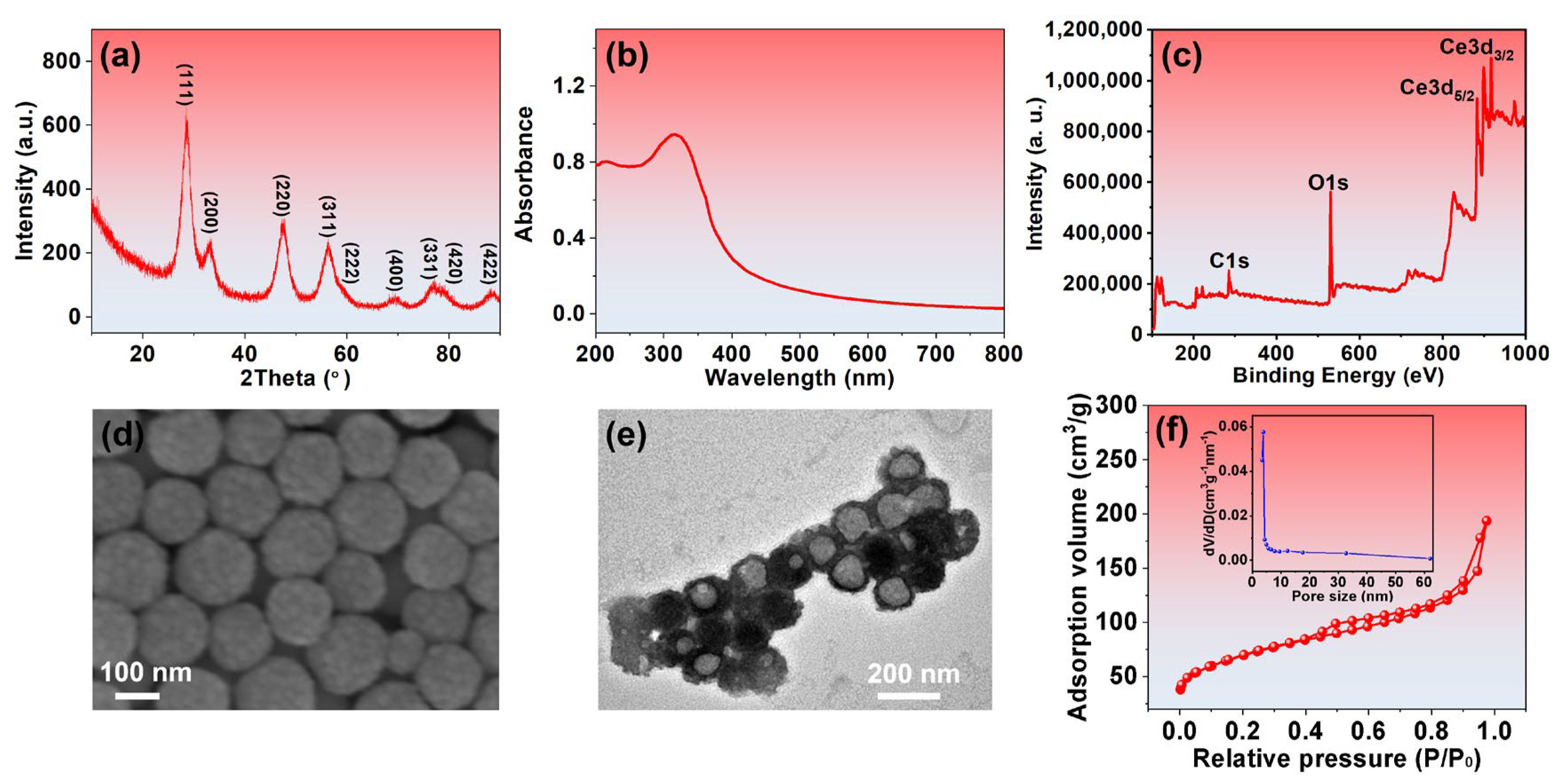
The X-ray diffraction (XRD) patterns for CeO2-phm revealed nine diffraction peaks (Figure 1a), which match well with the (111), (200), (220), (311), (222), (400), (331), (420) and (422) planes of cubic fluorite CeO2 (JCPDS No. 34-0394) [28]. In the FTIR spectrum of CeO2-phm (Figure S1), the absorption band at 3381 cm−1 is attributed to the stretching vibration of Ce–OH groups, while the peak at 1600 cm−1 corresponds to the stretching vibrations of Ce–O–C and C–O bonds [29]. The UV–Vis spectra of the CeO2-phm were shown in Figure 1b. The CeO2-phm exhibits a prominent absorption peak at 350 nm, which is attributed to the charge transfer transition from the O2− 2p valence band to the Ce4+ 4f conduction band in CeO2 [30]. X-ray photoelectron spectroscopy (XPS) characterization of CeO2-phm confirmed the existence of cerium species (Figure 1c). The characteristic peaks observed at 900.7, 882.3, 907.0, 888.6, 916.6, and 898.3 eV (denoted as u, v, u″, v″, u‴, and v‴, respectively) were assigned to Ce4+ species, while the additional signals at 902.9 eV (u′) and 885.3 eV (v′) corresponded to Ce3+ (Figure S2). Based on the XPS deconvolution analysis, the ratio of Ce3+/Ce4+ reaches 48.4%. The enzyme-mimetic activity of ceria is closely associated with the Ce3+/Ce4+ ratio. The elevated Ce3+ content in CeO2 correlates with a higher density of oxygen vacancies, as each pair of Ce3+ ions compensates for one oxygen vacancy [31,32]. In addition, the calculated atomic ratio of O and Ce by XPS is about 4.7, which is 2.35 times higher than that (2) in CeO2, also suggesting that CeO2-phm possesses a large amount of oxygen vacancies [22]. The presence of abundant oxygen vacancies facilitates oxygen adsorption and promotes rapid redox reactions, both of which are critical for enhancing the sensitivity and response time of CeO2-based electrochemical biosensors. These results confirm the successful synthesis of CeO2.
The hollow mesoporous structure of CeO2-phm was subsequently elucidated by electron microscopy and surface area analysis. SEM images (Figure 1d) revealed that the as-synthesized CeO2-phm exhibited uniform spherical morphology with average diameters of ~140 nm. The surface texture appears rough, indicating the aggregation of primary nanoparticles on the sphere surfaces, which suggests the formation of a porous shell. The closely packed structure with distinguishable interparticle boundaries is indicative of well-assembled nanocrystals constituting the microsphere shells. To further investigate the elemental distribution and compositional homogeneity, energy-dispersive X-ray spectroscopy (EDS) elemental mapping was performed. The EDS layered image (Figure S3a) of CeO2-phm shows the overall elemental distribution. Elemental mapping images for (Figure S3b) Ce, (Figure S3c) O, (Figure S3d) N, and (Figure S3e) C illustrate a uniform distribution of these elements throughout the sample. The corresponding EDS spectrum of CeO2-phm (Figure S3f), with the inset presenting the quantified elemental composition, confirms that Ce and O are the major constituents. As shown in Figure 1e, the individual particles exhibit a well-defined spherical morphology with a darker periphery and a relatively brighter center, indicating the presence of a hollow interior structure. Moreover, the shell of each microsphere appears to be composed of aggregated nanoparticles, forming a porous network. Nitrogen adsorption–desorption isotherms (Figure 1f) of CeO2-phm exhibited typical type IV behavior with an H3-type hysteresis loop, confirming the mesoporous nature of the sample [33,34]. The BET specific surface area up to 168.6 m2/g, and the Barrett–Joyner–Halenda (BJH) pore size distribution (inset of Figure 1f) showed a narrow peak centered at 3.4 nm. Such a porous structure not only increases the number of electroactive sites but also facilitates ion diffusion in sensing applications.
The hydrodynamic size and colloidal stability of CeO2-phm were assessed using dynamic light scattering (DLS) (Figure S4) and zeta potential measurements. DLS results revealed an average hydrodynamic diameter of approximately 152.2 nm with a low polydispersity index (PDI) of 0.154, indicating uniform particle distribution and good dispersion stability in aqueous media. The observed particle size from DLS was larger than that obtained by TEM, which can be attributed to the fact that DLS evaluates particles in their hydrated and dynamic state, while TEM measures the dried and immobilized counterparts under vacuum conditions [35]. The zeta potential measurement revealed a surface charge of −20.6 mV, confirming good electrostatic repulsion among particles and good colloidal stability [36], which is particularly beneficial for preparing modified electrodes through the droplet coating method.

3.2. Electrochemical Characterization of Modified Electrodes

The electrocatalytic performance of CeO2-phm/cMWCNTs/SPCE toward H2O2 reduction was evaluated by cyclic voltammetry (CV) in 0.1 M PBS (pH 7.0) containing 50 μM H2O2 at a scan rate of 50 mV·s−1. For comparison, CV responses of cMWCNTs/SPCE and CeO2-c/cMWCNTs/SPCE were also recorded under identical conditions to serve as control electrodes. As shown in Figure 2a, cMWCNTs/SPCE (curve a), CeO2-c/cMWCNTs/SPCE (curve c) and CeO2-phm/cMWCNTs/SPCE (curve e) exhibited negligible cathodic current and no discernible reduction peak in PBS without H2O2. However, compared to curve a, the CV response of cMWCNTs/SPCE in PBS with 50 μM H2O2 (curve b) also showed no obvious reduction peak, indicating the poor electrochemical response towards H2O2. As expected, the CeO2-c/cMWCNTs/SPCE electrode exhibited a noticeable increase in reduction current only at relatively high overpotentials; however, no distinct cathodic peak corresponding to H2O2 reduction was observed within the scanned potential window of 0 to –0.8 V. In contrast, the CeO2-phm/cMWCNTs/SPCE displayed a pronounced cathodic peak centered at −0.6 V, indicating efficient electrocatalytic reduction of H2O2. Notably, the peak reduction current at −0.6 V for CeO2-phm/cMWCNTs/SPCE was approximately 4.3 times higher than that observed for CeO2-c/cMWCNTs/SPCE, underscoring the superior catalytic performance of the porous hollow structure.
The enhanced current response and positively shifted reduction peak potential observed at CeO2-phm/cMWCNTs/SPCE can be ascribed to the unique structural characteristics of CeO2-phm. To gain insight into the electrochemical reduction mechanism of H2O2, the influence of scan rate on the CeO2-phm/cMWCNTs/SPCE was investigated in the presence of 50 μM H2O2 (Figure 2b). As the scan rate increased from 10 to 60 mV·s−1, a noticeable enhancement in peak current was observed, accompanied by a cathodic peak shift toward more negative potentials. Also, the cathodic peaks are in linear relationship with the square root of the scan rate (Figure S5), which indicates the reduction of H2O2 at CeO2-phm is under diffusion control electrochemical process [37,38]. The excellent electrocatalytic activity toward H2O2 reduction is mainly attributed to the hollow mesoporous structure and abundant oxygen vacancies. Based on these excellent performances, we can expect that the CeO2-phm/cMWCNTs/SPCE-based H2O2 biosensor has remarkable sensitivity, strong anti-interference performance and outstanding stability.

3.3. Electrochemical Sensing Performance of H2O2

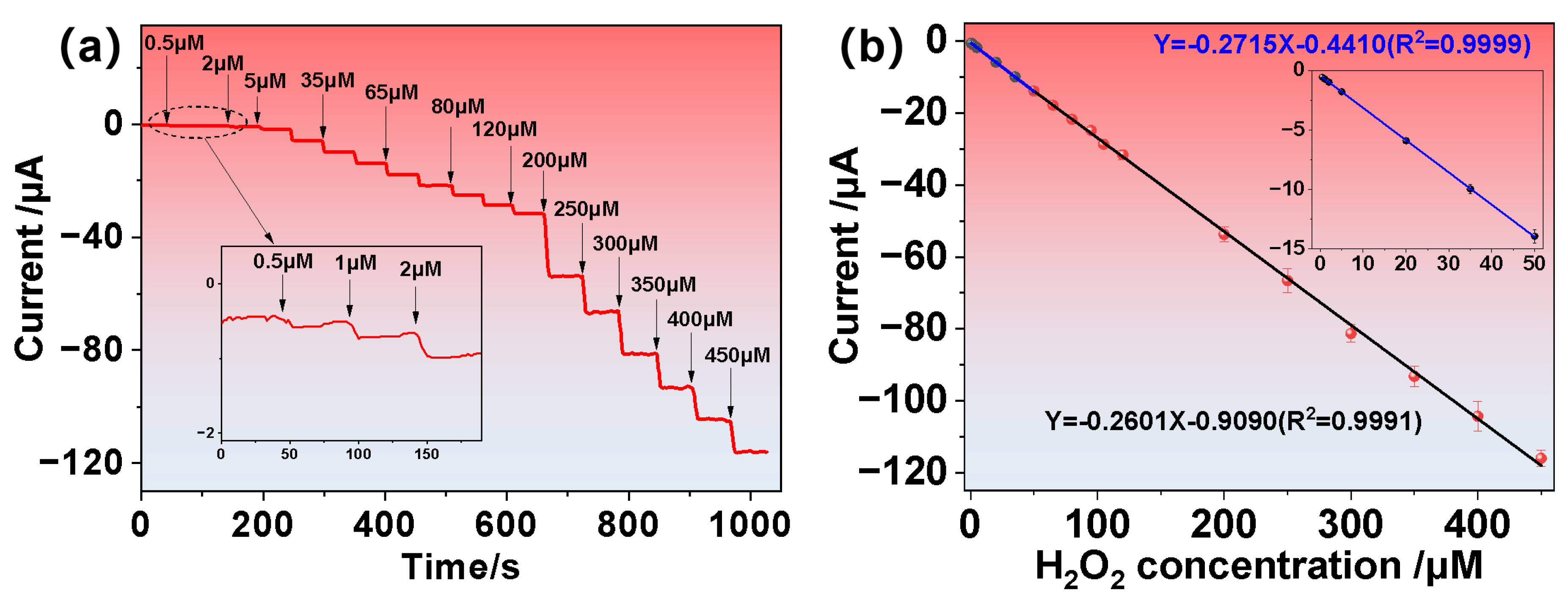
To optimize the electrochemical performance of the biosensor, the influence of the applied potential on the amperometric response of the CeO2-phm/cMWCNTs/SPCE-based H2O2 biosensor was investigated, revealing that the highest current response was observed at an applied potential of −0.55 V (Figure S6). Consequently, −0.55 V was selected as the optimal operating potential for subsequent electrochemical measurements. The analytical performance of the fabricated biosensor was assessed by constructing a calibration curve. As illustrated in Figure 3a, the amperometric i–t responses were recorded upon successive additions of H2O2 at a relatively low working potential of −0.55 V (vs. Ag/AgCl). The corresponding calibration plot, depicted in Figure 3b, reveals a well-defined linear relationship between the current response and H2O2 concentration. The sensor exhibited two distinct linear ranges: 0.5–50 μM and 50–450 μM, with respective sensitivities of 2161.6 μA·mM−1·cm−2 and 2070.9 μA·mM−1·cm−2. The presence of two linear ranges is mainly attributed to the transition of the sensing process from a kinetic-controlled regime to a diffusion-controlled regime [39,40], and 50 μM is exactly the critical point. The detection limit was calculated to be 0.017 μM, using a signal-to-noise ratio criterion of 3 (S/N = 3). These performance metrics are either superior to or on par with those reported for other electrochemical H2O2 biosensors (Table 1), underscoring the excellent sensitivity and wide detection range of the CeO2-phm/cMWCNTs/SPCE-based biosensor.
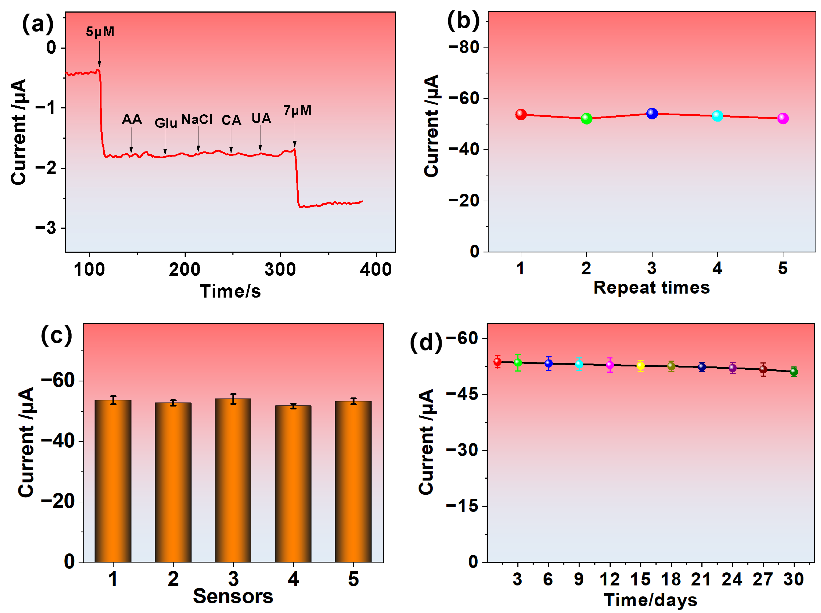
To further evaluate the sensing capabilities of the CeO2-phm/cMWCNTs/SPCE-based biosensor, its selectivity toward H2O2 was examined in the presence of common electroactive interferents. As illustrated in Figure 4a, a series of potentially interfering species—such as ascorbic acid (AA), citric acid (CA), uric acid (UA), glucose (Glu), and sodium chloride (NaCl)—were added to a 5 μM H2O2 solution. The current response remained nearly unchanged following the addition of these species, indicating minimal interference. In contrast, the current response exhibited a distinct stepwise enhancement upon successive additions of H2O2, which remained consistent even in the presence of potential interfering substances. These results confirm that the CeO2-phm-modified electrode possesses excellent selectivity for H2O2 detection, even in complex environments containing potentially interfering analytes. The excellent anti-interference performance of the CeO2-phm/cMWCNTs/SPCE-based biosensor is mainly attributed to the abundant oxygen vacancies of CeO2-phm, the hollow mesoporous structure that promotes selective H2O2 diffusion, the optimized low detection potential, and the synergistic electron transfer with cMWCNTs.
The amperometric responses of CeO2-phm/cMWCNTs/SPCE and CeO2-c/cMWCNTs/SPCE were evaluated through successive additions of 5 μM H2O2 in 0.1 M PBS (Figure S7). The results clearly indicate that the CeO2-phm-based electrode exhibited a markedly enhanced sensing performance compared to its CeO2-c counterpart. This enhancement is primarily attributed to the distinctive porous hollow architecture of CeO2-phm, which offers an enlarged electroactive surface area and abundant accessible catalytic sites. These structural advantages promote more efficient electron transfer and enhance the overall electrocatalytic activity toward H2O2 detection.
The performance consistency of the CeO2-phm/cMWCNTs/SPCE sensor was evaluated in terms of repeatability, reproducibility and stability. For repeatability, five successive measurements were carried out using the same electrode, yielding relative standard deviation (RSD) values below 1.68% (Figure 4b). Reproducibility was assessed using five independently fabricated electrodes, with RSD values remaining under 1.73% (Figure 4c). These findings confirm the reliable and consistent performance of the sensor across repeated and independent tests. Additionally, the long-term stability of the sensor was examined by conducting H2O2 detection every three days over a 30-day period. On day 30, the current response retained 95.04% of its initial value (Figure 4d), indicating strong temporal stability. These results demonstrate that the CeO2-phm/cMWCNTs/SPCE biosensor possesses excellent repeatability, reproducibility and stability.

3.4. Real Sample Analysis

To evaluate the real-world applicability of the proposed biosensor, hydrogen peroxide was quantified in various complex matrices—including groundwater, commercial drinking water, milk and fetal bovine serum—via the standard addition method [66]. Known concentrations of H2O2 were spiked into each sample, and the recovery rates were calculated accordingly, as summarized in Table 2. The recoveries ranged from 100.5% to 102.6%, demonstrating the sensor’s high accuracy in complex sample matrices. These results confirm that the CeO2-phm/cMWCNTs/SPCE-based biosensor not only performs well under controlled laboratory conditions but also retains its sensitivity and reliability in real-world environments. This highlights its strong potential for use in environmental monitoring and biomedical diagnostics involving H2O2 detection.

4. Conclusions

In summary, in this report, a porous ceria hollow microsphere (CeO2-phm) with a uniform pore size (~3.4 nm) and a high specific surface area (168.6 m2/g) was synthesized via a facile solvothermal method. These unique structural characteristics, along with abundant oxygen vacancies, significantly enhanced electrocatalytic activity toward H2O2 reduction. When integrated into a flexible screen-printed electrode functionalized with cMWCNTs, the resulting CeO2-phm/cMWCNTs/SPCE biosensor exhibited excellent electrochemical sensing performance, including a wide linear range (0.5–450 μM), an ultralow detection limit (0.017 μM), and a high sensitivity (up to 2070.9 and 2161.6 μA·mM−1·cm−2). Furthermore, the sensor demonstrated excellent selectivity against common interferents, repeatability and reproducibility (RSD < 1.73%), and long-term stability (retaining over 95% of its signal after 30 days). Importantly, the biosensor was successfully applied in the quantification of H2O2 in complex real-world samples such as groundwater, milk, commercial drinking water, and fetal bovine serum, with excellent recovery rates (100.5–102.6%). These results collectively validate the CeO2-phm/cMWCNTs/SPCE biosensor as a promising platform for real-time, cost-effective, and high-accuracy monitoring of H2O2 in biomedical diagnostics, environmental surveillance, and food safety applications.

Supplementary Materials

The following supporting information can be downloaded at: https://www.mdpi.com/article/10.3390/bios15100664/s1, Figure S1: FT-IR spectrum of CeO2-phm; Figure S2: XPS analysis of CeO2-phm; Figure S3: (a) EDS layer image, and element mapping of the CeO2-phm representing as (b) Ce, (c) O, (d) N, and (e) C; (f) The EDS representation of the CeO2-phm and insert image showing element composition of the CeO2-phm. Figure S4: The DLS analysis of CeO2-phm; Figure S5: Linear relationship between square roots of canning speed and peak current; Figure S6: Current responses of CeO2-phm/cMWCNTs/SPCEs towards 200 μM H2O2 at different applied operating potential; Figure S7: Amperometric response of continuously addition 5 μM H2O2 for 4 times of CeO2-phm/cMWCNTs/SPCEs (curve a) and CeO2-c /cMWCNTs/SPCEs (curve b).

Author Contributions

J.H.: Conceptualization, Methodology, Investigation, Writing—original draft, Project administration. X.H.: Methodology, Formal analysis, Validation, Writing. S.Z.: Investigation, Data Curation, Formal Analysis, Visualization. K.L.: Resources, Investigation, Writing. H.Z.: Investigation, Data Curation, Writing—Review and Editing. Q.J.: Formal analysis, Validation. Z.F.: Data curation, Methodology, Formal Analysis. Y.Z.: Methodology, Instrumentation, Data analysis. P.W.: Methodology, Instrumentation, Data analysis. X.D.: Data curation, Methodology, Formal Analysis. H.L.: Conceptualization, Supervision, Funding acquisition, Writing—review and editing. Z.Y.: Conceptualization, Resources, Supervision, Writing—review and editing. Y.L.: Resources, Project administration, Writing—review and editing. J.T.: Conceptualization, Supervision, Writing—review and editing. All authors have read and agreed to the published version of the manuscript.

Funding

This work was supported by the Science Research Project of the Hunan Provincial Department of Education (No. 23B0534), the Natural Science Foundation of Hunan Province (No. 2023JJ40254, 2024JJ6207 and 2023JJ50171), the Natural Science Foundation Young Student Research Foundation Project of Hunan Provincial (No. 2025JJ60934), the general project of Hunan Education Department (No. 23C0198) and the Student Innovation and Entrepreneurship Training Program of China (No. S202412604004).

Institutional Review Board Statement

Not applicable.

Informed Consent Statement

Not applicable.

Data Availability Statement

The datasets generated during the current study are available from the first author on reasonable request.

Conflicts of Interest

The authors declare that they have no known competing financial interests or personal relationships that could have appeared to influence the work reported in this paper.

Abbreviations

The following abbreviations are used in this manuscript:
CeO2-phmporous ceria hollow microspheres
CeO2-ccommercial CeO2 nanospheres with solid cores
SPCEscreen-printed carbon electrode
ROSreactive oxygen species
CeO2cerium dioxide
cMWCNTscarboxylated multi-walled carbon nanotubes
Ce(NO3)3·6H2OCerium nitrate hexahydrate
EGethylene glycol
NHSN-hydroxysuccinimide
EDC1-ethyl-3-(3-dimethylaminopropyl)carbodiimide hydrochloride
AAascorbic acid
Gluglucose
CAcitric acid
UAuric acid
NaClsodium chloride
PBSPhosphate-buffered saline
CeO2-ccommercial CeO2 nanospheres
DLSdynamic light scattering
PDIpolydispersity index
FBSFetal Bovine Serum

References

  1. Sies, H.; Jones, D.P. Reactive oxygen species (ROS) as pleiotropic physiological signalling agents. Nat. Rev. Mol. Cell Biol. 2020, 21, 363–383. [Google Scholar] [CrossRef] [PubMed]
  2. Averill-Bates, D. Reactive oxygen species and cell signaling. Review. Biochim. Biophys. Acta Mol. Cell Res. 2024, 1871, 119573. [Google Scholar] [CrossRef] [PubMed]
  3. You, R.; Mu, Y.; Zhou, J.; Wang, C.; Fang, Z.; Liu, Y.; Liu, S.; Zhai, Q.; Zhang, C. Ferroptosis is involved in trophoblast cells cytotoxicity induced by black phosphorus nanoparticles. Toxicology 2024, 505, 153810. [Google Scholar] [CrossRef]
  4. Giorgi, C.; Marchi, S.; Simoes, I.C.M.; Ren, Z.; Morciano, G.; Perrone, M.; Patalas-Krawczyk, P.; Borchard, S.; Jędrak, P.; Pierzynowska, K.; et al. Mitochondria and reactive oxygen species in aging and age-related diseases. Int. Rev. Cell Mol. Biol. 2018, 340, 209–344. [Google Scholar]
  5. Prasad, V.; Siddiqui, L.; Mishra, P.K.; Ekielski, A.; Talegaonkar, S. Recent advancements in lignin valorization and biomedical applications: A patent review. Recent Pat. Nanotechnol. 2022, 16, 107–127. [Google Scholar] [CrossRef]
  6. Devi, A.; Saran, C.; Saratale, G.D.; Saratale, R.G.; Ferreira, L.F.R.; Mulla, S.I.; Bharagava, R.N. Sustainable Algal Industrial Wastewater Treatment: Applications and Challenges. Microbes Based Approaches Manag. Hazard. Contam. 2024, 12, 190–205. [Google Scholar]
  7. Wang, F.; Van Halem, D.; Van der Hoek, J. The fate of H2O2 during managed aquifer recharge: A residual from advanced oxidation processes for drinking water production. Chemosphere 2016, 148, 263–269. [Google Scholar] [CrossRef] [PubMed]
  8. Kurniawan, T.A.; Lo, W.H. Removal of refractory compounds from stabilized landfill leachate using an integrated H2O2 oxidation and granular activated carbon (GAC) adsorption treatment. Water Res. 2009, 43, 4079–4091. [Google Scholar] [CrossRef]
  9. Ullah, M.; Kanjariya, P.; Rekha, M.M.; Kundlas, M.; Prasad, G.V.S.; Chahar, M.; Algahtani, A.; Tirth, V.; Zhengxin, L. Correction to: Fabrication of an electrochemical sensor based on g-C3N4–NiO nanocomposite for sensitive and selective detection of hydrogen peroxide. J. Mater. Sci. Mater. Electron. 2025, 36, 1342. [Google Scholar] [CrossRef]
  10. Ahmad, T.; Iqbal, A.; Halim, S.A.; Uddin, J.; Khan, A.; El Deeb, S.; Al-Harrasi, A. Recent advances in electrochemical sensing of hydrogen peroxide (H2O2) released from cancer cells. Nanomaterials 2022, 12, 1475. [Google Scholar] [CrossRef]
  11. Pundir, C.S.; Deswal, R.; Narwal, V. Quantitative analysis of hydrogen peroxide with special emphasis on biosensors. Bioprocess Biosyst. Eng. 2018, 41, 313–329. [Google Scholar] [CrossRef] [PubMed]
  12. Shen, Y.; Zhao, S.; Chen, F.; Lv, Y.; Fu, L. Enhancing sensitivity and selectivity: Current trends in electrochemical immunosensors for organophosphate analysis. Biosensors 2024, 14, 496. [Google Scholar] [CrossRef] [PubMed]
  13. Thatikayala, D.; Ponnamma, D.; Sadasivuni, K.K.; Cabibihan, J.J.; Al-Ali, A.K.; Malik, R.A.; Min, B. Progress of advanced nanomaterials in the non-enzymatic electrochemical sensing of glucose and H2O2. Biosensors 2020, 10, 151. [Google Scholar] [CrossRef] [PubMed]
  14. Liu, Z.; Wang, L.; Liu, P.; Zhao, K.; Ye, S.; Liang, G. Rapid, ultrasensitive and non-enzyme electrochemiluminescence detection of hydrogen peroxide in food based on the ssDNA/g-C3N4 nanosheets hybrid. Food Chem. 2021, 357, 129753. [Google Scholar] [CrossRef]
  15. Wu, S.; Duan, N.; Qiu, Y.; Li, J.; Wang, Z. Colorimetric aptasensor for the detection of Salmonella enterica serovar typhimurium using ZnFe2O4-reduced graphene oxide nanostructures as an effective peroxidase mimetics. Int. J. Food Microbiol. 2017, 261, 42–48. [Google Scholar] [CrossRef]
  16. Liang, M.; Yan, X. Nanozymes: From new concepts, mechanisms, and standards to applications. Acc. Chem. Res. 2019, 52, 2190–2200. [Google Scholar] [CrossRef]
  17. Cai, Y.; Ren, W.; Du, C.; Zhang, J.; Zhao, Y.; Yu, J.; Du, H.; Kang, H.; Ge, X.; Lu, M.; et al. Cerium Oxide Nanozymes: Exploring Synthesis, Catalytic Activity, and Biomedical Potential. Small 2025, 21, 2504436. [Google Scholar] [CrossRef]
  18. Wang, R.; Du, Y.; Fu, Y.; Guo, Y.; Gao, X.; Guo, X.; Wei, J.; Yang, Y. Ceria-based nanozymes in point-of-care diagnosis: An emerging futuristic approach for biosensing. ACS Sens. 2023, 8, 4442–4467. [Google Scholar] [CrossRef]
  19. Shi, Y.; Lyu, Z.; Zhao, M.; Chen, R.; Nguyen, Q.N.; Xia, Y. Noble-metal nanocrystals with controlled shapes for catalytic and electrocatalytic applications. Chem. Rev. 2020, 121, 649–735. [Google Scholar] [CrossRef]
  20. Wu, T.; Han, M.-Y.; Xu, Z.J. Size effects of electrocatalysts: More than a variation of surface area. ACS Nano 2022, 16, 8531–8539. [Google Scholar] [CrossRef]
  21. Chen, J.; Lim, B.; Lee, E.P.; Xia, Y. Shape-controlled synthesis of platinum nanocrystals for catalytic and electrocatalytic applications. Nano Today 2009, 4, 81–95. [Google Scholar] [CrossRef]
  22. Wang, S.; Zhang, J.; Jiang, J.; Liu, R.; Zhu, B.; Xu, M.; Wang, Y.; Cao, J.; Li, M.; Yuan, Z.; et al. Porous ceria hollow microspheres: Synthesis and characterization. Micropor. Mesopor. Mater. 2009, 123, 349–353. [Google Scholar] [CrossRef]
  23. Li, H.; Lu, G.; Dai, Q.; Wang, Y.; Guo, Y.; Guo, Y. Hierarchical Organization and Catalytic Activity of High-Surface-Area Mesoporous Ceria Microspheres Prepared Via Hydrothermal Routes. ACS Appl. Mater. Interfaces 2010, 2, 838–846. [Google Scholar] [CrossRef]
  24. Khan, A.; DeVoe, E.; Andreescu, S. Carbon-based electrochemical biosensors as diagnostic platforms for connected decentralized healthcare. Sens. Diagn. 2023, 2, 529–558. [Google Scholar] [CrossRef]
  25. Zhou, C.; Zhang, W.; Richardson-Barlow, C. Navigating ecological civilisation: Polycentric environmental governance and policy regulatory framework in China. Energy Res. Soc. Sci. 2025, 128, 104347. [Google Scholar] [CrossRef]
  26. Huang, J.; Chen, J.; Lv, L.; Yu, Z.; Yao, W.; Cheng, H.; Niu, W.; Wang, J.; Zhang, J.; Qi, H. Design and Verification of a Wearable Micro-Capacitance Test System for POC Biosensing. IEEE Trans. Instrum. Meas. 2025, 74, 1–11. [Google Scholar]
  27. Jeerapan, I.; Poorahong, S. Flexible and stretchable electrochemical sensing systems: Materials, energy sources, and integrations. J. Electrochem. Soc. 2020, 167, 037573. [Google Scholar] [CrossRef]
  28. Yang, Y.; Mao, Z.; Huang, W.; Liu, L.; Li, J.; Li, J.; Wu, Q. Redox enzyme-mimicking activities of CeO2 nanostructures: Intrinsic influence of exposed facets. Sci. Rep. 2016, 6, 35344. [Google Scholar] [CrossRef] [PubMed]
  29. Culica, M.E.; Chibac-Scutaru, A.L.; Melinte, V.; Coseri, S. Cellulose Acetate Incorporating Organically Functionalized CeO2 NPs: Efficient Materials for UV Filtering Applications. Materials 2020, 13, 2955. [Google Scholar] [CrossRef] [PubMed]
  30. Atisme, T.B.; Yu, C.-Y.; Tseng, E.N.; Chen, Y.-C.; Shu, P.-K.; Chen, S.Y. Interface Interactions in Conjugated Polymer Composite with Metal Oxide Nanoparticles. Nanomaterials 2019, 9, 1534. [Google Scholar] [CrossRef]
  31. Qin, J.; Feng, Y.; Cheng, D.; Liu, B.; Wang, Z.; Zhao, Y.; Wei, J. Construction of a Mesoporous Ceria Hollow Sphere/Enzyme Nanoreactor for Enhanced Cascade Catalytic Antibacterial Therapy. ACS Appl. Mater. Interfaces 2021, 13, 40302–40314. [Google Scholar] [CrossRef]
  32. Bortamuly, R.; Konwar, G.; Boruah, P.K.; Das, M.R.; Mahanta, D.; Saikia, P. CeO2-PANI-HCl and CeO2-PANI-PTSA composites: Synthesis, characterization, and utilization as supercapacitor electrode materials. Ionics 2020, 26, 5747–5756. [Google Scholar] [CrossRef]
  33. Dang-Bao, T.; Bao, N.H.; Anh, N.P.; Phuong, P.H.; Nguyen, T.-T.; Tri, N. Green-synthesized silver nanoparticles decorated on ceria nanorods for room-temperature p-nitrophenol hydrogenation. Green Chem. Lett. Rev. 2022, 15, 449–459. [Google Scholar] [CrossRef]
  34. Zhou, C.; Richardson-Barlow, C.; Fan, L.; Cai, H.; Zhang, W.; Zhang, Z. Towards organic collaborative governance for a more sustainable environment: Evolutionary game analysis within the policy implementation of China’s net-zero emissions goals. J. Environ. Manage. 2025, 373, 123765. [Google Scholar] [CrossRef] [PubMed]
  35. Pecora, R. Dynamic light scattering measurement of nanometer particles in liquids. J. Nanopart. Res. 2000, 2, 123–131. [Google Scholar] [CrossRef]
  36. Foroutan, Z.; Afshari, A.R.; Sabouri, Z.; Mostafapour, A.; Far, B.F.; Jalili-Nik, M.; Darroudi, M. Plant-based synthesis of cerium oxide nanoparticles as a drug delivery system in improving the anticancer effects of free temozolomide in glioblastoma (U87) cells. Ceram. Int. 2022, 48, 30441–30450. [Google Scholar] [CrossRef]
  37. Gao, J.; Gong, L.; Fan, X.; Yu, K.; Zheng, Z.; Zhou, B. {P2W18O62}-Encapsulated Potassium-Ion Nanotubes Intercalated in Copper Biimidazole Frameworks for Supercapacitors and Hydrogen Peroxide Sensing. ACS Appl. Nano Mater. 2020, 3, 1497–1507. [Google Scholar] [CrossRef]
  38. Rajendran, S.; Manoj, D.; Suresh, R.; Vasseghian, Y.; Ghfar, A.A.; Sharma, G.; Soto-Moscoso, M. Electrochemical detection of hydrogen peroxide using micro and nanoporous CeO2 catalysts. Environ. Res. 2022, 214, 113961. [Google Scholar] [CrossRef]
  39. Huang, J.; Zhang, Y.; Ding, F.; Chen, D.; Wang, Y.; Jin, X.; Zhu, X. Rational design of electroactive redox enzyme nanocapsules for high-performance biosensors and enzymatic biofuel cell. Biosens. Bioelectron. 2021, 174, 112805. [Google Scholar] [CrossRef]
  40. Zhang, J.; Qi, H.; Wu, J.J.; Mao, X.; Zhang, H.; Amin, N.; Xu, F.; Dong, C.; Wang, C.; Wang, P.; et al. Disposable peptidoglycan-specific biosensor for noninvasive real-time detection of broad-spectrum Gram-positive bacteria in exhaled breath condensates. Anal. Chem. 2024, 96, 9817–9825. [Google Scholar] [CrossRef]
  41. Uzunoglu, A.; Ipekci, H.H. The use of CeO2-modified Pt/C catalyst inks for the construction of high-performance enzyme-free H2O2 sensors. J. Electroanal. Chem. 2019, 848, 113302. [Google Scholar] [CrossRef]
  42. Jha, S.K.; Kumar, C.N.; Raj, R.P.; Jha, N.S.; Mohan, S. Synthesis of 3D porous CeO2/reduced graphene oxide xerogel composite and low level detection of H2O2. Electrochim. Acta 2014, 120, 308–313. [Google Scholar] [CrossRef]
  43. Remani, K.C.; Binitha, N.N. Cobalt doped ceria catalysts for the oxidative abatement of gaseous pollutants and colorimetric detection of H2O2. Mater. Res. Bull. 2021, 139, 111253. [Google Scholar] [CrossRef]
  44. Dong, W.; Huang, Y. CeO2/C nanowire derived from a cerium(III) based organic framework as a peroxidase mimic for colorimetric sensing of hydrogen peroxide and for enzymatic sensing of glucose. Microchim. Acta 2019, 187, 11. [Google Scholar] [CrossRef] [PubMed]
  45. Liu, H.; Ding, Y.; Yang, B.; Liu, Z.; Liu, Q.; Zhang, X. Colorimetric and ultrasensitive detection of H2O2 based on Au/Co3O4-CeOx nanocomposites with enhanced peroxidase-like performance. Sens. Actuators, B 2018, 271, 336–345. [Google Scholar] [CrossRef]
  46. Liu, Q.; Ding, Y.; Yang, Y.; Zhang, L.; Sun, L.; Chen, P.; Gao, C. Enhanced peroxidase-like activity of porphyrin functionalized ceria nanorods for sensitive and selective colorimetric detection of glucose. Mater. Sci. Eng. C 2016, 59, 445–453. [Google Scholar] [CrossRef]
  47. Zhu, Y.; Yang, Z.; Song, L.; Chi, M.; Li, M.; Wang, C.; Lu, X. Encapsulation of Co3O4 Nanoparticles Inside CeO2 Nanotubes: An Efficient Biocatalyst for the Ultrasensitive Detection of Ascorbic Acid. Part. Part. Syst. Char. 2018, 35, 1800049. [Google Scholar] [CrossRef]
  48. Rahman, M.M.; Adeosun, W.A.; Asiri, A.M. Fabrication of selective and sensitive chemical sensor development based on flower-flake La2ZnO4 nanocomposite for effective non-enzymatic sensing of hydrogen peroxide by electrochemical method. Microchem. J. 2020, 159, 105536. [Google Scholar] [CrossRef]
  49. Sudhakara, S.M.; Devendrachari, M.C.; Kotresh, H.M.N.; Khan, F. Phthalocyanine pendented polyaniline via amide linkage for an electrochemical sensing of H2O2. Microchem. J. 2021, 161, 105781. [Google Scholar] [CrossRef]
  50. Dang, W.; Sun, Y.; Jiao, H.; Xu, L.; Lin, M. AuNPs-NH2/Cu-MOF modified glassy carbon electrode as enzyme-free electrochemical sensor detecting H2O2. J. Electroanal. Chem. 2020, 856, 113592. [Google Scholar] [CrossRef]
  51. Wei, X.; Song, S.; Song, W.; Xu, W.; Jiao, L.; Luo, X.; Wu, N.; Yan, H.; Wang, X.; Gu, W.; et al. Fe3C-Assisted Single Atomic Fe Sites for Sensitive Electrochemical Biosensing. Anal. Chem. 2021, 93, 5334–5342. [Google Scholar] [CrossRef]
  52. Othmani, A.; Derbali, M.; Kalfat, R.; Touati, F.; Dhaouadi, H. A novel 1D/2D Bi2S3/g-C3N4 core–shell nanocomposite as a highly performing H2O2 non-enzymatic electrochemical sensor. J. Mater. Res. Technol. 2021, 15, 5762–5775. [Google Scholar] [CrossRef]
  53. Samadi-Maybodi, A.; Ghasemi, S.; Ghaffari-Rad, H. Ag-doped zeolitic imidazolate framework-8 nanoparticles modified CPE for efficient electrocatalytic reduction of H2O2. Electrochim. Acta 2015, 163, 280–287. [Google Scholar] [CrossRef]
  54. Lee, P.K.; Nia, P.M.; Woi, P.M. Facile self-assembled Prussian blue-polypyrrole nanocomposites on glassy carbon: Comparative synthesis methods and its electrocatalytic reduction towards H2O2. Electrochim. Acta 2017, 246, 841–852. [Google Scholar] [CrossRef]
  55. Chen, S.; Xie, Y.; Guo, X.; Sun, D. Self-supporting electrochemical sensors for monitoring of cell-released H2O2 based on metal nanoparticle/MOF nanozymes. Microchem. J. 2022, 181, 107715. [Google Scholar] [CrossRef]
  56. Riaz, M.A.; Yuan, Z.; Mahmood, A.; Liu, F.; Sui, X.; Chen, J.; Huang, Q.; Liao, X.; Wei, L.; Chen, Y. Hierarchically porous carbon nanofibers embedded with cobalt nanoparticles for efficient H2O2 detection on multiple sensor platforms. Sens. Actuators, B 2020, 319, 128243. [Google Scholar] [CrossRef]
  57. Zhou, W.; Huang, S.; Sun, C. Ni3Mo3N coupled with nitrogen-rich carbon microspheres as an efficient hydrogen evolution reaction catalyst and electrochemical sensor for H2O2 detection. Int. J. Hydrog. Energy 2022, 47, 14906–14915. [Google Scholar] [CrossRef]
  58. Zhou, J.; Min, M.; Liu, Y.; Tang, J.; Tang, W. Layered assembly of NiMn-layered double hydroxide on graphene oxide for enhanced non-enzymatic sugars and hydrogen peroxide detection. Sens. Actuators, B 2018, 260, 408–417. [Google Scholar] [CrossRef]
  59. Woo, S.; Kim, Y.-R.; Chung, T.D.; Piao, Y.; Kim, H. Synthesis of a graphene–carbon nanotube composite and its electrochemical sensing of hydrogen peroxide. Electrochim. Acta 2012, 59, 509–514. [Google Scholar] [CrossRef]
  60. Temur, E.; Eryiğit, M.; Kurt Urhan, B.; Demir, Ü.; Öznülüer Özer, T. Cu/Electrochemically reduced graphene oxide layered nanocomposite for non-enzymatic H2O2 sensor. Mater. Today Proc. 2021, 46, 6971–6975. [Google Scholar] [CrossRef]
  61. Gopalakrishnan, A.; Vishnu, N.; Badhulika, S. Cuprous oxide nanocubes decorated reduced graphene oxide nanosheets embedded in chitosan matrix: A versatile electrode material for stable supercapacitor and sensing applications. J. Electroanal. Chem. 2019, 834, 187–195. [Google Scholar] [CrossRef]
  62. Jia, W.; Guo, M.; Zheng, Z.; Yu, T.; Rodriguez, E.G.; Wang, Y.; Lei, Y. Electrocatalytic oxidation and reduction of H2O2 on vertically aligned Co3O4 nanowalls electrode: Toward H2O2 detection. J. Electroanal. Chem. 2009, 625, 27–32. [Google Scholar] [CrossRef]
  63. Liang, Y.; Liu, Y.; Zhao, P.; Chen, Y.; Lei, J.; Hou, J.; Hou, C.; Huo, D. An electrochemical sensor based on FeCo bimetallic single-atom nanozyme for sensitive detection of H2O2. Anal. Chim. Acta 2023, 1281, 341867. [Google Scholar] [CrossRef]
  64. Li, T.; Bao, Y.; Qiu, H.; Tong, W. Boosted peroxidase-like activity of metal-organic framework nanoparticles with single atom Fe(Ⅲ) sites at low substrate concentration. Anal. Chim. Acta 2021, 1152, 338299. [Google Scholar] [CrossRef] [PubMed]
  65. Yang, X.; Qiu, W.; Gao, R.; Wang, Y.; Bai, Y.; Xu, Z.; Bao, S.-J. MIL-47(V) catalytic conversion of H2O2 for sensitive H2O2 detection and tumor cell inhibition. Sens. Actuators, B 2022, 354, 131201. [Google Scholar] [CrossRef]
  66. Bhangoji, J.C.; Sahoo, S.; Satpati, A.K.; Shendage, S.S. Facile and green synthesis of silver nanoparticle-reduced graphene oxide composite and its application as nonenzymatic electrochemical sensor for hydrogen peroxide. Curr. Chem. Lett. 2021, 10, 295–308. [Google Scholar] [CrossRef]
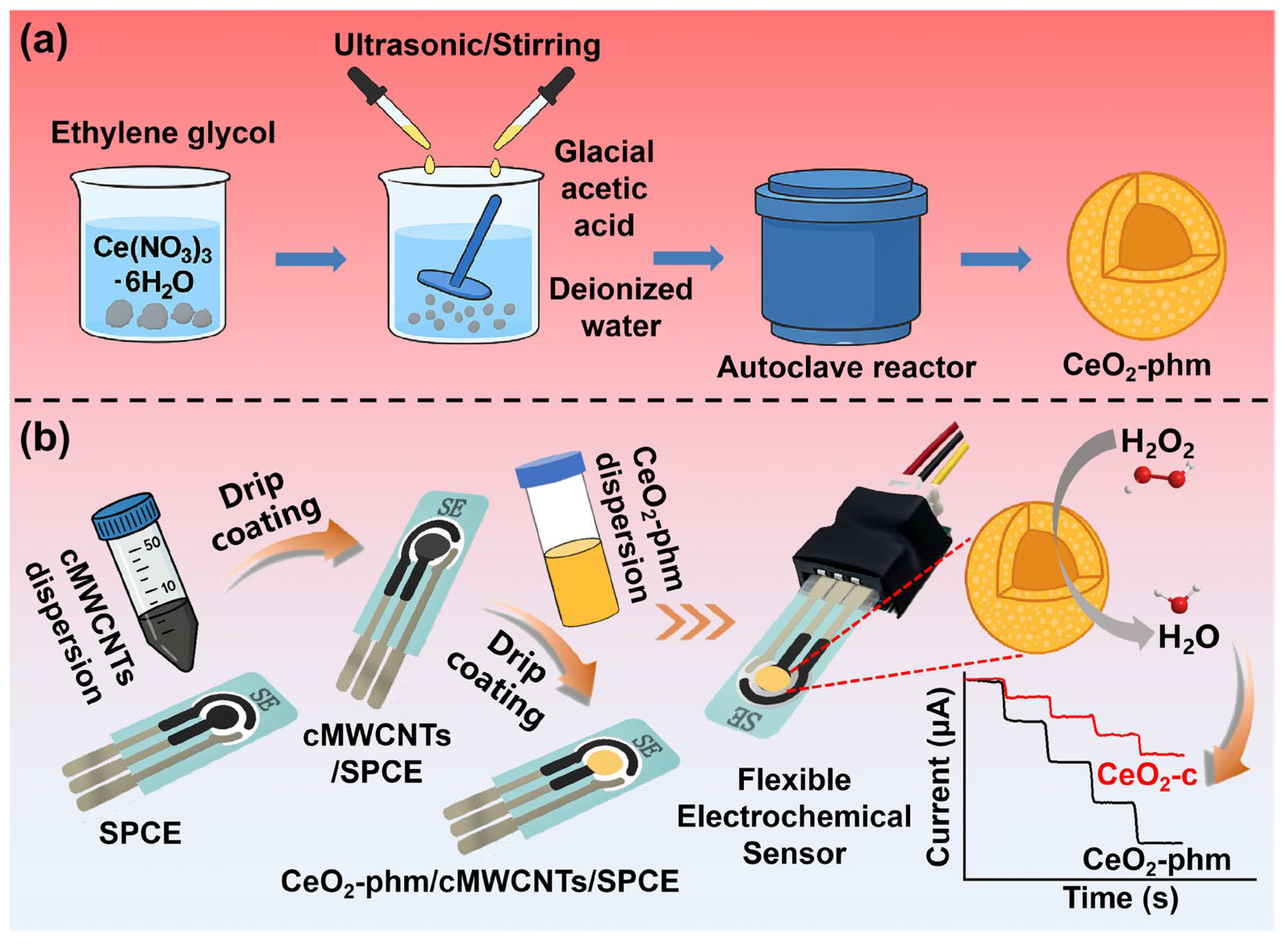
Scheme 1. Schematics of the preparation of CeO2-phm (a) and schematic for fabrication of the H2O2 biosensor and electrochemical detection of H2O2 (b).
Biosensors 15 00664 sch001
Figure 1. (a) XRD patterns of CeO2-phm; (b) UV-Visible absorbance spectrum of CeO2-phm; (c) XPS analysis of CeO2-phm; (d) SEM image of CeO2-phm; (e) TEM image of CeO2-phm; (f) N2 sorption isotherms and pore size distributions (inset) for CeO2-phm.
Biosensors 15 00664 g001
Figure 2. (a) Cyclic voltammograms (CVs) of cMWCNTs/SPCE, CeO2-c/cMWCNTs/SPCE and CeO2-phm/cMWCNTs/SPCE in 0.1 M PBS (pH 7.0) containing 50 μM H2O2 (a, c and e) or without H2O2 (b, d and f), respectively; (b) CVs of CeO2-phm/cMWCNTs/SPCE in presence of 50 μM H2O2 recorded at different scan rates.
Biosensors 15 00664 g002
Figure 3. (a) Current-time plot for micro CeO2-phm/cMWCNTs/SPCE with successive addition of H2O2 at −0.55 V. (b) Corresponding calibration plot between current response and H2O2 concentration, based on the average of three independent measurements, with error bars representing the standard deviation (mean ± SD).
Biosensors 15 00664 g003
Figure 4. (a) Amperometric response of CeO2-phm in presence of interference species in 5 μM H2O2. Supporting electrolyte: 0.1 M PBS (pH 7.0); (b) five electrochemical experiments on the same electrode; (c) electrochemical tests of five different electrodes under the same conditions; (d) electrochemical stability test in 30 days.
Biosensors 15 00664 g004
Table 1. Comparison of CeO2-phm/cMWCNTs/SPCE-based biosensor with other sensors reported in the literature.
Sensor MaterialOperating Potential (V, vs. Ag/AgCl))Linear RangeSensitivityDetection LimitReference
CeO2-phm/cMWCNTs−0.550.5–50 μM;
50–450 μM
2161.6 μA·mM−1·cm−2;
2070.9 μA·mM−1·cm−2
0.017 μMThis work
CeO2/Pt/C−0.40.01–30 mM185.4 ± 6.5 μA mM−1 cm−22 μM[41]
CeO2/rGO
xerogel
−0.360.7 nM–3.0 μM1.978 × 10−1 μA mM−130.40 nM[42]
CeO2−0.391.88 μM–2.0 mM2.9346 × 10−5 μA mM−131.29 μM[42]
Co/CeO2-3.33–100 μM;
100–1166 μM;
1166–5000 μM
-3.33 μM[43]
CeO2/C nanowires-0.5–100 μM-0.42 μM[44]
porphyrin functionalized CeO2-10–100 μM-5.29 μM[45]
porphyrin functionalized CeO2 nanorods-10–100 μM-6.1 μM[46]
Co3O4 nanoparticles inside CeO2 nanotubes-2–80 μM-1.2 μM[47]
La2ZnO4-3.0–85.0 μM25,000 μA mM−1 cm−20.04 μM[48]
Phthalocyanine pendented polyaniline0.0150.2–52 μM2317.5 μA mM−1 cm−20.15 μM[49]
AuNPs-NH2/Cu-MOF−0.155–850 μM1710 μA mM−1 cm−21.2 μM[50]
Au/ZnO0.051 μM–3.0 mM1336.7 μA mM−1 cm−20.1 μM[48]
Fe3C@C/Fe-N-C-1–6000 μM1225 μA mM−1 cm−20.26 μM[51]
Bi2S3/g-C3N4+0.260.5–950 μM1011 μA mM−1 cm−278 nM[52]
Ag/ZIF-8−0.620 μM–5 mM;
5.5–10 mM
398.47 and 145.21 μA mM−1 cm−26.2 μM[53]
Prussian blue-polypyrrole composite−0.10–3.5 mM377.43 μA mM−1 cm−2-[54]
Ag/2D Zn-MOFs−0.555.0 μM–70 mM358.7 μA mM−1 cm−21.67 μM[55]
Co-NC/CNF−0.510–5000 μM300 μA mM−1 cm−210 μM[56]
Ni3Mo3N/NC MSs−0.605 μM–40 mM120.3 μA mM−1 cm−21 μM[57]
NiMn-LDH/GO−0.4520–5860 μM96.82 μA mM−1 cm−24.4 μM[58]
Graphene-MWCNT−0.420–2000 μM32.91 μA mM−1 cm−29.4 μM[59]
Cu nanoparticles/ERGO−0.20.01–1 mM20 μA mM−1 cm−21.87 × 10⁻⁹ M[60]
CNC-rGO−0.220–160 μM0.333 μA mM−1 cm−25.28 μM[61]
Co3O4 nanowalls+0.80–1.4 mM100.3 μA mM−12.8 μM[62]
Co3O4 nanowalls−0.20–5.35 mM4.844 μA mM−110 μM[62]
Fe SAs/Co CNs-1–400 μM-0.36 μM[63]
Fe–HCl–NH2-UiO-66-3.125–100 μM-1.0 μM[64]
MIL-47(V)-OH-4.38–43.97 μM;
50.33–2240 μM
-5.84 μM[65]
Table 2. Recovery for the detection of H2O2 in groundwater, commercial drinking water, milk and fetal bovine serum samples.
SampleAdded (μM)Relative Standard Deviation (%, n = 3)Measured (μM)Recovery (%)
Groundwater5.02.465.13102.6%
Commercial drinking water2.01.342.01100.5%
Milk1.00.961.01101.0%
Fetal Bovine Serum101.0210.13101.3%
Disclaimer/Publisher’s Note: The statements, opinions and data contained in all publications are solely those of the individual author(s) and contributor(s) and not of MDPI and/or the editor(s). MDPI and/or the editor(s) disclaim responsibility for any injury to people or property resulting from any ideas, methods, instructions or products referred to in the content.