Recent Status on Lactate Monitoring in Sweat Using Biosensors: Can This Approach Be an Alternative to Blood Detection?


Abstract
1. Introduction and Literature Search Methods
2. Electrochemical Biosensors and Nanomaterials for Lactate Analysis in Sweat
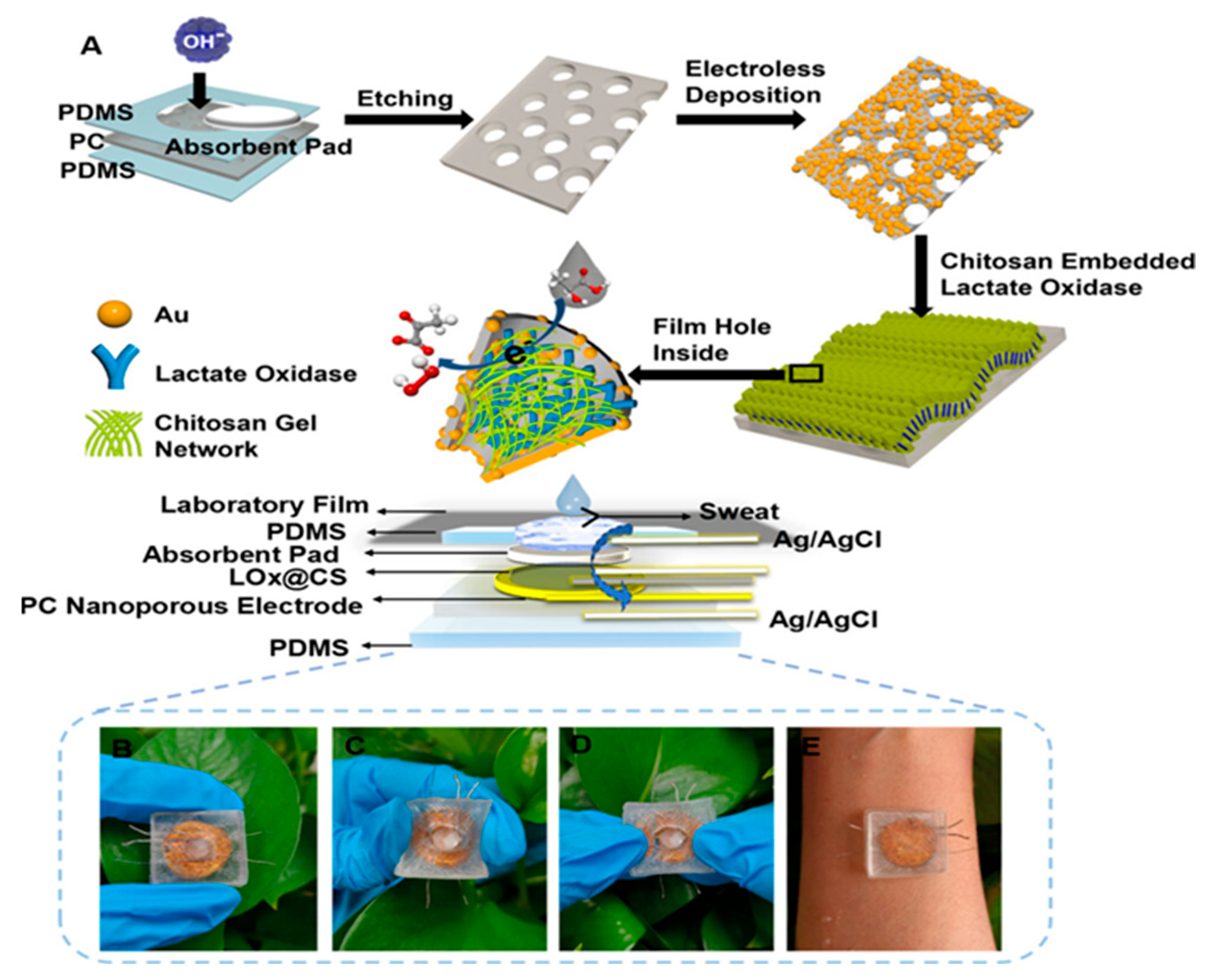
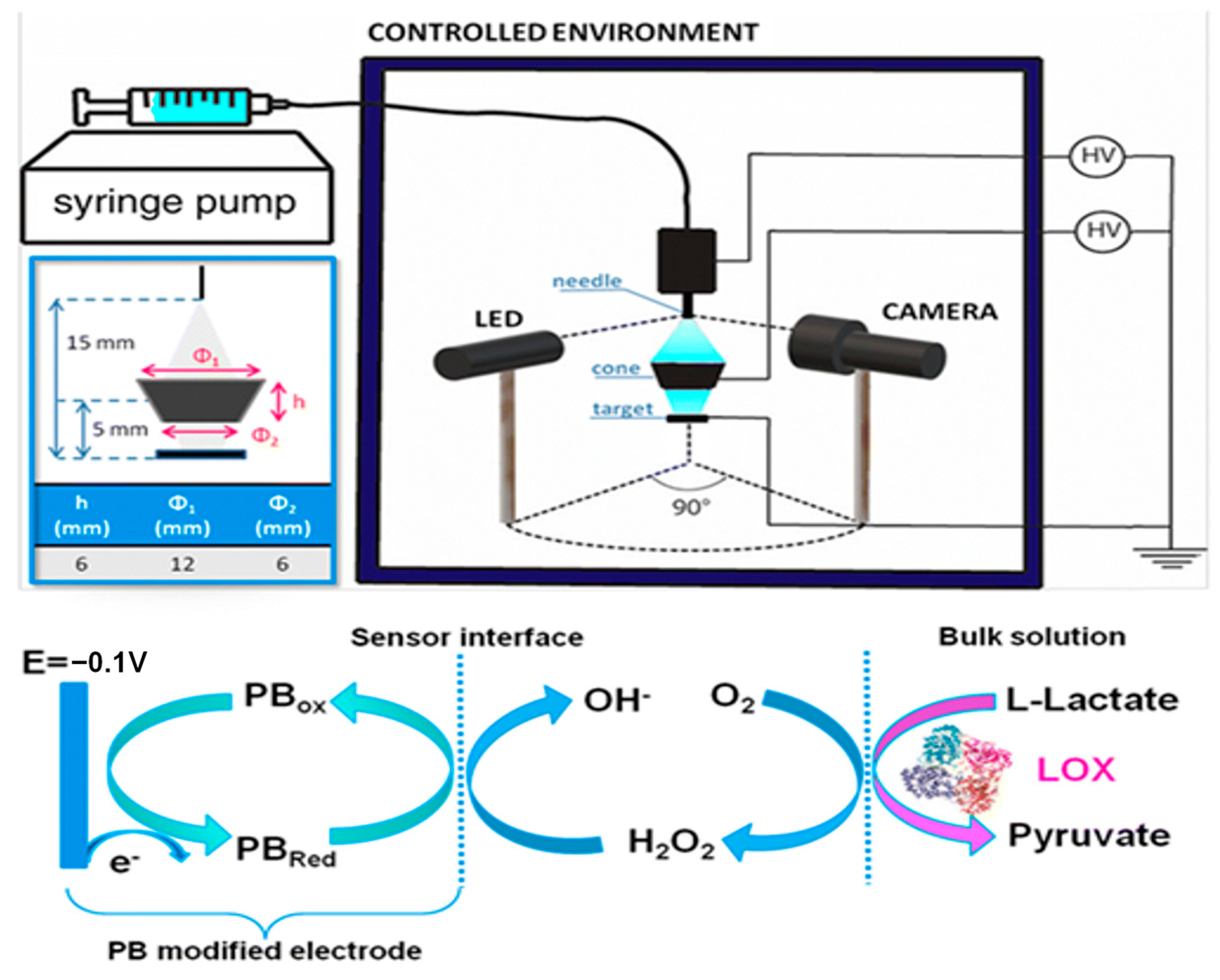
2.1. Enzymatic Electrochemical Biosensors
2.2. Enzyme-Free Electrochemical Biosensors—Semiconductors
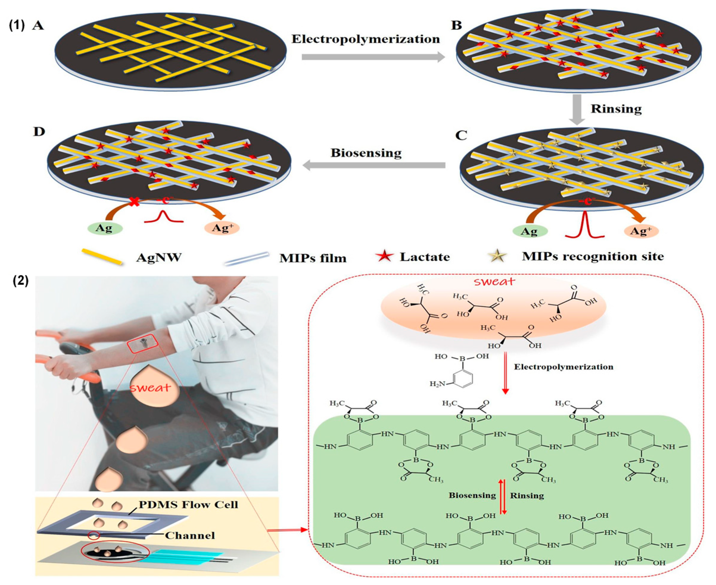
2.3. MIPs Electrochemical Biosensors
3. Comparative Analysis of Non-Invasive Electrochemical Biosensors for the Detection of Lactate in Sweat

| Working Electrodes | Measurement Techniques | Typology | Sensitivity | Linearity | Detection Limits | Ref. |
|---|---|---|---|---|---|---|
| Nano–Au/CS PC | Amperometry | LOx enzyme | 0.8 µA/mMcm2 | 0.01–35 mM | 0.144 µM | [10] |
| Au/GLAD NiO | Amperometry | Enzyme-free | 80 µA/mMcm2 | 1–65 mM | 16 µM | [12] |
| NiOOH/Ni(OH)2 | Amperometry | Enzyme-free | 80 µA/mMcm2 | 0.02–53 mM | 0.13 µM | [14] |
| SPCE/PB | Amperometry | LOx enzyme | −0.01 µA/mMcm2 | 1–20 mM | 0.2 mM | [15] |
| SPCE | EIS | LDH enzyme | N/A | 0.100 mM | 0.1 mM | [16] |
| SPCE/MXCeO2 | Amperometry | LOx enzyme | N/A | 0.01–12 mM | 0.4 mM | [18] |
| CeO2/MoS2AuNPs | Amperometry | LOx enzyme | 0.027 µA/mMcm2 | 0–100 mM | N/A | [19] |
| GO | EIS | LOx enzyme | N/A | 0.1–80 mM | N/A | [20] |
| NiCo2O4/SWCNTs | Amperometry | LOx enzyme | N/A | 0–30 mM | 39.9 µM | [21] |
| SPGE | Amperometry | LOx enzyme | 25.58 µA/mMcm2 | 0–5 mM | 0.135 mM | [22] |
| NGQD/NiCo LDH | Amperometry | Enzyme-free | 62.63 µA/mMcm2 | 0–15 mM | 0.3 mM | [24] |
| SPCE/PB/MIPs | DPV/EIS | MIPs | N/A | 1–35 mM | 0.62 mM | [26] |
| SPCE/PtNPs/Pt@MI | Amperometry | MIPs | N/A | 0–1.5 mM | 1.1 mM | [27] |
| MCF/PBPPY | Amperometry | MIPs | 0.11 µA/mMcm2 | 0.01–25 mM | N/A | [28] |
| SPCE | Amperometry | LOx enzyme | 0.9 µA/mMcm2 | 0.03–0.5 mM | N/A | [29] |
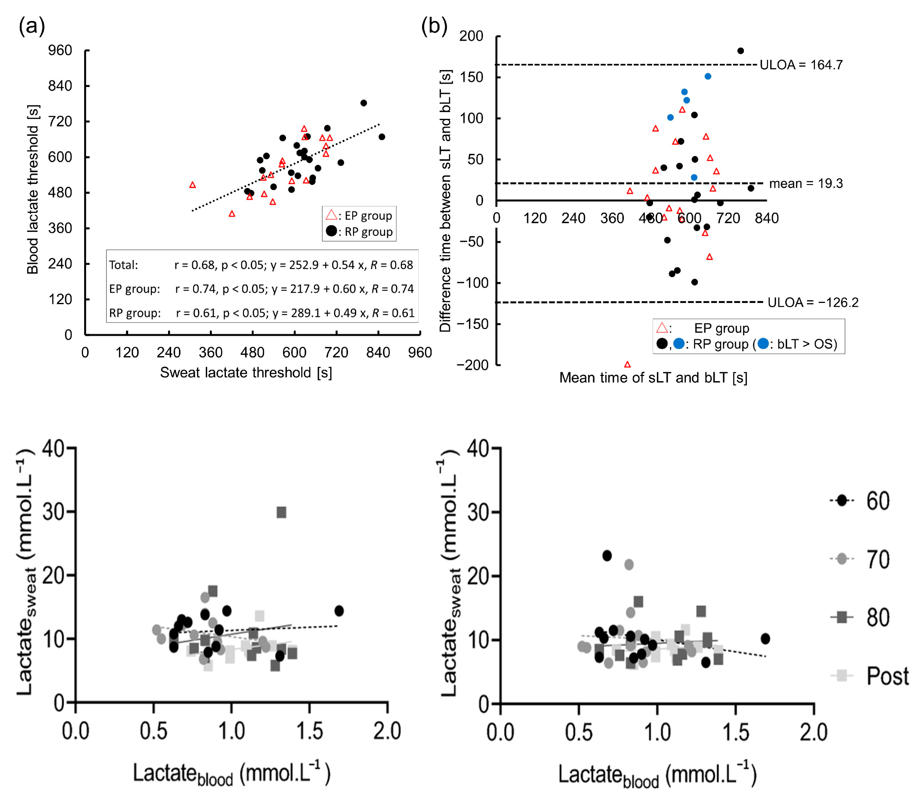
4. Blood and Sweat Lactate Analysis Using Electrochemical Biosensors
5. Conclusions
| Company | Sample Type | Stage of Development | Measuring Range | Sample Volume | Testing Time | Operating Temperature |
|---|---|---|---|---|---|---|
| Zimmer & Peacock | Sweat | Research | N/A | N/A | N/A | N/A |
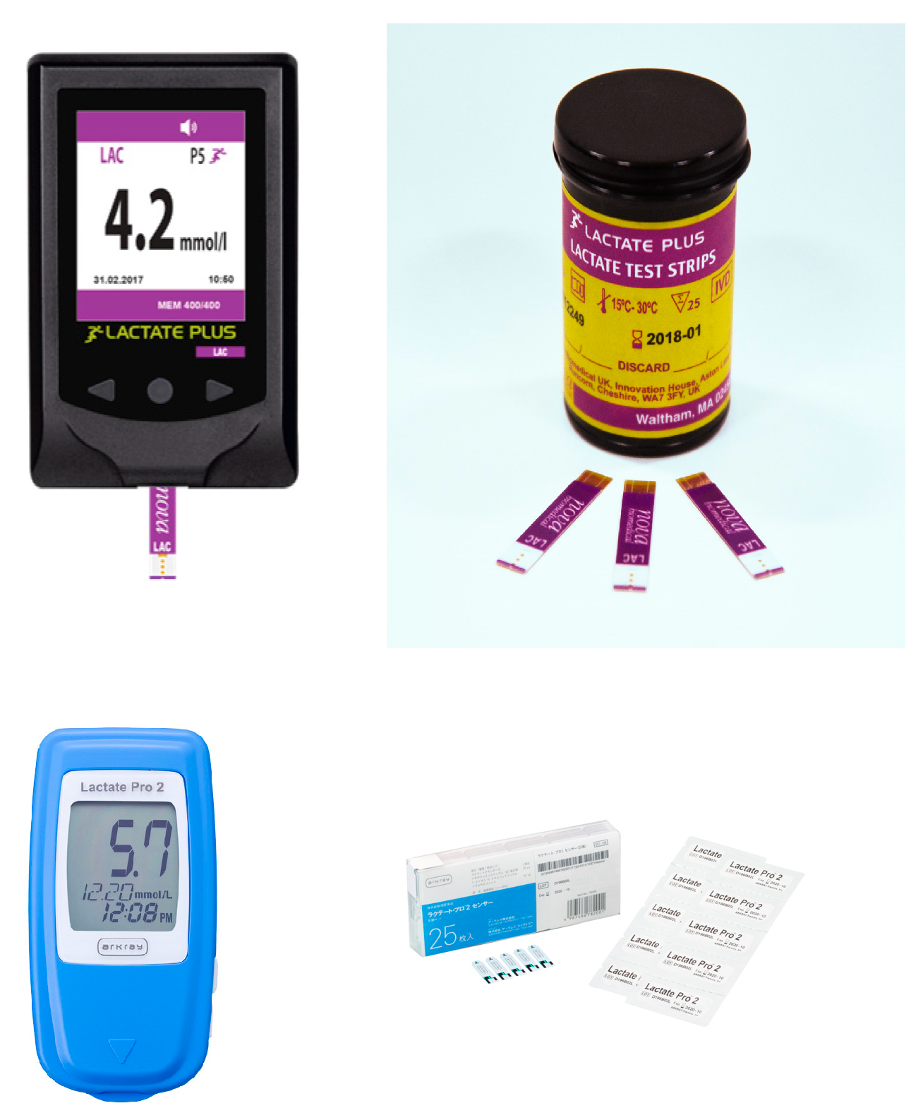
| Nova Niomedical | Blood | Market | 0.5–25 mM | 0.5 µL | 10 s | 10–45 °C |
| PKvitality | Sweat | Research | N/A | N/A | N/A | N/A |
| IST | Sweat | Market | 0.05–25 mM | N/A | <90 s | 15–42 °C |
| Cosmed | Blood | Market | 0.5–25 mM | 0.3 µL | 15 s | N/A |
| APExBIO | Blood | Market | 0.7–22.2 mM | 3 µL | 45 s | 10–40 °C |
Author Contributions
Funding
Institutional Review Board Statement
Informed Consent Statement
Data Availability Statement
Conflicts of Interest
References
- Sonner, Z.; Wilder, E.; Heikenfeld, J.; Kasting, G.; Beyette, F.; Swaile, D.; Sherman, F.; Joyce, J.; Hagen, J.; Kelley-Loughnane, N.; et al. The microfluidics of the eccrine sweat gland, including biomarker partitioning, transport, and biosensing implications. Biomicrofluidics 2015, 9, 031301. [Google Scholar] [CrossRef] [PubMed]
- Gubala, V.; Harris, L.F.; Ricco, A.J.; Tan, M.X.; Williams, D.E. Point of Care Diagnostics: Status and Future. Anal. Chem. 2011, 84, 487–515. [Google Scholar] [CrossRef] [PubMed]
- Robergs, R.A.; Ghiasvand, F.; Parker, D. Biochemistry of exercise-induced metabolic acidosis. Am. J. Physiol. Integr. Comp. Physiol. 2004, 287, R502–R516. [Google Scholar] [CrossRef]
- Zymliński, R.; Biegus, J.; Sokolski, M.; Siwołowski, P.; Nawrocka-Millward, S.; Todd, J.; Jankowska, E.A.; Banasiak, W.; Cotter, G.; Cleland, J.G.; et al. Increased blood lactate is prevalent and identifies poor prognosis in patients with acute heart failure without overt peripheral hypoperfusion. Eur. J. Heart Fail. 2018, 20, 1011–1018. [Google Scholar] [CrossRef]
- Green, J.M.; Pritchett, R.C.; Crews, T.R.; McLester, J.R.; Tucker, D.C. Sweat lactate response between males with high and low aerobic fitness. Eur. J. Appl. Physiol. 2003, 91, 1–6. [Google Scholar] [CrossRef]
- García, E.T. Development of a Flexible Biosensor for the Monitoring of Lactate in Human Sweat for Its Medical Use in Pressure Ischemia. 2014. Available online: http://dspace.lib.cranfield.ac.uk/handle/1826/9254 (accessed on 26 July 2024).
- Yang, G.; Hong, J.; Park, S.-B. Wearable device for continuous sweat lactate monitoring in sports: A narrative review. Front. Physiol. 2024, 15, 1376801. [Google Scholar] [CrossRef]
- Pérez, D.; Orozco, J. Wearable electrochemical biosensors to measure biomarkers with complex blood-to-sweat partition such as proteins and hormones. Microchim. Acta 2022, 189, 127. [Google Scholar] [CrossRef]
- Shen, Y.; Liu, C.; He, H.; Zhang, M.; Wang, H.; Ji, K.; Wei, L.; Mao, X.; Sun, R.; Zhou, F. Recent Advances in Wearable Biosensors for Non-Invasive Detection of Human Lactate. Biosensors 2022, 12, 1164. [Google Scholar] [CrossRef]
- Wu, Z.-Q.; Cao, X.-Q.; Hua, Y.; Yu, C.-M. A Bifunctional Wearable Sensor Based on a Nanoporous Membrane for Simultaneous Detection of Sweat Lactate and Temperature. Anal. Chem. 2024, 96, 3087–3095. [Google Scholar] [CrossRef]
- Zhang, Q.; Jiang, D.; Xu, C.; Ge, Y.; Liu, X.; Wei, Q.; Huang, L.; Ren, X.; Wang, C.; Wang, Y. Wearable electrochemical biosensor based on molecularly imprinted Ag nanowires for noninvasive monitoring lactate in human sweat. Sens. Actuators B Chem. 2020, 320, 128325. [Google Scholar] [CrossRef]
- Li, P.; Kalambate, P.K.; Harris, K.D.; Jemere, A.B.; Tang, X. Robust and flexible electrochemical lactate sensors for sweat analysis based on nanozyme-enhanced electrode. Biosens. Bioelectron. X 2024, 17, 100455. [Google Scholar] [CrossRef]
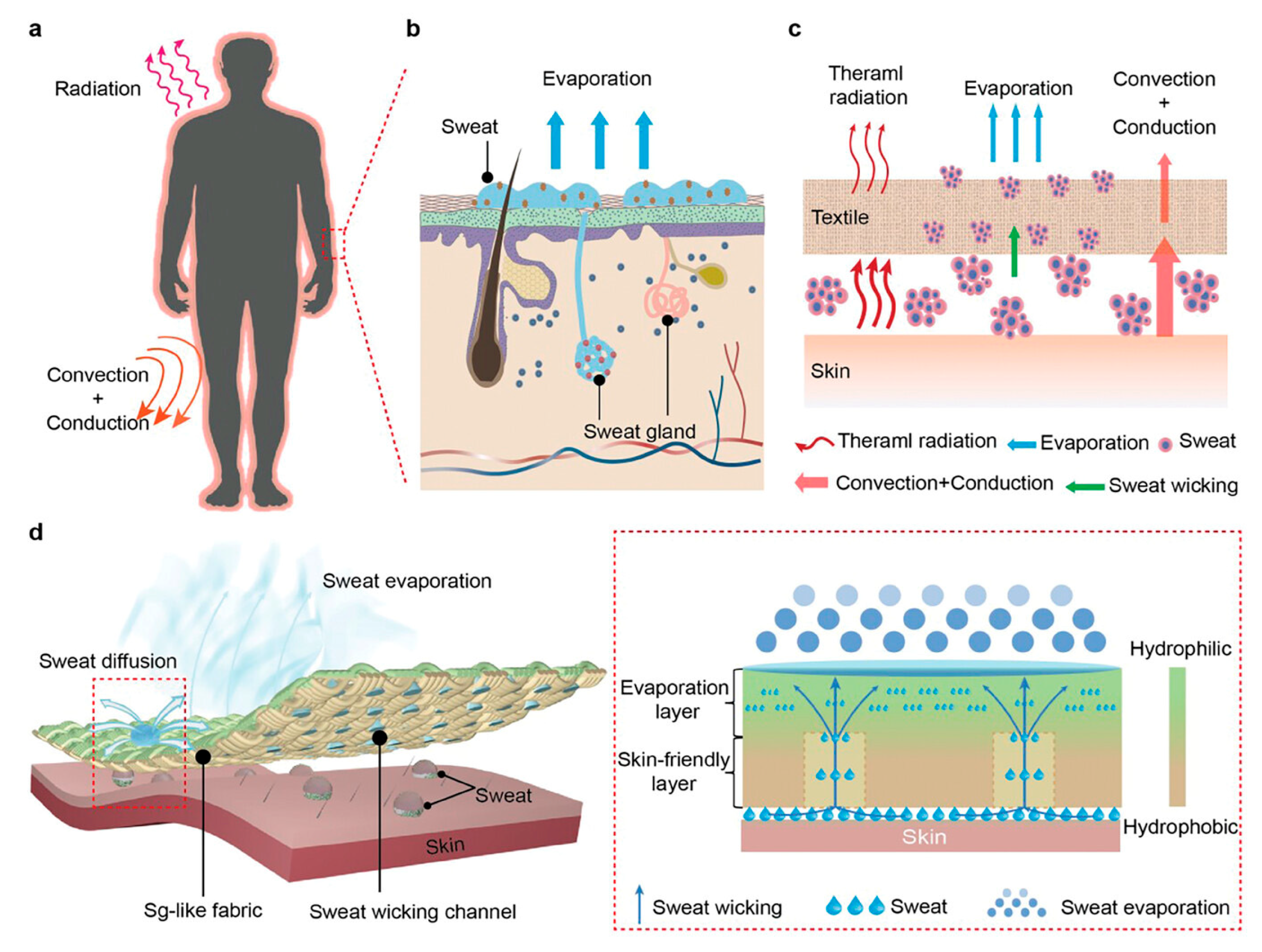
- Zhang, Q.; Wang, M.; Chen, T.; Chen, Z.; Liu, D.; Zhang, Z.; Zhuo, L.; Wang, Y.; Xiao, X.; Zhu, B.; et al. Sweat Gland-Like Fabric for Personal Thermal-Wet Comfort Management. Adv. Funct. Mater. 2024, 2409807. [Google Scholar] [CrossRef]
- Gao, W.; Guan, X.; Wang, J.; Heinig, N.F.; Thomas, J.P.; Zhang, L.; Ding, K.; Leung, K.T. Supported NiOx Nanocatalysts on Graphene for Nonenzymatic Lactate Sensing. ACS Appl. Nano Mater. 2024, 7, 7162–7171. [Google Scholar] [CrossRef]
- Xuan, X.; Chen, C.; Molinero-Fernandez, A.; Ekelund, E.; Cardinale, D.; Swarén, M.; Wedholm, L.; Cuartero, M.; Crespo, G.A. Fully Integrated Wearable Device for Continuous Sweat Lactate Monitoring in Sports. ACS Sens. 2023, 8, 2401–2409. [Google Scholar] [CrossRef]
- Kumar, N.; Lin, Y.-J.; Huang, Y.-C.; Liao, Y.-T.; Lin, S.-P. Detection of lactate in human sweat via surface-modified, screen-printed carbon electrodes. Talanta 2023, 265, 124888. [Google Scholar] [CrossRef]
- Xu, P.; Yano, T.; Yamamoto, K.; Suzuki, H.; Kumagai, H. Characterization of a lactate oxidase from a strain of gram negative bacterium from soil. Appl. Biochem. Biotechnol. 1996, 56, 277–288. [Google Scholar] [CrossRef]
- Khan, R.; Andreescu, S. Catalytic MXCeO2 for enzyme based electrochemical biosensors: Fabrication, characterization and application towards a wearable sweat biosensor. Biosens. Bioelectron. 2023, 248, 115975. [Google Scholar] [CrossRef]
- Weng, X.; Li, M.; Chen, L.; Peng, B.; Jiang, H. A wearable nanozyme–enzyme electrochemical biosensor for sweat lactate monitoring. Talanta 2024, 279, 126675. [Google Scholar] [CrossRef]
- Deng, S. Application of graphene oxide nanosheet lactate biosensors in continuous assessment of athlete fitness. Alex. Eng. J. 2024, 88, 31–35. [Google Scholar] [CrossRef]
- Tian, L.; Cai, L.; Ding, Z.; Zhou, Y.; Zhang, Y.; Liu, Q.; Ge, X.; Yu, C. Sweat lactate biosensor based on lactate oxidase immobilized with flower-like NiCo2O4 and carbon nanotubes. Microchem. J. 2024, 200, 110417. [Google Scholar] [CrossRef]
- He, Q.; Wang, C.; Jain, R.; Byrnes, J.; Farquhar, E.R.; Reed, E.; Berezovsky, E.; Chance, M.R.; Lodowski, D.; An, R. An engineered lactate oxidase based electrochemical sensor for continuous detection of biomarker lactic acid in human sweat and serum. Heliyon 2024, 10, e34301. [Google Scholar] [CrossRef] [PubMed]
- Castrovilli, M.C.; Scognamiglio, V.; Tempesta, E.; Chiarinelli, J.; Parracino, M.; Frisulli, V.; Giardi, M.T.; Avaldi, L.; Rossi, D.; Cartoni, A. Improved reuse and storage performances at room temperature of a new environmentally friendly lactate oxidase biosensor prepared by ambient electrospray immobilization. Green Chem. 2023, 25, 5257–5266. [Google Scholar] [CrossRef]
- Chang, L.-Y.; Rinawati, M.; Guo, Y.-T.; Lin, Y.-C.; Chang, C.-Y.; Su, W.-N.; Mizuguchi, H.; Huang, W.-H.; Chen, J.-L.; Yeh, M.-H. Nitrogen-Doped Graphene Quantum Dots Incorporated into MOF-Derived NiCo Layered Double Hydroxides for Nonenzymatic Lactate Detection in Noninvasive Biosensors. ACS Appl. Nano Mater. 2024, 7, 14431–14442. [Google Scholar] [CrossRef]
- Wang, Y.-X.; Tsao, P.-K.; Rinawati, M.; Chen, K.-J.; Chen, K.-Y.; Chang, C.; Yeh, M.-H. Designing ZIF-67 derived NiCo layered double hydroxides with 3D hierarchical structure for Enzyme-free electrochemical lactate monitoring in human sweat. Chem. Eng. J. 2021, 427, 131687. [Google Scholar] [CrossRef]
- Dykstra, G.; Chapa, I.; Liu, Y. Reagent-Free Lactate Detection Using Prussian Blue and Electropolymerized-Molecularly Imprinted Polymers-Based Electrochemical Biosensors. ACS Appl. Mater. Interfaces 2024, 16, 66921–66931. [Google Scholar] [CrossRef]
- Pei, S.; Ji, W.; Yang, Y.; Liu, T.; Yang, S.; Wu, J.; Dai, J.; Hou, X.; Wu, Q.; Li, L. Flexible Dual-Channel Molecularly Imprinted Electrochemical Sensor for Simultaneously Monitoring Sweat Cortisol and Lactate Levels. Anal. Sens. 2024, 4, e202400003. [Google Scholar] [CrossRef]
- Chen, Y.; Hu, X.; Liang, Q.; Wang, X.; Zhang, H.; Jia, K.; Li, Y.; Zhang, A.; Chen, P.; Lin, M.; et al. Large-Scale Flexible Fabric Biosensor for Long-Term Monitoring of Sweat Lactate. Adv. Funct. Mater. 2024, 34, 2401270. [Google Scholar] [CrossRef]
- Konno, S.; Kudo, H. Fundamental Study of a Wristwatch Sweat Lactic Acid Monitor. Biosensors 2024, 14, 187. [Google Scholar] [CrossRef]
- Klous, L.; de Ruiter, C.J.; Scherrer, S.; Gerrett, N.; Daanen, H.A.M. The (in)dependency of blood and sweat sodium, chloride, potassium, ammonia, lactate and glucose concentrations during submaximal exercise. Eur. J. Appl. Physiol. 2020, 121, 803–816. [Google Scholar] [CrossRef]
- Rabost-Garcia, G.; Colmena, V.; Aguilar-Torán, J.; Galí, J.V.; Punter-Villagrasa, J.; Casals-Terré, J.; Miribel-Catala, P.; Muñoz, X.; Cadefau, J.; Padullés, J.; et al. Non-Invasive Multiparametric Approach To Determine Sweat–Blood Lactate Bioequivalence. ACS Sens. 2023, 8, 1536–1541. [Google Scholar] [CrossRef]
- Sakharov, D.A.; Shkurnikov, M.U.; Vagin, M.Y.; Yashina, E.I.; Karyakin, A.A.; Tonevitsky, A.G. Relationship between Lactate Concentrations in Active Muscle Sweat and Whole Blood. Bull. Exp. Biol. Med. 2010, 150, 83–85. [Google Scholar] [CrossRef] [PubMed]
- Karpova, E.V.; Laptev, A.I.; Andreev, E.A.; Karyakina, E.E.; Karyakin, A.A. Relationship Between Sweat and Blood Lactate Levels During Exhaustive Physical Exercise. ChemElectroChem 2020, 7, 191–194. [Google Scholar] [CrossRef]
- Maeda, Y.; Okawara, H.; Sawada, T.; Nakashima, D.; Nagahara, J.; Fujitsuka, H.; Ikeda, K.; Hoshino, S.; Kobari, Y.; Katsumata, Y.; et al. Implications of the Onset of Sweating on the Sweat Lactate Threshold. Sensors 2023, 23, 3378. [Google Scholar] [CrossRef] [PubMed]
- Teunissen, H. H2TRAIN. Inside. Available online: https://www.inside-association.eu/post/h2train (accessed on 8 October 2024).
- PONS IP. H2TRAIN, the Largest Project Funded Under the European Chips Act for the Application of Digital Technologies in Smart Sports and Health Care, Has Been Launched with the Participation of PONS IP. Available online: https://ponsip.com/en/ip-news/news/h2train-the-largest-project-funded-under-the-european-chips-act-for-the-application-of-digital-technologies-in-smart-sports-and-health-care-has-been-launched-with-the-participation-of-pons-ip/ (accessed on 8 October 2024).


Disclaimer/Publisher’s Note: The statements, opinions and data contained in all publications are solely those of the individual author(s) and contributor(s) and not of MDPI and/or the editor(s). MDPI and/or the editor(s) disclaim responsibility for any injury to people or property resulting from any ideas, methods, instructions or products referred to in the content. |
© 2024 by the authors. Licensee MDPI, Basel, Switzerland. This article is an open access article distributed under the terms and conditions of the Creative Commons Attribution (CC BY) license (https://creativecommons.org/licenses/by/4.0/).
Share and Cite
Messina, L.; Giardi, M.T. Recent Status on Lactate Monitoring in Sweat Using Biosensors: Can This Approach Be an Alternative to Blood Detection? Biosensors 2025, 15, 3. https://doi.org/10.3390/bios15010003
Messina L, Giardi MT. Recent Status on Lactate Monitoring in Sweat Using Biosensors: Can This Approach Be an Alternative to Blood Detection? Biosensors. 2025; 15(1):3. https://doi.org/10.3390/bios15010003
Chicago/Turabian StyleMessina, Leonardo, and Maria Teresa Giardi. 2025. "Recent Status on Lactate Monitoring in Sweat Using Biosensors: Can This Approach Be an Alternative to Blood Detection?" Biosensors 15, no. 1: 3. https://doi.org/10.3390/bios15010003
APA StyleMessina, L., & Giardi, M. T. (2025). Recent Status on Lactate Monitoring in Sweat Using Biosensors: Can This Approach Be an Alternative to Blood Detection? Biosensors, 15(1), 3. https://doi.org/10.3390/bios15010003