SERS Sensors with Bio-Derived Substrates Under the Way to Agricultural Monitoring of Pesticide Residues


Abstract
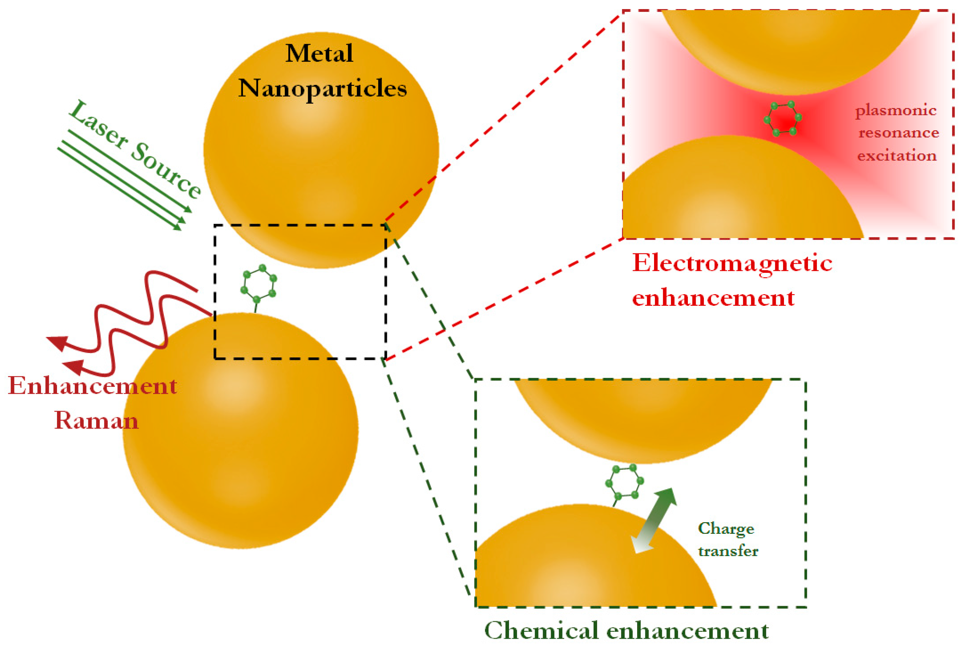
1. Introduction
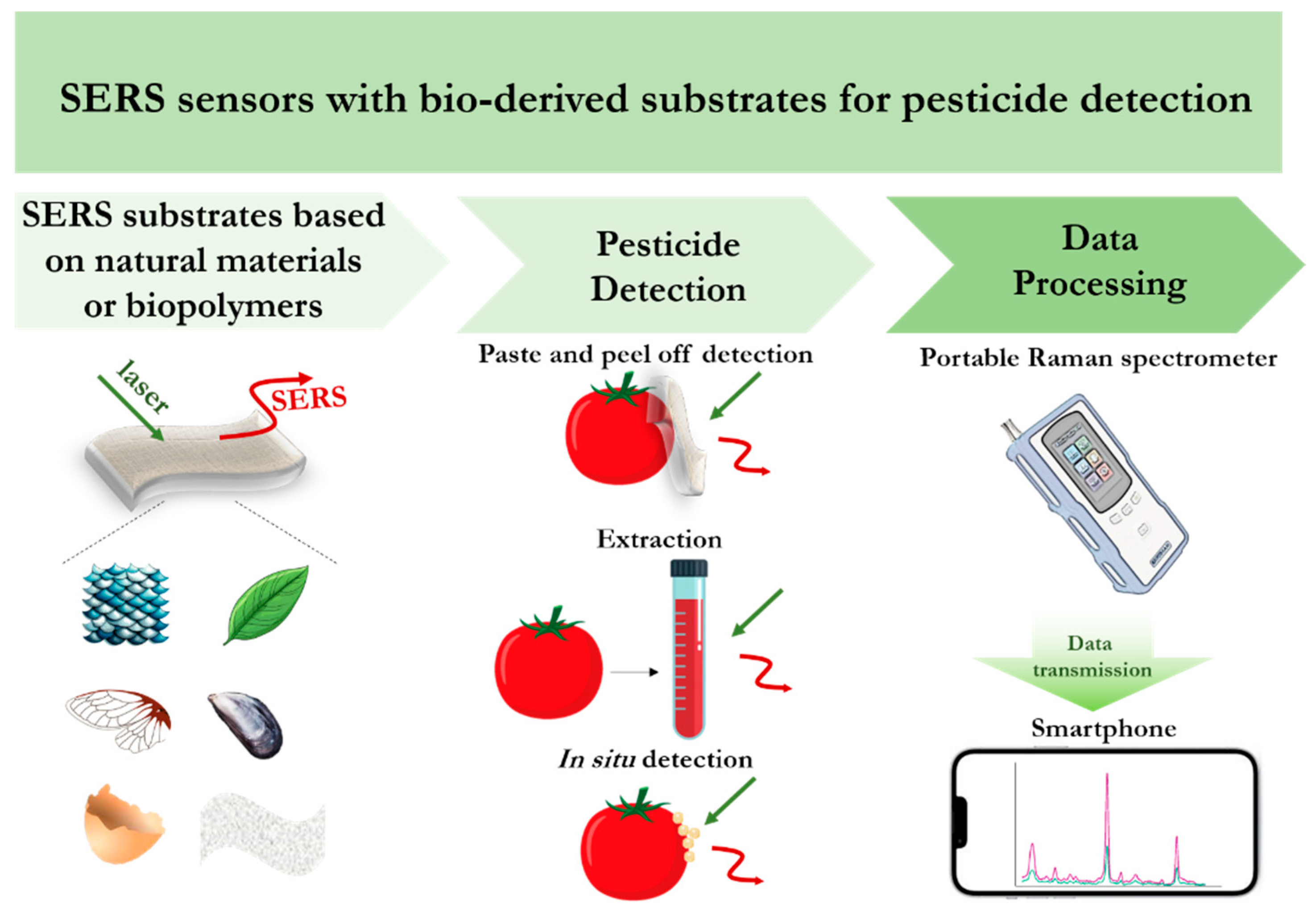
2. Exploring the Diversity of Biomaterials and Design of Biocompatible SERS Substrates
3. SERS Detection of Pesticides Using Biomaterial-Based Substrates

| Substrate | Plasmonic Nanostructures | Analyte | Detection Limit | Samples | Ref. |
|---|---|---|---|---|---|
| Natural biomaterials | |||||
| lotus leaf | AgNPs | paraquat | 4.7 × 10⁻⁹ M | lake, tap and drinking waters | [98] |
| cicada wing | AgNPs | difenoconazole | 3.9 × 10⁻⁸ M | potato | [103] |
| dragonfly wing | Au nanoislands | cypermethrin | 10−3 ng/cm2 | tomato peels | [85] |
| fish scale bio-wastes | Au nanoflowers | glyphosate | 2.6 × 10⁻⁶ M | agricultural soil | [99] |
| aminomethylphosphonic acid | 2.4 × 10⁻⁶ M | ||||
| cicada wing | Au nanofilm | acephate | 10−9 mg/mL | pear peels | [86] |
| lotus leaf mastoid | Ag micro/nanoarrays on PDMS film | thiram | 10−6 M | dendrobium leaves and stem | [93] |
| fonofos | 10−5 M | ||||
| triadophos | 10−7 M | ||||
| capsicum, celery, cole | Ag nanoislands | paraquat | 10−9 M | capsicum, celery, cole | [94] |
| fenthion | 10−8 M | ||||
| tea leaves | AuNPs | ferbam | - | tea leaves | [95] |
| soybean leaves | AuNPs | acetamiprid | 4.5 × 10⁻⁷ M | soybean leaves | [96] |
| chlorothalonil | 3.7 × 10⁻⁶ M | ||||
| spinach leaf | AuNPs | dimethoate | 4 × 10⁻⁶ M | spinach leaf | [97] |
| Biopolymer | |||||
| bacterial cellulose nanocrystal | AuNPs | thiram | 1.5 × 10⁻⁷ M | peach juice | [90] |
| 2 × 10⁻⁷ M | apple juice | ||||
| 2 × 10⁻⁷ M | grape juice | ||||
| bacterial nanocellulose paper | AgNPs | methomyl | 3.6 × 10⁻⁷ M | orange and apple peels | [81] |
| bacterial nanocellulose | Ag nanorods | thiram | 10−9 M | grape | [87] |
| chitosan | AgNPs | thiram | 3.2 × 10⁻⁵ M | water samples | [100] |
| cellulose | AgNPs | chlorfenapyr | 2.5 × 10⁻⁶ M | - | [101] |
| bacterial nanocellulose | succulent-like Ag nanoflowers | thiram | 10−10 M | apple | [88] |
| nanocellulose fiber | AgNPs | carbendazim | 10−8 M | - | [104] |
| nanocellulose paper | Au-Ag bimetallic NPs | thiram | 10−6 M | apple | [89] |
| chitosan foam | AgNPs | triazophos | 3.2 × 10⁻⁵ M | - | [51] |
| alginate–chitosan porous gel | Ag nanocubes | thiram | 1.43 × 10⁻⁸ M | apple | [75] |
| alginate hydrogel | Au@Ag NPs | thiram | 10−10 M | fruit juices, apple peels and cabbage leaves | [64] |
| gelatine hydrogel | AgNPs | sodium diethyldithiocarbamate | 10−5 M | - | [91] |
| jellylike nitrocellulose texture | AgNPs | thiram | 0.5 ng/cm2 | apple peels | [105] |
| gelatin gel | AgNPs | malachite green | 10−9 M | lake water | [67] |
4. Functionalization of SERS Substrates for Improving Detection Capabilities
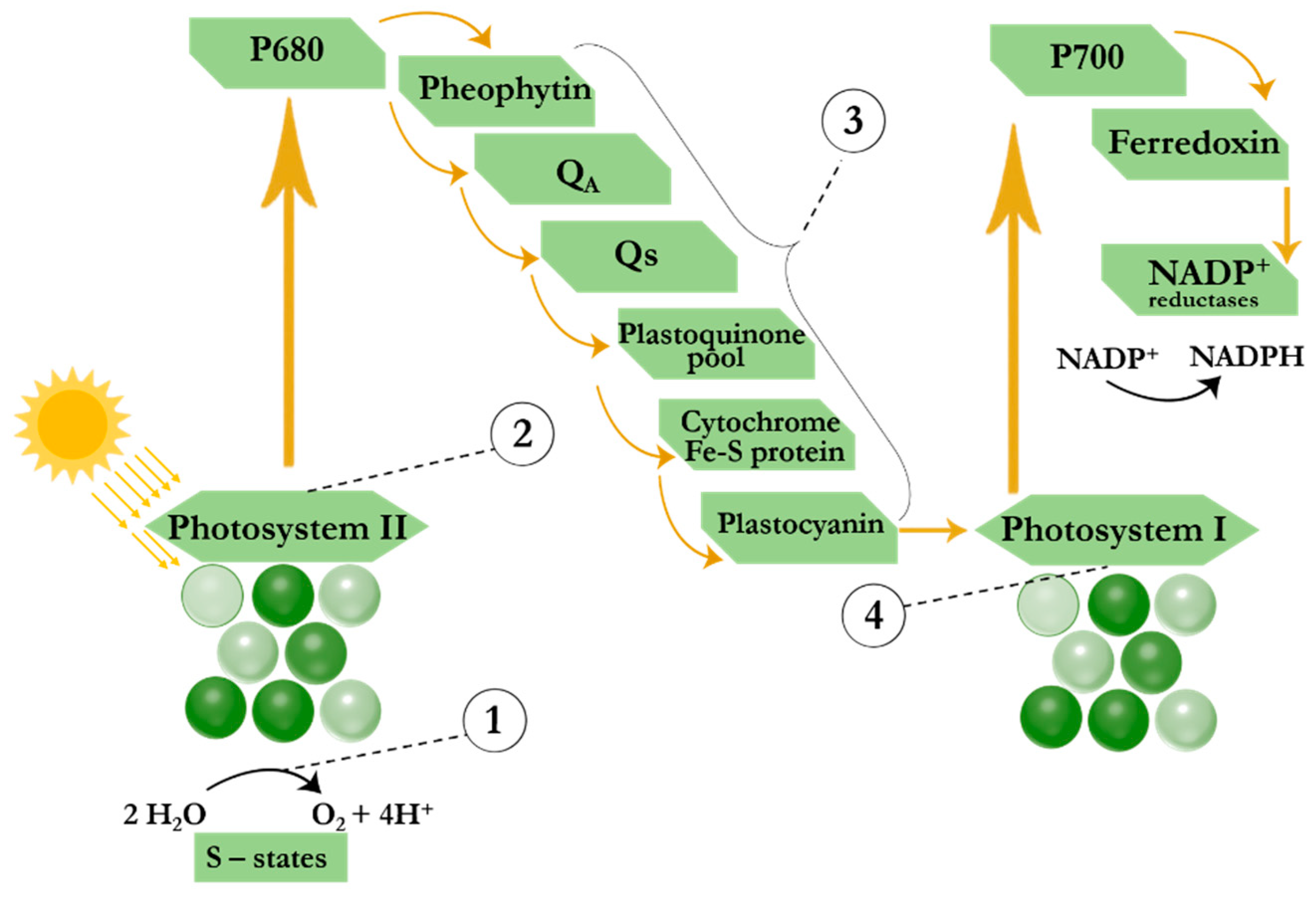
5. The Potential of Raman Spectroscopy in Detecting Photosynthesis Inhibitor Substances Using Biomaterials
6. Conclusions and Outlook
- The unique structure, natural pattern, surface hydrophobicity and gaps provided by natural materials form many “hot spots” and create the necessary conditions for the detection of trace pesticides;
- Biopolymer-based SERS substrates, combining flexibility, stability and low costs, demonstrate high sensitivity and allow pre-concentration of pesticides directly from the sample;
- Functionalization of SERS substrates extracts target analytes from the complex organic sample environment and enhance the selectivity of pesticide analysis.
- Despite the unique surface morphology of natural biomaterials (petals, eggshells, leaves, cicada wings, etc.), which provide hierarchical micro/nanostructures with a large number of gaps in the designed substrates to enhance the electromagnetic field, as well as the natural hydrophobicity of materials, mechanical strength, durability and repeatability of SERS substrates are the weak points of natural biomaterials. Inspired by the pattern and morphology of the surface of natural biomaterials and considering their weaknesses when designing SERS substrates, scientists obtain multifunctional flexible SERS substrates with replicated micro/nanostructure of natural materials, for example, using nanoimprinting technology [144,145].
- A common drawback of all types of SERS substrates in the direct analysis is the presence of compounds that contribute to the recorded spectrum and complicate the interpretation of the assay results. The solution to this problem is the functionalization of SERS substrates with aptamers, antibodies or other receptors to enhance the substrate specificity [111].
- Fluorescence arising from close interactions between plasmonic nanoparticles and the target molecule or causing by the analysis of fluorophore-containing samples is a competing effect that suppresses the Raman signal and reduces the sensitivity of sensing [146,147]. To improve the performance of SERS substrates, it is necessary to design substrates based on bio-inspired materials using components that inhibit fluorescence while maintaining or even increasing achievable EF values. For example, a graphene-containing composite SERS substrate provides improved SERS efficiency through a chemical enhancement mechanism in addition to reducing the fluorescence background [147].
- The disadvantage of flexible SERS substrates is the low accuracy of testing, since some pesticides penetrate deeply into the sample tissue and soft treatment of the surface is insufficient. In this case, deep extraction procedures are still required for sample preparation. In this regard, the development of a hybrid substrate combining a flexible SERS substrate with separation methods including microfluidic systems and thin layer chromatography allows for the sensing procedure to be simplified and its efficiency to be increased [148,149,150].
- The uniformity and reproducibility of SERS substrates are key parameters determining the practical applicability of the method, but achieving these characteristics remains a challenge. Standard methods such as incorporation of plasmonic nanoparticles into the structure of flexible substrates or in situ growth of nanoparticles on the surface of bio-derived materials do not provide homogeneity. Therefore, new fabrication techniques are still in demand.
- The standard analysis procedure consists of depositing a probe molecule on a bio-derived-based SERS substrate and disposing of the substrate after measuring the Raman spectra. Fabrication of reusable SERS substrates in a low-cost and easy-to-use manner is a relatively new direction that presents favorable opportunities for sensing applications. The proposed approaches to ensure reusability of SERS substrates include rinsing the analyte from the surface using solvents and degradation or decomposition of the probe molecule using various techniques (e.g., ultrasound, plasma cleaning, catalytic degradation) [151].
- Further application of SERS substrates for monitoring agricultural products requires engineering of portable and handheld Raman systems. In the field, it is important to quickly assess pesticide levels outside the laboratory to detect when toxicants reach dangerous concentrations in a timely manner. The use of portable SERS spectrometers at the sampling site requires large-scale production of reproducible SERS substrates [152,153]. In addition, the design of portable spectrometers should be improved to variability of the excitation laser wavelength range (from visible to near infrared). Finally, the use of SERS substrates in combination with a portable recording device for on-site pesticide analysis requires the expansion of the database/library with spectra of all pesticides.
Author Contributions
Funding
Institutional Review Board Statement
Informed Consent Statement
Data Availability Statement
Conflicts of Interest
References
- Tudi, M.; Daniel Ruan, H.; Wang, L.; Lyu, J.; Sadler, R.; Connell, D.; Chu, C.; Phung, D.T. Agriculture Development, Pesticide Application and Its Impact on the Environment. Int. J. Environ. Res. Public Health 2021, 18, 1112. [Google Scholar] [CrossRef] [PubMed]
- Bernardes, M.F.F.; Pazin, M.; Pereira, L.C.; Dorta, D.J. Impact of pesticides on environmental and human health. In Toxicology Studies—Cells, Drugs and Environment; IntechOpen: London, UK, 2015; pp. 195–233. [Google Scholar] [CrossRef]
- Popp, J.; Pető, K.; Nagy, J. Pesticide productivity and food security. A review. Agron. Sustain. Dev. 2013, 33, 243–255. [Google Scholar] [CrossRef]
- Carrasco Cabrera, L.; Medina Pastor, P. The 2019 European Union report on pesticide residues in food. EFSA J. 2021, 19, e06491. [Google Scholar] [CrossRef] [PubMed]
- Jeyaseelan, A.; Murugesan, K.; Thayanithi, S.; Palanisamy, S.B. A review of the impact of herbicides and insecticides on the microbial communities. Environ. Res. 2024, 245, 118020. [Google Scholar] [CrossRef] [PubMed]
- Sousa, S.; Maia, M.L.; Correira-Sá, L.; Fernandes, V.C.; Delerue-Matos, C.; Calhau, C.; Domingues, V.F. Chemistry and Toxicology Behind Insecticides and Herbicides. In Controlled Release of Pesticides for Sustainable Agriculture; Rakhimol, K.R., Thomas, S., Volova, T., Jayachandran., K., Eds.; Springer International Publishing: Cham, Switzerland, 2020; pp. 59–109. [Google Scholar] [CrossRef]
- Reeves, W.R.; McGuire, M.K.; Stokes, M.; Vicini, J.L. Assessing the Safety of Pesticides in Food: How Current Regulations Protect Human Health. Adv. Nutr. 2019, 10, 80–88. [Google Scholar] [CrossRef]
- Narenderan, S.T.; Meyyanathan, S.N.; Babu, B. Review of pesticide residue analysis in fruits and vegetables. Pre-treatment, extraction and detection techniques. Food Res. Int. 2020, 133, 109141. [Google Scholar] [CrossRef]
- Mandal, S.; Poi, R.; Hazra, D.K.; Ansary, I.; Bhattacharyya, S.; Karmakar, R. Review of extraction and detection techniques for the analysis of pesticide residues in fruits to evaluate food safety and make legislative decisions: Challenges and anticipations. J. Chromatogr. B 2023, 1215, 123587. [Google Scholar] [CrossRef]
- Fang, L.; Liao, X.; Jia, B.; Shi, L.; Kang, L.; Zhou, L.; Kong, W. Recent progress in immunosensors for pesticides. Biosens. Bioelectron. 2020, 164, 112255. [Google Scholar] [CrossRef]
- Jara, M.D.L.; Alvarez, L.A.C.; Guimarães, M.C.C.; Antunes, P.W.P.; de Oliveira, J.P. Lateral flow assay applied to pesticides detection: Recent trends and progress. Environ. Sci. Pollut. Res. 2022, 29, 46487–46508. [Google Scholar] [CrossRef]
- Xu, L.; Abd El-Aty, A.M.; Eun, J.-B.; Shim, J.-H.; Zhao, J.; Lei, X.; Gao, S.; She, Y.; Jin, F.; Wang, J.; et al. Recent Advances in Rapid Detection Techniques for Pesticide Residue: A Review. J. Agric. Food Chem. 2022, 70, 13093–13117. [Google Scholar] [CrossRef]
- Umapathi, R.; Park, B.; Sonwal, S.; Rani, G.M.; Cho, Y.; Huh, Y.S. Advances in optical-sensing strategies for the on-site detection of pesticides in agricultural foods. Trends Food Sci. Technol. 2022, 119, 69–89. [Google Scholar] [CrossRef]
- Jain, U.; Saxena, K.; Hooda, V.; Balayan, S.; Singh, A.P.; Tikadar, M.; Chauhan, N. Emerging vistas on pesticides detection based on electrochemical biosensors—An update. Food Chem. 2022, 371, 131126. [Google Scholar] [CrossRef] [PubMed]
- Wang, X.; Huang, S.-C.; Hu, S.; Yan, S.; Ren, B. Fundamental understanding and applications of plasmon-enhanced Raman spectroscopy. Nat. Rev. Phys. 2020, 2, 253–271. [Google Scholar] [CrossRef]
- Cialla-May, D.; Schmitt, M.; Popp, J. Theoretical principles of Raman spectroscopy. Phys. Sci. Rev. 2019, 4, 20170040. [Google Scholar] [CrossRef]
- Ong, T.T.X.; Blanch, E.W.; Jones, O.A.H. Surface Enhanced Raman Spectroscopy in environmental analysis, monitoring and assessment. Sci. Total Environ. 2020, 720, 137601. [Google Scholar] [CrossRef]
- Huang, Z.; Peng, J.; Xu, L.; Liu, P. Development and Application of Surface-Enhanced Raman Scattering (SERS). Nanomaterials 2024, 14, 1417. [Google Scholar] [CrossRef]
- Mahanty, S.; Majumder, S.; Paul, R.; Boroujerdi, R.; Valsami-Jones, E.; Laforsch, C. A review on nanomaterial-based SERS substrates for sustainable agriculture. Sci. Total Environ. 2024, 950, 174252. [Google Scholar] [CrossRef]
- Zhang, D.; Pu, H.; Huang, L.; Sun, D.-W. Advances in flexible surface-enhanced Raman scattering (SERS) substrates for nondestructive food detection: Fundamentals and recent applications. Trends Food Sci. Technol. 2021, 109, 690–701. [Google Scholar] [CrossRef]
- Li, H.; Haruna, S.A.; Sheng, W.; Bei, Q.; Ahmad, W.; Zareef, M.; Chen, Q.; Ding, Z. SERS-activated platforms for chemical contaminants in food: Probes, encoding methods, and detection. TrAC Trends Anal. Chem. 2023, 169, 117365. [Google Scholar] [CrossRef]
- Zhang, Q.; Fang, L.; Jia, B.; Long, N.; Shi, L.; Zhou, L.; Zhao, H.; Kong, W. Optical lateral flow test strip biosensors for pesticides: Recent advances and future trends. TrAC Trends Anal. Chem. 2021, 144, 116427. [Google Scholar] [CrossRef]
- Lin, D.-Y.; Yu, C.-Y.; Ku, C.-A.; Chung, C.-K. Design, Fabrication, and Applications of SERS Substrates for Food Safety Detection: Review. Micromachines 2023, 14, 1343. [Google Scholar] [CrossRef] [PubMed]
- Xie, X.; Pu, H.; Sun, D.-W. Recent advances in nanofabrication techniques for SERS substrates and their applications in food safety analysis. Crit. Rev. Food Sci. Nutr. 2018, 58, 2800–2813. [Google Scholar] [CrossRef] [PubMed]
- Liu, X.; Guo, J.; Li, Y.; Wang, B.; Yang, S.; Chen, W.; Wu, X.; Guo, J.; Ma, X. SERS substrate fabrication for biochemical sensing: Towards point-of-care diagnostics. J. Mater. Chem. B 2021, 9, 8378–8388. [Google Scholar] [CrossRef] [PubMed]
- Jebakumari, K.A.E.; Murugasenapathi, N.K.; Palanisamy, T. Engineered Two-Dimensional Nanostructures as SERS Substrates for Biomolecule Sensing: A Review. Biosensors 2023, 13, 102. [Google Scholar] [CrossRef] [PubMed]
- Peng, R.; Zhang, T.; Yan, S.; Song, Y.; Liu, X.; Wang, J. Recent Development and Applications of Stretchable SERS Substrates. Nanomaterials 2023, 13, 2968. [Google Scholar] [CrossRef]
- Yadav, S.; Satija, J. The current state of the art of plasmonic nanofibrous mats as SERS substrates: Design, fabrication and sensor applications. J. Mater. Chem. B 2021, 9, 267–282. [Google Scholar] [CrossRef]
- Li, B.; Liu, S.; Huang, L.; Jin, M.; Wang, J. Nanohybrid SERS substrates intended for food supply chain safety. Coord. Chem. Rev. 2023, 494, 215349. [Google Scholar] [CrossRef]
- Serafinelli, C.; Fantoni, A.; Alegria, E.C.B.A.; Vieira, M. Plasmonic Metal Nanoparticles Hybridized with 2D Nanomaterials for SERS Detection: A Review. Biosensors 2022, 12, 225. [Google Scholar] [CrossRef]
- López-Lorente, Á.I. Recent developments on gold nanostructures for surface enhanced Raman spectroscopy: Particle shape, substrates and analytical applications. A review. Anal. Chim. Acta 2021, 1168, 338474. [Google Scholar] [CrossRef]
- Mosier-Boss, P.A. Review of SERS Substrates for Chemical Sensing. Nanomaterials 2017, 7, 142. [Google Scholar] [CrossRef]
- Restaino, S.M.; White, I.M. A critical review of flexible and porous SERS sensors for analytical chemistry at the point-of-sample. Analytica Chimica Acta 2019, 1060, 17–29. [Google Scholar] [CrossRef] [PubMed]
- Zhu, H.; Huang, Y.; Lou, X.; Xia, F. Bioinspired superwetting surfaces for biosensing. View 2021, 2, 20200053. [Google Scholar] [CrossRef]
- Ying, Y.; Tang, Z.; Liu, Y.J.N. Material design, development, and trend for surface-enhanced Raman scattering substrates. Nanoscale 2023, 15, 10860–10881. [Google Scholar] [CrossRef]
- Pérez-Jiménez, A.I.; Lyu, D.; Lu, Z.; Liu, G.; Ren, B. Surface-enhanced Raman spectroscopy: Benefits, trade-offs and future developments. Chem. Sci. 2020, 11, 4563–4577. [Google Scholar] [CrossRef]
- Sharma, V.; Balaji, R.; Walia, R.; Krishnan, V. Au Nanoparticle Aggregates Assembled on 3D Mirror-like Configuration Using Canna generalis Leaves for SERS Applications. Colloid Interface Sci. Commun. 2017, 18, 9–12. [Google Scholar] [CrossRef]
- Yan, X.; Wang, Y.; Shi, G.; Wang, M.; Zhang, J.; Sun, X.; Xu, H. Flower-like Cu nanoislands decorated onto the cicada wing as SERS substrates for the rapid detection of crystal violet. Optik 2018, 172, 812–821. [Google Scholar] [CrossRef]
- Kumar, J.; Jinachandran, A.; Ponnusamy, V.K.; Huang, G.G.; Suresh, A.K.; Noothalapati, H.; Panneerselvam, R. Ag nanoparticle-embedded fish scales as SERS substrates for sensitive detection of forever chemical in real samples. Appl. Surf. Sci. 2024, 674, 160961. [Google Scholar] [CrossRef]
- Yuan, K.; Zheng, J.; Yang, D.; Jurado Sánchez, B.; Liu, X.; Guo, X.; Liu, C.; Dina, N.E.; Jian, J.; Bao, Z.; et al. Self-Assembly of Au@Ag Nanoparticles on Mussel Shell To Form Large-Scale 3D Supercrystals as Natural SERS Substrates for the Detection of Pathogenic Bacteria. ACS Omega 2018, 3, 2855–2864. [Google Scholar] [CrossRef]
- Dong, J.; Zhou, W.; Yang, C.; Wu, H.; Han, Q.; Zhang, C.; Gao, W.; Yan, X.; Sun, M. Preparation of a Three-Dimensional Composite Structure Based on a Periodic Au@Ag Core–Shell Nanocube with Ultrasensitive Surface-Enhanced Raman Scattering for Rapid Detection. ACS Appl. Mater. Interfaces 2023, 15, 28840–28848. [Google Scholar] [CrossRef]
- Lu, Z.; Liu, Y.; Wang, M.; Zhang, C.; Li, Z.; Huo, Y.; Li, Z.; Xu, S.; Man, B.; Jiang, S. A novel natural surface-enhanced Raman spectroscopy (SERS) substrate based on graphene oxide-Ag nanoparticles-Mytilus coruscus hybrid system. Sens. Actuators B Chem. 2018, 261, 1–10. [Google Scholar] [CrossRef]
- Arnob, M.M.P.; Shih, W.-C. 3-Dimensional Plasmonic Substrates Based on Chicken Eggshell Bio-Templates for SERS-Based Bio-Sensing. Micromachines 2017, 8, 196. [Google Scholar] [CrossRef]
- Chamuah, N.; Chetia, L.; Zahan, N.; Dutta, S.; Ahmed, G.A.; Nath, P. A naturally occurring diatom frustule as a SERS substrate for the detection and quantification of chemicals. J. Phys. D Appl. Phys. 2017, 50, 175103. [Google Scholar] [CrossRef]
- Ge, N.; Hu, X.; Pan, Z.; Cai, S.; Fu, F.; Wang, Z.; Yao, J.; Liu, X. Sustainable fabrication of cellulose aerogel embedded with ZnO@noble metal (Ag, Au, Ag-Au) NPs for sensitive and reusable SERS application. Colloids Surf. A Physicochem. Eng. Asp. 2023, 671, 131650. [Google Scholar] [CrossRef]
- Xu, F.; Ma, F.; Ding, Z.; Xiao, L.; Zhang, X.; Lu, Q.; Lu, G.; Kaplan, D.L. SERS Substrate with Silk Nanoribbons as Interlayer Template. ACS Appl. Mater. Interfaces 2019, 11, 42896–42903. [Google Scholar] [CrossRef]
- Viriyakitpattana, N.; Rattanabut, C.; Lertvachirapaiboon, C.; Pimalai, D.; Bamrungsap, S. Layer-by-Layer Biopolymer Assembly for the In Situ Fabrication of AuNP Plasmonic Paper—A SERS Substrate for Food Adulteration Detection. ACS Omega 2024, 9, 10099–10109. [Google Scholar] [CrossRef]
- Puente, C.; Sánchez-Domínguez, M.; Brosseau, C.L.; López, I. Silver-chitosan and gold-chitosan substrates for surface-enhanced Raman spectroscopy (SERS): Effect of nanoparticle morphology on SERS performance. Mater. Chem. Phys. 2021, 260, 124107. [Google Scholar] [CrossRef]
- Vafakish, B.; Wilson, L.D. A Highly Sensitive Chitosan-Based SERS Sensor for the Trace Detection of a Model Cationic Dye. Int. J. Mol. Sci. 2024, 25, 9327. [Google Scholar] [CrossRef]
- Fu, F.; Yang, B.; Hu, X.; Tang, H.; Zhang, Y.; Xu, X.; Zhang, Y.; Touhid, S.S.; Liu, X.; Zhu, Y.; et al. Biomimetic synthesis of 3D Au-decorated chitosan nanocomposite for sensitive and reliable SERS detection. Chem. Eng. J. 2020, 392, 123693. [Google Scholar] [CrossRef]
- Wang, C.; Wong, K.W.; Wang, Q.; Zhou, Y.; Tang, C.; Fan, M.; Mei, J.; Lau, W.-M. Silver-nanoparticles-loaded chitosan foam as a flexible SERS substrate for active collecting analytes from both solid surface and solution. Talanta 2019, 191, 241–247. [Google Scholar] [CrossRef]
- Zhang, Q.; Xu, G.; Guo, N.; Wang, T.; Song, P.; Xia, L. In-Situ Synthesis of Methyl Cellulose Film Decorated with Silver Nanoparticles as a Flexible Surface-Enhanced Raman Substrate for the Rapid Detection of Pesticide Residues in Fruits and Vegetables. Materials 2021, 14, 5750. [Google Scholar] [CrossRef]
- Liou, P.; Nayigiziki, F.X.; Kong, F.; Mustapha, A.; Lin, M. Cellulose nanofibers coated with silver nanoparticles as a SERS platform for detection of pesticides in apples. Carbohydr. Polym. 2017, 157, 643–650. [Google Scholar] [CrossRef] [PubMed]
- Oh, K.; Lee, M.; Lee, S.G.; Jung, D.H.; Lee, H.L. Cellulose nanofibrils coated paper substrate to detect trace molecules using surface-enhanced Raman scattering. Cellulose 2018, 25, 3339–3350. [Google Scholar] [CrossRef]
- Sun, L.; Yu, Z.; Alsammarraie, F.K.; Lin, M.-H.; Kong, F.; Huang, M.; Lin, M. Development of cellulose Nanofiber-based substrates for rapid detection of ferbam in kale by Surface-enhanced Raman spectroscopy. Food Chem. 2021, 347, 129023. [Google Scholar] [CrossRef] [PubMed]
- Kang, Y.; Kim, H.J.; Lee, S.H.; Noh, H. Paper-Based Substrate for a Surface-Enhanced Raman Spectroscopy Biosensing Platform—A Silver/Chitosan Nanocomposite Approach. Biosensors 2022, 12, 266. [Google Scholar] [CrossRef] [PubMed]
- Zhou, X.; Zhao, Z.; He, Y.; Ye, Y.; Zhou, J.; Zhang, J.; Ouyang, Q.; Tang, B.; Wang, X. Photoinduced synthesis of gold nanoparticle–bacterial cellulose nanocomposite and its application for in-situ detection of trace concentration of dyes in textile and paper. Cellulose 2018, 25, 3941–3953. [Google Scholar] [CrossRef]
- Qiu, H.; Wang, M.; Li, L.; Li, J.; Yang, Z.; Cao, M. Hierarchical MoS2-microspheres decorated with 3D AuNPs arrays for high-efficiency SERS sensing. Sens. Actuators B Chem. 2018, 255, 1407–1414. [Google Scholar] [CrossRef]
- Barbosa, I.B.; Barbosa-Dekker, A.M.; Dekker, R.F.H.; Bezerra, A.G.; de Santana, H.; Orsato, A. Polysaccharide-based substrate for surface-enhanced Raman spectroscopy. Spectrochim. Acta Part A Mol. Biomol. Spectrosc. 2021, 249, 119255. [Google Scholar] [CrossRef]
- Zhang, S.; Jin, K.; Xu, J.; Xu, J.; Ding, L.; Wu, L.; Liu, X.; Du, Z.; Jiang, S. Cotton swabs wrapped with three-dimensional silver nanoflowers as SERS substrates for the determination of food colorant carmine on irregular surfaces. Microchim. Acta 2024, 191, 222. [Google Scholar] [CrossRef]
- Fu, H.; Chen, J.; Chen, L.; Zhu, X.; Chen, Z.; Qiu, B.; Lin, Z.; Guo, L.; Chen, G. A calcium alginate sponge with embedded gold nanoparticles as a flexible SERS substrate for direct analysis of pollutant dyes. Microchim. Acta 2019, 186, 1–7. [Google Scholar] [CrossRef]
- Chen, J.; Yang, Y.; Lin, B.; Wei, X.; Lin, Z.; Hong, G. Calcium Alginate Gel Beads Containing Gold Nanobipyramids for Surface-Enhanced Raman Scattering Detection in Aqueous Samples. ACS Appl. Nano Mater. 2021, 4, 10287–10295. [Google Scholar] [CrossRef]
- Zhang, Y.; Zhou, J.; He, Y.; Ye, Y.; An, J. SERS active fibers from wet-spinning of alginate with gold nanoparticles for pH sensing. Spectrochim. Acta Part A Mol. Biomol. Spectrosc. 2022, 271, 120848. [Google Scholar] [CrossRef] [PubMed]
- Qi, G.; Wang, Y.; Liu, T.; Sun, D. “On-site” analysis of pesticide residues in complex sample matrix by plasmonic SERS nanostructure hybridized hydrogel. Anal. Chim. Acta 2023, 1282, 341903. [Google Scholar] [CrossRef] [PubMed]
- Lin, B.; Wang, Y.; Yao, Y.; Chen, L.; Zeng, Y.; Li, L.; Lin, Z.; Guo, L. Oil-free gold nanobipyramid@ Ag microgels as a functional SERS substrate for direct detection of small molecules in a complex sample matrix. Anal. Chem. 2021, 93, 16727–16733. [Google Scholar] [CrossRef]
- Wang, W.; Vikesland, P.J. SERS-Active Printable Hydrogel for 3D Cell Culture and Imaging. Anal. Chem. 2023, 95, 18055–18064. [Google Scholar] [CrossRef] [PubMed]
- Wang, T.; Sun, B.; Wang, W.; Dong, J.; Qu, J.; Zhang, Z. Controlled shrinkage surface driven by solvent evaporation for highly sensitive residual fungicides detection. Appl. Surf. Sci. 2024, 648, 158982. [Google Scholar] [CrossRef]
- Wang, X.; Chen, C.; Wang, R.; Qiao, X.; Waterhouse, G.I.N.; Xu, Z. Performance evaluation of novel Ag@GO-biomaterial SERS substrates for the ultrasensitive detection of neomycin in foods. Sens. Actuators B Chem. 2023, 380, 133250. [Google Scholar] [CrossRef]
- Linh, V.T.N.; Ja’farawy, M.S.A.; Koh, E.H.; Lee, M.Y.; Park, S.G.; Kim, D.H.; Jung, H.S. Flexible surface-enhanced Raman scattering substrates toward sampling approaches for on-site sensing and diagnosis applications. Appl. Spectrosc. Rev. 2024, 59, 90–123. [Google Scholar] [CrossRef]
- Hu, F.; Li, Y.; Zhang, Y.; Li, Y.; Li, H.; Ai, S. Flexible Ag NCs/CNFs film for colorimetric and SERS dual-mode ultrasensitive detection of mercury ions (II). Vib. Spectrosc. 2022, 118, 103342. [Google Scholar] [CrossRef]
- Li, J.; Lu, D.; Yang, J.; You, R.; Chen, J.; Weng, J.; Lu, Y. Three-dimensional flexible SERS substrate based on bacterial cellulose membrane for detection of glutathione in serum. Cellulose 2023, 30, 5187–5200. [Google Scholar] [CrossRef]
- Marpu, S.B.; Benton, E.N. Shining Light on Chitosan: A Review on the Usage of Chitosan for Photonics and Nanomaterials Research. Int. J. Mol. Sci. 2018, 19, 1795. [Google Scholar] [CrossRef]
- Choi, C.; Nam, J.-P.; Nah, J.-W. Application of chitosan and chitosan derivatives as biomaterials. J. Ind. Eng. Chem. 2016, 33, 1–10. [Google Scholar] [CrossRef]
- Desbrières, J.; Guibal, E. Chitosan for wastewater treatment. Polym. Int. 2018, 67, 7–14. [Google Scholar] [CrossRef]
- Guo, Z.; Zheng, Y.; Wang, C.; Jayan, H.; Yin, L.; El-Seedi, H.R.; Gong, Y.; Zou, X. Flexible label-free SERS substrate with alginate-chitosan@ silver nanocube for in situ nondestructive detection of thiram on apples. Talanta 2024, 283, 127168. [Google Scholar] [CrossRef]
- Kim, D.J.; Jeon, T.Y.; Park, S.G.; Han, H.J.; Im, S.H.; Kim, D.H.; Kim, S.H. Uniform microgels containing agglomerates of silver nanocubes for molecular size-selectivity and high SERS activity. Small 2017, 13, 1604048. [Google Scholar] [CrossRef] [PubMed]
- Ogundare, S.A.; van Zyl, W.E. A review of cellulose-based substrates for SERS: Fundamentals, design principles, applications. Cellulose 2019, 26, 6489–6528. [Google Scholar] [CrossRef]
- Hu, B.; Pu, H.; Sun, D.-W. Multifunctional cellulose based substrates for SERS smart sensing: Principles, applications and emerging trends for food safety detection. Trends Food Sci. Technol. 2021, 110, 304–320. [Google Scholar] [CrossRef]
- Noremylia, M.B.; Hassan, M.Z.; Ismail, Z. Recent advancement in isolation, processing, characterization and applications of emerging nanocellulose: A review. Int. J. Biol. Macromol. 2022, 206, 954–976. [Google Scholar] [CrossRef]
- Yang, X.; Reid, M.S.; Olsén, P.; Berglund, L.A. Eco-friendly cellulose nanofibrils designed by nature: Effects from preserving native state. ACS Nano 2020, 14, 724–735. [Google Scholar] [CrossRef]
- Parnsubsakul, A.; Ngoensawat, U.; Wutikhun, T.; Sukmanee, T.; Sapcharoenkun, C.; Pienpinijtham, P.; Ekgasit, S. Silver nanoparticle/bacterial nanocellulose paper composites for paste-and-read SERS detection of pesticides on fruit surfaces. Carbohydr. Polym. 2020, 235, 115956. [Google Scholar] [CrossRef]
- Wang, H.; Xu, P.; Chen, Y.; Wang, C.; Chen, S.; Ren, J.; Lu, Y.; Chen, J.; Zhang, L.; Liu, Y.; et al. “Partner” cellulose gel with “dialysis” function: Achieve the integration of filtration-enrichment-SERS detection. Biosens. Bioelectron. 2025, 267, 116775. [Google Scholar] [CrossRef]
- Wu, J.; Feng, Y.; Zhang, L.; Wu, W. Nanocellulose-based Surface-enhanced Raman spectroscopy sensor for highly sensitive detection of TNT. Carbohydr. Polym. 2020, 248, 116766. [Google Scholar] [CrossRef] [PubMed]
- Pang, S.; Yang, T.; He, L. Review of surface enhanced Raman spectroscopic (SERS) detection of synthetic chemical pesticides. TrAC Trends Anal. Chem. 2016, 85, 73–82. [Google Scholar] [CrossRef]
- Shi, G.C.; Wang, M.L.; Zhu, Y.Y.; Shen, L.; Ma, W.L.; Wang, Y.H.; Li, R.F. Dragonfly wing decorated by gold nanoislands as flexible and stable substrates for surface-enhanced Raman scattering (SERS). Sci. Rep. 2018, 8, 6916. [Google Scholar] [CrossRef]
- Shi, G.; Wang, M.; Zhu, Y.; Wang, Y.; Ma, W. Synthesis of flexible and stable SERS substrate based on Au nanofilms/cicada wing array for rapid detection of pesticide residues. Opt. Commun. 2018, 425, 49–57. [Google Scholar] [CrossRef]
- Zhang, S.; Xu, J.; He, M.; Sun, Z.; Li, Y.; Ding, L.; Wu, L.; Liu, X.; Du, Z.; Jiang, S. Flexible, scalable and simple-fabricated silver nanorod-decorated bacterial nanocellulose SERS substrates cooperated with portable Raman spectrometer for on-site detection of pesticide residues. Spectrochim. Acta Part A Mol. Biomol. Spectrosc. 2024, 315, 124300. [Google Scholar] [CrossRef] [PubMed]
- Zhang, S.; Xu, J.; Liu, Z.; Huang, Y.; Fu, R.; Jiang, S. Facile and scalable preparation of solution-processed succulent-like silver nanoflowers for 3D flexible nanocellulose-based SERS sensors. Surf. Interfaces 2022, 34, 102391. [Google Scholar] [CrossRef]
- Xiang, Z.; He, M.; Li, L.; Bobokalonov, J.; Dzhonmurodov, A.; Ji, X. A xylan assisted surface-enhanced Raman scattering substrate for rapid food safety detection. Front. Bioeng. Biotechnol. 2022, 10, 1031152. [Google Scholar] [CrossRef]
- Xiao, L.; Hua, M.Z.; Lu, X. Determination of thiram in fruit juices using a bacterial cellulose nanocrystal-based SERS substrate. Int. J. Biol. Macromol. 2024, 255, 128207. [Google Scholar] [CrossRef]
- Fateixa, S.; Soares, S.F.; Daniel-da-Silva, A.L.; Nogueira, H.I.S.; Trindade, T. Silver-gelatine bionanocomposites for qualitative detection of a pesticide by SERS. Analyst 2015, 140, 1693–1701. [Google Scholar] [CrossRef]
- Jiang, L.; Gu, K.; Liu, R.; Jin, S.; Wang, H.; Pan, C. Rapid detection of pesticide residues in fruits by surface-enhanced Raman scattering based on modified QuEChERS pretreatment method with portable Raman instrument. SN Appl. Sci. 2019, 1, 627. [Google Scholar] [CrossRef]
- Zhu, Z.; Shi, X.; Feng, Y.; He, M.; Ye, C.; Zhou, H.; Zhang, M.; Zhang, W.; Li, J.; Jiang, C. Lotus leaf mastoid inspired Ag micro/nanoarrays on PDMS film as flexible SERS sensor for in-situ analysis of pesticide residues on nonplanar surfaces. Spectrochim. Acta Part A Mol. Biomol. Spectrosc. 2023, 288, 122211. [Google Scholar] [CrossRef] [PubMed]
- Liu, X.; Zong, C.; Ai, K.; He, W.; Lu, L. Engineering natural materials as surface-enhanced Raman spectroscopy substrates for in situ molecular sensing. ACS Appl. Mater. Interfaces 2012, 4, 6599–6608. [Google Scholar] [CrossRef] [PubMed]
- Hou, R.; Zhang, Z.; Pang, S.; Yang, T.; Clark, J.M.; He, L. Alteration of the nonsystemic behavior of the pesticide ferbam on tea leaves by engineered gold nanoparticles. Environ. Sci. Technol. 2016, 50, 6216–6223. [Google Scholar] [CrossRef] [PubMed]
- Wu, G.; Li, W.; Du, W.; Yue, A.; Zhao, J.; Liu, D. In-situ monitoring of nitrile-bearing pesticide residues by background-free surface-enhanced Raman spectroscopy. Chin. Chem. Lett. 2022, 33, 519–522. [Google Scholar] [CrossRef]
- Hou, R.; Tong, M.; Gao, W.; Wang, L.; Yang, T.; He, L. Investigation of degradation and penetration behaviors of dimethoate on and in spinach leaves using in situ SERS and LC-MS. Food Chem. 2017, 237, 305–311. [Google Scholar] [CrossRef]
- Yao, L.; Dai, P.; Ouyang, L.; Zhu, L. A sensitive and reproducible SERS sensor based on natural lotus leaf for paraquat detection. Microchem. J. 2021, 160, 105728. [Google Scholar] [CrossRef]
- Parimi, D.S.; Kumar, J.; Panneerselvam, R.; Sreenivasulu, T.; Suresh, A.K. Sustainable golden nanoflowers grafted food-waste derived biotemplate for the direct SERS-detection of carcinogenic herbicides from agro-farms. Mater. Today Chem. 2024, 36, 101985. [Google Scholar] [CrossRef]
- Martins, N.C.T.; Fateixa, S.; Nogueira, H.I.S.; Trindade, T. Surface-enhanced Raman scattering detection of thiram and ciprofloxacin using chitosan–silver coated paper substrates. Analyst 2024, 149, 244–253. [Google Scholar] [CrossRef]
- Nguyen Thi, H.; Nguyen, D.H.; Vu, M.T.; Tran, H.N.; Pham Tran, L.P.; Nguyen-Thi, N.-T.; Le, N.T.T.; Le Minh Tri, N. Fabrication process and characterization of AgNPs/PVA/cellulose as a SERS platform for in-situ detection of residual pesticides in fruit. Mater. Res. Express 2020, 7, 035019. [Google Scholar] [CrossRef]
- Wang, C.Y.; Yang, J.; Qin, J.C.; Yang, Y.W. Eco-Friendly Nanoplatforms for Crop Quality Control, Protection, and Nutrition. Adv. Sci. 2021, 8, 2004525. [Google Scholar] [CrossRef]
- Thanh Nguyen, D.; Phuong Nguyen, L.; Duc Luu, P.; Quoc Vu, T.; Quynh Nguyen, H.; Phat Dao, T.; Nhut Pham, T.; Quoc Tran, T. Surface-enhanced Raman scattering (SERS) from low-cost silver nanoparticle-decorated cicada wing substrates for rapid detection of difenoconazole in potato. Spectrochim. Acta Part A Mol. Biomol. Spectrosc. 2022, 275, 121117. [Google Scholar] [CrossRef] [PubMed]
- Huang, L.; Wu, C.; Xie, L.; Yuan, X.; Wei, X.; Huang, Q.; Chen, Y.; Lu, Y. Silver-nanocellulose composite used as SERS substrate for detecting carbendazim. Nanomaterials 2019, 9, 355. [Google Scholar] [CrossRef] [PubMed]
- Chen, J.; Huang, M.; Kong, L.; Lin, M. Jellylike flexible nanocellulose SERS substrate for rapid in-situ non-invasive pesticide detection in fruits/vegetables. Carbohydr. Polym. 2019, 205, 596–600. [Google Scholar] [CrossRef] [PubMed]
- Yaseen, T.; Pu, H.; Sun, D.-W. Functionalization techniques for improving SERS substrates and their applications in food safety evaluation: A review of recent research trends. Trends Food Sci. Technol. 2018, 72, 162–174. [Google Scholar] [CrossRef]
- Liu, R.; Zhang, F.; Sang, Y.; Katouzian, I.; Jafari, S.M.; Wang, X.; Li, W.; Wang, J.; Mohammadi, Z. Screening, identification, and application of nucleic acid aptamers applied in food safety biosensing. Trends Food Sci. Technol. 2022, 123, 355–375. [Google Scholar] [CrossRef]
- Phopin, K.; Tantimongcolwat, T. Pesticide aptasensors—State of the art and perspectives. Sensors 2020, 20, 6809. [Google Scholar] [CrossRef]
- Ashiagbor, K.; Jayan, H.; Yosri, N.; Amaglo, N.K.; Zou, X.; Guo, Z. Advancements in SERS based systematic evolution of ligands by exponential enrichment for detection of pesticide residues in fruits and vegetables. Food Chem. 2025, 463, 141394. [Google Scholar] [CrossRef]
- Muhammad, M.; Huang, Q. A review of aptamer-based SERS biosensors: Design strategies and applications. Talanta 2021, 227, 122188. [Google Scholar] [CrossRef]
- Zhou, J.; Wang, D.; Yang, H.; Wang, F. Specific detection of acetamiprid with aptamer based on flexible and adhesive SERS membrane. Spectrochim. Acta Part A Mol. Biomol. Spectrosc. 2022, 270, 120801. [Google Scholar] [CrossRef]
- Li, X.; Li, G.; Wu, M.; Pang, Y.; Nie, H.; Wu, S.; Tang, X.; Luo, Y.; Li, X. Study on SERS methods for detection of chlorpyrifos in tea based on aptamer controlled gold-doped fullerence carbon dots catalyzed nanogold reaction. Mater. Express 2023, 13, 574–583. [Google Scholar] [CrossRef]
- Zhi, S.; Shi, J.; Liang, A.; Jiang, Z. MXene nanosheet loaded gold nanocluster catalytic amplification–aptamer SERS quantitative assay platform for isocarbophos. Talanta 2023, 251, 123771. [Google Scholar] [CrossRef] [PubMed]
- Li, C.; Yu, F.; Yang, J.; Bai, H.; Ma, X.; Jiang, Z. SERS- and absorbance-based catalytic assay for determination of isocarbophos using aptamer-modified FeMOF nanozyme and in situ generated silver nanoparticles. Microchim. Acta 2022, 190, 4. [Google Scholar] [CrossRef] [PubMed]
- Nie, Y.; Teng, Y.; Li, P.; Liu, W.; Shi, Q.; Zhang, Y. Label-free aptamer-based sensor for specific detection of malathion residues by surface-enhanced Raman scattering. Spectrochim. Acta Part A Mol. Biomol. Spectrosc. 2018, 191, 271–276. [Google Scholar] [CrossRef] [PubMed]
- Kamkrua, N.; Ngernsutivorakul, T.; Limwichean, S.; Eiamchai, P.; Chananonnawathorn, C.; Pattanasetthakul, V.; Ricco, R.; Choowongkomon, K.; Horprathum, M.; Nuntawong, N.; et al. Au nanoparticle-based surface-enhanced Raman spectroscopy aptasensors for paraquat herbicide detection. ACS Appl. Nano Mater. 2023, 6, 1072–1082. [Google Scholar] [CrossRef]
- Pang, S.; Labuza, T.P.; He, L. Development of a single aptamer-based surface enhanced Raman scattering method for rapid detection of multiple pesticides. Analyst 2014, 139, 1895–1901. [Google Scholar] [CrossRef]
- Barahona, F.; Bardliving, C.L.; Phifer, A.; Bruno, J.G.; Batt, C.A. An aptasensor based on polymer-gold nanoparticle composite microspheres for the detection of malathion using surface-enhanced raman spectroscopy. Ind. Biotechnol. 2013, 9, 42–50. [Google Scholar] [CrossRef]
- Zharmukhamedov, S.K.; Allakhverdiev, S.I. Chemical inhibitors of photosystem II. Russ. J. Plant Physiol. 2021, 68, 212–227. [Google Scholar] [CrossRef]
- Fuerst, E.P.; Norman, M.A. Interactions of herbicides with photosynthetic electron transport. Weed Sci. 1991, 39, 458–464. [Google Scholar] [CrossRef]
- Mets, L.; Thiel, A. Biochemistry and genetic control of the photosystem II herbicide target site. In Target Sites of Herbicide Action; CRC Press: Boca Raton, FL, USA, 2020; pp. 1–24. [Google Scholar]
- Sharma, A.; Kumar, V.; Shahzad, B.; Ramakrishnan, M.; Singh Sidhu, G.P.; Bali, A.S.; Handa, N.; Kapoor, D.; Yadav, P.; Khanna, K.; et al. Photosynthetic response of plants under different abiotic stresses: A review. J. Plant Growth Regul. 2020, 39, 509–531. [Google Scholar] [CrossRef]
- Singh, S.; Tiwari, S. Chapter 10—Responses of Plants to Herbicides: Recent Advances and Future Prospectives. In Plant Life Under Changing Environment; Tripathi, D.K., Pratap Singh, V., Chauhan, D.K., Sharma, S., Prasad, S.M., Dubey, N.K., Ramawat, N., Eds.; Academic Press: Cambridge, MA, USA, 2020; pp. 237–250. [Google Scholar]
- Duke, S.; Dayan, F. Bioactivity of Herbicides; Elsevier: Amsterdam, The Netherlands, 2011; pp. 23–35. [Google Scholar]
- Chen, M.; Yin, G.; Zhao, N.; Gan, T.; Feng, C.; Gu, M.; Qi, P.; Ding, Z. Rapid and sensitive detection of water toxicity based on photosynthetic inhibition effect. Toxics 2021, 9, 321. [Google Scholar] [CrossRef]
- Bucur, B.; Munteanu, F.-D.; Marty, J.-L.; Vasilescu, A. Advances in enzyme-based biosensors for pesticide detection. Biosensors 2018, 8, 27. [Google Scholar] [CrossRef] [PubMed]
- Modak, N.; Friebe, V.M. Amperometric biosensors: Harnessing photosynthetic reaction centers for herbicide detection. Curr. Opin. Electrochem. 2023, 42, 101414. [Google Scholar] [CrossRef]
- Grasso, G.; Cocco, G.; Zane, D.; Frazzoli, C.; Dragone, R. Microalgae-based fluorimetric bioassays for studying interferences on photosynthesis induced by environmentally relevant concentrations of the herbicide diuron. Biosensors 2022, 12, 67. [Google Scholar] [CrossRef]
- Lázár, D.; Takács, E.; Mörtl, M.; Klátyik, S.; Barócsi, A.; Kocsányi, L.; Lenk, S.; Domján, L.; Szarvas, G.; Lengyel, E.; et al. Application of a fluorescence-based instrument prototype for chlorophyll measurements and its utility in an herbicide algal ecotoxicity assay. Water 2023, 15, 1866. [Google Scholar] [CrossRef]
- Octobre, G.; Delprat, N.; Doumèche, B.; Leca-Bouvier, B. Herbicide detection: A review of enzyme- and cell-based biosensors. Environ. Res. 2024, 249, 118330. [Google Scholar] [CrossRef]
- Varsamis, D.G.; Touloupakis, E.; Morlacchi, P.; Ghanotakis, D.F.; Giardi, M.T.; Cullen, D.C. Development of a photosystem II-based optical microfluidic sensor for herbicide detection. Talanta 2008, 77, 42–47. [Google Scholar] [CrossRef] [PubMed]
- Rasmussen, M.; Minteer, S.D. Self-powered herbicide biosensor utilizing thylakoid membranes. Anal. Methods 2013, 5, 1140–1144. [Google Scholar] [CrossRef]
- Salouti, M.; Khadivi Derakhshan, F. Biosensors and Nanobiosensors in Environmental Applications, in Biogenic Nano-Particles and Their Use in Agro-Ecosystems; Ghorbanpour, M., Bhargava, P., Varma, A., Choudhary, D., Eds.; Springer: Singapore, 2020; pp. 515–591. [Google Scholar]
- Wang, X.; Sun, X.; Liu, Z.; Zhao, Y.; Wu, G.; Wang, Y.; Li, Q.; Yang, C.; Ban, T.; Liu, Y.; et al. Surface-enhanced Raman scattering imaging assisted by machine learning analysis: Unveiling pesticide molecule permeation in crop tissues. Adv. Sci. 2024, 11, 2405416. [Google Scholar] [CrossRef]
- Yang, T.; Zhang, Z.; Zhao, B.; Hou, R.; Kinchla, A.; Clark, J.M.; He, L. Real-time and in situ monitoring of pesticide penetration in edible leaves by surface-enhanced Raman scattering mapping. Anal. Chem. 2016, 88, 5243–5250. [Google Scholar] [CrossRef]
- Yang, T.; Doherty, J.; Guo, H.; Zhao, B.; Clark, J.M.; Xing, B.; Hou, R.; He, L. Real-time monitoring of pesticide translocation in tomato plants by surface-enhanced Raman spectroscopy. Anal. Chem. 2019, 91, 2093–2099. [Google Scholar] [CrossRef]
- Tognaccini, L.; Ricci, M.; Gellini, C.; Feis, A.; Smulevich, G.; Becucci, M. Surface enhanced Raman spectroscopy for in-field detection of pesticides: A test on dimethoate residues in water and on olive leaves. Molecules 2019, 24, 292. [Google Scholar] [CrossRef] [PubMed]
- Schulz, H.; Baranska, M. Identification and quantification of valuable plant substances by IR and Raman spectroscopy. Vib. Spectrosc. 2007, 43, 13–25. [Google Scholar] [CrossRef]
- Weng, S.; Hu, X.; Wang, J.; Tang, L.; Li, P.; Zheng, S.; Zheng, L.; Huang, L.; Xin, Z. Advanced application of Raman spectroscopy and surface-enhanced Raman spectroscopy in plant disease diagnostics: A review. J. Agric. Food Chem. 2021, 69, 2950–2964. [Google Scholar] [CrossRef] [PubMed]
- Vítek, P.; Novotná, K.; Hodaňová, P.; Rapantová, B.; Klem, K. Detection of herbicide effects on pigment composition and PSII photochemistry in Helianthus annuus by Raman spectroscopy and chlorophyll a fluorescence. Spectrochim. Acta Part A Mol. Biomol. Spectrosc. 2017, 170, 234–241. [Google Scholar] [CrossRef]
- Vítek, P.; Veselá, B.; Klem, K. Spatial and temporal variability of plant leaf responses cascade after PSII Inhibition: Raman, chlorophyll fluorescence and infrared thermal imaging. Sensors 2020, 20, 1015. [Google Scholar] [CrossRef]
- Singh, V.; Dou, T.; Krimmer, M.; Singh, S.; Humpal, D.; Payne, W.Z.; Sanchez, L.; Voronine, D.V.; Prosvirin, A.; Scully, M.; et al. Raman Spectroscopy can distinguish glyphosate-susceptible and -resistant Palmer Amaranth (Amaranthus palmeri). Front. Plant Sci. 2021, 12, 657963. [Google Scholar] [CrossRef]
- Qin, X.; Zhu, J.; Wang, W.; Ding, X.; Wang, K.; Fang, Y.; Kuang, T. A stable ‘sandwich’ system of surface-enhanced resonance Raman scattering for the analysis of β-carotenes in a photosynthetic pigment-protein complex. J. Raman Spectrosc. 2013, 44, 1111–1119. [Google Scholar] [CrossRef]
- Fu, J.; Zhang, H.; Xiang, Z.; Wu, L.; Jin, S. Biologically Inspired Superwetting Surface Enhanced Raman Scattering (SERS) Substrates. ACS Appl. Nano Mater. 2024, 7, 23337–23367. [Google Scholar] [CrossRef]
- Li, K.; Zhang, L.; Wang, L.; Guo, L.; Li, Y. Fabrication of Surface-Enhanced Raman Scattering (SERS) Substrates in Analytical Science by Natural-inspired Materials: A Review. Curr. Anal. Chem. 2024, in press. [Google Scholar] [CrossRef]
- Samodelova, M.V.; Kapitanova, O.O.; Evdokimov, P.V.; Eremina, O.E.; Goodilin, E.A.; Veselova, I.A. Plasmonic features of free-standing chitosan nanocomposite film with silver and graphene oxide for SERS applications. Nanotechnology 2022, 33, 335501. [Google Scholar] [CrossRef]
- Zhou, Q.; Jin, M.; Wu, W.; Fu, L.; Yin, C.; Karimi-Maleh, H. Graphene-Based Surface-Enhanced Raman Scattering (SERS) Sensing: Bibliometrics Based Analysis and Review. Chemosensors 2022, 10, 317. [Google Scholar] [CrossRef]
- Guo, J.; Zeng, F.; Guo, J.; Ma, X. Preparation and application of microfluidic SERS substrate: Challenges and future perspectives. J. Mater. Sci. Technol. 2020, 37, 96–103. [Google Scholar] [CrossRef]
- Zhang, M.; Yu, Q.; Guo, J.; Wu, B.; Kong, X. Review of Thin-Layer Chromatography Tandem with Surface-Enhanced Raman Spectroscopy for Detection of Analytes in Mixture Samples. Biosensors 2022, 12, 937. [Google Scholar] [CrossRef] [PubMed]
- Kang, Y.; Hu, Y.; Ha, T.; Ma, Y.; Yu, X. Fabrication of encapsulated and sticky surface-enhanced Raman spectroscopy substrate coupling with thin layer chromatography for determination of pesticide. Vib. Spectrosc. 2023, 125, 103499. [Google Scholar] [CrossRef]
- Yavuz, E.; Sakir, M.; Onses, M.S.; Salem, S.; Yilmaz, E. Advancements in reusable SERS substrates for trace analysis applications. Talanta 2024, 279, 126640. [Google Scholar] [CrossRef]
- Tripathy, S.; Chavva, S.; Coté, G.L.; Mabbott, S. Modular and handheld Raman systems for SERS-based point-of-care diagnostics. Curr. Opin. Biomed. Eng. 2023, 28, 100488. [Google Scholar] [CrossRef]
- Wang, W.; Ma, P.; Song, D. Applications of surface-enhanced Raman spectroscopy based on portable Raman spectrometers: A review of recent developments. Luminescence 2022, 37, 1822–1835. [Google Scholar] [CrossRef]


| Substrate | Plasmonic Nanostructures | Raman Reporter Molecule | Limit of Detection, M | RSD of SERS Intensity, % | Ref. | |
|---|---|---|---|---|---|---|
| Natural biomaterials | ||||||
| Canna generalis leaf | Au film and AuNPs | Rh6G | 10−5 | - | [37] | |
| cicada wings | CuNPs | crystal violet (CV) | 10−7 | 16 | [38] | |
| fish scale substrate | AgNPs | perfluorooctane sulfonamide | 10−7 | 6.4 | [39] | |
| mussel shell | Au@Ag NPs | Rh6G | 10−9 | 6.5 | [40] | |
| cicada wings | Ag-coated Au nanocubes | Rh6G | 5 × 10⁻⁹ | 8.2 | [41] | |
| Mytilus coruscus | graphene oxide-Ag NPs | Rh6G | 10−9 | 6.6 | [42] | |
| chicken eggshell | AuNPs | Rh6G | 10−8 | 10.056–11.924 | [43] | |
| diatom frustule | AuNPs | malachite green | 10−9 | - | [44] | |
| Biopolymers | ||||||
| cellulose aerogel | ZnO@Ag NPs | Rh6G | 10−10 | - | [45] | |
| silk nanoribbons | AuNPs | 4-Aminothiophenol (4-ATP) | 10−15 | 11.2 | [46] | |
| filter paper coated with chitosan and alginate | AuNPs | 4-mercaptobenzoic acid | 1.37 × 10⁻¹² | 8.2 | [47] | |
| chitosan | Ag spheres | p-ATP | 10−4 | - | [48] | |
| Ag nanocubes | p-ATP | 10−9 | 26.11 | |||
| Au nanospheres | p-ATP | 10−5 | - | |||
| Au nanorods | p-ATP | 10−4 | - | |||
| chitosan | AgNPs | methylene blue | 1.6 × 10⁻⁹ | 5.2 | [49] | |
| chitosan | AuNPs | 4-MBA | 10−8 | 5.66 | [50] | |
| chitosan foam | AgNPs | Nile blue A | 3 × 10⁻¹¹ | 16.4 | [51] | |
| Rh6G | 2 × 10⁻⁷ | - | ||||
| methylcellulose | AgNPs | Nile blue A | 10−8 | 7.47–9.95 | [52] | |
| cellulose nanofibers | AgNPs | 4-ATP | 8 × 10⁻⁵ | - | [53] | |
| cellulose nanofibrils-coated filter paper | AgNPs | 4-ATP | 1 × 10⁻¹⁰ | 9 | [54] | |
| cellulose nanofibers deposited on quartz paper | AgNPs and Au nanostars | 4-ATP | 8 × 10⁻⁸ | - | [55] | |
| cellulose paper | AgNPs | 4-ATP | 41 × 10−9 | 17.7 | [56] | |
| cellulose aerogel | ZnO@Ag NPs | Rh6G | 10−10 | - | [45] | |
| bacterial cellulose hydrogel | AuNPs | Rh6G | 10−10 | - | [57] | |
| cellulose acetate hydrogel | cauliflower-like AuNPs | MB | 10−12 | - | [58] | |
| fungal β-D-glucan, botryosphaeran | AgNPs | CV | 1.2 × 10⁻¹¹ | - | [59] | |
| cotton swabs | Ag nanoflowers | carmine | 10−8 | 11.2 | [60] | |
| calcium alginate sponge | Au nanorods | Rh6G | 0.1× 10⁻⁹ | 7.94 | [61] | |
| calcium alginate gel beads | Au nanobipyramids | Rh6G | 0.4× 10⁻⁹ | 6.57 | [62] | |
| calcium alginate fiber | AuNPs | CV, Rh6G | 10−8, 10−9 | - | [63] | |
| sodium alginate hydrogels | Au@Ag NPs |
4-mercapto- benzoic acid | 1 × 10⁻¹⁰ | 3.56 | [64] | |
| hyaluronic acid microgel | Au nanobipyramids@Ag | Rh6G | 1 × 10⁻⁹ | 2.82 | [65] | |
| alginate/gelatin hydrogel | AuNPs | 4-mercaptophenyl-boronic acid | 10−8 | - | [66] | |
| gelatin gel | AgNPs | Rh6G | 10−9 | 3.45 | [67] | |
| Functionalized SERS Substrate | Analyte | Limit of Detection | Samples | Ref. |
|---|---|---|---|---|
| AuNPs modified by aptamer PQ77-SH | paraquat | 0.10 × 10⁻⁶ M | natural water | [116] |
| PCR sealing membranes dotted with AgNPs and aptamer | acetamiprid | 10−8 M | N/A | [111] |
| Ag dendrites modified by thiolated aptamer | isocarbophos | 3.4 × 10⁻⁶ M | apple juice | [117] |
| omethoate | 2.4 × 10⁻⁵ M | |||
| phorate | 4 × 10⁻⁷ M | |||
| profenofos | 1.4 × 10⁻⁵ M | |||
| Au-doped fullerene carbon dots combined with chlorpyrifos aptamer | chlorpyrifos | 2.40 × 10⁻⁷ mg/kg | tea | [112] |
| Au nanocluster doped nanosheets sol coupled with aptamer | isocarbophos | 4.5 × 10⁻¹⁴ M | water | [113] |
| Fe metal–organic framework-loaded liquid crystal 4-octoxybenzoic acid coupled with bimodal nanosilver modified by aptamer | isocarbophos | 10−11M | rice | [114] |
| AgNPs modified by aptamer | malathion | 5 × 10⁻⁷ M | tap water | [115] |
| AuNPs-doted polymer particles modified by thiolated aptamer | malathion | 10−5M | N/A | [118] |
Disclaimer/Publisher’s Note: The statements, opinions and data contained in all publications are solely those of the individual author(s) and contributor(s) and not of MDPI and/or the editor(s). MDPI and/or the editor(s) disclaim responsibility for any injury to people or property resulting from any ideas, methods, instructions or products referred to in the content. |
© 2024 by the authors. Licensee MDPI, Basel, Switzerland. This article is an open access article distributed under the terms and conditions of the Creative Commons Attribution (CC BY) license (https://creativecommons.org/licenses/by/4.0/).
Share and Cite
Serebrennikova, K.V.; Komova, N.S.; Zherdev, A.V.; Dzantiev, B.B. SERS Sensors with Bio-Derived Substrates Under the Way to Agricultural Monitoring of Pesticide Residues. Biosensors 2024, 14, 573. https://doi.org/10.3390/bios14120573
Serebrennikova KV, Komova NS, Zherdev AV, Dzantiev BB. SERS Sensors with Bio-Derived Substrates Under the Way to Agricultural Monitoring of Pesticide Residues. Biosensors. 2024; 14(12):573. https://doi.org/10.3390/bios14120573
Chicago/Turabian StyleSerebrennikova, Kseniya V., Nadezhda S. Komova, Anatoly V. Zherdev, and Boris B. Dzantiev. 2024. "SERS Sensors with Bio-Derived Substrates Under the Way to Agricultural Monitoring of Pesticide Residues" Biosensors 14, no. 12: 573. https://doi.org/10.3390/bios14120573
APA StyleSerebrennikova, K. V., Komova, N. S., Zherdev, A. V., & Dzantiev, B. B. (2024). SERS Sensors with Bio-Derived Substrates Under the Way to Agricultural Monitoring of Pesticide Residues. Biosensors, 14(12), 573. https://doi.org/10.3390/bios14120573