Article

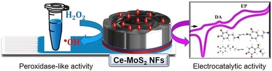
Highly Conductive Peroxidase-like Ce-MoS2 Nanoflowers for the Simultaneous Electrochemical Detection of Dopamine and Epinephrine

Department of BioNano Technology, Gachon University, 1342 Seongnamdae-ro, Sujeong-gu, Seongnam 13120, Gyeonggi, Republic of Korea
*
Author to whom correspondence should be addressed.
Biosensors 2023, 13(12), 1015; https://doi.org/10.3390/bios13121015
Submission received: 5 November 2023 / Revised: 2 December 2023 / Accepted: 4 December 2023 / Published: 6 December 2023

Abstract

:
The accurate and simultaneous detection of neurotransmitters, such as dopamine (DA) and epinephrine (EP), is of paramount importance in clinical diagnostic fields. Herein, we developed cerium–molybdenum disulfide nanoflowers (Ce-MoS2 NFs) using a simple one-pot hydrothermal method and demonstrated that they are highly conductive and exhibit significant peroxidase-mimicking activity, which was applied for the simultaneous electrochemical detection of DA and EP. Ce-MoS2 NFs showed a unique structure, comprising MoS2 NFs with divalent Ce ions. This structural design imparted a significantly enlarged surface area of 220.5 m2 g−1 with abundant active sites as well as enhanced redox properties, facilitating electron transfer and peroxidase-like catalytic action compared with bare MoS2 NFs without Ce incorporation. Based on these beneficial features, Ce-MoS2 NFs were incorporated onto a screen-printed electrode (Ce-MoS2 NFs/SPE), enabling the electrochemical detection of H2O2 based on their peroxidase-like activity. Ce-MoS2 NFs/SPE biosensors also showed distinct electrocatalytic oxidation characteristics for DA and EP, consequently yielding the highly selective, sensitive, and simultaneous detection of target DA and EP. Dynamic linear ranges for both DA and EP were determined to be 0.05~100 μM, with detection limits (S/N = 3) of 28 nM and 44 nM, respectively. This study shows the potential of hierarchically structured Ce-incorporated MoS2 NFs to enhance the detection performances of electrochemical biosensors, thus enabling extensive applications in healthcare, diagnostics, and environmental monitoring.

Graphical Abstract

1. Introduction

In recent years, electrochemical detection has witnessed remarkable advancements fueled by the development of nanomaterials with extraordinary conductivity and catalytic properties [1]. These nanomaterials, often endowed with enzyme-like activities, have opened new horizons for the highly selective and sensitive detection of analytes of interest [2]. The advantageous features of nanomaterials also made it possible to simultaneously detect multiple targets, providing more comprehensive sensing information with lowered total cost and thus, expanding usability in diagnostic areas [3,4,5,6,7]. For this, many different kinds of nanomaterials composed of noble metals, metal oxides, metal sulfides, carbon with doped elements, and their hybrids, with diverse morphologies have been reported [8]. Among these, MoS2-based nanomaterials have emerged as a promising candidate for the construction of electrochemical biosensors since they exhibit high conductivity as well as peroxidase-like activity from their synergistic elemental composition [9,10,11]. Further research is being conducted to further engineer MoS2-based nanomaterials to improve their electrical and catalytic properties.
DA and EP are representative catecholamine neurotransmitters that play pivotal roles in regulating various physiological processes in the human body, including the central nervous and cardiovascular systems [12,13,14]. Aberrations in their levels are associated with a range of neurological and neuropsychiatric disorders, including Parkinson’s disease, schizophrenia, and attention deficit hyperactivity disorder (ADHD) [15,16,17]. Therefore, the reliable, selective, sensitive, and simultaneous detection of both DA and EP has garnered substantial attention for realizing their applications in the clinical field [18,19]. Conventionally, DA and EP have been analyzed using instrumentation-based methods including high-performance liquid chromatography [15,20], capillary electrophoresis [21], and mass spectrometry [22]. Enzyme-linked immunosorbent assay, which needs a microplate reader for the quantitative analysis, has also been used to produce target-specific colorimetric responses [23]. While these methods generally provide accurate and sensitive assay results, they are often laborious, time-consuming, and expensive to perform, which hinders their widespread utilization, particularly in resource-limited environments. For a more convenient detection of DA and EP, biosensors that are based on enzymes, nanozymes, and other nanomaterials having unique physicochemical or electrocatalytic properties have been reported [24,25]. They display colorimetric, fluorescent, or electrochemical signals proportional to the amount of target substances, such as DA and EP, without any sophisticated procedure or instrumentation, and thus, can be recognized as potent methods to be practically used in point-of-care testing (POCT) environments. Among them, electrochemical detection methods can exhibit higher sensitivity than those of optical ones, with simple and rapid procedures at lower cost. Thus, they have been extensively studied for detecting DA and EP [26]. Diverse nanomaterials composed of noble metals [27], metal oxides [28], and carbon [29] have been used to construct electrodes for efficient electrochemical detection of neurotransmitter molecules based on their affirmative electrocatalytic property with large surface area. Although these examples demonstrate the potential of nanomaterials, including MoS2-based ones, to electrochemically detect DA and EP, further engineering of nanomaterials is required to achieve higher detection performance, particularly for the simultaneous detection of neurotransmitters.
In this regard, we developed Ce-MoS2 NFs as highly efficient electrocatalysts for the simultaneous detection of DA and EP. Ce-MoS2 NFs were synthesized simply with a one-pot hydrothermal method, yielding the successful incorporation of Ce3+ and Ce4+ ions on MoS2 NFs. The synthesized Ce-MoS2 NFs were demonstrated to exhibit both higher conductivity and peroxidase-like activity, which were much higher than those of bare MoS2 NFs without Ce incorporation. With the incorporation of Ce-MoS2 NFs on a screen-printed electrode (Ce-MoS2 NFs/SPE), target H2O2 and neurotransmitters including DA and EP were successfully detected. Various analytical features, including selectivity and sensitivity, were investigated.

2. Experimental Section

2.1. Materials

Thiourea, ammonium molybdate, ethanol, cerium nitrate, dopamine hydrochloride, epinephrine, 3,3′,5,5′-tetramethylbenzidine (TMB), dimethyl sulfoxide (DMSO), phosphate-buffered saline (PBS), potassium ferricyanide, and human serum were purchased from Sigma-Aldrich (St. Louis, MO, USA). Hydrogen peroxide (H2O2, 35%) was purchased from Junsei Chemical Co. (Tokyo, Japan). Deionized water, purified using a Milli-Q Purification System (Millipore, Darmstadt, Germany), was used to prepare all solutions. PBS buffers containing 0.1 M KCl (pH 6.0) were used as electrolytes by properly mixing the solutions of Na2HPO4, NaH2PO4, and KCl.

2.2. Synthesis and Characterization of MoS2 NFs and Ce-MoS2 NFs

Figure S1 illustrates the synthesis of both MoS2 NFs and Ce-MoS2 NFs using the one-pot hydrothermal method. In brief, a solution was prepared by dissolving 20 mmol of thiourea and ammonium molybdate in a 50 mL water–ethanol mixture (with a volumetric ratio of Vwater:Vethanol = 5:1). The solution was stirred for 10 min, followed by the addition of 0.5 mmol of cerium nitrate with a further reaction for 10 min with stirring. The combined solution was then put into a 100 mL autoclave with a Teflon lining, and it was treated hydrothermally for 24 h at 200 °C. Following the hydrothermal process, the product was collected with centrifugation (10,000× g, 5 min), followed by three washes in ethanol and deionized water and overnight drying at 70 °C. For comparative purposes, MoS2 NFs were synthesized using the same procedure, with the exception that cerium nitrate was not included.
An X-ray diffractometer (D/MAX-2500, Rigaku Corporation, Tokyo, Japan) was used to determine the crystal structure of the produced materials using powder X-ray diffraction (XRD). In order to investigate the nanostructured morphologies of Ce-MoS2 NFs, two types of microscopes were used: a scanning electron microscope (Magellan 400 Microscope, FEI Co., Eindhoven, The Netherlands) and a transmission electron microscope (FEI Tecnai, OR, USA). By combining an energy dispersive spectrometer (EDS; Elementar, Vario Macro, Langenselbold Germany) with a scanning electron microscope, elemental mapping was achieved. Using the Brunauer–Emmett–Teller (BET) method, a nitrogen physisorption study was carried out to measure the specific surface area, pore size, and pore volume (3Flex, Micromeritics, Norcross, GA, USA). The elemental composition and electronic states were examined using X-ray photoelectron spectroscopy (XPS) (Sigma Probe, Thermo Scientific, Fitchburg, WI, USA). To clarify the functional groups of the synthesized materials, Fourier transform infrared spectroscopy (FT-IR) was used with an FT-IR spectrophotometer (FT/IR-4600, JASCO, Easton, MD, USA).

2.3. Evaluation of the Peroxidase-like Activity of Ce-MoS2 NFs

The oxidation of TMB in the presence of H2O2 was measured to assess the peroxidase-like activity of Ce-MoS2 NFs. Ce-MoS2 NFs (100 µg mL−1) were introduced into a sodium acetate buffer (0.1 M, pH 6) containing 1 mM TMB and 2 mM H2O2 in a typical experiment. After 20 min of incubation at room temperature (RT), the supernatant was separated with centrifugation (10,000× g, 1 min). The absorbance at 652 nm, which is the wavelength at which TMB oxidizes, was measured using a microplate reader (Synergy H1, BioTek, Winooski, VT, USA), provided by the Center for Bionano Materials Research at Gachon University (Seongnam, Republic of Korea), to analyze the blue color evolution. Steady-state kinetic assays of Ce-MoS2 NFs were further conducted using TMB and H2O2 as substrates. These experiments involved systematically varying the concentrations of TMB while keeping the H2O2 concentration constant, and vice versa. The equation v = Vmax × [S]/(Km + [S]) was utilized to compute the kinetic parameters with the use of Lineweaver–Burk plots. In this equation, v represents the initial velocity, Vmax denotes the maximal reaction velocity, [S] represents the substrate concentration, and Km represents the Michaelis constant.

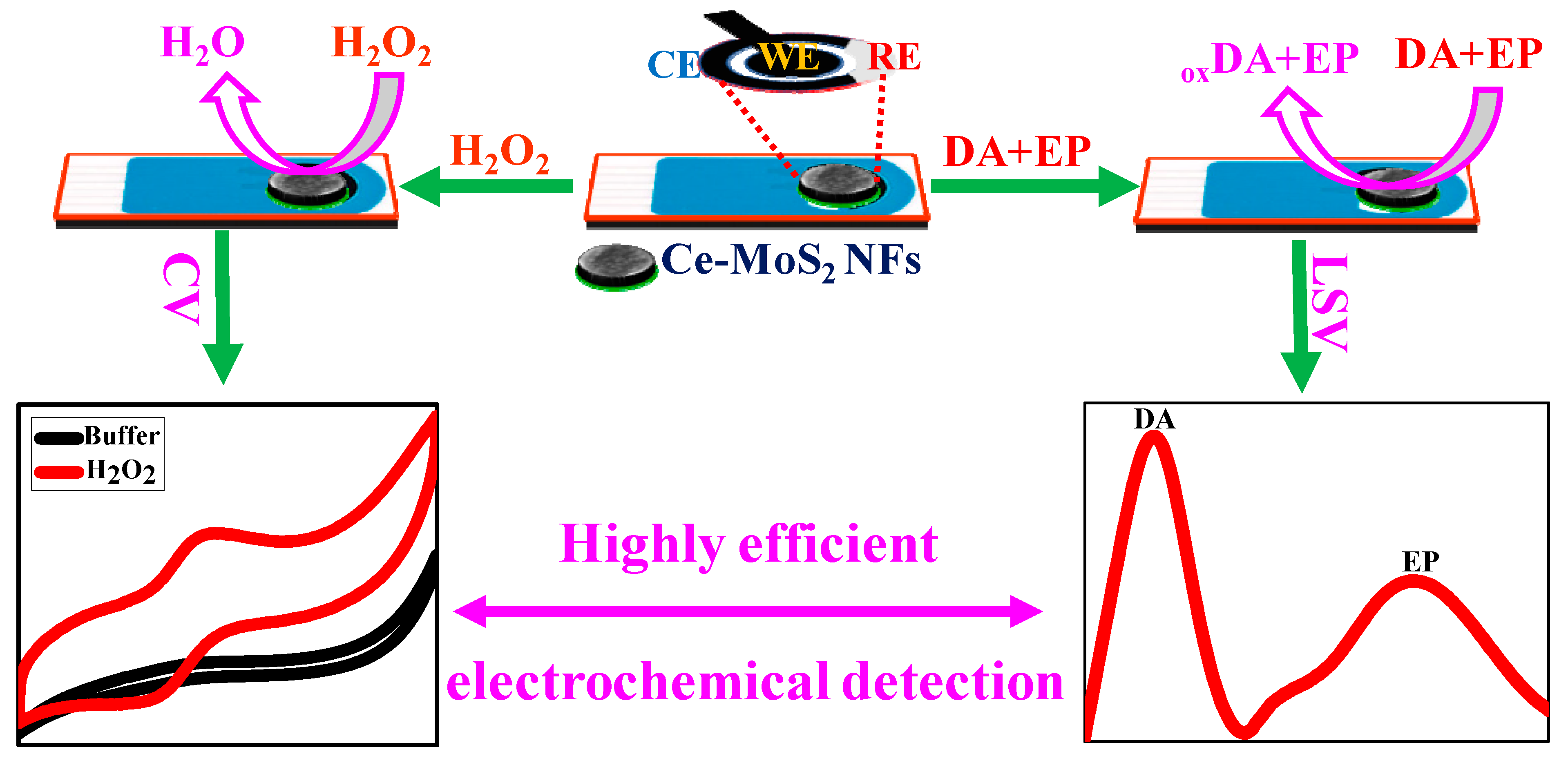
2.4. Fabrication of Ce-MoS2 NFs/SPE Biosensors and Electrochemical Analysis

A carbon-based SPE (DropSens, Metrohm, Oviedo, Spain) was obtained and customized with the developed Ce-MoS2 NFs for preparing the working electrode. In brief, a blend of 2 mg of Ce-MoS2 NFs was dispersed in 0.5 mL ethanol. The resulting dark suspension was subjected to 2 h of ultrasonication at RT to achieve a uniform slurry. Subsequently, this slurry was deposited onto the surface of the SPE to yield the Ce-MoS2 NFs/SPE biosensor.
Electrochemical analyses were conducted using a CHI600E Potentiostat (IJ Cambria Scientific Ltd., Austin, TX, USA). A conventional three-electrode electrochemical system was used, with the Ce-MoS2 NFs/SPE as the working electrode. Ag/AgCl saturated in KCl served as the reference electrode, and a platinum wire was used as the counter electrode. This study used electroanalytical techniques such as cyclic voltammetry (CV) and linear sweep voltammetry (LSV) to detect H2O2 and simultaneously detect DA and EP. Electrochemical experiments were carried out by adding 50 µL of sample solutions to the Ce-MoS2 NFs/SPE. They included H2O2 or DA and EP at various concentrations, which were dissolved in an aqueous PBS solution (0.1 M, pH 7.0) containing 0.1 M KCl electrolyte to enhance solution conductivity and prevent ion migration. Before each test, all sample solutions were freshly prepared and stored at 4 °C until the analysis to preserve the stability of DA and EP.

3. Results and Discussion

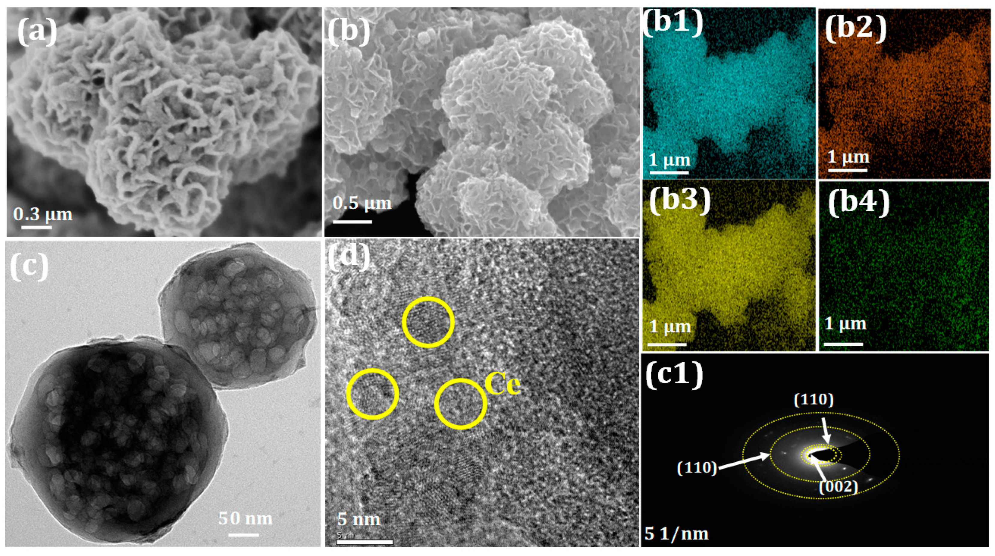
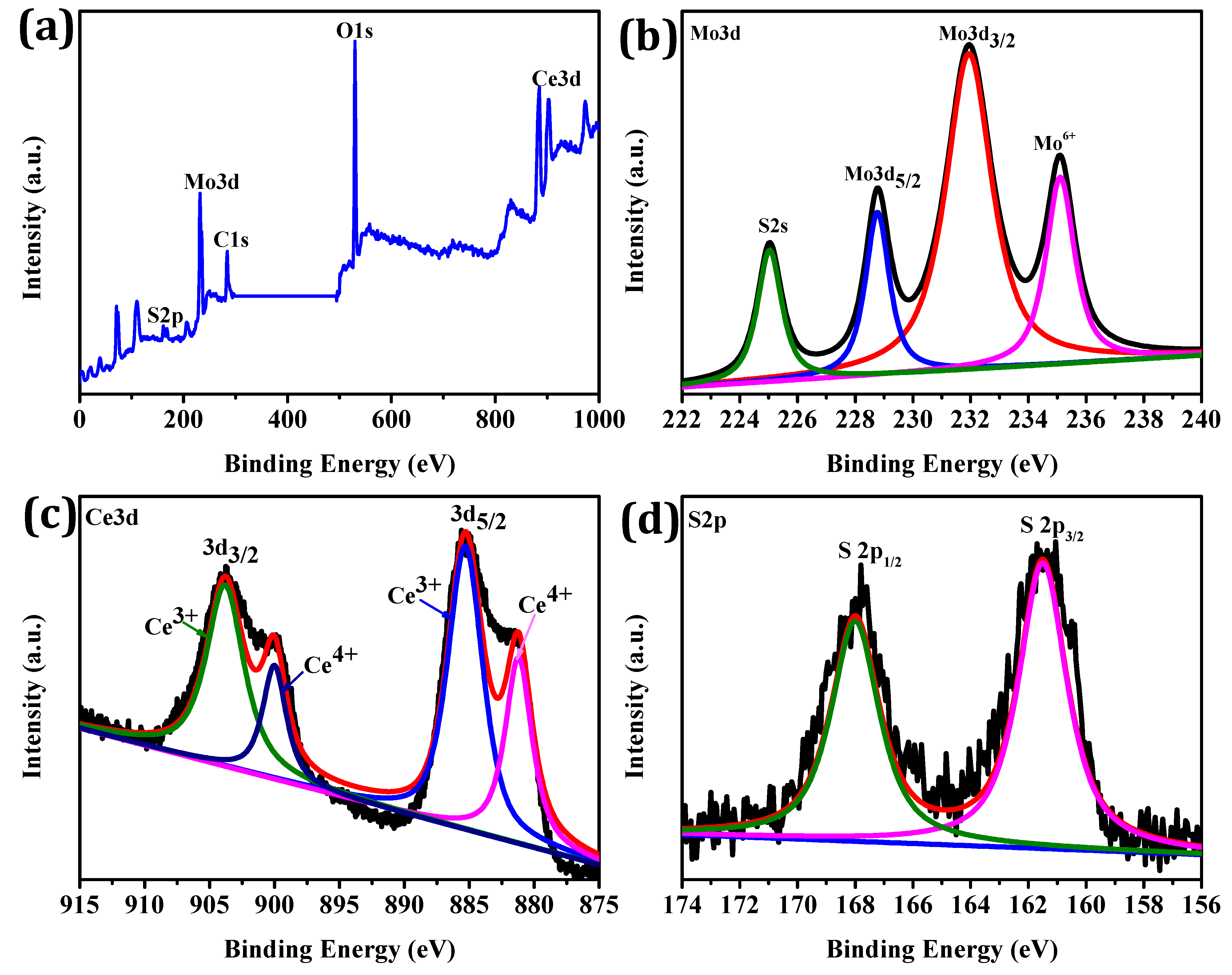
3.1. Synthesis of Highly-Conductive Peroxidase-like Ce-MoS2 NFs

MoS2 NFs were proven to exhibit peroxidase-like and conductive properties [30], and herein, we hypothesized that Ce-MoS2 NFs, simply synthesized with one-pot hydrothermal treatment, might have higher peroxidase-like activity as well as conductivity due to the affirmative effects of Ce ions with enlarged surface area. We then incorporated Ce-MoS2 NFs onto the SPE to yield the Ce-MoS2 NFs/SPE biosensor, which successfully detected H2O2 as well as both DA and EP simultaneously, based on their high peroxidase-like activity and electrocatalytic activity, respectively. In proportion to the amount of DA and EP, characteristic oxidation peak intensity was selectively and sensitively detected, enabling the simultaneous quantification of both DA and EP (Scheme 1).
The structural characteristics of Ce-MoS2 NFs were analyzed and compared with those of bare MoS2 NFs without Ce incorporation. The XRD patterns of both bare MoS2 NFs and Ce-MoS2 NFs reveal the formation of hexagonal crystal phases characterized by the JCPDS card number 24-0513 (Figure 1a) [31]. The XRD patterns of bare MoS2 NFs precisely match the standard diffraction peaks at the (002), (100), and (110) planes. With the incorporation of Ce, the characteristic peaks related to Ce were clearly observed, such as the (110), (220), (311), (222), and (400) planes, aligning well with the JCPDS card number 81-0792 and indicating pure cubic fluorite CeO2 [32]. The presence of the highest valences in Ce-MoS2 NFs was attributed to intramolecular electron transfer occurring during synthesis [33]. Notably, the peak intensity at (002) decreases with the incorporation of Ce. This observation shows the successful incorporation of Ce into the MoS2 NFs, resulting in Ce-MoS2 NFs with a marginally modified crystal structure. The nitrogen physisorption analysis proves that the Ce-MoS2 NFs have a remarkably enlarged surface area of 220.5 m2 g−1, as determined using the BET method, which is notably larger than that of the bare MoS2 NFs (132.22 m2 g−1) (Figure 1b). The enlarged surface area of Ce-MoS2 NFs might play a pivotal role in enhancing their electrochemical detection performance since increasing surface area at the electrode–electrolyte interface provides a greater number of reaction sites and promotes faster diffusion kinetics. In addition, FT-IR provides clear evidence for the functional groups present in Ce-MoS2 NFs. The peak observed at 620 cm−1 is attributed to the stretching vibration of Mo-S. Multiple peaks spanning the range of 700–1150 cm−1 are indicative of sulfate groups, and the peaks at 3440 and 1610 cm−1 correspond to the stretching of O-H bonds and the bending of water (Figure S2).
MoS2-based nanoparticles synthesized using a hydrothermal method normally tend to form sheets due to their inherent lamellar structures. These sheets subsequently aggregate, driven by van der Waals interactions, and ultimately self-assemble into three-dimensional nanostructures up to NFs. In our study, the flower-like morphology of MoS2 NFs was clearly observed, with diameters in ranges of 0.5 to 2 μm (Figure 2a). With the addition of cerium nitrate during the hydrothermal treatment, the flower-like morphology was still detected, and moreover, it was observed that Ce elements were uniformly distributed through the entire surface (Figure 2b). From the high-resolution TEM images, Ce ions were observed on the Ce-MoS2 NFs (Figure 2c,d) while showing clear diffraction rings attributed to (110), (002), and (100), all of which were also detected with the XRD analyses.
The XPS analysis of Ce-MoS2 NFs was further performed to confirm their elemental composition and electronic states. The full survey shows that the synthesized Ce-MoS2 NFs are composed of Ce, Mo, S, C, and O, which is in agreement with the EDS mapping (Figure 3a). The atomic percentages of Ce and Mo in Ce-MoS2 NFs were determined to be ~22% and ~30%, respectively, indicating the abundance of active sites (Figure 3a). Mo 3d shows a weak satellite peak (2 s) of S species at 225.3 eV and two peaks of Mo 3d3/2 (232.15 eV) and Mo 3d5/2 (228.77 eV), which can be ascribed to Mo4+ (Figure 3b). The other Mo 3d3/2 peak observed at 235.32 eV corresponds to Mo6+, indicating that a certain degree of oxidation occurs due to the formation of MoO3. The two main characteristic peaks of Mo 3d3/2 and Mo 3d5/2 are near the meta-stable T phase of MoS2, which can improve the conductivity and electrocatalytic activity. The Ce 3d spectra (Figure 3c) display four distinctive peaks. The two prominent peaks correspond to Ce3+ 3d5/2, while the other two peaks are attributed to Ce4+ 3d3/2, indicating the presence of both Ce3+ and Ce4+ in the Ce-MoS2 NFs. The existence of Ce4+ can be linked to the oxidation on the surface from Ce3+ to Ce4+, due to inevitable exposure to air and moisture. Furthermore, the peaks at 162.3 eV and 163.5 eV in the S 2p spectra (Figure 3d) can be identified as the S 2p3/2 and S 2p1/2 orbitals of S2−, respectively. These characterizations support the successful incorporation of Ce ions onto the MoS2 NFs, with the multivalent electronic states helping to enhance the conductivity and electrocatalytic activity.

3.2. Enhanced Peroxidase-like Activity of Ce-MoS2 NFs

The peroxidase-like activities of Ce-MoS2 NFs and bare MoS2 NFs were evaluated based on the oxidation of TMB in the presence of H2O2, and the responses were monitored using absorption spectroscopy to analyze the corresponding blue color intensities. The results clearly show that the Ce-MoS2 NFs have a significant peroxidase-like activity, which is ~2-fold higher than that of the bare MoS2 NFs (Figure S3a,b). According to the investigations of the effects of pH on the peroxidase-like activity, pH 6 yielded the best results and was used for further studies at RT for practical convenience (Figure S3c).
To elucidate the peroxidase-like catalytic mechanism of Ce-MoS2 NFs, steady-state kinetic assays were performed using TMB and H2O2 as substrates, and the resulting kinetic parameters were compared with those of HRP and previously reported peroxidase-like nanozymes including Fe3O4 nanoparticles and N-MoS2 NFs (Table 1, Figure S4). The apparent Km values of the Ce-MoS2 NFs for TMB and H2O2 were 0.25 and 0.52 mM, respectively, both of which are lower than those for HRP. In particular, the Km value of H2O2 was only ~14% of that of HRP and over two orders lower than that of Fe3O4 nanoparticles. This result indicates that the developed Ce-MoS2 NFs exhibit an excellent level of affinity toward the substrates. The enhanced substrate affinity and peroxidase-like activity of Ce-MoS2 NFs can be primarily attributed to the creation of additional sulfur vacancies with the incorporation of Ce elements, making higher numbers of reaction sites throughout the NFs matrices.

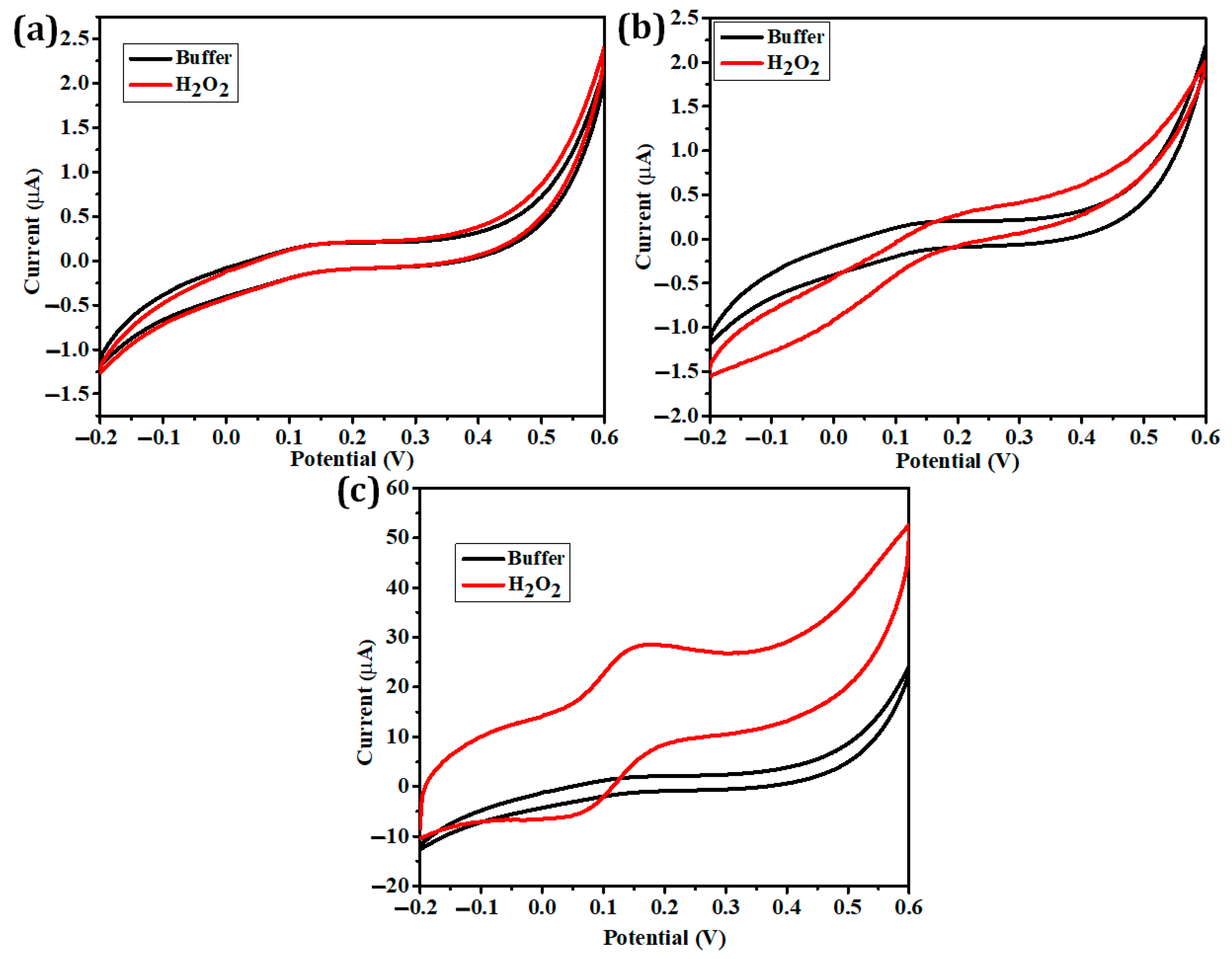
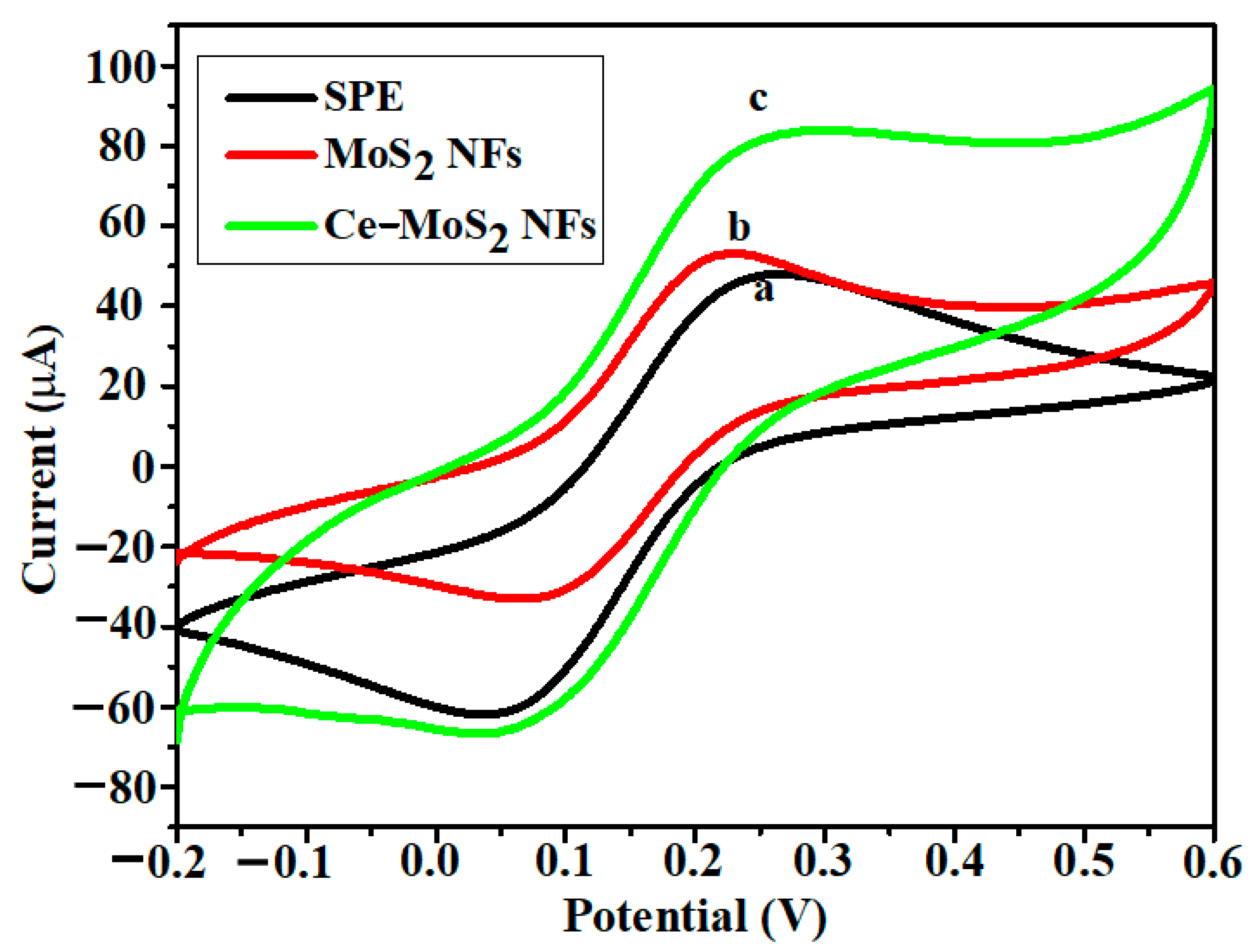
3.3. Enhanced Electrocatalytic Performance of Ce-MoS2 NFs

Based on the enhanced peroxidase-like activity of Ce-MoS2 NFs, the detection of H2O2 based on its electrocatalytic reduction was performed. To explore this applicability, the detection performances of the bare SPE, the bare MoS2 NFs/SPE, and the Ce-MoS2/SPE were assessed and compared in the absence and presence of H2O2 (0.2 mM) (Figure 4). The results vividly show that the Ce-MoS2/SPE biosensor yielded a notably enhanced reduction peak with the addition of H2O2, while the bare SPE and the bare MoS2 NFs/SPE showed a negligible and a significantly low peak, respectively (Figure 4), signifying the excellent electrocatalytic performance of Ce-MoS2 NFs toward H2O2. The difference in peak current (ΔI = IH2O2 − Iwithout H2O2) at 0.12 V was also assessed for the above three electrodes. The ΔI for the Ce-MoS2/SPE reached 28.35 μA, marking over a 10-fold increase compared with that of MoS2/GCE. Additionally, we investigated the electrochemical performance of the Ce-MoS2/SPE biosensor in detecting 0.2 mM H2O2 by varying the scan rate from 10 to 100 mV s−1 (Figure S5). The investigation indicated that the electrocatalytic reaction followed a diffusion-controlled process, as evidenced by the relationship between peak current and scan rates.
Cyclic voltammetry (CV) was conducted on the bare SPE, the bare MoS2 NFs/SPE, and the Ce-MoS2/SPE in PBS solutions containing 0.1 M KCl and 5.0 mM [Fe(CN)6]3−/4− as a redox probe. The sweep rate was set at 50 mV s−1, with scanning from −0.2 to 0.6 V. As shown in Figure 5, we observed a typical reversible redox peak for the bare SPE at 0.09 V and 0.21 V, with a peak current of 40 µA (curve a). Upon the incorporation of bare MoS2 NFs onto the SPE, the peak current increased to 55 µA, accompanied by an augmented ΔEp of 180 mV (curve b). Conversely, when Ce-MoS2 NFs were introduced onto the SPE, the peak current consistently rose to 95 µA, accompanied by a reduction in ΔEp by 130 mV (curve c). This significant increase in peak current in the Ce-MoS2 NFs/SPE configuration confirms the pivotal roles played by both MoS2 NFs and Ce in augmenting electrocatalytic activity and the detection current response. Moreover, Ce-MoS2 NFs significantly affect the material’s electrical conductivity and porosity, thereby improving its wettability, which might induce faster electron transfer at the electrode surface. The effective surface area of the Ce-MoS2 NF electrode can be estimated using the Randles–Sevcik equation for a reversible process observed during CV using K3[Fe(CN)6] as the oxidation–reduction probe.
Ipa = (2.69 × 10⁵) n3/2D1/2 v1/2 AC
Here, the effective surface area (A) is calculated using the Randles–Sevcik equation, where C represents the bulk concentration of K3[Fe(CN)6], n is the number of electrons transferred (n = 1), D is the diffusion coefficient of K3[Fe(CN)6] (measuring 7.6 × 10⁻⁶ cm2 s−1), and ν is the sweep rate. In this study, the effective surface area of Ce-MoS2 NFs was determined to be 0.83 cm2. Notably, this represents a substantial increase compared with the bare SPE, which has an area of 0.07 cm2.

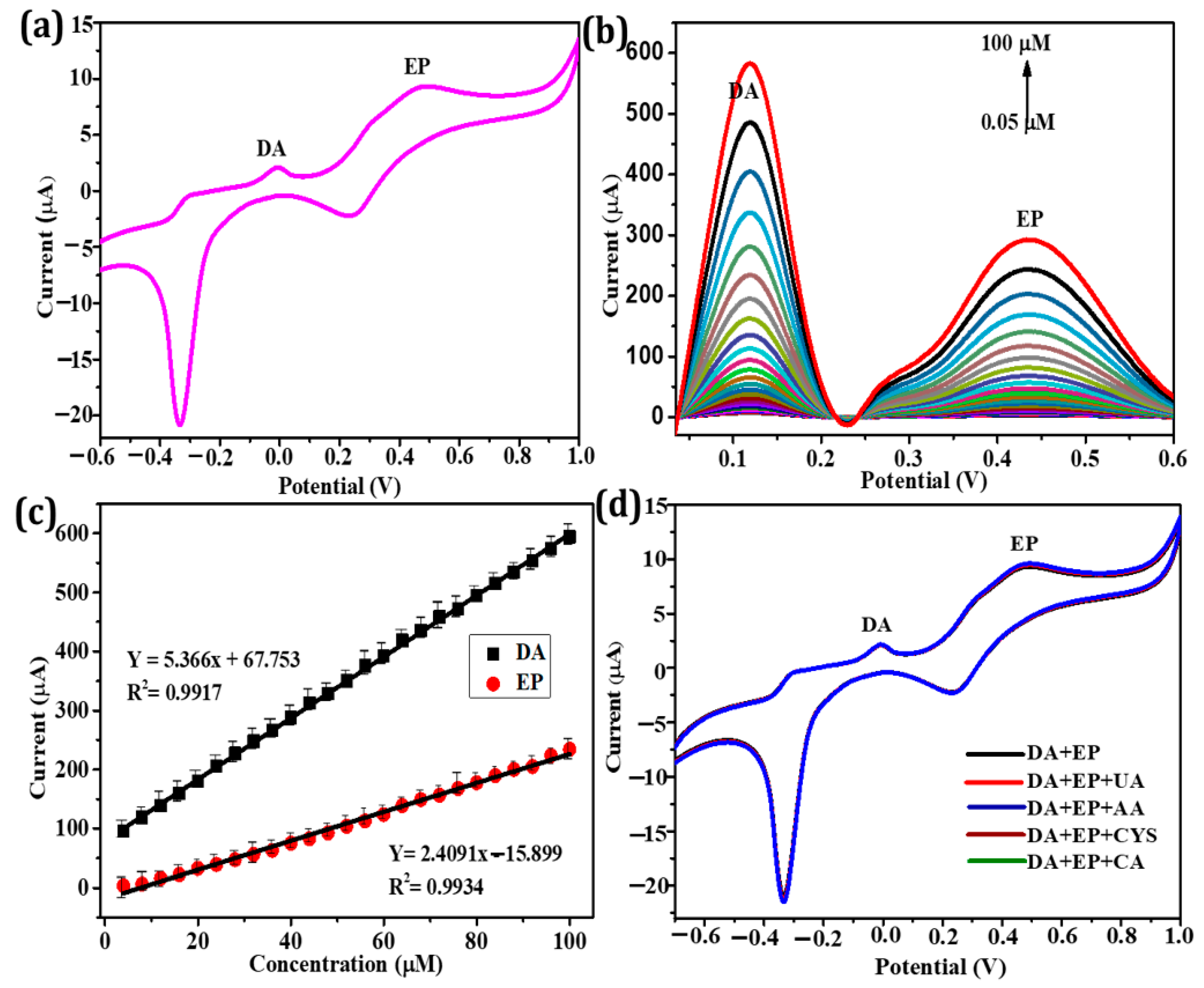
3.4. Simultaneous Electrochemical Detection of DA and EP Using the Ce-MoS2 NFs/SPE Biosensor

For demonstrating the simultaneous detection capability for DA and EP using the Ce-MoS2 NFs/SPE biosensor, the CV of the fabricated electrodes was conducted using a sample solution containing both DA and EP. When both neurotransmitters were present together in 0.1 M KCl (pH 7.0), distinct redox peaks were observed, with a significant increase in the oxidation and reduction currents (Figure 6a). These peaks corresponded to the epinephrine–epinephrine quinone and dopamine–dopamine quinone redox couples, appearing at potentials of −0.005, 0.30, −2.28, and 0.24 V, respectively (Figure S6). These results indicate that the Ce-MoS2 NFs electrode exhibits the capability for the simultaneous detection of both DA and EP. In addition, individual DA or EP could be detected by producing distinct redox peaks during CV (Figure S7).
To confirm the practical applicability of the Ce-MoS2 NFs/SPE biosensor for the simultaneous electrochemical detection of DA and EP using reversible processes, the LSV technique was used as the electroanalytical tool. The LSV experimental results with varying concentrations of DA and EP (ranging from 0.05 to 100 µM) are presented in Figure 6b. An evident increase in the oxidation currents corresponding to DA and EP was observed as their concentrations increased. This rise in the respective current can be attributed to the electrochemical behaviors of Ce-MoS2 NFs regarding the oxidation of DA and EP. The calibration plots revealed a linear relationship between the oxidation peak current and the concentrations of DA and EP within the range of 0.05 to 100 µM. The linear regression equations, I (µA) = 5.37 (DA) (µA µM−1) + 67.75 and I (µA) = 2.41 (EP) (µA µM−1) + 15.90, along with regression coefficients of over 0.99 (Figure 6c), confirm the clear linear relationship and high detection sensitivity. Calculated based on the signal-to-noise ratio (S/N = 3), the limit of detection (LOD) values for DA and EP on the Ce-MoS2 NFs/SPE biosensors were determined to be ~28 and 44 nM, respectively. When compared with recently reported DA and EP sensors, it becomes evident that the Ce-MoS2 NFs/SPE biosensors exhibited the highest level of sensitivity in the simultaneous detection of DA and EP (Table S1) [3,13,35,36,37,38,39,40,41]. The developed biosensors also showed high selectivity toward target DA and EP, while possible interfering molecules did not yield any considerable signal (Figure 6d). These investigations clearly demonstrate the significant potential of Ce-MoS2 NFs to construct efficient electrodes and biosensors for the simultaneous detection of both DA and EP, with sufficiently high selectivity and sensitivity.
Reusability and storage stability are important characteristics in POCT aspects. As displayed in Figure S8, the developed biosensor efficiently retained its original detection capability for both DA and EP, during a 10-time iterative utilization and 7-day storage at RT, further supporting its practical applicability (Figure S8). To further corroborate the practical efficacy of the biosensor, it was applied to detect DA and EP in spiked human serum samples (Table S2). The measured concentrations of DA and EP were in good agreement with the spiked levels, yielding recovery rates ranging from 99.4% to 102.6%. These results indicate that the developed electrochemical biosensors could potentially be used as a reliable device for the detection of DA and EP in human serum samples.

4. Conclusions

We developed highly conductive and peroxidase-like Ce-MoS2 NFs based on the synergistic incorporation of Ce onto MoS2 NFs, yielding enlarged surface area and enhanced redox electrocatalytic activity. The Ce-MoS2 NFs followed typical Michaelis–Menten kinetics and their Km values for TMB and H2O2 were much lower than those of HRP. By incorporating the Ce-MoS2 NFs onto the SPE (Ce-MoS2 NFs/SPE), H2O2 or both DA and EP were conveniently detected by just dropping sample solutions on the biosensors. The Ce-MoS2 NFs/SPE enabled the successful simultaneous quantification of both DA and EP with excellent selectivity and sensitivity. Moreover, the developed biosensor exhibited excellent storage stability and reusability, as well as detection accuracy when spiked human serum samples were used. This study serves as a basis for the continued efforts toward the development of nanocomposite-incorporated electrochemical devices, which have significant potential in biosensing areas, particularly in POCT environments.

Supplementary Materials

The following supporting information can be downloaded at: https://www.mdpi.com/article/10.3390/bios13121015/s1, Table S1: Comparison of the electroanalytical sensitivity of the Ce-MoS2 NFs/SPE biosensor with those of recent DA and EP sensors; Figure S1: Schematic illustration of the synthesis of bare MoS2 NFs and Ce-MoS2 NFs; Figure S2: FT-IR spectra of the bare MoS2 NFs and Ce-MoS2 NFs; Figure S3: Evaluation of the peroxidase-like activity of (a) Ce-MoS2 NFs, (b) comparison of the activities of Ce-MoS2 NFs and the bare MoS2 NFs, and (c) effects of the reaction pH on catalytic activity; Figure S4: Michaelis–Menten curves of Ce-MoS2 NFs for (a) TMB and (b) H2O2 and (c,d) their corresponding Lineweaver–Burk plots (n = 3); Figure S5: CVs of the Ce-MoS2 NFs/SPE in 0.1 M PBS (pH 6.0) containing 0.2 mM H2O2 at different scan rates from 10 to 100 mV s−1 and their calibration plots; Figure S6: Reversible oxidation-reduction mechanism of DA and EP; Figure S7: Cyclic voltammograms of the Ce-MoS2 NFs/SPE at a scan rate of 100 mV s−1 for (a) 0.05 µM DA and (b) 0.05 µM EP; Figure S8: (a) Reusability and (b) storage stability of the Ce-MoS2 NFs/SPE biosensor. Excessive washing with 0.1 M PBS solution was used to evaluate reusability, and the electrode was kept in 0.1 M PBS solution at RT to evaluate storage stability; Table S2: Quantitative analysis of DA and EP spiked in human serum samples using the Ce-MoS2 NFs/SPE biosensor (n = 3).

Author Contributions

Conceptualization, methodology, investigation, and writing—original draft, A.T.; investigation and validation, T.V.D.; conceptualization, supervision, and writing—review and editing, M.I.K. All authors have read and agreed to the published version of the manuscript.

Funding

This work was supported by a National Research Foundation of Korea (NRF) grant funded by the Korean government (Ministry of Science and ICT (NRF-2023R1A2C2007833) and the Basic Science Research Program through the NRF funded by the Ministry of Education (Grant No. 2021R1A6A1A03038996). This research was also supported by Korea Basic Science Institute (KBSI) research (grant number: C280300).

Institutional Review Board Statement

Not applicable.

Informed Consent Statement

Not applicable.

Data Availability Statement

Data are contained within the article.

Conflicts of Interest

The authors declare no conflict of interest.

References

  1. Fatma, S.; Prasad, B.B.; Jaiswal, S.; Singh, R.; Singh, K. Electrochemical simultaneous analysis of dopamine and epinephrine using double imprinted one monomer acryloylated graphene oxide-carbon black composite polymer. Biosens. Bioelectron. 2019, 135, 36–44. [Google Scholar] [CrossRef] [PubMed]
  2. Chen, J.; Xu, F.; Zhang, Q.; Li, S. N-doped MoS2-nanoflowers as peroxidase-like nanozymes for total antioxidant capacity assay. Anal. Chim. Acta 2021, 1180, 338740. [Google Scholar] [CrossRef]
  3. de Fatima Ulbrich, K.; Winiarski, J.P.; Jost, C.L.; Maduro de Campos, C.E. Mechanochemical synthesis of a Ni3−XTe2 nanocrystalline composite and its application for simultaneous electrochemical detection of dopamine and adrenaline. Compos. B Eng. 2020, 183, 107649. [Google Scholar] [CrossRef]
  4. Banerjee, S.; McCracken, S.; Faruk Hossain, M.; Slaughter, G. Electrochemical detection of neurotransmitters. Biosensors 2020, 10, 101. [Google Scholar] [CrossRef] [PubMed]
  5. Sajid, M.; Nazal, M.K.; Mansha, M.; Alsharaa, A.; Jillani, S.M.S.; Basheer, C. Chemically modified electrodes for electrochemical detection of dopamine in the presence of uric acid and ascorbic acid: A review. TrAC-Trends Anal. Chem. 2016, 76, 15–29. [Google Scholar] [CrossRef]
  6. Madhurantakam, S.; Karnam, J.B.; Brabazon, D.; Takai, M.; Ahad, I.U.; Balaguru Rayappan, J.B.; Krishnan, U.M. “nano”: An emerging avenue in electrochemical detection of neurotransmitters. ACS Chem. Neurosci. 2020, 11, 4024–4047. [Google Scholar] [CrossRef] [PubMed]
  7. Moon, J.M.; Thapliyal, N.; Hussain, K.K.; Goyal, R.N.; Shim, Y.B. Conducting polymer-based electrochemical biosensors for neurotransmitters: A review. Biosens. Bioelectron. 2018, 102, 540–552. [Google Scholar] [CrossRef]
  8. Qian, L.; Durairaj, S.; Prins, S.; Chen, A. Nanomaterial-based electrochemical sensors and biosensors for the detection of pharmaceutical compounds. Biosens. Bioelectron. 2021, 175, 112836. [Google Scholar] [CrossRef]
  9. Wang, W.; Zeng, X.; Warner, J.H.; Guo, Z.; Hu, Y.; Zeng, Y.; Lu, J.; Jin, W.; Wang, S.; Lu, J.; et al. Photoresponse-bias modulation of a high-performance MoS2 photodetector with a unique vertically stacked 2H-MoS2/1T@2H-MoS2 structure. ACS Appl. Mater. Interfaces 2020, 12, 33325–33335. [Google Scholar] [CrossRef]
  10. Yang, L.; Loh, L.; Nandakumar, D.K.; Lu, W.; Gao, M.; Wee, X.L.C.; Zeng, K.; Bosman, M.; Tan, S.C. Sustainable fuel production from ambient moisture via ferroelectrically driven MoS2 nanosheets. Adv. Mater. 2020, 32, 2000971. [Google Scholar] [CrossRef]
  11. Sang, Y.; Li, W.; Liu, H.; Zhang, L.; Wang, H.; Liu, Z.; Ren, J.; Qu, X. Construction of nanozyme-hydrogel for enhanced capture and elimination of bacteria. Adv. Funct. Mater. 2019, 29, 1900518. [Google Scholar] [CrossRef]
  12. He, C.; Tao, M.; Zhang, C.; He, Y.; Xu, W.; Liu, Y.; Zhu, W. Microelectrode-based electrochemical sensing technology for in vivo detection of dopamine: Recent developments and future prospects. Crit. Rev. Anal. Chem. 2022, 52, 544–554. [Google Scholar] [CrossRef] [PubMed]
  13. da Silva, L.V.; dos Santos, N.D.; de Almeida, A.K.A.; dos Santos, D.D.E.R.; Santos, A.C.F.; França, M.C.; Lima, D.J.P.; Lima, P.R.; Goulart, M.O.F. A new electrochemical sensor based on oxidized capsaicin/multi-walled carbon nanotubes/glassy carbon electrode for the quantification of dopamine, epinephrine, and xanthurenic, ascorbic and uric acids. J. Electroanal. Chem. 2021, 881, 114919. [Google Scholar] [CrossRef]
  14. Kiranmai, S.; Kuchi, C.; Sravani, B.; Ƚuczak, T.; Kim, M.J.; Madhavi, G.; Veera Manohara Reddy, Y. Construction of ultrasensitive electrochemical sensor using TiO2-reduced graphene oxide nanofibers nanocomposite for epinephrine detection. Surf. Interfaces 2022, 35, 102455. [Google Scholar] [CrossRef]
  15. Zhang, Y.; Ren, W.; Fan, Y.Z.; Luo, H.Q.; Li, N.B. Chemically-modulated turn-on fluorescence for rapid and visual discrimination of norepinephrine and epinephrine and its application for dopamine-β-hydroxylase detection. Sens. Actuators B Chem. 2020, 305, 127463. [Google Scholar] [CrossRef]
  16. Cubells, J.F.; Sun, X.; Li, W.; Bonsall, R.W.; McGrath, J.A.; Avramopoulos, D.; Lasseter, V.K.; Wolyniec, P.S.; Tang, Y.L.; Mercer, K.; et al. Linkage analysis of plasma dopamine β-hydroxylase activity in families of patients with schizophrenia. Hum. Genet. 2011, 130, 635–643. [Google Scholar] [CrossRef] [PubMed]
  17. Das, D.; Dutta, R.K. Ethylene glycol and alanine anhydride based nitrogen doped fluorescent carbon nanoparticles as probe for detection of epinephrine, nor-epinephrine and dopamine. Dyes Pigm. 2022, 203, 110314. [Google Scholar] [CrossRef]
  18. Bergmann, M.L.; Sadjadi, S.; Schmedes, A. Analysis of catecholamines in urine by unique LC/MS suitable ion-pairing chromatography. J. Chromatogr. B Anal. Technol. Biomed. Life Sci. 2017, 1057, 118–123. [Google Scholar] [CrossRef]
  19. Takahashi, R.; Ishii, K.; Sousa, K.; Marumoto, K.; Kashibayashi, T.; Fujita, J.; Yokoyama, K. Distinctive regional asymmetry in dopaminergic and serotoninergic dysfunction in degenerative Parkinsonisms. J. Neurol. Sci. 2021, 423, 117363. [Google Scholar] [CrossRef]
  20. Costas-Ferreira, C.; Silva, A.C.d.J.; Hage-Melim, L.I.d.S.; Faro, L.R.F. Role of voltage-dependent calcium channels on the striatal in vivo dopamine release induced by the organophosphorus pesticide glyphosate. Environ. Toxicol. Pharmacol. 2023, 104, 104285. [Google Scholar] [CrossRef]
  21. Roychoudhury, A.; Francis, K.A.; Patel, J.; Jha, S.K.; Basu, S. A decoupler-free simple paper microchip capillary electrophoresis device for simultaneous detection of dopamine, epinephrine and serotonin. RSC Adv. 2020, 10, 25487–25495. [Google Scholar] [CrossRef] [PubMed]
  22. Luo, W.; van Beek, T.A.; Chen, B.; Zuilhof, H.; Salentijn, G.I. Boronate affinity paper spray mass spectrometry for determination of elevated levels of catecholamines in urine. Anal. Chim. Acta 2022, 1235, 340508. [Google Scholar] [CrossRef] [PubMed]
  23. Chau, P.B.K.; Dang, T.V.; Kim, M.I. Highly crystalline oxidase-like MnOOH nanowire-incorporated paper dipstick for one-step colorimetric detection of dopamine. Chemosensors 2023, 11, 11070382. [Google Scholar] [CrossRef]
  24. Li, Z.; Liang, L.; Lin, W.; Huang, Y.; Huang, T.; Wang, W.; Ma, J.; Li, J.; Sun, L.P.; Guan, B.O. Optofluidic laser sensor for the detection of dopamine. Sens. Actuators B Chem. 2023, 390, 133941. [Google Scholar] [CrossRef]
  25. Desagani, D.; Ben-Yoav, H. Chemometrics meets electrochemical sensors for intelligent in vivo bioanalysis. TrAC-Trends Anal. Chem. 2023, 164, 117089. [Google Scholar] [CrossRef]
  26. Lakshmanakumar, M.; Nesakumar, N.; Kulandaisamy, A.J.; Rayappan, J.B.B. Principles and recent developments in optical and electrochemical sensing of dopamine: A comprehensive review. Measurement 2021, 183, 109873. [Google Scholar] [CrossRef]
  27. De Bortoli, L.S.; Vanoni, C.R.; Jost, C.L.; Mezalira, D.Z.; Fredel, M.C. Stable and ligand-free gold nanoparticles produced by laser ablation as efficient electrocatalysts for electrochemical sensing of dopamine. J. Electroanal. Chem. 2023, 947, 117744. [Google Scholar] [CrossRef]
  28. Thamilselvan, A.; Rajagopal, V.; Suryanarayanan, V. Highly sensitive and selective amperometric determination of BPA on carbon black/f-MWCNT composite modified GCE. J Alloys. Compd. 2019, 786, 698–706. [Google Scholar] [CrossRef]
  29. Thamilselvan, A.; Manivel, P.; Rajagopal, V.; Nesakumar, N.; Suryanarayanan, V. Improved electrocatalytic activity of Au@Fe3 O4 magnetic nanoparticles for sensitive dopamine detection. Colloids Surf. B Biointerfaces 2019, 180, 1–8. [Google Scholar] [CrossRef]
  30. Yin, W.; Yu, J.; Lv, F.; Yan, L.; Zheng, L.R.; Gu, Z.; Zhao, Y. Functionalized nano-MoS2 with peroxidase catalytic and near-infrared photothermal activities for safe and synergetic wound antibacterial applications. ACS Nano 2016, 10, 11000–11011. [Google Scholar] [CrossRef]
  31. Sabarinathan, M.; Hayakawa, Y.; Harish, S. Cerium-doped MoS2 layered nanostructures for enhanced photocatalytic activity under visible light illumination. J. Mater. Sci. 2022, 33, 13988–14000. [Google Scholar] [CrossRef]
  32. Malleshappa, J.; Nagabhushana, H.; Sharma, S.C.; Vidya, Y.S.; Anantharaju, K.S.; Prashantha, S.C.; Daruka Prasad, B.; Raja Naika, H.; Lingaraju, K.; Surendra, B.S. Leucas aspera mediated multifunctional CeO2 nanoparticles: Structural, photoluminescent, photocatalytic and antibacterial properties. Spectrochim. Acta A Mol. Biomol. Spectrosc. 2015, 149, 452–462. [Google Scholar] [CrossRef]
  33. Zhang, X.; Shao, C.; Li, X.; Miao, F.; Wang, K.; Lu, N.; Liu, Y. 3D MoS2 nanosheet/TiO2 nanofiber heterostructures with enhanced photocatalytic activity under UV irradiation. J. Alloys Compd. 2016, 686, 137–144. [Google Scholar] [CrossRef]
  34. Gao, L.; Zhuang, J.; Nie, L.; Zhang, J.; Zhang, Y.; Gu, N.; Wang, T.; Feng, J.; Yang, D.; Perrett, S.; et al. Intrinsic peroxidase-like activity of ferromagnetic nanoparticles. Nat. Nanotechnol. 2007, 2, 577–583. [Google Scholar] [CrossRef] [PubMed]
  35. Cincotto, F.H.; Canevari, T.C.; Campos, A.M.; Landers, R.; Machado, S.A.S. Simultaneous determination of epinephrine and dopamine by electrochemical reduction on the hybrid material SiO2/graphene oxide decorated with Ag nanoparticles. Analyst 2014, 139, 4634–4640. [Google Scholar] [CrossRef] [PubMed]
  36. Vinoth, V.; Natarajan, L.N.; Mangalaraja, R.V.; Valdés, H.; Anandan, S. Simultaneous electrochemical determination of dopamine and epinephrine using gold nanocrystals capped with graphene quantum dots in a silica network. Microchim. Acta 2019, 186, 681. [Google Scholar] [CrossRef]
  37. Ma, W.; Sun, D.M. Simultaneous determination of epinephrine and dopamine with poly(l-arginine) modified electrode. Chin. J. Anal. Chem. 2007, 35, 66–70. [Google Scholar] [CrossRef]
  38. Manivel, P.; Thamilselvan, A.; Rajagopal, V.; Nesakumar, N.; Suryanarayanan, V. Enhanced electrocatalytic activity of Ni-CNT nanocomposites for simultaneous determination of epinephrine and dopamine. Electroanal 2019, 31, 2387–2396. [Google Scholar] [CrossRef]
  39. Valentini, F.; Palleschi, G.; Morales, E.L.; Orlanducci, S.; Tamburri, E.; Terranova, M.L. Functionalized single-walled carbon nanotubes modified microsensors for the selective response of epinephrine in presence of ascorbic acid. Electroanal 2007, 19, 859–869. [Google Scholar] [CrossRef]
  40. Agboola, B.O.; Vilakazi, S.L.; Ozoemena, K.I. Electrochemistry at cobalt(II)tetrasulfophthalocyanine-multi-walled carbon nanotubes modified glassy carbon electrode: A sensing platform for efficient suppression of ascorbic acid in the presence of epinephrine. J. Solid State Electrochem. 2009, 13, 1367–1379. [Google Scholar] [CrossRef]
  41. Fernandes, D.M.; Costa, M.; Pereira, C.; Bachiller-Baeza, B.; Rodríguez-Ramos, I.; Guerrero-Ruiz, A.; Freire, C. Novel electrochemical sensor based on N-doped carbon nanotubes and Fe3O4 nanoparticles: Simultaneous voltammetric determination of ascorbic acid, dopamine and uric acid. J. Colloid. Interface Sci. 2014, 432, 207–213. [Google Scholar] [CrossRef] [PubMed]
Scheme 1. Schematic representation of Ce-MoS2 NF-based electrochemical detection of H2O2 or DA and EP simultaneously.
Biosensors 13 01015 sch001
Figure 1. (a) XRD pattern and (b) N2 adsorption–desorption isotherms of Ce-MoS2 NFs and bare MoS2 NFs. In (a), blue and red spectra lines indicate Ce-MoS2 NFs and bare MoS2 NFs, respectively.
Biosensors 13 01015 g001
Figure 2. SEM of (a) MoS2 NFs and (b) Ce-MoS2 NFs, (c,d) high-resolution TEM of Ce-MoS2 NFs. Elemental mappings ((b1) Mo, (b2) O, (b3) S, and (b4) Ce) and selected area electron diffraction (SAED) pattern (c1) of Ce-MoS2 NFs are shown.
Biosensors 13 01015 g002
Figure 3. XPS spectra of (a) Ce-MoS2 NFs, (b) Mo 3d, (c) Ce 3d, and (d) S 2p.
Biosensors 13 01015 g003
Figure 4. Electrochemical performances of the (a) bare SPE, (b) bare MoS2 NFs/SPE, and (c) Ce-MoS2 NFs/SPE in the presence (red curve) and absence (black curve) of 0.2 mM H2O2.
Biosensors 13 01015 g004
Figure 5. Cyclic voltammograms at a scan rate of 50 mV s−1 for 5 mM [K3Fe(CN)6]/[K4Fe(CN)6] depicted for the (a) bare SPE, (b) bare MoS2 NFs/SPE, and (c) Ce-MoS2 NFs/SPE.
Biosensors 13 01015 g005
Figure 6. (a) Cyclic voltammogram, (b) linear sweep voltammograms, (c) calibration plots of current as a function of the concentrations of DA and EP, and (d) CV responses of DA and EP (100 nM each) in the presence of 1 μM of uric acid (UA), ascorbic acid (AA), cysteine (CYS), and critic acid (CA), using the Ce-MoS2 NFs/SPE biosensor.
Biosensors 13 01015 g006
Table 1. Comparison of the kinetic parameters of peroxidase-like Ce-MoS2 NFs for TMB and H2O2 with those of HRP and peroxidase-like nanozymes.
CatalystKm (mM)Vm (10−8 M s−1)Ref.
TMBH2O2TMBH2O2
HRP0.4343.7108.71[34]
Fe3O4 nanoparticles0.0981543.449.78[34]
N-MoS2 NFs0.0653.1310.7616.06[2]
Ce-MoS2 NFs0.250.5210.517.81This work
Disclaimer/Publisher’s Note: The statements, opinions and data contained in all publications are solely those of the individual author(s) and contributor(s) and not of MDPI and/or the editor(s). MDPI and/or the editor(s) disclaim responsibility for any injury to people or property resulting from any ideas, methods, instructions or products referred to in the content.

