A Framework for Biosensors Assisted by Multiphoton Effects and Machine Learning

 ,
, 
Abstract
:
1. Introduction
2. SARS-CoV-2 Biosensors
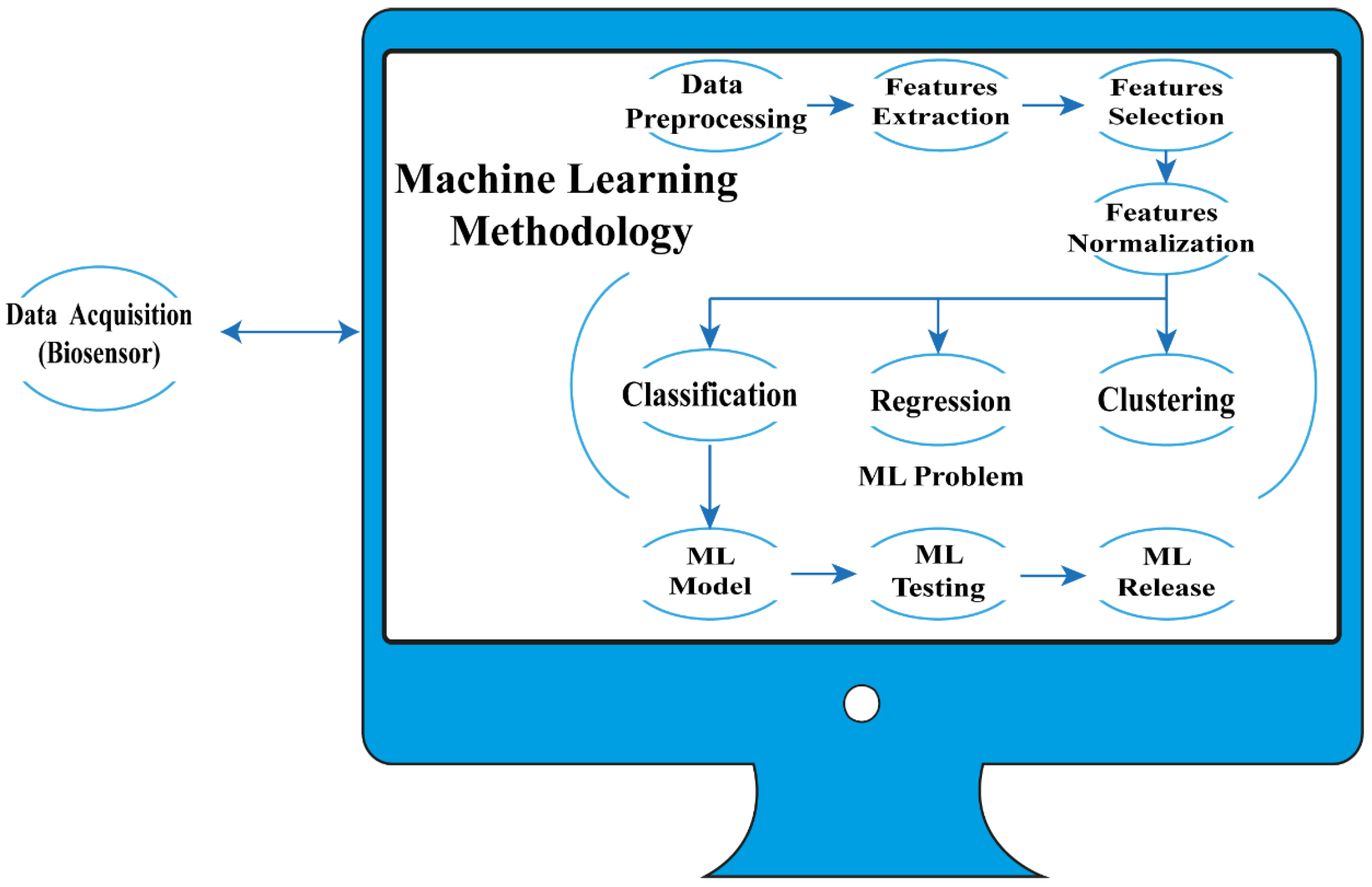
3. Biosensors Assisted by Machine Learning
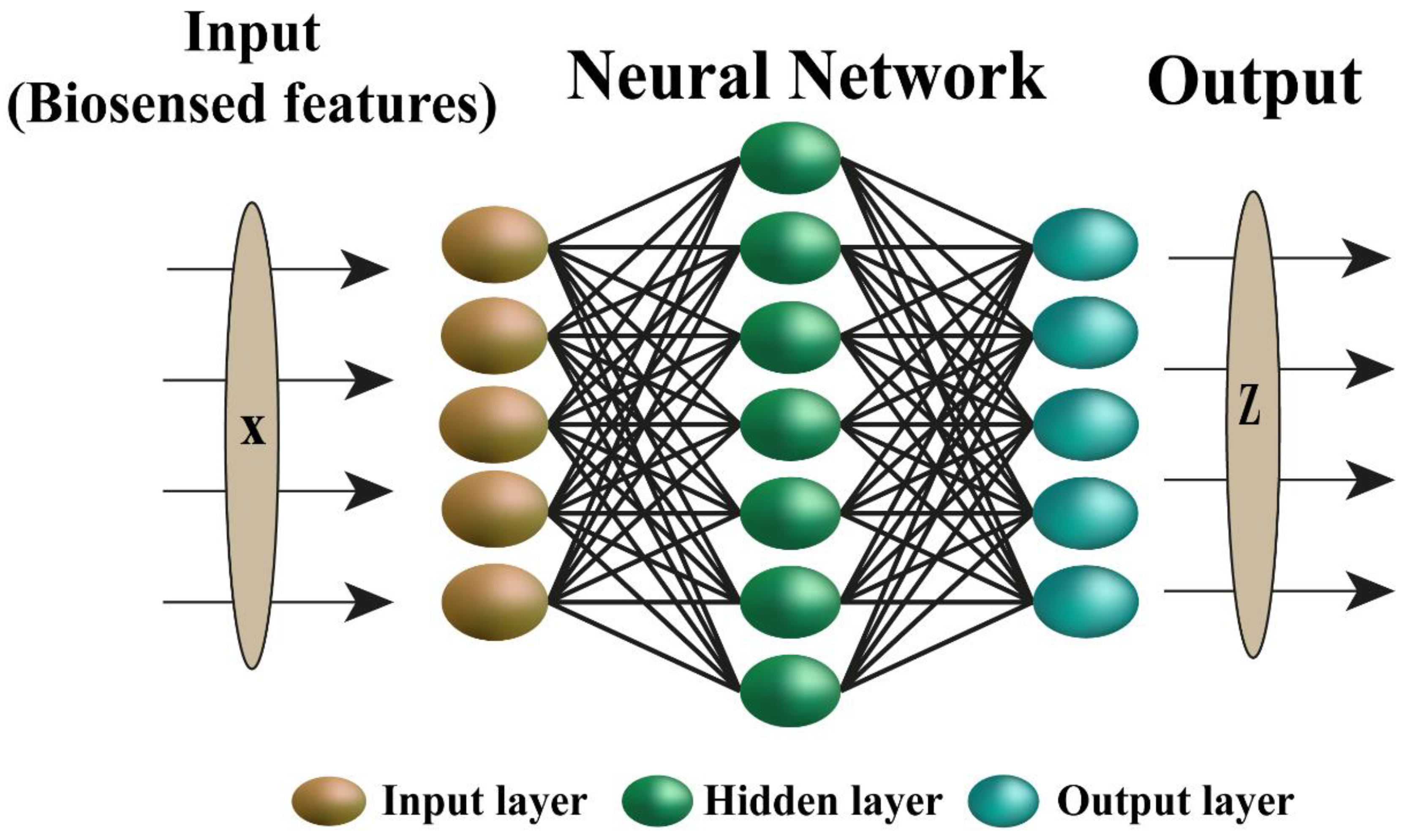
Neural Networks in Biosensors
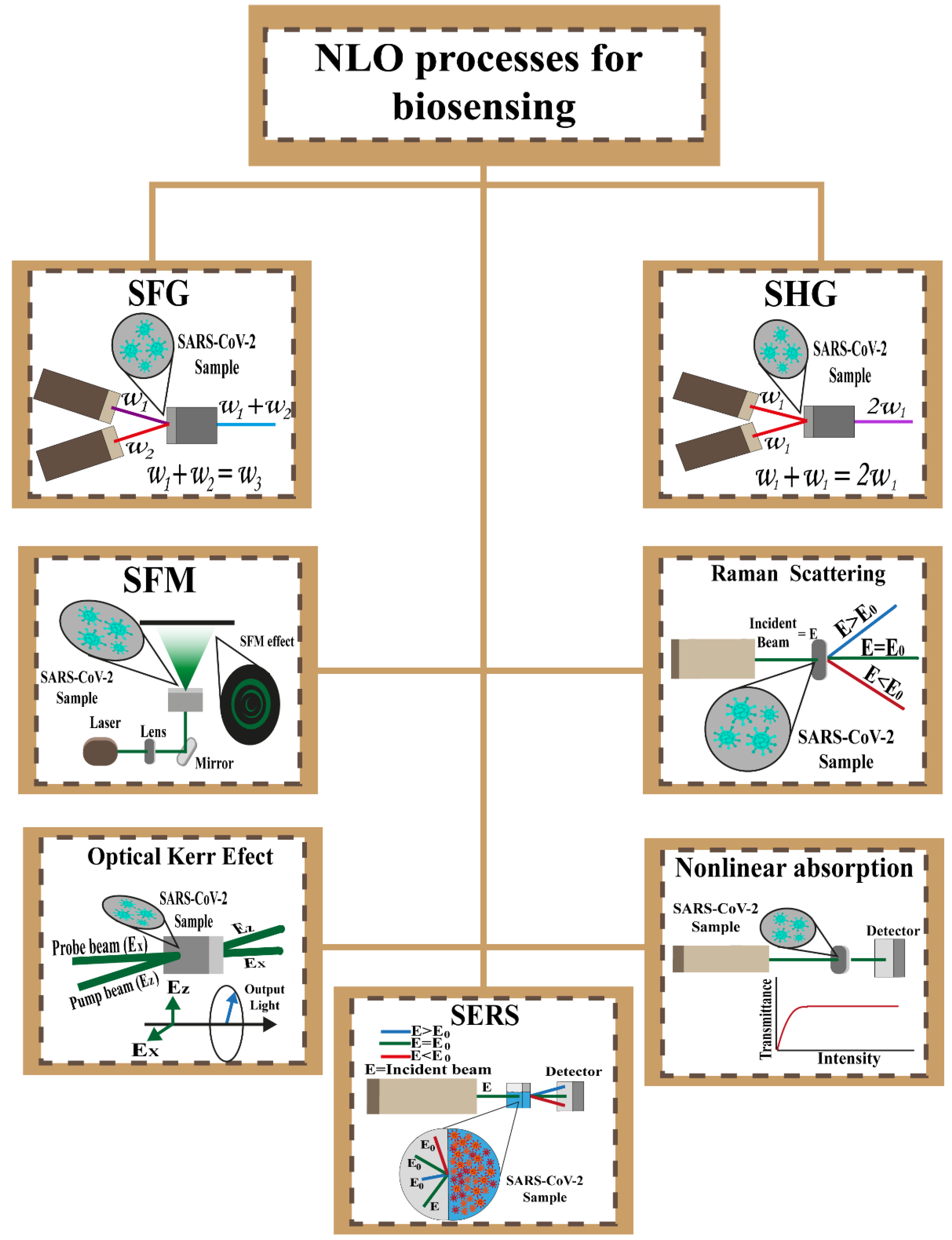
4. NLO Processes Analyzed with ML
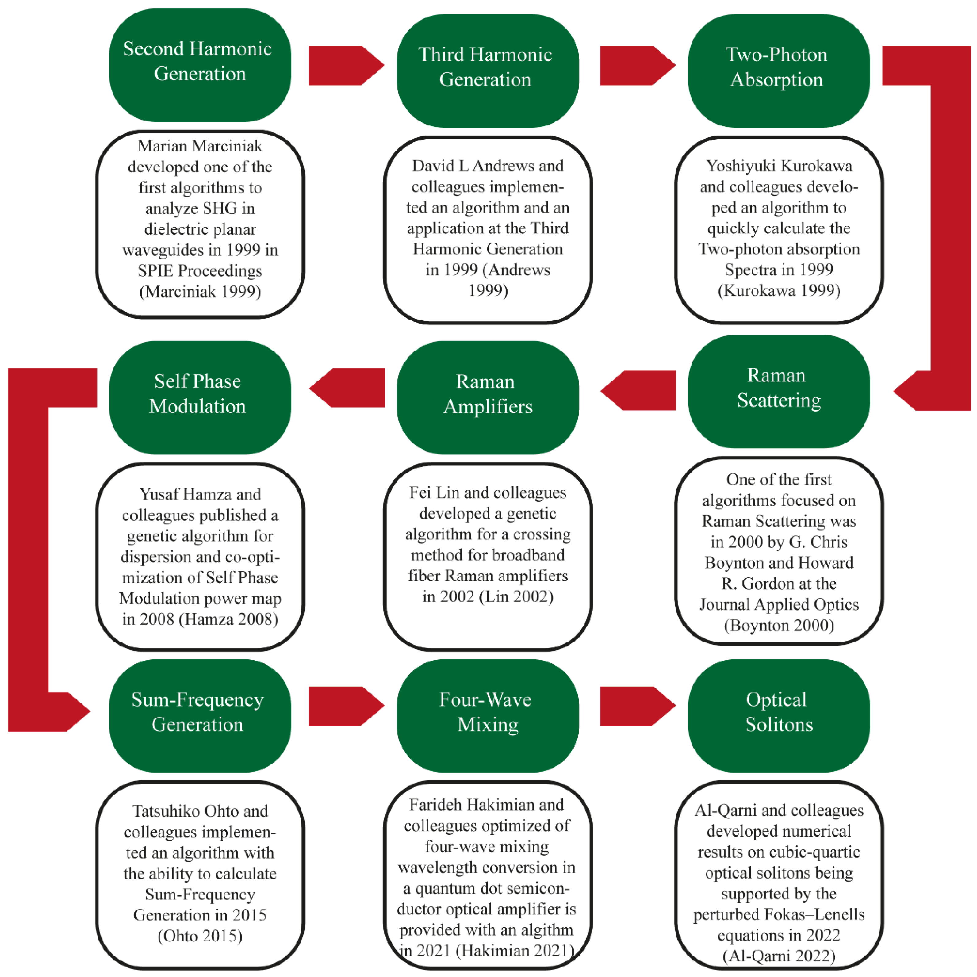
4.1. Second-Harmonic Generation
4.2. Nonlinear Optical Absorption
4.3. Optical Kerr Effect
4.4. Sum Frequency Generation
4.5. Self-Phase Modulation
4.6. Raman Amplifiers
4.7. Surface-Enhanced Raman Scattering
4.8. Summary of Representative Nonlinear Optical Effects Assisted by ML Algorithms
5. Perspectives
6. Conclusions
Author Contributions
Funding
Institutional Review Board Statement
Informed Consent Statement
Data Availability Statement
Acknowledgments
Conflicts of Interest
References
- Lei, Z.-L.; Guo, B. 2D Material-Based Optical Biosensor: Status and Prospect. Adv. Sci. 2022, 9, 2102924. [Google Scholar] [CrossRef] [PubMed]
- Khalil, I.; Julkapli, N.; Yehye, W.; Basirun, W.; Bhargava, S. Graphene–Gold Nanoparticles Hybrid—Synthesis, Functionalization, and Application in a Electrochemical and Surface-Enhanced Raman Scattering Biosensor. Materials 2016, 9, 406. [Google Scholar] [CrossRef] [PubMed]
- Malhotra, B.D.; Ali, M.A. Nanomaterials in Biosensors. In Nanomaterials for Biosensors; Elsevier: Amsterdam, The Netherlands, 2018; pp. 1–74. [Google Scholar]
- Shumeiko, V.; Malach, E.; Helman, Y.; Paltiel, Y.; Bisker, G.; Hayouka, Z.; Shoseyov, O. A Nanoscale Optical Biosensor Based on Peptide Encapsulated SWCNTs for Detection of Acetic Acid in the Gaseous Phase. Sens. Actuators B Chem. 2021, 327, 128832. [Google Scholar] [CrossRef]
- Samani, S.S.; Khojastehnezhad, A.; Ramezani, M.; Alibolandi, M.; Yazdi, F.T.; Mortazavi, S.A.; Khoshbin, Z.; Abnous, K.; Taghdisi, S.M. Ultrasensitive Detection of Micrococcal Nuclease Activity and Staphylococcus Aureus Contamination Using Optical Biosensor Technology-A Review. Talanta 2021, 226, 122168. [Google Scholar] [CrossRef]
- Portes, A.V.R.; Martins, A.J.L.; Guerrero, J.A.; Carvalho, M.M.; Amaya-Fernandez, F.O.; Saito, L.A.M.; Ramirez, J.C. Electro-Optical Biosensor Based on Embedded Double-Monolayer of Graphene Capacitor in Polymer Technology. Polymers 2021, 13, 3564. [Google Scholar] [CrossRef]
- Aldaya, I.; Gil-Molina, A.; Pita, J.L.; Gabrielli, L.H.; Fragnito, H.L.; Dainese, P. Nonlinear Carrier Dynamics in Silicon Nano-Waveguides. Optica 2017, 4, 1219. [Google Scholar] [CrossRef]
- Ramirez, J.C.; Lechuga, L.M.; Gabrielli, L.H.; Hernandez-Figueroa, H.E. Study of a Low-Cost Trimodal Polymer Waveguide for Interferometric Optical Biosensors. Opt. Express 2015, 23, 11985. [Google Scholar] [CrossRef]
- Singh, P. Surface Plasmon Resonance: A Boon for Viral Diagnostics. In Reference Module in Life Sciences; Elsevier: Amsterdam, The Netherlands, 2017; ISBN 978-0-12-809633-8. [Google Scholar]
- Su, Y.; Xia, S.; Wang, R.; Xiao, L. Phytohormonal Quantification Based on Biological Principles. In Hormone Metabolism and Signaling in Plants; Li, J., Li, C., Smith, S.M., Eds.; Elsevier: Amsterdam, The Netherlands, 2017; pp. 431–470. ISBN 978-0-12-811562-6. [Google Scholar]
- Cajigas, S.; Soto, D.; Orozco, J. Biosensors: Biosensors With Signal Amplification. In Reference Module in Biomedical Sciences; Elsevier: Amsterdam, The Netherlands, 2021; ISBN 978-0-12-801238-3. [Google Scholar]
- Shrivastav, A.M.; Cvelbar, U.; Abdulhalim, I. A Comprehensive Review on Plasmonic-Based Biosensors Used in Viral Diagnostics. Commun. Biol. 2021, 4, 70. [Google Scholar] [CrossRef] [PubMed]
- Li, D.; Zhou, H.; Hui, X.; He, X.; Mu, X. Plasmonic Biosensor Augmented by a Genetic Algorithm for Ultra-Rapid, Label-Free, and Multi-Functional Detection of COVID-19. Anal. Chem. 2021, 93, 9437–9444. [Google Scholar] [CrossRef]
- Sharma, S.; Kumar, A. Design of a Biosensor for the Detection of Dengue Virus Using 1D Photonic Crystals. Plasmonics 2022, 17, 675–680. [Google Scholar] [CrossRef]
- Sharma, S.; Kumar, A.; Singh, K.S.; Tyagi, H.K. 2D Photonic Crystal Based Biosensor for the Detection of Chikungunya Virus. Optik 2021, 237, 166575. [Google Scholar] [CrossRef]
- Zhou, J.; Yang, T.; Chen, J.; Wang, C.; Zhang, H.; Shao, Y. Two-Dimensional Nanomaterial-Based Plasmonic Sensing Applications: Advances and Challenges. Coord. Chem. Rev. 2020, 410, 213218. [Google Scholar] [CrossRef]
- Meradi, K.A.; Tayeboun, F.; Guerinik, A.; Zaky, Z.A.; Aly, A.H. Optical Biosensor Based on Enhanced Surface Plasmon Resonance: Theoretical Optimization. Opt. Quantum Electron. 2022, 54, 124. [Google Scholar] [CrossRef]
- Lertvachirapaiboon, C.; Baba, A.; Ekgasit, S.; Shinbo, K.; Kato, K.; Kaneko, F. Transmission Surface Plasmon Resonance Techniques and Their Potential Biosensor Applications. Biosens. Bioelectron. 2018, 99, 399–415. [Google Scholar] [CrossRef]
- Kushwaha, A.S.; Kumar, A.; Kumar, R.; Srivastava, S.K. A Study of Surface Plasmon Resonance (SPR) Based Biosensor with Improved Sensitivity. Photonics Nanostruct.-Fundam. Appl. 2018, 31, 99–106. [Google Scholar] [CrossRef]
- Lu, Y.; Tan, Y.; Xiao, Y.; Li, Z.; Sheng, E.; Dai, Z. A Silver@gold Nanoparticle Tetrahedron Biosensor for Multiple Pesticides Detection Based on Surface-Enhanced Raman Scattering. Talanta 2021, 234, 122585. [Google Scholar] [CrossRef] [PubMed]
- Calvo-Lozano, O.; Sierra, M.; Soler, M.; Estévez, M.C.; Chiscano-Camón, L.; Ruiz-Sanmartin, A.; Ruiz-Rodriguez, J.C.; Ferrer, R.; González-López, J.J.; Esperalba, J.; et al. Label-Free Plasmonic Biosensor for Rapid, Quantitative, and Highly Sensitive COVID-19 Serology: Implementation and Clinical Validation. Anal. Chem. 2022, 94, 975–984. [Google Scholar] [CrossRef]
- Sidhu, R.; Zheng, R.; Rasheed, A.; Khan, M.A. The Development of Point-of-Care Plasmonic-Based Biosensor for Early Detection of COVID-19 Virus. In Proceedings of the 2021 IEEE 15th International Conference on Nano/Molecular Medicine & Engineering (NANOMED), Taipei, Taiwan, 15–17 November 2021; IEEE: Manhattan, NY, USA, 2021; pp. 23–27. [Google Scholar]
- Wang, J.; Yao, Z.; Lei, T.; Poon, A.W. Silicon Coupled-Resonator Optical-Waveguide-Based Biosensors Using Light-Scattering Pattern Recognition with Pixelized Mode-Field-Intensity Distributions. Sci. Rep. 2015, 4, 7528. [Google Scholar] [CrossRef]
- Wang, J.; Yao, Z.; Poon, A.W. Silicon-Nitride-Based Integrated Optofluidic Biochemical Sensors Using a Coupled-Resonator Optical Waveguide. Front. Mater. 2015, 2, 34. [Google Scholar] [CrossRef]
- Zhang, P.; Ma, G.; Dong, W.; Wan, Z.; Wang, S.; Tao, N. Plasmonic Scattering Imaging of Single Proteins and Binding Kinetics. Nat. Methods 2020, 17, 1010–1017. [Google Scholar] [CrossRef]
- Song, M.K.; Chen, S.X.; Hu, P.P.; Huang, C.Z.; Zhou, J. Automated Plasmonic Resonance Scattering Imaging Analysis via Deep Learning. Anal. Chem. 2021, 93, 2619–2626. [Google Scholar] [CrossRef] [PubMed]
- Pang, Y.; Wan, N.; Shi, L.; Wang, C.; Sun, Z.; Xiao, R.; Wang, S. Dual-Recognition Surface-Enhanced Raman Scattering(SERS)Biosensor for Pathogenic Bacteria Detection by Using Vancomycin-SERS Tags and Aptamer-Fe3O4@Au. Anal. Chim. Acta 2019, 1077, 288–296. [Google Scholar] [CrossRef] [PubMed]
- Mao, K.; Zhou, Z.; Han, S.; Zhou, X.; Hu, J.; Li, X.; Yang, Z. A Novel Biosensor Based on Au@Ag Core-Shell Nanoparticles for Sensitive Detection of Methylamphetamine with Surface Enhanced Raman Scattering. Talanta 2018, 190, 263–268. [Google Scholar] [CrossRef] [PubMed]
- He, X.; Zhou, X.; Liu, Y.; Wang, X. Ultrasensitive, Recyclable and Portable Microfluidic Surface-Enhanced Raman Scattering (SERS) Biosensor for Uranyl Ions Detection. Sens. Actuators B Chem. 2020, 311, 127676. [Google Scholar] [CrossRef]
- Khalil, I.; Yehye, W.A.; Muhd Julkapli, N.; Ibn Sina, A.A.; Islam Chowdhury, F.; Khandaker, M.U.; Hsiao, V.K.S.; Basirun, W.J. Simultaneous Detection of Dual Food Adulterants Using Graphene Oxide and Gold Nanoparticle Based Surface Enhanced Raman Scattering Duplex DNA Biosensor. Vib. Spectrosc. 2021, 116, 103293. [Google Scholar] [CrossRef]
- Vendrell, M.; Maiti, K.K.; Dhaliwal, K.; Chang, Y.-T. Surface-Enhanced Raman Scattering in Cancer Detection and Imaging. Trends Biotechnol. 2013, 31, 249–257. [Google Scholar] [CrossRef]
- Dey, S.; Ahmed, E.; Somvanshi, P.S.; Sina, A.A.I.; Wuethrich, A.; Trau, M. An Electrochemical and Raman Scattering Dual Detection Biosensor for Rapid Screening and Biomolecular Profiling of Cancer Biomarkers. Chemosensors 2022, 10, 93. [Google Scholar] [CrossRef]
- Granger, J.H.; Granger, M.C.; Firpo, M.A.; Mulvihill, S.J.; Porter, M.D. Toward Development of a Surface-Enhanced Raman Scattering (SERS)-Based Cancer Diagnostic Immunoassay Panel. Analyst 2013, 138, 410–416. [Google Scholar] [CrossRef]
- Lopatynskyi, A.M.; Lopatynska, O.G.; Guo, L.J.; Chegel, V.I. Localized Surface Plasmon Resonance Biosensor—Part I: Theoretical Study of Sensitivity—Extended Mie Approach. IEEE Sens. J. 2011, 11, 361–369. [Google Scholar] [CrossRef]
- Manikandan, M.; Revathi, C.; Senthilkumar, P.; Amreetha, S.; Dhanuskodi, S.; Rajendra Kumar, R.T. CdTe Nanorods for Nonenzymatic Hydrogen Peroxide Biosensor and Optical Limiting Applications. Ionics 2020, 26, 2003–2010. [Google Scholar] [CrossRef]
- Prasanna Kumaar, S.; Sivasubramanian, A. Optimization of the Transverse Electric Photonic Strip Waveguide Biosensor for Detecting Diabetes Mellitus from Bulk Sensitivity. J. Healthc. Eng. 2021, 2021, 6081570. [Google Scholar] [CrossRef]
- Panda, A.; Puspa Devi, P. Photonic Crystal Biosensor for Refractive Index Based Cancerous Cell Detection. Opt. Fiber Technol. 2020, 54, 102123. [Google Scholar] [CrossRef]
- Efimov, I.M.; Vanyushkin, N.A.; Gevorgyan, A.H.; Golik, S.S. Optical Biosensor Based on a Photonic Crystal with a Defective Layer Designed to Determine the Concentration of SARS-CoV-2 in Water. Phys. Scr. 2022, 97, 055506. [Google Scholar] [CrossRef]
- Abrego-Martinez, J.C.; Jafari, M.; Chergui, S.; Pavel, C.; Che, D.; Siaj, M. Aptamer-Based Electrochemical Biosensor for Rapid Detection of SARS-CoV-2: Nanoscale Electrode-Aptamer-SARS-CoV-2 Imaging by Photo-Induced Force Microscopy. Biosens. Bioelectron. 2022, 195, 113595. [Google Scholar] [CrossRef]
- Ranjan, A.K.; Patra, A.K.; Gorai, A.K. Effect of Lockdown Due to SARS COVID-19 on Aerosol Optical Depth (AOD) over Urban and Mining Regions in India. Sci. Total Environ. 2020, 745, 141024. [Google Scholar] [CrossRef]
- Cennamo, N.; D’Agostino, G.; Perri, C.; Arcadio, F.; Chiaretti, G.; Parisio, E.M.; Camarlinghi, G.; Vettori, C.; Di Marzo, F.; Cennamo, R.; et al. Proof of Concept for a Quick and Highly Sensitive On-Site Detection of SARS-CoV-2 by Plasmonic Optical Fibers and Molecularly Imprinted Polymers. Sensors 2021, 21, 1681. [Google Scholar] [CrossRef]
- Liu, Y.-N.; Lv, Z.-T.; Yang, S.-Y.; Liu, X.-W. Optical Tracking of the Interfacial Dynamics of Single SARS-CoV-2 Pseudoviruses. Environ. Sci. Technol. 2021, 55, 4115–4122. [Google Scholar] [CrossRef]
- Gomez-Gonzalez, E.; Barriga-Rivera, A.; Fernandez-Muñoz, B.; Navas-Garcia, J.M.; Fernandez-Lizaranzu, I.; Munoz-Gonzalez, F.J.; Parrilla-Giraldez, R.; Requena-Lancharro, D.; Gil-Gamboa, P.; Rosell-Valle, C.; et al. Optical Imaging Spectroscopy for Rapid, Primary Screening of SARS-CoV-2: A Proof of Concept. Sci. Rep. 2022, 12, 2356. [Google Scholar] [CrossRef]
- Daoudi, K.; Ramachandran, K.; Alawadhi, H.; Boukherroub, R.; Dogheche, E.; El Khakani, M.A.; Gaidi, M. Ultra-Sensitive and Fast Optical Detection of the Spike Protein of the SARS-CoV-2 Using AgNPs/SiNWs Nanohybrid Based Sensors. Surf. Interfaces 2021, 27, 101454. [Google Scholar] [CrossRef]
- Li, Z.; Hirst, J.D. Computed Optical Spectra of SARS-CoV-2 Proteins. Chem. Phys. Lett. 2020, 758, 137935. [Google Scholar] [CrossRef]
- Rabiee, N.; Fatahi, Y.; Ahmadi, S.; Abbariki, N.; Ojaghi, A.; Rabiee, M.; Radmanesh, F.; Dinarvand, R.; Bagherzadeh, M.; Mostafavi, E.; et al. Bioactive Hybrid Metal-Organic Framework (MOF)-Based Nanosensors for Optical Detection of Recombinant SARS-CoV-2 Spike Antigen. Sci. Total Environ. 2022, 825, 153902. [Google Scholar] [CrossRef] [PubMed]
- Minopoli, A.; Scardapane, E.; Acunzo, A.; Campanile, R.; Della Ventura, B.; Velotta, R. Analysis of the Optical Response of a SARS-CoV-2-Directed Colorimetric Immunosensor. AIP Adv. 2021, 11, 065319. [Google Scholar] [CrossRef]
- Diaz, L.M.; Johnson, B.E.; Jenkins, D.M. Real-Time Optical Analysis of a Colorimetric LAMP Assay for SARS-CoV-2 in Saliva with a Handheld Instrument Improves Accuracy Compared with Endpoint Assessment. J. Biomol. Tech. 2021, 32, 158–171. [Google Scholar] [CrossRef] [PubMed]
- Tao, Y.; Bian, S.; Wang, P.; Zhang, H.; Bi, W.; Zhu, P.; Sawan, M. Rapid Optical Biosensing of SARS-CoV-2 Spike Proteins in Artificial Samples. Sensors 2022, 22, 3768. [Google Scholar] [CrossRef]
- Xu, W.; Liu, J.; Song, D.; Li, C.; Zhu, A.; Long, F. Rapid, Label-Free, and Sensitive Point-of-Care Testing of Anti-SARS-CoV-2 IgM/IgG Using All-Fiber Fresnel Reflection Microfluidic Biosensor. Microchim. Acta 2021, 188, 261. [Google Scholar] [CrossRef]
- Doulou, S.; Leventogiannis, K.; Tsilika, M.; Rodencal, M.; Katrini, K.; Antonakos, N.; Kyprianou, M.; Karofylakis, E.; Karageorgos, A.; Koufargyris, P.; et al. A Novel Optical Biosensor for the Early Diagnosis of Sepsis and Severe Covid-19: The PROUD Study. BMC Infect. Dis. 2020, 20, 860. [Google Scholar] [CrossRef]
- Courtney, S.; Stromberg, Z.; Myers y Gutiérrez, A.; Jacobsen, D.; Stromberg, L.; Lenz, K.; Theiler, J.; Foley, B.; Gans, J.; Yusim, K.; et al. Optical Biosensor Platforms Display Varying Sensitivity for the Direct Detection of Influenza RNA. Biosensors 2021, 11, 367. [Google Scholar] [CrossRef]
- Schotter, J.; Schrittwieser, S.; Muellner, P.; Melnik, E.; Hainberger, R.; Koppitsch, G.; Schrank, F.; Soulantika, K.; Lentijo-Mozo, S.; Pelaz, B.; et al. Optical Biosensor Technologies for Molecular Diagnostics at the Point-of-Care. Proc. SPIE 2015, 9490, 94900B. [Google Scholar] [CrossRef]
- El-Sherif, D.M.; Abouzid, M.; Gaballah, M.S.; Ahmed, A.A.; Adeel, M.; Sheta, S.M. New Approach in SARS-CoV-2 Surveillance Using Biosensor Technology: A Review. Environ. Sci. Pollut. Res. 2022, 29, 1677–1695. [Google Scholar] [CrossRef]
- Lee, S.-L.; Kim, J.; Choi, S.; Han, J.; Seo, G.; Lee, Y.W. Fiber-Optic Label-Free Biosensor for SARS-CoV-2 Spike Protein Detection Using Biofunctionalized Long-Period Fiber Grating. Talanta 2021, 235, 122801. [Google Scholar] [CrossRef]
- Sun, Y.; Shi, L.; Mi, L.; Guo, R.; Li, T. Recent Progress of SERS Optical Nanosensors for MiRNA Analysis. J. Mater. Chem. B 2020, 8, 5178–5183. [Google Scholar] [CrossRef] [PubMed]
- Yildirim, D.U.; Ghobadi, A.; Ozbay, E. Nanosensors Based on Localized Surface Plasmon Resonance. In Plasmonic Sensors and Their Applications; Wiley Online Books; Wiley: Hoboken, NJ, USA, 2021; pp. 23–54. ISBN 9783527830343. [Google Scholar]
- Dziekan, Z.; Pituła, E.; Kwietniewski, N.; Stonio, B.; Janik, M.; Śmiarowski, T.; Koba, M.; Parzuchowski, P.; Niedziółka-Jönsson, J.; Śmietana, M. Performance of Nanoimprinted and Nanocoated Optical Label-Free Biosensor-Nanocoating Properties Perspective. Opt. Lasers Eng. 2022, 153, 107009. [Google Scholar] [CrossRef]
- Threm, D.; Nazirizadeh, Y.; Gerken, M. Photonic Crystal Biosensors towards On-Chip Integration. J. Biophotonics 2012, 5, 601–616. [Google Scholar] [CrossRef]
- Fernández Gavela, A.; Grajales García, D.; Ramirez, J.; Lechuga, L. Last Advances in Silicon-Based Optical Biosensors. Sensors 2016, 16, 285. [Google Scholar] [CrossRef] [PubMed]
- Srivastava, A.; Sharma, A.K.; Kumar Prajapati, Y. On the Sensitivity-Enhancement in Plasmonic Biosensor with Photonic Spin Hall Effect at Visible Wavelength. Chem. Phys. Lett. 2021, 774, 138613. [Google Scholar] [CrossRef]
- Li, N.; Tang, T.; Li, J.; Luo, L.; Li, C.; Shen, J.; Yao, J. Highly Sensitive Biosensor with Graphene-MoS2 Heterostructure Based on Photonic Spin Hall Effect. J. Magn. Magn. Mater. 2019, 484, 445–450. [Google Scholar] [CrossRef]
- Leuermann, J.; Stamenkovic, V.; Ramirez-Priego, P.; Sánchez-Postigo, A.; Fernández-Gavela, A.; Chapman, C.A.; Bailey, R.C.; Lechuga, L.M.; Perez-Inestrosa, E.; Collado, D.; et al. Coherent Silicon Photonic Interferometric Biosensor with an Inexpensive Laser Source for Sensitive Label-Free Immunoassays. Opt. Lett. 2020, 45, 6595. [Google Scholar] [CrossRef]
- Xie, L.; Zhang, Z.; Du, J. The Photonic Spin Hall Effect Sensor. In Applied Optical Metrology II; Novak, E., Trolinger, J.D., Eds.; SPIE: Bellingham, WA, USA, 2017; Volume 10373, p. 10. [Google Scholar]
- Mavrikou, S.; Moschopoulou, G.; Tsekouras, V.; Kintzios, S. Development of a Portable, Ultra-Rapid and Ultra-Sensitive Cell-Based Biosensor for the Direct Detection of the SARS-CoV-2 S1 Spike Protein Antigen. Sensors 2020, 20, 3121. [Google Scholar] [CrossRef]
- Wrapp, D.; Wang, N.; Corbett, K.S.; Goldsmith, J.A.; Hsieh, C.-L.; Abiona, O.; Graham, B.S.; McLellan, J.S. Cryo-EM Structure of the 2019-NCoV Spike in the Prefusion Conformation. Science 2020, 367, 1260–1263. [Google Scholar] [CrossRef]
- Ruiz-Vega, G.; Soler, M.; Lechuga, L.M. Nanophotonic Biosensors for Point-of-Care COVID-19 Diagnostics and Coronavirus Surveillance. J. Phys. Photonics 2021, 3, 011002. [Google Scholar] [CrossRef]
- Moznuzzaman, M.; Khan, I.; Islam, M.R. Nano-Layered Surface Plasmon Resonance-Based Highly Sensitive Biosensor for Virus Detection: A Theoretical Approach to Detect SARS-CoV-2. AIP Adv. 2021, 11, 065023. [Google Scholar] [CrossRef] [PubMed]
- Bai, H.; Wang, R.; Hargis, B.; Lu, H.; Li, Y. A SPR Aptasensor for Detection of Avian Influenza Virus H5N1. Sensors 2012, 12, 12506–12518. [Google Scholar] [CrossRef] [PubMed]
- Prabowo, B.A.; Wang, R.Y.L.; Secario, M.K.; Ou, P.-T.; Alom, A.; Liu, J.-J.; Liu, K.-C. Rapid Detection and Quantification of Enterovirus 71 by a Portable Surface Plasmon Resonance Biosensor. Biosens. Bioelectron. 2017, 92, 186–191. [Google Scholar] [CrossRef]
- Murugan, D.; Bhatia, H.; Sai, V.V.R.; Satija, J. P-FAB: A Fiber-Optic Biosensor Device for Rapid Detection of COVID-19. Trans. Indian Natl. Acad. Eng. 2020, 5, 211–215. [Google Scholar] [CrossRef]
- Alathari, M.J.A.; Al Mashhadany, Y.; Mokhtar, M.H.H.; Burham, N.; Bin Zan, M.S.D.; A Bakar, A.A.; Arsad, N. Human Body Performance with COVID-19 Affectation According to Virus Specification Based on Biosensor Techniques. Sensors 2021, 21, 8362. [Google Scholar] [CrossRef]
- Taha, B.A.; Al Mashhadany, Y.; Hafiz Mokhtar, M.H.; Dzulkefly Bin Zan, M.S.; Arsad, N. An Analysis Review of Detection Coronavirus Disease 2019 (COVID-19) Based on Biosensor Application. Sensors 2020, 20, 6764. [Google Scholar] [CrossRef] [PubMed]
- Ma, R.-M.; Oulton, R.F. Applications of Nanolasers. Nat. Nanotechnol. 2019, 14, 12–22. [Google Scholar] [CrossRef]
- Rodriguez, G.A.; Markov, P.; Cartwright, A.P.; Choudhury, M.H.; Afzal, F.O.; Cao, T.; Halimi, S.I.; Retterer, S.T.; Kravchenko, I.I.; Weiss, S.M. Photonic Crystal Nanobeam Biosensors Based on Porous Silicon. Opt. Express 2019, 27, 9536. [Google Scholar] [CrossRef]
- Kim, H.; Hwang, J.; Kim, J.H.; Lee, S.; Kang, M. Sensitive Detection of Multiple Fluoresence Probes Based on Surface-Enhanced Raman Scattering (SERS) for MERS-CoV. In Proceedings of the 2019 IEEE 14th International Conference on Nano/Micro Engineered and Molecular Systems (NEMS), Bangkok, Thailand, 11–14 April 2019; IEEE: Manhattan, NY, USA, 2019; pp. 498–501. [Google Scholar]
- Kumar, A.; Kumar, A.; Kushwaha, A.S.; Dubey, S.K.; Srivastava, S.K. A Comparative Study of Different Types of Sandwiched Structures of SPR Biosensor for Sensitive Detection of SsDNA. Photonics Nanostruct.-Fundam. Appl. 2022, 48, 100984. [Google Scholar] [CrossRef]
- Syed Nor, S.N.; Rasanang, N.S.; Karman, S.; Zaman, W.S.W.K.; Harun, S.W.; Arof, H. A Review: Surface Plasmon Resonance-Based Biosensor for Early Screening of SARS-CoV2 Infection. IEEE Access 2022, 10, 1228–1244. [Google Scholar] [CrossRef]
- Yang, Y.; Liu, J.; Zhou, X. A CRISPR-Based and Post-Amplification Coupled SARS-CoV-2 Detection with a Portable Evanescent Wave Biosensor. Biosens. Bioelectron. 2021, 190, 113418. [Google Scholar] [CrossRef] [PubMed]
- Cavalera, S.; Colitti, B.; Rosati, S.; Ferrara, G.; Bertolotti, L.; Nogarol, C.; Guiotto, C.; Cagnazzo, C.; Denina, M.; Fagioli, F.; et al. A Multi-Target Lateral Flow Immunoassay Enabling the Specific and Sensitive Detection of Total Antibodies to SARS CoV-2. Talanta 2021, 223, 121737. [Google Scholar] [CrossRef] [PubMed]
- Song, D.; Liu, J.; Xu, W.; Han, X.; Wang, H.; Cheng, Y.; Zhuo, Y.; Long, F. Rapid and Quantitative Detection of SARS-CoV-2 IgG Antibody in Serum Using Optofluidic Point-of-Care Testing Fluorescence Biosensor. Talanta 2021, 235, 122800. [Google Scholar] [CrossRef]
- Han, H.; Wang, C.; Yang, X.; Zheng, S.; Cheng, X.; Liu, Z.; Zhao, B.; Xiao, R. Rapid Field Determination of SARS-CoV-2 by a Colorimetric and Fluorescent Dual-Functional Lateral Flow Immunoassay Biosensor. Sens. Actuators B Chem. 2022, 351, 130897. [Google Scholar] [CrossRef]
- Zhang, K.; Fan, Z.; Ding, Y.; Xie, M. A PH-Engineering Regenerative DNA Tetrahedron ECL Biosensor for the Assay of SARS-CoV-2 RdRp Gene Based on CRISPR/Cas12a Trans-Activity. Chem. Eng. J. 2022, 429, 132472. [Google Scholar] [CrossRef]
- Zhang, K.; Fan, Z.; Ding, Y.; Zhu, S.; Xie, M.; Hao, N. Exploring the Entropy-Driven Amplification Reaction and Trans -Cleavage Activity of CRISPR-Cas12a for the Development of an Electrochemiluminescence Biosensor for the Detection of the SARS-CoV-2 RdRp Gene in Real Samples and Environmental Surveillance. Environ. Sci. Nano 2022, 9, 162–172. [Google Scholar] [CrossRef]
- Xi, H.; Juhas, M.; Zhang, Y. G-Quadruplex Based Biosensor: A Potential Tool for SARS-CoV-2 Detection. Biosens. Bioelectron. 2020, 167, 112494. [Google Scholar] [CrossRef]
- O’Brien, A.; Chen, D.-Y.; Hackbart, M.; Close, B.J.; O’Brien, T.E.; Saeed, M.; Baker, S.C. Detecting SARS-CoV-2 3CLpro Expression and Activity Using a Polyclonal Antiserum and a Luciferase-Based Biosensor. Virology 2021, 556, 73–78. [Google Scholar] [CrossRef]
- Zhang, K.; Fan, Z.; Huang, Y.; Ding, Y.; Xie, M.; Wang, M. Hybridization Chain Reaction Circuit-Based Electrochemiluminescent Biosensor for SARS-Cov-2 RdRp Gene Assay. Talanta 2022, 240, 123207. [Google Scholar] [CrossRef] [PubMed]
- Fan, Z.; Yao, B.; Ding, Y.; Xu, D.; Zhao, J.; Zhang, K. Rational Engineering the DNA Tetrahedrons of Dual Wavelength Ratiometric Electrochemiluminescence Biosensor for High Efficient Detection of SARS-CoV-2 RdRp Gene by Using Entropy-Driven and Bipedal DNA Walker Amplification Strategy. Chem. Eng. J. 2022, 427, 131686. [Google Scholar] [CrossRef]
- Yang, X.; Liu, L.; Hao, Y.; So, E.; Emami, S.S.; Zhang, D.; Gong, Y.; Sheth, P.M.; Wang, Y. A Bioluminescent Biosensor for Quantifying the Interaction of SARS-CoV-2 and Its Receptor ACE2 in Cells and In Vitro. Viruses 2021, 13, 1055. [Google Scholar] [CrossRef] [PubMed]
- Yang, X.; Liu, L.; Hao, Y.; So, Y.W.; Emami, S.S.; Zhang, D.; Gong, Y.; Sheth, P.M.; Wang, Y.T. An Ultrasensitive Biosensor for Quantifying the Interaction of SARS-CoV-2 and Its Receptor ACE2 in Cells and In Vitro. bioRxiv 2020, 424698. [Google Scholar] [CrossRef]
- Murillo, A.M.M.; Tomé-Amat, J.; Ramírez, Y.; Garrido-Arandia, M.; Valle, L.G.; Hernández-Ramírez, G.; Tramarin, L.; Herreros, P.; Santamaría, B.; Díaz-Perales, A.; et al. Developing an Optical Interferometric Detection Method Based Biosensor for Detecting Specific SARS-CoV-2 Immunoglobulins in Serum and Saliva, and Their Corresponding ELISA Correlation. Sens. Actuators B Chem. 2021, 345, 130394. [Google Scholar] [CrossRef]
- Liu, N.; Wang, S.; Wang, J.; Lv, J.; Cheng, Q.; Ma, W.; Lu, Y. Promising Refractive Index and Temperature Biosensor Based on Hybrid Gmr/Fp System Employed for the Detection of SARS-CoV-2. SSRN Electron. J. 2022. [Google Scholar] [CrossRef]
- Gutiérrez-Gálvez, L.; del Caño, R.; Menéndez-Luque, I.; García-Nieto, D.; Rodríguez-Peña, M.; Luna, M.; Pineda, T.; Pariente, F.; García-Mendiola, T.; Lorenzo, E. Electrochemiluminescent Nanostructured DNA Biosensor for SARS-CoV-2 Detection. Talanta 2022, 240, 123203. [Google Scholar] [CrossRef] [PubMed]
- Jiang, C.; Mu, X.; Liu, S.; Liu, Z.; Du, B.; Wang, J.; Xu, J. A Study of the Detection of SARS-CoV-2 ORF1ab Gene by the Use of Electrochemiluminescent Biosensor Based on Dual-Probe Hybridization. Sensors 2022, 22, 2402. [Google Scholar] [CrossRef]
- Karakuş, E.; Erdemir, E.; Demirbilek, N.; Liv, L. Colorimetric and Electrochemical Detection of SARS-CoV-2 Spike Antigen with a Gold Nanoparticle-Based Biosensor. Anal. Chim. Acta 2021, 1182, 338939. [Google Scholar] [CrossRef]
- Xu, W.; Zhuo, Y.; Song, D.; Han, X.; Xu, J.; Long, F. Development of a Novel Label-Free All-Fiber Optofluidic Biosensor Based on Fresnel Reflection and Its Applications. Anal. Chim. Acta 2021, 1181, 338910. [Google Scholar] [CrossRef] [PubMed]
- Kim, M. Detecting SARS-CoV-2 with a Rapid, Cost-Effective Colorimetric Biosensor. Scilight 2021, 2021, 251103. [Google Scholar] [CrossRef]
- Sampad, M.J.N.; Zhang, H.; Yuzvinsky, T.D.; Stott, M.A.; Hawkins, A.R.; Schmidt, H. Optical Trapping Assisted Label-Free and Amplification-Free Detection of SARS-CoV-2 RNAs with an Optofluidic Nanopore Sensor. Biosens. Bioelectron. 2021, 194, 113588. [Google Scholar] [CrossRef]
- Ahmad, M.; Sharma, P.; Kamai, A.; Agrawal, A.; Faruq, M.; Kulshreshtha, A. HRPZyme Assisted Recognition of SARS-CoV-2 Infection by Optical Measurement (HARIOM). Biosens. Bioelectron. 2021, 187, 113280. [Google Scholar] [CrossRef]
- Ravalin, M.; Roh, H.; Suryawanshi, R.; Kumar, G.R.; Pak, J.E.; Ott, M.; Ting, A.Y. A Single-Component Luminescent Biosensor for the SARS-CoV-2 Spike Protein. J. Am. Chem. Soc. 2022, 144, 13663–13672. [Google Scholar] [CrossRef]
- Huang, L.; Ding, L.; Zhou, J.; Chen, S.; Chen, F.; Zhao, C.; Xu, J.; Hu, W.; Ji, J.; Xu, H.; et al. One-Step Rapid Quantification of SARS-CoV-2 Virus Particles via Low-Cost Nanoplasmonic Sensors in Generic Microplate Reader and Point-of-Care Device. Biosens. Bioelectron. 2021, 171, 112685. [Google Scholar] [CrossRef] [PubMed]
- Yao, B.; Zhang, J.; Fan, Z.; Ding, Y.; Zhou, B.; Yang, R.; Zhao, J.; Zhang, K. Rational Engineering of the DNA Walker Amplification Strategy by Using a Au@Ti3C2@PEI-Ru(Dcbpy)32+ Nanocomposite Biosensor for Detection of the SARS-CoV-2 RdRp Gene. ACS Appl. Mater. Interfaces 2021, 13, 19816–19824. [Google Scholar] [CrossRef]
- Bhattacharjee, A.; Sabino, R.M.; Gangwish, J.; Manivasagam, V.K.; James, S.; Popat, K.C.; Reynolds, M.; Li, Y.V. A Novel Colorimetric Biosensor for Detecting SARS-CoV-2 by Utilizing the Interaction between Nucleocapsid Antibody and Spike Proteins. Vitr. Model. 2022, 1, 241–247. [Google Scholar] [CrossRef]
- Lee, C.Y.; Degani, I.; Cheong, J.; Lee, J.-H.; Choi, H.-J.; Cheon, J.; Lee, H. Fluorescence Polarization System for Rapid COVID-19 Diagnosis. Biosens. Bioelectron. 2021, 178, 113049. [Google Scholar] [CrossRef] [PubMed]
- Roda, A.; Cavalera, S.; Di Nardo, F.; Calabria, D.; Rosati, S.; Simoni, P.; Colitti, B.; Baggiani, C.; Roda, M.; Anfossi, L. Dual Lateral Flow Optical/Chemiluminescence Immunosensors for the Rapid Detection of Salivary and Serum IgA in Patients with COVID-19 Disease. Biosens. Bioelectron. 2021, 172, 112765. [Google Scholar] [CrossRef] [PubMed]
- Zheng, Y.; Song, K.; Cai, K.; Liu, L.; Tang, D.; Long, W.; Zhai, B.; Chen, J.; Tao, Y.; Zhao, Y.; et al. B-Cell-Epitope-Based Fluorescent Quantum Dot Biosensors for SARS-CoV-2 Enable Highly Sensitive COVID-19 Antibody Detection. Viruses 2022, 14, 1031. [Google Scholar] [CrossRef]
- Büyüksünetçi, Y.T.; Çitil, B.E.; Tapan, U.; Anık, Ü. Development and Application of a SARS-CoV-2 Colorimetric Biosensor Based on the Peroxidase-Mimic Activity of γ-Fe2O3 Nanoparticles. Microchim. Acta 2021, 188, 335. [Google Scholar] [CrossRef]
- Divagar, M.; Gayathri, R.; Rasool, R.; Shamlee, J.K.; Bhatia, H.; Satija, J.; Sai, V.V.R. Plasmonic Fiberoptic Absorbance Biosensor (P-FAB) for Rapid Detection of SARS-CoV-2 Nucleocapsid Protein. IEEE Sens. J. 2021, 21, 22758–22766. [Google Scholar] [CrossRef]
- Zheng, Y.; Bian, S.; Sun, J.; Wen, L.; Rong, G.; Sawan, M. Label-Free LSPR-Vertical Microcavity Biosensor for On-Site SARS-CoV-2 Detection. Biosensors 2022, 12, 151. [Google Scholar] [CrossRef] [PubMed]
- Zhang, M.; Li, X.; Pan, J.; Zhang, Y.; Zhang, L.; Wang, C.; Yan, X.; Liu, X.; Lu, G. Ultrasensitive Detection of SARS-CoV-2 Spike Protein in Untreated Saliva Using SERS-Based Biosensor. Biosens. Bioelectron. 2021, 190, 113421. [Google Scholar] [CrossRef] [PubMed]
- Li, Y.; Lin, C.; Peng, Y.; He, J.; Yang, Y. High-Sensitivity and Point-of-Care Detection of SARS-CoV-2 from Nasal and Throat Swabs by Magnetic SERS Biosensor. Sens. Actuators B Chem. 2022, 365, 131974. [Google Scholar] [CrossRef]
- Hojjat Jodaylami, M.; Djaileb, A.; Live, L.S.; Boudreau, D.; Pelletier, J.; Masson, J.-F. Rapid Quantification of SARS-CoV-2 Antibodies with a Portable Surface Plasmon Resonance Biosensor. ECS Meet. Abstr. 2021, MA2021-01, 2026. [Google Scholar] [CrossRef]
- Cady, N.C.; Tokranova, N.; Minor, A.; Nikvand, N.; Strle, K.; Lee, W.T.; Page, W.; Guignon, E.; Pilar, A.; Gibson, G.N. Multiplexed Detection and Quantification of Human Antibody Response to COVID-19 Infection Using a Plasmon Enhanced Biosensor Platform. Biosens. Bioelectron. 2021, 171, 112679. [Google Scholar] [CrossRef] [PubMed]
- Cognetti, J.S.; Miller, B.L. Monitoring Serum Spike Protein with Disposable Photonic Biosensors Following SARS-CoV-2 Vaccination. Sensors 2021, 21, 5857. [Google Scholar] [CrossRef] [PubMed]
- Wu, Q.; Wu, W.; Chen, F.; Ren, P. Highly Sensitive and Selective Surface Plasmon Resonance Biosensor for the Detection of SARS-CoV-2 Spike S1 Protein. Analyst 2022, 147, 2809–2818. [Google Scholar] [CrossRef]
- Ebrem Bilgin, B.; Torun, H.; Ilgü, M.; Yanik, C.; Batur, S.N.; Çelik, S.; Öztürk, M.; Dogan, Ö.; Ergönül, Ö.; Solaroglu, I.; et al. Clinical Validation of SERS Metasurface SARS-CoV-2 Biosensor. In Biomedical Vibrational Spectroscopy 2022: Advances in Research and Industry; Huang, Z., Ed.; SPIE: Bellingham, WA, USA, 2022; Volume 11957, p. 36. [Google Scholar]
- Kumar, A.; Kumar, A.; Srivastava, S.K. Silicon Nitride-BP-Based Surface Plasmon Resonance Highly Sensitive Biosensor for Virus SARS-CoV-2 Detection. Plasmonics 2022, 17, 1065–1077. [Google Scholar] [CrossRef]
- Akib, T.B.A.; Mou, S.F.; Rahman, M.M.; Rana, M.M.; Islam, M.R.; Mehedi, I.M.; Mahmud, M.A.P.; Kouzani, A.Z. Design and Numerical Analysis of a Graphene-Coated SPR Biosensor for Rapid Detection of the Novel Coronavirus. Sensors 2021, 21, 3491. [Google Scholar] [CrossRef]
- Anshori, I.; Nugroho, A.E.; Jessika, A.S.; Yusuf, M.; Hartati, Y.W.; Sari, S.P.; Tohari, T.R.; Yuliarto, B.; Gumilar, G.; Nuraviana, L.; et al. Single-Chained Fragment Variable (ScFv) Recombinant as a Potential Receptor for SARS-CoV-2 Biosensor Based on Surface Plasmon Resonance (SPR). In 2021 IEEE Sensors; IEEE: Piscataway, NJ, USA, 2021; p. 21487738. [Google Scholar]
- Dai, Z.; Xu, X.; Wang, Y.; Li, M.; Zhou, K.; Zhang, L.; Tan, Y. Surface Plasmon Resonance Biosensor with Laser Heterodyne Feedback for Highly-Sensitive and Rapid Detection of COVID-19 Spike Antigen. Biosens. Bioelectron. 2022, 206, 114163. [Google Scholar] [CrossRef]
- Peng, Y.; Lin, C.; Li, Y.; Gao, Y.; Wang, J.; He, J.; Huang, Z.; Liu, J.; Luo, X.; Yang, Y. Identifying Infectiousness of SARS-CoV-2 by Ultra-Sensitive SnS2 SERS Biosensors with Capillary Effect. Matter 2022, 5, 694–709. [Google Scholar] [CrossRef] [PubMed]
- Lee, W.-I.; Subramanian, A.; Mueller, S.; Levon, K.; Nam, C.-Y.; Rafailovich, M.H. Potentiometric Biosensors Based on Molecular-Imprinted Self-Assembled Monolayer Films for Rapid Detection of Influenza A Virus and SARS-CoV-2 Spike Protein. ACS Appl. Nano Mater. 2022, 5, 5045–5055. [Google Scholar] [CrossRef] [PubMed]
- Qiu, G.; Gai, Z.; Tao, Y.; Schmitt, J.; Kullak-Ublick, G.A.; Wang, J. Dual-Functional Plasmonic Photothermal Biosensors for Highly Accurate Severe Acute Respiratory Syndrome Coronavirus 2 Detection. ACS Nano 2020, 14, 5268–5277. [Google Scholar] [CrossRef] [PubMed]
- Forssén, P.; Samuelsson, J.; Lacki, K.; Fornstedt, T. Advanced Analysis of Biosensor Data for SARS-CoV-2 RBD and ACE2 Interactions. Anal. Chem. 2020, 92, 11520–11524. [Google Scholar] [CrossRef]
- Behrouzi, K.; Lin, L. Gold Nanoparticle Based Plasmonic Sensing for the Detection of SARS-CoV-2 Nucleocapsid Proteins. Biosens. Bioelectron. 2022, 195, 113669. [Google Scholar] [CrossRef]
- Basso, C.R.; Malossi, C.D.; Haisi, A.; de Albuquerque Pedrosa, V.; Barbosa, A.N.; Grotto, R.T.; Araujo Junior, J.P. Fast and Reliable Detection of SARS-CoV-2 Antibodies Based on Surface Plasmon Resonance. Anal. Methods 2021, 13, 3297–3306. [Google Scholar] [CrossRef]
- Saada, H.; Pagneux, Q.; Wei, J.; Live, L.; Roussel, A.; Dogliani, A.; Die Morini, L.; Engelmann, I.; Alidjinou, E.K.; Rolland, A.S.; et al. Sensing of COVID-19 Spike Protein in Nasopharyngeal Samples Using a Portable Surface Plasmon Resonance Diagnostic System. Sens. Diagn. 2022. [Google Scholar] [CrossRef]
- Moitra, P.; Chaichi, A.; Abid Hasan, S.M.; Dighe, K.; Alafeef, M.; Prasad, A.; Gartia, M.R.; Pan, D. Probing the Mutation Independent Interaction of DNA Probes with SARS-CoV-2 Variants through a Combination of Surface-Enhanced Raman Scattering and Machine Learning. Biosens. Bioelectron. 2022, 208, 114200. [Google Scholar] [CrossRef]
- Bong, J.-H.; Kim, T.-H.; Jung, J.; Lee, S.J.; Sung, J.S.; Lee, C.K.; Kang, M.-J.; Kim, H.O.; Pyun, J.-C. Pig Sera-Derived Anti-SARS-CoV-2 Antibodies in Surface Plasmon Resonance Biosensors. BioChip J. 2020, 14, 358–368. [Google Scholar] [CrossRef]
- Achadu, O.J.; Nwaji, N.; Lee, D.; Lee, J.; Akinoglu, E.M.; Giersig, M.; Park, E.Y. 3D Hierarchically Porous Magnetic Molybdenum Trioxide@gold Nanospheres as a Nanogap-Enhanced Raman Scattering Biosensor for SARS-CoV-2. Nanoscale Adv. 2022, 4, 871–883. [Google Scholar] [CrossRef]
- Funari, R.; Chu, K.-Y.; Shen, A.Q. Detection of Antibodies against SARS-CoV-2 Spike Protein by Gold Nanospikes in an Opto-Microfluidic Chip. Biosens. Bioelectron. 2020, 169, 112578. [Google Scholar] [CrossRef] [PubMed]
- Gutgsell, A.R.; Gunnarsson, A.; Forssén, P.; Gordon, E.; Fornstedt, T.; Geschwindner, S. Biosensor-Enabled Deconvolution of the Avidity-Induced Affinity Enhancement for the SARS-CoV-2 Spike Protein and ACE2 Interaction. Anal. Chem. 2022, 94, 1187–1194. [Google Scholar] [CrossRef] [PubMed]
- Saad, Y.; Gazzah, M.H.; Mougin, K.; Selmi, M.; Belmabrouk, H. Sensitive Detection of SARS-CoV-2 Using a Novel Plasmonic Fiber Optic Biosensor Design. Plasmonics 2022. [Google Scholar] [CrossRef]
- Bistaffa, M.J.; Camacho, S.A.; Pazin, W.M.; Constantino, C.J.L.; Oliveira, O.N.; Aoki, P.H.B. Immunoassay Platform with Surface-Enhanced Resonance Raman Scattering for Detecting Trace Levels of SARS-CoV-2 Spike Protein. Talanta 2022, 244, 123381. [Google Scholar] [CrossRef] [PubMed]
- Zhao, B.; Che, C.; Wang, W.; Li, N.; Cunningham, B.T. Single-Step, Wash-Free Digital Immunoassay for Rapid Quantitative Analysis of Serological Antibody against SARS-CoV-2 by Photonic Resonator Absorption Microscopy. Talanta 2021, 225, 122004. [Google Scholar] [CrossRef] [PubMed]
- Cennamo, N.; Pasquardini, L.; Arcadio, F.; Lunelli, L.; Vanzetti, L.; Carafa, V.; Altucci, L.; Zeni, L. SARS-CoV-2 Spike Protein Detection through a Plasmonic D-Shaped Plastic Optical Fiber Aptasensor. Talanta 2021, 233, 122532. [Google Scholar] [CrossRef] [PubMed]
- Zhang, Z.-Y.; Liu, X.; Shen, L.; Chen, L.; Fang, W.-H. Machine Learning with Multilevel Descriptors for Screening of Inorganic Nonlinear Optical Crystals. J. Phys. Chem. C 2021, 125, 25175–25188. [Google Scholar] [CrossRef]
- Wang, X.; Wang, H.; Zhou, W.; Zhang, T.; Huang, H.; Song, Y.; Li, Y.; Liu, Y.; Kang, Z. Carbon Dots with Tunable Third-Order Nonlinear Coefficient Instructed by Machine Learning. J. Photochem. Photobiol. A Chem. 2022, 426, 113729. [Google Scholar] [CrossRef]
- Fairbairn, C.E.; Kang, D.; Bosch, N. Using Machine Learning for Real-Time BAC Estimation from a New-Generation Transdermal Biosensor in the Laboratory. Drug Alcohol Depend. 2020, 216, 108205. [Google Scholar] [CrossRef]
- Robison, H.M.; Chapman, C.A.; Zhou, H.; Erskine, C.L.; Theel, E.; Peikert, T.; Lindestam Arlehamn, C.S.; Sette, A.; Bushell, C.; Welge, M.; et al. Risk Assessment of Latent Tuberculosis Infection through a Multiplexed Cytokine Biosensor Assay and Machine Learning Feature Selection. Sci. Rep. 2021, 11, 20544. [Google Scholar] [CrossRef]
- Kim, H.; Seong, W.; Rha, E.; Lee, H.; Kim, S.K.; Kwon, K.K.; Park, K.-H.; Lee, D.-H.; Lee, S.-G. Machine Learning Linked Evolutionary Biosensor Array for Highly Sensitive and Specific Molecular Identification. Biosens. Bioelectron. 2020, 170, 112670. [Google Scholar] [CrossRef] [PubMed]
- Pennacchio, A.; Giampaolo, F.; Piccialli, F.; Cuomo, S.; Notomista, E.; Spinelli, M.; Amoresano, A.; Piscitelli, A.; Giardina, P. A Machine Learning-Enhanced Biosensor for Mercury Detection Based on an Hydrophobin Chimera. Biosens. Bioelectron. 2022, 196, 113696. [Google Scholar] [CrossRef] [PubMed]
- Green, E.M.; van Mourik, R.; Wolfus, C.; Heitner, S.B.; Dur, O.; Semigran, M.J. Machine Learning Detection of Obstructive Hypertrophic Cardiomyopathy Using a Wearable Biosensor. Npj Digit. Med. 2019, 2, 57. [Google Scholar] [CrossRef]
- Vakilian, K.A. A Nitrate Enzymatic Biosensor Based on Optimized Machine Learning Techniques. In 2022 9th Iranian Joint Congress on Fuzzy and Intelligent Systems (CFIS); IEEE: Piscataway, NJ, USA, 2022; p. 21758368. [Google Scholar]
- Khor, S.M.; Choi, J.; Won, P.; Ko, S.H. Challenges and Strategies in Developing an Enzymatic Wearable Sweat Glucose Biosensor as a Practical Point-Of-Care Monitoring Tool for Type II Diabetes. Nanomaterials 2022, 12, 221. [Google Scholar] [CrossRef] [PubMed]
- Gonzalez-Navarro, F.; Stilianova-Stoytcheva, M.; Renteria-Gutierrez, L.; Belanche-Muñoz, L.; Flores-Rios, B.; Ibarra-Esquer, J. Glucose Oxidase Biosensor Modeling and Predictors Optimization by Machine Learning Methods. Sensors 2016, 16, 1483. [Google Scholar] [CrossRef] [PubMed]
- Boscolo, S.; Finot, C. Artificial Neural Networks for Nonlinear Pulse Shaping in Optical Fibers. Opt. Laser Technol. 2020, 131, 106439. [Google Scholar] [CrossRef]
- Chicea, D.; Rei, S.M. A Fast Artificial Neural Network Approach for Dynamic Light Scattering Time Series Processing. Meas. Sci. Technol. 2018, 29, 105201. [Google Scholar] [CrossRef]
- Talebi-Moghaddam, S.; Bauer, F.J.; Huber, F.J.T.; Will, S.; Daun, K.J. Inferring Soot Morphology through Multi-Angle Light Scattering Using an Artificial Neural Network. J. Quant. Spectrosc. Radiat. Transf. 2020, 251, 106957. [Google Scholar] [CrossRef]
- Pelenis, D.; Barauskas, D.; Vanagas, G.; Dzikaras, M.; Viržonis, D. CMUT-Based Biosensor with Convolutional Neural Network Signal Processing. Ultrasonics 2019, 99, 105956. [Google Scholar] [CrossRef]
- Byun, S.-J.; Kim, D.-G.; Park, K.-D.; Choi, Y.-J.; Kumar, P.; Ali, I.; Kim, D.-G.; Yoo, J.-M.; Huh, H.-K.; Jung, Y.-J.; et al. A Low-Power Analog Processor-in-Memory-Based Convolutional Neural Network for Biosensor Applications. Sensors 2022, 22, 4555. [Google Scholar] [CrossRef]
- Mennel, L.; Symonowicz, J.; Wachter, S.; Polyushkin, D.K.; Molina-Mendoza, A.J.; Mueller, T. Ultrafast Machine Vision with 2D Material Neural Network Image Sensors. Nature 2020, 579, 62–66. [Google Scholar] [CrossRef] [PubMed]
- Chen, C.; Wang, J. Optical Biosensors: An Exhaustive and Comprehensive Review. Analyst 2020, 145, 1605–1628. [Google Scholar] [CrossRef] [PubMed]
- Yan, Y.; Gong, J.; Chen, J.; Zeng, Z.; Huang, W.; Pu, K.; Liu, J.; Chen, P. Recent Advances on Graphene Quantum Dots: From Chemistry and Physics to Applications. Adv. Mater. 2019, 31, 1808283. [Google Scholar] [CrossRef]
- Zhao, Y.; Li, Y.; Zhang, P.; Yan, Z.; Zhou, Y.; Du, Y.; Qu, C.; Song, Y.; Zhou, D.; Qu, S.; et al. Cell-Based Fluorescent Microsphere Incorporated with Carbon Dots as a Sensitive Immunosensor for the Rapid Detection of Escherichia Coli O157 in Milk. Biosens. Bioelectron. 2021, 179, 113057. [Google Scholar] [CrossRef]
- Hu, Q.; Wang, S.; Duan, H.; Liu, Y. A Fluorescent Biosensor for Sensitive Detection of Salmonella Typhimurium Using Low-Gradient Magnetic Field and Deep Learning via Faster Region-Based Convolutional Neural Network. Biosensors 2021, 11, 447. [Google Scholar] [CrossRef] [PubMed]
- Zhu, X.; Liu, P.; Xue, T.; Ge, Y.; Ai, S.; Sheng, Y.; Wu, R.; Xu, L.; Tang, K.; Wen, Y. A Novel Graphene-like Titanium Carbide MXene/Au–Ag Nanoshuttles Bifunctional Nanosensor for Electrochemical and SERS Intelligent Analysis of Ultra-Trace Carbendazim Coupled with Machine Learning. Ceram. Int. 2021, 47, 173–184. [Google Scholar] [CrossRef]
- Chen, G.; Du, J.; Sun, L.; Zhang, W.; Xu, K.; Chen, X.; Reed, G.T.; He, Z. Nonlinear Distortion Mitigation by Machine Learning of SVM Classification for PAM-4 and PAM-8 Modulated Optical Interconnection. J. Light. Technol. 2018, 36, 650–657. [Google Scholar] [CrossRef]
- Lin, J.; Shen, G.; Zhai, Z.; Zheng, D.; Li, Y.; Chang, Z.; Zong, L.; Deng, N.; Chang, T. Delivering Distributed Machine Learning Services in All-Optical Datacenter Networks with Torus Topology. In Asia Communications and Photonics Conference 2021; Chang-Hasnain Willner, A., Shieh, W., Shum, P., Su, Y., Li, G., Eggleton, B., Essiambre, R., Dai, D., Ma, D.C., Eds.; Optica Publishing Group: Washington, DC, USA, 2021; p. W3C.5. [Google Scholar]
- McConnon, A. Deep Learning Characterizes Optical Pulses Using Speckle Patterns at the End of Multimode Fibers. Scilight 2020, 2020, 381102. [Google Scholar] [CrossRef]
- Noble, J.; Zhou, C.; Murray, W.T.; Liu, Z. Convolutional Neural Network Reconstruction of Ultrashort Optical Pulses. Ultrafast Nonlinear Imaging Spectrosc. VIII 2020, 11497, 20. [Google Scholar] [CrossRef]
- Närhi, M.; Salmela, L.; Toivonen, J.; Billet, C.; Dudley, J.M.; Genty, G. Machine Learning Analysis of Extreme Events in Optical Fibre Modulation Instability. Nat. Commun. 2018, 9, 4923. [Google Scholar] [CrossRef] [Green Version]
- Kokhanovskiy, A.; Kuprikov, E.; Bednyakova, A.; Popkov, I.; Smirnov, S.; Turitsyn, S. Inverse Design of Mode-Locked Fiber Laser by Particle Swarm Optimization Algorithm. Sci. Rep. 2021, 11, 13555. [Google Scholar] [CrossRef] [PubMed]
- Woodward, R.I.; Kelleher, E.J.R. Towards ‘Smart Lasers’: Self-Optimisation of an Ultrafast Pulse Source Using a Genetic Algorithm. Sci. Rep. 2016, 6, 37616. [Google Scholar] [CrossRef] [PubMed]
- Zahavy, T.; Dikopoltsev, A.; Moss, D.; Haham, G.I.; Cohen, O.; Mannor, S.; Segev, M. Deep Learning Reconstruction of Ultrashort Pulses. Optica 2018, 5, 666. [Google Scholar] [CrossRef]
- Underwood, K.J.; Jones, A.M.; Gopinath, J.T. Synthesis of Coherent Optical Pulses Using a Field-Programmable Gate Array (FPGA)-Based Gradient Descent Phase-Locking Algorithm with Three Semiconductor Lasers. In CLEO: 2015; OSA: Washington, DC, USA, 2015; p. 15380458. [Google Scholar]
- Wang, D.; Zhang, M.; Cai, Z.; Cui, Y.; Li, Z.; Han, H.; Fu, M.; Luo, B. Combatting Nonlinear Phase Noise in Coherent Optical Systems with an Optimized Decision Processor Based on Machine Learning. Opt. Commun. 2016, 369, 199–208. [Google Scholar] [CrossRef]
- Marciniak, M. Two-Beam-Propagation Method Algorithm for Second-Harmonic Generation in Dielectric Planar Waveguides. In Proceedings of SPIE; Tuchin, V.V., Ryabukho, V.P., Zimnyakov, D.A., Eds.; SPIE: Bellingham, WA, USA, 1999; Volume 3726, p. 32. [Google Scholar]
- Andrews, D.L.; Romero, L.C.D.; Meath, W.J. An Algorithm for the Nonlinear Optical Susceptibilities of Dipolar Molecules, and an Application to Third Harmonic Generation. J. Phys. B At. Mol. Opt. Phys. 1999, 32, 1. [Google Scholar] [CrossRef]
- Kurokawa, Y.; Nomura, S.; Takemori, T.; Aoyagi, Y. Fast Algorithm for Calculating Two-Photon Absorption Spectra. Phys. Rev. E 1999, 59, 3694–3697. [Google Scholar] [CrossRef]
- Boynton, G.C.; Gordon, H.R. Irradiance Inversion Algorithm for Estimating the Absorption and Backscattering Coefficients of Natural Waters: Raman-Scattering Effects. Appl. Opt. 2000, 39, 3012–3022. [Google Scholar] [CrossRef]
- Lin, F.; Gong, Y.; Shum, P. Optimization Design By Genetic Algorithm With A New Crossover Method For Broadband Fiber Raman Amplifiers. In Optical Communications and Networks; World Scientific: Singapore, 2002; pp. 249–252. ISBN 978-981-238-232-0. [Google Scholar]
- Hamza, M.Y.; Tariq, S.; Awais, M.M.; Yang, S. Mitigation of the Effects of Self Phase Modulation and Group-Velocity Dispersion in Fiber Optic Communications: Dispersion- and Power-Map Cooptimization Using the Genetic Algorithm. Opt. Eng. 2008, 47, 1–12. [Google Scholar] [CrossRef]
- Ohto, T.; Usui, K.; Hasegawa, T.; Bonn, M.; Nagata, Y. Toward Ab Initio Molecular Dynamics Modeling for Sum-Frequency Generation Spectra; an Efficient Algorithm Based on Surface-Specific Velocity-Velocity Correlation Function. J. Chem. Phys. 2015, 143, 124702. [Google Scholar] [CrossRef]
- Hakimian, F.; Shayesteh, M.R.; Moslemi, M.R. Optimization of Four-Wave Mixing Wavelength Conversion in a Quantum-Dot Semiconductor Optical Amplifier Based on the Genetic Algorithm. Opt. Quantum Electron. 2021, 53, 140. [Google Scholar] [CrossRef]
- Al-Qarni, A.A.; Bakodah, H.O.; Alshaery, A.A.; Biswas, A.; Yıldırım, Y.; Moraru, L.; Moldovanu, S. Numerical Simulation of Cubic-Quartic Optical Solitons with Perturbed Fokas–Lenells Equation Using Improved Adomian Decomposition Algorithm. Mathematics 2022, 10, 138. [Google Scholar] [CrossRef]
- Moreno-Larios, J.A.; Ramírez-Guerra, C.; Contreras-Martínez, R.; Rosete-Aguilar, M.; Garduño-Mejía, J. Algorithm to Filter the Noise in the Spectral Intensity of Ultrashort Laser Pulses. Appl. Opt. 2020, 59, 7233–7241. [Google Scholar] [CrossRef] [PubMed]
- Fan, Q.; Zhou, G.; Gui, T.; Lu, C.; Lau, A.P.T. Advancing Theoretical Understanding and Practical Performance of Signal Processing for Nonlinear Optical Communications through Machine Learning. Nat. Commun. 2020, 11, 3694. [Google Scholar] [CrossRef] [PubMed]
- Smolyaninov, I.I. Nonlinear Optics of Photonic Hyper-Crystals: Optical Limiting and Hyper-Computing. J. Opt. Soc. Am. B 2019, 36, 1629–1636. [Google Scholar] [CrossRef]
- Dikopoltsev, A.; Zahavy, T.; Ziv, R.; Rubinstein, I.; Sidorenko, P.; Mannor, S.; Cohen, O.; Segev, M. Reconstruction of Ultrashort Pulses Using Deep Neural Networks. In 2018 2nd URSI Atlantic Radio Science Meeting (AT-RASC); IEEE: Piscataway, NJ, USA, 2018; p. 18144568. [Google Scholar]
- Wang, Q.; Liu, W.; Chen, X.; Wang, X.; Chen, G.; Zhu, X. Quantification of Scar Collagen Texture and Prediction of Scar Development via Second Harmonic Generation Images and a Generative Adversarial Network. Biomed. Opt. Express 2021, 12, 5305–5319. [Google Scholar] [CrossRef] [PubMed]
- Wang, G.; Sun, Y.; Jiang, S.; Wu, G.; Liao, W.; Chen, Y.; Lin, Z.; Liu, Z.; Zhuo, S. Machine Learning-Based Rapid Diagnosis of Human Borderline Ovarian Cancer on Second-Harmonic Generation Images. Biomed. Opt. Express 2021, 12, 5658–5669. [Google Scholar] [CrossRef]
- Wu, B.; Judd, N.B.; Smith, J.T.; Icaza, M.; Mukherjee, S.; Jain, M.; Gallagher, R.M.; Szeligowski, R.V. A Pilot Study for Distinguishing Chromophobe Renal Cell Carcinoma and Oncocytoma Using Second Harmonic Generation Imaging and Convolutional Neural Network Analysis of Collagen Fibrillar Structure. In Optical Biopsy XVI: Toward Real-Time Spectroscopic Imaging and Diagnosis; Alfano, R.R., Demos, S.G., Eds.; SPIE: Bellingham, WA, USA, 2018; Volume 10489, p. 44. [Google Scholar]
- Vidal-Codina, F.; Nguyen, N.-C.; Ciracì, C.; Oh, S.-H.; Peraire, J. A Nested Hybridizable Discontinuous Galerkin Method for Computing Second-Harmonic Generation in Three-Dimensional Metallic Nanostructures. J. Comput. Phys. 2021, 429, 110000. [Google Scholar] [CrossRef]
- Xu, Z.; Wang, P.; Zhao, M.; Yang, M.; Zhao, W.; Hu, K.; Dong, L.; Wang, S.; Li, X.; Yang, P.; et al. Prediction of Second-Harmonic Generation Wave-Front Distribution by Extreme Learning Machine. IEEE Photonics Technol. Lett. 2020, 32, 693–696. [Google Scholar] [CrossRef]
- Jafari, R.; Jones, T.; Trebino, R. 100% Reliable Algorithm for Second-Harmonic-Generation Frequency-Resolved Optical Gating. Opt. Express 2019, 27, 2112–2124. [Google Scholar] [CrossRef]
- Jafari, R.; Jones, T.; Trebino, R. 100% Reliable Frequency-Resolved-Optical-Gating Pulse Retrieval Algorithmic Approach. In Frontiers in Optics + Laser Science APS/DLS; OSA: Washington, DC, USA, 2019; p. JW3A.33. [Google Scholar]
- Saito, K.; Tanabe, T.; Oyama, Y. Numerical Analysis of Second Harmonic Generation for THz-Wave in a Photonic Crystal Waveguide Using a Nonlinear FDTD Algorithm. Opt. Commun. 2016, 365, 164–167. [Google Scholar] [CrossRef]
- Schneidereit, D.; Nübler, S.; Prölß, G.; Reischl, B.; Schürmann, S.; Müller, O.J.; Friedrich, O. Optical Prediction of Single Muscle Fiber Force Production Using a Combined Biomechatronics and Second Harmonic Generation Imaging Approach. Light Sci. Appl. 2018, 7, 79. [Google Scholar] [CrossRef] [PubMed]
- Liu, Q.; Mukhopadhyay, S.; Bastidas Rodriguez, M.X.; Fu, X.; Sahu, S.; Burk, D.; Gartia, M. A One-Shot Learning Framework for Assessment of Fibrillar Collagen from Second Harmonic Generation Images of an Infarcted Myocardium. In 2020 IEEE 17th International Symposium on Biomedical Imaging (ISBI); IEEE: Piscataway, NJ, USA, 2020; pp. 839–843. [Google Scholar]
- Diatta, A.; Rouquette, J.; Armand, P.; Hermet, P. Density Functional Theory Prediction of the Second Harmonic Generation and Linear Pockels Effect in Trigonal BaZnO2. J. Phys. Chem. C 2018, 122, 21277–21283. [Google Scholar] [CrossRef]
- Comin, A.; Hartschuh, A. Efficient Optimization of SHG Hotspot Switching in Plasmonic Nanoantennas Using Phase-Shaped Laser Pulses Controlled by Neural Networks. Opt. Express 2018, 26, 33678–33686. [Google Scholar] [CrossRef] [PubMed]
- Hall, G.; Liang, W.; Li, X. Efficient and Unbiased Fit-Free Algorithm for Quantification of Collagen Fiber Alignment for SHG Imaging Applications. In Biomedical Optics 2016; OSA: Washington, DC, USA, 2016. [Google Scholar]
- Preißler, D.; Kiefer, D.; Führer, T.; Walther, T. Evolutionary Algorithm-Assisted Design of a UV SHG Cavity with Elliptical Focusing to Avoid Crystal Degradation. Appl. Phys. B 2019, 125, 220. [Google Scholar] [CrossRef]
- Hall, G.; Liang, W.; Li, X. Fitting-Free Algorithm for Efficient Quantification of Collagen Fiber Alignment in SHG Imaging Applications. Biomed. Opt. Express 2017, 8, 4609–4620. [Google Scholar] [CrossRef]
- Kang, L.; Liang, F.; Gong, P.; Lin, Z.; Liu, F.; Huang, B. Two Novel Deep-Ultraviolet Nonlinear Optical Crystals with Shorter Phase-Matching Second Harmonic Generation than KBe2BO3F2: A First-Principles Prediction. Phys. Status Solidi–Rapid Res. Lett. 2018, 12, 1800276. [Google Scholar] [CrossRef]
- Zhang, B.; Tikhonov, E.; Xie, C.; Yang, Z.; Pan, S. Prediction of Fluorooxoborates with Colossal Second Harmonic Generation (SHG) Coefficients and Extremely Wide Band Gaps: Towards Modulating Properties by Tuning the BO3/BO3F Ratio in Layers. Angew. Chem. 2019, 131, 11852–11856. [Google Scholar] [CrossRef]
- McLean, J.; DiMarzio, C. A Linear Algorithm for Quantitative Measure of Corneal Collagen Fiber Orientation Using Second Harmonic Generation Microscopy. Proc. SPIE 2016, 9713, 971317. [Google Scholar] [CrossRef]
- Kumar, A.; Yadav, M.P.S. Computational Studies of Third-Order Nonlinear Optical Properties of Pyridine Derivative 2-Aminopyridinium p-Toluenesulphonate Crystal. Pramana 2017, 89, 7. [Google Scholar] [CrossRef]
- Salem, M.A.; Twelves, I.; Brown, A. Prediction of Two-Photon Absorption Enhancement in Red Fluorescent Protein Chromophores Made from Non-Canonical Amino Acids. Phys. Chem. Chem. Phys. 2016, 18, 24408–24416. [Google Scholar] [CrossRef]
- Eybposh, M.H.; Caira, N.W.; Atisa, M.; Chakravarthula, P.; Pégard, N.C. Enhanced Two-Photon Absorption with Deep Learning-Based Computer Generated Holography. In Frontiers in Optics/Laser Science; Lee Mazzali, C., Corwin, K., Jason Jones, R.B., Eds.; OSA: Washington, DC, USA, 2020. [Google Scholar]
- Deng, A.; Huang, J.; Liu, H.; Cai, W. Deep Learning Algorithms for Temperature Field Reconstruction of Nonlinear Tomographic Absorption Spectroscopy. Meas. Sens. 2020, 10–12, 100024. [Google Scholar] [CrossRef]
- Yadav, C.; Roy, S. Ultrafast Nonlinear Absorption in Hemoprotein Cytochrome-c and Its Application to Computing. Opt. Quantum Electron. 2016, 48, 377. [Google Scholar] [CrossRef]
- Wang, Z.; Zhu, N.; Wang, W.; Chao, X. Y-Net: A Dual-Branch Deep Learning Network for Nonlinear Absorption Tomography with Wavelength Modulation Spectroscopy. Opt. Express 2022, 30, 2156–2172. [Google Scholar] [CrossRef] [PubMed]
- Neskorniuk, V.; Freire, P.J.; Napoli, A.; Spinnler, B.; Schairer, W.; Prilepsky, J.E.; Costa, N.; Turitsyn, S.K. Simplifying the Supervised Learning of Kerr Nonlinearity Compensation Algorithms by Data Augmentation. In 2020 European Conference on Optical Communications (ECOC); IEEE: Piscataway, NJ, USA, 2020; pp. 1–4. [Google Scholar]
- Yıldırım, Y.; Çelik, N.; Yaşar, E. Nonlinear Schrödinger Equations with Spatio-Temporal Dispersion in Kerr, Parabolic, Power and Dual Power Law Media: A Novel Extended Kudryashov’s Algorithm and Soliton Solutions. Results Phys. 2017, 7, 3116–3123. [Google Scholar] [CrossRef]
- Solli, D.R.; Ropers, C.; Koonath, P.; Jalali, B. Optical Rogue Waves. Nature 2007, 450, 1054–1057. [Google Scholar] [CrossRef]
- Gopalakrishnan, S.S.; Panajotov, K.; Taki, M.; Tlidi, M. Dissipative Light Bullets in Kerr Cavities: Multistability, Clustering, and Rogue Waves. Phys. Rev. Lett. 2021, 126, 153902. [Google Scholar] [CrossRef]
- Jhangeer, A.; Faridi, W.A.; Asjad, M.I.; Akgül, A. Analytical Study of Soliton Solutions for an Improved Perturbed Schrödinger Equation with Kerr Law Non-Linearity in Non-Linear Optics by an Expansion Algorithm. Partial Differ. Equ. Appl. Math. 2021, 4, 100102. [Google Scholar] [CrossRef]
- Chase, H.M.; Rudshteyn, B.; Psciuk, B.T.; Upshur, M.A.; Strick, B.F.; Thomson, R.J.; Batista, V.S.; Geiger, F.M. Assessment of DFT for Computing Sum Frequency Generation Spectra of an Epoxydiol and a Deuterated Isotopologue at Fused Silica/Vapor Interfaces. J. Phys. Chem. B 2016, 120, 1919–1927. [Google Scholar] [CrossRef]
- Shah, S.A.; Pikalov, A.A.; Baldelli, S. ChemSpecNet: A Neural Network for Chemical Analysis of Sum Frequency Generation Spectroscopic Imaging. Opt. Commun. 2022, 507, 127691. [Google Scholar] [CrossRef]
- Jackson, W.; Zishan, W.; Xiong, W. Imaging Orientation of a Single Molecular Hierarchical Self-Assembled Sheet: The Combined Power of a Vibrational Sum Frequency Generation Microscopy and Neural Network. ChemRxiv 2022. [Google Scholar] [CrossRef]
- Cai, M.; Zhuge, Q.; Lun, H.; Fu, M.; Yi, L.; Hu, W. Pilot-Aided Self-Phase Modulation Noise Monitoring Based on Artificial Neural Network. In Asia Communications and Photonics Conference (ACPC) 2019; Optica Publishing Group: Chengdu, China, 2019; p. M4A.9. [Google Scholar]
- Fumumoto, Y.; Owaki, S.; Nakamura, M. Effect of Number of Neurons of a Neural-Network on Compensation Performance of SPM Non-Linear Waveform Distortion. In 2017 Opto-Electronics and Communications Conference (OECC) and Photonics Global Conference (PGC); IEEE: Piscataway, NJ, USA, 2017; pp. 1–2. [Google Scholar]
- Owaki, S.; Nakamura, M. Simultaneous Compensation of Waveform Distortion Caused by Chromatic Dispersion and SPM Using a Three-Layer Neural-Network. In 2017 Opto-Electronics and Communications Conference (OECC) and Photonics Global Conference (PGC); IEEE: Piscataway, NJ, USA, 2017; p. 17373621. [Google Scholar]
- Caballero, F.J.V.; Ives, D.J.; Laperle, C.; Charlton, D.; Zhuge, Q.; O’Sullivan, M.; Savory, S.J. Machine Learning Based Linear and Nonlinear Noise Estimation. J. Opt. Commun. Netw. 2018, 10, 42–51. [Google Scholar] [CrossRef]
- Brusin, A.M.R.; de Moura, U.C.; Curri, V.; Zibar, D.; Carena, A. Introducing Load Aware Neural Networks for Accurate Predictions of Raman Amplifiers. J. Light. Technol. 2020, 38, 6481–6491. [Google Scholar] [CrossRef]
- Soltani, M.; Da Ros, F.; Carena, A.; Zibar, D. Spectral and Spatial Power Evolution Design With Machine Learning-Enabled Raman Amplification. J. Light. Technol. 2022, 40, 3546–3556. [Google Scholar] [CrossRef]
- Soltani, M.; Da Ros, F.; Carena, A.; Zibar, D. Distance and Spectral Power Profile Shaping Using Machine Learning Enabled Raman Amplifiers. In 2021 IEEE Photonics Society Summer Topicals Meeting Series (SUM); IEEE: Piscataway, NJ, USA, 2021; p. 21048928. [Google Scholar]
- de Moura, U.C.; Da Ros, F.; Zibar, D.; Rosa Brusin, A.M.; Carena, A. Optimization of Raman Amplifiers Using Machine Learning. In 2021 IEEE Photonics Society Summer Topicals Meeting Series (SUM); IEEE: Piscataway, NJ, USA, 2021; p. 21048937. [Google Scholar]
- Marcon, G.; Galtarossa, A.; Palmieri, L.; Santagiustina, M. Model-Aware Deep Learning Method for Raman Amplification in Few-Mode Fibers. J. Light. Technol. 2021, 39, 1371–1380. [Google Scholar] [CrossRef]
- Huang, Y.; Du, J.; Chen, Y.; Xu, K.; He, Z. Machine Learning Assisted Inverse Design for Ultrafine, Dynamic and Arbitrary Gain Spectrum Shaping of Raman Amplification. Photonics 2021, 8, 260. [Google Scholar] [CrossRef]
- Ionescu, M.; Ghazisaeidi, A.; Renaudier, J. Machine Learning Assisted Hybrid EDFA-Raman Amplifier Design for C+L Bands. In Proceedings of the 2020 European Conference on Optical Communications (ECOC), Virtual Event, 6–10 December 2020; IEEE: Piscataway, NJ, USA, 2020; p. 20349557. [Google Scholar]
- Marcon, G.; Galtarossa, A.; Palmieri, L.; Santagiustina, M. C+L Band Gain Design in Few-Mode Fibers Using Raman Amplification and Machine Learning. In 2020 Italian Conference on Optics and Photonics (ICOP); IEEE: Piscataway, NJ, USA, 2020; p. 20264995. [Google Scholar]
- Soltani, M.; Da Ros, F.; Carena, A.; Zibar, D. Inverse Design of a Raman Amplifier in Frequency and Distance Domains Using Convolutional Neural Networks. Opt. Lett. 2021, 46, 2650–2653. [Google Scholar] [CrossRef]
- Zibar, D.; de Moura, U.C.; Rosa Brusin, A.M.; Carena, A.; Da Ros, F. Machine Learning Enabled Raman Amplifiers. In Proceedings of the 2021 Conference on Lasers and Electro-Optics Europe and European Quantum Electronics Conference, Virtual Event, 21–25 June 2021; Optica Publishing Group: Munich, Germany, 2021; p. ci_1_3. [Google Scholar]
- Marcon, G.; Galtarossa, A.; Palmieri, L.; Santagiustina, M. Gain Design of Few-Mode Fiber Raman Amplifiers Using an Autoencoder-Based Machine Learning Approach. In 2020 European Conference on Optical Communications (ECOC); IEEE: Piscataway, NJ, USA, 2020; p. 20349595. [Google Scholar]
- Gong, J.; Liu, F.; Wu, Y.; Zhang, Y.; Lei, S.; Zhu, Z. Raman Fiber Amplifier Design Scheme Based on Back Propagation Neural Network Algorithm. Opt. Eng. 2021, 60, 37103. [Google Scholar] [CrossRef]
- Hunter, R.A.; Asare-Werehene, M.; Mandour, A.; Tsang, B.K.; Anis, H. Determination of Chemoresistance in Ovarian Cancer by Simultaneous Quantification of Exosomes and Exosomal Cisplatin with Surface Enhanced Raman Scattering. Sens. Actuators B Chem. 2022, 354, 131237. [Google Scholar] [CrossRef]
- Chen, T.; Cheng, Q.; Lee, H.J. Machine-Learning-Mediated Single-Cell Classification by Hyperspectral Stimulated Raman Scattering Imaging. Opt. Heal. Care Biomed. Opt. XI 2021, 11900, 30. [Google Scholar] [CrossRef]
- Viljoen, R.; Neethling, P.; Spangenberg, D.; Heidt, A.; Frey, H.-M.; Feurer, T.; Rohwer, E. Implementation of Temporal Ptychography Algorithm, I2PIE, for Improved Single-Beam Coherent Anti-Stokes Raman Scattering Measurements. J. Opt. Soc. Am. B 2020, 37, A259–A265. [Google Scholar] [CrossRef]
- Melnikov, G.; Ignatenko, N.; Petrova, L.; Manzhos, O.; Gromkov, A. On the Influence of Clustering Processes in the Liquid Structure on Raman Scattering. MATEC Web Conf. 2021, 344, 01010. [Google Scholar] [CrossRef]
- Fang, X.; Zeng, Q.; Yan, X.; Zhao, Z.; Chen, N.; Deng, Q.; Zhu, M.; Zhang, Y.; Li, S. Fast Discrimination of Tumor and Blood Cells by Label-Free Surface-Enhanced Raman Scattering Spectra and Deep Learning. J. Appl. Phys. 2021, 129, 123103. [Google Scholar] [CrossRef]
- Weng, S.; Xu, X.; Li, J.; Wong, S.T.C. Combining Deep Learning and Coherent Anti-Stokes Raman Scattering Imaging for Automated Differential Diagnosis of Lung Cancer. J. Biomed. Opt. 2017, 22, 106017. [Google Scholar] [CrossRef] [PubMed]
- Yamato, N.; Matsuya, M.; Niioka, H.; Miyake, J.; Hashimoto, M. Nerve Segmentation with Deep Learning from Label-Free Endoscopic Images Obtained Using Coherent Anti-Stokes Raman Scattering. Biomolecules 2020, 10, 1012. [Google Scholar] [CrossRef] [PubMed]
- Brusin, A.M.R.; Zefreh, M.R.; Poggiolini, P.; Piciaccia, S.; Forghieri, F.; Carena, A. Machine Learning for Power Profiles Prediction in Presence of Inter-Channel Stimulated Raman Scattering. In 2021 European Conference on Optical Communication (ECOC); IEEE: Piscataway, NJ, USA, 2021; p. 21549326. [Google Scholar]
- Yamato, N.; Niioka, H.; Miyake, J.; Hashimoto, M. Improvement of Nerve Imaging Speed with Coherent Anti-Stokes Raman Scattering Rigid Endoscope Using Deep-Learning Noise Reduction. Sci. Rep. 2020, 10, 15212. [Google Scholar] [CrossRef]
- Yin, P.; Li, G.; Zhang, B.; Farjana, H.; Zhao, L.; Qin, H.; Hu, B.; Ou, J.; Tian, J. Facile PEG-Based Isolation and Classification of Cancer Extracellular Vesicles and Particles with Label-Free Surface-Enhanced Raman Scattering and Pattern Recognition Algorithm. Analyst 2021, 146, 1949–1955. [Google Scholar] [CrossRef]
- Shi, H.; Wang, H.; Meng, X.; Chen, R.; Zhang, Y.; Su, Y.; He, Y. Setting Up a Surface-Enhanced Raman Scattering Database for Artificial-Intelligence-Based Label-Free Discrimination of Tumor Suppressor Genes. Anal. Chem. 2018, 90, 14216–14221. [Google Scholar] [CrossRef]
- Sun, Y.W.; Liu, C.; Chan, K.L.; Xie, P.H.; Liu, W.Q.; Zeng, Y.; Wang, S.M.; Huang, S.H.; Chen, J.; Wang, Y.P.; et al. Stack Emission Monitoring Using Non-Dispersive Infrared Spectroscopy with an Optimized Nonlinear Absorption Cross Interference Correction Algorithm. Atmos. Meas. Tech. 2013, 6, 1993–2005. [Google Scholar] [CrossRef]
- Leong, Y.X.; Lee, Y.H.; Koh, C.S.L.; Phan-Quang, G.C.; Han, X.; Phang, I.Y.; Ling, X.Y. Surface-Enhanced Raman Scattering (SERS) Taster: A Machine-Learning-Driven Multireceptor Platform for Multiplex Profiling of Wine Flavors. Nano Lett. 2021, 21, 2642–2649. [Google Scholar] [CrossRef]
- Buzalewicz, I.; Suchwałko, A.; Karwańska, M.; Wieliczko, A.; Podbielska, H. Development of the Correction Algorithm to Limit the Deformation of Bacterial Colonies Diffraction Patterns Caused by Misalignment and Its Impact on the Bacteria Identification in the Proposed Optical Biosensor. Sensors 2020, 20, 5797. [Google Scholar] [CrossRef]
- Hu, J.; Ma, L.; Wang, S.; Yang, J.; Chang, K.; Hu, X.; Sun, X.; Chen, R.; Jiang, M.; Zhu, J.; et al. Biomolecular Interaction Analysis Using an Optical Surface Plasmon Resonance Biosensor: The Marquardt Algorithm vs Newton Iteration Algorithm. PLoS ONE 2015, 10, e0132098. [Google Scholar] [CrossRef] [PubMed]
- Mirsanaye, K.; Uribe Castaño, L.; Kamaliddin, Y.; Golaraei, A.; Augulis, R.; Kontenis, L.; Done, S.J.; Žurauskas, E.; Stambolic, V.; Wilson, B.C.; et al. Machine Learning-Enabled Cancer Diagnostics with Widefield Polarimetric Second-Harmonic Generation Microscopy. Sci. Rep. 2022, 12, 10290. [Google Scholar] [CrossRef] [PubMed]
- Desa, D.E.; Strawderman, R.L.; Wu, W.; Hill, R.L.; Smid, M.; Martens, J.W.M.; Turner, B.M.; Brown, E.B. Intratumoral Heterogeneity of Second-Harmonic Generation Scattering from Tumor Collagen and Its Effects on Metastatic Risk Prediction. BMC Cancer 2020, 20, 1217. [Google Scholar] [CrossRef]
- Jafari, R.; Jones, T.; Trebino, R. 100% Robust and Fast Algorithm for Second-Harmonic-Generation Frequency-Resolved Optical Gating. Real-Time Meas. Rogue Phenom. Single-Shot Appl. IV SPIE LASE 2019, 10903, 22. [Google Scholar] [CrossRef]
- Tan, T.; Peng, C.; Yuan, Z.; Xie, X.; Liu, H.; Xie, Z.; Huang, S.-W.; Rao, Y.; Yao, B. Predicting Kerr Soliton Combs in Microresonators via Deep Neural Networks. J. Light. Technol. 2020, 38, 6591–6599. [Google Scholar] [CrossRef]
- Costa, C.; Borges, L.; Penchel, R.A.; Abbade, M.L.F.; Giacoumidis, E.; Wei, J.; de Oliveira, J.A.; Santos, M.; Marconi, J.D.; Pita, J.L.; et al. Self-Phase Modulation and Inter-Polarization Cross-Phase Modulation Mitigation in Single-Channel Dp-16qam Coherent Pon Employing 4d Clustering. SSRN Electron. J. 2022. [Google Scholar] [CrossRef]
- Chen, Y.; Du, J.; Huang, Y.; Xu, K.; He, Z. Intelligent Gain Flattening in Wavelength and Space Domain for FMF Raman Amplification by Machine Learning Based Inverse Design. Opt. Express 2020, 28, 11911–11920. [Google Scholar] [CrossRef]
- Chen, Y.; Du, J.; Huang, Y.; Xu, K.; He, Z. Intelligent Gain Flattening of FMF Raman Amplification by Machine Learning Based Inverse Design. In Proceedings of the Optical Fiber Communication Conference (OFC) 2020, San Diego, CA, USA, 8–12 March 2020; Optica Publishing Group: San Diego, CA, USA, 2020; p. T4B.1. [Google Scholar]
- Jiang, L.; Mehedi Hassan, M.; Jiao, T.; Li, H.; Chen, Q. Rapid Detection of Chlorpyrifos Residue in Rice Using Surface-Enhanced Raman Scattering Coupled with Chemometric Algorithm. Spectrochim. Acta Part A Mol. Biomol. Spectrosc. 2021, 261, 119996. [Google Scholar] [CrossRef]
- Li, H.; Mehedi Hassan, M.; Wang, J.; Wei, W.; Zou, M.; Ouyang, Q.; Chen, Q. Investigation of Nonlinear Relationship of Surface Enhanced Raman Scattering Signal for Robust Prediction of Thiabendazole in Apple. Food Chem. 2021, 339, 127843. [Google Scholar] [CrossRef]
- Lussier, F.; Missirlis, D.; Spatz, J.P.; Masson, J.-F. Machine-Learning-Driven Surface-Enhanced Raman Scattering Optophysiology Reveals Multiplexed Metabolite Gradients Near Cells. ACS Nano 2019, 13, 1403–1411. [Google Scholar] [CrossRef]
- Burzynski, N.; Yuan, Y.; Felsen, A.; Reitano, D.; Wang, Z.; Sethi, K.A.; Lu, F.; Chiu, K. Deep Learning Techniques for Unmixing of Hyperspectral Stimulated Raman Scattering Images. In 2021 IEEE International Conference on Big Data (Big Data); IEEE: Piscataway, NJ, USA, 2021; pp. 5862–5864. [Google Scholar]
- Fang, Z.; Wang, W.; Lu, A.; Wu, Y.; Liu, Y.; Yan, C.; Han, C. Rapid Classification of Honey Varieties by Surface Enhanced Raman Scattering Combining with Deep Learning. In Proceedings of the 2018 Cross Strait Quad-Regional Radio Science and Wireless Technology Conference (CSQRWC), Xuzhou, China, 21–24 July 2018; IEEE: Piscataway, NJ, USA, 2018; p. 18092502. [Google Scholar]
- Sha, P.; Dong, P.; Deng, J.; Wu, X. Rapid Identification and Quantitative Analysis of Anthrax Protective Antigen Based on Surface-Enhanced Raman Scattering and Convolutional Neural Networks. In 2021 IEEE 21st International Conference on Nanotechnology (NANO); IEEE: Piscataway, NJ, USA, 2021; pp. 155–158. [Google Scholar]
- Paryanti, G.; Faig, H.; Rokach, L.; Sadot, D. A Direct Learning Approach for Neural Network Based Pre-Distortion for Coherent Nonlinear Optical Transmitter. J. Light. Technol. 2020, 38, 3883–3896. [Google Scholar] [CrossRef]
- Lee, G.-G.C.; Haung, K.-W.; Sun, C.-K.; Liao, Y.-H. Stem Cell Detection Based on Convolutional Neural Network via Third Harmonic Generation Microscopy Images. In 2017 International Conference on Orange Technologies (ICOT); IEEE: Piscataway, NJ, USA, 2017; pp. 45–48. [Google Scholar]
- Gupta, A.K.; Hsu, C.-H.; Lai, C.-S. Enhancement of the Au/ZnO-NA Plasmonic SERS Signal Using Principal Component Analysis as a Machine Learning Approach. IEEE Photonics J. 2020, 12, 20013846. [Google Scholar] [CrossRef]
- Rajput, S.K.; Nishchal, N.K. Fresnel Domain Nonlinear Optical Image Encryption Scheme Based on Gerchberg–Saxton Phase-Retrieval Algorithm. Appl. Opt. 2014, 53, 418–425. [Google Scholar] [CrossRef] [PubMed]
- Aşırım, Ö.E.; Yolalmaz, A.; Kuzuoğlu, M. High-Fidelity Harmonic Generation in Optical Micro-Resonators Using BFGS Algorithm. Micromachines 2020, 11, 686. [Google Scholar] [CrossRef]
- Bresci, A.; Guizzardi, M.; Valensise, C.M.; Marangi, F.; Scotognella, F.; Cerullo, G.; Polli, D. Removal of Cross-Phase Modulation Artifacts in Ultrafast Pump–Probe Dynamics by Deep Learning. APL Photonics 2021, 6, 76104. [Google Scholar] [CrossRef]
- Wang, L.; Gao, M.; Zhang, Y.; Cao, F.; Huang, H. Optical Phase Conjugation with Complex-Valued Deep Neural Network for WDM 64-QAM Coherent Optical Systems. IEEE Photonics J. 2021, 13, 21200412. [Google Scholar] [CrossRef]
- Shan, M.; Cheng, Q.; Zhong, Z.; Liu, B.; Zhang, Y. Deep-Learning-Enhanced Ice Thickness Measurement Using Raman Scattering. Opt. Express 2020, 28, 48–56. [Google Scholar] [CrossRef]
- Pereira, V.R.; Pereira, D.R.; de Melo Tavares Vieira, K.C.; Ribas, V.P.; Constantino, C.J.L.; Antunes, P.A.; Favareto, A.P.A. Sperm Quality of Rats Exposed to Difenoconazole Using Classical Parameters and Surface-Enhanced Raman Scattering: Classification Performance by Machine Learning Methods. Environ. Sci. Pollut. Res. 2019, 26, 35253–35265. [Google Scholar] [CrossRef]
- Owaki, S.; Nakamura, M. XPM Compensation in Optical Fiber Transmission Systems Using Neural-Network-Based Digital Signal Processing. IEICE Commun. Express 2018, 7, 31–36. [Google Scholar] [CrossRef]
- Yildiz, N.; San, S.E.; Polat, Ö. Light-Scattering Experiments in Dye-Doped Liquid Crystals Both to Determine Crystal Parameters and to Construct Consistent Neural Network Empirical Physical Formulas for Scattering Amplitudes. Opt. Commun. 2011, 284, 2173–2181. [Google Scholar] [CrossRef]
- Owaki, S.; Nakamura, M. Compensation of Optical Nonlinear Waveform Distortion Using Neural-Network Based Digital Signal Processing. IEICE Commun. Express 2017, 6, 484–489. [Google Scholar] [CrossRef]
- Wang, R.; Liang, F.; Lin, Z. Data-Driven Prediction of Diamond-like Infrared Nonlinear Optical Crystals with Targeting Performances. Sci. Rep. 2020, 10, 3486. [Google Scholar] [CrossRef] [PubMed]
- Williamson, I.A.D.; Hughes, T.W.; Minkov, M.; Bartlett, B.; Pai, S.; Fan, S. Reprogrammable Electro-Optic Nonlinear Activation Functions for Optical Neural Networks. IEEE J. Sel. Top. Quantum Electron. 2020, 26, 18881115. [Google Scholar] [CrossRef]
- Khulbe, M.; Kumar, S. Role of Nonlinear Optics in Big Data Transmission and Next Generation Computing Technologies. In 2019 9th International Conference on Cloud Computing, Data Science & Engineering (Confluence); IEEE: Piscataway, NJ, USA, 2019; pp. 234–238. [Google Scholar]
- Berry, M.E.; McCabe, S.M.; Shand, N.C.; Graham, D.; Faulds, K. Depth Prediction of Nanotags in Tissue Using Surface Enhanced Spatially Offset Raman Scattering (SESORS). Chem. Commun. 2022, 58, 1756–1759. [Google Scholar] [CrossRef] [PubMed]
- Sun, Y.; Dong, M.; Yu, M.; Xia, J.; Zhang, X.; Bai, Y.; Lu, L.; Zhu, L. Nonlinear All-Optical Diffractive Deep Neural Network with 10.6 Μm Wavelength for Image Classification. Int. J. Opt. 2021, 2021, 6667495. [Google Scholar] [CrossRef]
- Xu, L.; Rahmani, M.; Ma, Y.; Smirnova, D.A.; Kamali, K.Z.; Deng, F.; Chiang, Y.K.; Huang, L.; Zhang, H.; Gould, S.; et al. Enhanced Light–Matter Interactions in Dielectric Nanostructures via Machine-Learning Approach. Adv. Photonics 2020, 2, 1. [Google Scholar] [CrossRef]
- Wright, L.G.; Onodera, T.; Stein, M.M.; Wang, T.; Schachter, D.T.; Hu, Z.; McMahon, P.L. Deep Nonlinear Optical Neural Networks Using Physics-Aware Training. In Conference on Lasers and Electro-Optics; Kang Tomasulo, S., Ilev, I., Müller, D., Litchinitser, N., Polyakov, S., Podolskiy, V., Nunn, J., Dorrer, C., Fortier, T., Gan, Q., et al., Eds.; Optica Publishing Group: Washington, DC, USA, 2021; p. FF1A.4. [Google Scholar]
- Miscuglio, M.; Mehrabian, A.; Hu, Z.; Azzam, S.I.; George, J.; Kildishev, A.V.; Pelton, M.; Sorger, V.J. All-Optical Nonlinear Activation Function for Photonic Neural Networks [Invited]. Opt. Mater. Express 2018, 8, 3851. [Google Scholar] [CrossRef]
- Girija, R.; Anshula; Singh, H. Security-Enhanced Optical Nonlinear Cryptosystem Based on Modified Gerchberg–Saxton Iterative Algorithm. Optik 2021, 244, 167568. [Google Scholar] [CrossRef]
- Singh, P.; Kumar, R.; Yadav, A.K.; Singh, K. Security Analysis and Modified Attack Algorithms for a Nonlinear Optical Cryptosystem Based on DRPE. Opt. Lasers Eng. 2021, 139, 106501. [Google Scholar] [CrossRef]
- Ghiasuddin; Akram, M.; Adeel, M.; Khalid, M.; Tahir, M.N.; Khan, M.U.; Asghar, M.A.; Ullah, M.A.; Iqbal, M. A Combined Experimental and Computational Study of 3-Bromo-5-(2,5-Difluorophenyl) Pyridine and 3,5-Bis(Naphthalen-1-Yl)Pyridine: Insight into the Synthesis, Spectroscopic, Single Crystal XRD, Electronic, Nonlinear Optical and Biological Properties. J. Mol. Struct. 2018, 1160, 129–141. [Google Scholar] [CrossRef]
- Agarwal, N.R.; Lucotti, A.; Tommasini, M.; Chalifoux, W.A.; Tykwinski, R.R. Nonlinear Optical Properties of Polyynes: An Experimental Prediction for Carbyne. J. Phys. Chem. C 2016, 120, 11131–11139. [Google Scholar] [CrossRef]
- Kutz, N. Deep Learning for Control of Nonlinear Optical Systems. In AI and Optical Data Sciences II; Kitayama, K., Jalali, B., Eds.; SPIE: Bellingham, WA, USA, 2021; Volume 11703, p. 41. [Google Scholar]
- Wu, J.; Li, Z.; Luo, J.; Jen, A.K.-Y. High-Performance Organic Second- and Third-Order Nonlinear Optical Materials for Ultrafast Information Processing. J. Mater. Chem. C 2020, 8, 15009–15026. [Google Scholar] [CrossRef]


| Journal | Detection Limit | Analyte Types | Optical Effect | Year | Reference |
|---|---|---|---|---|---|
| Biosensors and Bioelectronics | 2 μL | The genes of S, N, and Orf1ab | Evanescent wave fluorescence | 2021 | [79] |
| Talanta | 1.0 mg/mL | Immunoglobulins (G, M, and A) | Colorimetric | 2021 | [80] |
| Talanta | 12.5 ng/mL | IgG antibody | Evanescent wave fluorescence | 2021 | [81] |
| Sensors and Actuators B: Chemical | 1 and 0.033 ng/mL | Spike 1 protein | Fluorescent bifunctional | 2022 | [82] |
| Chemical Engineering Journal | 43.70 aM | RNA-dependent RNA polymerase gene | Electrochemiluminescence | 2022 | [83] |
| Environmental Science: Nano | 32.80 aM | RNA-dependent RNA polymerase gene | Electrochemiluminescence | 2022 | [84] |
| Biosensors and Bioelectronics | 2.75 fM | Spike protein, matrix protein, envelope protein, and nucleocapsid | Colorimetry G-quadruplex | 2020 | [85] |
| Virology | - | Nucleocapsid protein | Luminescence | 2021 | [86] |
| Talanta | 59 aM | Nucleic acid | Electrochemiluminescence | 2022 | [87] |
| Chemical Engineering Journal | 7.8 aM | RNA-dependent RNA polymerase gene | Electrochemiluminescence | 2022 | [88] |
| Viruses | 50 μg/mL | Angiotensin-converting enzyme 2 | Bioluminescent | 2021 | [89] |
| Cold Spring Harbor Laboratory | 50 μg/mL | Angiotensin-converting enzyme 2 | Bioluminescent | 2020 | [90] |
| Physica Scripta | 1020 nm/refractive index unit (RIU) | Pathogens of SARS-CoV-2 | Refractive index | 2022 | [38] |
| Sensors and Actuators B: Chemical | - | Spike protein | Optical interferometry | 2021 | [91] |
| SSRN Electronic Journal | 833.33 nm/RIU | Spike glycoprotein | Refractive index | 2022 | [92] |
| Talanta | 514 aM | spike protein, nucleocapsid protein, the RNA-dependent RNA polymerase gene | Electrochemiluminescence | 2022 | [93] |
| Sensors | 0.1 fM | Open reading frames 1ab gene | Electrochemiluminescence | 2022 | [94] |
| Talanta | 0.22 pM | Spike protein | Refractive index | 2021 | [55] |
| Analytica Chimica Acta | 48 ng/mL | SARS-CoV-2 spike antigen | Colorimetric | 2021 | [95] |
| Analytica Chimica Acta | 1.0 × 10⁻⁶ RIU | Spike protein receptor-binding domain | Fresnel reflection | 2021 | [96] |
| 2021 IEEE 15th International Conference on Nano/Molecular Medicine & Engineering (NANOMED) | COVID-19 virus detection by delivering quick, dependable results | Refractive index | 2021 | [22] | |
| Scilight | ∼106 virions/mL | SARS-CoV-2 proteins (membrane, envelope, and spike) | Colorimetric | 2021 | [97] |
| Biosensors and Bioelectronics | 17 aM | SARS-CoV-2 RNAs with single molecule sensitivity | Electro-optofluidic | 2021 | [98] |
| Biosensors and Bioelectronics | - | Nucleic-acid-based testing | Colorimetric | 2021 | [99] |
| Journal of the American Chemical Society | - | Spike antigen and cultured virus | Luminescent | 2022 | [100] |
| Biosensors and Bioelectronics | 370 vp/mL | SARS-CoV-2 virus particles in one step | Nanoplasmonic resonance | 2021 | [101] |
| ACS Applied Materials & Interfaces | 0.21 fM | RNA-dependent RNA polymerase gene | Electrochemiluminescence | 2021 | [102] |
| In vitro models | 1 μg/mL | S protein of SARS-CoV-2 | Colorimetric | 2022 | [103] |
| Biosensors and Bioelectronics | 3 copies/μL | Two regions in nucleocapsid gene (N1 and N2 genes) | Fluorescence polarization | 2021 | [104] |
| Biosensors and Bioelectronics | 1 mg/mL | Immunoglobulins G and M | Optical/chemiluminescence | 2021 | [105] |
| Viruses | 100 pM | Spike proteins, nucleocapsid proteins | Fluorescent | 2022 | [106] |
| Microchimica Acta | Angiotensin-converting enzyme 2 | Colorimetric | 2021 | [107] |
| Journal | Detection Limit | Analyte Types | Optical Effect | Year | Reference |
|---|---|---|---|---|---|
| IEEE Sensors Journal | 2.5 ng/mL | Nucleocapsid protein | Plasmonic fiber optic absorbance | 2021 | [108] |
| Biosensors | 0.047 μg/mL | SARS-CoV-2 pseudovirus | Surface plasmon resonance | 2022 | [109] |
| Biosensors and Bioelectronics | Spike protein | Raman scattering | 2021 | [110] | |
| Sensors and Actuators B: Chemical | 50 and 10 pfu/mL | Angiotensin-converting enzyme 2 | Raman scattering | 2022 | [111] |
| ECS Meeting Abstracts | - | Antibodies to SARS-CoV-2 | Surface plasmon resonance | 2021 | [112] |
| Analytical Chemistry | Nucleocapsid protein | Plasmonics | 2022 | [21] | |
| Biosensors and Bioelectronics | 2 ng/spot | spike S1, spike S1 S2, and the nucleocapsid protein | Fluorescent plasmonics | 2021 | [113] |
| Sensors | 4.2 μg/mL | Spike protein | Photonics | 2021 | [114] |
| Analyst | Spike S1 protein | Surface plasmon resonance | 2022 | [115] | |
| Biomedical Vibrational Spectroscopy 2022: Advances in Research and Industry | - | Spike protein | Raman spectroscopy | 2022 | [116] |
| Plasmonics | 152°/RIU | Spike proteins, membrane proteins, envelop proteins, and nucleoprotein | Surface plasmon resonance | 2022 | [117] |
| Sensors | 250 μg/mL | Spike (S1 and S2) proteins | Surface plasmon resonance | 2021 | [118] |
| IEEE SENSORS 2021 | 8.34 ng/mL | Spike protein | Surface plasmon resonance | 2021 | [119] |
| Biosensors and Bioelectronics | 1 μg/mL | Nucleocapsid antibody | Surface plasmon resonance | 2022 | [120] |
| AIP Advances | Spike glycoprotein | Surface plasmon resonance | 2021 | [67] | |
| Analytical Chemistry | - | Spike surface glycoprotein | Surface-enhanced infrared absorption | 2021 | [13] |
| Matter | 10 PFU/mL | Spike glycoprotein and membrane protein | Raman scattering | 2022 | [121] |
| ACS Applied Nano Materials | 200 PFU/mL | Spike proteins | Raman scattering | 2022 | [122] |
| ACS Nano | 0.22 pM | RNA-dependent RNA polymerase | Localized surface plasmon resonance | 2020 | [123] |
| Analytical Chemistry | - | Angiotensin-converting enzyme 2 | Surface plasmon resonance | 2020 | [124] |
| Biosensors and Bioelectronics | 150 ng/ml | Detect SARS-CoV-2 nucleocapsid proteins | Localized surface Plasmon resonance | 2022 | [125] |
| Analytical Methods | 200 μL | Spike and nucleocapsid proteins | Surface plasmon resonance | 2021 | [126] |
| Sensors & Diagnostics | 10 RU | Spike protein | Surface plasmon resonance | 2022 | [127] |
| Biosensors and Bioelectronics | particles/mL | Nucleocapsid phosphoprotein gene | Raman scattering | 2022 | [128] |
| BioChip Journal | 1.02 pM | Antibodies against nucleoprotein | Surface plasmon resonance | 2020 | [129] |
| Nanoscale Advances | SARS-CoV-2 spike protein | Raman scattering | 2022 | [130] | |
| Biosensors and Bioelectronics | 0.08 ng/mL | SARS-CoV-2 spike protein | Localized surface plasmon resonance | 2020 | [131] |
| Analytical Chemistry | 4 mg/mL | SARS-CoV-2 spike protein | Surface plasmon resonance | 2021 | [132] |
| Plasmonics | Thiol-tethered DNA of SARS-CoV-2 | Surface plasmon resonance | 2021 | [133] | |
| Talanta | 0.046 ng/mL | SARS-CoV-2 spike protein | Raman scattering | 2022 | [134] |
| Talanta | Measurement of SARS-CoV-2 antibody | Photonic resonator absorption | 2021 | [135] | |
| Talanta | 37 nM | SARS-CoV-2 spike glycoprotein | Surface plasmon resonance | 2021 | [136] |
| Journal or Conference Event | Application | Optical Effect | Year | Reference |
|---|---|---|---|---|
| Sensors | Optical biosensors supported by algorithms for rigorous monitoring and control in the identification of bacteria | Light Diffraction | 2020 | [242] |
| PLOS ONE | Comparison between Marquardt Algorithm vs. Newton Iteration Algorithm for biomolecular interaction process between antigen and antibodies or receptors | Optical Surface Plasmon Resonance | 2015 | [243] |
| Scientific Reports | Improves the image difference between normal tissues and tumors | SHG | 2021 | [244] |
| BMC Cancer | An independent predictive measure of metastasis-free survival in patients with invasive ductal cancer | SHG | 2020 | [245] |
| SPIE LASE | Enhanced Pulse Extraction Algorithm FROG used for geometry | SHG | 2019 | [246] |
| Atmospheric Measurement Techniques | Measures the error between CO and CO2 by nonlinear absorption and fluctuations in interference coefficients | Nonlinear Absorption | 2013 | [240] |
| Journal of Lightwave Technology | A scheme allowing the soliton comb to be determined under a specific pump scan, with an error of <8%, verified by experimental measurements | Optical Kerr Effect | 2020 | [247] |
| SSRN Electronic Journal | Tackling the effects of the intra-polarization self-phase modulation and inter-polarization cross-phase modulation | SFM | 2022 | [248] |
| Optics Express | Optimizes the pump wavelength | Raman Amplifiers | 2020 | [249] |
| Optical Fiber Communication Conference (OFC) 2020 | Gains improvements for a few mode fiber amplifiers | Raman Amplifiers | 2020 | [250] |
| Spectrochimica Acta Part A: Molecular and Biomolecular Spectroscopy | Detection of quantity of chlorpyrifos in rice. | Raman Scattering | 2021 | [251] |
| Food Chemistry | Quantifies the systemic fungicide residues of Benzimidazole (Thiabendazole) in apples | Raman Scattering | 2021 | [252] |
| ACS Nano | Performs the measurement simultaneously from gradients, at least eight in vitro metabolites along with different cell lines | Raman Scattering | 2019 | [253] |
| 2021 IEEE International Conference on Big Data (Big Data) | Improves rapidity in the inspection of the techniques of images of cellular and tissue pathology | Raman Scattering | 2021 | [254] |
| 2018 Cross Strait Quad-Regional Radio Science and Wireless Technology Conference (CSQRWC) | Sorts different varieties of honey | Raman Scattering | 2018 | [255] |
| In Proceedings of the 2021 IEEE 21st International Conference on Nanotechnology | Label-free method for detection of protective anthrax antigens based on SERS | Raman Scattering | 2021 | [256] |
| Journal | Application | Algorithm | Nonlinear Optical | Year | Reference |
|---|---|---|---|---|---|
| Journal of Lightwave Technology | A standard for optical quality | A new approach to direct-learning-based pre-distortion using ANN | High throughput coherent optical | 2020 | [257] |
| 2017 International Conference on Orange Technologies (ICOT) | Using a Computer-Aided Diagnosis (CAD) system, stem cells in the stratum basale are studied | Convolutional ANN | Third-harmonic generation | 2017 | [258] |
| Optical and Quantum Electronics | Optimizing the wavelength conversion for four-wave mixing in a quantum dot semiconductor optical amplifier | A fresh method based on ANN and genetic algorithms | Four-wave mixing | 2021 | [176] |
| IEEE Photonics Journal | Extrapolates helpful characteristics and details from the SERS signal | A novel approach to enhance SERS signals using principal component analysis as an ML approach | Raman scattering | 2020 | [259] |
| Applied Optics | Encryption scheme | A fresh nonlinear picture encryption method using the Fresnel transform domain’s Gerchberg–Saxton phase retrieval algorithm | Fresnel transform domain | 2014 | [260] |
| Micromachines | In optical micro-resonators, achieves high-fidelity harmonic production | Algorithm Broyden Fletcher Goldfarb Shanno | High-fidelity harmonic | 2020 | [261] |
| APL Photonics | A unique method for eliminating Cross-Phase Modulation (XPM) coherent artifacts in ultrafast pumping | XPMnet algorithm | Cross-phase modulation | 2021 | [262] |
| IEEE Photonics Journal | Showcases an optical phase conjugation photoelectric nonlinear compensation method | Complex-valued deep NN | Optical phase conjugation | 2021 | [263] |
| Optics Express | The deep residual network is used to forecast the Raman spectra of ice and water to detect the ice-water contact as an identification challenge | Deep-learning-based component identification for mixed Raman spectra | Raman scattering | 2019 | [264] |
| Environmental Science and Pollution Research | Examines the impact of the fungicide difenoconazole on the quality of rat sperm | Compare the effectiveness of three categorization algorithms | Raman scattering | 2019 | [265] |
| IEICE Communications Express | Improved performance in terms of bit error rate and error vector magnitude by effectively compensating for the nonlinear distortion brought on by cross-phase modulation | A cutting-edge digital signal processing method based on ANN for cross-phase modulation correction | Cross-phase modulation | 2018 | [266] |
| Optics Communications | Creates empirical physical formulations based on experimental evidence for the light-scattering amplitude response functions of nematic liquid crystals, which are intrinsically nonlinear | Layered feedforward ANN | Light-scattering | 2011 | [267] |
| IEICE Communications Express | Compensates nonlinear distortion in optical communication systems | A three-layer ANN | Self-phase modulation | 2017 | [268] |
| Scientific Reports | SHG coefficients of NLO crystals with different diamond-like features are being studied | Random forests regression | Second-harmonic generation | 2020 | [269] |
| IEEE Journal of Selected Topics in Quantum Electronics | A nonlinear activation function in a feed forward optical NN | Optical ANN | Electro-optic | 2019 | [270] |
| 2019 9th International Conference on Cloud Computing | Provides scenarios that demonstrate the relationship between quantum computers and a light of light in the NLO | Algorithm assisting photonic operations | Four-wave mixing and cross-phase | 2019 | [271] |
| Chemical Communications | Ratiometric analysis is used to provide a model for the prediction of the depth of two “flavors” of SERS active nanotags buried inside pig tissue | A proof-of-concept approach for the prediction | Raman scattering | 2022 | [272] |
| International Journal of Optics | Inference abilities for the task of classifying images | The deep NN with all-optical diffraction | Nonlinear diffraction | 2021 | [273] |
| Advanced Photonics | Enhancement of the third- harmonic generation in optimized metasurfaces and contributes to improving the amplitude of optomechanical vibrations | Deep learning techniques for the inverse design of nanophotonics | Third-harmonic generation | 2020 | [274] |
| Conference on Lasers and Electro-Optics | Performs image and audio classification | A universal algorithm for backpropagating | Second-harmonic generation | 2021 | [275] |
| Optical Materials Express | Activation functions for fully connected ANN, emulated in tensor flow | Photonic ANN | Induced transparency and reverse saturated absorption | 2018 | [276] |
| Optik | Encryption security has been improved to the greatest extent possible to fend off attempts | Modified Gerchberg Saxton Iterative Algorithm | Optical nonlinear cryptosystem | 2021 | [277] |
| Optics and Lasers in Engineering | Checks the security of a dual random-phase-coding-based nonlinear optical cryptosystem | chosen-plaintext attack algorithm and known-plaintext attack algorithm modifications | Based on double random phase encoding, the NLO cryptosystem | 2021 | [278] |
Publisher’s Note: MDPI stays neutral with regard to jurisdictional claims in published maps and institutional affiliations. |
© 2022 by the authors. Licensee MDPI, Basel, Switzerland. This article is an open access article distributed under the terms and conditions of the Creative Commons Attribution (CC BY) license (https://creativecommons.org/licenses/by/4.0/).
Share and Cite
Arano-Martinez, J.A.; Martínez-González, C.L.; Salazar, M.I.; Torres-Torres, C. A Framework for Biosensors Assisted by Multiphoton Effects and Machine Learning. Biosensors 2022, 12, 710. https://doi.org/10.3390/bios12090710
Arano-Martinez JA, Martínez-González CL, Salazar MI, Torres-Torres C. A Framework for Biosensors Assisted by Multiphoton Effects and Machine Learning. Biosensors. 2022; 12(9):710. https://doi.org/10.3390/bios12090710
Chicago/Turabian StyleArano-Martinez, Jose Alberto, Claudia Lizbeth Martínez-González, Ma Isabel Salazar, and Carlos Torres-Torres. 2022. "A Framework for Biosensors Assisted by Multiphoton Effects and Machine Learning" Biosensors 12, no. 9: 710. https://doi.org/10.3390/bios12090710
APA StyleArano-Martinez, J. A., Martínez-González, C. L., Salazar, M. I., & Torres-Torres, C. (2022). A Framework for Biosensors Assisted by Multiphoton Effects and Machine Learning. Biosensors, 12(9), 710. https://doi.org/10.3390/bios12090710