Electrochemical Biosensors for Tracing Cyanotoxins in Food and Environmental Matrices


Abstract
:1. Introduction
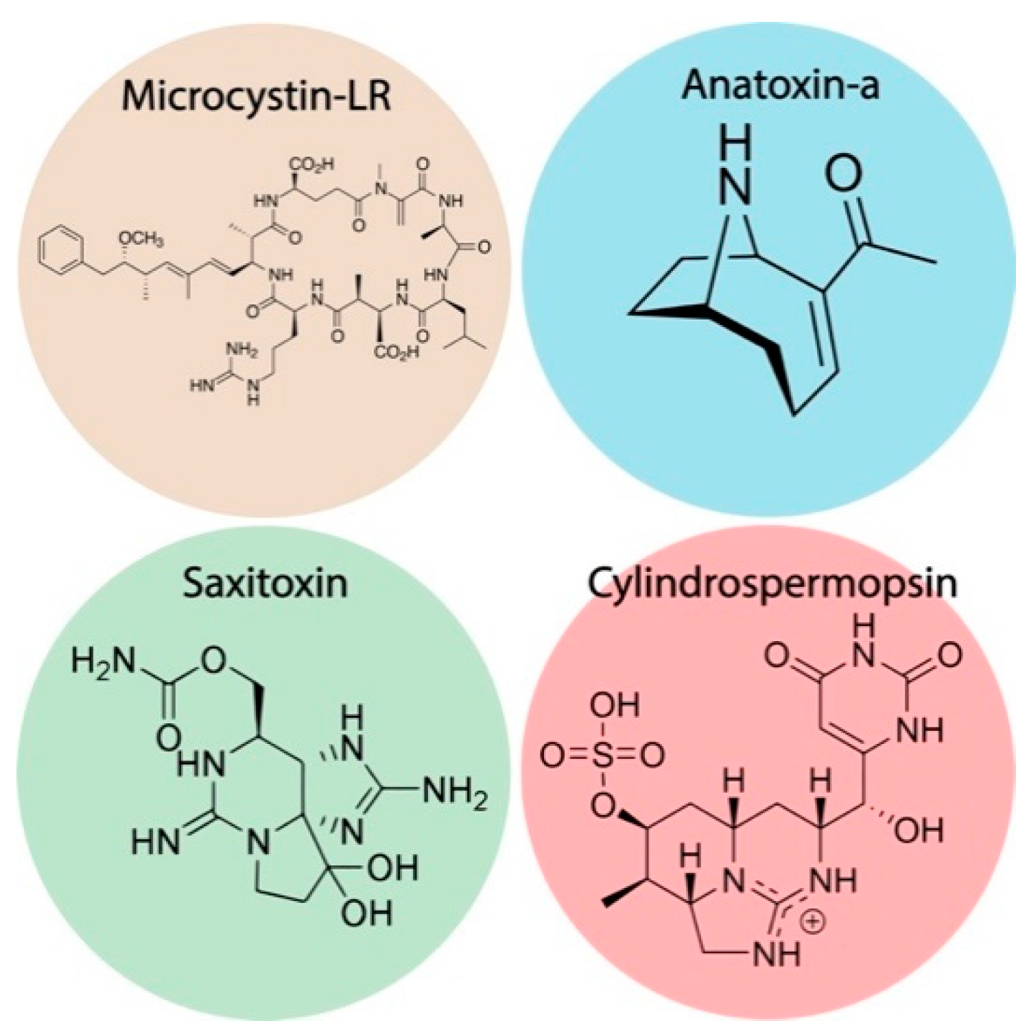
2. Microcystins
2.1. Microcystin-LR
2.2. Anatoxin-a
2.3. Saxitoxin
2.4. Cylindrospermopsin
3. Discussion and Future Outlooks
4. Conclusions
Author Contributions
Funding
Institutional Review Board Statement
Informed Consent Statement
Data Availability Statement
Conflicts of Interest
References
- Anderson, D.M.; Glibert, P.M.; Burkholder, J.M. Harmful Algal Blooms and Eutrophication: Nutrient Sources, Composition, and Consequences. Estuaries 2002, 25, 704–726. [Google Scholar] [CrossRef]
- Masó, M.; Garcés, E. Harmful Microalgae Blooms (HAB); Problematic and Conditions That Induce Them. Mar. Pollut. Bull. 2006, 53, 620–630. [Google Scholar] [CrossRef] [PubMed]
- McCarthy, M.; Bane, V.; García-Altares, M.; Van Pelt, F.N.A.M.; Furey, A.; O’Halloran, J. Assessment of Emerging Biotoxins (Pinnatoxin G and Spirolides) at Europe’s First Marine Reserve: Lough Hyne. Toxicon 2015, 108, 202–209. [Google Scholar] [CrossRef]
- Smayda, T.J. Reflections on the Ballast Water Dispersal—Harmful Algal Bloom Paradigm. Harmful Algae 2007, 6, 601–622. [Google Scholar] [CrossRef]
- Moreira, C.; Vasconcelos, V.; Antunes, A. Phylogeny and Biogeography of Cyanobacteria and Their Produced Toxins. Mar. Drugs 2013, 11, 4350–4369. [Google Scholar] [CrossRef] [Green Version]
- Moreira, C.; Ramos, V.; Azevedo, J.; Vasconcelos, V. Methods to Detect Cyanobacteria and Their Toxins in the Environment. Appl. Microbiol. Biotechnol. 2014, 98, 8073–8082. [Google Scholar] [CrossRef] [PubMed]
- Catherine, A.; Bernard, C.; Spoof, L.; Bruno, M. Microcystins and Nodularins. In Handbook of Cyanobacterial Monitoring and Cyanotoxin Analysis; John Wiley & Sons, Ltd.: Hoboken, NJ, USA, 2016; pp. 107–126. [Google Scholar] [CrossRef]
- World Health Organization. Guidelines for Drinking-Water Quality; World Health Organization: Geneva, Switzerland, 1993. [Google Scholar]
- Lawton, L.A.; Chambers, H.; Edwards, C.; Nwaopara, A.A.; Healy, M. Rapid Detection of Microcystins in Cells and Water. Toxicon 2010, 55, 973–978. [Google Scholar] [CrossRef] [PubMed]
- Toxins, C. Microcystin-LR in Drinking-Water. In Background Document for Development of WHO Guidelines for Drinking-Water Quality; WHO: Geneva, Switzerland, 2003. [Google Scholar]
- Schaefer, A.M.; Yrastorza, L.; Stockley, N.; Harvey, K.; Harris, N.; Grady, R.; Sullivan, J.; McFarland, M.; Reif, J.S. Exposure to Microcystin among Coastal Residents during a Cyanobacteria Bloom in Florida. Harmful Algae 2020, 92, 101769. [Google Scholar] [CrossRef]
- Poste, A.E.; Hecky, R.E.; Guildford, S.J. Evaluating Microcystin Exposure Risk through Fish Consumption. Environ. Sci. Technol. 2011, 45, 5806–5811. [Google Scholar] [CrossRef] [Green Version]
- Backer, L.C.; Carmichael, W.; Kirkpatrick, B.; Williams, C.; Irvin, M.; Zhou, Y.; Johnson, T.B.; Nierenberg, K.; Hill, V.R.; Kieszak, S.M.; et al. Recreational Exposure to Low Concentrations of Microcystins During an Algal Bloom in a Small Lake. Mar. Drugs 2008, 6, 389–406. [Google Scholar] [CrossRef]
- Devlin, J.P.; Edwards, O.E.; Gorham, P.R.; Hunter, N.R.; Pike, R.K.; Stavric, B. Anatoxin-a, a Toxic Alkaloid from Anabaena Flos-Aquae NRC-44h. Can. J. Chem. 1977, 55, 1367–1371. [Google Scholar] [CrossRef]
- Osswald, J.; Rellán, S.; Gago, A.; Vasconcelos, V. Toxicology and Detection Methods of the Alkaloid Neurotoxin Produced by Cyanobacteria, Anatoxin-a. Environ. Int. 2007, 33, 1070–1089. [Google Scholar] [CrossRef]
- Hackett, J.D.; Wisecaver, J.H.; Brosnahan, M.L.; Kulis, D.M.; Anderson, D.M.; Bhattacharya, D.; Plumley, F.G.; Erdner, D.L. Evolution of Saxitoxin Synthesis in Cyanobacteria and Dinoflagellates. Mol. Biol. Evol. 2013, 30, 70–78. [Google Scholar] [CrossRef] [Green Version]
- McElhiney, J.; Lawton, L.A. Detection of the Cyanobacterial Hepatotoxins Microcystins. Toxicol. Appl. Pharmacol. 2005, 203, 219–230. [Google Scholar] [CrossRef] [PubMed]
- Zaffiro, A.; Rosenblum, L.; Wendelken, S.C. Method 546: Determination of Total Microcystins and Nodularins in Drinking Water and Ambient Water by Adda Enzyme-Linked Immunosorbent Assay; US EPA (United States Environmental Protection Agency), Standards and Risk Management Division: Cincinnati, OH, USA, 2016; pp. 1–21. [Google Scholar]
- Shoemaker, J.A.; Tettenhorst, D.R.; De la Cruz, A. Determination of Microcystins and Nodularin in Drinking Water by Solid Phase Extraction and Liquid Chromatography/Tandem Mass Spectrometry (LC/MS/MS); EPA: Washington, DC, USA, 2015; EPA method 544. EPA Document #: EPA/600/R-14/474. Version 1.0. [Google Scholar]
- Campbell, D.L.; Lawton, L.A.; Beattie, K.A.; Codd, G.A. Comparative Assessment of the Specificity of the Brine Shrimp and Microtox Assays to Hepatotoxic (Microcystin-LR-Containing) Cyanobacteria. Environ. Toxicol. Water Qual. 1994, 9, 71–77. [Google Scholar] [CrossRef]
- Falconer, I.R. CHAPTER 10—Measurement of Toxins from Blue-Green Algae in Water and Foodstuffs. In Algal Toxins in Seafood and Drinking Water; Falconer, I.R., Ed.; Academic Press: San Diego, CA, USA, 1993; pp. 165–175. [Google Scholar] [CrossRef]
- MacKintosh, C.; Beattie, K.A.; Klumpp, S.; Cohen, P.; Codd, G.A. Cyanobacterial Microcystin-LR Is a Potent and Specific Inhibitor of Protein Phosphatases 1 and 2A from Both Mammals and Higher Plants. FEBS Lett. 1990, 264, 187–192. [Google Scholar] [CrossRef] [Green Version]
- An, J.; Carmichael, W.W. Use of a Colorimetric Protein Phosphatase Inhibition Assay and Enzyme Linked Immunosorbent Assay for the Study of Microcystins and Nodularins. Toxicon 1994, 32, 1495–1507. [Google Scholar] [CrossRef]
- Bouaïcha, N.; Maatouk, I.; Vincent, G.; Levi, Y. A Colorimetric and Fluorometric Microplate Assay for the Detection of Microcystin-LR in Drinking Water without Preconcentration. Food Chem. Toxicol. 2002, 40, 1677–1683. [Google Scholar] [CrossRef]
- Heresztyn, T.; Nicholson, B.C. Determination of Cyanobacterial Hepatotoxins Directly in Water Using a Protein Phosphatase Inhibition Assay. Water Res. 2001, 35, 3049–3056. [Google Scholar] [CrossRef]
- Rivasseau, C.; Racaud, P.; Deguin, A.; Hennion, M.-C. Development of a Bioanalytical Phosphatase Inhibition Test for the Monitoring of Microcystins in Environmental Water Samples. Anal. Chim. Acta 1999, 394, 243–257. [Google Scholar] [CrossRef]
- Wong, B.S.F.; Lam, P.K.S.; Xu, L.; Zhang, Y.; Richardson, B.J. A Colorimetric Assay for Screening Microcystin Class Compounds in Aquatic Systems. Chemosphere 1999, 38, 1113–1122. [Google Scholar] [CrossRef]
- Ward, C.J.; Beattie, K.A.; Lee, E.Y.C.; Codd, G.A. Colorimetric Protein Phosphatase Inhibition Assay of Laboratory Strains and Natural Blooms of Cyanobacteria: Comparisons with High-Performance Liquid Chromatographic Analysis for Microcystins. FEMS Microbiol. Lett. 1997, 153, 465–473. [Google Scholar] [CrossRef]
- Lawton, L.A.; Edwards, C.; Codd, G.A. Extraction and High-Performance Liquid Chromatographic Method for the Determination of Microcystins in Raw and Treated Waters. Analyst 1994, 119, 1525–1530. [Google Scholar] [CrossRef]
- Tsuji, K.; Naito, S.; Kondo, F.; Watanabe, M.F.; Suzuki, S.; Nakazawa, H.; Suzuki, M.; Shimada, T.; Harada, K.-I. A Clean-up Method for Analysis of Trace Amounts of Microcystins in Lake Water. Toxicon 1994, 32, 1251–1259. [Google Scholar] [CrossRef]
- Edwards, C.; Lawton, L.A.; Beattie, K.A.; Codd, G.A.; Pleasance, S.; Dear, G.J. Analysis of Microcystins from Cyanobacteria by Liquid Chromatography with Mass Spectrometry Using Atmospheric-Pressure Ionization. Rapid Commun. Mass Spectrom. 1993, 7, 714–721. [Google Scholar] [CrossRef]
- Pelander, A.; Ojanperä, I.; Lahti, K.; Niinivaara, K.; Vuori, E. Visual Detection of Cyanobacterial Hepatotoxins by Thin-Layer Chromatography and Application to Water Analysis. Water Res. 2000, 34, 2643–2652. [Google Scholar] [CrossRef]
- Vasas, G.; Szydlowska, D.; Gáspár, A.; Welker, M.; Trojanowicz, M.; Borbély, G. Determination of Microcystins in Environmental Samples Using Capillary Electrophoresis. J. Biochem. Biophys. Methods 2006, 66, 87–97. [Google Scholar] [CrossRef]
- Sirén, H.; Jussila, M.; Liu, H.; Peltoniemi, S.; Sivonen, K.; Riekkola, M.-L. Separation, Purity Testing and Identification of Cyanobacterial Hepatotoxins with Capillary Electrophoresis and Electrospray Mass Spectrometry. J. Chromatogr. A 1999, 839, 203–215. [Google Scholar] [CrossRef]
- Tsuji, K.; Masui, H.; Uemura, H.; Mori, Y.; Harada, K. Analysis of Microcystins in Sediments Using MMPB Method. Toxicon 2001, 39, 687–692. [Google Scholar] [CrossRef]
- Cinti, S.; Moscone, D.; Arduini, F. Preparation of Paper-Based Devices for Reagentless Electrochemical (Bio)Sensor Strips. Nat. Protoc. 2019, 14, 2437–2451. [Google Scholar] [CrossRef]
- Parolo, C.; Sena-Torralba, A.; Bergua, J.F.; Calucho, E.; Fuentes-Chust, C.; Hu, L.; Rivas, L.; Álvarez-Diduk, R.; Nguyen, E.P.; Cinti, S.; et al. Tutorial: Design and Fabrication of Nanoparticle-Based Lateral-Flow Immunoassays. Nat. Protoc. 2020, 15, 3788–3816. [Google Scholar] [CrossRef]
- Salentijn, G.I.J.; Grajewski, M.; Verpoorte, E. Reinventing (Bio)Chemical Analysis with Paper. Anal. Chem. 2018, 90, 13815–13825. [Google Scholar] [CrossRef] [Green Version]
- Cinti, S.; Cinotti, G.; Parolo, C.; Nguyen, E.P.; Caratelli, V.; Moscone, D.; Arduini, F.; Merkoci, A. Experimental Comparison in Sensing Breast Cancer Mutations by Signal ON and Signal OFF Paper-Based Electroanalytical Strips. Anal. Chem. 2020, 92, 1674–1679. [Google Scholar] [CrossRef] [PubMed]
- Noiphung, J.; Songjaroen, T.; Dungchai, W.; Henry, C.S.; Chailapakul, O.; Laiwattanapaisal, W. Electrochemical Detection of Glucose from Whole Blood Using Paper-Based Microfluidic Devices. Anal. Chim. Acta 2013, 788, 39–45. [Google Scholar] [CrossRef]
- Bagheri, N.; Mazzaracchio, V.; Cinti, S.; Colozza, N.; Di Natale, C.; Netti, P.A.; Saraji, M.; Roggero, S.; Moscone, D.; Arduini, F. Electroanalytical Sensor Based on Gold-Nanoparticle-Decorated Paper for Sensitive Detection of Copper Ions in Sweat and Serum. Anal. Chem. 2021, 93, 5225–5233. [Google Scholar] [CrossRef] [PubMed]
- Eissa, S.; Ng, A.; Siaj, M.; Zourob, M. Label-Free Voltammetric Aptasensor for the Sensitive Detection of Microcystin-LR Using Graphene-Modified Electrodes. Anal. Chem. 2014, 86, 7551–7557. [Google Scholar] [CrossRef] [PubMed]
- Lebogang, L.; Jantra, J.; Hedström, M.; Mattiasson, B. Electrochemical Flow-ELISA for Rapid and Sensitive Determination of Microcystin-LR Using Automated Sequential Injection System. Sensors 2017, 17, 1639. [Google Scholar] [CrossRef] [Green Version]
- Lin, Z.; Huang, H.; Xu, Y.; Gao, X.; Qiu, B.; Chen, X.; Chen, G. Determination of Microcystin-LR in Water by a Label-Free Aptamer Based Electrochemical Impedance Biosensor. Talanta 2013, 103, 371–374. [Google Scholar] [CrossRef]
- Reverté, L.; Garibo, D.; Flores, C.; Diogène, J.; Caixach, J.; Campàs, M. Magnetic Particle-Based Enzyme Assays and Immunoassays for Microcystins: From Colorimetric to Electrochemical Detection. Environ. Sci. Technol. 2013, 47, 471–478. [Google Scholar] [CrossRef]
- Du, X.; Jiang, D.; Dai, L.; Zhou, L.; Hao, N.; Qian, J.; Qiu, B.; Wang, K. Fabricating Photoelectrochemical Aptasensor for Selectively Monitoring Microcystin-LR Residues in Fish Based on Visible Light-Responsive BiOBr Nanoflakes/N-Doped Graphene Photoelectrode. Biosens. Bioelectron. 2016, 81, 242–248. [Google Scholar] [CrossRef]
- Bilibana, M.P.; Williams, A.R.; Rassie, C.; Sunday, C.E.; Makelane, H.; Wilson, L.; Ntshongontshi, N.; Jijana, A.N.; Masikini, M.; Baker, P.G.L.; et al. Electrochemical Aptatoxisensor Responses on Nanocomposites Containing Electro-Deposited Silver Nanoparticles on Poly(Propyleneimine) Dendrimer for the Detection of Microcystin-LR in Freshwater. Sensors 2016, 16, 1901. [Google Scholar] [CrossRef] [PubMed] [Green Version]
- Zhang, J.; Lei, J.; Pan, R.; Leng, C.; Hu, Z.; Ju, H. In Situ Assembly of Gold Nanoparticles on Nitrogen-Doped Carbon Nanotubes for Sensitive Immunosensing of Microcystin-LR. Chem. Commun. 2010, 47, 668–670. [Google Scholar] [CrossRef]
- Lotierzo, M.; Abuknesha, R.; Davis, F.; Tothill, I.E. A Membrane-Based ELISA Assay and Electrochemical Immunosensor for Microcystin-LR in Water Samples. Environ. Sci. Technol. 2012, 46, 5504–5510. [Google Scholar] [CrossRef]
- Zhang, W.; Han, C.; Jia, B.; Saint, C.; Nadagouda, M.; Falaras, P.; Sygellou, L.; Vogiazi, V.; Dionysiou, D.D. A 3D Graphene-Based Biosensor as an Early Microcystin-LR Screening Tool in Sources of Drinking Water Supply. Electrochim. Acta 2017, 236, 319–327. [Google Scholar] [CrossRef]
- Abnous, K.; Danesh, N.M.; Nameghi, M.A.; Ramezani, M.; Alibolandi, M.; Lavaee, P.; Taghdisi, S.M. An Ultrasensitive Electrochemical Sensing Method for Detection of Microcystin-LR Based on Infinity-Shaped DNA Structure Using Double Aptamer and Terminal Deoxynucleotidyl Transferase. Biosens. Bioelectron. 2019, 144, 111674. [Google Scholar] [CrossRef]
- Wu, J.; Yu, C.; Yu, Y.; Chen, J.; Zhang, C.; Gao, R.; Mu, X.; Geng, Y.; He, J. Ultra-Sensitive Detection of Microcystin-LR with a New Dual-Mode Aptasensor Based on MoS2-PtPd and ZIF-8-Thi-Au. Sens. Actuators B Chem. 2020, 305, 127280. [Google Scholar] [CrossRef]
- Elshafey, R.; Siaj, M.; Zourob, M. DNA Aptamers Selection and Characterization for Development of Label-Free Impedimetric Aptasensor for Neurotoxin Anatoxin-a. Biosens. Bioelectron. 2015, 68, 295–302. [Google Scholar] [CrossRef]
- Fawell, J.K.; Mitchell, R.E.; Hill, R.E.; Everett, D.J. The Toxicity of Cyanobacterial Toxins in the Mouse: II Anatoxin-a. Hum. Exp. Toxicol. 1999, 18, 168–173. [Google Scholar] [CrossRef] [PubMed]
- Devic, E.; Li, D.; Dauta, A.; Henriksen, P.; Codd, G.A.; Marty, J.-L.; Fournier, D. Detection of Anatoxin-a(s) in Environmental Samples of Cyanobacteria by Using a Biosensor with Engineered Acetylcholinesterases. Appl. Environ. Microbiol. 2002, 68, 4102–4106. [Google Scholar] [CrossRef] [Green Version]
- Cinti, S. Novel Paper-Based Electroanalytical Tools for Food Surveillance. Anal. Bioanal. Chem. 2019, 411, 4303–4311. [Google Scholar] [CrossRef]
- Patočka, J.; Gupta, R.C.; Kuča, K. Anatoxin-a(s): Natural Organophosphorus Anticholinesterase Agent. Mil. Med. Sci. Lett. 2011, 80, 129–139. [Google Scholar] [CrossRef]
- Villatte, F.; Schulze, H.; Schmid, R.; Bachmann, T. A Disposable Acetylcholinesterase-Based Electrode Biosensor to Detect Anatoxin-a(s) in Water. Anal. Bioanal. Chem. 2002, 372, 322–326. [Google Scholar] [CrossRef]
- Wharton, R.E.; Feyereisen, M.C.; Gonzalez, A.L.; Abbott, N.L.; Hamelin, E.I.; Johnson, R.C. Quantification of Saxitoxin in Human Blood by ELISA. Toxicon 2017, 133, 110–115. [Google Scholar] [CrossRef]
- Bragg, W.A.; Garrett, A.; Hamelin, E.I.; Coleman, R.M.; Campbell, K.; Elliott, C.T.; Johnson, R.C. Quantitation of Saxitoxin in Human Urine Using Immunocapture Extraction and LC–MS. Bioanalysis 2018, 10, 229–239. [Google Scholar] [CrossRef] [PubMed]
- Munday, R.; Thomas, K.; Gibbs, R.; Murphy, C.; Quilliam, M.A. Acute Toxicities of Saxitoxin, Neosaxitoxin, Decarbamoyl Saxitoxin and Gonyautoxins 1&4 and 2&3 to Mice by Various Routes of Administration. Toxicon 2013, 76, 77–83. [Google Scholar] [CrossRef]
- Bratakou, S.; Nikoleli, G.-P.; Siontorou, C.G.; Nikolelis, D.P.; Karapetis, S.; Tzamtzis, N. Development of an Electrochemical Biosensor for the Rapid Detection of Saxitoxin Based on Air Stable Lipid Films with Incorporated Anti-STX Using Graphene Electrodes. Electroanalysis 2017, 29, 990–997. [Google Scholar] [CrossRef]
- Hou, L.; Jiang, L.; Song, Y.; Ding, Y.; Zhang, J.; Wu, X.; Tang, D. Amperometric Aptasensor for Saxitoxin Using a Gold Electrode Modified with Carbon Nanotubes on a Self-Assembled Monolayer, and Methylene Blue as an Electrochemical Indicator Probe. Microchim. Acta 2016, 183, 1971–1980. [Google Scholar] [CrossRef]
- Jin, X.; Chen, J.; Zeng, X.; Xu, L.; Wu, Y.; Fu, F. A Signal-on Magnetic Electrochemical Immunosensor for Ultra-Sensitive Detection of Saxitoxin Using Palladium-Doped Graphitic Carbon Nitride-Based Non-Competitive Strategy. Biosens. Bioelectron. 2019, 128, 45–51. [Google Scholar] [CrossRef]
- Serrano, P.C.; Nunes, G.E.; Avila, L.B.; Reis, C.P.S.; Gomes, A.M.C.; Reis, F.T.; Sartorelli, M.L.; Melegari, S.P.; Matias, W.G.; Bechtold, I.H. Electrochemical Impedance Biosensor for Detection of Saxitoxin in Aqueous Solution. Anal. Bioanal. Chem. 2021. [Google Scholar] [CrossRef]
- Elshafey, R.; Siaj, M.; Zourob, M. In Vitro Selection, Characterization, and Biosensing Application of High-Affinity Cylindrospermopsin-Targeting Aptamers. Anal. Chem. 2014, 86, 9196–9203. [Google Scholar] [CrossRef]
- Valério, E.; Tenreiro, A.; Abrantes, L.M. Preliminary Studies Towards the Development of DNA Biosensors for Detection of Cylindrospermopsin—A Cyanobacterial Toxin. Port. Electrochim. Acta 2007, 26, 77–87. [Google Scholar] [CrossRef]
- Valério, E.; Abrantes, L.M.; Viana, A.S. 4-Aminothiophenol Self-Assembled Monolayer for the Development of a DNA Biosensor Aiming the Detection of Cylindrospermopsin Producing Cyanobacteria. Electroanalysis 2008, 20, 2467–2474. [Google Scholar] [CrossRef]
- Zhao, Z.; Chen, H.; Ma, L.; Liu, D.; Wang, Z. A Label-Free Electrochemical Impedance Aptasensor for Cylindrospermopsin Detection Based on Thionine–Graphene Nanocomposites. Analyst 2015, 140, 5570–5577. [Google Scholar] [CrossRef]
- Vogiazi, V.; De la Cruz, A.; Mishra, S.; Shanov, V.; Heineman, W.R.; Dionysiou, D.D. A comprehensive review: Development of electrochemical biosensors for detection of cyanotoxins in freshwater. ACS Sens. 2019, 4, 1151–1173. [Google Scholar] [CrossRef]
- Zhang, W.; Dixon, M.B.; Saint, C.; Teng, K.S.; Furumai, H. Electrochemical biosensing of Algal toxins in water: The current state-of-the-art. ACS Sens. 2018, 3, 1233–1245. [Google Scholar] [CrossRef]
- Reverté, L.; Prieto-Simón, B.; Campàs, M. New advances in electrochemical biosensors for the detection of toxins: Nanomaterials, magnetic beads and microfluidics systems. A review. Anal. Chim. Acta 2016, 908, 8–21. [Google Scholar] [CrossRef]
- Tortorella, S.; Cinti, S. How Can Chemometrics Support the Development of Point of Need Devices? Anal. Chem. 2021, 93, 2713–2722. [Google Scholar] [CrossRef]
- Paulovich, F.V.; De Oliveira, M.C.F.; Oliveira, O.N. A Future with Ubiquitous Sensing and Intelligent Systems. ACS Sens. 2018, 3, 1433–1438. [Google Scholar] [CrossRef] [PubMed] [Green Version]
- Cui, F.; Yue, Y.; Zhang, Y.; Zhang, Z.; Zhou, H.S. Advancing Biosensors with Machine Learning. ACS Sens. 2020, 5, 3346–3364. [Google Scholar] [CrossRef] [PubMed]
- Cinti, S.; Valdés-Ramírez, G.; Gao, W.; Li, J.; Palleschi, G.; Wang, J. Microengine-Assisted Electrochemical Measurements at Printable Sensor Strips. Chem. Commun. 2015, 51, 8668–8671. [Google Scholar] [CrossRef] [PubMed] [Green Version]
- Medina-Sánchez, M.; Mayorga-Martinez, C.C.; Watanabe, T.; Ivandini, T.A.; Honda, Y.; Pino, F.; Nakata, A.; Fujishima, A.; Einaga, Y.; Merkoçi, A. Microfluidic Platform for Environmental Contaminants Sensing and Degradation Based on Boron-Doped Diamond Electrodes. Biosens. Bioelectron. 2016, 75, 365–374. [Google Scholar] [CrossRef] [PubMed]
- Cioffi, A.; Mancini, M.; Gioia, V.; Cinti, S. Office Paper-Based Electrochemical Strips for Organophosphorus Pesticide Monitoring in Agricultural Soil. Environ. Sci. Technol. 2021, 55, 8859–8865. [Google Scholar] [CrossRef] [PubMed]
- Cinti, S.; Minotti, C.; Moscone, D.; Palleschi, G.; Arduini, F. Fully Integrated Ready-to-Use Paper-Based Electrochemical Biosensor to Detect Nerve Agents. Biosens. Bioelectron. 2017, 93, 46–51. [Google Scholar] [CrossRef] [PubMed]


| Cyanotoxin | Electrochemical Method | Sensing Platform | Electrode Materials/Nanomaterials | LOD | Time of Measure | Real Matrix | Ref |
|---|---|---|---|---|---|---|---|
| Microcystin-LR | Voltammetry | Aptamer assembled on a modified electrode with graphene | Carbon nanomaterial graphene | 0.0019 nM | 10 min | Drinking water | [42] |
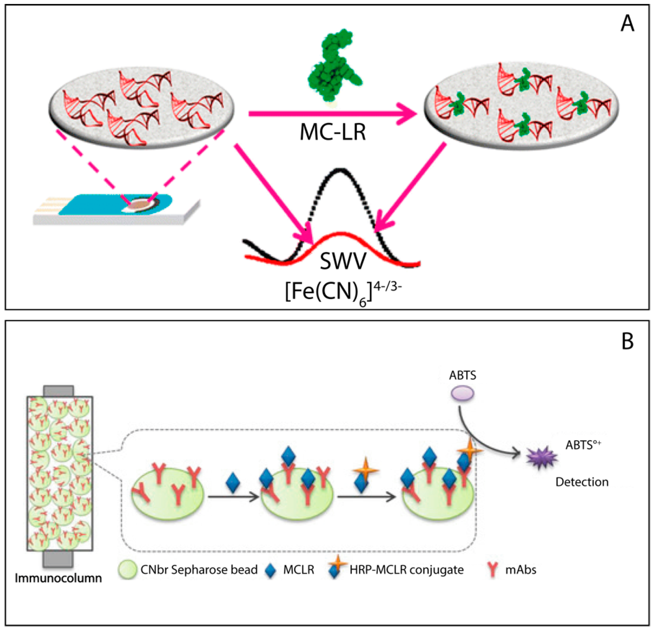
| Microcystin-LR | Amperometry | Immobilized monoclonal antibodies on Sepharose beads | Sepharose beads on a screen printed electrode | 10 nM 1000 nM | 20 min | Fresh water | [43] |
| Microcystin-LR | Impedance | Modified electrode (Au-S) | Self-assembled monolayer | 0.018 nM | 10 min | Fresh water | [44] |
| Microcystin-LR | Amperometry | Monoclonal antibodies conjugated to protein G-coated MPs | Carbon screen printed electrode | 3.9 μg/L | 10 min | Cyanobacterial culture and a natural bloom | [45] |
| Microcystin-LR | Photoelectrochem. | Photoelectrode of graphene doped with nitrogen | BiOBr nanoflakes/N-doped graphene p–n heterojunction electrode | 3.0 × 10⁻⁵ nM | 15 min | Fish | [46] |
| Microcystin-LR | Voltammetry | Drugged aptamer with electro-synthesized silver nanoparticles | AgNPs with cobalt(II) salicylaldiiminemetallodendrimer on a glassy carbon electrode | 0.04 μg/L | 10 min | Fresh water | [47] |
| Microcystin-LR | Elisa | Nitrogen-doped carbon nanotubes assembled on gold nanoparticles | AuNPs; Nitrogen-doped carbon nanotubes (Au/CNx-MWNTs) | 0.004 μg/L | 10 min | Fresh water | [48] |
| Microcystin-LR | Elisa | Polyclonal antibodies (produced in sheep) | SPE with a membrane containing an immobilized isoproturon-ovalbumin conjugate | 0.06 μg/L | 10 min | Fresh water | [49] |
| Microcystin-LR | Impedance | 3D graphene-based electrochemical impedance spectroscopy biosensor using antibodies. | 3D graphene foam (GF) sheets as working electrode | 0.05 mg/L | 30 min | Drinking water supply | [50] |
| Microcystin-LR | Voltammetry | Electrochemical detection of MC-LR based on infinity-shaped DNA structure using double aptamer and terminal deoxynucleotidyl transferase | Screen printed gold electrode | 15 pM | 90 min | Serum and tap water samples | [51] |
| Microcystin-LR | Amperometry (direct method) Voltammetry (indirect method) | Dual-mode aptasensor based on MoS2-PtPd (direct method) and ZIF-8-Thi-Au (indirect method) | GCE modified with MoS2-PtPd-NPs or (ZIF)-8-thionine (Thi)-Au | 0.006 ng/mL 0.045 ng/mL | 60 min | Water environment | [52] |
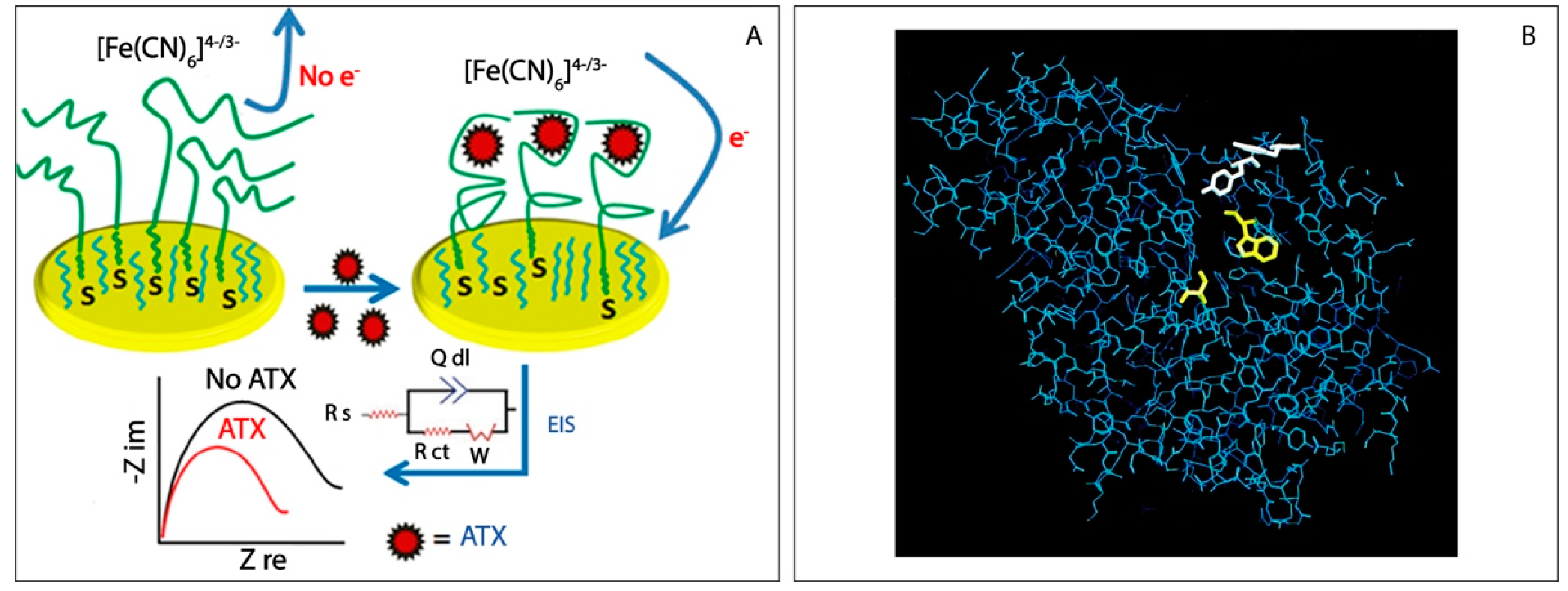
| Anatoxin-a | Impedance | Modified disulfide aptamer assembled on a gold electrode | Self-assembled monolayer | 0.05 nM | 10 min | Drinking water | [53] |
| Anatoxin-a | Amperometry | Inhibition enzymes (inhibition of acetylcholinesterase activity) | Graphite working electrode | 0.5 nmol/L | 10 min | Fresh water | [55] |
| Anatoxin-a | Amperometry | Inhibition enzymes (inhibition of acetylcholinesterase activity) | 7,7,8,8,-tetracyanoquinodimethane (TCNQ)-graphite working electrode | 1 μg/L | 10 min | Drinking water | [58] |
| Saxitoxin | Potentiometry | Anti-STX incorporated lipid films on graphene nanosheets | Graphene nanosheets. Anti-STX, immobilized on a stabilized lipid films | 1 nM | 5 min | Lake water samples, fresh shellfish samples. | [62] |
| Saxitoxin | Amperometry | Gold electrode modified with carbon nanotubes on a self-assembled monolayer | Monolaer of octadecanethiol deposited on a gold electrode, coated with MWCNTs | 0.38 nM | 30 min | Mussel samples | [63] |
| Saxitoxin | Amperometry | STX-specific antibody-functionalized magnetic beads (MBs). Palladium-doped graphitic carbon nitride (g-C3N4-PdNPs) peroxidase mimetic | Palladium-doped graphitic carbon nitride nanosheets on a magnetic gold electrode | 1.2 pg/mL | 75 min | Seawater and shellfish samples | [64] |
| Saxitoxin | Impedance | Label-free impedimetric aptasensor | Self-assembled monolayer on Au electrode | 0.3 μg/L | 60 min | Aqueous solution | [65] |
| Cylindrospermopsin | Impedance | Modified disulfide aptamer assembled on a gold electrode | Self-assembled monolayer from a disulfide-derivatized aptamer on a gold electrode | 0.1 nM | 10 min | Fresh water | [66] |
| Cylindrospermopsin | Voltammetry | Electrodes modified with polytiramine (PTy) | Pt disk | 25 pg/mL | 10 min | Fresh water | [67] |
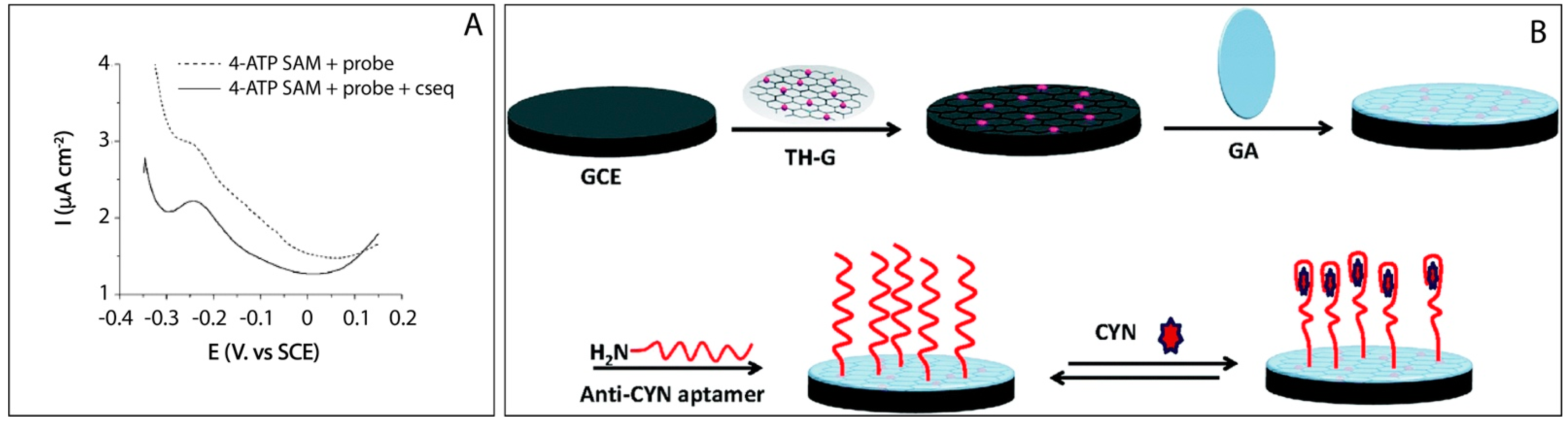
| Cylindrospermopsin | Impedance | Covalent immobilization of the aptamer of CYN on the thionine–graphene (TH–G) nanocomposite | Glassy carbon electrode | 0.117 ng/mL | 120 min | Lake water | [69] |
Publisher’s Note: MDPI stays neutral with regard to jurisdictional claims in published maps and institutional affiliations. |
© 2021 by the authors. Licensee MDPI, Basel, Switzerland. This article is an open access article distributed under the terms and conditions of the Creative Commons Attribution (CC BY) license (https://creativecommons.org/licenses/by/4.0/).
Share and Cite
Miglione, A.; Napoletano, M.; Cinti, S. Electrochemical Biosensors for Tracing Cyanotoxins in Food and Environmental Matrices. Biosensors 2021, 11, 315. https://doi.org/10.3390/bios11090315
Miglione A, Napoletano M, Cinti S. Electrochemical Biosensors for Tracing Cyanotoxins in Food and Environmental Matrices. Biosensors. 2021; 11(9):315. https://doi.org/10.3390/bios11090315
Chicago/Turabian StyleMiglione, Antonella, Maria Napoletano, and Stefano Cinti. 2021. "Electrochemical Biosensors for Tracing Cyanotoxins in Food and Environmental Matrices" Biosensors 11, no. 9: 315. https://doi.org/10.3390/bios11090315
APA StyleMiglione, A., Napoletano, M., & Cinti, S. (2021). Electrochemical Biosensors for Tracing Cyanotoxins in Food and Environmental Matrices. Biosensors, 11(9), 315. https://doi.org/10.3390/bios11090315