A Facile Aptasensor for Instantaneous Determination of Cadmium Ions Based on Fluorescence Amplification Effect of MOPS on FAM-Labeled Aptamer

 ,
,
Abstract
1. Introduction
2. Materials and Methods
2.1. Chemicals and Apparatus
2.2. Optimization of Experimental Conditions
2.3. Sensitivity and Selectivity of the Sensing System
2.4. Application in Water Samples
2.5. Calculation of Fluorescence Quenching and Fluorescence Quenching Efficiency
3. Results and Discussion
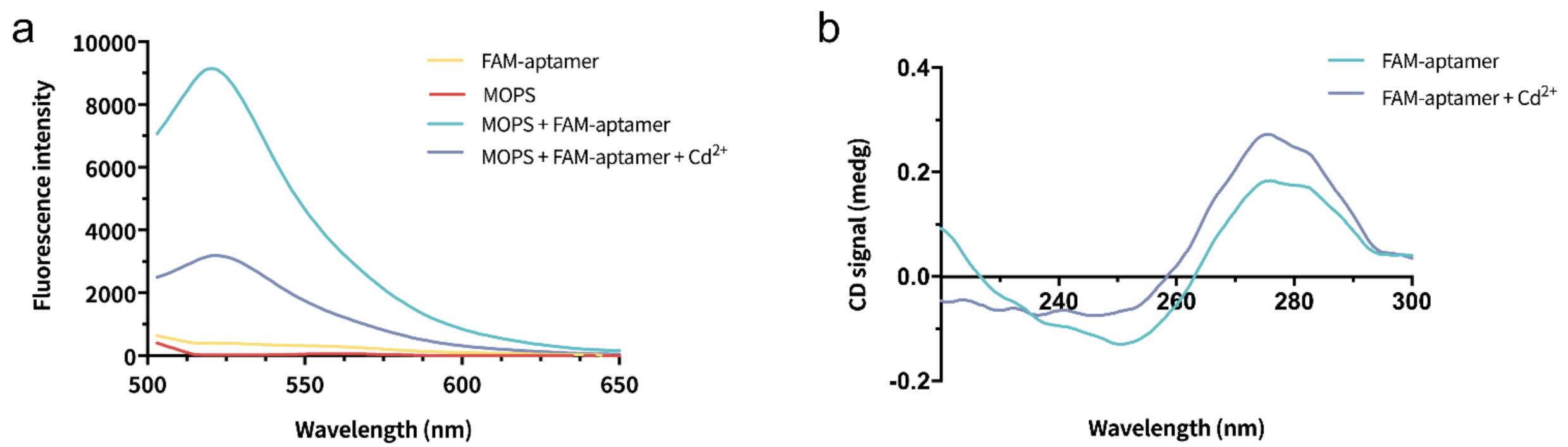
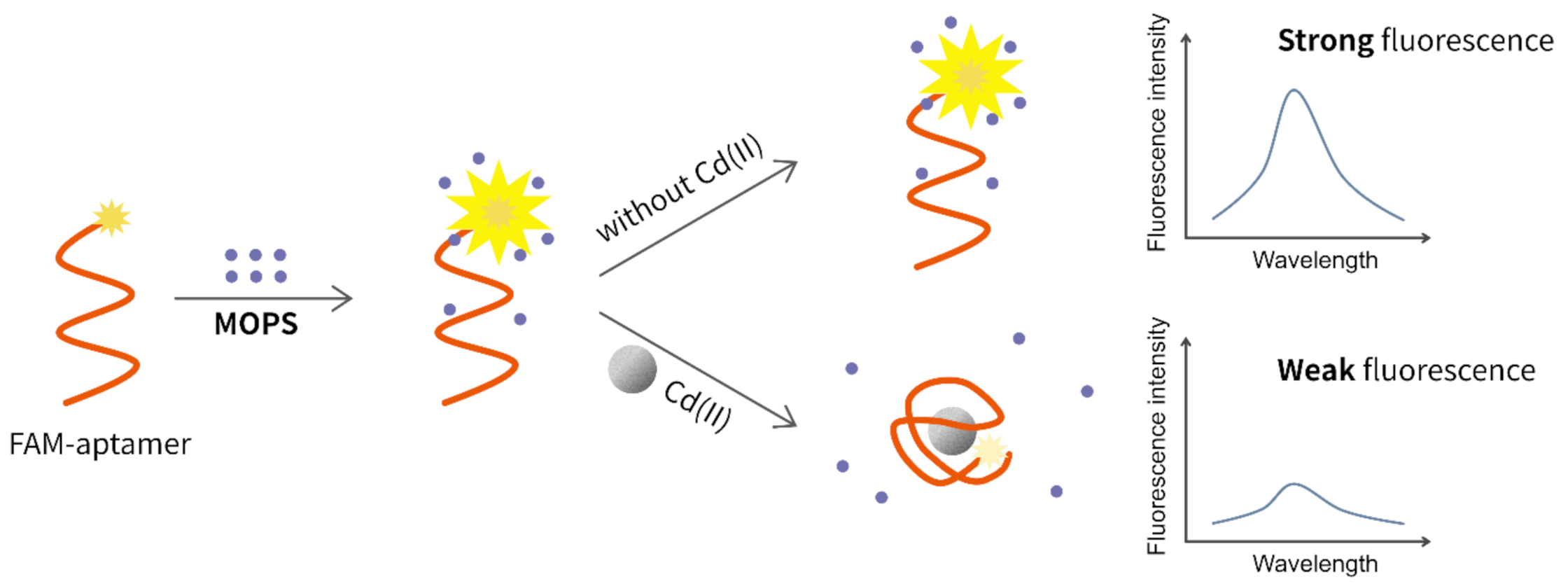
3.1. Sensing Mechanism
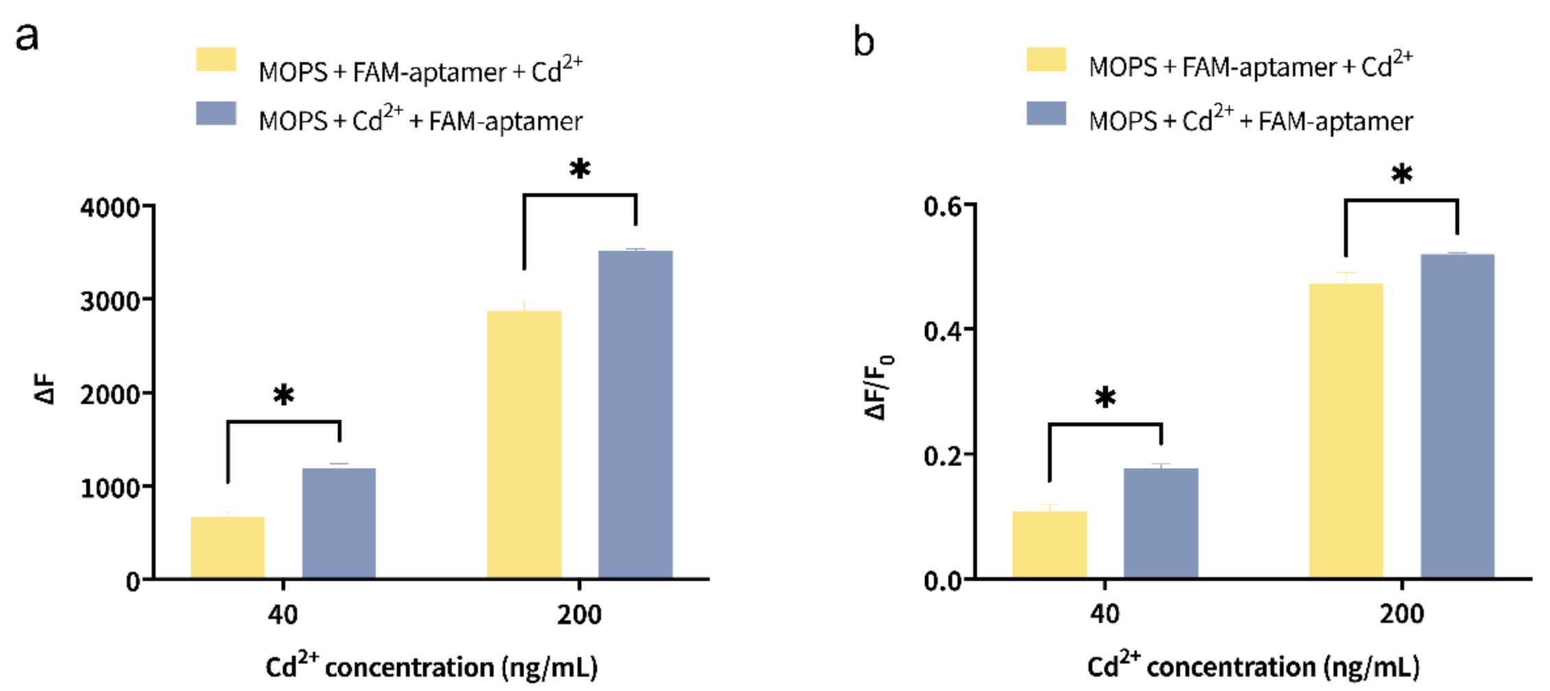
3.2. Optimization of Addition Order
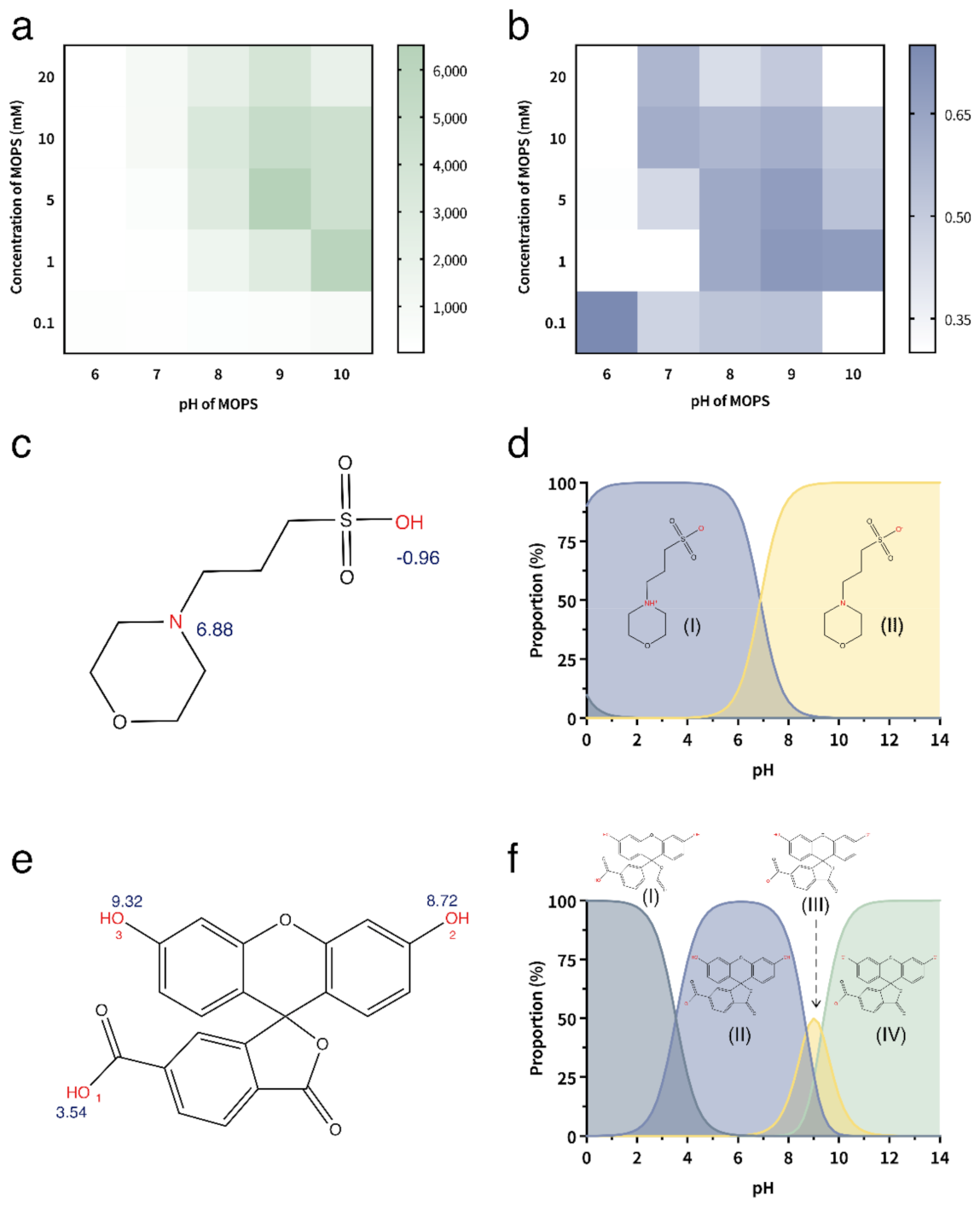
3.3. Optimization of Concentration and pH of MOPS
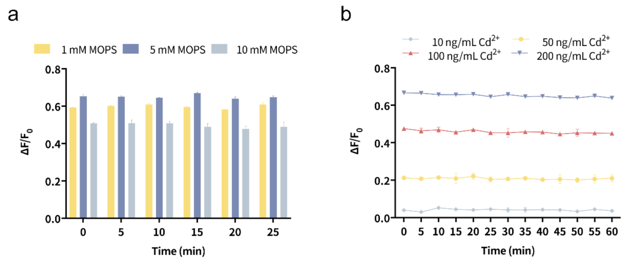
3.4. Optimization of Incubation Time
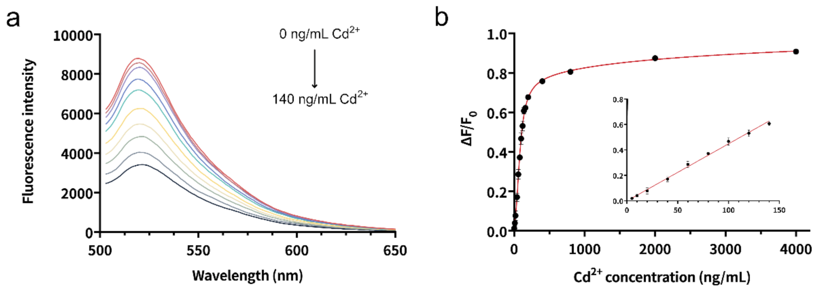
3.5. Sensitivity
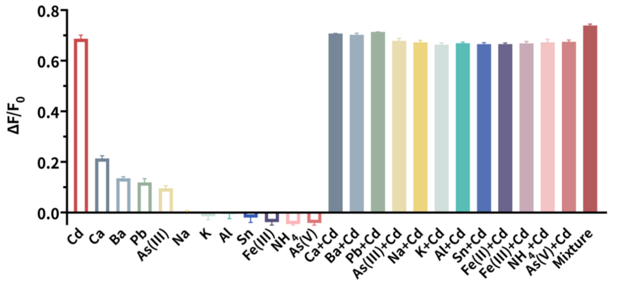
3.6. Selectivity
3.7. Reproducibility
3.8. Determination of Cd2+ in Water Samples
4. Conclusions
Author Contributions
Funding
Institutional Review Board Statement
Informed Consent Statement
Data Availability Statement
Conflicts of Interest
References
- Guo, Y.; Zhang, Y.; Shao, H.; Wang, Z.; Wang, X.; Jiang, X. Label-free colorimetric detection of cadmium ions in rice samples using gold nanoparticles. Anal. Chem. 2014, 86, 8530–8534. [Google Scholar] [CrossRef] [PubMed]
- Movaghgharnezhad, S.; Mirabi, A.; Toosi, M.R.; Rad, A.S. Synthesis of cellulose nanofibers functionalized by dithiooxamide for preconcentration and determination of trace amounts of Cd(II) ions in water samples. Cellulose 2020, 27, 8885–8898. [Google Scholar] [CrossRef]
- Priya, T.; Dhanalakshmi, N.; Thennarasu, S.; Thinakaran, N. Ultra sensitive detection of Cd (II) using reduced graphene oxide/carboxymethyl cellulose/glutathione modified electrode. Carbohydr. Polym. 2018, 197, 366–374. [Google Scholar] [CrossRef] [PubMed]
- Ashrafzadeh Afshar, E.; Taher, M.A.; Fazelirad, H. Ultra-trace determination of thallium(I) using a nanocomposite consisting of magnetite, halloysite nanotubes and dibenzo−18-crown−6 for preconcentration prior to its quantitation by ET-AAS. Microchim. Acta 2017, 184, 791–797. [Google Scholar] [CrossRef]
- Junior, M.M.; Silva, L.O.; Leao, D.J.; Ferreira, S.L. Analytical strategies for determination of cadmium in Brazilian vinegar samples using ET AAS. Food Chem. 2014, 160, 209–213. [Google Scholar] [CrossRef]
- Alvarez, M.A.; Carrillo, G. Simultaneous determination of arsenic, cadmium, copper, chromium, nickel, lead and thallium in total digested sediment samples and available fractions by electrothermal atomization atomic absorption spectroscopy (ET AAS). Talanta 2012, 97, 505–512. [Google Scholar] [CrossRef] [PubMed]
- Wang, M.; Ma, H.; Chi, Q.; Li, Q.; Li, M.; Zhang, H.; Li, C.; Fang, H. A monolithic copolymer prepared from N-(4-vinyl)-benzyl iminodiacetic acid, divinylbenzene and N,N’-methylene bisacrylamide for preconcentration of cadmium(II) and cobalt(II) from biological samples prior to their determination by ICP-MS. Microchim. Acta 2019, 186, 537. [Google Scholar] [CrossRef] [PubMed]
- Li, Y.; Guo, W.; Hu, Z.; Jin, L.; Hu, S.; Guo, Q. Method Development for Direct Multielement Quantification by LA-ICP-MS in Food Samples. J. Agric. Food Chem. 2019, 67, 935–942. [Google Scholar] [CrossRef] [PubMed]
- Zhang, N.; Shen, K.; Yang, X.; Li, Z.; Zhou, T.; Zhang, Y.; Sheng, Q.; Zheng, J. Simultaneous determination of arsenic, cadmium and lead in plant foods by ICP-MS combined with automated focused infrared ashing and cold trap. Food Chem. 2018, 264, 462–470. [Google Scholar] [CrossRef] [PubMed]
- Nunes, M.A.; Voss, M.; Corazza, G.; Flores, E.M.; Dressler, V.L. External calibration strategy for trace element quantification in botanical samples by LA-ICP-MS using filter paper. Anal. Chim. Acta 2016, 905, 51–57. [Google Scholar] [CrossRef] [PubMed]
- Paixao, L.B.; Brandao, G.C.; Araujo, R.G.O.; Korn, M.G.A. Assessment of cadmium and lead in commercial coconut water and industrialized coconut milk employing HR-CS GF AAS. Food Chem. 2019, 284, 259–263. [Google Scholar] [CrossRef] [PubMed]
- Dos Santos, J.M.; Quinaia, S.P.; Felsner, M.L. Fast and direct analysis of Cr, Cd and Pb in brown sugar by GF AAS. Food Chem. 2018, 260, 19–26. [Google Scholar] [CrossRef] [PubMed]
- Tuerk, C.; Gold, L. Systematic evolution of ligands by exponential enrichment: RNA ligands to bacteriophage T4 DNA polymerase. Science 1990, 249, 505–510. [Google Scholar] [CrossRef] [PubMed]
- Ellington, A.D.; Szostak, J.W. In vitro selection of RNA molecules that bind specific ligands. Nature 1990, 346, 818–822. [Google Scholar] [CrossRef] [PubMed]
- Yuce, M.; Ullah, N.; Budak, H. Trends in aptamer selection methods and applications. Analyst 2015, 140, 5379–5399. [Google Scholar] [CrossRef] [PubMed]
- Zhu, C.; Li, L.; Wang, Z.; Irfan, M.; Qu, F. Recent advances of aptasensors for exosomes detection. Biosens. Bioelectron. 2020, 160, 112213. [Google Scholar] [CrossRef] [PubMed]
- Zeng, L.; Gong, J.; Rong, P.; Liu, C.; Chen, J. A portable and quantitative biosensor for cadmium detection using glucometer as the point-of-use device. Talanta 2019, 198, 412–416. [Google Scholar] [CrossRef]
- Zhu, Y.F.; Wang, Y.S.; Zhou, B.; Yu, J.H.; Peng, L.L.; Huang, Y.Q.; Li, X.J.; Chen, S.H.; Tang, X.; Wang, X.F. A multifunctional fluorescent aptamer probe for highly sensitive and selective detection of cadmium(II). Anal. Bioanal. Chem. 2017, 409, 4951–4958. [Google Scholar] [CrossRef]
- Lotfi Zadeh Zhad, H.R.; Rodríguez Torres, Y.M.; Lai, R.Y. A reagentless and reusable electrochemical aptamer-based sensor for rapid detection of Cd(II). J. Electroanal. Chem. 2017, 803, 89–94. [Google Scholar] [CrossRef]
- Wang, H.; Cheng, H.; Wang, J.; Xu, L.; Chen, H.; Pei, R. Selection and characterization of DNA aptamers for the development of light-up biosensor to detect Cd(II). Talanta 2016, 154, 498–503. [Google Scholar] [CrossRef]
- Wu, Y.; Zhan, S.; Wang, L.; Zhou, P. Selection of a DNA aptamer for cadmium detection based on cationic polymer mediated aggregation of gold nanoparticles. Analyst 2014, 139, 1550–1561. [Google Scholar] [CrossRef]
- Solra, M.; Bala, R.; Wangoo, N.; Soni, G.K.; Kumar, M.; Sharma, R.K. Optical pico-biosensing of lead using plasmonic gold nanoparticles and a cationic peptide-based aptasensor. Chem. Commun. 2019, 56, 289–292. [Google Scholar] [CrossRef] [PubMed]
- Xiao, Q.; Feng, J.; Li, J.; Feng, M.; Huang, S. A label-free and ultrasensitive electrochemical aptasensor for lead(II) using a N,P dual-doped carbon dot-chitosan composite as a signal-enhancing platform and thionine as a signaling molecule. Analyst 2018, 143, 4764–4773. [Google Scholar] [CrossRef] [PubMed]
- Ding, J.; Liu, Y.; Zhang, D.; Yu, M.; Zhan, X.; Zhang, D.; Zhou, P. An electrochemical aptasensor based on gold@polypyrrole composites for detection of lead ions. Microchim. Acta 2018, 185, 545. [Google Scholar] [CrossRef]
- Chen, Y.; Li, H.H.; Gao, T.; Zhang, T.T.; Xu, L.J.; Wang, B.; Wang, J.N.; Pei, R.J. Selection of DNA aptamers for the development of light-up biosensor to detect Pb(II). Sens. Actuators B Chem. 2018, 254, 214–221. [Google Scholar] [CrossRef]
- Yuan, M.; Song, Z.H.; Fei, J.Y.; Wang, X.L.; Xu, F.; Cao, H.; Yu, J.S. Aptasensor for lead(II) based on the use of a quartz crystal microbalance modified with gold nanoparticles. Microchim. Acta 2017, 184, 1397–1403. [Google Scholar] [CrossRef]
- Sharifi, A.; Hallaj, R.; Bahar, S.; Babamiri, B. Indirect determination of mercury(II) by using magnetic nanoparticles, CdS quantum dots and mercury(II)-binding aptamers, and quantitation of released CdS by graphite furnace AAS. Microchim. Acta 2020, 187, 91. [Google Scholar] [CrossRef]
- Ning, Y.; Hu, J.; Wei, K.; He, G.; Wu, T.; Lu, F. Fluorometric determination of mercury(II) via a graphene oxide-based assay using exonuclease III-assisted signal amplification and thymidine-Hg(II)-thymidine interaction. Microchim. Acta 2019, 186, 216. [Google Scholar] [CrossRef]
- Berlina, A.N.; Zherdev, A.V.; Dzantiev, B.B. Progress in rapid optical assays for heavy metal ions based on the use of nanoparticles and receptor molecules. Microchim. Acta 2019, 186, 172. [Google Scholar] [CrossRef]
- Wang, H.; Zhang, Y.; Ma, H.; Ren, X.; Wang, Y.; Zhang, Y.; Wei, Q. Electrochemical DNA probe for Hg2+ detection based on a triple-helix DNA and Multistage Signal Amplification Strategy. Biosens. Bioelectron. 2016, 86, 907–912. [Google Scholar] [CrossRef]
- Cui, L.; Wu, J.; Ju, H. Label-free signal-on aptasensor for sensitive electrochemical detection of arsenite. Biosens. Bioelectron. 2016, 79, 861–865. [Google Scholar] [CrossRef]
- Wu, Y.; Liu, L.; Zhan, S.; Wang, F.; Zhou, P. Ultrasensitive aptamer biosensor for arsenic(III) detection in aqueous solution based on surfactant-induced aggregation of gold nanoparticles. Analyst 2012, 137, 4171–4178. [Google Scholar] [CrossRef]
- Zhu, Y.F.; Wang, Y.S.; Zhou, B.; Huang, Y.Q.; Li, X.J.; Chen, S.H.; Wang, X.F.; Tang, X. Ultrasensitive detection of Ag(I) based on the conformational switching of a multifunctional aptamer probe induced by silver(I). Spectrochim. Acta A Mol. Biomol. Spectrosc. 2018, 189, 190–194. [Google Scholar] [CrossRef] [PubMed]
- Zhou, W.; Ding, J.; Liu, J. 2-Aminopurine-modified DNA homopolymers for robust and sensitive detection of mercury and silver. Biosens. Bioelectron. 2017, 87, 171–177. [Google Scholar] [CrossRef] [PubMed]
- Banerjee, S.; Kumar, N.P.; Srinivas, A.; Roy, S. Core-shell Fe3O4@Au nanocomposite as dual-functional optical probe and potential removal system for arsenic (III) from Water. J. Hazard Mater. 2019, 375, 216–223. [Google Scholar] [CrossRef]
- Song, J.; Huang, M.; Jiang, N.; Zheng, S.; Mu, T.; Meng, L.; Liu, Y.; Liu, J.; Chen, G. Ultrasensitive detection of amoxicillin by TiO2-g-C3N4@AuNPs impedimetric aptasensor: Fabrication, optimization, and mechanism. J. Hazard Mater. 2020, 391, 122024. [Google Scholar] [CrossRef]
- Liu, Y.; Zhang, D.; Ding, J.; Hayat, K.; Yang, X.; Zhan, X.; Zhang, D.; Lu, Y.; Zhou, P. Label-Free and Sensitive Determination of Cadmium Ions Using a Ti-Modified Co3O4-Based Electrochemical Aptasensor. Biosensors 2020, 10, 195. [Google Scholar] [CrossRef] [PubMed]
- Wang, S.; Zhang, G.; Chen, Q.; Zhou, J.; Wu, Z. Sensing of cocaine using polarized optical microscopy by exploiting the conformational changes of an aptamer at the water/liquid crystal interface. Microchim. Acta 2019, 186, 724. [Google Scholar] [CrossRef]
- Xiao, F.; Tan, H.; Wu, Y.; Liao, S.; Wu, Z.; Shen, G.; Yu, R. A novel logic gate based on liquid-crystals responding to the DNA conformational transition. Analyst 2016, 141, 2870–2873. [Google Scholar] [CrossRef]
- Chang, Y.; Liu, M.; Liu, J. Highly Selective Fluorescent Sensing of Phosphite through Recovery of Poisoned Nickel Oxide Nanozyme. Anal. Chem. 2020, 92, 3118–3124. [Google Scholar] [CrossRef]
- Zhang, X.; Servos, M.R.; Liu, J. Instantaneous and quantitative functionalization of gold nanoparticles with thiolated DNA using a pH-assisted and surfactant-free route. J. Am. Chem. Soc. 2012, 134, 7266–7269. [Google Scholar] [CrossRef]
- Zhang, X.; Fan, X.; Wang, Y.; Lei, F.; Li, L.; Liu, J.; Wu, P. Highly Stable Colorimetric Sensing by Assembly of Gold Nanoparticles with SYBR Green I: From Charge Screening to Charge Neutralization. Anal. Chem. 2020, 92, 1455–1462. [Google Scholar] [CrossRef]
- Zhang, Y.; Zhang, Z.; Yin, D.; Li, J.; Xie, R.; Yang, W. Turn-on fluorescent InP nanoprobe for detection of cadmium ions with high selectivity and sensitivity. ACS Appl. Mater. Interfaces 2013, 5, 9709–9713. [Google Scholar] [CrossRef]
- Xu, L.; Liang, J.; Wang, Y.; Ren, S.; Wu, J.; Zhou, H.; Gao, Z. Highly Selective, Aptamer-Based, Ultrasensitive Nanogold Colorimetric Smartphone Readout for Detection of Cd(II). Molecules 2019, 24, 2745. [Google Scholar] [CrossRef]
- Zhou, B.; Chen, Y.T.; Yang, X.Y.; Wang, Y.S.; Hu, X.J.; Suo, Q.L. An Ultrasensitive Colorimetric Strategy for Detection of Cadmium Based on the Peroxidase-like Activity of G-Quadruplex-Cd(II) Specific Aptamer. Anal. Sci. 2019, 35, 277–282. [Google Scholar] [CrossRef] [PubMed]
- Zhou, D.H.; Wu, W.; Li, Q.; Pan, J.F.; Chen, J.H. A label-free and enzyme-free aptasensor for visual Cd2+ detection based on split DNAzyme fragments. Anal. Methods 2019, 11, 3546–3551. [Google Scholar] [CrossRef]
- Wang, J.; Wang, J.; Zhou, P.; Tao, H.; Wang, X.; Wu, Y. Oligonucleotide-induced regulation of the oxidase-mimicking activity of octahedral Mn3O4 nanoparticles for colorimetric detection of heavy metals. Microchim. Acta 2020, 187, 99. [Google Scholar] [CrossRef] [PubMed]
- Li, Y.; Ran, G.; Lu, G.; Ni, X.; Liu, D.; Sun, J.; Xie, C.; Yao, D.; Bai, W. Highly Sensitive Label-Free Electrochemical Aptasensor Based on Screen-Printed Electrode for Detection of Cadmium(II) Ions. J. Electrochem. Soc. 2019, 166, B449–B455. [Google Scholar] [CrossRef]
- Luan, Y.X.; Lu, A.X.; Chen, J.Y.; Fu, H.L.; Xu, L. A Label-Free Aptamer-Based Fluorescent Assay for Cadmium Detection. Appl. Sci. Basel 2016, 6, 432. [Google Scholar] [CrossRef]
- Zhou, B.; Yang, X.Y.; Wang, Y.S.; Yi, J.C.; Zeng, Z.; Zhang, H.; Chen, Y.T.; Hu, X.J.; Suo, Q.L. Label-free fluorescent aptasensor of Cd2+ detection based on the conformational switching of aptamer probe and SYBR green I. Microchem. J. 2019, 144, 377–382. [Google Scholar] [CrossRef]
- Wang, W.; Jin, Y.; Zhao, Y.; Yue, X.; Zhang, C. Single-labeled hairpin probe for highly specific and sensitive detection of lead(II) based on the fluorescence quenching of deoxyguanosine and G-quartet. Biosens. Bioelectron. 2013, 41, 137–142. [Google Scholar] [CrossRef] [PubMed]
- Li, W.; Zhang, Z.; Zhou, W.; Liu, J. Kinetic Discrimination of Metal Ions Using DNA for Highly Sensitive and Selective Cr3+ Detection. ACS Sens. 2017, 2, 663–669. [Google Scholar] [CrossRef] [PubMed]


| Approaches | Linear Range (ng mL−1) | LOD (ng mL−1) | Incubation Time | Reference |
|---|---|---|---|---|
| Colorimetry | 1.12–44.8 | 0.52 | 65 min | [21] |
| 1–400 | 1 | 80 min | [44] | |
| 0.056–22.48, 33.72–112.41 | 0.017 | 105 min | [45] | |
| 1.12 × 10⁻³–112 | 1.12 × 10⁻³ | >150 min | [46] | |
| 5–100 | 2.4 | 50 min | [47] | |
| Electrochemistry | 0.1–1000 | 0.05 | >45 min | [48] |
| 28–112 | 10.3 | >19 h | [19] | |
| Glucometer | 2.24 × 10⁻³–22.4 | 5.6 × 10⁻⁴ | 180 min | [17] |
| Fluorometry | 0–112 | 4.48 | 30 min | [20] |
| 100–105 | 0.038 | 20 min | [49] | |
| 0.81–22.4, 22.4–560 | 0.24 | 30 min | [18] | |
| 1.12–224.82 | 0.34 | 70 min | [50] | |
| 5–140 | 1.92 | <1 min | This work |
| Spiked (ng mL−1) | Interfering Ions (ng mL−1) | Found (ng mL−1) | Recovery (%) | RSD (%) |
|---|---|---|---|---|
| 10 | Pb2+ (10), Hg2+ (10), As5+ (10), As3+ (10), Na+ (10), K+ (10), Al3+ (10), Fe2+ (10), Sn2+ (10), NH4+ (10) | 9.68 | 96.79 | 2.36 |
| 50 | Hg2+ (50), As5+ (50), As3+ (50), Na+ (50), K+ (50), Al3+ (50), Fe2+ (50), Sn2+ (50), NH4+ (50) | 48.43 | 96.85 | 3.12 |
| 100 | Pb2+ (100), As5+ (100), As3+ (100), Na+ (100), K+ (100), Al3+ (100), Fe2+(100), Sn2+ (100), NH4+ (100) | 105.22 | 105.22 | 0.64 |
| Background (ng mL−1) | Spiked (ng mL−1) | Found (ng mL−1) | Recovery (%) | RSD (%) |
|---|---|---|---|---|
| 0.02 | 10 | 9.15 | 91.29 | 7.94 |
| 50 | 53.89 | 107.74 | 4.80 | |
| 100 | 97.55 | 97.53 | 5.07 |
Publisher’s Note: MDPI stays neutral with regard to jurisdictional claims in published maps and institutional affiliations. |
© 2021 by the authors. Licensee MDPI, Basel, Switzerland. This article is an open access article distributed under the terms and conditions of the Creative Commons Attribution (CC BY) license (https://creativecommons.org/licenses/by/4.0/).
Share and Cite
Liu, Y.; Zhang, D.; Ding, J.; Hayat, K.; Yang, X.; Zhan, X.; Zhang, D.; Lu, Y.; Zhou, P. A Facile Aptasensor for Instantaneous Determination of Cadmium Ions Based on Fluorescence Amplification Effect of MOPS on FAM-Labeled Aptamer. Biosensors 2021, 11, 133. https://doi.org/10.3390/bios11050133
Liu Y, Zhang D, Ding J, Hayat K, Yang X, Zhan X, Zhang D, Lu Y, Zhou P. A Facile Aptasensor for Instantaneous Determination of Cadmium Ions Based on Fluorescence Amplification Effect of MOPS on FAM-Labeled Aptamer. Biosensors. 2021; 11(5):133. https://doi.org/10.3390/bios11050133
Chicago/Turabian StyleLiu, Yang, Dongwei Zhang, Jina Ding, Kashif Hayat, Xijia Yang, Xuejia Zhan, Dan Zhang, Yitong Lu, and Pei Zhou. 2021. "A Facile Aptasensor for Instantaneous Determination of Cadmium Ions Based on Fluorescence Amplification Effect of MOPS on FAM-Labeled Aptamer" Biosensors 11, no. 5: 133. https://doi.org/10.3390/bios11050133
APA StyleLiu, Y., Zhang, D., Ding, J., Hayat, K., Yang, X., Zhan, X., Zhang, D., Lu, Y., & Zhou, P. (2021). A Facile Aptasensor for Instantaneous Determination of Cadmium Ions Based on Fluorescence Amplification Effect of MOPS on FAM-Labeled Aptamer. Biosensors, 11(5), 133. https://doi.org/10.3390/bios11050133