Review

Synthesis of Copper Nanocluster and Its Application in Pollutant Analysis

1
State Key Laboratory of Quality Research in Chinese Medicine, Institute of Chinese Medical Sciences, University of Macau, Macau 999078, China
2
Affiliated Foshan Maternity & Child Healthcare Hospital, Southern Medical University, Foshan 528000, China
3
School of Pharmaceutical Sciences, Southern Medical University, Guangzhou 510150, China
*
Authors to whom correspondence should be addressed.
These authors contributed equally to this work.
Biosensors 2021, 11(11), 424; https://doi.org/10.3390/bios11110424
Submission received: 16 October 2021 / Revised: 26 October 2021 / Accepted: 26 October 2021 / Published: 28 October 2021
(This article belongs to the Special Issue Biosensors in Food Analysis and Environmental Detection)

Abstract

:
Copper nanoclusters (Cu NCs) with their inherent optical and chemical advantages have gained increasing attention as a kind of novel material that possesses great potential, primarily in the use of contaminants sensing and bio-imaging. With a focus on environmental safety, this article comprehensively reviews the recent advances of Cu NCs in the application of various contaminants, including pesticide residues, heavy metal ions, sulfide ions and nitroaromatics. The common preparation methods and sensing mechanisms are summarized. The typical high-quality sensing probes based on Cu NCs towards various target contaminants are presented; additionally, the challenges and future perspectives in the development and application of Cu NCs in monitoring and analyzing environmental pollutants are discussed.

1. Introduction

Metal nanoclusters (MNCs) with ultra-small and tunable sizes, excellent photoluminescent efficiency, long fluorescence lifespan, desirable physical and biochemical stability and relatively low toxicity, have prompted the great advancement of research in both theoretical and practical fields [1,2]. The last decade witnessed the successful synthesis of novel MNCs and their applications in fluorescent sensors mainly based on gold (Au) and silver (Ag) nanoclusters. Meanwhile, copper nanoclusters (Cu NCs) have gradually gained increasing attention due to their chemical similarity with Ag NCs and Au NCs, distinct fluorescent characteristics, and in particular the low-cost and easier accessibility of their precursors as well as facile preparation procedures [3,4]. With the help of various functional ligands, it is possible to tune their emission wavelengths and obtain highly photoluminescent nanoclusters, providing potential for large-scale applications. More importantly, Cu NCs possess additional merits over other noble metal clusters with their excellent biocompatibility [5,6]. The fluorescent probes based on Cu NCs have demonstrated their versatility in sensing, lighting and bioimaging in clinical diagnosis and treatment [6,7,8,9]. Meanwhile, the significance of monitoring and analyzing various contaminants for the purpose of environmental safety should also be emphasized due to the widespread application of Cu NCs in this field.
In this review, we emphasize the recent progress of Cu NCs for application in environmental analysis. We first make a brief introduction of the common synthesis approaches of Cu NCs with a highlight on their intriguing optical properties. In the second section, we categorize the mainstream strategies based on Cu NCs in terms of sensing mechanisms. In the following part, we mainly present several typical novel Cu NCs targeting various contaminants in the environment, including pesticides, heavy metals, sulfide anions, as well as aromatic compounds. In the end, we conclude the article by discussing the challenges and prospects in the future development of Cu NCs as sensors for environmental pollutants.

2. Preparation Methods and Sensing Mechanism of Cu NCs

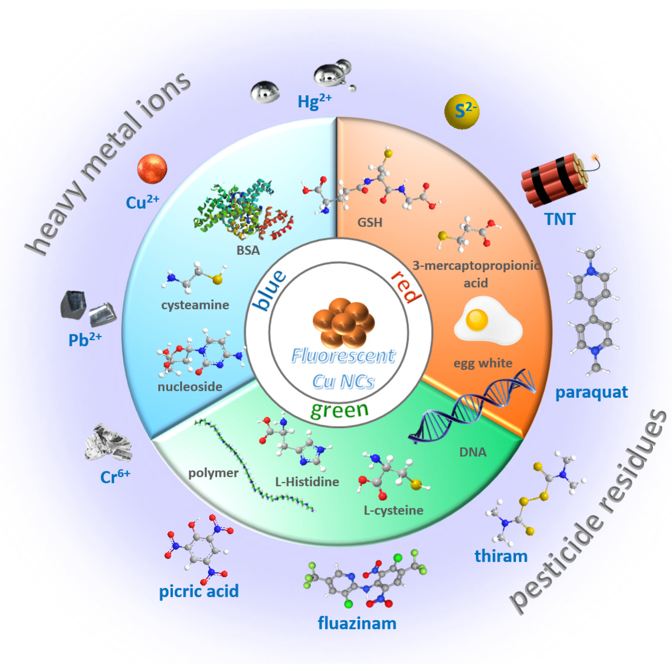
2.1. Preparation Methods

2.1.1. Blue Emission

In the recent decade, various fluorescent Cu NCs with a wide range of emission wavelengths from blue to red were developed with employment of different ligands and templates (Scheme 1). Cu NCs with blue emitting fluorescence have been covered most frequently in the latest research [7,10,11,12,13]. They are reported to have a broad range of applications, including detection of various contaminants and bioimaging [9,14,15,16,17]. The first blue emitting Cu NCs were reported in 2011 with bovine serum albumin (BSA) as template [18]. This novel type of nanocluster was synthesized in a one-pot method by using CuSO4 and BSA aqueous solution in the presence of NaOH. The as-prepared Cu NCs showed luminescence emission maxima at 410 nm with a high quantum yield of 15%. MALDI-TOF mass spectrometry was utilized to determine the precise atom number of the copper core. And the composition of this Cu NCs include Cu5 and Cu13. Since BSA is a very popular protein template in the synthesis of metal nanoclusters, many other BSA-Cu NCs with blue luminescence were subsequently reported [12,19,20]. Apart from proteins, thiolate ligands were also used widely to prepare Cu NCs [17,21,22,23,24]. In this category, glutathione (GSH) was a typical one [25,26,27]. In a study, Cu NCs with strong blue fluorescence were synthesized with GSH as both stabilizing and reducing agent [13]. These GSH-Cu NCs exhibit good photostability and storage stability with a quantum yield of 10.6%. Yang’s group reported their successful fabrication of Cu NCs stabilized with L-cysteine for the first time in a simple and green method. The most notable advantage of this material is that the L-cysteine-capped Cu NCs could be stored for more than 3 months in powder form without perceptible changes in their optical property. Moreover, a series of bi-ligand Cu NCs were developed using both thiosalicylic acid and cysteamine as protective and reducing agents [21]. The quantum yields of the products varied from 18.9% to 34.0% with the increase in cysteamine portion. This phenomenon results from the electron transfer to copper inducted by cysteamine’s terminal amine. Simultaneously, the thiosalicylic acid ligand absorbs energy through aromatic ring. Thus, more energy was released in the case of a higher cysteamine ratio. Interestingly, a pH-dependent synthesis method was reported that could produce Cu NCs with blue or yellow fluorescence emission under basic or acidic environments [28]. The two nanoclusters were obtained with the same method using trypsin as template and hydrazine hydrate as a reducing agent. The difference in pH value influences the conformational states of trypsin, and consequently results in the formation of Cu NCs with different sizes and fluorescent properties. And the fluorescence intensity of the trypsin-templated Cu NCs was further improved by adjustment of several synthesis conditions, such as molar ratio of CuSO4/N2H4, reaction temperature and reaction time. A pH-dependent Cu NC was synthesized in the presence of lysozyme and NaBH4. The Cu NCs showed potential in pH sensing since its fluorescence intensity increases with the decrease in pH [29]. In another study, a bright-blue Cu NCs was prepared with lysozyme as stabilizer and N2H4 as reducing agent. The Cu NCs were pH-stable and successfully applied in Hela cell labeling [30].

2.1.2. Green Emission

Green emitting Cu/Ag bimetallic nanoclusters were synthesized by Mao et al. using DNA as template with the assistance of NaBH4 [31]. In this work, a series of DNA-Ag NCs and DNA-Cu/Ag NCs were prepared with several different single-stranded C-rich DNA templates. The fluorescence emission of the nanoclusters varies from green to orange. The study found that the nanoclusters with longer emission wavelengths tend to be constructed by templates with more consecutive cytosine bases. Another research designed Cu NCs protected by a polymer via a special UV-light-induced synthesis approach [32]. The synthesis procedure was simple and facile. Cu NCs were obtained through reduction of Cu(NO3)2 with ascorbic acid (AA) in the presence of polyethyleneimine (PEI) under UV light irradiation. Compared with stirring method, the PEI-Cu NCs prepared by UV-light-induced approach displayed higher quantum yield and significantly enhanced fluorescence intensity. By the means of UV-light irradiation, the absolute quantum yield of PEI-Cu NCs was raised from 0.98% to 2.63% and its fluorescence intensity increased 3.7 times. Additionally, some amino acid ligands such as cysteine and histidine were also employed in the development of green emitting Cu NCs [33,34]. In an intriguing case, pH-stimulus-responsive Cu NCs were fabricated using L-cysteine and chitosan as ligands [35]. The as-prepared Cu NCs exhibited orange-red luminescence emission at pH 4.5, and showed cyan-green emission with aggregation-induced emission (AIE) behavior when the pH was above 7.4. The special phenomenon results from the weakening of H-bonding between carboxylic group of cysteine and the protonated amine moiety of chitosan caused by pH adjustment to 7.4. The stable overall environment around the cluster core was disrupted, leading to the formation of aggregates. Furthermore, the change in interatomic bond distance in the aggregation state brought about a blue shift of emission, and as a result, green luminescence emission was generated.

2.1.3. Orange/Red Emission

Many research groups developed GSH capped Cu NCs with orange or red fluorescence emission [36,37,38]. A one-pot sonochemical synthesis method was established for the preparation of GSH-Cu NCs by Wang et al. [39]. In this method, Cu (NO3)2 and GSH were mixed in water by 1: 4 ratio, and the pH of solution was adjusted to 6 with NaOH. The reaction was conducted in 15 min via ultrasonic irradiation, which is facile, fast and easy to operate. The as-synthesized GSH-Cu NCs exhibits bright red luminescence at λmax = 606 nm and quantum yield up to 5.3%. As mentioned above, there are also many fluorescent GSH-Cu NCs that have been reported with blue emission. The main difference in the synthesis process is that red emitting GSH-Cu NCs utilized GSH as both a stabilizing and a reducing agent in acidic condition, while blue emitting ones were synthesized in basic condition or reduced by AA. DNA template is also frequently exploited in the fabrication of Cu NCs with red emission [40,41,42,43]. Li et al. developed copper nanoclusters templated by poly(thymine)-DNA with a fluorescence emission of 627 nm [44]. The DNA-Cu NCs were produced by a facile reaction between copper sulfate and poly-T DNA in 3-(N-morpholino) propanesulfonic acid buffer with the help of sodium ascorbate. Intriguingly, a research group successfully synthesized Cu NCs with strong orange emission utilized egg white as template [45]. The formation of Cu NCs relies on the interaction between multiple functional groups in egg white and CuSO4. Hydrazine hydrate was employed as reducing agent and NaOH was used to provide the basic environment. The reaction proceeded extremely fast, since it is carried out under microwave.

2.1.4. Near Infrared Emission

In recent years, some Cu NCs showing near infrared emission have also been developed and applied in fluorescent sensors. Li et al. reported novel Cu NCs using denatured bovine serum albumin (dBSA) as templates [46]. The reaction was carried out at room temperature with hydrazine hydrate as reducing agent. In particular, the denatured BSA could form Cu NCs more efficiently, since it possesses more free cysteine with –SH groups than BSA. The as-prepared nanoclusters were successfully utilized to detect heparin by aggregation-induced quenching mechanism. Another study synthesized a near infrared emitting copper nanocluster co-protected by thiolate and phosphine ligands [47]. The structure of this nanocluster is atomically precise with the formula of [Cu15(PPh3)6(PET)13]2+. The product shows bright photoluminescence at 720 nm with crystallization induced emission enhancement property. More importantly, this research established a controllable synthesis strategy for atomically precise Cu NCs, which is meaningful and instructive to further study of their optical properties.

2.2. Sensing Mechanisms

2.2.1. Turn Off

On account of the prominent fluorescence property and cost-effectiveness of Cu NCs, numerous Cu NCs-based sensing probes have been developed for multitude of analytes. The majority of Cu NCs-based fluorescent sensors follow turn-off mechanism that the analytes are detected due to the decrease in fluorescence intensity.
Inner filter effect (IFE) is a most common strategy in the development of turn-off sensors [48,49,50,51]. The inner filter effect refers to the fluorescence quenching process that the quencher absorbs the emission or excitation light of fluorophore. A study in this context established a label-free assay for nitrofurantoin using adenosine-stabilized Cu NCs [16]. Nitrofurantoin’s UV absorption band located at 250–430 nm happens to overlap the Cu NCs’ fluorescence excitation and emission spectra. In this way, the excitation and emission light could be shielded by nitrofurantoin, leading to decrease in fluorescence intensity.
Some sensing methods for the detection of electron-rich species were constructed based on the electron transfer mechanism [44,52,53]. Cao et al. developed tannic acid (TA) capped Cu NCs that could selectively detect ferric ions [52]. Fe3+ could form donor-accepter complex with the TA on the surface of Cu NCs, leading to fluorescence quenching through electron transfer process, and the quenching obeys a static quenching mechanism since the fluorescence lifespan was independent of the concentration of Fe3+. The high selectivity of this sensor is related to the ferric ions’ electronic structure. Fe3+ has five half-filled d orbits so that it exhibits stronger electron-accepting ability than other common ions.
Another major cause of fluorescence quenching in turn-off sensing methods is aggregation-induced quenching, which is widely employed in analysis of metal ions [26,39,54]. A GSH-Cu NCs based probe for Hg2+ was constructed based on Hg2+-induced aggregation of the nanoclusters [13]. Hg2+ linked the Cu NCs through the interaction with GSH ligands, leading to fluorescence quenching. According to the zeta potential measurement results, the surface charge of GSH-Cu NCs became less negative in the presence of Hg2+, which destroyed the stability of Cu NCs and generatesd aggregates. The aggregation was further evidenced by increased diameter in TEM and enhanced light scattering signal.

2.2.2. Turn On

The main strategy for turn-on sensors is a signal off-on process that the fluorescence would be quenched in the first place and subsequently recovered via the addition of analyte. Surfactant-free copper nanoclusters were exploited as a turn-on probe for quantification of vitamin C [55]. In the first step, the fluorescence was quenched by the formation of Fe-Cu NCs conjugate through a static quenching mechanism. On account of the strong reducing property of vitamin C, the Fe3+ were converted into Fe2+, which consequently resulted in the dissociation of Fe-Cu NCs conjugate and restored fluorescent intensity.
As AIE has become an enticing topic in recent years, many turn-on sensors were developed inspired by this phenomenon. Boonmee et al. established a fluorescent assay based on Al3+ induced AIE of cysteamine-capped Cu NCs [56]. Al3+ could interact with the amino groups on the surface of Cu NCs, causing aggregation of nanoclusters. The detection was realized through fluorescence enhancement triggered by the aggregation. Similar strategy was also applied in detection of S2− and Pb2+ [10,57]. Specially, some studies directly utilized the formation of copper nanoclusters as a selective Cu2+ turn-on sensing method [38,58]. Apart from AIE, confinement-induced enhanced emission (CIEE) is also a strategy that frequently used to provide highly fluorescent Cu NCs with high quantum yield [36,59,60]. The improved fluorescence property was achieved by restricting the free movement of fluorescent molecules with the host materials [61]. Yang et al. constructed a turn-on sensing method for bio-enzyme based on CIEE with ultra-high sensitivity [62]. In this work, a fluorescent composite GS-Cu NCs and LDH (GS-Cu NCs/LDH) was fabricated on the basis of surface confinement effect, affording excellent quantum yield, improved stability and long fluorescence lifetime. The surface confinement effect of this material was firstly inhibited by hyaluronic acid (HA). In the presence of hyaluronidase (HAase), the HA would be hydrolyzed, leading to the recovery of fluorescence enhancement. In this way, a fluorescent assay for cancer biomarker HAase was successfully developed.

2.2.3. Ratiometric Analysis

Ratiometric fluorescence sensors have also attracted increasing research interest since it exhibits dominant advantages in accuracy, sensitivity and stability. In a ratiometric sensing approach, the detection is achieved by the intensity ratio of dual-emission peaks, which could eliminate the interference of environment and probe concentrations by self-calibration. A CuNC@AF660 sensor was fabricated for ratiometric sensing of calcium ions. The fluorescence intensity of Cu NCs emission peak was enhanced gradually with the increase in Ca2+ concentration through ion-induced AIE mechanism. The Alex Fluor 660 NHS ester fabricated on Cu NCs provides the inner-reference signal.
In some ratiometric sensing systems, one emission peak is quenched while the other is enhanced in the presence of analytes. In these cases, the underlying mechanism is usually ascribed to Förster resonance energy transfer (FRET). A probe based on aptamer-modified Copper @ Gold nanoclusters (apt-Cu@Au NCs) was designed for ratiometric detection of Hg2+ [63]. The mercury ions were captured by T-rich nucleic acid single strands on the surface of nanoclusters. The formation of T-Hg2+-T structure led to aggregation between the two fluorophores and affected the fluorescence intensity via FRET between Au NCs and Cu NCs.

3. Sensing Applications Based on Cu NCs

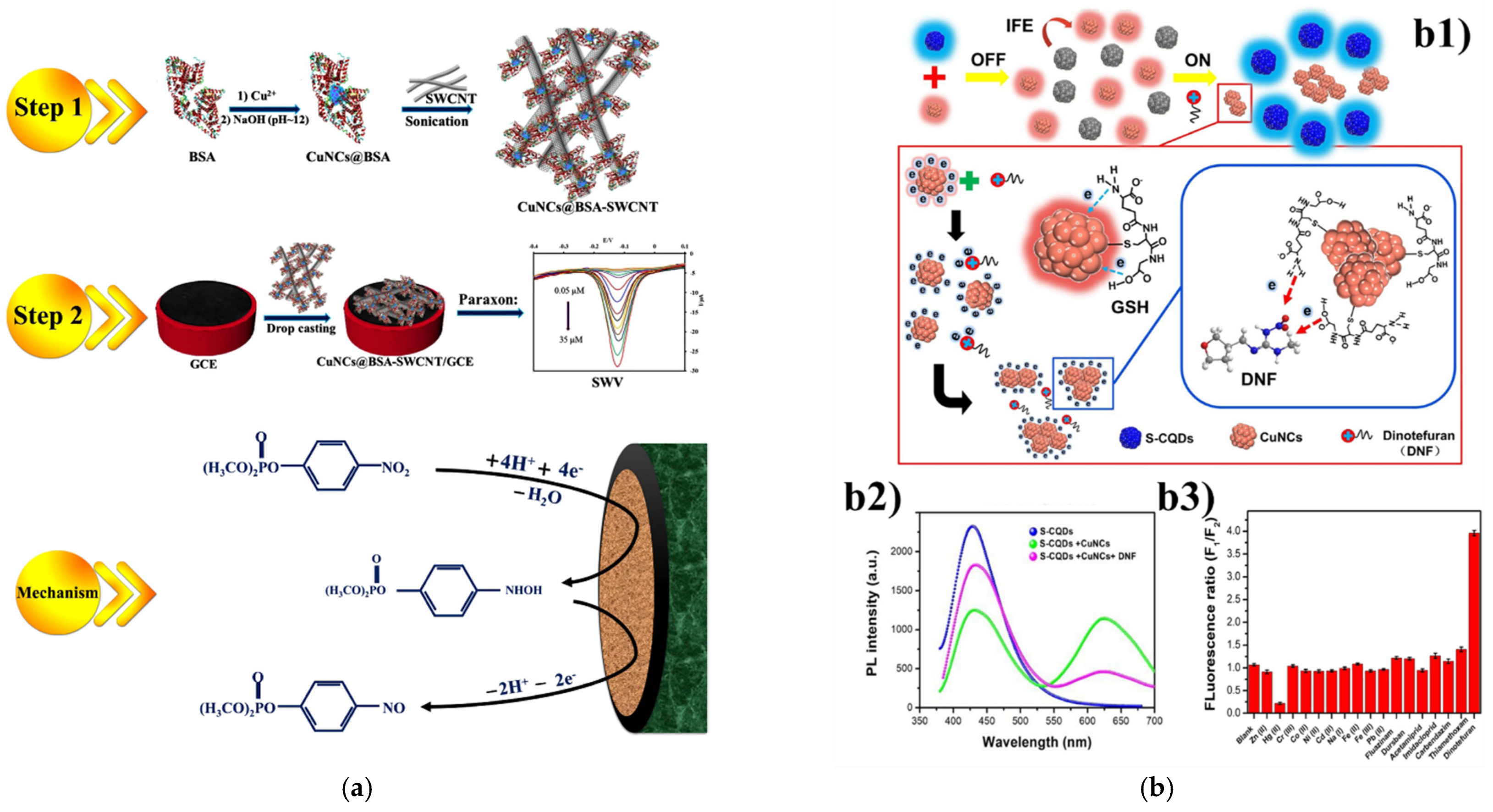
3.1. Pesticides as Target Analytes

Pesticide residues have been widespread in the environment due to the excessive use of pesticides in agricultural production and horticulture [64,65,66]. It has been found that even trace amounts of highly toxic pollutants could greatly threaten the ecosystem and human health.
In this section, we mainly highlight the Cu NCs-based detection systems targeting common pesticides (Table 1), e.g., paraoxon, dinotefuran (DNF), o-phenylenediamine (OPD), dithiocarbamates (DTCs), thiram, paraquat, fluazina, nitrofurantoin (NFT). Copper nanocluster was employed in the construction of an enzyme-free electrochemical biosensor toward paraoxon as the model of organophosphates (OP) [12]. The biocompatible nanocomposite Cu NCs@BSA-SWCNT was synthesized by combining bovine serum albumin (BSA) template-capped Cu nanoclusters (Cu NCs@BSA) and single-walled carbon nanotubes (SWCNT), which demonstrated remarkable sensitivity and high electrocatalytic property toward the reduction of paraoxon (Figure 1a). The entrapped Cu NCs rendered high electrical conductivity and concentrated the redox active centers on the surface of the probe, while the SWCNT further enhanced the electrocatalytic activity along with conductivity of the glassy carbon electrode (GCE) surface. The linear range was 0.5–35 μM, with a limit of detection of 12.8 nM. Moreover, this electrochemical nanocomposite was found to be able to effectively determine paraoxon with satisfied recoveries ranged from 93% to 104% in a real water sample. In order to detect and monitor the residues of dinotefuran (DNF), which has been widely used in agriculture, novel sensing probes and platforms based on fluorescent copper nanoclusters have been constructed. Yang et al. [67] established a dual-emission ratiometric fluorescent probe by integrating sulfur-doped carbon quantum dots (S-CQDs) and Cu NCs with mixed fluorescent signals (Figure 1b). The as-developed hybrid (S-CQDs/Cu NCs) was observed to demonstrate desirable sensitivity and selectivity towards DNF with linear range from 10 to 500 μM. In this nanocomposite, IFE caused the decrease in fluorescent signals of S-CQDs with the addition of Cu NCs. In the presence of dinitefuran, the IFE between S-CQDs and Cu NCs would be weakened due to the aggregation of Cu NCs, leading to the restoration of S-CODs fluorescence. In the case of honey as the real sample, this ratiometric fluorescent S-CQDs/Cu NCs showed good analysis performance for the detection of DNF. Besides, another ratiometric detection system was proposed for o-phenylenediamine (OPD) based on the use of copper nanoclusters [27]. This method achieved signal amplification and ideal sensitivity through the combined influence of the oxidation reaction and FRET effect. With addition of OPD into Cu NCs, the fluorescence intensity of the Cu NCs at 432 nm decreased, while the oxidized OPD (oxOPD) showed strong fluorescence at 557 nm. This detection strategy was able to determine OPD in real water samples with an ultralow limit of detection of 0.096 g L−1. Furthermore, a rapid and sensitive detection method of dithiocarbamates (DTCs) with dual functionality in fluorescence and colorimetry was established utilizing CTAB -entrapped Cu NCs [68]. Owing to the fluorescence quenching of the Cu NCs with addition of DTCs, the detection system demonstrated remarkable sensitivity and selectivity toward DTCs with a linear range from 1 to 100 mg kg−1 and a low limit of detection of 0.63 mg kg−1.
Acetylcholinesterase (AChE) as a key enzyme in relation to Alzheimer’s disease (AD) has been a hot detection target for many novel sensing probes based on copper nanoclusters. Due to the intrinsic mechanism of AChE inhibition in the presence of organophosphates, these strategies could be extended for pesticide detection. Yang et al. [32] reported the successful establishment of a fluorescence sensing method for the determination of multiple analytes through the induction of UV-light. The polyethyleneimine-protected copper nanoclusters (PEI-Cu NCs) presented strong stability and fluorescence intensity, which was employed in the development a label-free assay for sensitive detection of Cu2+ (Figure 2a). The interaction between Cu2+ with the -SH functional groups in biothiols generated the RSH-Cu2+ complex, which could trigger the fluorescence recovery of the PEI-Cu NCs. GSH and cysteine (Cy) as two biothiols were consequently detected within a linear range of 1–25 μM and 0.5–25 μM, respectively. In terms of the AChE activity, thiocholine (TCh) containing -SH group was produced with the hydrolyzation of acetylthiocholine by AChE. TCh interacted with Cu2+ to generate TCh-Cu2+ complex, which triggered the fluorescence recovery of the PEI-Cu NCs. The response toward AChE was linear ranging from 3–200 UL−1 with a limit of detection of 1.38 UL−1. Furthermore, this sensing platform was also employed in the detection of tacrine, a typical inhibitor of AChE with the IC50 value at 53.4 nM. A novel ratiometric florescence probe toward the evaluation of AChE activity was established through incorporating single-atom nanozyme (SAzyme) with the polyvinylpyrrolidone-protected copper nanocluster (PVP-Cu NCs) [14]. The SAzyme was first achieved through conjugating single-atom iron with N-doped porous carbon (Fe-SAs/NC) with peroxidase-like activity (Figure 2b). The obtained Fe-SAs/NC was observed to catalyze oxidation of OPD to 2,3-diaminophenazine (DAP) in the presence of H2O2. The fluorescent DAP displayed fluorescence emission at 566 nm, while the PVP-Cu NCs exhibited fluorescence emission peak at 438 nm. With the increase in the DAP concentration, the fluorescence intensity of PVP-Cu NCs was found to be quenched. The mechanism behind the quenching effect was confirmed to be FRET between PVP-Cu NCs and DAP through the measurement of fluorescence lifetime. This dual-emission signal system demonstrated high sensitivity toward AChE activity with a linear range between 2–70 UL−1 and a low limit of detection at 0.56 UL−1. Although this exploration proved the effectiveness of SAzymes in AChE analysis, the further investigation into its feasibility in its inhibitor evaluation, especially pesticide residues, is still urgent. Based on the IFE, another sensitive analytical method was proposed based on 5′-Dithiobis-(2-nitrobenzoic acid) (DTNB) and copper/silver nanoclusters [34]. This method exhibited high sensitivity with strong fluorescence within the linear range between 0.1–1.0 U L−1 and with a very low detection limit of 0.03 UL−1. Furthermore, the sensor constructed in this work showed distinct color change that could be distinguished by naked eyes, which further improved its feasibility in detecting AChE from other substance selectively.

3.2. Heavy Metals as Target Analytes
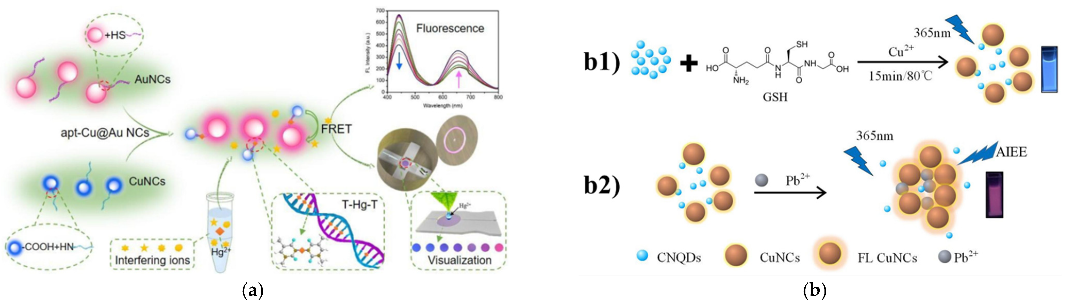
Heavy metals are notorious and hazardous contaminants in environment due to their low degradability, acute toxicity, high bioaccumulation, and other factors. Herein, we summarized nanoprobes based on Cu NCs for the determination of several representative heavy metal ions including mercury ions (Hg2+), lead ions (Pb2+), chromate anions (Cr6+) and copper ions (Cu2+) (Table 2).

3.2.1. Mercury Ions

Mercury has been found to be prevalent in the food chain, which poses a huge threat to the health of the ecosystem and humans. The conventional monitoring methods to Hg2+ could barely satisfy the soaring demand of rapid, facile and sensitive determination of this substrate in real samples [70,71,72]. An analytical method with high signal-to-noise ratio was constructed by regulating the fluorescence of copper nanoclusters through engineering the reticular poly(T) DNA template [40]. The fluorescence intensity of as-designed copper-based nanoclusters was observed to be enhanced to a great extent by using this DNA template with its unique structure and its resistance towards enzyme digestion. This novel DNA-templated nanomaterial exhibited excellent sensitivity and selectivity toward mercury ions with a desirably linear range from 50 pM to 500 μM and a remarkably low limit of detection at 16 pM (Figure 3a). Meanwhile, owning to the interplay between Hg2+ and GSH, this sensory probe could also be employed to detect the inhibition of GSH by Hg2+ and thus to determine the GSH in real biological samples. In the real sample analysis, this biosensor was found to be a reliable and feasible analytical method. An instant-response and facile analysis platform targeting Hg2+ and S2− ions was established through incorporating copper nanoclusters with 1-Thio-β-Dglucose as a capping ligand via ultrasonication, the synthesis of which could be completed in as fast as one minute [73]. As shown in Figure 3b, the as-synthesized TG-Cu NCs demonstrated the emission maximum at 430 nm, and through fluorescence quenching, this probe is able to detect Hg2+ and S2− ions with very low detection limits at 1.7 and 1.02 nM, respectively. This on-site detection and monitoring of the target analytes in real samples (e.g., tap, river and pond water) could be achieved easily with the developed cheap smartphone-aided and paper-based kit. Shi et al. [63] developed a ratiometric sensing strategy toward mercury ions by modifying copper nanoclusters @ gold nanoclusters via aptamer (apt-Cu@Au NCs). The changes in the fluorescence of this novel probe is attributed to the FRET strategy, which was achieved through the thymidine–Hg–thymidine (T–Hg–T) structure generated by addition of Hg2+ into the solution containing apt-Cu@Au NCs. Hg2+ ions could be detected with a range from 0.1 to 9.0 μM with the detection limit of 4.92 nM (Figure 4a). Meanwhile, to improve the feasibility, practicality, cost-effectiveness and the user-friendliness of this sensor, a smartphone-aided visualization method based on the fluorescent microfluidic chip was proposed to enable the facile and on-site determination of the target analytes. This sensing platform exhibited good performance in real samples with recovery rate at 101.83–114.00%. In another Hg2+ sensor, Cu NCs were assembled with polymers with multi-thiol groups as templates (Cu NCs@P-8B) through the simple one-pot approach [74]. This nanohybrid demonstrated satisfactory water stability, high sensitivity and responsiveness to pH and temperature, with a linear range between 10–100 μM and a limit of detection of 10 μM. Its performance in the real sample test with human urine indicated its potential in practical applications.

3.2.2. Lead Ions

Similarly, numerous novel probes have also been developed and investigated to monitor lead ions (Pb2+) in the environment and in the drinking water system, as lead is highly biotoxic and threatens the health of children, in particular. Intake of extremely low levels of lead can cause cardiovascular, nervous, and reproductive diseases [75,76]. Based on the nanocomposite of copper nanoclusters and carbon quantum dots (Cu NCs–CNQDs) blended in nitrogen, a novel ratiometric fluorescence determination method was reported to detect and analyze the trace amount of hypertoxic lead ions in a popular aquatic product porphyra [77]. As illustrated in Figure 4b, the fluorescence emission of the Cu NCs-CNQDs was observed to peak at 468 nm and 632 nm. The fluorescence of this Cu NCs was considerably enhanced when Pb2+ was added into the solution owing to the aggregation-induced emission enhancement (AIEE) between Pb2+ and Cu NCs. Meanwhile, the fluorescence of CNQDs remained the same, which could be taken as the self-calibration signal, and the Cu NCs-CNQDs demonstrated remarkable sensitivity and selectivity toward Pb2+ in porphyra ranging from 0.010 mg/L to 2.5 mg/L, with a low detection limit of 0.0031 mgL−1. Goswami and co-workers [18] proposed a BSA-capped Cu quantum clusters (CuQC@BSA) as the probe targeting Pb2+. The as-developed nanocomposite was highly sensitive toward Pb2+, which is attributed to the fluorescence quenching effect in ultra-low concentration of hydrogen peroxide at 200 ppm. It also performed well, even in the presence of interference from other metal ions. In another work, GSH-stabilized Cu NCs (GSH-Cu NCs) was fabricated successfully via the simple sonochemical synthesis method [39]. Based on the fluorescence quenching mechanism, this hybrid showed excellent performance in the label-free detection of Pb2+ with a linear range between 1 and 160 nM and a detection limit as low as 1.0 nM.

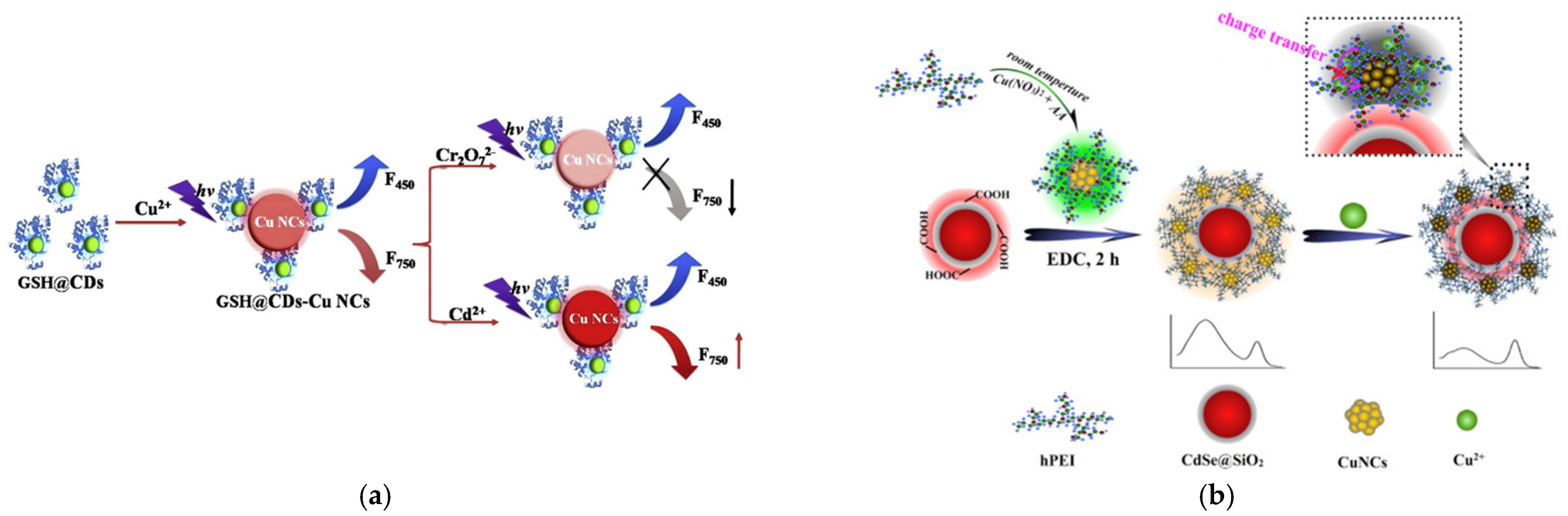
3.2.3. Chromate Anions
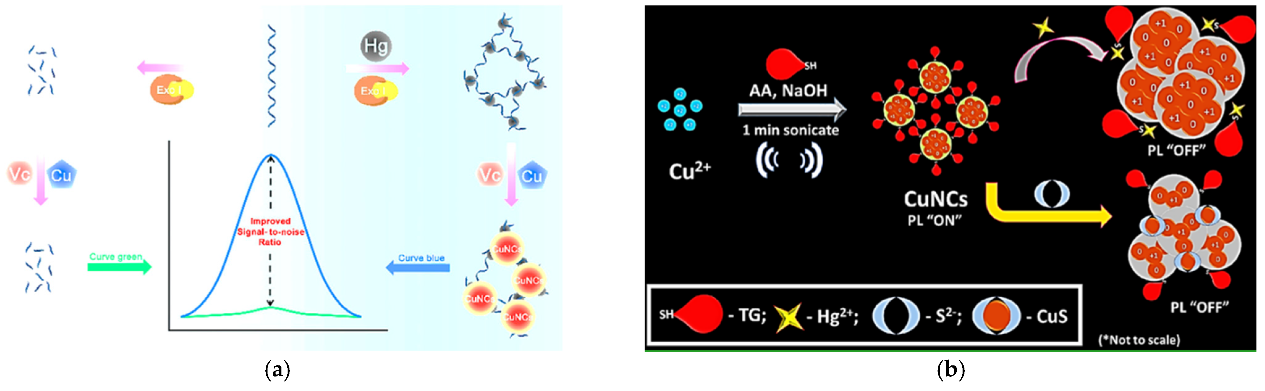
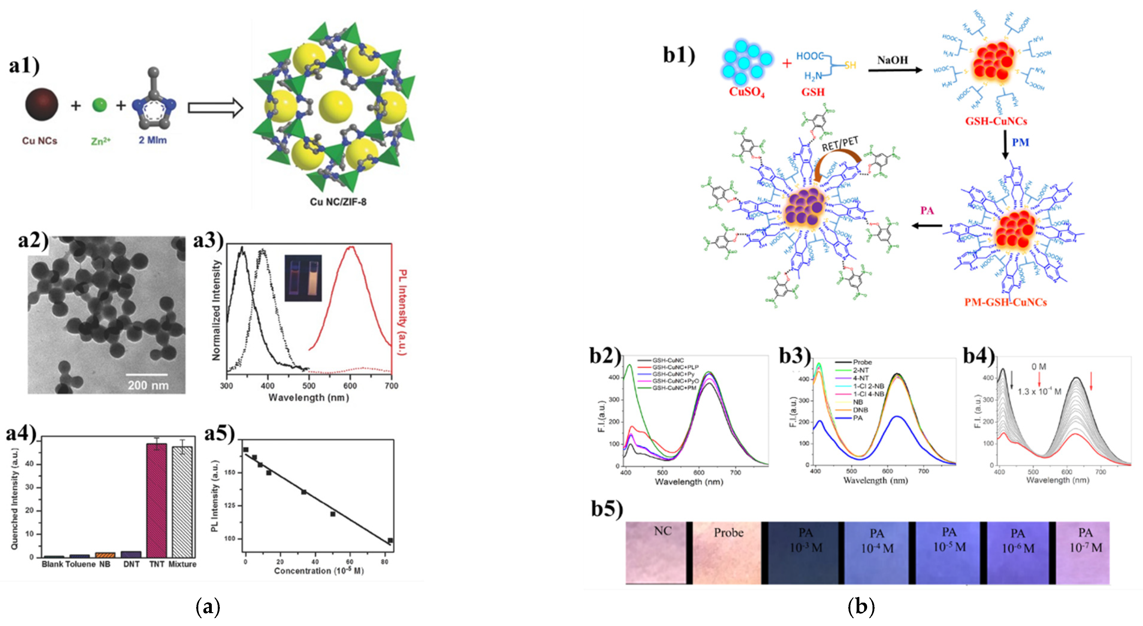
Chromium (Cr (VI)) has been extensively used in the modern industrial production and has also been a common contaminant in the environment. Considering its hugely harmful effect on human health, various analytical methods for the detection of chromium have been designed and, similar to other heavy metal contaminants, many novel sensors based on metal nanoclusters for Cr (VI) have been constructed. Bai and coworkers [78] proposed a ratiometric fluorescent probe for the convenient and effective detection of Cr2O72− or Cd2+ by integrating GSH-based carbon dots with copper nanoclusters. As shown in Figure 5a, the carbon dots-stabilized copper nanoclusters (GSH@CDs-Cu NCs) exhibited two obvious emission peaks at 450 nm and 750 nm, respectively. Owing to fluorescence quenching or enhancement of the nanohybrid, the GSH@CDs-Cu NCs showed good sensitivity and selectivity toward Cr2O72− with a linear range from 2 to 40 μmol L−1 and a detection limit of 0.9 μmol L−1, and Cd2+ with a linear range from 0 to 20 μmol L−1 and a detection limit of 0.6 μmol L−1. In addition, successful application of the fluorescence test strips was achieved in the rapid detection of Cr2O72− ions with distinct fluorescent color changes from pink to purple under UV light. In the test of real samples, the recovery rates of the target analytes in various water samples were in the range from 102% to 109% with the relative standard deviations (RSD) smaller than 4.5%. And the recovery rates of the target analytes ranged from 97%to 108% with the RSD smaller than 3.5%. Through the one-pot galvanic reduction approach, gold-copper nanoclusters capped by cysteamine (CA-AuCu NCs) were prepared for the detection of chromium(VI) and dopamine, the levels of which are critical to the development of severe diseases [23]. The distinct optical properties of this probe made it an ideal candidate in the determination of the target analytes in real samples, such as tap water, lake water, sea water and human urine. In particular, this switch-off probe was responsive in the linear range of 0.2–100 μm for Cr (VI) and with a detection limit of 80 nM.

3.2.4. Copper Ions

The prevalence of copper ion residues in the food and in the environment is another looming danger due to damage to the human liver and the nervous system caused by excessive Cu2+. Typically, Alzheimer’s disease (AD), as a common neurodegenerative disease, might be caused by excessive accumulation of copper ion and corresponding oxidative stress [79,80]. Hyperbranched polyethyleneimine-protected copper nanoclusters (hPEI-Cu NCs) were integrated with the silica-coated CdSe quantum dots (QDs) to obtain a novel ratiometric and visual analytical probe toward copper ions through the simple and green one-pot chemical reduction method at room temperature [81]. This QDs@Cu NCs probe exhibited high sensitivity and visual detection capability toward Cu2+, which could be attributed to the self-calibration function of the dual-emission fluorescence signals inherent in the ratio-based fluorescence probe. The fluorescence of hPEI-Cu NCs was quenched due to the interaction of its amine groups with Cu2+, while the fluorescence intensity of QDs remained unchanged, and the distinct color changes could be easily observed by the naked eye under the UV-light. The linear range of the probe in Cu2+ concentrations was, approximately, from 22 nM to 8.8 μM, with an estimated detection limit of 8.9 nM (Figure 5b). When applied in the real water samples with different concentrations of Cu2+, the as-synthesized probe exhibited satisfactory recovery.

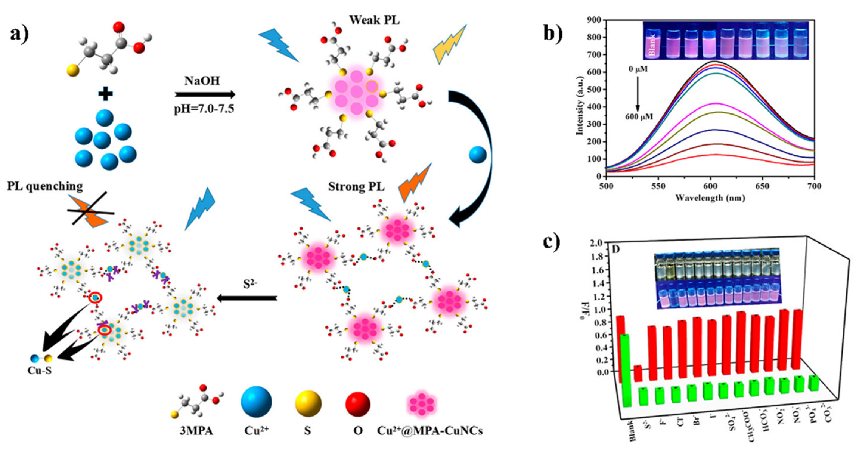
3.3. Sulfide as Target Analytes

Another main category of environmental pollutants is sulfide ions, which are also by-products of massive industrial development. Their wide distribution in different formats (i.e., H2S, HS and S2−) in nature makes them a latent crisis, threatening human health. Cu NCs have also been extensively utilized to determine sulfide ions (Table 3). A “turn-on” determination strategy toward sulfide ion (S2−) was developed based on fluorescent copper nanoclusters with natural silk fibroin (SF) protein as template (SF@Cu NCs), which was synthesized via utilizing the environmental friendly and user-friendly one-pot method [10]. With the addition of S2− ions into the aqueous solutions, the fluorescence intensity of the probe was observed to be increased, due to the inherent feature of aggregation/assembly-induced emission enhancement (AIEE). The S2− could induce the self-assembly of copper nanoclusters into large rod-shape nanoparticles. This SF@Cu NCs nanohybrid demonstrated good sensitivity and selectivity toward S2− with a linear range of 5.0 μM–110.0 μM and the limit of detection at 0.286 μM. In the real sample test, this analytical approach was found to be powerful in determining the sulfide ions at different concentrations with a satisfactory recovery rate of 95.6–101.6%. Another sensing platform for the determination of sulfide ions was reported. The main component of this approach was synthesized through modifying copper nanoclusters by 3-mercaptopropionic acid and with AIE induced by Cu2+ (Cu2+@MPA-Cu NCs) [92]. The preparation process of this luminescence probe was easy-to-operate and facile. The probe exhibited good linear detection of S2− in the range from 0 to 600 μM, with a detection limit of 26.3 nM (Figure 6). In the quantitative detection of S2−, the Cu2+@MPA-Cu NCs sensor presented high selectivity and good accuracy in the simulated real environment, with recovery rates ranging from 92.5% to 104.5% and RSD less than 4%. Ratio-based sensing systems for the monitoring of sulfide ions have also been established based on all Cu NCs (G-R-Cu NCs) [93]. The fluorescent probe was obtained through rapid and simple synthesis procedure and exhibited remarkable selectivity and sensitivity in the real sample.

3.4. Others

Cu NCs with their fascinating fluorescence properties as well as desirable stability have also been widely employed as sensing probes to determine aromatic compounds (Table 4). Trinitrotoluene (TNT), a popular explosive commonly used in military work, is indeed a dangerous contaminant in water. For the detection of TNT, a luminescent sensor was established by embedding copper nanoclusters (Cu NCs) into metal-organic frameworks (MOF) ZIF-8 [36]. Thanks to the confinement-assisted emission enhancement similar to AIE of fluorescence, the stability and emission intensity of the composite was largely improved, with an orange-color emission peak at 600 nm (Figure 7a). This Cu NC/ZIF-8 hybrid exhibited capability of detecting trace amounts of TNT, since it was selectively quenched in the presence of TNT. Similarly, there has also been an urgent need for the facile and sensitive detection of picric acid (2,4,6- trinitrophenol, PA). Patel et al. [37] reported an analytical system for PA based on the fluorescence quenching of glutathione stabilized copper nanoclusters and a vitamin B6 cofactor pyridoxamine (PM-GSH-Cu NCs) (Figure 7b). This nanoprobe exhibited excellent sensitivity and selectivity to PA in different solutions and real samples including tap water, river water and matchstick. To be specific, it could detect PA down to 27.4 × 10⁻⁷ M, and the paper strips based on this nanoprobe could determine PA with the naked eye at as low as 1 μM.
Artificial food coloring has contributed greatly to the food manufacturing industry to increase the attractiveness of food by vivid colors. Due to the looming danger and risks to human health, some artificial colorants are banned from being added in food, while the use of others in food are strictly regulated. Quinoline yellow (QY) belongs to the one of the above two categories in various countries that ban colorants and strictly regulate substances. Analytical methods based on Cu NCs have been thus developed toward quinoline yellow in food products. Sivasankaran et al. reported their successful construction of a fluorescent-based probe for QY [97]. Owing to the quenching effect, the blue-fluorescence-emitting-L-cysteine stabilized Cu NCs (L-Cys-Cu NCs) demonstrated excellent sensitivity and selectivity toward QY with a low detection limit of 0.11 μM and a linear concentration range between 5.50 and 0.20 μM. Cu NCs-based sensing probes or systems have also been used in detection and monitoring of overuse or abuse of broad-spectrum antibiotics. An ultrasensitive biosensor based on glutathione-capped copper nanoclusters (GSH-Cu NCs) was designed to determine the trace level of furazolidone in the environment and control the overuse [98]. The blue-emitting fluorescent probe was prepared in a very short time. Due to the inner filter effect and static quenching between GSH-Cu NCs and furazolidone, the as-prepared probe exhibited a favorable linear range from 0.05 to 60 μM and a low detection limit at 0.012 μM. Furthermore, Cu NCs have also proven effectiveness in the detection of residues of 2,6-pyridinedicarboxylic acid (DPA) in the environment, which is notorious for its hazards toward human health and safety, as well as in vulnerable ecosystems. A portable sensing strategy was developed based on the Cu NC fluorescent test strip. The as-fabricated ratiometric sensing probe (GSH-Cu NCs/Eu3+) demonstrated its sensitivity, timeliness and convenience in determining DPA in the complicated solution with an ultra-low detection limit of 8 nM [99].

4. Conclusions and Perspectives

This review summarized and reported the latest development of fluorescent copper nanoclusters utilized in monitoring various types of environmental contaminants. Despite the fascinating advances, there is still a long and arduous way ahead in further improving Cu NCs to make them ideal materials for pollutant detection, quantitative determination, and, even, decomposition and removal. Firstly, the sizes of the current Cu NCs designed fall into a rather wide range, and the desirable optical properties of most Cu NCs heavily rely on excitation. Both of these two aspects may largely constrain the possibility of efficient application of Cu NCs in environmental analysis on a large scale. Secondly, efforts are imperative to further enhance the chemical and optical stability, as well as quantum yield of Cu NCs, so as to improve their feasibility as sensors in more complex, real environments, even with complicated, interfering factors. Lastly, the functions of the current probes are expected to be extended from pure detection of environmental pollutants to simultaneous detection, degradation and, even, removal.

Author Contributions

Investigation, Y.X., Z.C. and M.L.; Writing–original draft, Y.X. and Z.C.; Methodology, Y.X.; Conceptualization, Funding acquisition and Writing–review& editing, H.H. and C.X. All authors have read and agreed to the published version of the manuscript.

Funding

This research is supported by the Foshan Medicine Dengfeng Project of China (2019–2021), Foshan Municipal Government (CP-013-2020). It is also partially supported by the grant from the University of Macau (MYRG2020-00230-ICMS).

Institutional Review Board Statement

Not applicable.

Informed Consent Statement

Not applicable.

Data Availability Statement

No new data were created or analyzed in this study. Data sharing is not applicable to this article.

Conflicts of Interest

The authors declare no conflict of interest.

References

  1. Jin, R.; Zeng, C.; Zhou, M.; Chen, Y. Atomically Precise Colloidal Metal Nanoclusters and Nanoparticles: Fundamentals and Opportunities. Chem. Rev. 2016, 116, 10346–10413. [Google Scholar] [CrossRef] [PubMed]
  2. Du, Y.; Sheng, H.; Astruc, D.; Zhu, M. Atomically Precise Noble Metal Nanoclusters as Efficient Catalysts: A Bridge between Structure and Properties. Chem. Rev. 2019, 120, 526–622. [Google Scholar] [CrossRef] [PubMed]
  3. Liu, X.; Astruc, D. Atomically precise copper nanoclusters and their applications. Coord. Chem. Rev. 2018, 359, 112–126. [Google Scholar] [CrossRef]
  4. Wang, Z.; Chen, B.; Rogach, A.L. Synthesis, optical properties and applications of light-emitting copper nanoclusters. Nanoscale Horiz. 2017, 2, 135–146. [Google Scholar] [CrossRef] [PubMed]
  5. Baghdasaryan, A.; Bürgi, T. Copper nanoclusters: Designed synthesis, structural diversity, and multiplatform applications. Nanoscale 2021, 13, 6283–6340. [Google Scholar] [CrossRef] [PubMed]
  6. Lai, W.F.; Wong, W.T.; Rogach, A.L. Development of Copper Nanoclusters for in vitro and in vivo Theranostic Applications. Adv. Mater. 2020, 32, 1906872. [Google Scholar] [CrossRef]
  7. Shahsavari, S.; Hadian-Ghazvini, S.; Hooriabad Saboor, F.; Menbari Oskouie, I.; Hasany, M.; Simchi, A.; Rogach, A.L. Ligand functionalized copper nanoclusters for versatile applications in catalysis, sensing, bioimaging, and optoelectronics. Mater. Chem. Front. 2019, 3, 2326–2356. [Google Scholar] [CrossRef]
  8. Hu, X.; Liu, T.; Zhuang, Y.; Wang, W.; Li, Y.; Fan, W.; Huang, Y. Recent advances in the analytical applications of copper nanoclusters. TrAC Trends Anal. Chem. 2016, 77, 66–75. [Google Scholar] [CrossRef]
  9. An, Y.; Ren, Y.; Bick, M.; Dudek, A.; Waworuntu, E.H.; Tang, J.; Chen, J.; Chang, B. Highly fluorescent copper nanoclusters for sensing and bioimaging. Biosens. Bioelectron. 2020, 154, 112078. [Google Scholar] [CrossRef] [Green Version]
  10. Zhang, G.; Wang, R.; Shi, L.; Zhang, C.; Zhang, Y.; Zhou, Y.; Dong, C.; Li, G.; Shuang, S. Aggregation/assembly induced emission based on silk fibroin-templated fluorescent copper nanoclusters for “turn-on” detection of S2−. Sens. Actuators B Chem. 2019, 279, 361–368. [Google Scholar] [CrossRef]
  11. Liu, Y.; Yao, D.; Zhang, H. Self-Assembly Driven Aggregation-Induced Emission of Copper Nanoclusters: A Novel Technology for Lighting. ACS Appl. Mater. Interfaces 2017, 10, 12071–12080. [Google Scholar] [CrossRef]
  12. Bagheri, H.; Afkhami, A.; Khoshsafar, H.; Hajian, A.; Shahriyari, A. Protein capped Cu nanoclusters-swcnt nanocomposite as a novel candidate of high performance platform for organophosphates enzymeless biosensor. Biosens. Bioelectron. 2017, 89, 829–836. [Google Scholar] [CrossRef] [PubMed]
  13. Hu, X.; Wang, W.; Huang, Y. Copper nanocluster-based fluorescent probe for sensitive and selective detection of Hg(2+) in water and food stuff. Talanta 2016, 154, 409–415. [Google Scholar] [CrossRef] [PubMed]
  14. Wang, M.; Liu, L.; Xie, X.; Zhou, X.; Lin, Z.; Su, X. Single-atom iron containing nanozyme with peroxidase-like activity and copper nanoclusters based ratio fluorescent strategy for acetylcholinesterase activity sensing. Sens. Actuators B Chem. 2020, 313, 128023. [Google Scholar] [CrossRef]
  15. Xia, J.; Wei, X.; Chen, X.; Shu, Y.; Wang, J. Folic acid modified copper nanoclusters for fluorescent imaging of cancer cells with over-expressed folate receptor. Microchim. Acta 2018, 185, 205. [Google Scholar] [CrossRef]
  16. Wang, Y.; Chen, T.; Zhuang, Q.; Ni, Y. Label-free photoluminescence assay for nitrofurantoin detection in lake water samples using adenosine-stabilized copper nanoclusters as nanoprobes. Talanta 2018, 179, 409–413. [Google Scholar] [CrossRef] [PubMed]
  17. Yang, X.; Feng, Y.; Zhu, S.; Luo, Y.; Zhuo, Y.; Dou, Y. One-step synthesis and applications of fluorescent Cu nanoclusters stabilized by l-cysteine in aqueous solution. Anal. Chim. Acta 2014, 847, 49–54. [Google Scholar] [CrossRef]
  18. Goswami, N.; Giri, A.; Bootharaju, M.S.; Xavier, P.L.; Pradeep, T.; Pal, S.K. Copper Quantum Clusters in Protein Matrix: Potential Sensor of Pb2+ Ion. Anal. Chem. 2011, 83, 9676–9680. [Google Scholar] [CrossRef]
  19. Xiaoqing, L.; Ruiyi, L.; Zaijun, L.; Xiulan, S.; Zhouping, W.; Junkang, L. Fast synthesis of copper nanoclusters through the use of hydrogen peroxide additive and their application for the fluorescence detection of Hg2+ in water samples. New J. Chem. 2015, 39, 5240–5248. [Google Scholar] [CrossRef]
  20. Zhong, Y.; Zhu, J.; Wang, Q.; He, Y.; Ge, Y.; Song, C. Copper nanoclusters coated with bovine serum albumin as a regenerable fluorescent probe for copper(II) ion. Microchim. Acta 2014, 182, 909–915. [Google Scholar] [CrossRef]
  21. Lin, Y.-S.; Chiu, T.-C.; Hu, C.-C. Fluorescence-tunable copper nanoclusters and their application in hexavalent chromium sensing. RSC Adv. 2019, 9, 9228–9234. [Google Scholar] [CrossRef] [Green Version]
  22. Bao, Z.; Zhang, K.; Jian, J.; Hu, Z.; Yuan, K.; Shao, H.; Peng, K.; Jiang, Z.; Zapien, J.A.; Yan, Y.; et al. Strongly fluorescent cysteamine-coated copper nanoclusters as a fluorescent probe for determination of picric acid. Microchim. Acta 2018, 185, 507. [Google Scholar] [CrossRef] [PubMed]
  23. Shellaiah, M.; Simon, T.; Thirumalaivasan, N.; Sun, K.W.; Ko, F.H.; Wu, S.P. Cysteamine-capped gold-copper nanoclusters for fluorometric determination and imaging of chromium(VI) and dopamine. Mikrochim. Acta 2019, 186, 788. [Google Scholar] [CrossRef]
  24. Khonkayan, K.; Sansuk, S.; Srijaranai, S.; Tuntulani, T.; Saiyasombat, C.; Busayaporn, W.; Ngeontae, W. New approach for detection of chromate ion by preconcentration with mixed metal hydroxide coupled with fluorescence sensing of copper nanoclusters. Microchim. Acta 2017, 184, 2965–2974. [Google Scholar] [CrossRef]
  25. Huang, H.; Li, H.; Feng, J.-J.; Feng, H.; Wang, A.-J.; Qian, Z. One-pot green synthesis of highly fluorescent glutathione-stabilized copper nanoclusters for Fe3+ sensing. Sens. Actuators B Chem. 2017, 241, 292–297. [Google Scholar] [CrossRef]
  26. Luo, T.; Zhang, S.; Wang, Y.; Wang, M.; Liao, M.; Kou, X. Glutathione-stabilized Cu nanocluster-based fluorescent probe for sensitive and selective detection of Hg2+ in water. Luminescence 2017, 32, 1092–1099. [Google Scholar] [CrossRef] [PubMed] [Green Version]
  27. Ma, Y.; Yu, Y.; Lin, B.; Zhang, L.; Cao, Y.; Guo, M. A novel signal amplification strategy based on the use of copper nanoclusters for ratiometric fluorimetric determination of o-phenylenediamine. Mikrochim. Acta 2019, 186, 206. [Google Scholar] [CrossRef] [PubMed]
  28. Feng, J.; Chen, Y.; Han, Y.; Liu, J.; Ma, S.; Zhang, H.; Chen, X. pH-Regulated Synthesis of Trypsin-Templated Copper Nanoclusters with Blue and Yellow Fluorescent Emission. ACS Omega 2017, 2, 9109–9117. [Google Scholar] [CrossRef] [PubMed] [Green Version]
  29. Pandit, S.; Kundu, S. pH-Dependent reversible emission behaviour of lysozyme coated fluorescent copper nanoclusters. J. Lumin. 2020, 228, 117607. [Google Scholar] [CrossRef]
  30. Ghosh, R.; Sahoo, A.K.; Ghosh, S.S.; Paul, A.; Chattopadhyay, A. Blue-Emitting Copper Nanoclusters Synthesized in the Presence of Lysozyme as Candidates for Cell Labeling. ACS Appl. Mater. Interfaces 2014, 6, 3822–3828. [Google Scholar] [CrossRef]
  31. Mao, A.; Wei, C. Cytosine-rich ssDNA-templated fluorescent silver and copper/silver nanoclusters: Optical properties and sensitive detection for mercury(II). Mikrochim. Acta 2019, 186, 541. [Google Scholar] [CrossRef] [PubMed]
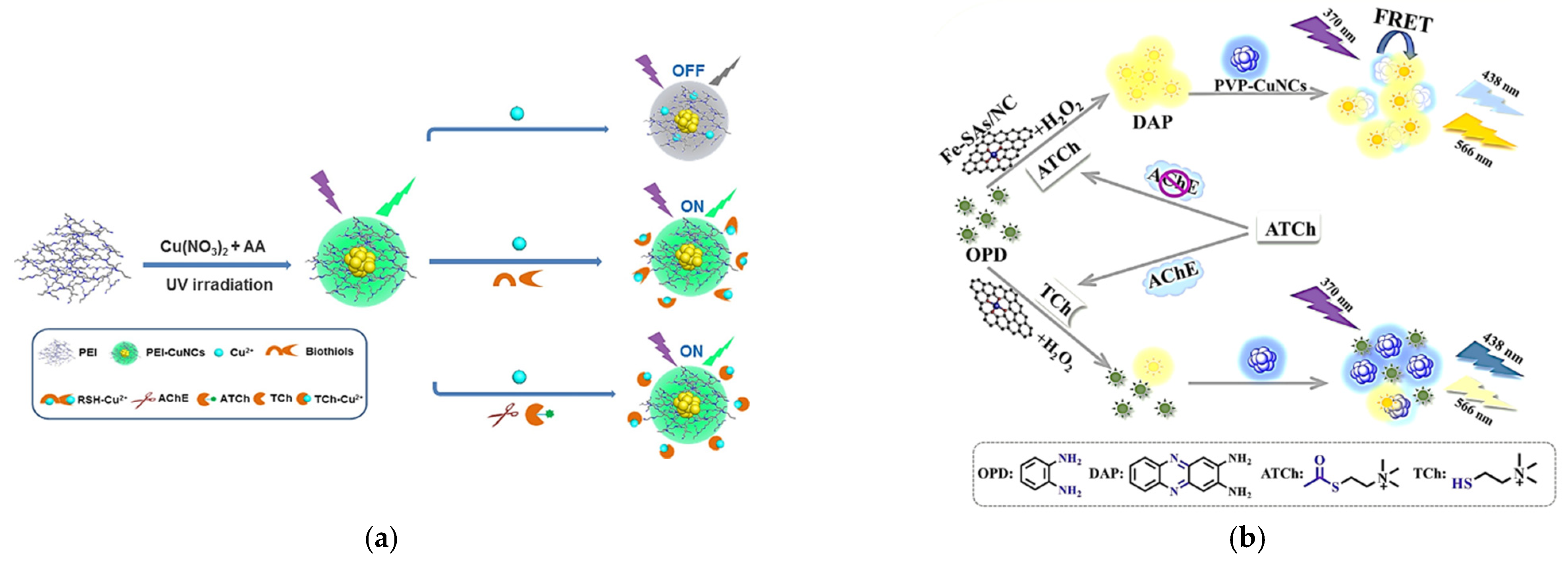
  32. Yang, J.; Song, N.; Lv, X.; Jia, Q. UV-light-induced synthesis of PEI-CuNCs based on Cu2+-quenched fluorescence turn-on assay for sensitive detection of biothiols, acetylcholinesterase activity and inhibitor. Sens. Actuators B Chem. 2018, 259, 226–232. [Google Scholar] [CrossRef]
  33. Li, L.; Hou, C.; Li, J.; Yang, Y.; Hou, J.; Ma, Y.; He, Q.; Luo, H.; Huo, D. Fluazinam direct detection based on the inner filter effect using a copper nanocluster fluorescent probe. Anal. Methods 2019, 11, 4637–4643. [Google Scholar] [CrossRef]
  34. Li, Y.; Hu, Y.; He, Y.; Ge, Y.; Song, G.; Zhou, J. Sensitive Naked-eye and Fluorescence Determination of Acetylcholinesterase Activity using Cu/Ag Nanoclusters Based on Inner Filter Effect. ChemistrySelect 2019, 4, 7639–7644. [Google Scholar] [CrossRef]
  35. Dutta, A.; Goswami, U.; Chattopadhyay, A. Probing Cancer Cells through Intracellular Aggregation-Induced Emission Kinetic Rate of Copper Nanoclusters. ACS Appl. Mater. Interfaces 2018, 10, 19459–19472. [Google Scholar] [CrossRef]
  36. Wang, Z.; Chen, R.; Xiong, Y.; Cepe, K.; Schneider, J.; Zboril, R.; Lee, C.-S.; Rogach, A.L. Incorporating Copper Nanoclusters into Metal-Organic Frameworks: Confinement-Assisted Emission Enhancement and Application for Trinitrotoluene Detection. Part. Part. Syst. Charact. 2017, 34, 1700029. [Google Scholar] [CrossRef]
  37. Patel, R.; Bothra, S.; Kumar, R.; Crisponi, G.; Sahoo, S.K. Pyridoxamine driven selective turn-off detection of picric acid using glutathione stabilized fluorescent copper nanoclusters and its applications with chemically modified cellulose strips. Biosens. Bioelectron. 2018, 102, 196–203. [Google Scholar] [CrossRef]
  38. Shen, Z.; Zhang, C.; Yu, X.L.; Li, J.; Liu, B.H.; Zhang, Z.P. A facile stage for Cu2+ ions detection by formation and aggregation of Cu nanoclusters. Microchem. J. 2019, 145, 517–522. [Google Scholar] [CrossRef]
  39. Wang, C.; Cheng, H.; Huang, Y.; Xu, Z.; Lin, H.; Zhang, C. Facile sonochemical synthesis of pH-responsive copper nanoclusters for selective and sensitive detection of Pb2+ in living cells. Analyst 2015, 140, 5634–5639. [Google Scholar] [CrossRef] [PubMed]
  40. Li, J.; Fu, W.; Bao, J.; Wang, Z.; Dai, Z. Fluorescence Regulation of Copper Nanoclusters via DNA Template Manipulation toward Design of a High Signal-to-Noise Ratio Biosensor. ACS Appl. Mater. Interfaces 2018, 10, 6965–6971. [Google Scholar] [CrossRef]
  41. He, J.-L.; Wang, X.-X.; Mei, T.-T.; Wu, L.; Zeng, J.-L.; Wang, J.-H.; Wang, J.; Yu, D.; Cao, Z. DNA-templated copper nanoclusters obtained via TdT isothermal nucleic acid amplification for mercury(ii) assay. Anal. Methods 2019, 11, 4165–4172. [Google Scholar] [CrossRef]
  42. Su, Y.-T.; Lan, G.-Y.; Chen, W.-Y.; Chang, H.-T. Detection of Copper Ions Through Recovery of the Fluorescence of DNA-Templated Copper/Silver Nanoclusters in the Presence of Mercaptopropionic Acid. Anal. Chem. 2010, 82, 8566–8572. [Google Scholar] [CrossRef]
  43. Ou, L.J.; Huang, J.K.; Lv, X.L.; Huang, N. DsDNA-templated fluorescent copper nanoclusters for ultrasensitive label-free detection of Pb2+ ion. Chin. J. Anal. Lab. 2016, 35, 899–902. [Google Scholar] [CrossRef]
  44. Li, H.; Chang, J.; Hou, T.; Ge, L.; Li, F. A facile, sensitive, and highly specific trinitrophenol assay based on target-induced synergetic effects of acid induction and electron transfer towards DNA-templated copper nanoclusters. Talanta 2016, 160, 475–480. [Google Scholar] [CrossRef] [PubMed]
  45. Bhamore, J.R.; Jha, S.; Mungara, A.K.; Singhal, R.K.; Sonkeshariya, D.; Kailasa, S.K. One-step green synthetic approach for the preparation of multicolor emitting copper nanoclusters and their applications in chemical species sensing and bioimaging. Biosens. Bioelectron. 2016, 80, 243–248. [Google Scholar] [CrossRef]
  46. Wu, X.; Zhang, F.; Li, Y. Facile synthesis of near-infrared emitting dBSA-templated Cu nanoclusters for sensitive detection of heparin. J. Mater. Chem. B 2018, 6, 5466–5475. [Google Scholar] [CrossRef] [PubMed]
  47. Nematulloev, S.; Huang, R.W.; Yin, J.; Shkurenko, A.; Dong, C.; Ghosh, A.; Alamer, B.; Naphade, R.; Hedhili, M.N.; Maity, P.; et al. [Cu15(PPh3)6(PET)13]2+: A Copper Nanocluster with Crystallization Enhanced Photoluminescence. Small 2021, 17, 2006839. [Google Scholar] [CrossRef]
  48. Dong, W.; Sun, C.; Sun, M.; Ge, H.; Asiri, A.M.; Marwani, H.M.; Ni, R.; Wang, S. Fluorescent Copper Nanoclusters for the Iodide-Enhanced Detection of Hypochlorous Acid. ACS Appl. Nano Mater. 2019, 3, 312–318. [Google Scholar] [CrossRef] [Green Version]
  49. Shojaeifard, Z.; Heidari, N.; Hemmateenejad, B. Bimetallic AuCu nanoclusters-based florescent chemosensor for sensitive detection of Fe(3+) in environmental and biological systems. Spectrochim. Acta. Part A Mol. Biomol. Spectrosc. 2019, 209, 202–208. [Google Scholar] [CrossRef]
  50. Luo, M.; Wei, J.; Zhao, Y.; Sun, Y.; Liang, H.; Wang, S.; Li, P. Fluorescent and visual detection of methyl-paraoxon by using boron-and nitrogen-doped carbon dots. Microchem. J. 2020, 154, 104547. [Google Scholar] [CrossRef]
  51. Wei, J.; Yang, Y.; Dong, J.; Wang, S.; Li, P. Fluorometric determination of pesticides and organophosphates using nanoceria as a phosphatase mimic and an inner filter effect on carbon nanodots. Microchim. Acta 2019, 186, 66. [Google Scholar] [CrossRef]
  52. Cao, H.; Chen, Z.; Zheng, H.; Huang, Y. Copper nanoclusters as a highly sensitive and selective fluorescence sensor for ferric ions in serum and living cells by imaging. Biosens. Bioelectron. 2014, 62, 189–195. [Google Scholar] [CrossRef]
  53. Das, N.K.; Ghosh, S.; Priya, A.; Datta, S.; Mukherjee, S. Luminescent Copper Nanoclusters as a Specific Cell-Imaging Probe and a Selective Metal Ion Sensor. J. Phys. Chem. C 2015, 119, 24657–24664. [Google Scholar] [CrossRef]
  54. Bhamore, J.R.; Deshmukh, B.; Haran, V.; Jha, S.; Singhal, R.K.; Lenka, N.; Kailasa, S.K.; Murthy, Z.V.P. One-step eco-friendly approach for the fabrication of synergistically engineered fluorescent copper nanoclusters: Sensing of Hg2+ ion and cellular uptake and bioimaging properties. New J. Chem. 2018, 42, 1510–1520. [Google Scholar] [CrossRef]
  55. Sahu, D.K.; Singha, D.; Sahu, K. Sensing of iron(III)-biomolecules by surfactant-free fluorescent copper nanoclusters. Sens. Bio-Sens. Res. 2019, 22, 100250. [Google Scholar] [CrossRef]
  56. Boonmee, C.; Promarak, V.; Tuntulani, T.; Ngeontae, W. Cysteamine-capped copper nanoclusters as a highly selective turn-on fluorescent assay for the detection of aluminum ions. Talanta 2018, 178, 796–804. [Google Scholar] [CrossRef]
  57. Han, B.-Y.; Hou, X.-F.; Xiang, R.-C.; Yu, M.-B.; Li, Y.; Peng, T.-T.; He, G.-H. Detection of Lead Ion Based on Aggregation-induced Emission of Copper Nanoclusters. Chin. J. Anal. Chem. 2017, 45, 23–27. [Google Scholar] [CrossRef]
  58. Wang, Y.; Chen, T.; Zhang, Z.; Ni, Y. Cytidine-stabilized copper nanoclusters as a fluorescent probe for sensing of copper ions and hemin. RSC Adv. 2018, 8, 9057–9062. [Google Scholar] [CrossRef] [Green Version]
  59. Yang, J.; Li, Z.; Jia, Q. Anchoring copper nanoclusters to Zn-containing hydroxy double salt: Construction of 2D surface confinement induced enhanced emission toward bio-enzyme sensing and light-emitting diode fabrication. Chem. Commun. 2020, 56, 3081–3084. [Google Scholar] [CrossRef]
  60. Hu, X.; Liu, X.; Zhang, X.; Chai, H.; Huang, Y. One-pot synthesis of the CuNCs/ZIF-8 nanocomposites for sensitively detecting H2O2 and screening of oxidase activity. Biosens. Bioelectron. 2018, 105, 65–70. [Google Scholar] [CrossRef] [PubMed]
  61. Meng, J.E.S.; Wei, X.; Chen, X.; Wang, J. Confinement of AuAg NCs in a Pomegranate-Type Silica Architecture for Improved Copper Ion Sensing and Imaging. ACS Appl. Mater. Interfaces 2019, 11, 21150–21158. [Google Scholar] [CrossRef] [PubMed]
  62. Yang, J.; Song, N.; Jia, Q. Investigation of the surface confinement effect of copper nanoclusters: Construction of an ultrasensitive fluorescence turn-on bio-enzyme sensing platform. Nanoscale 2019, 11, 21927–21933. [Google Scholar] [CrossRef] [PubMed]
  63. Shi, Y.; Li, W.; Feng, X.; Lin, L.; Nie, P.; Shi, J.; Zou, X.; He, Y. Sensing of mercury ions in Porphyra by Copper @ Gold nanoclusters based ratiometric fluorescent aptasensor. Food Chem. 2021, 344, 128694. [Google Scholar] [CrossRef]
  64. Sun, Y.; Wei, J.; Zou, J.; Cheng, Z.; Huang, Z.; Gu, L.; Zhong, Z.; Li, S.; Wang, Y.; Li, P. Electrochemical detection of methyl-paraoxon based on bifunctional nanozyme with catalytic activity and signal amplification effect. J. Pharm. Anal. 2020. [Google Scholar] [CrossRef]
  65. Wang, W.; Gunasekaran, S. Nanozymes-based biosensors for food quality and safety. TrAC Trends Anal. Chem. 2020, 126, 115841. [Google Scholar] [CrossRef]
  66. Wang, T.; Wang, S.; Cheng, Z.; Wei, J.; Yang, L.; Zhong, Z.; Hu, H.; Wang, Y.; Zhou, B.; Li, P. Emerging core–shell nanostructures for surface-enhanced Raman scattering (SERS) detection of pesticide residues. Chem. Eng. J. 2021, 424, 130323. [Google Scholar] [CrossRef]
  67. Yang, Y.; Wei, Q.; Zou, T.; Kong, Y.; Su, L.; Ma, D.; Wang, Y. Dual-emission ratiometric fluorescent detection of dinotefuran based on sulfur-doped carbon quantum dots and copper nanocluster hybrid. Sens. Actuators B Chem. 2020, 321, 128534. [Google Scholar] [CrossRef]
  68. Chen, S.; Wang, Y.; Feng, L. Specific detection and discrimination of dithiocarbamates using CTAB-encapsulated fluorescent copper nanoclusters. Talanta 2020, 210, 120627. [Google Scholar] [CrossRef]
  69. Li, W.; Li, W.; Hu, Y.; Xia, Y.; Shen, Q.; Nie, Z.; Huang, Y.; Yao, S. A fluorometric assay for acetylcholinesterase activity and inhibitor detection based on DNA-templated copper/silver nanoclusters. Biosens. Bioelectron. 2013, 47, 345–349. [Google Scholar] [CrossRef]
  70. Zhang, H.; Guan, Y.; Li, X.; Lian, L.; Wang, X.; Gao, W.; Zhu, B.; Liu, X.; Lou, D. Ultrasensitive Biosensor for Detection of Mercury(II) Ions Based on DNA-Cu Nanoclusters and Exonuclease III-assisted Signal Amplification. Anal. Sci. 2018, 34, 1155–1161. [Google Scholar] [CrossRef] [Green Version]
  71. Liu, H.; Gao, X.; Zhuang, X.; Tian, C.; Wang, Z.; Li, Y.; Rogach, A.L. A specific electrochemiluminescence sensor for selective and ultra-sensitive mercury(ii) detection based on dithiothreitol functionalized copper nanocluster/carbon nitride nanocomposites. Analyst 2019, 144, 4425–4431. [Google Scholar] [CrossRef] [PubMed]
  72. Cai, Z.; Zhu, R.; Pang, S.; Tian, F.; Zhang, C. One-step Green Synthetic Approach for the Preparation of Orange Light Emitting Copper Nanoclusters for Sensitive Detection of Mercury(II) Ions. ChemistrySelect 2020, 5, 165–170. [Google Scholar] [CrossRef]
  73. Maruthupandi, M.; Thiruppathi, D.; Vasimalai, N. One minute synthesis of green fluorescent copper nanocluster: The preparation of smartphone aided paper-based kit for on-site monitoring of nanomolar level mercury and sulfide ions in environmental samples. J. Hazard. Mater. 2020, 392, 122294. [Google Scholar] [CrossRef]
  74. Benavides, J.; Quijada-Garrido, I.; Garcia, O. The synthesis of switch-off fluorescent water-stable copper nanocluster Hg(2+) sensors via a simple one-pot approach by an in situ metal reduction strategy in the presence of a thiolated polymer ligand template. Nanoscale 2020, 12, 944–955. [Google Scholar] [CrossRef] [PubMed] [Green Version]
  75. Meyer, D.N.; Crofts, E.J.; Akemann, C.; Gurdziel, K.; Farr, R.; Baker, B.B.; Weber, D.; Baker, T.R. Developmental exposure to Pb2+ induces transgenerational changes to zebrafish brain transcriptome. Chemosphere 2020, 244, 125527. [Google Scholar] [CrossRef]
  76. Vineeth Daniel, P.; Kamthan, M.; Gera, R.; Dogra, S.; Gautam, K.; Ghosh, D.; Mondal, P. Chronic exposure to Pb2+ perturbs ChREBP transactivation and coerces hepatic dyslipidemia. FEBS Lett. 2019, 593, 3084–3097. [Google Scholar] [CrossRef] [PubMed]
  77. Li, W.; Hu, X.; Li, Q.; Shi, Y.; Zhai, X.; Xu, Y.; Li, Z.; Huang, X.; Wang, X.; Shi, J.; et al. Copper nanoclusters @ nitrogen-doped carbon quantum dots-based ratiometric fluorescence probe for lead (II) ions detection in porphyra. Food Chem. 2020, 320, 126623. [Google Scholar] [CrossRef]
  78. Bai, H.; Tu, Z.; Liu, Y.; Tai, Q.; Guo, Z.; Liu, S. Dual-emission carbon dots-stabilized copper nanoclusters for ratiometric and visual detection of Cr2O7(2−) ions and Cd(2+) ions. J. Hazard. Mater. 2020, 386, 121654. [Google Scholar] [CrossRef]
  79. Maayan, G.; Behar, A.E.; Sabater, L.; Baskin, M.; Hureau, C. A Water-Soluble Peptoid Chelator that Can Remove Cu2+ from Amyloid-β and Stop the Formation of Reactive Oxygen Species Associated with Alzheimer’s Disease. Angew. Chem. Int. Ed. 2021, 60, 2–12. [Google Scholar] [CrossRef]
  80. Migliorini, C.; Porciatti, E.; Luczkowski, M.; Valensin, D. Structural characterization of Cu2+, Ni2+ and Zn2+ binding sites of model peptides associated with neurodegenerative diseases. Coord. Chem. Rev. 2012, 256, 352–368. [Google Scholar] [CrossRef]
  81. Liu, Z.C.; Qi, J.W.; Hu, C.; Zhang, L.; Song, W.; Liang, R.P.; Qiu, J.D. Cu nanoclusters-based ratiometric fluorescence probe for ratiometric and visualization detection of copper ions. Anal. Chim. Acta 2015, 895, 95–103. [Google Scholar] [CrossRef] [PubMed]
  82. Liu, R.; Zuo, L.; Huang, X.; Liu, S.; Yang, G.; Li, S.; Lv, C. Colorimetric determination of lead(II) or mercury(II) based on target induced switching of the enzyme-like activity of metallothionein-stabilized copper nanoclusters. Mikrochim. Acta 2019, 186, 250. [Google Scholar] [CrossRef]
  83. Wang, H.-B.; Bai, H.-Y.; Wang, Y.-S.; Gan, T.; Liu, Y.-M. Highly selective fluorimetric and colorimetric sensing of mercury(II) by exploiting the self-assembly-induced emission of 4-chlorothiophenol capped copper nanoclusters. Microchim. Acta 2020, 187. [Google Scholar] [CrossRef] [PubMed]
  84. Xu, J.; Han, B. Synthesis of Protein-Directed Orange/Red-Emitting Copper Nanoclusters via Hydroxylamine Hydrochloride Reduction Approach and Their Applications on Hg2+ Sensing. Nano 2016, 11, 1650108. [Google Scholar] [CrossRef] [Green Version]
  85. Cai, Y.; Wang, J.; Niu, L.; Zhang, Y.; Liu, X.; Liu, C.; Yang, S.; Qi, H.; Liu, A. Selective colorimetric sensing of sub-nanomolar Hg2+ based on its significantly enhancing peroxidase mimics of silver/copper nanoclusters. Analyst 2021, 146, 4630–4635. [Google Scholar] [CrossRef] [PubMed]
  86. Li, Z.; Pang, S.; Wang, M.; Wu, M.; Li, P.; Bai, J.; Yang, X. Dual-emission carbon dots-copper nanoclusters ratiometric photoluminescent nano-composites for highly sensitive and selective detection of Hg2+. Ceram. Int. 2021, 47, 18238–18245. [Google Scholar] [CrossRef]
  87. Zhong, K.; Hao, C.; Liu, H.; Yang, H.; Sun, R. Synthesis of dual-emissive ratiometric probe of BSA-Au NCs and BSA-Cu NCs and their sensitive and selective detection of copper and mercury ions. J. Photochem. Photobiol. A Chem. 2021, 408. [Google Scholar] [CrossRef]
  88. Feng, D.-Q.; Zhu, W.; Liu, G.; Wang, W. Dual-modal light scattering and fluorometric detection of lead ion by stimuli-responsive aggregation of BSA-stabilized copper nanoclusters. RSC Adv. 2016, 6, 96729–96734. [Google Scholar] [CrossRef]
  89. Li, M.; Cai, Y.; Peng, C.; Wei, X.; Wang, Z. DNA dendrimer–templated copper nanoparticles: Self-assembly, aggregation-induced emission enhancement and sensing of lead ions. Microchim. Acta 2021, 188. [Google Scholar] [CrossRef]
  90. Cao, X.; Bai, Y.; Liu, F.; Li, F.; Luo, Y. ‘Turn-off’ fluorescence strategy for determination of hexavalent chromium ions based on copper nanoclusters. Luminescence 2020, 36, 229–236. [Google Scholar] [CrossRef]
  91. Li, D.; Li, B.; Yang, S.I. A selective fluorescence turn-on sensing system for evaluation of Cu2+ polluted water based on ultra-fast formation of fluorescent copper nanoclusters. Anal. Methods 2015, 7, 2278–2282. [Google Scholar] [CrossRef]
  92. Wang, D.; Wang, Z.; Wang, X.; Zhuang, X.; Tian, C.; Luan, F.; Fu, X. Functionalized Copper Nanoclusters-Based Fluorescent Probe with Aggregation-Induced Emission Property for Selective Detection of Sulfide Ions in Food Additives. J. Agric. Food Chem. 2020, 68, 11301–11308. [Google Scholar] [CrossRef] [PubMed]
  93. Li, D.; Wang, G.; Peng, Y.; Chen, Z.; Gao, X.; Cheng, L.; Mei, X. Development of ratiometric sensing and white light-harvesting materials based on all-copper nanoclusters. Nanoscale Adv. 2019, 1, 1086–1095. [Google Scholar] [CrossRef] [Green Version]
  94. Chen, P.C.; Li, Y.C.; Ma, J.Y.; Huang, J.Y.; Chen, C.F.; Chang, H.T. Size-tunable copper nanocluster aggregates and their application in hydrogen sulfide sensing on paper-based devices. Sci. Rep. 2016, 6, 24882. [Google Scholar] [CrossRef] [Green Version]
  95. Shojaeifard, Z.; Hemmateenejad, B.; Shamsipur, M.; Ahmadi, R. Dual fluorometric and colorimetric sensor based on quenching effect of copper (II) sulfate on the copper nanocluster for determination of sulfide ion in water samples. J. Photochem. Photobiol. A Chem. 2019, 384, 112030. [Google Scholar] [CrossRef]
  96. Wen, Z.; Song, S.; Hu, T.; Wang, C.; Qu, F.; Wang, P.; Yang, M. A dual emission nanocomposite prepared from copper nanoclusters and carbon dots as a ratiometric fluorescent probe for sulfide and gaseous H2S. Mikrochim. Acta 2019, 186, 258. [Google Scholar] [CrossRef] [PubMed]
  97. Sivasankaran, U.; Radecki, J.; Radecka, H.; Girish Kumar, K. Copper nanoclusters: An efficient fluorescence sensing platform for quinoline yellow. Luminescence 2019, 34, 243–248. [Google Scholar] [CrossRef]
  98. Cai, Z.; Wu, L.; Qi, K.; Deng, C.; Zhang, C. Blue-emitting glutathione-capped copper nanoclusters as fluorescent probes for the highly specific biosensing of furazolidone. Spectrochim. Acta Part A Mol. Biomol. Spectrosc. 2021, 247, 119145. [Google Scholar] [CrossRef] [PubMed]
  99. Zhao, J.; Chen, Y.; Du, P.; Li, J.; Zhang, Z.; Lu, X. Portable smartphone platform integrated with fluorescent test strip based on Eu3+-functionalized copper nanoclusters for on-site visual recognition of a pathogenic biomarker. Sens. Actuators B Chem. 2021, 332, 129495. [Google Scholar] [CrossRef]
  100. Shanmugaraj, K.; John, S.A. Inner filter effect based selective detection of picric acid in aqueous solution using green luminescent copper nanoclusters. New J. Chem. 2018, 42, 7223–7229. [Google Scholar] [CrossRef]
  101. Zhang, W.J.; Liu, S.G.; Han, L.; Ling, Y.; Liao, L.L.; Mo, S.; Luo, H.Q.; Li, N.B. Copper nanoclusters with strong fluorescence emission as a sensing platform for sensitive and selective detection of picric acid. Anal. Methods 2018, 10, 4251–4256. [Google Scholar] [CrossRef]
  102. Lian, N.; Zhang, Y.; Liu, D.; Tang, J.; Wu, H. Copper nanoclusters as a turn-on fluorescent probe for sensitive and selective detection of quinolones. Microchem. J. 2021, 164, 105989. [Google Scholar] [CrossRef]
  103. Ling, Y.; Li, J.X.; Qu, F.; Li, N.B.; Luo, H.Q. Rapid fluorescence assay for Sudan dyes using polyethyleneimine-coated copper nanoclusters. Microchim. Acta 2014, 181, 1069–1075. [Google Scholar] [CrossRef]
  104. Xu, S.; Deng, M.; Sui, Y.; Zhang, Y.; Chen, F. Ultrasensitive determination of bisphenol A in water by inhibition of copper nanoclusters-enhanced chemiluminescence from the luminol–KMnO4 system. RSC Adv. 2014, 4, 44644–44649. [Google Scholar] [CrossRef]
  105. Zhu, H.; Dai, W.; Yu, X.; Xu, J.; Chen, H. Poly thymine stabilized copper nanoclusters as a fluorescence probe for melamine sensing. Talanta 2015, 144, 642–647. [Google Scholar] [CrossRef]
Scheme 1. Summary of ligands and templates for Cu NCs and their typical applications.
Biosensors 11 00424 sch001
Figure 1. (a) Schematic representation of biosensor fabrication and suggested mechanism for paraoxon reduction; (b) (b1) The schematic illustration of ratiometric determination of dinotefuran based on S-CQDs/CuNCs probe; (b2) the emission spectra of S-CQDs and S-CQDs/CuNCs with and without the addition of DNF; (b3) selectivity analyses of as-developed ratiometric fluorescent probe mixed various interfering chemicals.
Biosensors 11 00424 g001
Figure 2. (a) Schematic illustration of Cu2+-quenched fluorescence turn-on assay for the detection of biothiols and AChE; (b) Schematic illustration of this ratio fluorescent sensing strategy for AChE activity sensing.
Biosensors 11 00424 g002
Figure 3. (a) Schematic Illustration of Hg2+ Quantification Based on Fluorescence Regulation of CuNCs via DNA Template Manipulation; (b) Schematic representation for the possible mechanism of Hg2+ and S2− detection using TG-CuNCs.
Biosensors 11 00424 g003
Figure 4. (a) Schematic of apt-Cu@Au NCs aptasensor for Hg2+ ions detection; (b) (b1) Schematic of the preparation of CuNCs–CNQDs nanohybrid; (b2) Schematic for the Pb2+ detection assay.
Biosensors 11 00424 g004
Figure 5. (a) Schematic illustration of the preparation of GSH@CDs-Cu NCs and the application in the detection of Cr2O72− and Cd2+ ions; (b) Schematic illustration of the preparation of dual-emission ratiometric fluorescence probe and its sensing mechanism to Cu2+ ions.
Biosensors 11 00424 g005
Figure 6. (a) Schematic illustration of the preparation of MPA-Capped Cu NCs and the aggregation induced PL weakening of Cu2+@MPA-Cu NCs in the presence of S2−; (b) fluorescence emission spectra of Cu2+@MPA-Cu NCs in the presence of various concentrations of S2− in Tris-HCl buffer solution; (c) effect of different anions on the fluorescence intensity of Cu2+@MPA-Cu NCs (red pillars), and the influence on the sensor in the presence of different anions coexisting with S2− ions (green pillars).
Biosensors 11 00424 g006
Figure 7. (a) (a1) Schematic illustration of the synthesis of Cu NC/ZIF-8 composites from Zn2+ ions, 2 Mlm and Cu NCs; (a2) TEM images of Cu NC/ZIF-8 composites; (a3) PL (red, excited at 365 nm) and PLE (black, detection wavelength 600 nm) spectra of Cu NCs (dotted lines) and Cu NC/ZIF-8 composites (solid lines); (a4) the decrease in PL intensity of Cu NC/ZIF-8 composites to blank, toluene, NB, DNT, TNT, and the mixture of all above (concentration of all compounds 500 × 10−6 M); (a5) Linear relationship between the PL intensity of Cu NC/ZIF-8 composites and the concentration of TNT; (b) (b1) Schematic representation for the synthesis of GSH-CuNCs and its interaction with PM and followed by with PA; (b2) fluorescent spectral changes of GSH-CuNCs upon addition of various vitamin B6 cofactors; (b3) fluorescence spectral changes of PM-GSH-CuNCs in the presence of various nitro-aromatics; (b4) fluorescence spectral changes of PM-GSH-CuNCs upon addition of increasing [PA]; (b5) fluorescence color changes of the modified paper strips upon interaction with different concentrations of PA (1 mM to 10−7 M) observed under UV light at 365 nm.
Biosensors 11 00424 g007
Table 1. List of fluorescent probes based on Cu NCs for pesticide detection.
NoAnalytesSensorsEx./Em. Maxima (nm)Sensing MechanismLinear RangeLimit of Detection (LOD)Real SampleRef.
1ParaoxonCu NCs @ BSA-SWCNT/GCE325/420electrochemical method0.05–0.5 μM 0.5–35 μM12.8 nMwater[12]
2Thiram
Paraquat
Egg white- Cu NCs344/600turn off0.5–1000 μM
0.2–1000 μM
70 nM 49 nMwater[45]
3Metham sodiumCTAB-Cu NCs254/620fluorescent based colorimetric method1–100 mg kg−10.63 mg kg−1apple, pear and cherry tomato[68]
4FluazinaL-Cys-Cu NCs365/497turn off0.05–25 µM1.4 nMpears and cabbage[33]
5o-PhenylenediamineGSH-Cu NCs334/432ratiometric0.15–110 μg L−193 ng L−1industry water[27]
6NitrofurantoinAdenosine-Cu NCs285/417turn off0.05–4.0 μM30 nMlake water[16]
7DinotefuranS-CQDs/Cu NCs330/430ratiometric10–500 μM7.04 μMhoney[67]
8AChE
Methamidophos
DNA-Cu/Ag NCs480/565turn off
turn on
0.05–2.0 U L−1
0.05 UL−1
0.075 mg L−1 (IC50)
water and vegetable[69]
10AChEL-His-Cu/Ag NCs390/485turn off0.1–1.0 UL−1 and 1.0–7.0 UL−10.03 UL−1[34]
9AChEPVP-Cu NCs370/438ratiometric2.0–70 UL−10.56 UL−1human serum sample[14]
11AChEPEI-Cu NCs365/495turn on3–200 UL−11.38 UL−1human serum sample[32]
Table 2. List of fluorescent probes based on Cu NCs for heavy metal detection.
NoAnalytesSensorsEx./Em. Maxima (nm)Sensing MechanismLinear RangeLimit of Detection (LOD)Real SampleRef.
1Hg2+Cu NCs@P-8B400/535turn off10–100 μM10 μMaqueous solution[74]
2Hg2+Curcuminoids-Cu NCs350/440turn off0.5 nM–25 µM0.12 nMwater[54]
3Hg2+Cu NCs340/560turn off2–40 μM23 nMwater[72]
4Hg2++Trypsin-Cu NCs360/567turn off0.1−100 μM30 nMhuman urine and serum samples[28]
5Hg2+TdT-INAA-DNA-Cu NCs343/600turn off0.2–500 nM76 pMenvironmental water[41]
6Hg2+GSH–Cu NCs360/445turn off10 nM–10 μM3.3 nMwater and rice[13]
7Hg2+poly(30T) DNA-Cu NCs340/650turn on50 pM–2.5 μM and 2.5–500 μM16 pMlake water[40]
8Hg2+DTT-Cu NCs/CNNS nanocomposite395/615electrochemiluminescence0.05–10 nM0.01 nMlake and tap water[71]
9Hg2+Metallothionein–Cu NCsUV-VIS97 nm–2.3 μM and 3.1–15.6 μM43.8 nMenvironmental water[82]
10Hg2+GSH-Cu NCs375/440turn off0.04−60 μM22 nMwater[26]
11Hg2+Cytosine rich- ssDNA-Cu/Ag NCs470/550turn off40–550 nM2.4 nMlake and tap water[31]
12Hg2+apt-Cu@Au NCs470/656ratiometric0.1–9.0 μM4.92 nMporphyra[63]
13Hg2+4-chlorothiophenol-Cu NCs330/605turn off1–500 nM0.3 nMenvironmental water[83]
14Hg2+BSA-Cu NCs320/420turn off0.01 nM–10 μM4.7 pMwater[19]
15Hg2+BSA-Cu NCs395/645turn off20–1000 nM0.2 nM[84]
16Hg2+L-Cys-Cu NCs375/480turn off0.1–1000 μM24 nMhuman urine sample[17]
17Hg2+dsDNA-Cu NCs570/595turn off0.04−8 nM4 pMwater[70]
18Hg2+Ag/Cu NCscolorimetricturn on0.1–700 nM0.05 nMaqueous sample[85]
19Hg2+CDs-CuNCs345/430,647ratiometric0–4000 nM0.31 nMTap, lake water[86]
20Hg2+BSA-Cu NCs/ BSA-Au NCs365/398,616 ratiometric0.06–1 µM and 1–4 µM19.4 nMTap, mineral, lake water[87]
21Pb2+BSA-Cu NCs324/401 (fluorescent);
324/396 (light scattering)
turn off;
turn on
30–180 nM;
3–21 nM
10 nM;
1 nM
environmental water[88]
22Pb2+BSA-Cu NCs325/410turn off0–200 ppm[18]
23Pb2+GSH-Cu NCs360/607turn on200–700 μM106 μMwater[57]
24Pb2+Cu NCs-CNQDs365/468, 632ratiometric0.01–2.5 mg L−10.0031 mg L−1porphyra[77]
25Pb2+Metallothionein–Cu NCsUV-VIS0.7–96 μM142 nMenvironmental water[82]
26Pb2+dsDNA-Cu NCs340/605turn off0–150 pM5.2 pMtap water[43]
27Pb2+GSH-Cu NCs420/606turn off1–160 nM1 nM[39]
28Pb2+Cu NASs340/590turn off2–100 nM0.75 nMaqueous sample[89]
29Cr2O72−GSH@CDs-Cu NCs360/450,750ratiometric0–20 μM0.9 μMtap water, spring water samples and human urine[78]
30Cr(VI)DAMP-Cu NCs357/428turn off0–150 μM8.5 μMwater[24]
31Cr(VI)Thiosalicylic acid/Cysteamine-Cu NCs355/411turn off0.1–1000 μM30 nMwater[21]
32Cr(VI)Cysteamine-Au/Cu NCs350/436turn off0.2–100 μM80 nMwater and human urine sample[23]
33Cr(Ⅵ)Cu NCs@TA360/430turn off0.03–60 µM5 nMwater sample[90]
34Cu2+D-Penicillamine -Cu NCs391/673turn on0.95–6.35 ppm0.3 ppmtap water[91]
35Cu2+CdSe QDs @ hPEI-Cu NCs380/495,625ratiometric0.022–8.8 μM8.9 nMriver water[81]
36Cu2+GSH- Cu NCs330/615turn on0.25–10 μM170 nMchalcocite[38]
37Cu2+DNA-Cu/Ag NCs480/576turn on5–200 nM2.7 nMsoil and pond water[42]
38Cu2+Cytidine-Cu NCs300/380turn on0.05–2.0 µM32 nMlake water[58]
39Cu2+BSA-Cu NCs340/420turn off0.02–34 μM1 nMtap water[20]
40Cu2+BSA-Cu NCs/ BSA-Au NCs365/398,616ratiometric0.1–1 µM and 1–5 µM23.4 nMTap, mineral, lake water[87]
Table 3. List of fluorescent probes based on Cu NCs for sulfide anions detection.
NoAnalytesSensorsEx./Em. Maxima (nm)Sensing MechanismLinear RangeLimit of Detection (LOD)Real SampleRef.
1H2SPSS-PA-Cu NCs325/655turn-off2–10 μM650 nMspring water[94]
2S2G-R-Cu NCs400/490, 610ratiometric0.1–10 μM and 0.1–10 mM;100 nMchicken blood[93]
3S2TA-Cu NCs360/441turn-off
colorimetric
0.7–80 μM
6–130 μM
0.1 μM
2.0 μM
natural water[95]
4S2Cu2+@MPA-Cu NCs350/610turn-off0−600 μM26.3 nMfood additives[92]
5S2 and H2SCu NCs-CQD365/469,622ratiometric26–128 nM4.3 nM[96]
6S2−Cu NCs326/422turn-on5–110 μM0.286 μMtap water and river water[10]
Table 4. List of fluorescent probes based on Cu NCs for detection of other analytes.
NoAnalytesSensorsEx./Em. Maxima (nm)Sensing MechanismLinear RangeLimit of Detection (LOD)Real SampleRef.
1picric acidCu NCs-CA393/480turn-off1–80 μM0.14 μMtap water, lake water and river water[22]
2trinitrophenolDNA-Cu NCs340/627turn-off0.1–100 μM0.03 μMwater samples[44]
3picric acidPM-GSH-Cu NCs360/625turn-off9.9–43 μM2.74 μMwater and matchstick[37]
4picric acidCys–Cu NCs370/494turn-off2.5–25 mM0.19 mMtap and lake water[100]
5trinitrotolueneCuNC/ZIF-8365/600turn-off5–80 μM8.5 μMtap water[36]
6picric acidCu NCs350/430turn-off2–40 mM0.98 mMwater samples[101]
7quinoline yellowL-Cys-Cu NCs 380/422,617ratiometric0.2–5.5 µM110 nMcandies and soft drink[97]
8furazolidoneGSH-Cu NCs366/426turn off0.05–60 µM12 nMaqueous sample[98]
9DPAGSH-Cu NCs380/422,617ratiometric0–20 µM8 nMaqueous sample[99]
10quinolonesCys-Cu NCs368/475turn on0.5–40 µM8 nMtablets[102]
11Sudan dyes Ⅰ
Sudan dyes Ⅱ
Sudan dyes Ⅲ
Sudan dyes Ⅳ
PEI-Cu NCs355/480turn off0.1–30 µM
0.1–30 µM
0.1–25 µM
0.1–25 µM
65 nM
70 nM
45 nM
50 nM
chilli powder sample[103]
12Bisphenol ABSA-Cu NCschemiluminescenceturn off0.001–10 µM0.12 nMwater sample[104]
13MelamineT30-Cu NCs345/598turn on0.1–6 µM95 nMMilk[105]
Publisher’s Note: MDPI stays neutral with regard to jurisdictional claims in published maps and institutional affiliations.

