Article

Pectobacterium atrosepticum Biosensor for Monitoring Blackleg and Soft Rot Disease of Potato

by
Mahdis Hashemi Tameh
1,*,
Elisabetta Primiceri
2,*,
Maria Serena Chiriacò
2,
Palmiro Poltronieri
3,
Masoud Bahar
1 and
Giuseppe Maruccio
2,4
1
Division of Plant Pathology, Department of Plant Protection, College of Agriculture, Isfahan University of Technology, Isfahan 8415683111, Iran
2
Institute of Nanotechnology, CNR-Nanotec, Via per Monteroni, 73100 Lecce, Italy
3
CNR-ISPA, Istituto di Scienze delle Produzioni Alimentari-Consiglio Nazionale delle Ricerche, 73100 Lecce, Italy
4
Department of Mathematics and Physics “Ennio De Giorgi”, University of Salento, Omnics Research Group, Via per Monteroni, 73100 Lecce, Italy
*
Authors to whom correspondence should be addressed.
Biosensors 2020, 10(6), 64; https://doi.org/10.3390/bios10060064
Submission received: 5 May 2020 / Revised: 6 June 2020 / Accepted: 9 June 2020 / Published: 15 June 2020
(This article belongs to the Section Biosensor and Bioelectronic Devices)

Abstract

:
Pectobacterium atrosepticum (Pba) is a quarantine and threatening phytopathogen known as the causal agent of blackleg and soft rot disease of potatoes in many areas. Its early detection is then important to have healthy potato tubers and reduce economic losses. Today, conventional methods such as enzyme-linked immunosorbent-assay (ELISA) and polymerase chain reaction (PCR) are typically used for Pba detection, but they are expensive and time-consuming. Here we report on the optimization of an alternative approach based on an electrochemical impedance immunosensor combining a microfluidic module and a microelectrodes array, and having advantages in terms of low cost, ease of use and portability. For validation and for assessing its performance, the lab-on-chip platform has been compared with two standard methods (ELISA and PCR).

1. Introduction

1.1. Emerging Needs in Phytodiagnostics

The agrifood field recently exhibited a strong need for new tools to fight pathogen infections by bacteria, fungi and/or viruses, which every year cause worldwide 20% crop losses and 10% post-harvest losses. Improvements are important in order to mitigate the problem and increase food production, taking also into account the present trend in population increase. To reduce crop losses, two actions are possible. The first one consists in the massive use of chemical compounds that has the side effect to increase the pathogen resistance. The second strategy, in line with present initiatives and laws to reduce pesticides use, focuses on improvement of diagnostic capability through the exploitation of new detection systems which permit identifying specific plant disease and applying treatment only when required [1].
Pectobacterium atrosepticum (P. atrosepticum, Pba) is a plant pathogen belonging to the Enterobacteriaceae family, which is predominantly found in temperate regions and leads to blackleg and soft rot diseases of potato. Around the world, P. atrosepticum is mainly limited to potato, unlike other blackleg pathogens such as P. carotovorum subsp. carotovorum, P. carotovorum subsp. brasiliense, P. parmentieri, and Dickeya spp. [2,3]. By affecting potato yield, tuber quality as well as food biosecurity, P. atrosepticum is one of the most dangerous potato pathogens [2,4] and can lead to significant economic losses ($20–100 million worldwide every year) [2,5,6].
The bacterium may be concentrated in potato stems and tubers and live there for a significant time in latent form without showing symptoms, but P. atrosepticum can produce huge amounts of extracellular pectinolytic enzymes which leads to specific decay symptoms. This can result in the destruction of the plant cell wall and the middle lamella of leaves, stems, roots, and tubers (Figure 1). As there are not sufficiently effective protocols for mass treatment, one of the best agro-technical approaches is to reject the infected plants. For all these reasons, timely detecting P. atrosepticum is crucial to effectively protect plants and produce potato seed tubers [7,8].
Currently, several techniques have been applied to diagnose Pba, including biochemical, immunochemical and molecular approaches [7,9,10]. Laboratory-based diagnostic methods such as culturing methods, enzyme-linked immunosorbent-assay (ELISA), and polymerase chain reaction (PCR) are the gold standard in diagnostics, but require long times, and transfer from the crop to equipped and specialized laboratories with trained personnel. In order to monitor Pba in the field, it would be desirable to have access to faster, simpler and low-cost detection methods.

1.2. New Technological Solutions in Phytodiagnostics and POC Testing

Point-of-care (POC) and on-field testing has revolutionized diagnostic methods in several fields included plant testing, offering the possibility to have user friendly tools for quick and on site phytophatogens detection. In this respect, lab-on-a-chip (LOC) devices comply with all these requirements and are gaining popularity in many fields of application, including clinical diagnostics [11,12,13] and agro-food monitoring, representing a valuable alternative also for plant pathogen monitoring [14,15,16,17]. Indeed, recently, this method has been tested to detect quarantine pests such as Citrus tristeza virus (CTV) [18] and plant pathogens such as the Xylella fastidiosa subsp. pauca strain, which is related to the olive quick decline syndrome [15]. Furthermore, by integrating sensors with microfluidics tools, LOC makes detection and isolation of bacterial cells possible by performing analytical and pre-analytical procedures on one platform, allowing on-site investigation about possible contamination of plants directly in the field or in customs checkbox to control imported goods.
Among potential technologies to implement sensitive biosensors for pathogens detection, electrochemical impedance spectroscopy (EIS) is particularly attractive because of its sensitivity, ease of miniaturization and low cost [13,15]. Indeed, electrochemical methods can allow bacterial identification and quantification by measuring changes in electrical parameters correlated to the release of ionic metabolites or the capture of bacterial cells by specific antibodies on the electrode surface.
In this study, we report the validation of an impedance LOC for the specific detection of P. atrosepticum and the performance of the designed devices is compared with standardized ones (including molecular and immunoassay tests). The platform consists of an array of interdigitated electrodes functionalized with specific antibodies against surface antigens of P. atrosepticum and aligned with microchannels and microchambers for fluids handling. The fabrication and structure of such devices is described in detail in the Materials and Methods section together with the protocols used for ELISA and PCR analysis. Then, in the Results and Discussion section, we show that LOC methods can be significantly helpful in monitoring and detecting how P. atrosepticum spreads, by providing a convenient inexpensive tool having better performance compared to standard ELISA (enzyme-linked immunosorbent assay) tests. In the conclusions, a comparison with the standard methods and other recent point of care devices is provided in order to demonstrate that the proposed approach is competitive for phytodiagnostics.

2. Materials and Methods

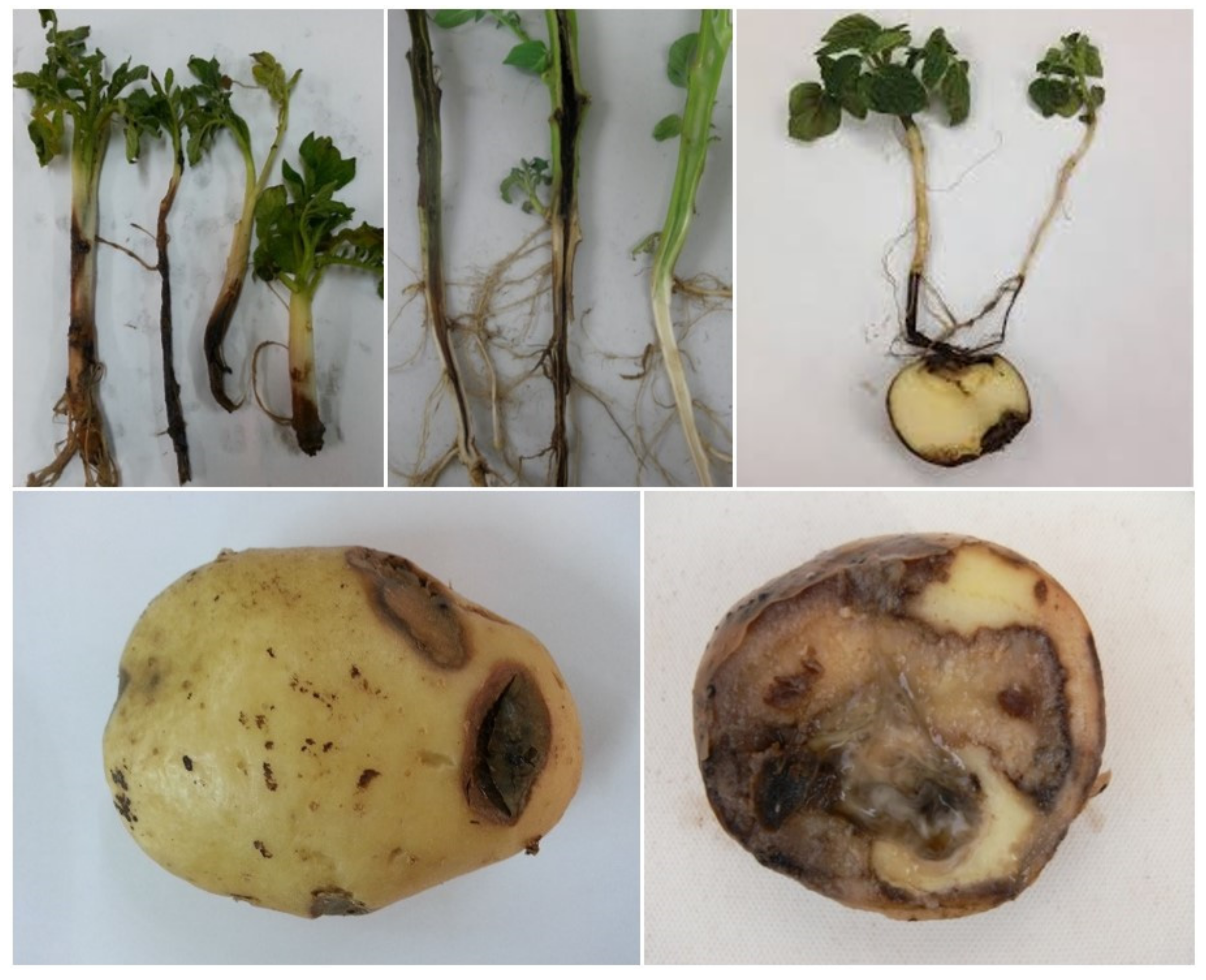
2.1. Bacterial Samples

Biological source (P. atrosepticum SCRI 1039) was obtained from the collection of the James Hutton Institute (Scotland, United Kingdom) and the dilutions in the test ranged from 109 to 102 CFU (colony-forming unit)/mL. Two other bacteria sources belonging to other Pectobacreium and Dickeya species (Pectobacterium parmentieri strain Wpp161, Dickeya solani strain 2222) were also obtained from the collection of the James Hutton Institute (Scotland, United Kingdom) and used as a negative control for diagnostic specificity. The sets of samples used for ELISA and PCR (Polymerase chain reaction) were the same.

2.2. ELISA and PCR Analysis

To assess the performance of the developed LOC, samples were evaluated using ELISA and PCR. Indirect-ELISA tests were carried out using the recommended antiserum (Agdia Inc., Elkhart, IN, USA) while dilutions ranged from 109 to 102 CFU/mL. ELISA cross-reaction was not observed against the plant pathogens: Agrobacterium tumefaciens; Erwinia amylovora, Pseudomonas marginalis, Ralstonia solanacearum; Xanthomonas campestris, Klebsiella oxycota, Escherichia coli, Rhodococcus rhodochrous, Curtobacterium flaccumfaciens, Pectobacterium parmentieri, P. carotovorum sub sp carotovourum, P. carotovorum sub sp brasiliense, Dickeya. Solani, D. dadantii, and D. zeae. The absorbance values at 405 nm (OD405) were recorded 4 h after adding the substrate, using an ASYS Hitech GmbH Expert 96 microplate reader. Numbers were normalized as R values (OD-sample/OD-negative control). R = 2.0 was used as a threshold to distinguish between positive and negative responses [18].
Genomic DNA was obtained from bacterial cultures by a standard protocol [19]. Primers ECA1f/ECA2r and Y45/Y46 were used in PCR reactions [20,21]. PCR amplifications were conducted as described using the following thermal regime: 94 °C/10 min, 34 × (94 °C/30 s, 62 °C/45 s, 72 °C/45 s) and 72 °C/10 min. The given primers are specific to the chromosomal DNA of P. atrosepticum and the amplification products show 690 bp and 439 bp, respectively. Agarose gel electrophoresis (1.5% agarose gel with ethidium bromide) was used to resolve amplicons.

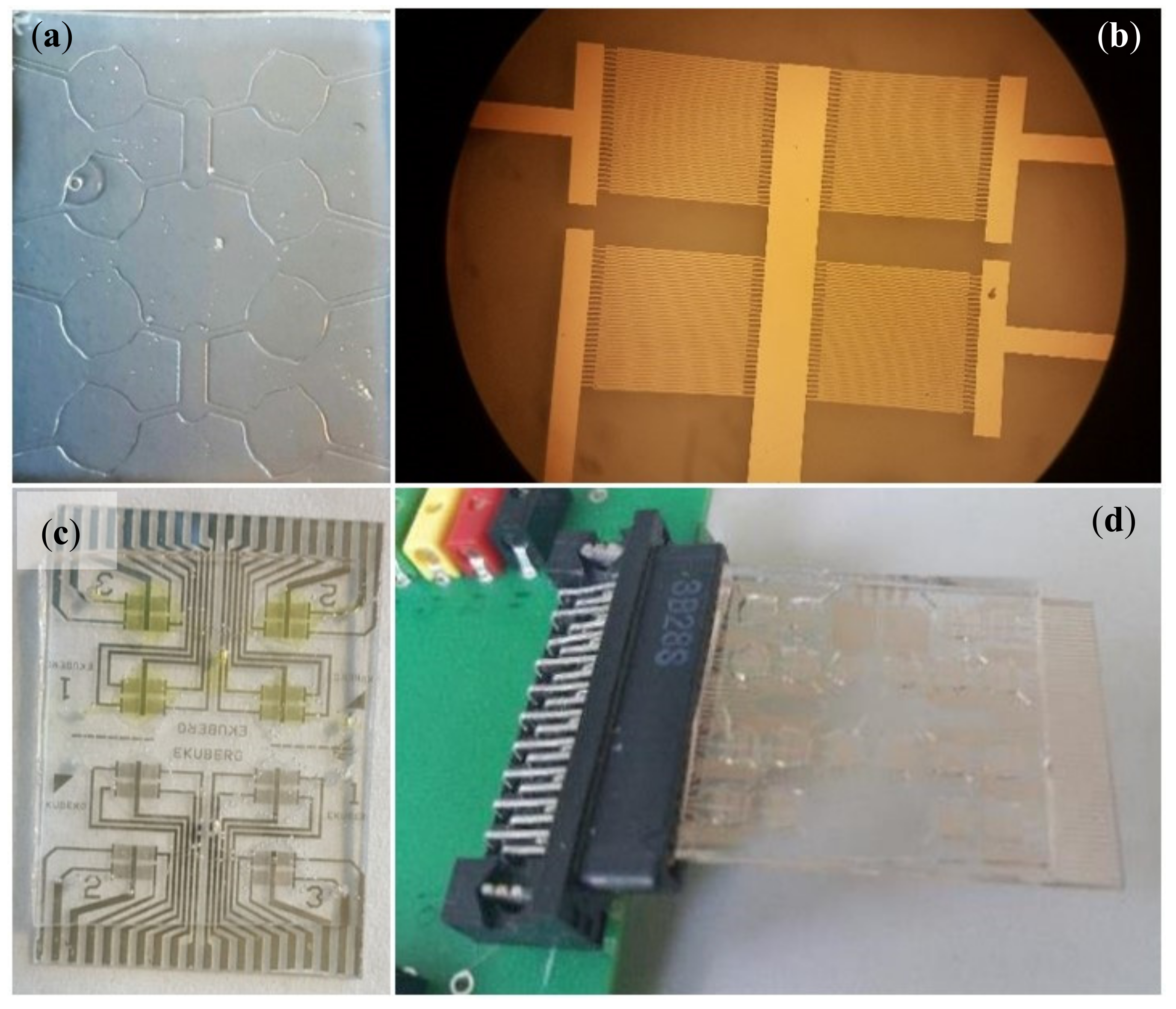
2.3. Lab-On-Chip Platform

The lab-on-chip optimized to detect P. atrosepticum (Figure 2) is similar in structure to the prototypes reported before for other applications [11,15,22,23] and consists of an array of gold interdigitated electrodes for EIS measurements combined to a microfluidic module which allows fluids handling and the confinement of the solutions (reagents for functionalization and sample to analyze). The microfluidic modules with microchannels and microchambers (with a volume of 20 μL) were obtained by PDMS replica molding, from an SU8 hard master. The layout includes a common central inlet and four peripheral outlets per side (half chip) that can be utilized to independently functionalize each chamber or to inject the sample to be tested in quadruplicate improving reproducibility and allowing to save reagents and samples (Figure 2a). The electrodes have a line-space period of 10 μm and cover a 2 mm × 1.5 mm area. They were fabricated by optical lithography (using a Karl Suss MJB3 mask aligner and AZ5214e resist) and thermal evaporation of Cr/Au (3 nm/15 nm) (Figure 2b). Here, highly specific antibodies (the same used for ELISA tests from Agdia Inc., Elkhart, IN, USA) were employed to functionalize the microelectrodes in order to make them capable to bind specifically to P. atrosepticum cells. Particularly, the functionalization includes the following layers and steps:
(1)
a mixed self-assembled monolayer (SAM) of beta-mercaptoethanol and mercaptoundecanoic acid obtained by overnight incubation in ethanol solution;
(2)
the activation of the COOH groups of 11-mercaptoundecanoic acid (MUA) by incubation for 30 min in a solution of N-hydroxysuccinimide and N-ethyl-N-3-dimethylaminopropyl carbodiimide hydrochloride at a ratio of 1:4 in milliQ water;
(3)
a protein A monolayer assembled by covalent binding to the mixed SAM by a 2 h incubation in a (50 μg/mL) phosphate buffered saline (PBS) solution;
(4)
the disactivation of ester reactive groups by incubation in ethanolamine (1 M);
(5)
the passivation of the unbound surface by incubation in bovine serum albumin (BSA) (1 mg/mL);
(6)
the oriented immobilization of the antibodies specific for P. atrosepticum (5 ng/mL) by binding to protein A through their Fc region (fragment crystallizable region).
In this way, protein A achieves a covalent binding to SAM of mercapto undecanoic acid. Then Protein A naturally binds the Fc region of antibodies allowing their oriented immobilization. Ethanolamine was used to block reactive groups while BSA was used to passivate and saturate the unbound surface. As the final step, each sensing area was functionalized using antibodies.
All reagents were purchased from Sigma-Aldrich Inc. (St. Louis, Missouri, USA), except the antibodies.
The calibration tests were carried out by known dilutions of bacteria suspensions in PBS to evaluate the sensitivity of the assay. For this purpose, bacteria cell dilutions ranging from 109 CFU/mL to 104 CFU/mL were delivered through the microfluidic channels and left in static conditions into the microchambers for 1 h to allow the biorecognition between the suspended cells of P. atrosepticum and antibodies immobilized on the surface. Afterward, 1 mL of washing PBS solution was delivered and chambers were filled with a solution of hexacyanoferrate (II/III) K3[Fe(CN)6]/K4[Fe(CN)6] (1:1) at a concentration of 10 mM to measure the electrochemical impedance with an Autolab PGSTAT30 under an applied sinusoidal 15 mV AC voltage at a frequency ranging from 105 Hz to 0.1 Hz. Impedance spectroscopy raw data were exported as ASCII files and OriginPro 2018 (OriginLab Corporation, Northampton, MA, USA) was used to process them.

3. Results and Discussion

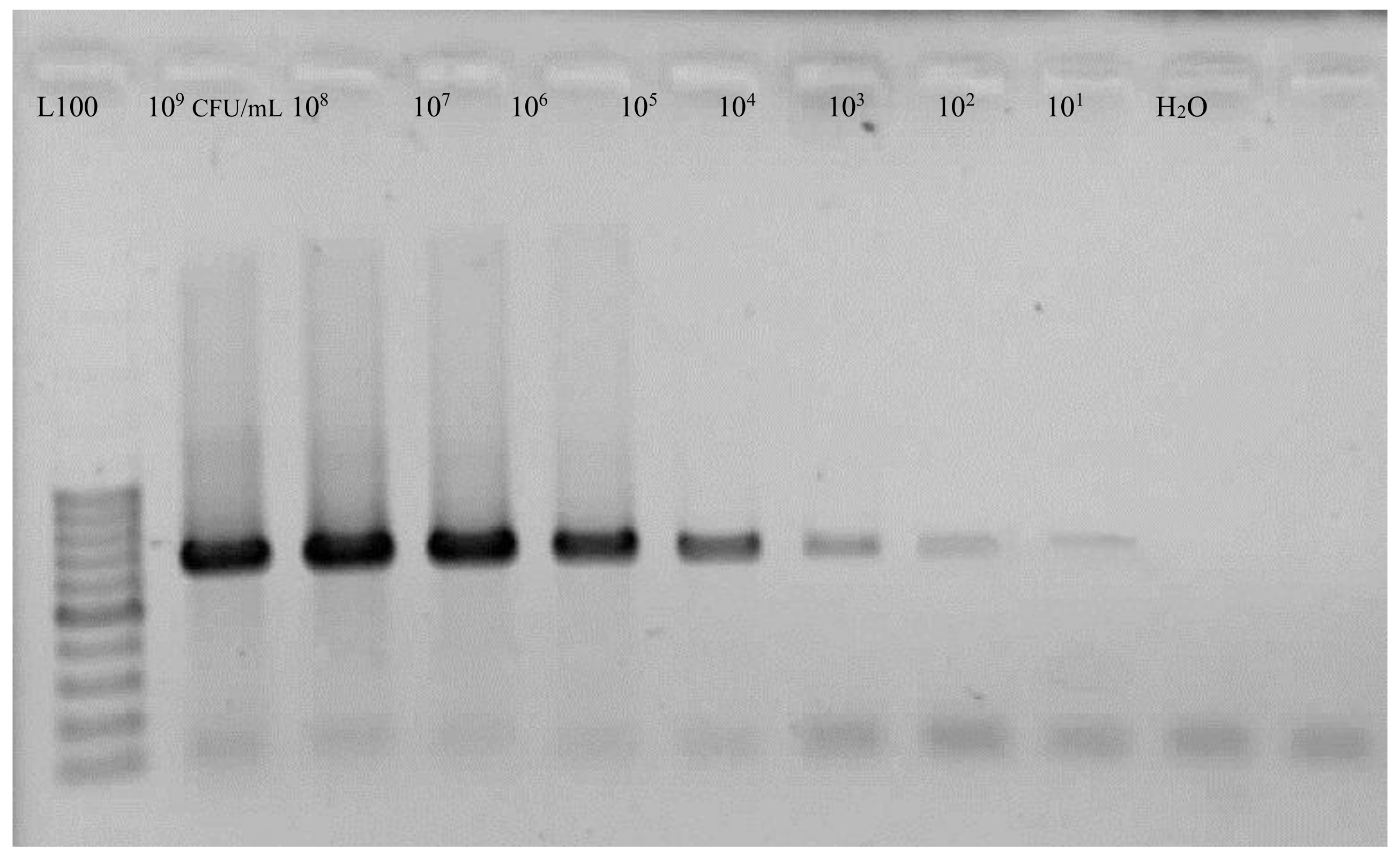
3.1. ELISA and PCR Assays
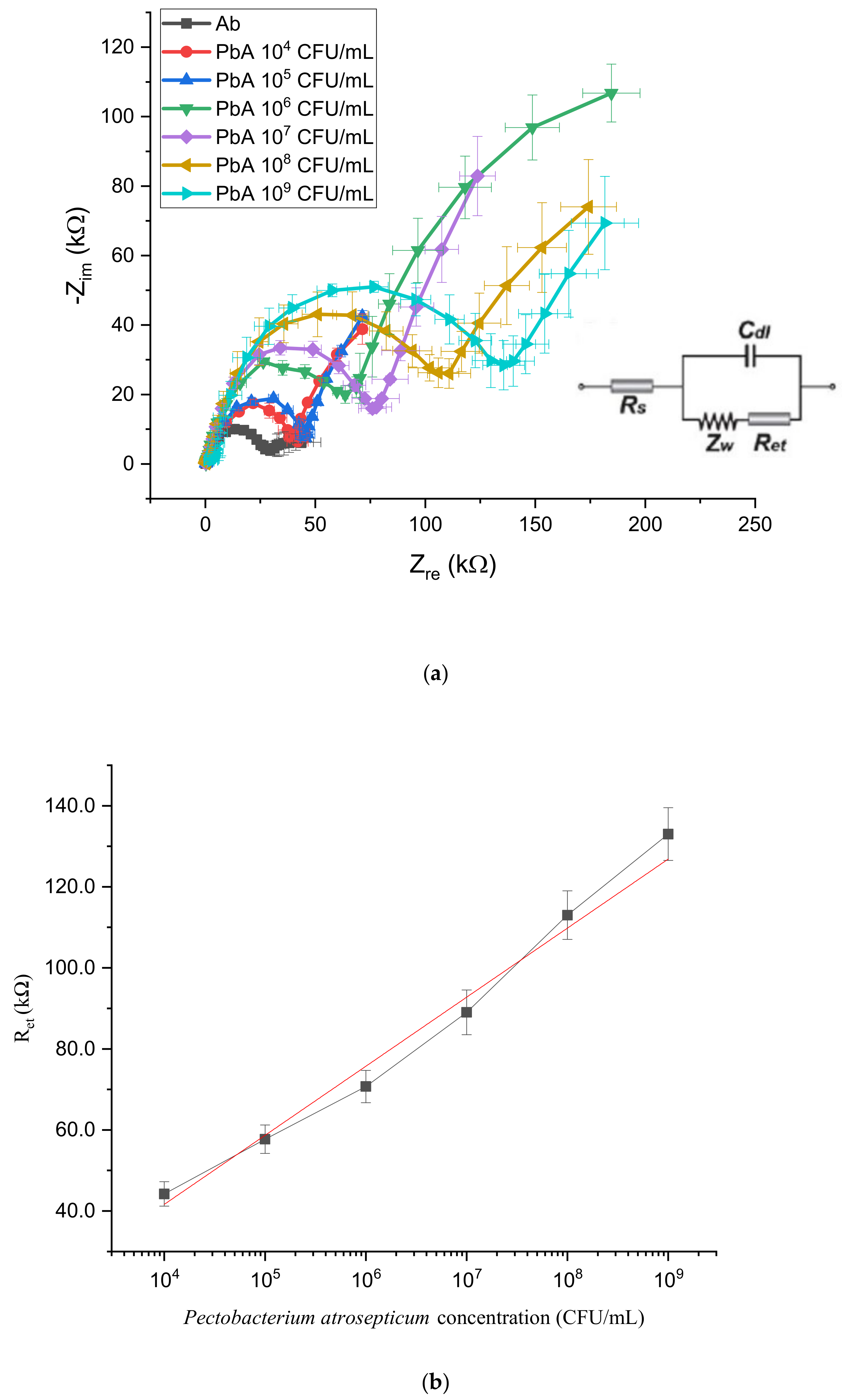
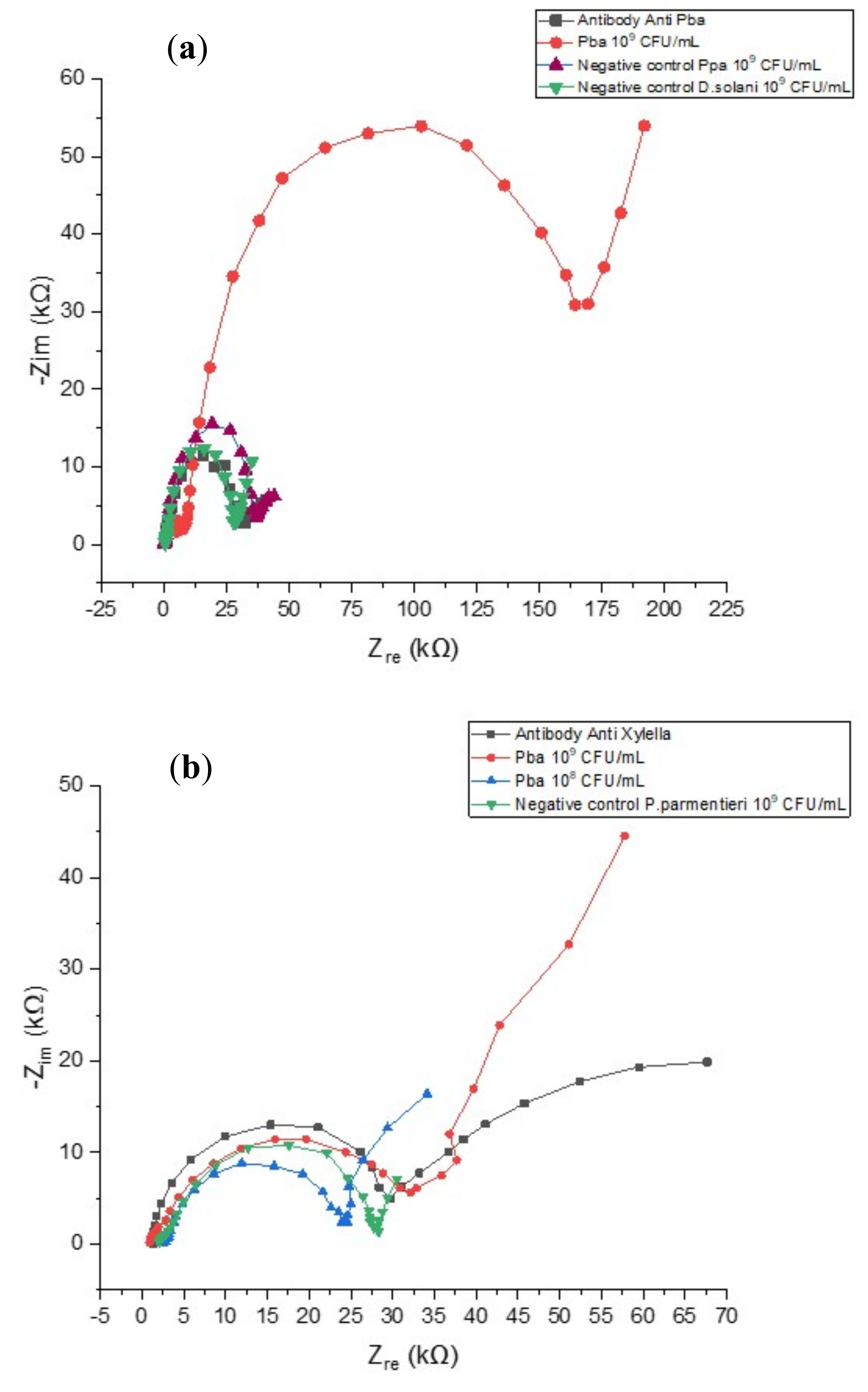
In ELISA assays (Table 1), cross-reaction against other plant pathogens and other species of Pectobacterium and Dickeya was not observed. Positive readings up to a dilution of 105 CFU/mL were obtained. The R value of positive control was 33, while the negative control yielded an absorbance value of less than 0.06.
In PCR assays, to determine the specificity of the primer sets, tests were performed using Eca1/Eca2 and Y45/Y46 primers with DNA from all samples. Two 690 bp and 439 bp DNA fragments were respectively produced for P. atrosepticum isolates, while no amplification was observed with isolates of other Pectobacterium spp. and Dickeya spp. Different concentrations of Pba isolates were used and the estimation for PCR sensitivity was 102 CFU/mL (Figure 3).

3.2. LOC Detection

To evaluate the sensitivity of the designed EIS lab-on-chip, impedance spectroscopy measurements were carried out in a frequency ranging from 105 to 0.1 Hz and plotted as Nyquist curves where the x-axis is the real component of impedance Zre while the y-axis is the complex component −Zim. The system can be modeled by an equivalent (Randles) circuit (inset of Figure 4a), describing the interfacial layer at the working electrode as an electron transfer resistance Ret in parallel combination with an electrical double layer capacitance Cdl. A series Warburg impedance Zw accounts for redox species depletion at the interface, while Rs is the uncompensated solution resistance. The electron transfer resistance (Ret) is very sensitive to electrode modifications and roughly corresponds the diameter of the semicircle-shaped curves.
As a first step, the sensing layer consisting of antibodies immobilized on the electrode surface was characterized to identify their impedance contribution. Anti- P. atrosepticum antibodies layer resulted in Ret ≈ 30 kΩ (dark gray line in Figure 4a). Then, to calibrate the sensors response, serial dilutions of P. atrosepticum in PBS (109, 108, 107, 106, 105, 104 CFU/mL) were prepared and tested. After incubation and washing steps, redox solution was added in the chamber and EIS measurements were collected. As shown in Figure 4a, a noticeable rise in impedance values with respect to the antibody baseline can be associated with incubation with a spiked solution on the sensing layer (colored curves) and impedance values are correlated with the bacterial P. atrosepticum concentration [C] in suspension (linearly in a Ret vs. log [C] plot) whose decrease results in smaller Ret values (Figure 4b). The suspension containing 104 CFU/mL was associated with Ret ≈ 42 kΩ (red curve), while the most concentrated suspension with 109 CFU/mL resulted in Ret ≈ 135 kΩ (light blue curve).
As a negative control, we incubated P. parmentieri or Dickeya solani suspensions containing 109 CFU/mL. As shown in Figure 5a, impedance curves obtained in these cases (purple and green curves respectively) resulted in Ret values close to the antibody baseline (≈30 kΩ) while the same concentration of Pba gave Ret ≈ 175 kΩ, demonstrating the high specificity of the optimized sensing system.
To further evaluate specificity of our LOC and a possible aspecific interaction between the bacterial cells and the electrode’s surface, we also immobilized a different antibody (Ab anti-Xyllella. Fastidiosa) not able to specifically interact with Pba. In this case, we obtained Ret ≈ 30 kΩ by adding different concentrations of P. atrosepticum, P. parmentieri and D. solani, showing again the high specificity of the LOC assay (Figure 5b).

4. Conclusions

Conventional diagnostic techniques usually need a long time for processing and a great deal of effort. Detection methods based on immunological approaches such as ELISA can be automated and therefore become effective in terms of time and effort, but there are some limitations which include low sensitivity and false-positive results. In the present work, a reliable, inexpensive and label-free LOC assay has been set to detect P. atrosepticum down to 104 CFU/mL. As reported by Czajkowski et al. [7], the usual level of lead accumulation in infected tubers is 105 and the density of bacteria in infected tissues is often more than 106, which is higher than the limit of detection of our device. Therefore, according to these results, the LOC can be used to detect Pba in infected tubers.
In Table 2, LOC sensitivity is compared to ELISA and PCR, demonstrating competitiveness of the developed assay as it provides a better sensitivity than the ELISA method. Indeed, ELISA tests are not able to detect pathogens at dilutions below 105 CFU/mL, while the developed LOC platform could identify bacteria cells down to 104 CFU/mL dilutions. On the other hand, PCR methods showed better performances compared to LOC devices with positive cases identified even at 102 CFU/mL, but this nucleic acid-based method is expensive and advanced equipment and experienced technicians are needed to perform it. The introduced device can provide a good compromise considering the need for sensitivity and assay accessibility. As mentioned earlier, the developed LOC assay also has a good specificity since it can detect 104 CFU/mL of Pba (below the typical 105 CFU/mL value in infected tubers) and differentiate it from other Pectobacterium and Dickeya species. Another advantage of the proposed LOC devices is associated with cost. Based on our estimations, it is almost 1.8 to 6 times less expensive than ELISA and PCR. The major factors contributing to the costs of LOC devices (around 5 €/device) are mainly the glass substrate and the metallization, since the volumes of employed reagents are very low with minimal use of antibodies.
Another useful comparison can be done with recent devices proposed for plant disease diagnosis. As shown in Table 3, the performance of our sensing platform is competitive with respect to other recent technologies in terms of sensitivity and the limit of detection. The two techniques based on lateral flow immune assays offer the great advantage to be very cheap and simple, two key aspects for on field analysis, but on the other hand they are not quantitative [24,25]. Papadakis and coworkers proposed an approach based on quartz crystal microbalance to detect DNA amplificons and they showed a LOD of 103 CFU/mL that is lower than our devices but their approach requires additional steps for DNA extraction from plant tissues and DNA amplification [26], which results in an increase in the cost and time necessary for the analysis. In the last work, Yazgan and coworkers optimized two sensing approaches based on cyclic voltammetry and surface plasmon resonance by using carbohydrate ligand as the bioreceptor. They modified with different moieties the ligands and obtained a very sensitive tool with improved LOD down to 1 CFU/mL with the SPR approach, but the limit of this smart solution is related with the possibility of cross reaction with other bacterial strains and the difficulties to translate an SPR approach into a point of care device [27].
On the contrary, our sensing platform is particularly suitable for POC applications thanks to its limited dimension (a few square centimeters). In addition, the number of sensors in the array can be increased by further miniaturization, permitting more analysis on the same chip without affecting its cost and enabling the simultaneous detection of several pathogens ranging from viruses to bacteria and fungi.
As next steps towards a fully integrated platform for POC phytodiagnostics, a read-out board for electronic connections with a portable potentiostat can be employed enabling a new and smart approach for on field plant diagnostics, including the use of a smartphone for data acquisition and sharing. In fact the current trend in automation in agriculture makes it more and more difficult to perform a manual inspection of plants in the crops for disease monitoring, so the use of smartphones and wireless technology together with the new developing internet of things (IoT) [28] open new frontiers in agriculture [29], offering the possibility to collect and analyze data about the diffusion of a disease in order to map the spreading of the pathogens and, for example, prevent the infection of healthy surrounding areas.

Author Contributions

Conceptualization: E.P., M.S.C., P.P., M.B. and G.M.; data curation: M.H.T., E.P. and M.S.C.; formal analysis: M.H.T., E.P., M.S.C., P.P., M.B. and G.M.; funding acquisition: M.B. and G.M.; investigation: M.H.T., E.P. and M.S.C.; methodology: M.H.T., E.P., M.S.C., P.P., M.B. and G.M.; supervision: P.P., M.B. and G.M.; writing—original draft: M.H.T.; writing—review and editing: E.P., M.S.C., P.P., M.B. and G.M. All authors have read and agreed to the published version of the manuscript.

Funding

This work was supported by Italian National FISR-CIPE Project “Inno-Sense”: Development of an innovative sensing platform for on-field analysis and monitoring (delibera CIPE n.78 del 07/08/2017) and by Fondazione Puglia (Prot n. 86/18).

Acknowledgments

We thank Ian Toth of the James Hutton Institute (Scotland, United Kingdom) for bacterial strains. We also thank Iolena Tarantini for helping with the fabrication of the sensor.

Conflicts of Interest

The authors declare no conflict of interest.

References

  1. Cassedy, A.; Mullins, E.; O’Kennedy, R. Sowing seeds for the future: The need for on-site plant diagnostics. Biotechnol. Adv. 2020, 39, 107358. [Google Scholar] [CrossRef] [PubMed]
  2. Ma, B.; Hibbing, M.E.; Kim, H.-S.; Reedy, R.M.; Yedidia, I.; Breuer, J.; Breuer, J.; Glasner, J.D.; Perna, N.T.; Kelman, A.; et al. Host Range and Molecular Phylogenies of the Soft Rot Enterobacterial Genera Pectobacterium and Dickeya. Phytopathology 2007, 97, 1150–1163. [Google Scholar] [CrossRef] [PubMed] [Green Version]
  3. Pérombelon, M.C.M. Potato diseases caused by soft rot erwinias: An overview of pathogenesis. Plant Pathol. 2002, 51, 1–12. [Google Scholar] [CrossRef]
  4. Hauben, L.; Moore, E.R.B.; Vauterin, L.; Steenackers, M.; Mergaert, J.; Verdonck, L.; Swings, J. Phylogenetic Position of Phytopathogens within the Enterobacteriaceae. Syst. Appl. Microbiol. 1998, 21, 384–397. [Google Scholar] [CrossRef]
  5. Czajkowski, R.L.; Grabe, G.J.; Wolf, J.M.v.d. Distribution of Dickeya spp. and Pectobacterium carotovorum subsp carotovorum in naturally infected seed potatoes. Eur. J. Plant Pathol. 2009, 125, 263–275. [Google Scholar] [CrossRef]
  6. Czajkowski, R.; Pérombelon, M.C.M.; van Veen, J.A.; van der Wolf, J.M. Control of blackleg and tuber soft rot of potato caused by Pectobacterium and Dickeya species: A review. Plant Pathol. 2011, 60, 999–1013. [Google Scholar] [CrossRef]
  7. Czajkowski, R.; Pérombelon, M.; Jafra, S.; Lojkowska, E.; Potrykus, M.; van der Wolf, J.; Sledz, W. Detection, identification and differentiation of Pectobacterium and Dickeya species causing potato blackleg and tuber soft rot: A review. Ann. Appl. Biol. 2015, 166, 18–38. [Google Scholar] [CrossRef] [Green Version]
  8. Humphris, S.N.; Cahill, G.; Elphinstone, J.G.; Kelly, R.; Parkinson, N.M.; Pritchard, L.; Toth, I.K.; Saddler, G.S. Detection of the Bacterial Potato Pathogens Pectobacterium and Dickeya spp. Using Conventional and Real-Time PCR. In Plant Pathology: Techniques and Protocols; Lacomme, C., Ed.; Springer: New York, NY, USA, 2015; pp. 1–16. [Google Scholar] [CrossRef]
  9. Alvarez, A.M. Integrated Approaches for Detection of Plant Pathogenic Bacteria and Diagnosis of Bacterial Diseases. Annu. Rev. Phytopathol. 2004, 42, 339–366. [Google Scholar] [CrossRef] [Green Version]
  10. Charlermroj, R.; Himananto, O.; Seepiban, C.; Kumpoosiri, M.; Warin, N.; Gajanandana, O.; Elliott, C.T.; Karoonuthaisiri, N. Antibody Array in a Multiwell Plate Format for the Sensitive and Multiplexed Detection of Important Plant Pathogens. Anal. Chem. 2014, 86, 7049–7056. [Google Scholar] [CrossRef] [PubMed]
  11. Chiriacò, M.S.; Primiceri, E.; De Feo, F.; Montanaro, A.; Monteduro, A.G.; Tinelli, A.; Megha, M.; Carati, D.; Maruccio, G. Simultaneous detection of multiple lower genital tract pathogens by an impedimetric immunochip. Biosens. Bioelectron. 2016, 79, 9–14. [Google Scholar] [CrossRef]
  12. Templier, V.; Livache, T.; Boisset, S.; Maurin, M.; Slimani, S.; Mathey, R.; Roupioz, Y. Biochips for Direct Detection and Identification of Bacteria in Blood Culture-Like Conditions. Sci. Rep. 2017, 7, 9457. [Google Scholar] [CrossRef] [PubMed]
  13. Paul, A.; Chiriacò, M.S.; Primiceri, E.; Srivastava, D.N.; Maruccio, G. Picomolar detection of retinol binding protein 4 for early management of type II diabetes. Biosens. Bioelectron. 2019, 128, 122–128. [Google Scholar] [CrossRef] [PubMed]
  14. Nezhad, A.S. Future of portable devices for plant pathogen diagnosis. Lab Chip 2014, 14, 2887–2904. [Google Scholar] [CrossRef] [PubMed]
  15. Chiriacò, M.S.; Luvisi, A.; Primiceri, E.; Sabella, E.; De Bellis, L.; Maruccio, G. Development of a lab-on-a-chip method for rapid assay of Xylella fastidiosa subsp pauca strain CoDiRO. Sci. Rep. 2018, 8, 7376. [Google Scholar] [CrossRef] [PubMed]
  16. Julich, S.; Riedel, M.; Kielpinski, M.; Urban, M.; Kretschmer, R.; Wagner, S.; Fritzsche, W.; Henkel, T.; Möller, R.; Werres, S. Development of a lab-on-a-chip device for diagnosis of plant pathogens. Biosens. Bioelectron. 2011, 26, 4070–4075. [Google Scholar] [CrossRef] [PubMed]
  17. Boer, S.H.D.; López, M.M. New Grower-Friendly Methods for Plant Pathogen Monitoring. Annu. Rev. Phytopathol. 2012, 50, 197–218. [Google Scholar] [CrossRef] [PubMed] [Green Version]
  18. Scuderi, G.; Lombardo, A.; Raspagliesi, D.; Russo, M.; Catara, A.; Licciardello, G. Development and Evaluation of a Novel Probe Microarray for Genotyping Citrus tristeza virus Using an Integrated Lab-on-Chip Device. J. Plant Pathol. 2016, 98, 25–34. [Google Scholar]
  19. Li, X.A.; Deboer, S.H. Selection of Polymerase Chain-Reaction Primers from an Rna Intergenic Spacer Region for Specific Detection of Clavibacter-michiganensis subsp sepedonicus. Phytopathology 1995, 85, 837–842. [Google Scholar] [CrossRef]
  20. Deboer, S.H.; Ward, L.J. PCR detection Of Erwinia-carotovora subsp atroseptica Associated with Potato Tissue. Phytopathology 1995, 85, 854–858. [Google Scholar] [CrossRef]
  21. Frechon, D.; Exbrayat, P.; Helias, V.; Hyman, L.J.; Jouan, B.; Llop, P.; Lopez, M.M.; Payet, N.; Perombelon, M.C.M.; Toth, I.K.; et al. Evaluation of a PCR kit for the detection of Erwinia carotovora subsp. atroseptica on potato tubers. Potato Res. 1998, 41, 163–173. [Google Scholar] [CrossRef]
  22. Chiriacò, M.S.; Primiceri, E.; Monteduro, A.G.; Bove, A.; Leporatti, S.; Capello, M.; Ferri-Borgogno, S.; Rinaldi, R.; Novelli, F.; Maruccio, G. Towards pancreatic cancer diagnosis using EIS biochips. Lab Chip 2013, 13, 730–734. [Google Scholar] [CrossRef] [PubMed] [Green Version]
  23. Primiceri, E.; Chiriacò, M.S.; D’Amone, E.; Urso, E.; Ionescu, R.E.; Rizzello, A.; Maffia, M.; Cingolani, R.; Rinaldi, R.; Maruccio, G. Real-time monitoring of copper ions-induced cytotoxicity by EIS cell chips. Biosens. Bioelectron. 2010, 25, 2711–2716. [Google Scholar] [CrossRef]
  24. Safenkova, I.V.; Pankratova, G.K.; Zaitsev, I.A.; Varitsev, Y.A.; Vengerov, Y.Y.; Zherdev, A.V.; Dzantiev, B.B. Multiarray on a test strip (MATS): Rapid multiplex immunodetection of priority potato pathogens. Anal. Bioanal. Chem. 2016, 408, 6009–6017. [Google Scholar] [CrossRef] [PubMed]
  25. Safenkova, I.V.; Zaitsev, I.A.; Varitsev, Y.A.; Byzova, N.A.; Drenova, N.V.; Zherdev, A.V.; Dzantiev, B.B. Development of a lateral flow immunoassay for rapid diagnosis of potato blackleg caused by Dickeya species. Anal. Bioanal. Chem. 2017, 409, 1915–1927. [Google Scholar] [CrossRef] [PubMed]
  26. Papadakis, G.; Skandalis, N.; Dimopoulou, A.; Glynos, P.; Gizeli, E. Bacteria Murmur: Application of an Acoustic Biosensor for Plant Pathogen Detection. PLoS ONE 2015, 10, e0132773. [Google Scholar] [CrossRef]
  27. Yazgan, I.; Noah, N.; Toure, O.; Zhang, S.; Sadik, O. Biosensor for selective detection of E-coli in spinach using the strong affinity of derivatized mannose with fimbrial lectin. Biosens. Bioelectron. 2014, 61C, 266–273. [Google Scholar] [CrossRef]
  28. Zikria, Y.B.; Afzal, M.K.; Kim, S.W. Internet of Multimedia Things (IoMT): Opportunities, Challenges and Solutions. Sensors 2020, 20, 2334. [Google Scholar] [CrossRef]
  29. Farooq, M.S.; Riaz, S.; Abid, A.; Umer, T.; Zikria, Y.B. Role of IoT Technology in Agriculture: A Systematic Literature Review. Electronics 2020, 9, 319. [Google Scholar] [CrossRef] [Green Version]
Figure 1. Damage induced by P. atrosepticum on seed tuber and stem of potatoes.
Biosensors 10 00064 g001
Figure 2. (a) Microfluidic modules for fluid handling consisting of four chambers per sides connected by a network of microchannels; (b) detail of the four interdigited electrodes available in each sensing area; (c,d) image of the lab-on-chip and its connection to the interface printed circuit board.
Biosensors 10 00064 g002
Figure 3. Agarose gel electrophoresis of polymerase chain reaction (PCR) sensitivity on bacterial isolates by Y45/Y46 primers.
Biosensors 10 00064 g003
Figure 4. (a) Nyquist spectra obtained in lab-on-a-chip (LOC) device calibration. Curves from cyan to red correspond to different concentrations of Pectobacterium atrosepticum in spiked solutions; each curve is the average of three different measurements. (b) Impedance values as a function of Pectobacterium atrosepticum concentration: each point is the average of three different measurements.
Biosensors 10 00064 g004
Figure 5. (a) Nyquist spectra obtained during negative testing with P. parmentieri and Dickeya solani at 109 CFU/mL and positive control (Pectobacterium atrosepticum 109 CFU/mL). (b) Nyquist spectra obtained by testing of P. atrosepticum, P. parmentieri and D. solani on a chip functionalized with antibodies anti-Xyllella. fastidiosa.
Biosensors 10 00064 g005
Table 1. Results of enzyme-linked immunosorbent-assay (ELISA) tests (− = negative, + = positive) on samples ranging from 109 to 102 CFU mL−1 (where CFU = colony-forming unit), R = OD-sample/OD-negative control.
CFU/mLELISA
ODRResult
109233+
1081.830+
1071.1218.67+
1060.213.5+
1050.162.6+
1040.0881.47-
1030.0631.05-
1020.0340.56-
1010.0150.25-
Control0.061-
Table 2. Performance comparison: LOC is more effective in detecting P. atrosepticum compared to the ELISA method, while PCR results in higher sensitivity, but at larger costs.
TechniqueLimit of Detection
ELISA105 CFU/mL
PCR102 CFU/mL
LOC104 CFU/mL
Table 3. Performance comparison with other devices for plant disease diagnosis.
Detection TechniqueBioreceptorPlantBacteriaLODRef.
Lateral Flow ImmunoassayAbPotatoDickeya dianthicola and Dickeya solani4 × 105 CFU/mLSafenkova et al. [25]
Quartz Crystal MicrobanceDNAtomatoRalstonia solanacearum,
Pseudomonas syringae pv.
tomato, Xanthomonas campestris pv. vesicatoria
103–104 CFU/mLPapadakis et al. [26]
Cyclic VoltammetryModified carbohydrate ligandSpinachE. coli6.25 × 102 CFU/mLYazgan et al. [27]
Surface Plasmon resonanceModified carbohydrate ligandSpinachE. coli1–104 CFU/mLYazgan et al. [27]
Lateral Flow immunoassay in multiarray on a test strip (MATS) formatAbPotatoClavibacter michiganensis104 CFU/mLSafenkova et al. [24]

