Comparison of Ti-Based Coatings on Silicon Nanowires for Phosphopeptide Enrichment and Their Laser Assisted Desorption/Ionization Mass Spectrometry Detection

 ,
, 
Abstract
:1. Introduction
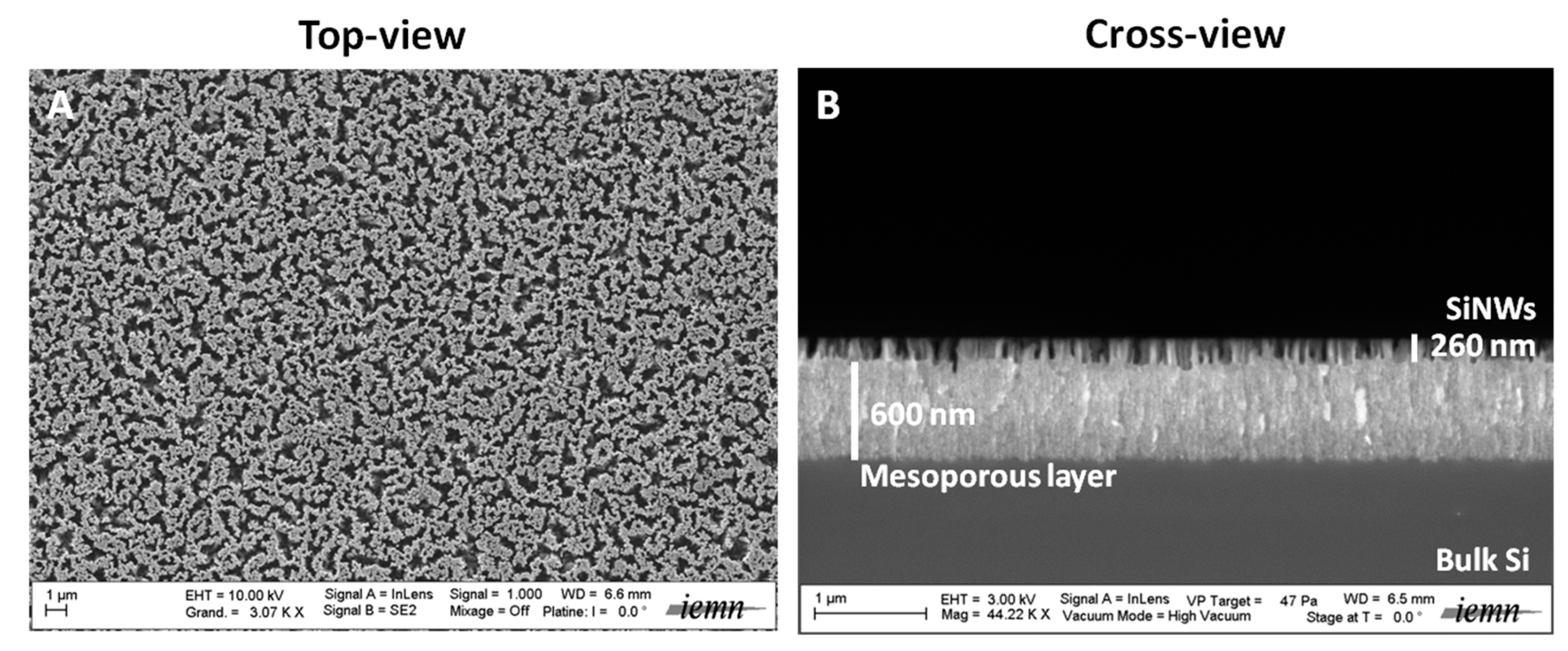
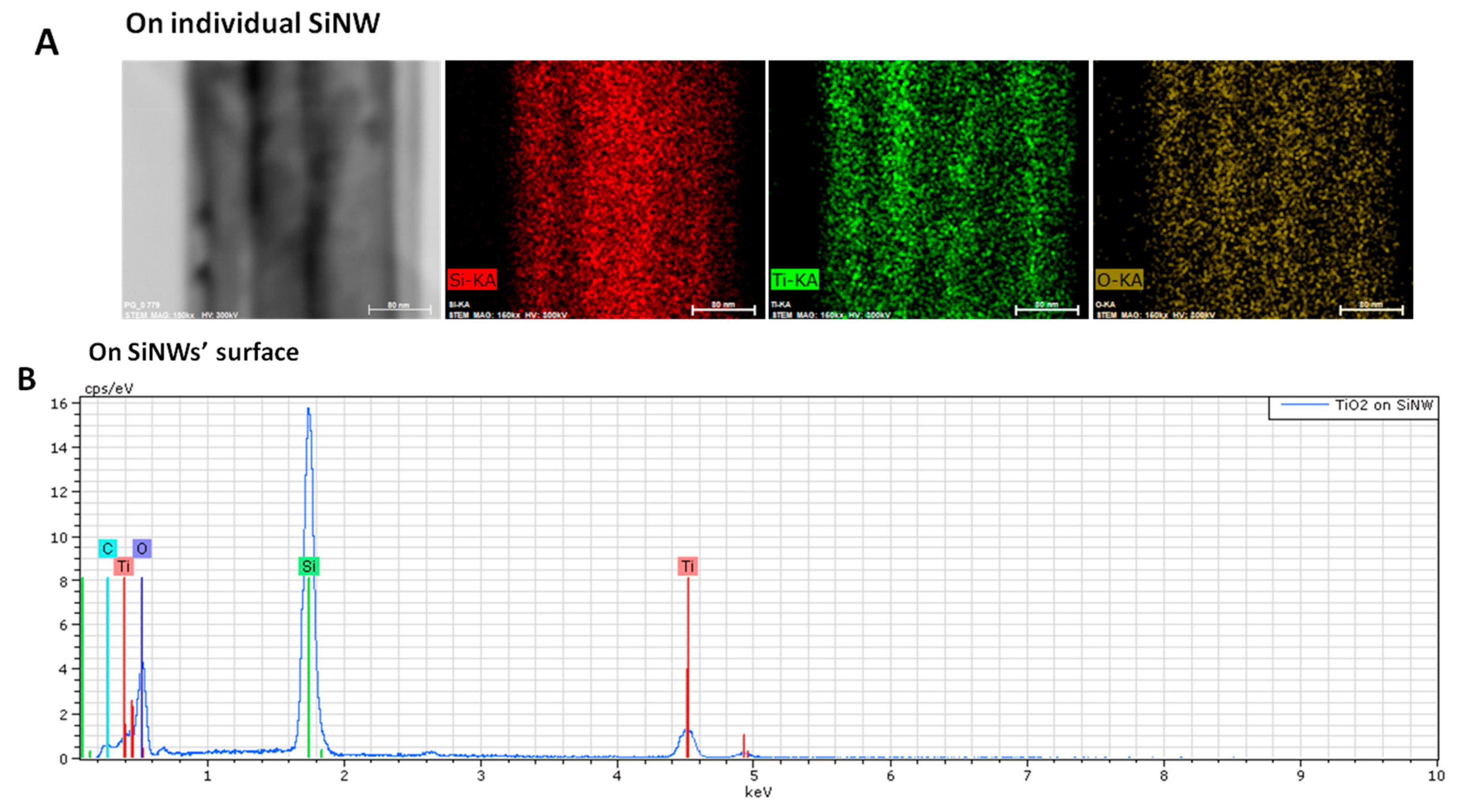
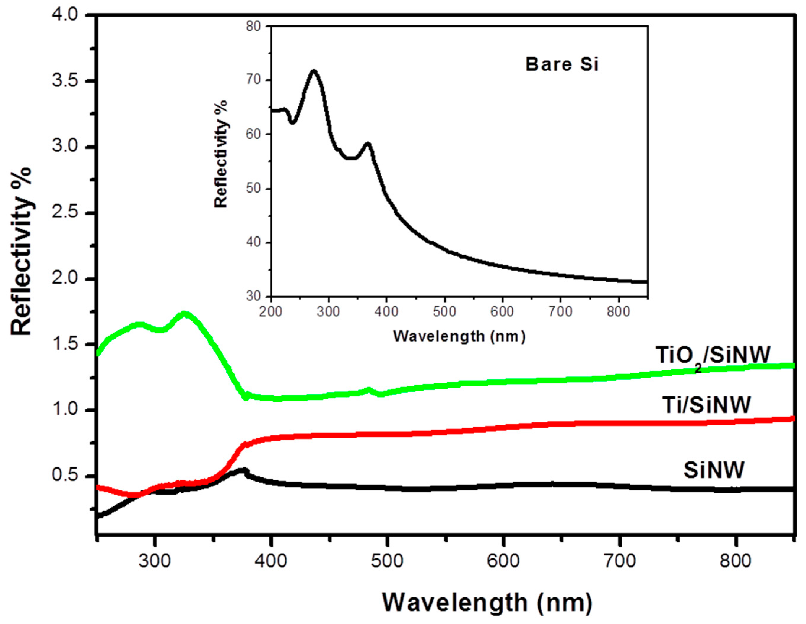
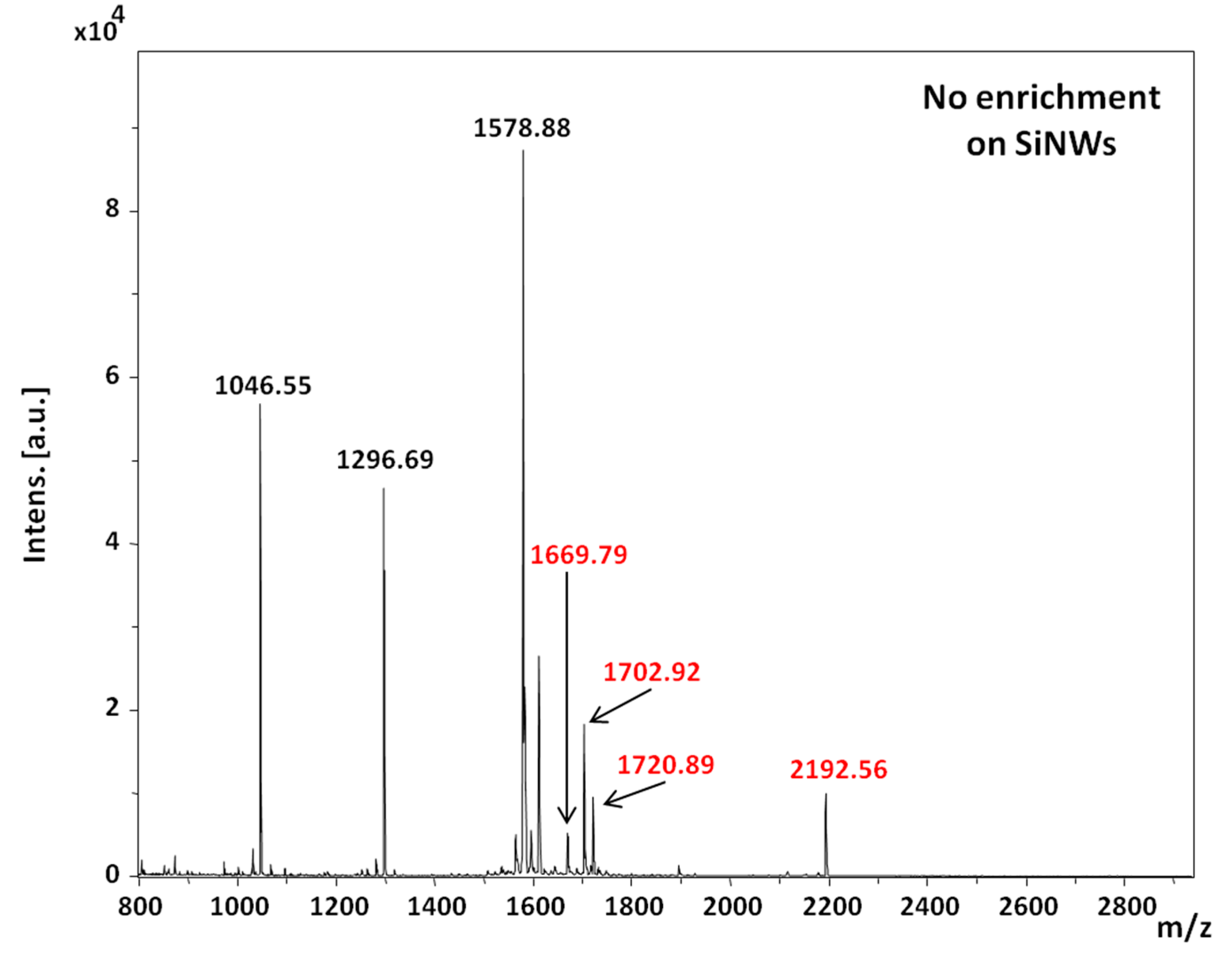
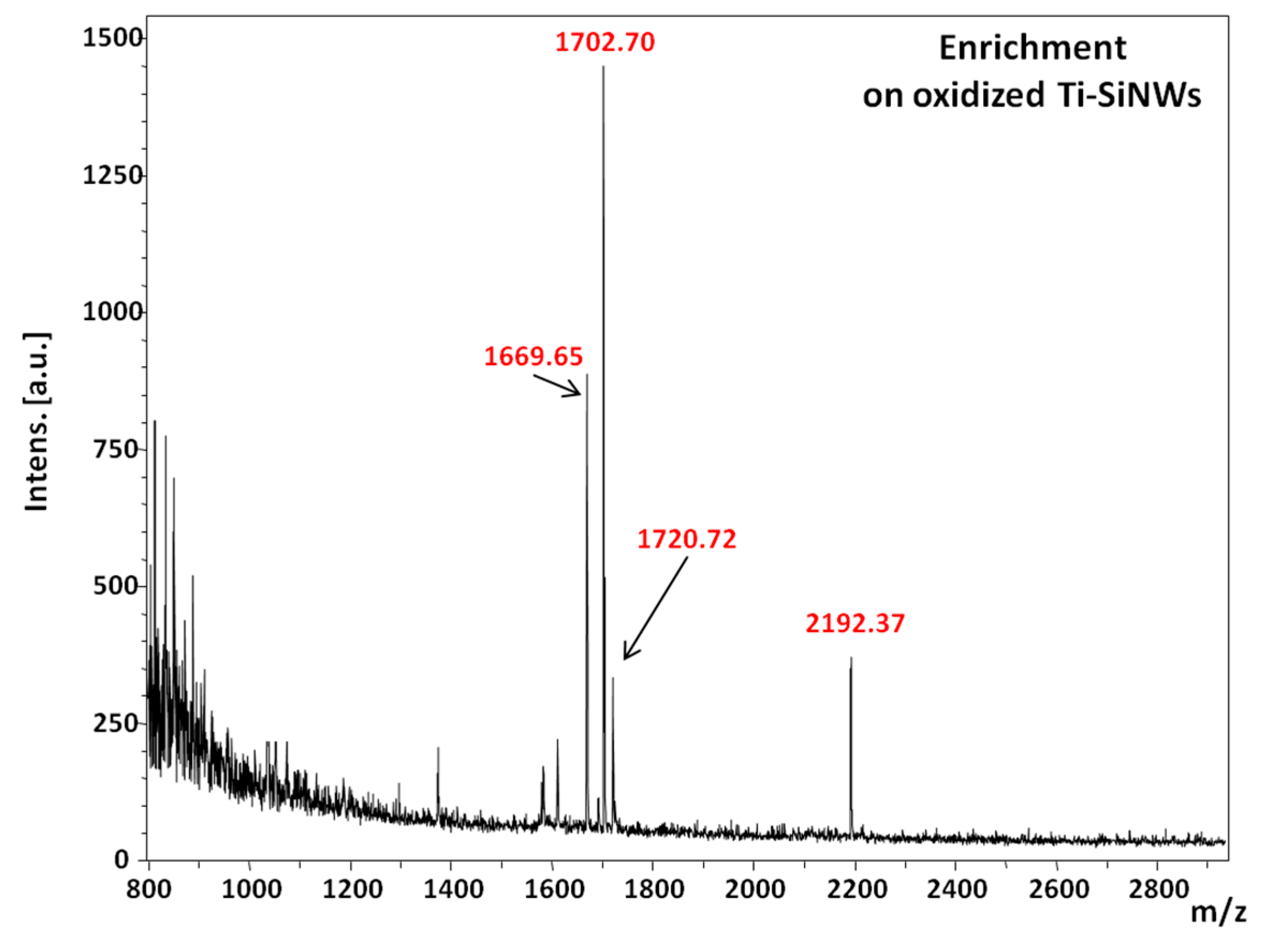
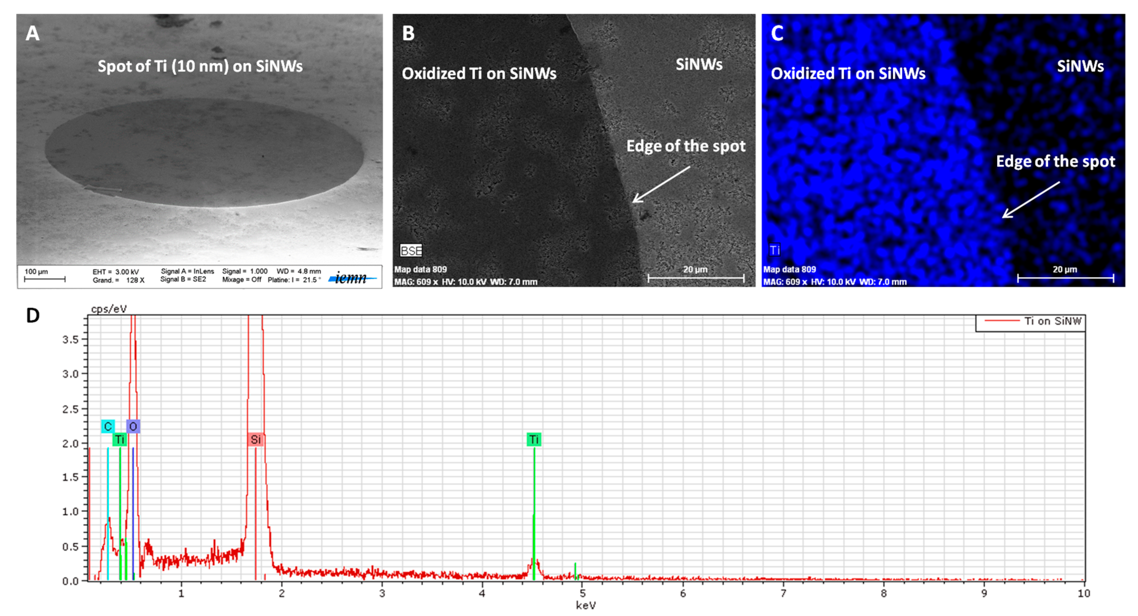
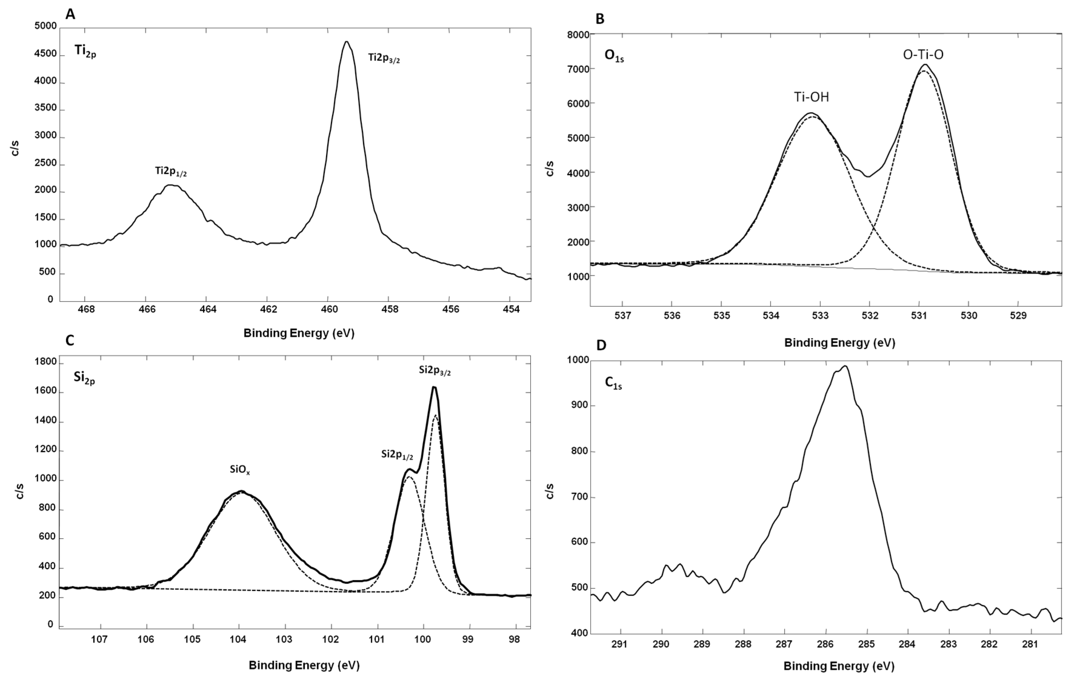
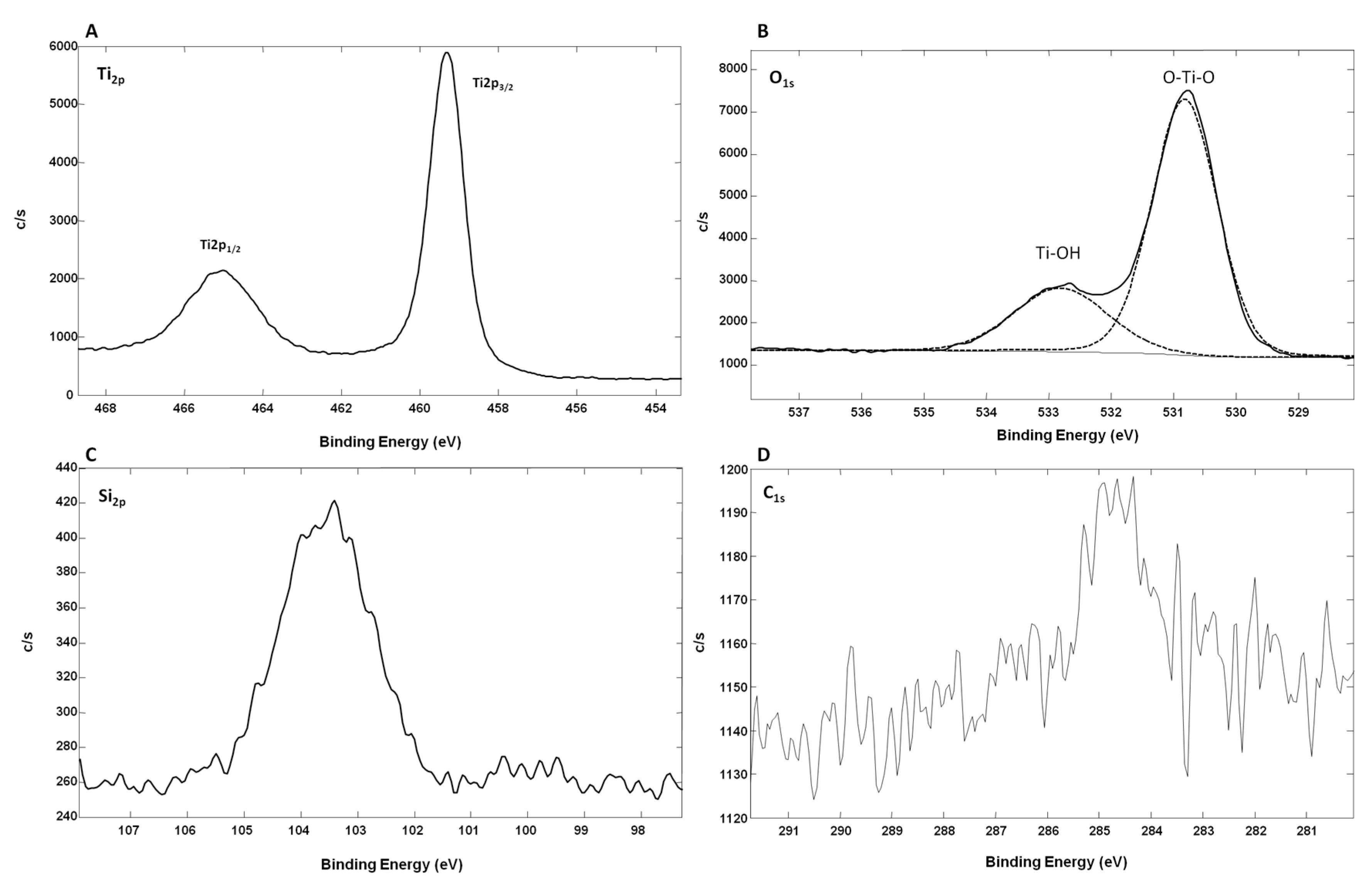
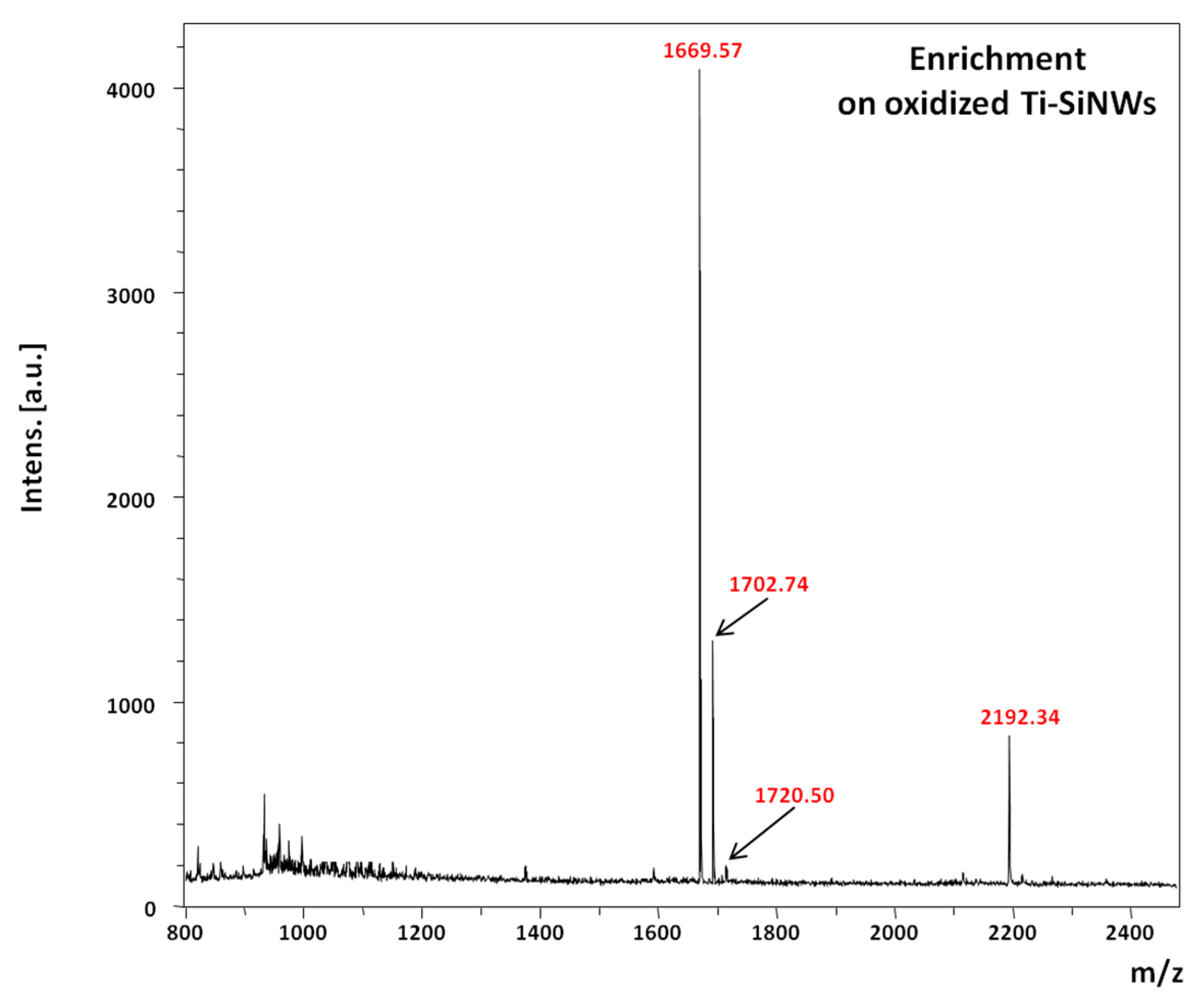
2. Results and Discussion
3. Materials and Methods
3.1. Materials
3.1.1. Nanostructure Fabrication
3.1.2. Safety Considerations
3.1.3. TiO2-SiNW Surface
3.1.4. Ti-SiNWs’ Surface
Thermal Evaporation
3.2. Characterization of Interfaces
3.2.1. Scanning Electron Microscopy (SEM)
3.2.2. Reflectivity Measurement
3.2.3. Transmission Electron Microscopy (TEM)
3.2.4. X-ray Photoelectron Spectroscopy (XPS)
3.2.5. Contact Angle Measurements
3.2.6. Sample Preparation
3.2.7. Specific Capture
3.2.8. LDI-MS Analysis
4. Conclusions
Acknowledgments
Author Contributions
Conflicts of Interest
References
- Morandell, S.; Stasyk, T.; Grosstessner-Hain, K.; Roitinger, E.; Mechtler, K.; Bonn, G.K.; Huber, L.A. Phosphoproteomics strategies for the functional analysis of signal transduction. Proteomics 2006, 6, 4047–4056. [Google Scholar] [CrossRef] [PubMed]
- Reinders, J.; Sickmann, A. State-of-the-art in phosphoproteomics. Proteomics 2005, 5, 4052–4061. [Google Scholar] [CrossRef] [PubMed]
- Graves, J.D.; Krebs, E.G. Protein phosphorylation and signal transduction. Pharmacol. Ther. 1999, 82, 111–121. [Google Scholar] [CrossRef]
- Hunter, T. Signaling–2000 and beyond. Cell 2000, 100, 113–127. [Google Scholar] [CrossRef]
- Thingholm, T.E.; Jensen, O.N.; Larsen, M.R. Analytical strategies for phosphoproteomics. Proteomics 2009, 9, 1451–1468. [Google Scholar] [CrossRef] [PubMed]
- Rush, J.; Moritz, A.; Lee, K.A.; Guo, A.; Goss, V.L.; Spek, E.J.; Zhang, H.; Zha, X.M.; Polakiewicz, R.D.; Comb, M.J. Immunoaffinity profiling of tyrosine phosphorylation in cancer cells. Nat. Biotechnol. 2005, 23, 94–101. [Google Scholar] [CrossRef] [PubMed]
- Hinsby, A.M.; Olsen, J.V.; Mann, M. Tyrosine phosphoproteomics of fibroblast growth factor signaling: A role for insulin receptor substrate-4. J. Biol. Chem. 2004, 279, 46438–46447. [Google Scholar] [CrossRef] [PubMed]
- Tao, W.A.; Wollscheid, B.; O’Brien, R.; Eng, J.K.; Li, X.J.; Bodenmiller, B.; Watts, J.D.; Hood, L.; Aebersold, R. Quantitative phosphoproteome analysis using a dendrimer conjugation chemistry and tandem mass spectrometry. Nat. Methods 2005, 2, 591–598. [Google Scholar] [CrossRef]
- Li, X.; Gerber, S.A.; Rudner, A.D.; Beausoleil, S.A.; Haas, W.; Villen, J.; Elias, J.E.; Gygi, S.P. Large-scale phosphorylation analysis of α-factor-arrested Saccharomyces cerevisiae. J. Proteome Res. 2007, 6, 1190–1197. [Google Scholar] [CrossRef] [PubMed]
- Blacken, G.R.; Volny, M.; Vaisar, T.; Sadilek, M.; Turecek, F. In Situ enrichment of phosphopeptides on MALDI plates functionalized by reactive landing of zirconium(IV)-n-propoxide ions. Anal. Chem. 2007, 79, 5449–5456. [Google Scholar] [CrossRef] [PubMed]
- Larsen, M.R.; Thingholm, T.E.; Jensen, O.N.; Roepstorff, P.; Jorgensen, T.J. Highly selective enrichment of phosphorylated peptides from peptide mixtures using titanium dioxide microcolumns. Mol. Cell. Proteom. 2005, 4, 873–886. [Google Scholar] [CrossRef] [PubMed]
- Imanishi, S.Y.; Kochin, V.; Ferraris, S.E.; de Thonel, A.; Pallari, H.M.; Corthals, G.L.; Eriksson, J.E. Fast and reliable phosphopeptide validation by microLC-ESI-Q-TOF MS/MS. Mol. Cell. Proteom. 2007, 6, 1380–1391. [Google Scholar] [CrossRef] [PubMed]
- Li, Y.; Leng, T.; Lin, H.; Deng, C.; Xu, X.; Yao, N.; Yang, P.; Zhang, X. Preparation of Fe3O4@ZrO2 core-shell microspheres as affinity probes for selective enrichment and direct determination of phosphopeptides using matrix-assisted laser desorption ionization mass spectrometry. J. Proteome Res. 2007, 6, 4498–4510. [Google Scholar] [CrossRef] [PubMed]
- Zhou, H.; Ye, M.; Dong, J.; Han, G.; Jiang, X.; Wu, R.; Zou, H. Specific phosphopeptide enrichment with immobilized titanium ion affinity chromatography adsorbent for phosphoproteome analysis. J. Proteome Res. 2008, 7, 3957–3967. [Google Scholar] [CrossRef] [PubMed]
- Loyet, K.M.; Stults, J.T.; Arnott, D. Mass spectrometric contributions to the practice of phosphorylation site mapping through 2003: A literature review. Mol. Cell. Proteom. 2005, 4, 235–245. [Google Scholar] [CrossRef] [PubMed]
- Dunn, J.D.; Igrisan, E.A.; Palumbo, A.M.; Reid, G.E.; Bruening, M.L. Phosphopeptide enrichment using MALDI plates modified with high-capacity polymer brushes. Anal. Chem. 2008, 80, 5727–5735. [Google Scholar] [CrossRef] [PubMed]
- Eriksson, A.; Bergquist, J.; Edwards, K.; Hagfeldt, A.; Malmström, D.; Hernandez, V.A. Optimized Protocol for On-Target Phosphopeptide Enrichment Prior to Matrix-Assisted Laser Desorption-Ionization Mass Spectrometry Using Mesoporous Titanium Dioxide. Anal. Chem. 2010, 82, 4577–4583. [Google Scholar] [CrossRef] [PubMed]
- Law, K.P.; Larkin, J.R. Recent advances in SALDI-MS techniques and their chemical and bioanalytical applications. Anal. Bioanal. Chem. 2011, 399, 2597–2622. [Google Scholar] [CrossRef] [PubMed]
- Wei, J.; Buriak, J.M.; Siuzdak, G. Desorption-ionization mass spectrometry on porous silicon. Nature 1999, 399, 243–246. [Google Scholar] [CrossRef] [PubMed]
- Coffinier, Y.; Kurylo, I.; Drobecq, H.; Szunerits, S.; Melnyk, O.; Zaitsev, V.N.; Boukherroub, R. Decoration of silicon nanostructures with copper particles for simultaneous selective capture and mass spectrometry detection of His-tagged model peptide. Analyst 2014, 139, 5155–5163. [Google Scholar] [CrossRef] [PubMed]
- Bi, H.; Qiao, L.; Busnel, J.M.; Devaud, V.; Liu, B.; Girault, H.H. TiO2 Printed Aluminum Foil: Single-Use Film for a Laser Desorption/Ionization Target Plate. Anal. Chem. 2009, 81, 1177–1183. [Google Scholar] [CrossRef] [PubMed]
- Dupré, M.; Coffinier, Y.; Boukherroub, R.; Cantel, S.; Martinez, J.; Enjalbal, C. Laser desorption ionization mass spectrometry of protein tryptic digests on nanostructured silicon plates. J. Proteom. 2012, 75, 1973–1990. [Google Scholar] [CrossRef] [PubMed]
- Dupré, M.; Enjalbal, C.; Cantel, S.; Martinez, J.; Megouda, N.; Hadjersi, T.; Boukherroub, R.; Coffinier, Y. Investigation of silicon-based nanostructure morphology and chemical termination on laser desorption ionization mass spectrometry performance. Anal. Chem. 2012, 84, 10637–10644. [Google Scholar] [CrossRef] [PubMed]
- Piret, G.; Drobecq, H.; Coffinier, Y.; Melnyk, O.; Boukherroub, R. Matrix-free laser desorption/ionization mass spectrometry on silicon nanowire arrays prepared by chemical etching of crystalline silicon. Langmuir 2010, 26, 1354–1361. [Google Scholar] [CrossRef] [PubMed]
- Nguyen, T.P.N.; Coffinier, Y.; Thomy, V.; Boukherroub, R. Fabrication of silicon nanostructures using metal-assisted etching in NaBF4. Phys. Status Solidi A 2013, 1–5. [Google Scholar] [CrossRef]
- Kurylo, I.; Dupré, M.; Cantel, S.; Enjalbal, C.; Drobecq, H.; Szunerits, S.; Melnyk, O.; Boukherroub, R.; Coffinier, Y. Characterization of peptide attachment on silicon nanowires by X-ray photoelectron spectroscopy and mass spectrometry. Analyst 2017, 142, 969–978. [Google Scholar] [CrossRef] [PubMed]
- Peterson, S.A. Matrix-free methods for laser desorption/ionization mass spectrometry. Mass Spectrom. Rev. 2007, 26, 19–34. [Google Scholar] [CrossRef] [PubMed]
- Hashimoto, S.; Tanaka, A. Alteration of Ti2p XPS spectrum for titanium oxide by low-energy Ar ion bombardment. Surf. Interface Anal. 2002, 34, 262–265. [Google Scholar] [CrossRef]
- Liu, B.; Wen, Q.H.L.; Zhao, X. The effect of sputtering power on the structure and photocatalytic activity of TiO2 films prepared by magnetron sputtering. Thin Solid Films 2009, 517, 6569–6575. [Google Scholar] [CrossRef]
- Hamdi, A.; Boussekey, L.; Roussel, P.; Addad, A.; Boukherroub, R.; Ezzaouia, H.; Coffinier, Y. Hydrothermal preparation of MoS2/TiO2/Si nanowires composite with enhanced photocatalytic performance under visible light. Mater. Des. 2016, 109, 634–643. [Google Scholar] [CrossRef]
- Feng, B.; Weng, J.; Yang, B.C.; Chen, J.Y.; Zhao, J.Z.; He, L.; Qi, S.K.; Zhang, X.D. Surface characterization of titanium and adsorption of bovine serum albumin. Mater. Charact. 2003, 49, 129–137. [Google Scholar] [CrossRef]
- Nawrocki, J.; Rigney, J.; McCormick, A.; Carr, P.W. Chemistry of zirconia and its use in chromatography. J. Chromatogr. A 1993, 657, 229–282. [Google Scholar] [CrossRef]
- Leitner, A. Enrichment Strategies in Phosphoproteomics. Methods Mol. Biol. 2016, 1355, 105–121. [Google Scholar] [CrossRef] [PubMed]
- Gates, M.B.; Tomer, K.B.; Deterding, L.J. Comparison of metal and metal oxide media for phosphopeptide enrichment prior to mass spectrometric analyses. J. Am. Soc. Mass Spectrom. 2010, 21, 1649–1659. [Google Scholar] [CrossRef] [PubMed]
- Svitasheva, S.N.; Gritsenko, V.A.; Kolesov, B.A. Optical properties of TiO2 films made by air oxidation of Ti. Phys. Stat. Sol. C 2008, 5, 1101–1104. [Google Scholar] [CrossRef]
- Llansola-Portoles, M.L.; Bergkamp, J.J.; Finkelstein-Shapiro, D.; Sherman, B.D.; Kodis, G.; Dimitrijevic, N.M.; Gust, D.; Moore, T.A.; Moore, A.L. Controlling Surface Defects and Photophysics in TiO2 Nanoparticles. J. Phys. Chem. A 2014, 118, 10631–10638. [Google Scholar] [CrossRef] [PubMed]


| Peptides | M + 1 | Sequence |
|---|---|---|
| Angiotensin II | 1046.54 | DRVYIHPF |
| Angiotensin I | 1296.68 | DRVYIHPFHL |
| Myelin basic protein, fragment 104–118 | 1578.85 | GKGRGLSLSRFSWGA |
| pTpY peptide (MAP kinase fragment 177–189) | 1669.67 | DHTGFLpTEpYVATR |
| pY peptide (insulin receptor, fragment 1142–1153) | 1702.75 | TRDIpYETDYYRK |
| pT peptide | 1720.89 | VPIPGRFDRRVpTVE |
| pS peptide (PKA RII peptide, fragment 81–99) | 2192.08 | DLDVPIPGRFDRRVpSVAAE |
| Layers on SiNWs | Thickness (nm) | Deposition Technique | Enrichment | Number of Phosphopeptides Detected |
|---|---|---|---|---|
| Oxidized Ti | 5 | Evaporation | Yes | 3 |
| Oxidized Ti | 10 | Evaporation | Yes | 4 |
| TiO2 | 2 | ALD | No | 0 |
| TiO2 | 5 | ALD | Yes | 2 |
| TiO2 | 10 | ALD | Yes | 3 |
| SiNWs | - | - | No | All peptides detected |
© 2017 by the authors. Licensee MDPI, Basel, Switzerland. This article is an open access article distributed under the terms and conditions of the Creative Commons Attribution (CC BY) license (http://creativecommons.org/licenses/by/4.0/).
Share and Cite
Kurylo, I.; Hamdi, A.; Addad, A.; Boukherroub, R.; Coffinier, Y. Comparison of Ti-Based Coatings on Silicon Nanowires for Phosphopeptide Enrichment and Their Laser Assisted Desorption/Ionization Mass Spectrometry Detection. Nanomaterials 2017, 7, 272. https://doi.org/10.3390/nano7090272
Kurylo I, Hamdi A, Addad A, Boukherroub R, Coffinier Y. Comparison of Ti-Based Coatings on Silicon Nanowires for Phosphopeptide Enrichment and Their Laser Assisted Desorption/Ionization Mass Spectrometry Detection. Nanomaterials. 2017; 7(9):272. https://doi.org/10.3390/nano7090272
Chicago/Turabian StyleKurylo, Ievgen, Abderrahmane Hamdi, Ahmed Addad, Rabah Boukherroub, and Yannick Coffinier. 2017. "Comparison of Ti-Based Coatings on Silicon Nanowires for Phosphopeptide Enrichment and Their Laser Assisted Desorption/Ionization Mass Spectrometry Detection" Nanomaterials 7, no. 9: 272. https://doi.org/10.3390/nano7090272
APA StyleKurylo, I., Hamdi, A., Addad, A., Boukherroub, R., & Coffinier, Y. (2017). Comparison of Ti-Based Coatings on Silicon Nanowires for Phosphopeptide Enrichment and Their Laser Assisted Desorption/Ionization Mass Spectrometry Detection. Nanomaterials, 7(9), 272. https://doi.org/10.3390/nano7090272