Article

Construction of Bi2MoO6/Ag2CrO4 Heterojunction Nanocomposites with Enhanced Visible-Light Photocatalytic Activity and Mechanistic Insight


by
Weijie Hua
*,
Songhua Huang
and
Huixin Yuan
School of Intelligent Equipment Engineering, Wuxi Taihu University, Wuxi 214064, China
*
Author to whom correspondence should be addressed.
Nanomaterials 2026, 16(17), 1079; https://doi.org/10.3390/nano16171079 (registering DOI)
Submission received: 23 July 2026 / Revised: 13 August 2026 / Accepted: 27 August 2026 / Published: 30 August 2026
(This article belongs to the Section Nanocomposite Materials)

Abstract

In this work, flower-like Bi2MoO6 microspheres were first prepared by solvothermal synthesis. Ag2CrO4 nanoparticles were then deposited in situ onto the Bi2MoO6 surface, leading to the formation of a Bi2MoO6/Ag2CrO4 n–n heterojunction nanocomposite photocatalyst. Through characterization technologies and visible-light degradation experiments, the photocatalytic behavior and degradation mechanisms of nanocomposites were explored. The results demonstrated that Ag2CrO4 block-like particles were uniformly anchored onto the surface of the flower-like Bi2MoO6 microspheres. The diffraction peaks, lattice fringes, XPS binding energies and FT-IR absorption bands of the composite samples were in good agreement with those of the individual components. Construction of the heterojunction remarkably broadened the optical response of Bi2MoO6, extending the absorption edge from 497 to 713 nm. The band gap decreased to 1.52 eV, lower than the values measured for either constituent semiconductor. Moreover, the nanocomposite showed a markedly lower photoluminescence emission intensity. The Bi2MoO6/Ag2CrO4 photocatalyst removed maximum 99.43% of Rhodamine B (RhB) within 60 min under visible-light illumination. The photocatalytic activity of nanocomposite with a Bi2MoO6:Ag2CrO4 molar ratio of 1:1 was 3.56 times that of pure Bi2MoO6, while the kinetic constant reached 0.0570 min−1, exceeding those of Bi2MoO6 and Ag2CrO4 by factors of 13.90 and 2.26, respectively. After five consecutive reuse cycles, the photocatalyst still removed more than 85% of RhB from aqueous solution. Optimal degradation performance was obtained using 0.50 g/L of photocatalyst and the initial RhB concentration of 10 mg/L. The photocatalytically generated active species h+ and ·O2 can effectively decompose the chromophores of RhB molecules in water. Finally, the degradation mechanism of RhB by nanocomposites was proposed.

1. Introduction

Wastewater containing synthetic dyes is widely produced during textile dyeing, leather finishing, papermaking and various chemical manufacturing processes. Such effluents usually contain high concentrations of persistent organic contaminants and exhibit strong coloration, complex compositions, and considerable toxicity, thereby presenting serious environmental concerns [1]. Global expansion of textile manufacturing has resulted in a substantial increase in the production of dye-laden wastewater. It has been reported that approximately 150–200 L of water is consumed to produce 1 kg of textiles during the dyeing and finishing process, while the wastewater generated during this stage accounts for approximately 70–80% of the total wastewater discharged by the textile industry [2]. Due to the limited dye fixation efficiency, approximately 10–15% of the applied dyes fail to bind to textile fibers during the dyeing process and are directly discharged into the environment with the wastewater [2]. The discharge of large volumes of untreated or inadequately treated dye wastewater into natural water bodies significantly increases water color intensity, reduces light penetration, inhibits the photosynthesis of algae and aquatic plants, decreases dissolved oxygen levels, and ultimately disrupts the stability of aquatic ecosystems. Moreover, azo dyes, anthraquinone dyes and their degradation products present in dye wastewater exhibit considerable toxicity. Some dye-related pollutants are characterized by mutagenicity, carcinogenicity and a strong tendency to bioaccumulate. Once introduced into food webs, they may progressively accumulate and threaten ecological integrity as well as public health [3]. Rhodamine B exhibits inhibitory effects on aquatic organisms, such as significant growth suppression and oxidative stress responses in the freshwater microalga chlorella vulgaris [4]. Driven by the pursuit of carbon neutrality and the principles of the circular economy, semiconductor photocatalytic technology has attracted widespread attention because of its environmental friendliness, low energy consumption and ability to utilize solar energy effectively [5]. Heterogeneous photocatalysis can directly drive the degradation of organic pollutants under ambient temperature and pressure using solar light, offering advantages such as easy catalyst separation, reusability, and a green, sustainable process. Semiconductor photocatalysis is of great theoretical significance and practical value for safeguarding aquatic ecosystems, promoting wastewater resource recovery and advancing sustainable industrial development. The photocatalytic performance of conventional semiconductor materials remains far from satisfactory because only a limited fraction of the solar spectrum can be effectively utilized, while photoexcited charge carriers readily undergo recombination before participating in surface reactions. In addition, sluggish charge transport across interfaces further reduces the utilization efficiency of photogenerated carriers [6]. Accordingly, developing semiconductor photocatalysts with enhanced visible-light utilization and efficient charge-separation capability has become an important objective in current photocatalysis research.
Benefiting from their favorable band structures, plasmon-enhanced light absorption and abundant surface-active sites, silver-based semiconductor nanomaterials have demonstrated considerable potential for visible-light-driven photocatalytic processes [7]. Owing to its band gap of about 1.8 eV, silver chromate (Ag2CrO4) can effectively harvest visible light. The valence- and conduction-band edge potentials are approximately 2.24 and 0.49 eV, respectively, enabling the generated holes to participate in strong oxidation reactions [8]. The crystal structure of Ag2CrO4 consists of AgO4 tetrahedra, CrO4 tetrahedra and AgO6 octahedra [9]. Its characteristic brick-red color reflects its excellent visible-light absorption capability, suggesting that Ag2CrO4 is a promising visible-light-responsive photocatalyst. Nevertheless, several issues remain to be further investigated. For instance, the photocorrosion of Ag2CrO4 and effective strategies for enhancing its photocatalytic performance are still major challenges. Luo et al. [10] coupled Ag2CrO4 with the narrow-band-gap semiconductor In2S3 to construct an In2S3/Ag2CrO4 heterostructure, in which the charge-transfer process followed an all-solid-state Z-scheme mechanism. Pirhashemi et al. [11] fabricated the ZnO/Ag2CrO4 heterojunction, and the resulting photocatalyst exhibited enhanced photocatalytic performance toward the degradation of organic dyes. Shaker et al. [12] synthesized the ternary ZnO/AgI/Ag2CrO4 photocatalyst, whose enhanced photocatalytic performance was attributed to the broadened light-response range and the accelerated migration of photogenerated charge carriers.
Bismuth-based semiconductors exhibit favorable electronic structures together with efficient light absorption, making them attractive materials for photocatalytic applications [13]. Bismuth molybdate is a visible-light-driven photocatalyst that has received extensive attention in recent years because of its low toxicity, high photocatalytic efficiency and excellent visible-light responsiveness [14,15]. γ-Bi2MoO6 is a typical Aurivillius-type ternary oxide. Its crystal structure belongs to the layered perovskite family and is composed of alternating perovskite-like (MoO4)2+ layers and fluorite-like (Bi2O2)2+ layers [16]. The optical absorption edge of γ-Bi2MoO6 reaches nearly 490 nm because of its moderate band gap (2.6–2.8 eV) [17]. Furthermore, the periodic stacking of atomic layers facilitates directional migration of photoexcited charge carriers, improving their separation efficiency [18,19]. Although Bi2MoO6 exhibits favorable photocatalytic properties, its practical performance is restricted by inefficient harvesting of visible light and fast recombination of charge carriers generated during photoexcitation [20]. As a semiconductor with a tunable electronic band structure, Bi2MoO6 can be coupled with other semiconductors to construct heterojunction photocatalysts. Feng et al. [21] synthesized the CdS/Bi2MoO6 composite with a type-II heterojunction structure via the hydrothermal method. The CS/BMO-1 composite achieved degradation efficiencies of 100% and 92% toward Rhodamine B and tetracycline, respectively, under visible-light irradiation. Chao et al. [22] reported the fabrication of Z-scheme Bi4O5I2/Bi2MoO6 nanocomposite using a solvothermal approach. Photocatalytic evaluation revealed that the BIB-2 sample exhibited the most efficient degradation of tetracycline, with 91.8% of the pollutant eliminated after 60 min of visible-light exposure. Yao et al. [23] reported the fabrication of an internal electric field-assisted S-scheme CdIn2S4/Bi2MoO6 heterojunction using a hydrothermal approach. The resulting 25–CIS/BMO microspheres exhibited outstanding activity toward levofloxacin degradation, eliminating 93.7% of the pollutants after 90 min of illumination.
To overcome the inherent drawbacks of single-component photocatalysts, an n–n Bi2MoO6/Ag2CrO4 heterojunction was designed by depositing Ag2CrO4 onto Bi2MoO6 through an in situ precipitation strategy. This modification restrains the photocorrosion behavior of Ag2CrO4 and meanwhile extends the spectral absorption region of Bi2MoO6. The resulting composite was applied to the visible-light degradation of Rhodamine B (RhB, typical cationic organic dye) in water. Its crystal structure, morphology, optical response and physicochemical properties were comprehensively analyzed using XRD, SEM, TEM, XPS, FT-IR, UV-Vis DRS and PL to establish the relationship between material properties and photocatalytic behavior. The photocatalytic activity of the heterojunction was further assessed through RhB degradation experiments, with particular attention given to its reusability, operational stability and charge-transfer mechanism. The influences of catalyst dosage and initial pollutant concentration on the degradation process were also examined. In addition, active-species quenching experiments together with the evolution of RhB UV–Vis absorption spectra were employed to clarify the degradation mechanism and identify the predominant active components involved in the photocatalytic reaction. The formation of the Bi2MoO6/Ag2CrO4 heterojunction effectively alleviated the photocorrosion of Ag2CrO4 while extending the visible-light response of Bi2MoO6. These characteristics highlight the potential of the Bi2MoO6/Ag2CrO4 heterojunction as an efficient visible-light photocatalyst for the removal of refractory organic contaminants from wastewater.

2. Materials and Methods

2.1. Chemicals

Bismuth nitrate pentahydrate (Bi(NO3)3·5H2O, 99%), sodium molybdate dihydrate (Na2MoO4·2H2O, 99%), ethylene glycol, potassium chromate (K2CrO4, 99.5%), Rhodamine B (RhB), tert-butanol (TBA), triethanolamine (TEOA) and p-benzoquinone (BQ) were obtained from Macklin Biochemical Co., Ltd. (Shanghai, China). Absolute ethanol and silver nitrate (AgNO3) were supplied by Meryer Chemical Technology Co., Ltd. (Shanghai, China) and Xijing Biotechnology Co., Ltd. (Fuzhou, China), respectively. Unless otherwise specified, commercially available analytical-grade chemicals were used as received. Deionized water (electrical conductivity: 1 μS/cm) was utilized for solution preparation and all subsequent experimental procedures.

2.2. Synthesis of Bi2MoO6

Flower-like microspherical Bi2MoO6 was successfully synthesized via a solvothermal method using Bi(NO3)3·5H2O and Na2MoO4·2H2O as the Bi and Mo precursors, respectively. First, 1.6866 g of Bi(NO3)3·5H2O and 0.4210 g of Na2MoO4·2H2O were separately dissolved in 5 mL of ethylene glycol under ultrasonic treatment until clear solutions were obtained. Subsequently, the two solutions were slowly mixed under continuous magnetic stirring. After thorough mixing, 20 mL of absolute ethanol was added dropwise to the mixed solution, followed by vigorous stirring for 30 min until the homogeneous transparent solution was formed. The precursor dispersion was introduced into a 100 mL Teflon-lined stainless-steel vessel for solvothermal treatment at 160 °C for 24 h. After the autoclave had cooled to room temperature, the precipitate was isolated by filtration, successively washed with deionized water and anhydrous ethanol, and subsequently dried at 60 °C for 12 h. The obtained solid was first dried, then cooled to room temperature under natural conditions and milled into a fine powder, resulting in pale-yellow Bi2MoO6.

2.3. Synthesis of Bi2MoO6/Ag2CrO4

Bi2MoO6/Ag2CrO4 heterojunction composites were synthesized via an in situ chemical deposition method. Four composite samples with Bi2MoO6-to-Ag2CrO4 molar ratios of 0.5:1, 1:1, 2:1 and 4:1 were prepared and denoted as BMAC0.5, BMAC1, BMAC2 and BMAC4, respectively. The preparation procedure of the BMAC1 sample is described as a representative example. Bi2MoO6 (0.2 mmol) was introduced into 40 mL of deionized water and subjected to 30 min of sonication to produce a well dispersed suspension, designated as Solution A. Separately, K2CrO4 (0.2 mmol) was completely dissolved in 15 mL of deionized water after 5 min of ultrasonic treatment, and the resulting solution was labeled as Solution B. Under continuous magnetic stirring, 4 mL of 0.1 M AgNO3 solution was added dropwise to Solution A and the suspension was further stirred for 30 min to ensure the uniform adsorption of Ag+ onto the surface of Bi2MoO6. Thereafter, Solution B was slowly added dropwise to the suspension under vigorous stirring, and the reaction was allowed to proceed for 2 h. During this process, the color of the suspension gradually changed from pale yellow to dark red. Following filtration, the precipitate was thoroughly washed with deionized water and anhydrous ethanol, then dried at 60 °C for 12 h. The dried solid was then milled into fine powder to produce the dark-red Bi2MoO6/Ag2CrO4 photocatalyst. Pure Ag2CrO4 was obtained by repeating the same synthetic procedure while omitting the Bi2MoO6.

2.4. Characterization

The phase compositions of the synthesized photocatalysts were identified by X-ray diffraction (XRD, SmartLab SE, Rigaku, Tokyo, Japan) using Cu Kα radiation. Surface morphology together with elemental distribution was analyzed using field-emission scanning electron microscopy coupled with energy-dispersive X-ray spectroscopy (SEM, Sigma 360, ZEISS, Oberkochen, Germany; EDS, Xplore 30, Oxford Instruments, Abingdon, UK). The microstructure and lattice characteristics were further examined by transmission electron microscopy (TEM, JEM-F200, JEOL, Yokohama, Japan). Fourier transform infrared spectroscopy (FT-IR, Nicolet iS20, Thermo Fisher Scientific, Waltham, MA, USA) was employed to investigate the chemical bonding environment, while X-ray photoelectron spectroscopy (XPS, K-Alpha, Thermo Scientific, Waltham, MA, USA) was used to determine the elemental chemical states and valence-band information. Optical absorption properties were evaluated from UV–Vis diffuse reflectance spectra (UV-Vis DRS) recorded on a UV-3600i Plus spectrophotometer (Shimadzu, Kyoto, Japan) with BaSO4 as the reflectance standard. An FLS1000 photoluminescence spectrometer (PL, Edinburgh Instruments, Abingdon, UK) was employed to acquire PL spectra under 370 nm excitation, providing information on the recombination behavior of photoinduced charge carriers.

2.5. Evaluation of Photocatalytic Activity

To evaluate the photocatalytic performance of Bi2MoO6, Ag2CrO4 and Bi2MoO6/Ag2CrO4 nanocomposites, Rhodamine B (RhB) aqueous solution was selected as the target contaminant for photocatalytic degradation. A 300 W xenon lamp (YM-GHX-XE-300, YuMing, Shanghai, China) fitted with the 400 nm cutoff filter served as the visible-light source. Photocatalytic reactions were performed in a water-cooled double-layer quartz reactor, where circulating water continuously regulated the reaction temperature close to ambient conditions. For each experiment, 50 mg of catalyst was introduced into 100 mL of a 10 mg/L RhB solution, followed by magnetic stirring in the dark for 30 min to reach adsorption equilibrium. Before light exposure, a 4 mL suspension was withdrawn as the initial sample. During irradiation, additional 4 mL aliquots were collected every 10 min and centrifuged at 10,000 r/min to remove the catalyst particles. The concentration of RhB was determined from the absorbance of the supernatant at 554 nm using the UV–Vis spectrophotometer (L8, YOKE, Shanghai, China). In the free radical scavenging experiments, TBA, TEOA and BQ were used as scavengers for ·OH, h+ and ·O2, respectively. The respective scavengers were added before the photoreaction began. The stability experiment procedure was as follows: After a single photocatalytic degradation reaction of RhB was completed, the suspension was filtered and washed three times with deionized water and anhydrous ethanol, and the sample was collected and dried at 60 °C for 12 h. The dried samples were then used in the next photodegradation experiment under the same experimental conditions. The degradation efficiency together with the apparent reaction rate constant was subsequently calculated using Equations (1) and (2).
η = C 0 C t C 0
ln ( C 0 C t ) = k t
In Equations (1) and (2), η refers to the percentage degradation of RhB; C0 and Ct indicate the initial and time-dependent RhB concentrations (mg/L), respectively; k represents the kinetic coefficient (min−1), and t corresponds to the irradiation period (min).

3. Results and Discussion

3.1. XRD Characterization

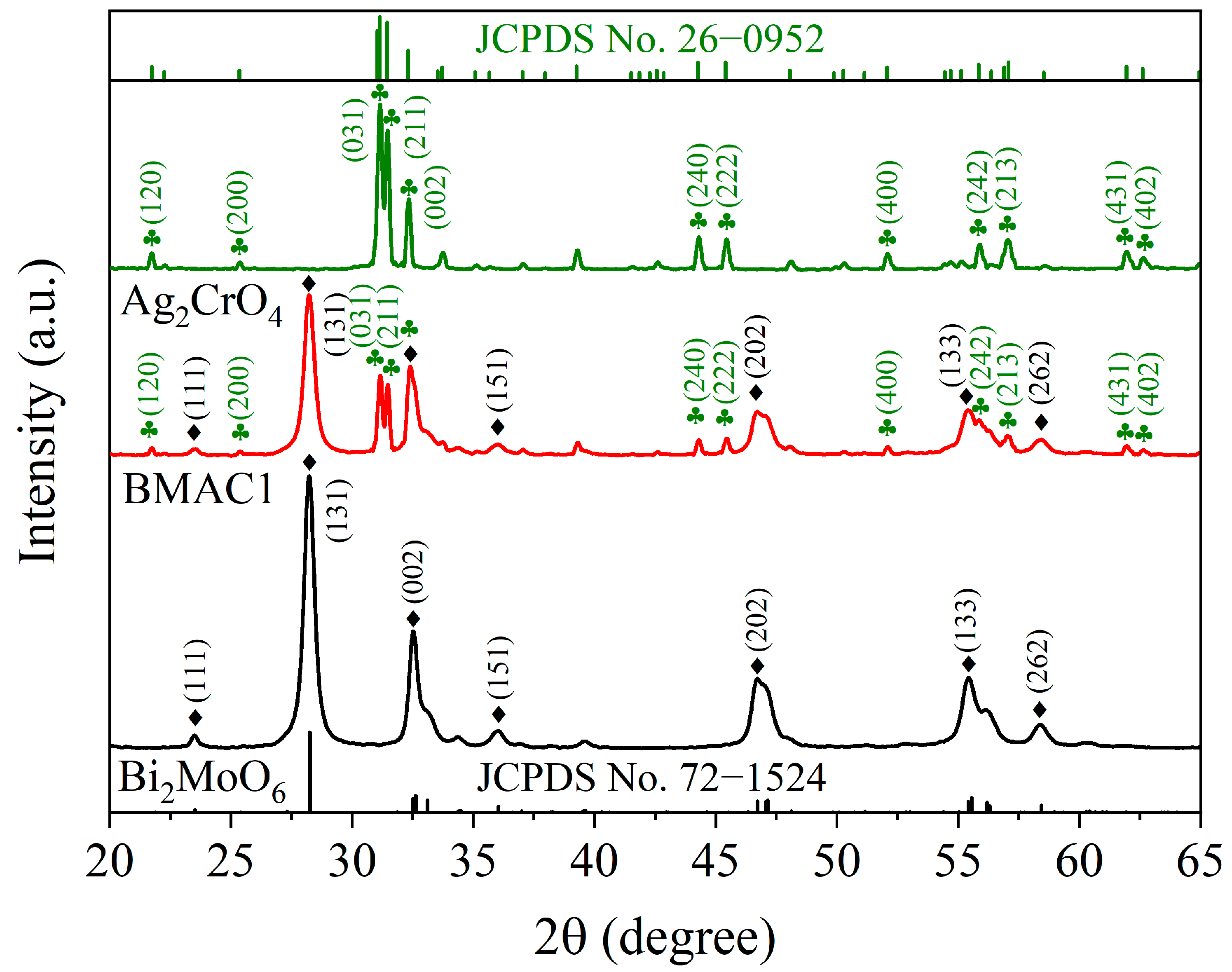
Figure 1 presents the XRD patterns of pristine Bi2MoO6, pristine Ag2CrO4 and the BMAC1 composite. Pristine Bi2MoO6 displays a series of diffraction peaks at 23.50°, 28.22°, 32.52°, 36.06°, 46.72°, 55.42° and 58.38°. These peaks can be attributed to the (111), (131), (002), (151), (202), (133) and (262) crystal planes of orthorhombic Bi2MoO6, in accordance with JCPDS card no. 72-1524. The diffraction pattern of Ag2CrO4 agrees well with the standard card (JCPDS no. 26-0952). The characteristic diffraction peaks located at 21.72°, 25.38°, 31.16°, 31.46°, 32.34°, 44.30°, 45.44°, 52.10°, 55.88°, 57.04°, 61.94° and 62.64° can be indexed to the (120), (200), (031), (211), (002), (240), (222), (400), (242), (213), (431) and (402) crystal planes of orthorhombic Ag2CrO4, respectively. The simultaneous appearance of the diffraction reflections assigned to Bi2MoO6 and Ag2CrO4 in the BMAC1 sample verifies that both phases were successfully integrated into the composite heterostructure. Moreover, no noticeable shift in the diffraction peak positions or impurity peaks was observed, indicating that the Bi2MoO6/Ag2CrO4 heterojunction was successfully synthesized with high crystallinity.

3.2. Microstructural Analysis

3.2.1. Morphological and Elemental Characterization

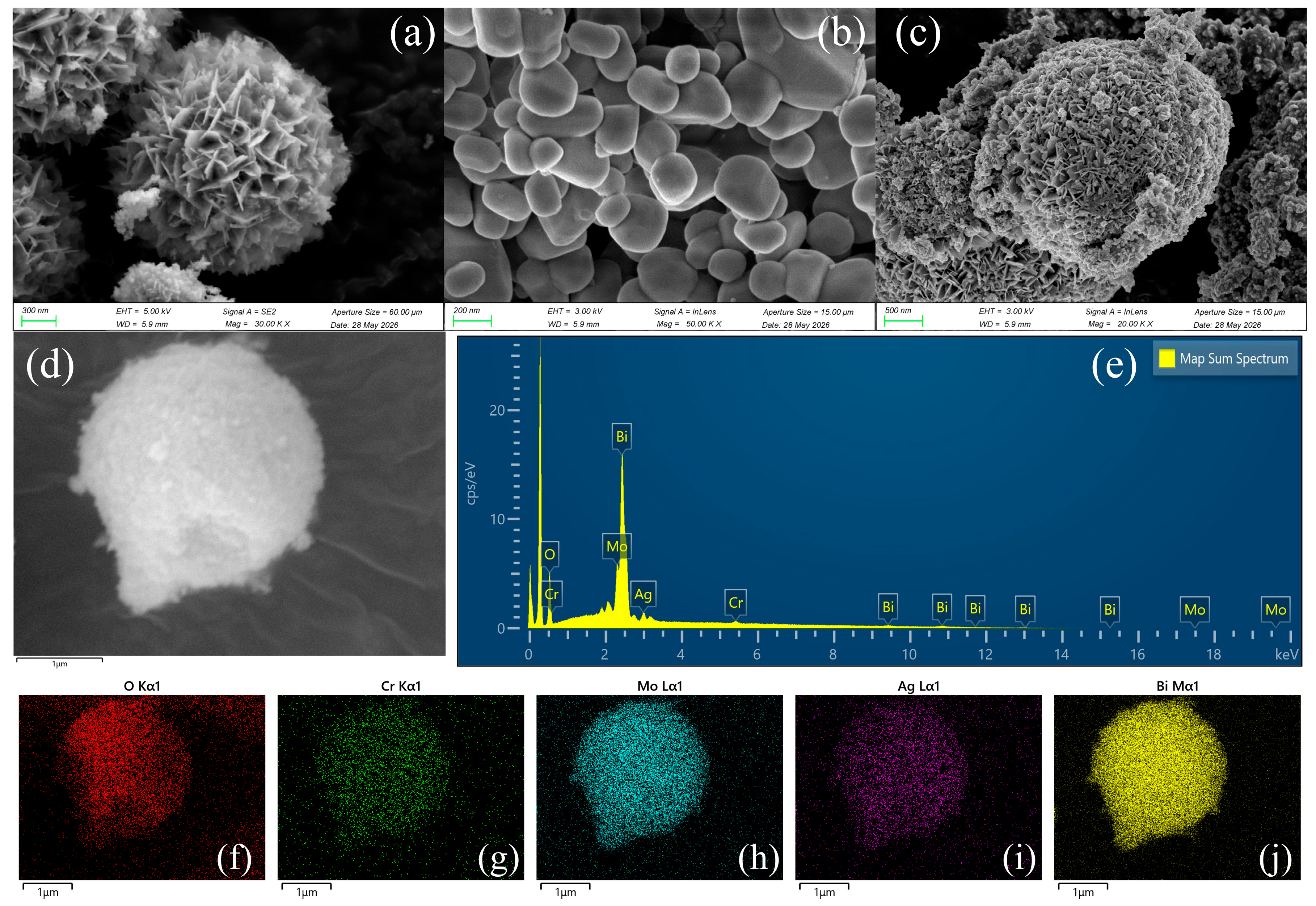
The surface morphologies of Bi2MoO6, Ag2CrO4 and BMAC1 were characterized by SEM. Figure 2 summarizes the characterization results. According to Figure 2a, as-prepared Bi2MoO6 exhibited a uniform flower-like microspherical morphology assembled from numerous interconnected nanosheets. The diameter of an individual flower-like microsphere was approximately 2 μm. Figure 2b shows that the synthesized Ag2CrO4 consisted of irregular block-like particles with different sizes, which were uniformly distributed despite a certain degree of aggregation. After the successful combination of Bi2MoO6 and Ag2CrO4, the block-like Ag2CrO4 particles were uniformly anchored onto the surface of the flower-like Bi2MoO6 microspheres, as shown in Figure 2c. In addition, some fine Ag2CrO4 nanoparticles were observed around the Bi2MoO6 nanosheets, resulting in an increased specific surface area. The formation of the heterojunction increased the interfacial contact area between Bi2MoO6 and Ag2CrO4, thereby providing more active sites for the photocatalytic reaction. The composite sample simultaneously retained the characteristic morphological features of both Bi2MoO6 and Ag2CrO4. The EDS spectra confirmed that the BMAC1 heterojunction photocatalyst was composed of Bi, Mo, O, Cr and Ag elements (Figure 2d,e). The elemental mapping images (Figure 2f–j) further revealed that O, Cr, Mo, Ag and Bi were uniformly distributed over the surface of the microspheres, with stronger signals for Bi, Mo and O, which are consistent with the theoretical composition. The morphology and elemental distribution analyses indicate that Ag2CrO4 block-like crystallites are well dispersed across the surface of Bi2MoO6 microspheres.

3.2.2. TEM Analysis

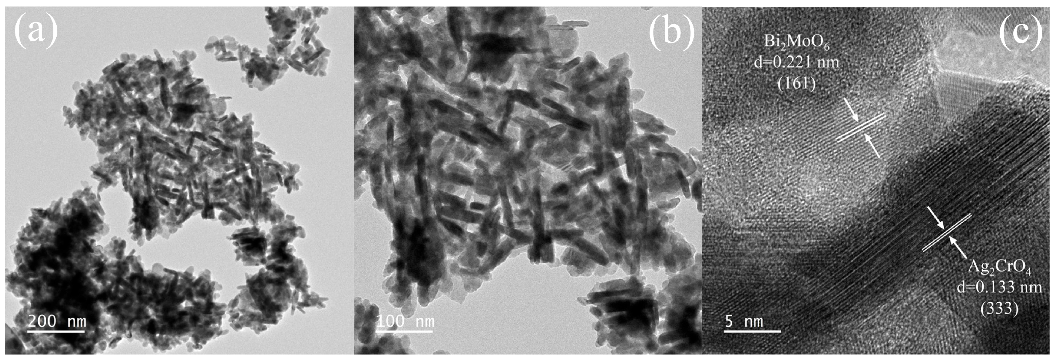
The microstructure of the BMAC1 composite was further investigated by TEM, and the results are shown in Figure 3. As shown in Figure 3a,b, the Ag2CrO4 nanoparticles were uniformly grown and anchored onto the surface of the Bi2MoO6 microspheres. An intimate interface is observed between Bi2MoO6 and Ag2CrO4, which favors charge transfer across the heterojunction. High-resolution TEM analysis clearly distinguishes the two semiconductor phases within the composite (Figure 3c). The lattice spacing measured from the Ag2CrO4 region is 0.133 nm, matching the (333) plane, while a spacing of 0.221 nm is identified in the Bi2MoO6 region and is attributed to the (161) plane. The agreement between the HRTEM measurements and XRD results confirms the coexistence of both crystalline phases and verifies the successful fabrication of the Bi2MoO6/Ag2CrO4 heterostructure.

3.3. XPS Analysis

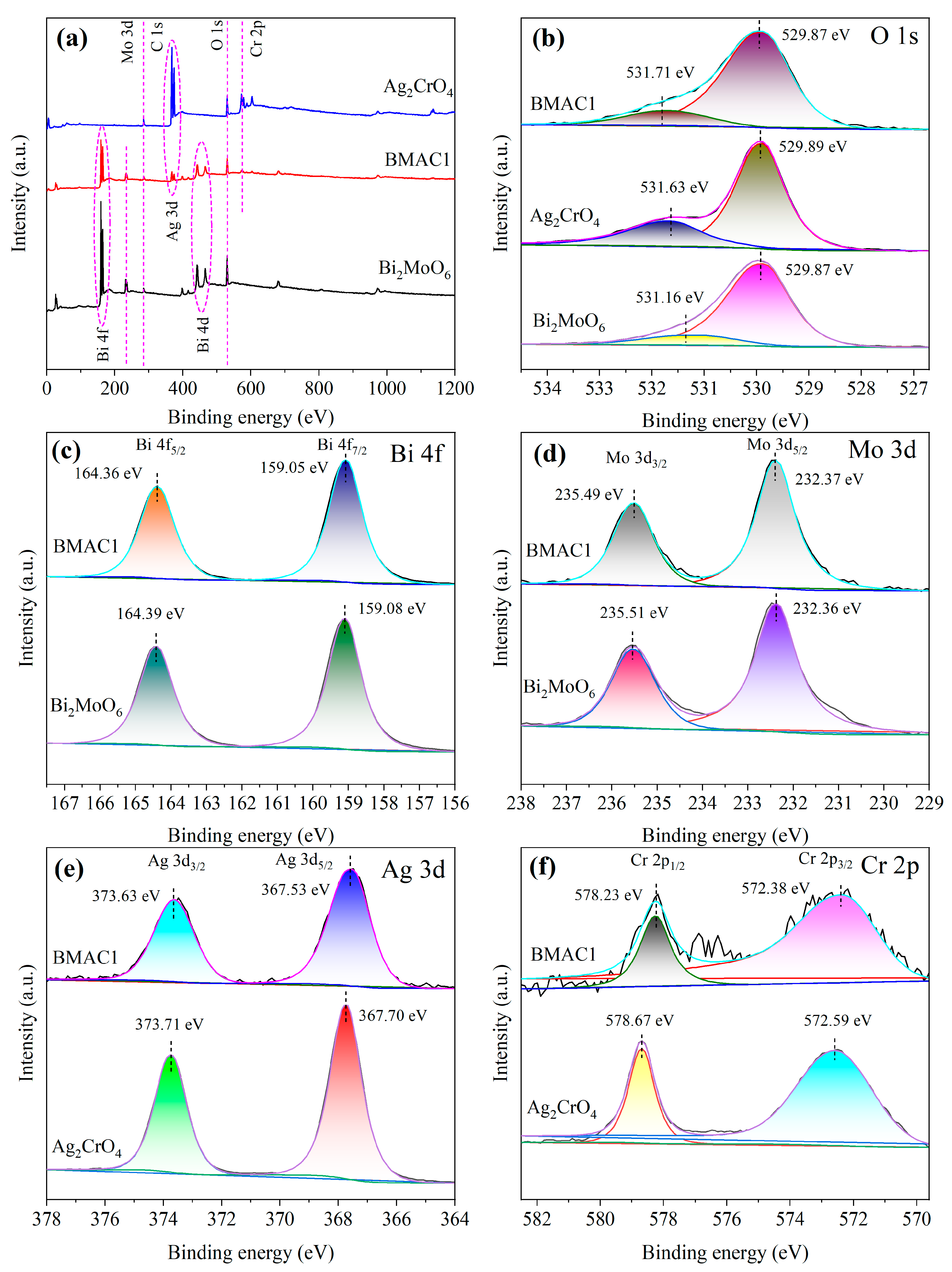
The surface chemical environments and binding-energy distributions of Bi2MoO6, Ag2CrO4 and BMAC1 were further characterized by XPS. The energy scale of the XPS measurements was corrected by setting the C 1s peak to 284.80 eV. The survey and high-resolution spectra are summarized in Figure 4. As shown in Figure 4a, the survey spectrum of Bi2MoO6 exhibits the characteristic photoelectron peaks of Bi, Mo and O, whereas that of Ag2CrO4 contains the characteristic peaks of Cr, Ag and O. More importantly, all of these characteristic photoelectron peaks are simultaneously observed in the survey spectrum of the BMAC1 composite. Figure 4b–f present the high-resolution XPS spectra of the O 1s, Bi 4f, Mo 3d, Ag 3d and Cr 2p orbitals. As shown in Figure 4b, the fitted peaks located at 529.87, 529.89 and 529.87 eV are assigned to lattice oxygen, whereas those located at 531.16, 531.63 and 531.71 eV are attributed to surface hydroxyl oxygen species [24]. The high-resolution Bi 4f and Mo 3d spectra of Bi2MoO6 and BMAC1 are presented in Figure 4c,d. For the Bi2MoO6 sample, the two characteristic peaks located at 159.08 and 164.39 eV correspond to the Bi 4f7/2 and Bi 4f5/2 orbitals, respectively, while the corresponding peaks of BMAC1 slightly shift to 159.05 and 164.36 eV, respectively [25]. For pristine Bi2MoO6, the Mo 3d spectrum is composed of two characteristic signals located at 232.36 and 235.51 eV, which are attributed to the Mo 3d5/2 and Mo 3d3/2 orbitals. In comparison, the BMAC1 composite displays these signals at 232.37 and 235.49 eV, respectively. The Bi 4f and Mo 3d spectra confirm the presence of Bi3+ and Mo6+ species [26]. The shifts in the binding energies of the Bi and Mo core levels after composite formation indicate enhanced chemical interactions between Bi2MoO6 and Ag2CrO4. The high-resolution Ag 3d and Cr 2p spectra of Ag2CrO4 and BMAC1 are shown in Figure 4e,f. In the XPS spectrum of pristine Ag2CrO4, the Ag element is characterized by two binding-energy peaks at 367.70 and 373.71 eV, corresponding to the Ag 3d5/2 and Ag 3d3/2 levels. Meanwhile, the Cr 2p spectrum contains two components at 572.59 and 578.67 eV, which are characteristic of Cr6+ species [27]. For the BMAC1 composite, the binding energies of the Ag 3d5/2 and Ag 3d3/2 orbitals shift to 367.53 and 373.63 eV, respectively, while those of the Cr 2p3/2 and Cr 2p1/2 orbitals shift to 572.38 and 578.23 eV, respectively. These results could confirm the existence of Ag1+ and Cr6+ species. Moreover, the binding energies of all the constituent elements exhibit different degrees of shift compared with those of the corresponding pristine components, further demonstrating the existence of strong interfacial chemical interactions between Bi2MoO6 and Ag2CrO4 and confirming the successful formation of the heterojunction.

3.4. FT-IR Analysis

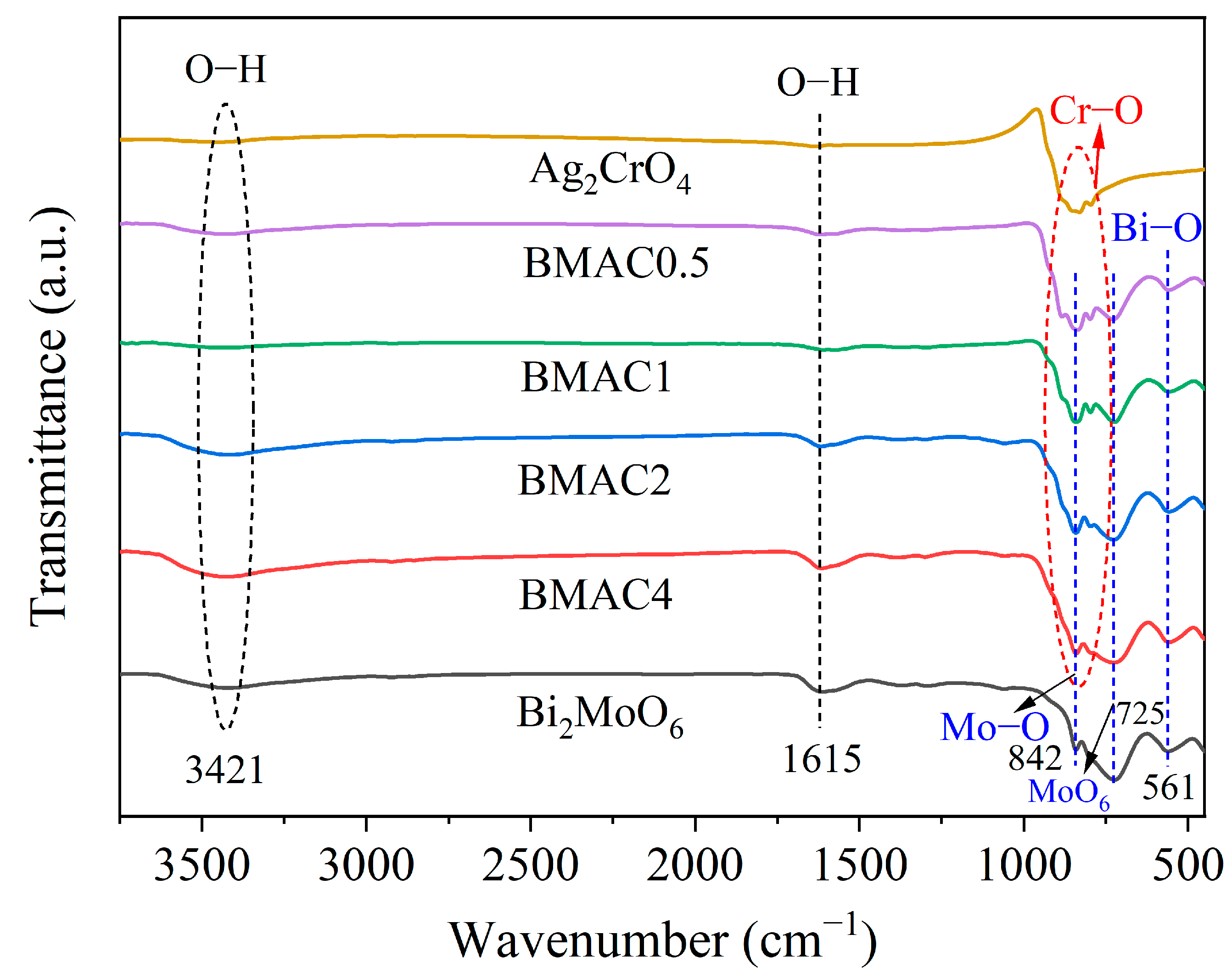
The functional groups of Bi2MoO6, Ag2CrO4 and the Bi2MoO6/Ag2CrO4 composite were characterized by FT-IR spectroscopy, and the corresponding spectra are presented in Figure 5. As shown in Figure 5, the FT-IR spectrum of the Bi2MoO6/Ag2CrO4 composite contains all the characteristic absorption bands of the individual components (Bi2MoO6 and Ag2CrO4). For both Bi2MoO6 and Bi2MoO6/Ag2CrO4, the absorption band centered at 561 cm−1 is assigned to the stretching vibration of the Bi–O bond [28]. The absorption band located at 725 cm−1 is attributed to the asymmetric stretching vibration of MoO6 octahedra. The absorption band at 842 cm−1 originates from the stretching vibration of the Mo–O bond [29]. For Ag2CrO4 and Bi2MoO6/Ag2CrO4, the broad absorption band in the range of 831–842 cm−1 is assigned to the stretching vibration of the Cr–O bond [30]. With increasing Ag2CrO4 loading, the intensity of this characteristic absorption band gradually increased. The FT-IR spectra of all samples display two broad absorption peaks near 1615 and 3421 cm−1, which are associated with the bending and stretching modes of O–H groups, respectively [31]. The intensity changes observed after composite formation reflect interactions between Bi2MoO6 and Ag2CrO4, indicating the establishment of an intimate heterojunction interface.

3.5. Optical Absorption Properties

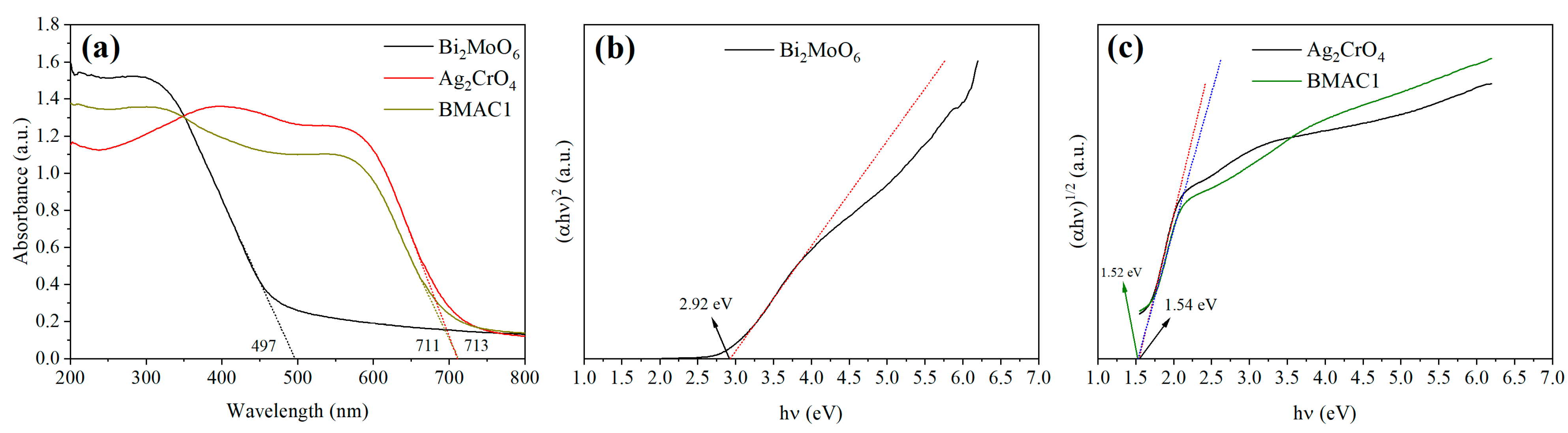
Figure 6a compares the UV–Vis DRS of Bi2MoO6, Ag2CrO4 and the BMAC1 composite, providing insight into their light-absorption characteristics. The absorption edge of pristine Bi2MoO6 was only 497 nm, indicating its limited visible-light response and relatively low efficiency of photogenerated charge carrier separation. The absorption edge of pristine Ag2CrO4 reached 711 nm, demonstrating its excellent visible-light harvesting capability for the generation of electron–hole pairs, although it is susceptible to photocorrosion. The BMAC1 composite exhibited an absorption edge of 713 nm, which is significantly longer than that of pristine Bi2MoO6, indicating the substantially enhanced visible-light response. Therefore, the in situ constructed heterojunction not only induced a distinct red shift in the optical absorption edge of Bi2MoO6 but also remarkably enhanced its visible-light absorption intensity. These results suggest that more incident photons could be utilized to generate photogenerated electron–hole pairs, thereby effectively suppressing the rapid recombination of charge carriers.
The band gap energies of Bi2MoO6, Ag2CrO4 and BMAC1 were further determined, and the results are presented in Figure 6b,c. The band gap energy (Eg) was calculated according to the Tauc equation [32]:
( α h v ) n = A ( h v E g )
In the above expression, α corresponds to the absorption coefficient; h denotes Planck’s constant; ν is the frequency of the incident light, and A is a material-specific parameter. The optical transition of Bi2MoO6 follows a direct band-gap model (n = 2), while Ag2CrO4 obeys an indirect transition model with n = 1/2.
Band-gap values derived from the UV–Vis DRS results are 2.92 eV for Bi2MoO6, 1.54 eV for Ag2CrO4 and 1.52 eV for BMAC1. The slight decrease in the band gap after heterojunction formation indicates an extended visible-light response, which enables more efficient absorption of solar energy and promotes the excitation of electron–hole pairs under irradiation.
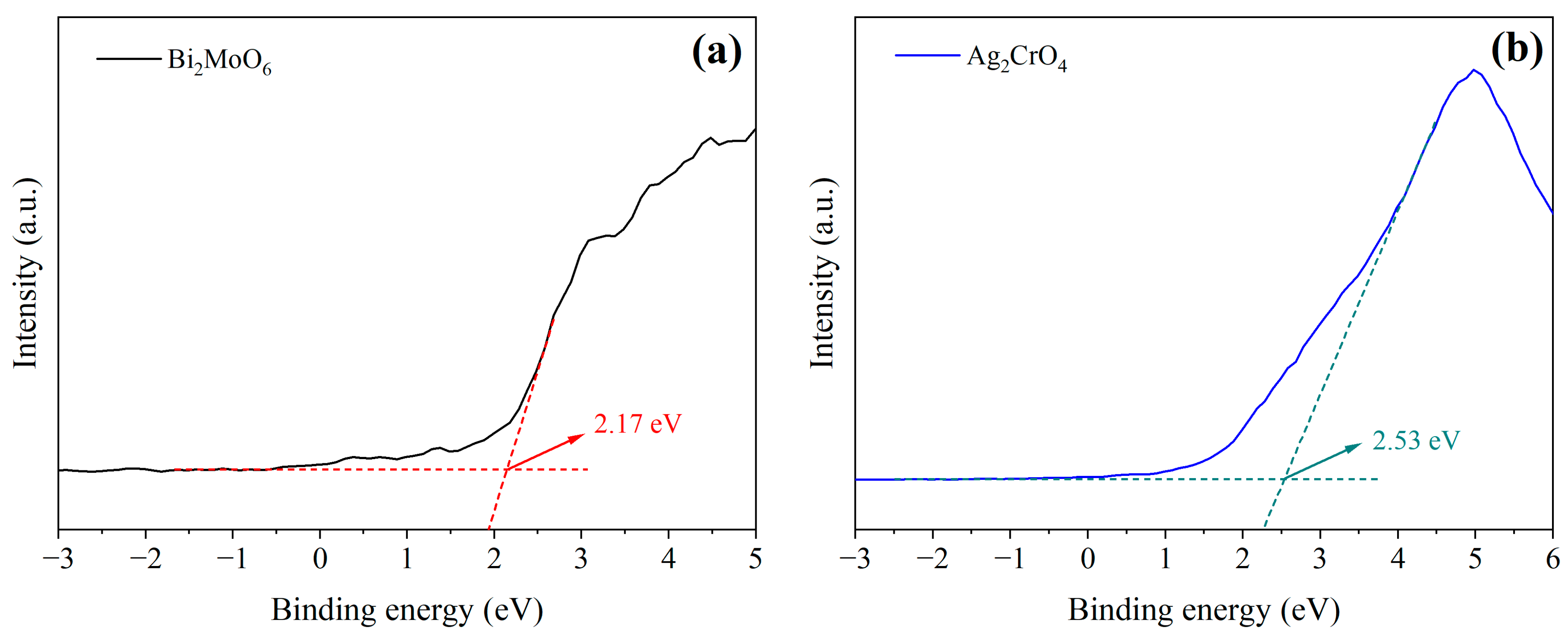
The band edge positions of Bi2MoO6 and Ag2CrO4 were determined by VB-XPS, and the results are presented in Figure 7. The valence band potentials (EVB) of Bi2MoO6 and Ag2CrO4 were determined to be 2.17 and 2.53 eV, respectively. According to the relationship ECB = EVB–Eg, the corresponding conduction band potentials (ECB) of Bi2MoO6 and Ag2CrO4 were calculated to be −0.75 and 0.99 eV, respectively.

3.6. Photoluminescence Analysis

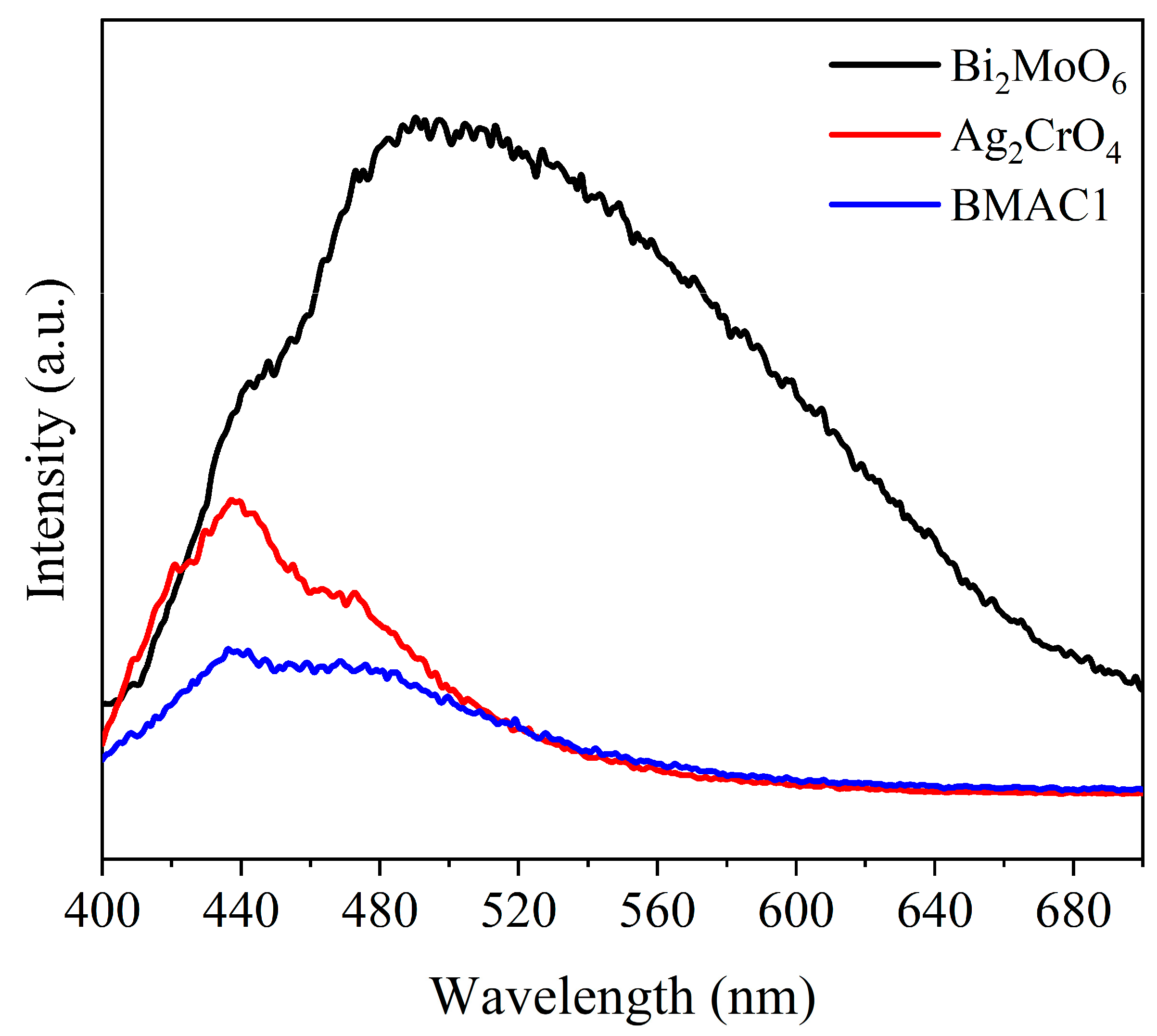
The photoluminescence (PL) spectra of Bi2MoO6, Ag2CrO4 and BMAC1 in the wavelength range of 400–700 nm are shown in Figure 8. The PL spectrum of Bi2MoO6 is characterized by the strongest emission peak among all samples, revealing pronounced recombination of photoexcited carriers. By comparison, the BMAC1 heterojunction exhibits substantially lower PL intensity than either Bi2MoO6 or Ag2CrO4, suggesting that the introduction of the heterointerface effectively inhibits carrier recombination. Moreover, BMAC1 exhibited the lowest PL emission intensity in the visible-light region. The pronounced decrease in PL intensity suggests that the recombination of photoinduced electrons and holes is effectively suppressed in BMAC1. Consequently, the heterojunction composite exhibits more efficient charge carrier separation than Bi2MoO6 and Ag2CrO4, providing a favorable basis for improved photocatalytic activity.

3.7. Photocatalytic Activity and Cycling Stability

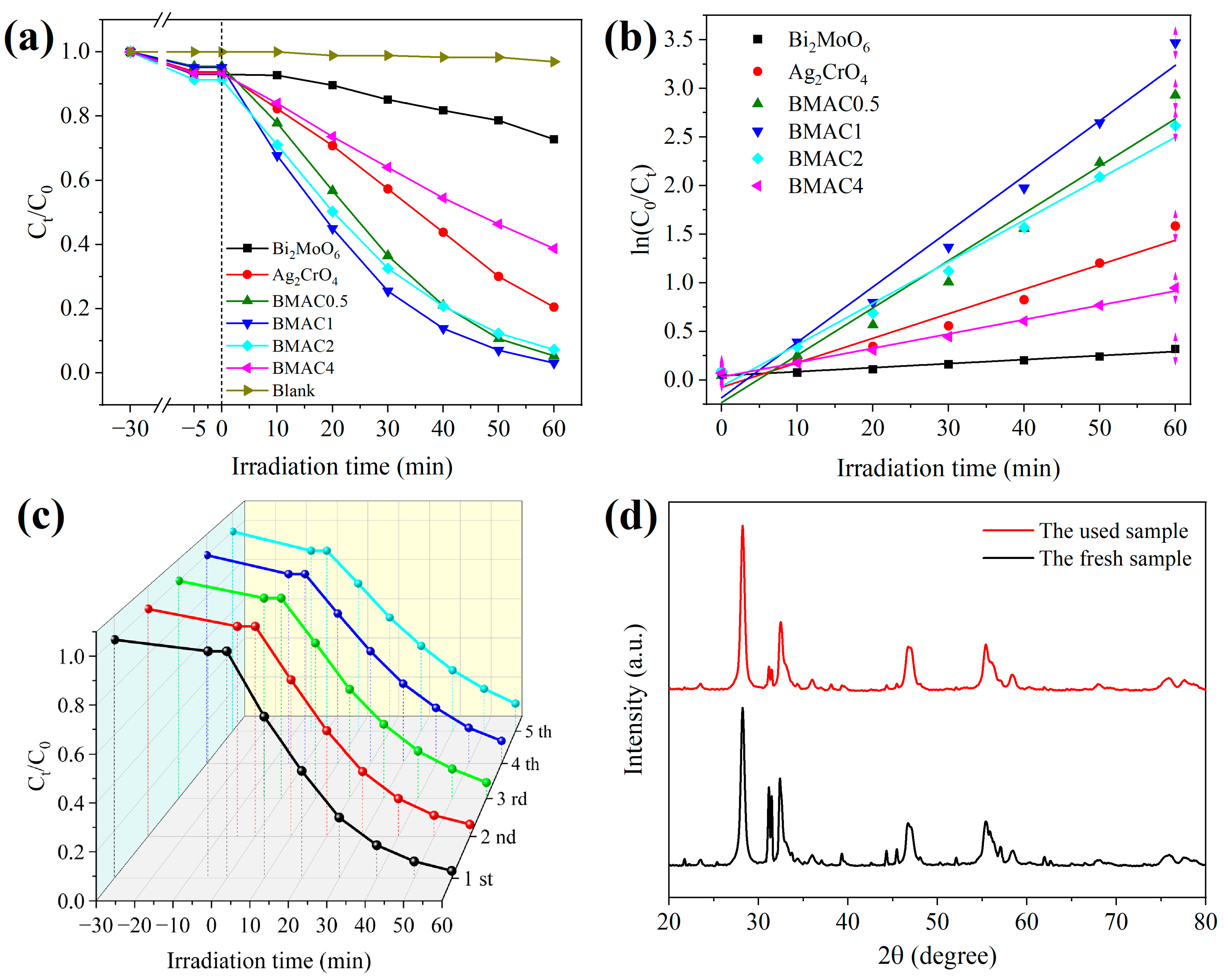
The photocatalytic activity and reusability of the Bi2MoO6/Ag2CrO4 heterojunction nanocomposites were evaluated by degrading RhB under visible-light illumination, and the results are compiled in Figure 9. As depicted in Figure 9a, in the absence of any catalyst, only 3.07% of RhB was removed within 60 min of light exposure. For the individual components, the dark-adsorption equilibria of Bi2MoO6 and Ag2CrO4 toward RhB were determined to be 7.02% and 6.18%, respectively. After 60 min of illumination with visible light, RhB removal reached 27.25% for pristine Bi2MoO6 and 79.49% for pristine Ag2CrO4. Upon coupling Bi2MoO6 with Ag2CrO4 to construct heterojunctions at various molar ratios, all the composite samples showed a pronounced enhancement in photocatalytic activity. Specifically, the BMAC1 sample exhibited a dark adsorption of 4.82% for RhB, while the RhB removal efficiency reached 96.88% after 60 min of visible-light irradiation. In other words, the photocatalytic activity of BMAC1 was approximately 3.56 times higher than that of pristine Bi2MoO6. The RhB degradation efficiencies of BMAC0.5, BMAC2 and BMAC4 after 60 min of reaction were 94.66%, 92.70% and 61.24%, respectively. These results clearly indicate that the molar ratio of Bi2MoO6 to Ag2CrO4 has a pronounced influence on the photocatalytic activity of heterojunction composites. For instance, photocatalytic degradation performance of BMAC4 toward RhB was even lower than that of pristine Ag2CrO4. Among all the prepared samples, BMAC1 exhibited the optimum photocatalytic performance.
Figure 9b and Table 1 present the photocatalytic degradation kinetics and the corresponding kinetic parameters for RhB degradation over Bi2MoO6, Ag2CrO4 and the Bi2MoO6/Ag2CrO4 composites. The degradation of RhB is well described by the pseudo-first-order kinetic model. As the Ag2CrO4 content increased, the apparent reaction rate constant first increased, reached a maximum at the optimal composition, and then declined with further addition of Ag2CrO4. After 60 min of visible-light irradiation, BMAC1 exhibited the highest apparent rate constant for RhB degradation, reaching 0.0570 min−1, which was 13.90, 2.26, 1.17, 1.33 and 3.88 times those of Bi2MoO6, Ag2CrO4, BMAC0.5, BMAC2 and BMAC4, respectively. The outstanding photocatalytic performance of the BMAC1 heterojunction nanocomposite under visible-light irradiation is consistent with the UV–Vis DRS results.
Figure 9c,d illustrate the reusability and cycling stability of the BMAC1 composite photocatalyst. The cycling experiments demonstrate that the photocatalytic activity of BMAC1 decreases only slightly after repeated operation. Following five reuse cycles under 60 min of visible-light irradiation, the RhB removal efficiencies were 96.88%, 94.62%, 92.63%, 89.24% and 86.12%, respectively. Notably, BMAC1 still removed more than 85% of RhB after five successive cycles, demonstrating its excellent reusability. These results indicate that the BMAC1 nanocomposite possesses good photocatalytic durability. Furthermore, XRD analysis was performed on BMAC1 before and after the cycling tests. As depicted in Figure 9d, although the diffraction peak intensities of the recycled photocatalyst decreased slightly, no discernible peak shift was observed, and the major diffraction peaks remained sharp and well preserved. Overall, these results demonstrate that the as-prepared Bi2MoO6/Ag2CrO4 heterojunction nanocomposite possesses excellent reusability and structural stability.

3.8. Effect of Photocatalyst Dosage

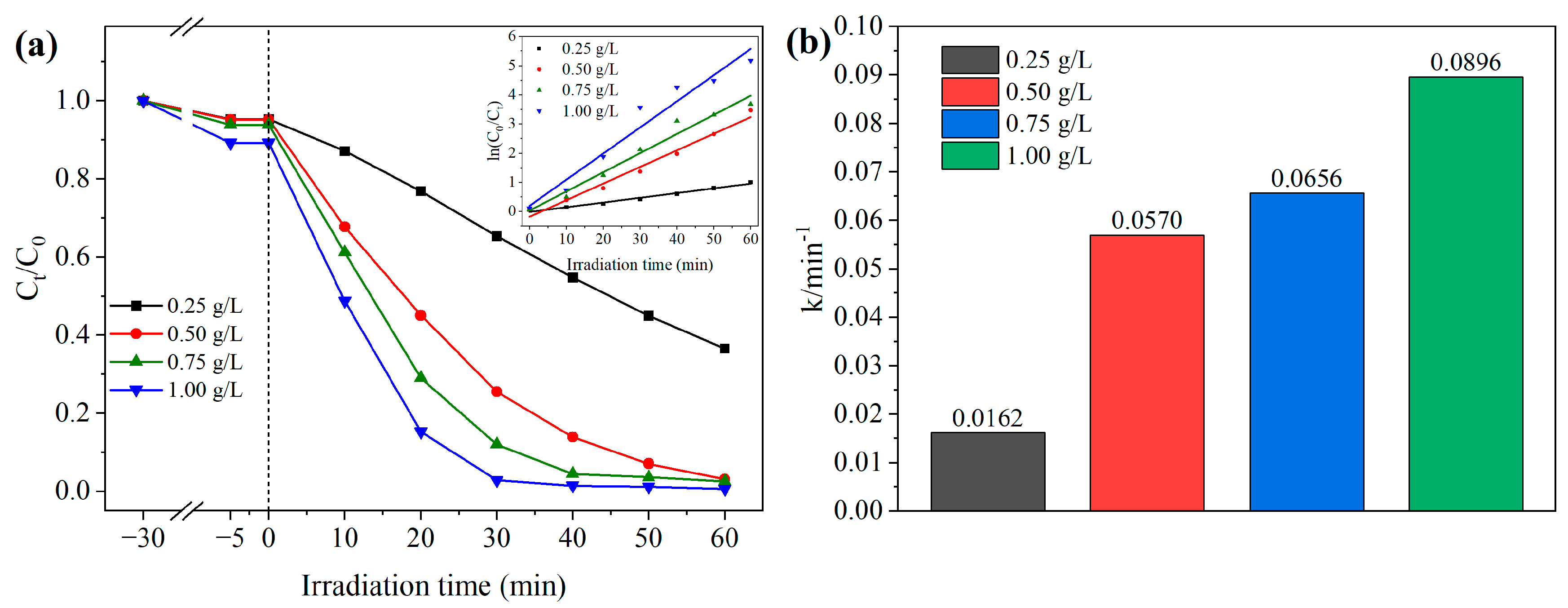
Figure 10 illustrates the influence of BMAC1 dosage on the photocatalytic degradation of RhB. An increase in catalyst dosage resulted in a progressive improvement in the dark adsorption of RhB by BMAC1. When the dosage was adjusted from 0.25 to 1.00 g/L, the adsorption efficiencies increased from 4.75% to 10.76%. The improved adsorption performance is attributed to the greater surface area and the higher density of adsorption-active sites available at elevated catalyst concentrations. As the photocatalyst dosage increased, the RhB degradation efficiency after 60 min of visible-light irradiation gradually improved. The final RhB removal efficiencies corresponding to BMAC1 dosages of 0.25, 0.50, 0.75 and 1.00 g/L were 63.41%, 96.88%, 97.49% and 99.43%, respectively. Obviously, a higher catalyst dosage could provide more active sites, thereby generating a greater amount of reactive species for the degradation of target pollutant. It is noteworthy that when the BMAC1 dosage increased from 0.50 to 1.00 g/L, the degradation efficiency improved by only 2.55%. Increasing the catalyst dosage further leads to stronger light scattering and shielding within the suspension, resulting in reduced photon availability for photocatalytic reactions on the BMAC1 surface. Therefore, the optimum photocatalyst dosage employed in this study was determined to be 0.50 g/L.
As illustrated by the kinetic fitting curves in the inset of Figure 10a, RhB degradation over BMAC1 can be satisfactorily described using a pseudo-first-order kinetic model. Increasing the catalyst dosage resulted in a progressive increase in the apparent reaction rate (Figure 10b), with rate constants of 0.0162, 0.0570, 0.0656 and 0.0896 min−1 for catalyst loadings of 0.25, 0.50, 0.75 and 1.00 g/L, respectively. The improved reaction kinetics are attributed to the higher density of accessible active sites, which enhances interfacial photocatalytic reactions.

3.9. Effect of Initial RhB Concentration

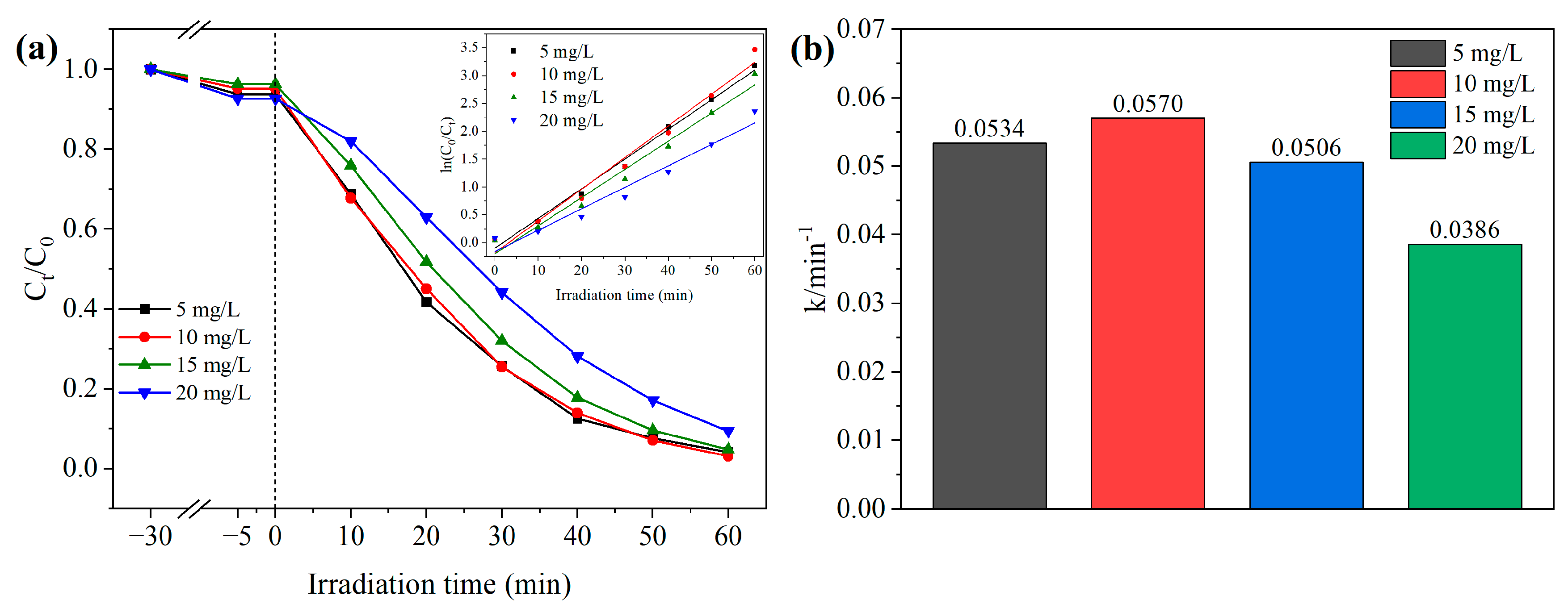
The effect of the initial RhB concentration on its photocatalytic degradation was investigated, and the results are presented in Figure 11. With increasing initial RhB concentration, the adsorption efficiency of BMAC1 toward RhB initially decreased and then increased. An adsorption efficiency of 7.35% was obtained when the initial RhB concentration was 20 mg/L. The photocatalytic degradation efficiency initially increased and then decreased with enhancing initial RhB concentration. Variation in the initial RhB concentration from 5 to 20 mg/L resulted in degradation efficiencies of 95.83%, 96.88%, 95.18% and 90.58%, respectively. These results indicate that an initial RhB concentration of 10 mg/L enabled BMAC1 to achieve the highest photocatalytic degradation efficiency. As the pollutant concentration increased to an appropriate level, the probability of contact between RhB molecules and the active sites on the BMAC1 surface increased, thereby promoting the photocatalytic degradation process. However, when the initial RhB concentration exceeded 10 mg/L, the generated reactive species were insufficient to rapidly degrade all RhB molecules. This is because the photocatalyst dosage remained constant, resulting in a nearly fixed amount of reactive species generated within a given period.
Pseudo-first-order kinetic fitting adequately describes the degradation behavior of RhB under different initial concentrations, as shown in the inset of Figure 11a. The degradation rates were calculated to be 0.0534, 0.0570, 0.0506 and 0.0386 min−1 for RhB concentrations ranging from 5 to 20 mg/L (Figure 11b). Both the degradation efficiency and kinetic constant reached their highest values at an initial concentration of 10 mg/L before declining at higher concentrations. Based on these results, 10 mg/L was chosen as the standard RhB concentration for subsequent investigations.

3.10. Photocatalytic Reaction Mechanism

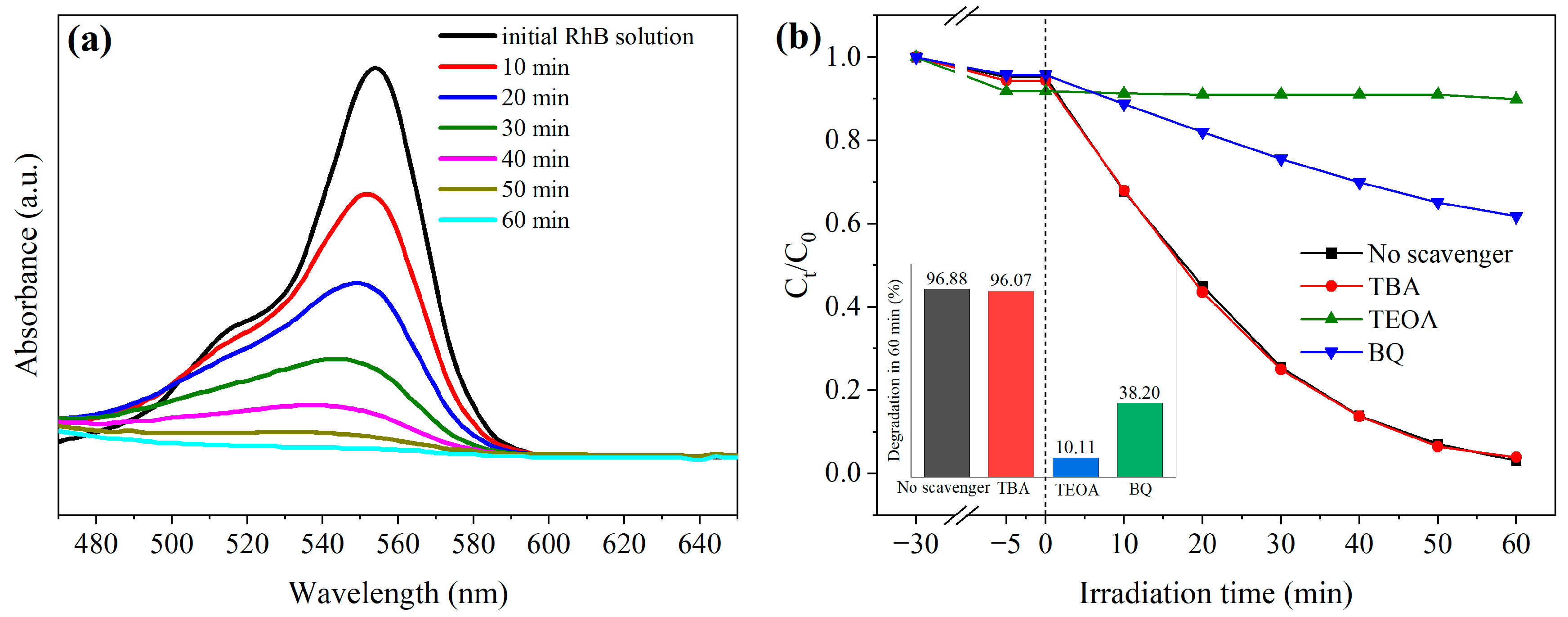
The complete degradation of RhB during the photocatalytic process was verified by monitoring its UV–Vis absorption spectra, and the results are presented in Figure 12a. The absorption spectra of the RhB solution were recorded over the wavelength range of 480–640 nm during visible-light irradiation. Monitoring of the UV–Vis spectra revealed a continuous decline in the absorbance of RhB at 554 nm throughout the photocatalytic process. After irradiation for 60 min, the absorption feature nearly disappeared, corresponding to the nearly complete decolorization of the reaction solution. The disappearance of the characteristic absorption peak indicates the complete destruction of the chromophoric xanthene ring structure of RhB molecules, suggesting extensive degradation of the dye molecules [33]. The involvement of these reactive species leads to the cleavage of the conjugated chromophoric system of RhB, thereby promoting its molecular degradation.
A series of quenching experiments was performed with various scavengers to clarify the roles of active species in the BMAC1-mediated degradation of RhB [34]. The RhB degradation efficiency remained nearly unchanged after adding TBA (Figure 12b). After the addition of TBA, RhB degradation efficiency remained as high as 96.07% after 60 min of irradiation, representing a decrease of only 0.81% compared with the control experiment. In contrast, when TEOA and BQ were introduced into the reaction system, the RhB degradation efficiencies decreased dramatically to 10.11% and 38.20%, respectively. The photocatalytic activity of BMAC1 was therefore markedly suppressed. Compared with the system without scavengers, the degradation efficiency decreased by 86.77% and 58.68% in the presence of TEOA and BQ, respectively. The significant inhibition caused by h+ and ·O2 scavengers confirms that these two species participate actively in RhB degradation. In contrast, the negligible variation observed after ·OH trapping indicates that ·OH makes only a minor contribution to the photocatalytic reaction over Bi2MoO6/Ag2CrO4.
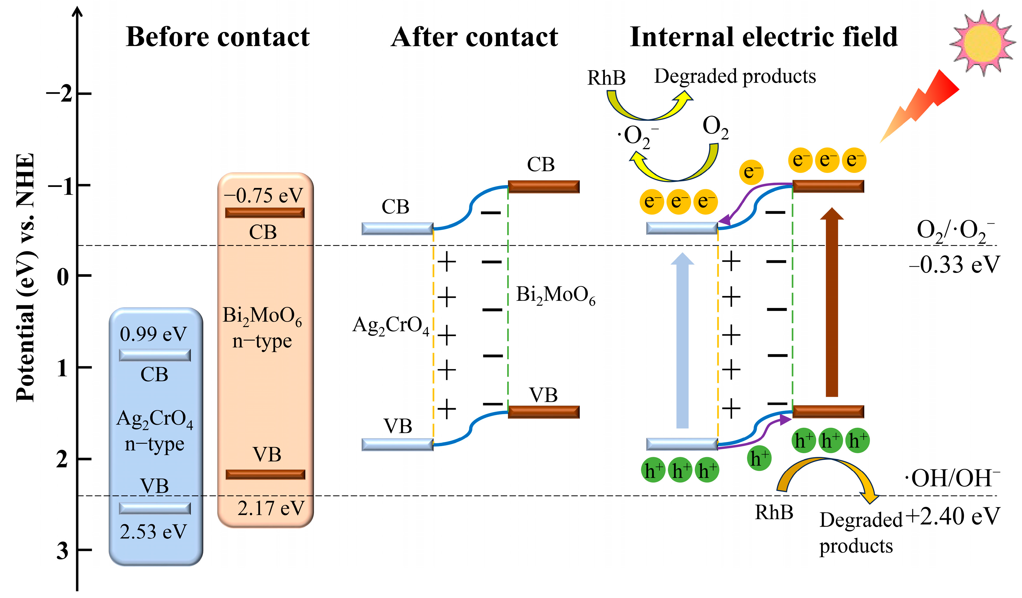
Based on the characterization results, photocatalytic performance analysis, RhB absorption spectra and reactive species trapping experiments, a possible photocatalytic mechanism for RhB degradation over the Bi2MoO6/Ag2CrO4 n–n heterojunction was proposed, as displayed in Figure 13. According to the band structure analysis, the VB edges of Bi2MoO6 and Ag2CrO4 are positioned at 2.17 and 2.53 eV, respectively, with CB edges of −0.75 and 0.99 eV. Light irradiation excites both components and triggers the generation of charge carriers. In the Bi2MoO6/Ag2CrO4 n–n heterostructure, interfacial equilibration between the two semiconductors induces an internal electric field, which regulates the carrier-transfer direction. Consequently, electrons accumulate on Ag2CrO4, whereas holes are enriched on Bi2MoO6. The resulting spatial separation of electrons and holes reduces recombination losses and provides more active carriers for photocatalytic reactions.
The photocatalytic degradation reactions are described by Equations (4)–(6). Following interfacial charge migration, the holes accumulated on the VB of Bi2MoO6 directly participate in the oxidation of RhB molecules. Meanwhile, the electrons enriched on the CB of Ag2CrO4 are captured by dissolved oxygen to generate ·O2. The highly reactive ·O2 species further accelerate the oxidative decomposition of RhB, ultimately leading to the formation of CO2, H2O and other innocuous products. This reaction pathway is consistent with the reactive species trapping results.
Bi 2 MoO 6 / Ag 2 CrO 4 + h v Bi 2 MoO 6 ( h VB + ) + Ag 2 CrO 4 ( e CB )
O 2 + Ag 2 CrO 4 ( e CB )   O 2
RhB + h + / O 2 ( main ) CO 2 + H 2 O   or   degradation   byproducts

4. Conclusions

In this study, flower-like Bi2MoO6 microspheres were successfully synthesized via the solvothermal method, followed by the fabrication of a visible-light-responsive Bi2MoO6/Ag2CrO4 n–n heterojunction photocatalyst through an in situ deposition strategy. Structural characterization demonstrated the intimate interfacial contact between Bi2MoO6 and Ag2CrO4, with Ag2CrO4 block-like particles uniformly anchored on the surface of the flower-like Bi2MoO6 microspheres. The XRD patterns, lattice fringes, XPS binding energies and FT-IR spectra of the composite were all consistent with those of the corresponding pristine components, confirming the successful construction of the Bi2MoO6/Ag2CrO4 heterojunction. Compared with pristine Bi2MoO6, BMAC1 exhibited a substantially extended light absorption edge (713 nm vs. 497 nm), and a significantly lower PL emission intensity, indicating enhanced visible-light harvesting capability and more efficient separation of photogenerated charge carriers. The optimal composite sample (BMAC1) achieved an RhB removal efficiency of 96.88% under visible-light exposure for 60 min. Its photocatalytic activity was 3.56 times that of pure Bi2MoO6, while its kinetic constant (0.0570 min−1) was 13.90 and 2.26 times those of Bi2MoO6 and Ag2CrO4, respectively. Moreover, the nanocomposite exhibited excellent reusability and structural stability after repeated cycling tests. The photocatalytic reaction was optimized at a BMAC1 concentration of 0.50 g/L and an RhB concentration of 10 mg/L. The active-species analysis indicated that h+ and ·O2 dominated the degradation mechanism. The remarkable photocatalytic enhancement of Bi2MoO6/Ag2CrO4 was attributed to the improved spatial separation of charge carriers arising from the n–n heterojunction structure, with electrons migrating toward Ag2CrO4 and holes transferring to Bi2MoO6. Future research should focus on assessing the risk of metal ion leaching, evaluating the mineralization degree of pollutants, and identifying degradation intermediates to further understand their environmental suitability for practical applications. Overall, this study offers a promising route for constructing efficient visible-light-responsive heterostructured nanocomposites for environmental purification applications.

Author Contributions

Conceptualization, W.H.; methodology, W.H.; validation, W.H.; formal analysis, W.H.; investigation, W.H.; resources, W.H. and H.Y.; funding acquisition, W.H.; writing—original draft preparation, W.H.; writing—review and editing, W.H. and S.H.; supervision and project administration, W.H. All authors have read and agreed to the published version of the manuscript.

Funding

This research was funded by the Natural Science Foundation of the Jiangsu Higher Education Institutions of China, grant number [25KJB530007].

Data Availability Statement

The original contributions presented in this study are included in the article. Further inquiries can be directed to the corresponding authors.

Conflicts of Interest

The authors declare no conflicts of interest.

References

  1. Enesca, A.; Andronic, L. The Influence of Photoactive Heterostructures on the Photocatalytic Removal of Dyes and Pharmaceutical Active Compounds: A Mini-Review. Nanomaterials 2020, 10, 1766. [Google Scholar] [CrossRef] [Scilit]
  2. Liu, Y.Q.; Chen, J.S.; Duan, D.R.; Zhang, Z.Y.; Liu, C.; Cai, W.; Zhao, Z.W. Environmental Impacts and Biological Technologies Toward Sustainable Treatment of Textile Dyeing Wastewater: A Review. Sustainability 2024, 16, 10867. [Google Scholar] [CrossRef] [Scilit]
  3. Periyasamy, A.P. Recent Advances in the Remediation of Textile-Dye-Containing Wastewater: Prioritizing Human Health and Sustainable Wastewater Treatment. Sustainability 2024, 16, 495. [Google Scholar] [CrossRef] [Scilit]
  4. Sudarshan, S.; Bharti, V.S.; Harikrishnan, S.; Shukla, S.P.; RathiBhuvaneswari, G. Eco-toxicological effect of a commercial dye Rhodamine B on freshwater microalgae Chlorella vulgaris. Arch. Microbiol. 2022, 204, 658. [Google Scholar] [CrossRef] [Scilit]
  5. Aftab, S.; Shabir, T.; Shah, A.; Nisar, J.; Shah, I.; Muhammad, H.; Shah, N.S. Highly Efficient Visible Light Active Doped ZnO Photocatalysts for the Treatment of Wastewater Contaminated with Dyes and Pathogens of Emerging Concern. Nanomaterials 2022, 12, 486. [Google Scholar] [CrossRef] [Scilit]
  6. Ren, G.M.; Han, H.T.; Wang, Y.X.; Liu, S.; Zhao, J.; Meng, X.; Li, Z. Recent Advances of Photocatalytic Application in Water Treatment: A Review. Nanomaterials 2021, 11, 1804. [Google Scholar] [CrossRef] [Scilit]
  7. Xu, H.; Cao, Y.; Xie, J.; Hu, J.; Li, Y.; Jia, D. A construction of Ag-modified raspberry-like AgCl/Ag2WO4, with excellent visible-light photocatalytic property and stability. Mater. Res. Bull. 2018, 102, 342–352. [Google Scholar] [CrossRef] [Scilit]
  8. Zhang, J.; Yu, W.; Liu, J.; Liu, B. Illustration of high-active Ag2CrO4 photocatalyst from the first-principle calculation of electronic structures and carrier effective mass. Appl. Surf. Sci. 2015, 358, 457–462. [Google Scholar] [CrossRef] [Scilit]
  9. Ouyang, S.; Li, Z.; Ouyang, Z.; Yu, T.; Ye, J.; Zou, Z. Correlation of Crystal Structures, Electronic Structures, and Photocatalytic Properties in a Series of Ag-based Oxides: AgAlO2, AgCrO2, and Ag2CrO4. J. Phys. Chem. C 2008, 112, 3134–3141. [Google Scholar] [CrossRef] [Scilit]
  10. Luo, J.; Zhou, X.S.; Ning, X.M.; Zhan, L.; Ma, L.; Xu, X.; Huang, Z.; Liang, J. Synthesis and characterization of Z-scheme In2S3/Ag2CrO4 composites with an enhanced visible-light photocatalytic performance. New J. Chem. 2017, 41, 845–856. [Google Scholar] [CrossRef] [Scilit]
  11. Pirhashemi, M.; Habibi-Yangjeh, A. Novel ZnO/Ag2CrO4 nanocomposites with n-n heterojunctions as excellent photocatalysts for degradation of different pollutants under visible light. J. Mater. Sci.-Mater. Electron. 2016, 27, 4098–4108. [Google Scholar] [CrossRef] [Scilit]
  12. Shaker-Agjekandy, S.; Habibi-Yangjeh, A. Ultrasonic-assisted preparation of novel ternary ZnO/AgI/Ag2CrO4 nanocomposites as visible-light-driven photocatalysts with excellent activity. Mater. Sci. Semicond. Process. 2016, 44, 48–56. [Google Scholar] [CrossRef] [Scilit]
  13. Zhang, L.; Li, Y.; Li, Q.; Fan, J.; Carabineiro, S.A.; Lv, K. Recent advances on Bismuth-based Photocatalysts: Strategies and mechanisms. Chem. Eng. J. 2021, 419, 129484. [Google Scholar] [CrossRef] [Scilit]
  14. Guo, J.H.; Shi, L.; Zhao, J.Y.; Wang, Y.; Tang, K.; Zhang, W.; Xie, C.; Yuan, X. Enhanced visible-light photocatalytic activity of Bi2MoO6 nanoplates with heterogeneous Bi2MoO6-x@Bi2MoO6 core-shell structure. Appl. Catal. B-Environ. 2018, 224, 692–704. [Google Scholar] [CrossRef] [Scilit]
  15. Reilly, L.M.; Sankar, G.; Catlow, C.R.A. Following the formation of γ-phase Bi2MoO6 catalyst by in situ XRD/XAS and thermogravimetric techniques. J. Solid. State Chem. 1999, 148, 178–185. [Google Scholar] [CrossRef] [Scilit]
  16. Stelo, F.; Kublik, N.; Ullah, S.; Wender, H. Recent advances in Bi2MoO6 based Z-scheme heterojunctions for photocatalytic degradation of pollutants. J. Alloys Compd. 2020, 829, 154591. [Google Scholar] [CrossRef] [Scilit]
  17. Wu, X.L.; Toe, C.Y.; Su, C.L.; Ng, Y.H.; Amal, R.; Scott, J. Preparation of Bi-based photocatalysts in the form of powdered particles and thin films: A review. J. Mater. Chem. A 2020, 8, 15302–15318. [Google Scholar] [CrossRef] [Scilit]
  18. Liu, X.T.; Gu, S.N.; Zhao, Y.J.; Zhou, G.; Li, W. BiVO4, Bi2WO6 and Bi2MoO6 photocatalysis: A brief review. J. Mater. Sci. Technol. 2020, 56, 45–68. [Google Scholar] [CrossRef] [Scilit]
  19. Lyu, M.; Wang, C.; Rong, Y.; Wei, J.; Yang, Y.; Liu, Y.; Wei, G.; Zhang, Q.; Wang, C.; Xiu, J. Advances in modification of Bi2MoO6 and its photocatalysis: A review. J. Alloys Compd. 2024, 982, 173759. [Google Scholar] [CrossRef] [Scilit]
  20. Wang, R.X.; Zhu, P.F.; Liu, M.; Xu, J.; Duan, M.; Luo, D. Synthesis and characterization of magnetic ZnFe2O4/Bi0- Bi2MoO6 with Z-scheme heterojunction for antibiotics degradation under visible light. Sep. Purif. Technol. 2021, 277, 119339. [Google Scholar] [CrossRef] [Scilit]
  21. Feng, L.J.; Ai, L.L.; Wang, L.X.; Guo, N.; Xu, M.; Leng, C.; Ma, Q.; Tan, C.; Shi, H. Constructing a Type-II CdS/Bi2MoO6 Heterostructure: Promoting Photocatalytic Degradation of Contaminants. Langmuir 2024, 40, 18896–18905. [Google Scholar] [CrossRef] [Scilit]
  22. Chao, M.R.; Hou, S.W.; Ding, S.N. Construction of Bi4O5I2/Bi2MoO6 Z-scheme heterojunction with enhanced photocatalytic performance to degrade antibiotics. Mater. Sci. Semicond. Process. 2025, 192, 109456. [Google Scholar] [CrossRef] [Scilit]
  23. Yao, S.; Meng, F.M.; Wei, H.N.; Yu, W.; Zhang, H. Internal electric field-mediated efficient photocatalytic degradation of levofloxacin by CdIn2S4/Bi2MoO6 S-scheme heterojunctions: Performance, degradation pathway and mechanism studies. J. Alloys Compd. 2024, 1005, 176021. [Google Scholar] [CrossRef] [Scilit]
  24. Hua, W.J.; Yuan, H.X.; Huang, S.H. Fabrication of Novel n-n Heterojunction Bi2O2CO3/AgVO3 Photocatalytic Materials with Visible-Light-Driven Photocatalytic Activity Enhancement. Materials 2025, 18, 4705. [Google Scholar] [CrossRef] [Scilit]
  25. Li, X.; Fang, S.; Ge, L.; Han, C.; Qiu, P.; Liu, W. Synthesis of flower-like Ag/AgCl-Bi2MoO6 plasmonic photocatalysts with enhanced visible-light photocatalytic performance. Appl. Catal. B Environ. 2015, 176–177, 62–69. [Google Scholar] [CrossRef] [Scilit]
  26. Zhang, L.W.; Xu, T.G.; Zhao, X.; Zhu, Y. Controllable synthesis of Bi2MoO6 and effect of morphology and variation in local structure on photocatalytic activities. Appl. Catal. B-Environ. 2010, 98, 138–146. [Google Scholar] [CrossRef] [Scilit]
  27. Zou, X.J.; Dong, Y.Y.; Li, S.J.; Ke, J.; Cui, Y. Facile anion exchange to construct uniform AgX (X = Cl, Br, I)/ Ag2CrO4 NR hybrids for efficient visible light driven photocatalytic activity. Sol. Energy 2018, 169, 392–400. [Google Scholar] [CrossRef] [Scilit]
  28. Huang, Y.; Fan, W.; Long, B.; Li, H.; Zhao, F.; Liu, Z.; Tong, Y.; Ji, H. Visible light Bi2S3/Bi2O3/Bi2O2CO3 photocatalyst for effective degradation of organic pollutions. Appl. Catal. B Environ. 2016, 185, 68–76. [Google Scholar] [CrossRef] [Scilit]
  29. Li, S.J.; You, C.J.; Yang, F.; Liang, G.; Zhuang, C.; Li, X. Interfacial Mo–S bond modulated S-scheme Mn0.5Cd0.5S/Bi2MoO6 heterojunction for boosted photocatalytic removal of emerging organic contaminants. Chin. J. Catal. 2025, 68, 259–271. [Google Scholar] [CrossRef] [Scilit]
  30. Liu, X.; Wang, Y.; Kang, Y. In situ synthesis of novel high-efficiency visible-light-driven p-n heterojunction BiOBr/Ag2CrO4 photocatalysts. Mater. Lett. 2022, 313, 131714. [Google Scholar] [CrossRef] [Scilit]
  31. Nguyen, V.H.; Mousavi, M.; Ghasemi, J.B.; Van Le, Q.; Delbari, S.A.; Namini, A.S.; Asl, M.S.; Shokouhimehr, M.; Mohammadi, M. Novel p-n Heterojunction Nanocomposite: TiO2 QDs/ZnBi2O4 Photocatalyst with Considerably Enhanced Photocatalytic Activity under Visible-Light Irradiation. J. Phys. Chem. C 2020, 124, 27519–27528. [Google Scholar] [CrossRef] [Scilit]
  32. Yuan, X.Z.; Jiang, L.B.; Liang, J.; Pan, Y.; Zhang, J.; Wang, H.; Leng, L.; Wu, Z.; Guan, R.; Zeng, G. In-situ synthesis of 3D microsphere-like In2S3/InVO4 heterojunction with efficient photocatalytic activity for tetracycline degradation under visible light irradiation. Chem. Eng. J. 2019, 356, 371–381. [Google Scholar] [CrossRef] [Scilit]
  33. Luo, M.H.; Xu, J.X.; Xu, W.J.; Zheng, Y.; Wu, G.; Jeong, T. Photocatalytic Activity of MoS2 Nanoflower-Modified CaTiO3 Composites for Degradation of RhB under Visible Light. Nanomaterials 2023, 13, 636. [Google Scholar] [CrossRef] [Scilit]
  34. Chang, H.; Wang, Y.; Qiao, P.; Sun, B.; Wang, Z.; Song, F. Formulating InVO4/α-Fe2O3 Heterojunction Composites for Photocatalytic Tetracycline Hydrochloride Degradation. Nanomaterials 2024, 14, 1441. [Google Scholar] [CrossRef] [Scilit]
Figure 1. XRD patterns of Bi2MoO6, Ag2CrO4 and BMAC1.
Nanomaterials 16 01079 g001
Figure 2. SEM characterization of Bi2MoO6 (a), Ag2CrO4 (b) and BMAC1 (c), along with the corresponding EDS spectra (d,e) and EDS mapping results of BMAC1 (fj).
Nanomaterials 16 01079 g002
Figure 3. TEM micrographs (a,b) and HRTEM micrograph (c) of BMAC1.
Nanomaterials 16 01079 g003
Figure 4. XPS results for Bi2MoO6, Ag2CrO4 and BMAC1, including the survey spectrum (a) and the corresponding high-resolution spectra of O 1s (b), Bi 4f (c), Mo 3d (d), Ag 3d (e) and Cr 2p (f).
Nanomaterials 16 01079 g004
Figure 5. FT-IR spectra of Bi2MoO6, Ag2CrO4 and the Bi2MoO6/Ag2CrO4 composite.
Nanomaterials 16 01079 g005
Figure 6. Diffuse reflectance spectra (a) together with the Tauc plots (b,c) for Bi2MoO6, Ag2CrO4 and BMAC1.
Nanomaterials 16 01079 g006
Figure 7. VB-XPS spectra of Bi2MoO6 (a) and Ag2CrO4 (b).
Nanomaterials 16 01079 g007
Figure 8. Photoluminescence spectra of Bi2MoO6, Ag2CrO4 and BMAC1.
Nanomaterials 16 01079 g008
Figure 9. Photocatalytic degradation efficiencies (a) and pseudo-first-order kinetic plots (b) for RhB degradation over Bi2MoO6, Ag2CrO4 and BMAC1; cycling photocatalytic degradation performance of BMAC1 toward RhB (c); and phase structure of BMAC1 before and after five consecutive photocatalytic runs (d).
Nanomaterials 16 01079 g009
Figure 10. Effect of BMAC1 dosage on RhB degradation efficiency (a) and reaction kinetics (b). CRhB: 10 mg/L.
Nanomaterials 16 01079 g010
Figure 11. Effect of initial concentration of the RhB solution on RhB degradation efficiency (a) and reaction kinetics (b). BMAC1 dosage: 0.50 g/L.
Nanomaterials 16 01079 g011
Figure 12. Variation in RhB absorption profiles under visible-light irradiation (a) and identification of reactive species through scavenging experiments using BMAC1 (b).
Nanomaterials 16 01079 g012
Figure 13. Schematic illustration of the photocatalytic degradation mechanism of RhB on Bi2MoO6/Ag2CrO4 under visible-light exposure.
Nanomaterials 16 01079 g013
Table 1. Pseudo-first-order kinetic parameters for RhB degradation over Bi2MoO6, Ag2CrO4 and Bi2MoO6/Ag2CrO4.
SamplePseudo-First-Order Kinetic Model Parameters
Fitting EquationsReaction Rate Constant (min−1)R2
Bi2MoO6y = 0.0446 + 0.0041x0.00410.9494
Ag2CrO4y = −0.0743 + 0.0252x0.02520.9510
BMAC0.5y = −0.2310 + 0.0486x0.04860.9578
BMAC1y = −0.1812 + 0.0169x0.05700.9774
BMAC2y = −0.0642 + 0.0226x0.04270.9851
BMAC4y = 0.0326 + 0.0147x0.01470.9924
Disclaimer/Publisher’s Note: The statements, opinions and data contained in all publications are solely those of the individual author(s) and contributor(s) and not of MDPI and/or the editor(s). MDPI and/or the editor(s) disclaim responsibility for any injury to people or property resulting from any ideas, methods, instructions or products referred to in the content.