Article

Rational Design of Core–Shell MoS2@ZIF-67 Nanocomposites for Enhanced Photocatalytic Degradation of Tetracycline

by
Maruthasalam Pannerselvam
1,2,
Vadivel Siva
1,2,*,
Anbazhagan Murugan
2,3,
Abdul Samad Shameem
2,4,
Thirugnanam Bavani
2,
Sahadevan Jhelai
1,2,
Sengottaiyan Shanmugan
5,
Imran Hussain Showkath Ali
6 and
Karthik Kannan
7,*
1
Department of Physics, Karpagam Academy of Higher Education, Coimbatore 641021, Tamil Nadu, India
2
Centre for Energy and Environment, Karpagam Academy of Higher Education, Coimbatore 641021, Tamil Nadu, India
3
Department of Science and Humanities, Karpagam College of Engineering, Coimbatore 641032, Tamil Nadu, India
4
Department of Science and Humanities, Karpagam Academy of Higher Education, Coimbatore 641021, Tamil Nadu, India
5
Research Centre for Solar Energy, Integrated Research and Discovery, Department of Physics, Koneru Lakshmaiah Education Foundation, Green Fields, Vaddeswaram, Guntur 522502, Andhra Pradesh, India
6
Centre for Micro Nano Design and Fabrication, Department of Electronics and Communication Engineering, Saveetha Engineering College, Chennai 602105, Tamil Nadu, India
7
Institute of Agricultural Engineering, Saveetha School of Engineering, Saveetha Institute of Medical and Technical Sciences, Chennai 602105, Tamil Nadu, India
*
Authors to whom correspondence should be addressed.
Nanomaterials 2025, 15(7), 545; https://doi.org/10.3390/nano15070545
Submission received: 1 February 2025 / Revised: 22 March 2025 / Accepted: 29 March 2025 / Published: 3 April 2025
(This article belongs to the Section Nanocomposite Materials)

Abstract

:
Zeolitic imidazolate frameworks (ZIFs) and their composites are attractive materials for photocatalytic applications due to their distinct characteristics. Core–shell ZIFs have lately emerged as a particularly appealing type of metal–organic frameworks, with improved light-absorption and charge-separation capabilities. In this study, hybrid nanocomposite materials comprising a zeolitic imidazolate framework-67 and molybdenum disulfide (MoS2) were fabricated with a core–shell structure. The prepared core–shell MoS2@ZIF-67 nanocomposites were studied using XRD, FTIR, XPS, and HR-TEM techniques. The crystalline nature and the presence of characteristic functional groups of the composites were analyzed using XRD and FTIR, respectively. The photocatalytic degradation of antibiotic tetracycline (TC) was measured using visible light irradiation. Compared to pristine MoS2 (12%) and ZIF-67 (34%), the most active MoS2@ZIF-67 nanocomposite (72%) exhibited a greater tetracycline degradation efficacy.

1. Introduction

With the social economy’s ongoing expansion and industrial capacity’s rapid development in recent decades, its associated concerns about the environment are becoming severe. Worldwide, the production of antibiotics has increased significantly in the last few decades [1,2]. Many river ditches have dark, foul-smelling water due to the vast volume of antibiotics and other organic effluent produced by the pharmaceutical sector. This has an immense adverse effect on people’s health [3]. The pollution of the water environment has gained increasing attention. A frequently used broad-spectrum antibiotic in clinical treatment, aquaculture, and other domains is tetracycline hydrochloride (TC). When TC is used excessively, it builds inside of wastewater in significant amounts, endangering both human health and ecosystems [4,5,6]. Finding more efficient methods to break down TC in wastewater is critically needed because of its stable chemical structure and antibacterial qualities, which make it difficult to biodegrade [7]. A variety of effective adsorbents, including metal organic frameworks (MOFs), metal oxides, zeolites, activated carbons, and graphene oxide, have been widely used for removing antibiotics from wastewater [8]. Despite the adsorption method’s superior qualities, a thorough breakdown of organic contaminants is still required. Semiconductor photocatalysis is a viable, economical, and ecologically benign method that holds tremendous promise for addressing energy and environmental pollution problems [9,10].
Because photocatalysis on semiconductor surfaces is effective, inexpensive, and environmentally benign in producing clean, renewable hydrogen and breaking down contaminants, it has garnered a lot of interest. WO3, TiO2, CdS, and ZnO are only a few of the many photocatalytic inorganic semiconductors that have been investigated [11,12,13]. They share several benefits, such as a visible response, abundance, and suitable bandgap and band location. However, because of their low specific surface areas, limited visible light harvesting capacity, simple charge carrier recombination, and non-photo corrosion resistance, pristine inorganic semiconductors continue to have inadequate photocatalytic performance [14,15]. The invention of excellent visible-light-driven photocatalysts is critical to their successful use. In general, photocatalyst systems must possess distinct properties to initiate a photocatalytic reaction, such as a bandgap energy that corresponds to the visible light spectrum (i.e., a high response to visible light), effective separation and migration of electron–hole pairs to prevent charge recombination, and appropriate redox potential [16,17,18]. Using two or more semiconductors in a hybrid photocatalytic heterostructure has been a popular strategy for meeting the conditions, resulting in increased photocatalytic activity.
Noble-metal-free molybdenum disulfide has fascinated researchers due to its extraordinary properties, including strong oxidizing activity, excellent stability and hardness, non-toxicity, huge surface area, and a high proportion of catalytically active sites. Some of the most common applications for MoS2 include photocatalytic hydrogen evolution, oxidative desulfurization, and the photocatalytic destruction of organic contaminants [19,20,21]. Furthermore, MoS2 has a high adsorption capacity and a customizable band structure. Based on the advantages listed above, MoS2 has been widely used as a co-photocatalyst in the creation of composites, particularly in the field of photocatalysis for the removal of organic pollutants. To improve the organic wastewater treatment strategy and more thoroughly investigate the use of MoS2 in the field of photocatalytic harm of organic materials [22,23].
Numerous organic materials have been developed as photocatalysts in addition to inorganic semiconductors. Because of their high specific surface area, tunable surface, and adjustable pore size, metal organic frameworks (MOFs), which are made up of metal clusters and organic connectors, have been extensively studied as promising photocatalysts [24,25]. However, pure MOFs are not good photocatalysts because of their high recombination rate of photo-generated electron–hole pairs, high electrical resistance, low photoresponsivity, and other characteristics [26]. A technique of creating heterogeneous structures between MOFs and inorganic semiconductors has been developed to ignite their photocatalytic activity under visible light or sunshine to overcome these drawbacks and fully utilize the benefits [27]. The fabrication of MOFs as efficient solid materials is regarded as one of the most exciting developments in contemporary material science because of their many significant catalytic and photocatalytic characteristics. Zeolitic imidazolate frameworks, a new subclass of MOFs made up of tetrahedral coordinated transition-metal cations (Zn, Co cations) and imidazolate linkers, have recently gained prominence in heterogeneous catalysis due to their abundant functionalities, rapid electron transfer ability, and exceptional chemical and thermal stabilities [28,29]. In recent years, zeolitic imidazolate framework (ZIF) based materials have gained significant attention as photocatalysts for hydrogen evolution and pollution remediation. Notable examples include ZIF-8, B–TiO2−x@ZIF-67, and ZIF-8/Fe2O3 [30,31,32]. Among various ZIF compounds, ZIF derived cobalt (Co) and zinc (Zn) nanoparticles have proven to be highly effective hydrogenation catalysts, particularly for CO2 reduction and the selective hydrogenation of unsaturated hydrocarbons. Their exceptionally high surface area enhances active site exposure, while the confinement effect within their porous structure facilitates efficient reactant diffusion. Furthermore, synergistic interactions with co-catalysts, such as MoS2, TiO2, and Fe2O3, further enhance their catalytic activity. One of the most promising heterogeneous photocatalysts is ZIF-67, which consists of imidazolate ligands and cobalt cations. It possesses a uniform pore structure, a large surface area, and a distinctive morphology, making it highly suitable for catalytic applications. These characteristics contribute to its superior performance in photocatalytic reactions, particularly in environmental remediation and sustainable energy applications.
Additionally, the cage-like structure of ZIF-67 makes it easier to create hollow and core–shell structures through recombination with other semiconductor materials [33,34]. As a result, ZIF-67 is widely used in the photocatalysis industry. The sandwich structure of alternating sulfur and molybdenum layers, or the distinctive “sandwich” structure of MoS2, garnered a lot of attention [35]. Based on these factors, the combination of MoS2 with ZIF-67 may result in increased photocatalytic activity and stability compared to the individual constituents. The MoS2@ZIF-67 heterojunction interface can facilitate the separation and transfer of photogenerated charge carriers, resulting in more efficient photocatalytic processes. In this study, we synthesized and characterized a new MoS2@ZIF-67 hybrid for the degradation of tetracycline hydrochloride under visible light (Figure 1).

2. Materials and Methods

2.1. Materials

AR grade of 2-methylimidazole (C4H6N2), cobaltous nitrate hexahydrate (Co(NO3)2·6H2O), and molybdenum disulfide (MoS2) were purchased from Sigma Aldrich, St. Louis, MO, USA. Methanol was used as a solvent for preparation of ZIF-67 and MoS2@ZIF-67 nanocomposite.

2.2. Preparation of Core–Shell MoS2@ZIF-67 Composites

According to earlier reports, the nanocrystals of ZIF-67 were prepared using in situ chemical method [36,37]. 2-Methylimidazole and cobalt nitrate hexahydrate were dissolved in 50 mL of methanolic solution. The C4H6N2 was treated with 0.4 g of MoS2 before adding a drop-by-drop solution of Co(NO3)2·6H2O and stirring for 2 h. The final solutions were carefully mixed and stirred at room temperature for 4 h, and the mixed materials were kept in 18 h. After, the precipitate was washed with methanol several times (to remove unreacted residues). Finally, the obtained MoS2@ZIF-67 nanocomposite was dried in 80 °C for 4 h in vacuum oven.

2.3. Photocatalytic Degradation Analysis

Photocatalytic degradation activity of the prepared photocatalysts was analyzed based on the degradation of the tetracycline hydrochloride (TC) under visible light. For this experiment, 100 mg of the catalyst was added in the TC (6 × 10⁻⁵ M) solution containing a vessel placed in the photocatalytic chamber with 160 W of tungsten halogen lamp as visible light source. To attain the sorption equilibrium, the solution was stirred for 30 min under dark circumstance, before irradiation of light. After that, 5 mL of aliquots was collected at regular time intervals under illumination of light. Then, the excess of catalyst was removed using centrifugation to further analyze its concentration at absorption maximum (λ) at 357 nm by using UV-vis spectrometer.

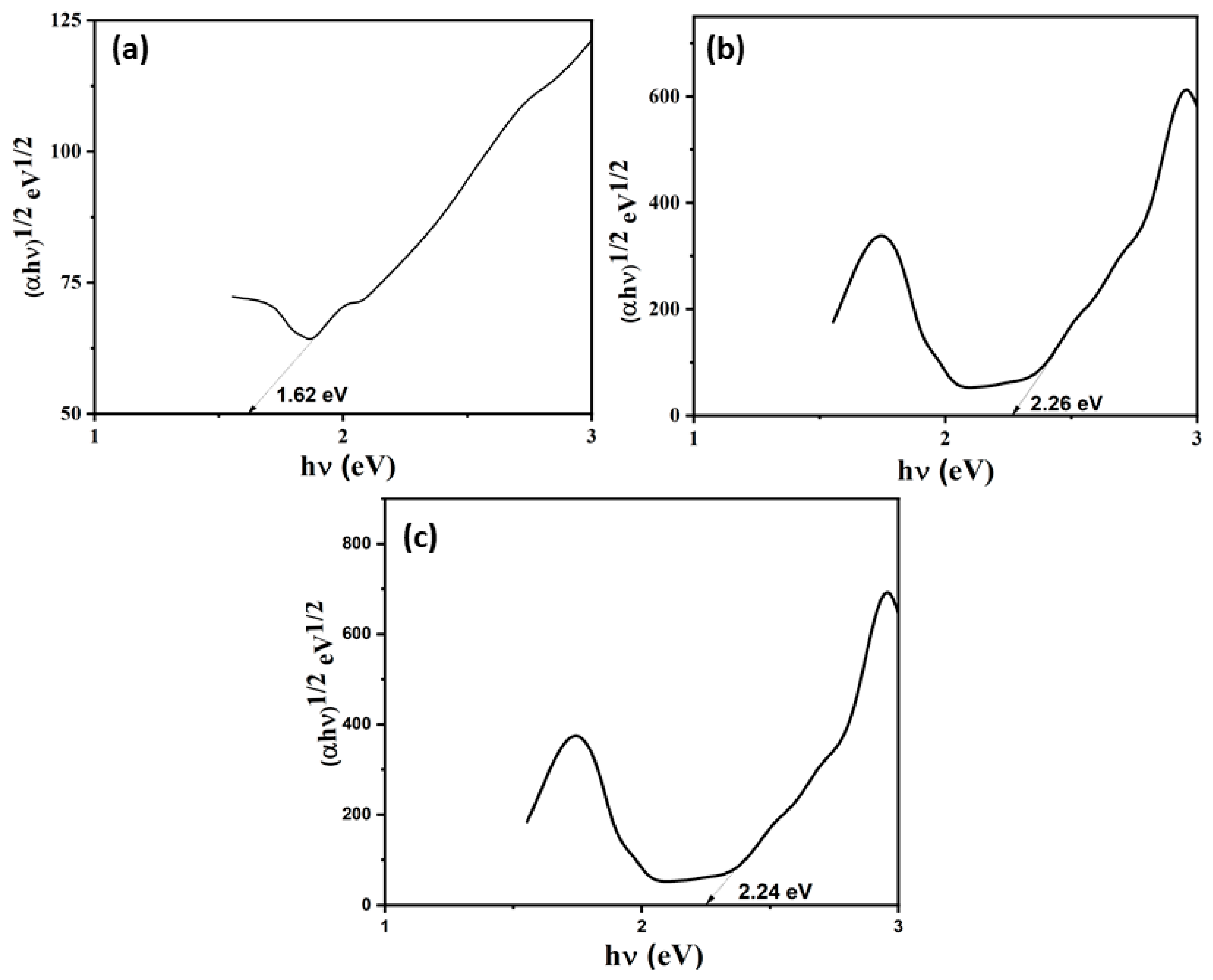
2.4. Band Gap Calculation

A standard relation Kubelka–Munk function was used to calculate the bandgap energy value of the prepared materials (Equation (1)) [38].
αhν = A (hν − Eg)n
where α, Eg, ν, h, n, and A signify the absorption coefficient, band gap energy, frequency of light, Planck’s constant, nature of the photocatalyst (n = 1/2 for direct bandgap semiconductors and n = 2 for indirect bandgap semiconductors), and proportionality constant, respectively.

2.5. Band Potential Calculation

The conduction band (CB) and valence band (VB) potentials are essential parameters for understanding the electronic structure and photocatalytic behavior of semiconductors. These potentials can be estimated using the following equations (Equations (2) and (3)) [39,40],
EVB = X − Ee + 0.5Eg
ECB = EVB − Eg
where ECB and EVB are the potentials of conduction and valence band, respectively, X represents the absolute electronegativity of the semiconductor, Ee is the energy of free electrons on the hydrogen scale (~4.5 eV), and Eg is the bandgap energy value of the semiconductors. This equation determines the VB and CB potential based on the material’s electronegativity and bandgap energy.

2.6. Characterization Details

The structural information was analyzed using Bruker D8 ECO advanced X-ray diffraction (Karlsruhe, Germany) with Cu Kα radiation (λ = 1.54 Å). JEOL JEM 2100 (Tokyo, Japan) Plus high-resolution transmission electron microscopy (HRTEM) was used to confirm the core–shell structure of the nanocomposites. The CHI660 F electrochemical workstation (Shanghai, China) was utilized to analyze the electrochemical properties using 3 electrode system in 1 mol KOH. Shimadzu IR tracer–100 (Kyoto, Japan) was used to confirm the vibrations of pure and composites in the range from 4000 to 400 cm−1. The chemical stability and composition were evaluated using Omicron ESCA, made by Oxford Instruments, Wiesbaden, Germany. The optical properties were studied with UV–Visible spectroscopy (JASCO V750, Tokyo, Japan) in the range of 200–800 nm.

3. Results and Discussion

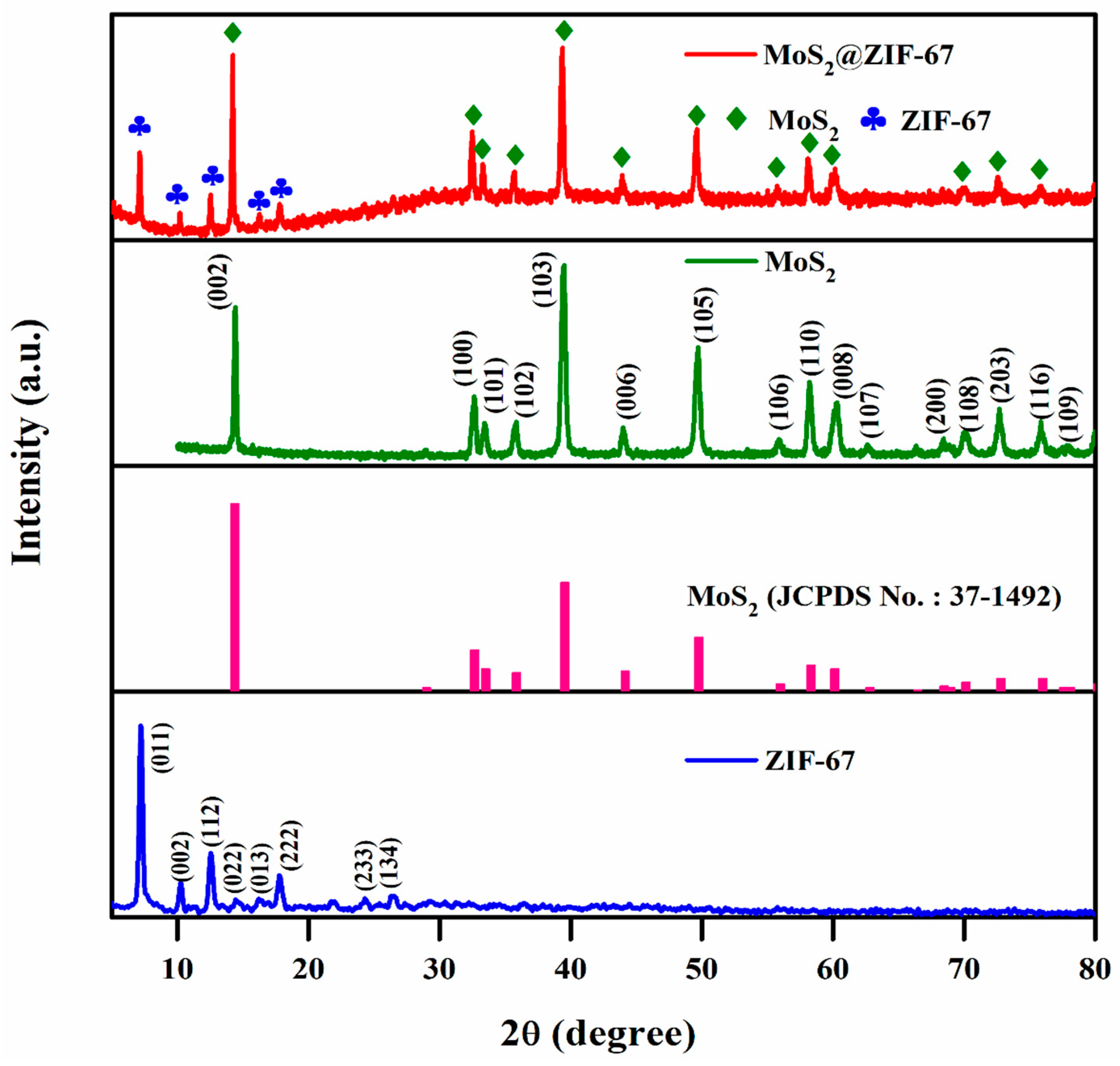
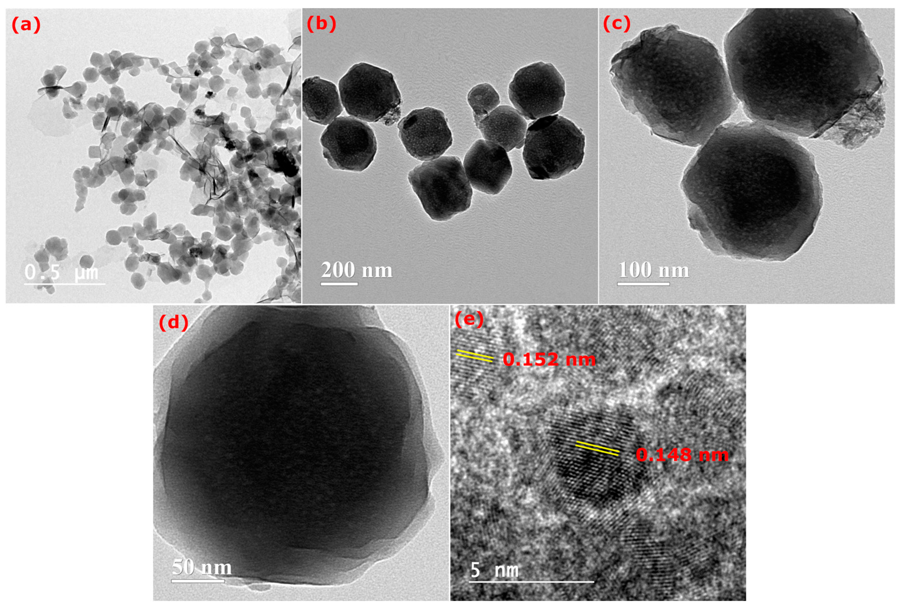
3.1. Structural and Surface Morphology
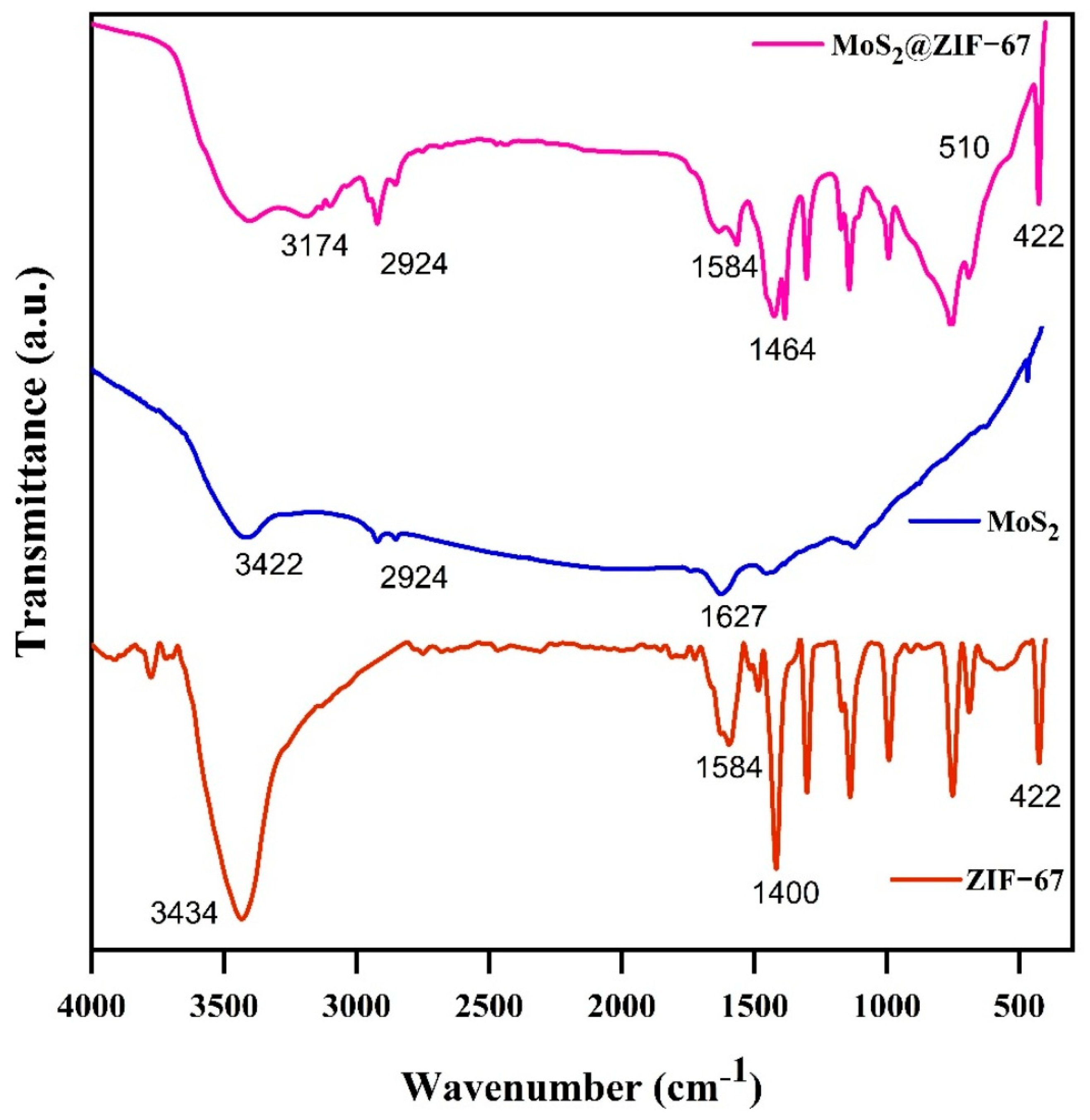
The structural information of the pristine MoS2, ZIF-67, and MoS2@ZIF-67 nanocomposites was studied using XRD, and the patterns are shown in Figure 2. The nanocomposite displayed peaks at 2θ values of 7.31° (0 1 1), 10.35° (0 0 2), 12.72° (1 1 2), 14.41° (0 2 2), 16.46° (0 1 3), 18.03° (2 2 2), 24.54° (2 3 3), and 26.72° (1 3 4), which match the crystalline structure of ZIF-67 and are in good agreement with previous reports [33,41]. Furthermore, high-intensity peaks are presented at 14.28° (0 2 2), 32.46° (1 0 0), 39.45° (1 0 3), 49.84° (1 0 5), and 57.36° (1 1 0). These planes were identified as MoS2 (JCPDS No.: 37-1492), indicating the existence of MoS2 in the MoS2@ZIF-67 nanocomposite [42]. The MoS2@ZIF-67 nanocomposite shows two distinct crystalline phases of ZIF-67 and MoS2. The strong and dominating peaks in the nanocomposite’s XRD diffractogram suggest that the MoS2 was extremely crystalline and maintained its crystallinity perfectly through the preparation of core–shell MoS2@ZIF-67. The average crystallite size of the materials was calculated using the Scherrer relation, and the values are MoS2 (49 nm), ZIF-67 (35 nm), and MoS2@ZIF-67 (40 nm).
HR-TEM images of the pristine ZIF-67 are presented in Figure 3a. Figure 3b–d present HR-TEM images of MoS2@ZIF-67, illustrating the irregular polygonal structures of ZIF-67 and the non-uniform nanosheets of MoS2, respectively. Additionally, Figure 3c shows that the MoS2 nanosheets are irregularly encapsulated on the ZIF-67 framework [43]. Figure 3d distinctly illustrates the core–shell architecture with polyhedral morphology, signifying the encapsulation of MoS2 nanoparticles (shell) around the core of ZIF-67. Figure 3e displays HRTEM images that exhibit lattice fringes with d-spacings of 0.152 nm and 0.148 nm, as indicated in the Figure (ZIF-67).

3.2. FT-IR Analysis

The functional group of materials was investigated using FT-IR in the wavenumber region of 4000–400 cm−1. The FT-IR spectra of MoS2, ZIF-67, and MoS2@ZIF-67 are displayed in Figure 4. The band of 3300–3500 cm−1 corresponds to the stretching vibration of O-H bonds in adsorbed water molecules [44]. Stretching vibrations at 1418 cm−1 and 1584 cm−1, attributed to the aromatic imidazole ring, are also observed in the spectra. Peaks occurring between 600 and 1500 cm−1 were associated with the aromatic bending and stretching vibrational modes. Additionally, the imidazole group exhibited out-of-plane bending vibrations at 751 cm−1, while in-plane bending vibrations are observed in the range of 1000–1300 cm−1 [43]. The same vibration is present in pure MoS2, ZIF-67, and MoS2@ZIF-67. The broad band at 3174 cm−1 is due to the C-H stretching vibrations of the methyl group in the 2-methylimidazole linker. The medium peak at 1584 cm−1 indicates the C=N stretching mode of 2-Methylimidazole [33]. The bands in the 1400–600 cm−1 region correspond to the stretching and bending modes of the imidazole ring. Most observed vibrational bands are related to the vibrations of imidazole, except for the Zn-N stretching at 422 cm−1 [37]. In addition, the peak at about 1464 cm−1 in the nanocomposite belongs to the S-Mo-S bond and a small peak at 510 cm−1 is due to the S–S bond [45]. From the FTIR analysis, all the characteristic vibrations of ZIF-67 and MoS2 were presented in the composite and confirm the formation of MoS2@ZIF-67.

3.3. X-Ray Photoelectron Spectral Analysis

To further substantiate the effective recombination of MoS2 and ZIF-67, we examined the elemental valence state of the sample. Figure 5a illustrates the XPS spectrum of the MoS2@ZIF-67 composite. Figure 5 shows that N, C, Co, Mo, O, and S were found in the full XPS spectrum of the composite material and the XPS content is shown in Table 1.
In Figure 5, the high-resolution N 1s spectrum shows peaks at 400.7 eV, 399.2 eV, and 395.2 eV, which are N=N, imidazole-N, and N3−, respectively, along with a small satellite peak. Except for the imidazole-N coordination bond from the ZIF-67 material, the previous peak displays a comparatively greater bonding energy than the others [46,47]. The amount of three nitrogen species was assessed by fitting the area to the nitrogen 1s curve. The proportion of each nitrogen functional group can be quantified using the XPS method, which detects the binding energy of chemical bonds. The binding energies of four nitrogen functional groups are as follows: pyrrolic N–H at 400 eV; pyridinic N at 398.4 eV; graphitic N at 401 eV; and pyridinic N–O at 402 and 404 eV [48,49]. The percentage of deconvoluted peaks is shown in Table 1. In total, 22% of the peaks were at 395.2 eV, 66% at 399.1 eV, and 12% 400.6 eV. The results indicate that the pyrolysis peak was diminished, the overall nitrogen content and pyridine nitrogen content was higher, but the graphite nitrogen concentration diminished [50,51]. The C 1s spectra (Figure 5c) were deconvoluted into two peaks: one at 284.8 eV attributed to the C=C bond, and another at 286 eV associated with the C-N bond [52]. Figure 5d illustrates the high-resolution Mo 3d spectra of MoS2@ZIF-67 composites. The distinct peaks at 228.9, 230.9, 232.5, and 235.4 eV are attributed to Mo4+ 3d3/2, Mo4+ 3d5/2, and Mo6+ 3d5/2, and Mo6+ 3d3/2, respectively [53,54]. This implies that Mo ions in the composite material coexist as Mo4+ and Mo6+, with the peak of Mo6+ being less pronounced than that of Mo4+, signifying a lower concentration of Mo6+. The simultaneous presence and transformation of Mo4+ and Mo6+ can enhance electron transport during the degradation process, thereby facilitating the generation of active compounds [55]. The doublet separation of Mo 3d5/2 and 3d3/2 is approximately 3.1 eV, which is in good agreement with Amin et al. [56]. We detected a minor peak at a binding energy of 226.3 eV, which corresponds to the S 2s spectrum [57]. The high-resolution S 2p spectra of the composite material are presented in Figure 5e, with two prominent peaks at 161.9 eV and 163.1 eV, corresponding to S2− 2p3/2 and S2− 2p1/2, respectively [58,59]. The findings indicate that a significant quantity of unsaturated sulfur in the composites enhances photo/electrocatalytic activity. The Co 2p spectrum of MoS2@ZIF 67 (Figure 5f) displays primary peaks at 781.8 eV and 797.4 eV, respectively, signifying the Co (II) oxidation state. Furthermore, we attribute the peaks observed at 782.8 eV and 798.5 eV to Co 2p3/2 and Co 2p1/2, respectively, indicating a Co (III) oxidation state. The binding energies of 786.5 eV and 802.8 eV correspond to the conventional Co 2p3/2 and Co 2p1/2 satellite peaks, respectively [60,61]. The doublet separation of Co 2p3/2 and 2p1/2 is approximately 15 eV [62]. The XPS survey revealed a small O1s peak at 531.5, indicative of C–O [63,64]. This signal may indicate the presence of chemisorbed oxygen or other hydroxyl-containing molecules [65]. The lack of a signal at 496.5 eV may result from surface oxidation in the sample exposed to the environment [66]. The presence of oxygen-containing functional groups may enhance the electrical conductivity and surface wettability in the composites, hence facilitating superior degradability. Figure 5g demonstrates that the O 1 s XPS signal can be deconvoluted into four peaks at 530.2, 531.4, 532.4, and 533.3 eV, which correspond to C=O, C–O, Mo–O, and C–OH bonds, respectively [67,68]. A chemical bond between molybdenum and oxygen promotes the attachment of paramolybdate ions to the ZIF-67 surface, leading to the formation of MoS2 nanosheets. This is corroborated by a study utilizing a transmission electron microscope.

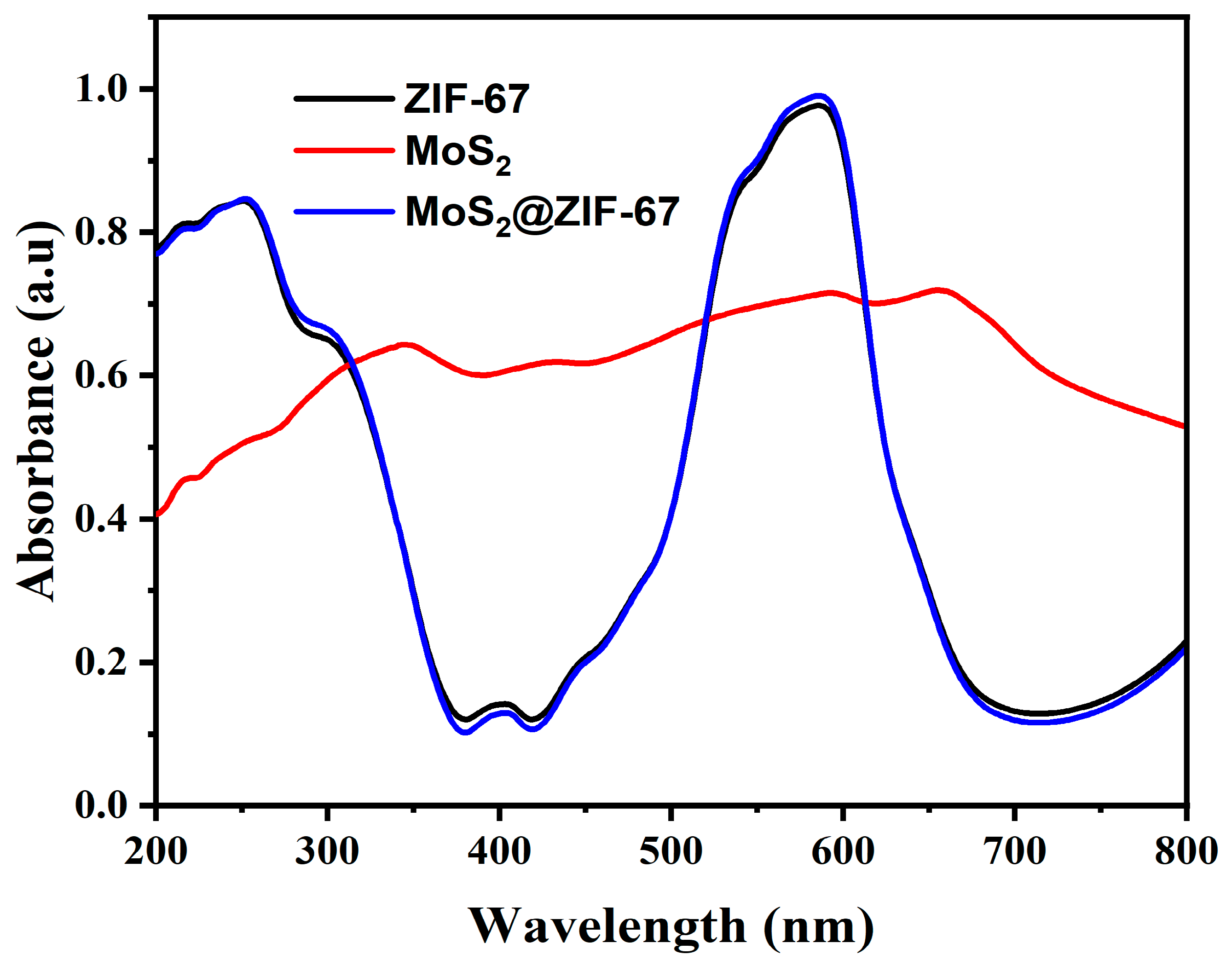
3.4. Optical Absorption Studies

The optical absorption properties of the photocatalysts were examined using a UV–visible diffuse reflectance spectral (DRS) study. Figure 6 illustrates the absorption spectra of the pure ZIF-67, MoS2, and MoS2@ZIF-67 hybrid. Herein, the absorption of ZIF-67 was observed at 252 and 585 nm, MoS2 at 656 nm, and MoS2@ZIF-67 composite at 251 and 584 nm, respectively. The shoulder peaks observed in the ZIF-67 are for the ligand metal charge transfer transition, while the strong absorption is due to the 4A2 (F) → 4T1 (P) transition of Co2+ of ZIF-67. The calculated band gap energy results for the pure ZIF-67 and MoS2, and the MoS2@ZIF-67 hybrid of 2.26, 1.62, and 2.24 eV, respectively, are shown in Figure 7. The prepared MoS2@ZIF-67 hybrid has a lower bandgap energy value when compared with pure ZIF-67, representing the greater absorption ability of the hybrid [43,69].
To the addition of the bandgap of the photocatalyst, the rate of reconnection for the photoinduced charge carriers also influences the photocatalytic capability; hence, the photoluminescence spectra was investigated for ZIF-67, MoS2, and MoS2@ZIF-67 hybrid photocatalysts under a 320 nm excitation wavelength. The results are shown in Figure 8. In general, the higher PL peak intensity suggests the lower carrier’s separation and migration. As seen in Figure 8, the greater peak intensity of ZIF-67 implies the greater reconnection of charges in the ZIF-67; at the same time, the pure MoS2 shows a lower PL intensity. In the case of the MoS2@ZIF-67 hybrid, the PL emission intensity was lower than the pure ZIF-67 due to its formation of a hybrid between ZIF-67 and MoS2, which signified effective separation and migration of the charge with an increased life span [70,71].

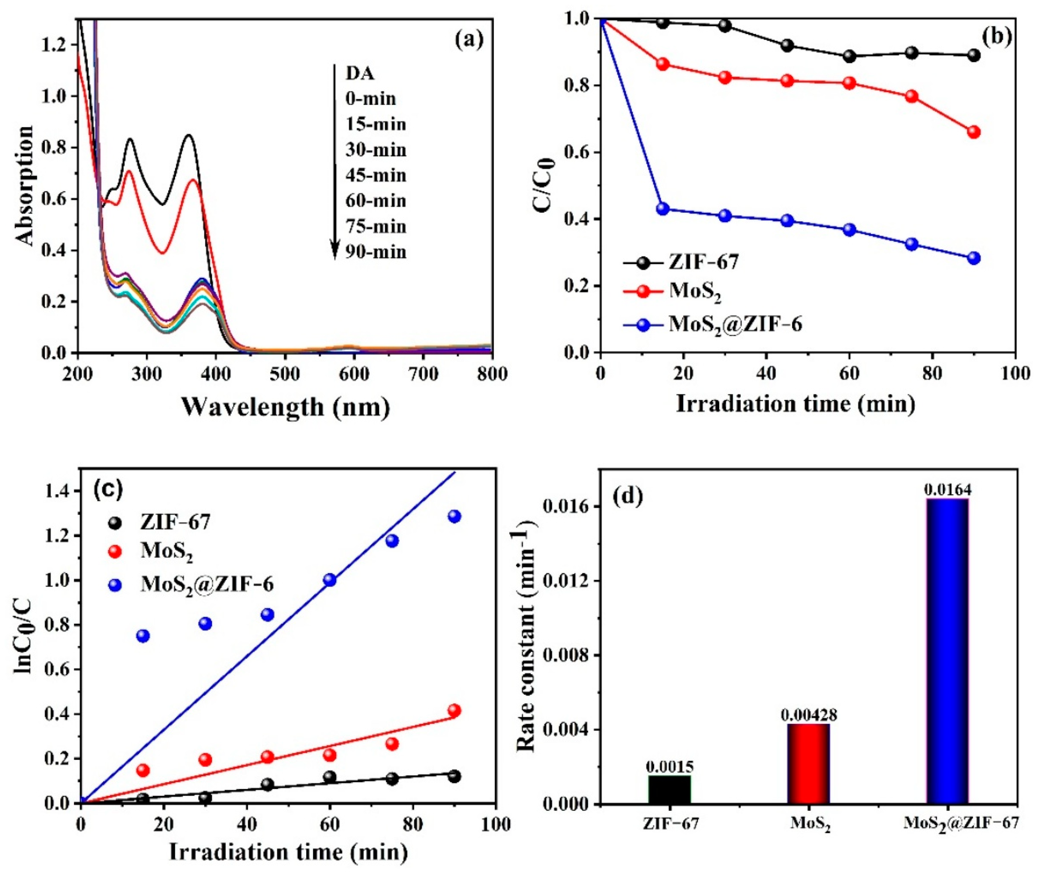
3.5. Photocatalytic Performance Analysis

The photocatalytic abilities of the fabricated materials were evaluated using the degradation of TC under simulated light illumination for 90 min. Figure 9a depicts the UV-vis absorption spectra for the MoS2@ZIF-67 hybrid towards the degradation of the TC under visible-light irradiation. This shows that the distinctive peak at maximum absorption of 357 nm steadily decreases with an increasing illumination time, resulting in the effective disappearance of the peak during the degradation of TC. Furthermore, the photocatalytic degradation efficiency of the pure ZIF-67 and MoS2, and the MoS2@ZIF-67 hybrid photocatalysts was 34, 12, and 72% respectively; the results are illustrated in Figure 9b. The MoS2@ZIF-67 hybrid revealed substantial enhancement in the degradation of TC within 90 min of irradiation, accredited to the effective formation of heterojunction that assists the quick separation and transfer of photoproduced carriers. The kinetics for the degradation of TC were analyzed by using the pseudo-first-order kinetics equation given in Equation (4) [43,72],
lnCt/C0 = kt
where Ct and C0 are the concentration of the dye at different time intervals and initially, respectively, k and t are the pseudo-first-order rate constant (min−1) and time (min), respectively. As seen in Figure 9c,d, the kinetic rate constant value of the pure ZIF-67 and MoS2, and the MoS2@ZIF-67 hybrid was found to be 0.0015, 0.00428, and 0.0164 min−1, respectively. The above results underscore the greater photocatalytic activity of the MoS2@ZIF-67 hybrid, indicating the effective formation of the heterojunction between the MoS2 nanoparticle and ZIF-67, which possibly encourages effective absorption in the visible-light region and improves the lifespan of the charges.
Table 2 compares ZIF-67- and MoS2-based composites for antibiotic degradation. ZIF-67 composites offer a high surface area, tunable porosity, and strong catalytic properties, enhancing the degradation efficiency. In contrast, MoS2 composites excel in photocatalytic degradation, which is influenced by the light frequency and material morphology. Both materials show promising results in antibiotic removal due to their edge-active sites and capacity to create ROS under UV light [73]. Depending on the catalytic mechanism, ZIF-67 composites degrade a wide range of antibiotics, such as tetracycline and ciprofloxacin, efficiently in both UV and visible environments. ZIF-67’s high surface area and porosity increase the contact area between antibiotics and catalysts, which improves the degradation process. The selection of MoS2@ZIF-67 composites for a given application is determined by the needed reaction conditions, stability, and degradation efficiency for tetracycline. As illustrated in Figure 10, radical scavenging was used to determine the activity of the key radical in the TC degradation over the MoS2@ZIF-67 hybrid, by adding various quenchers, such as isopropanol (IPA), potassium iodide (KI), and potassium persulfate (K2S2O8), at a concentration of 1 mmol L−1 to quench the hydroxyl (OH), holes (h+) and electrons (e), respectively. A sudden decrease in the photocatalytic activity was observed following the addition of KI and IPA, a scavenger of h+ and OH. radicals. The addition of the K2S2O8 scavenger of e exhibited a negligible impact on the degradation of TC. These results revealed that the h+ and OH. radicals are majorly active radicals in the degradation of TC over the MoS2@ZIF-67 hybrid.
Additionally, the recyclability of the photocatalyst is a crucial factor for practical applications, which determines their long-term reusability and cost-effectiveness. Figure 11 exposes the recycling runs of the MoS2@ZIF-67 hybrid over the degradation of TC. For the experiment, the MoS2@ZIF-67 hybrid photocatalyst was subjected to three consecutive degradation cycles, with the degradation rate monitored after each cycle. From Figure 10, negligible changes were observed even after three cycles. This demonstrates the excellent photostability and recyclability of the MoS2@ZIF-67 hybrid.
Table 3 shows a comparison of the radical’s activity experiments of MoS2 and ZIF-67 degradation of tetracycline. Photocatalytic radical trapping experiments are essential for identifying and understanding the reactive species involved in photocatalytic processes. These experiments typically employ specific scavengers to selectively quench reactive oxygen species (ROS), such as hydroxyl radicals (OH), superoxide radicals (O2−), and photogenerated holes (h+), by observing changes in photocatalytic activity upon the introduction of these scavengers.

3.6. Electrochemical Performance

In this study, cyclic voltammetry (CV) was performed to investigate the electrochemical performance of the MoS2@ZIF-67 hybrid nanocomposites; the results are shown in Figure 12a. the prepared electrode was examined in a 1 M KOH alkaline electrolyte and examined in a three-electrode configuration at a CHI 660F electrochemical workstation. We prepared the electrode material, Pt wire, and Hg/HgO for the working, counter, and reference electrode, respectively. This technique provides insights into the charge transfer mechanisms and the interaction of the electrocatalyst with the electrolyte [40]. The CV was conducted at a scan rate of 100 mV/s and ran continuously for 100 cycles, optimized to evaluate the reaction kinetics. With an increasing number of cycles, the peak current showed a proportional increase, suggesting a diffusion-controlled charge transfer process [33]. The electrocatalyst material was more stable, with a slight difference between the area of the 1st and 100th cycles, which denotes the excellent material stability. To analyze the role of oxygen and water molecules, moreover, the Linear Sweep Voltammetry (LSV) technique is used for the electrocatalyst of MoS2@ZIF67 nanocomposites and the results are shown in Figure 12b. It clearly shows an enhanced catalytic performance, with a lower overpotential of 373 mV at the current density of 10 mA/cm2 and 534 mV at the current density of 50 mA/cm2 at a scan rate of 5 mV/s.
The LSV analysis at various scan rates, such as 10, 15, 20, 25, and 50 mV/s, is shown in Figure 12c. Generally, in an oxygen evolution reaction, OH ions from the electrolyte adsorb onto the active sites of the catalyst, initiating the oxidation process [76]. Water molecules can also act as reactants, undergoing oxidation to form hydroxyl radicals and molecular oxygen [77]. The catalyst facilitates faster charge transfer, as evident from the LSV results at different scan rates (10–50 mV/s), showing variations in the current response and overpotential. The improved catalytic efficiency suggests strong interactions between the MoS2@ZIF-67 active sites and the OH/H2O species, reducing the energy barrier for oxygen evolution [77], which was confirmed with the obtained lower overpotentials. The bar diagram (Figure 12c) further illustrates how different scan rates influence the current density, confirming the catalyst’s stability and efficiency under varied electrochemical conditions. In Figure 12d, EIS was used to measure the response of the system to a small AC perturbation over a wide frequency range [78].
The prepared electrocatalytic materials were examined using electrochemical impedance spectroscopy (EIS) in a 1 M KOH aqueous solution at a frequency range of 1 to 100 kHz with an applied amplitude potential of 5 mV. A Nyquist plot, where the real part (Z′) corresponds to the charge transfer resistance, and the imaginary part (Z″) reflects the capacitive behavior of the electrode [73], is shown in Figure 12d. In the high- to mid-frequency region, a small semicircle is observed, which also confirmed that the electrocatalytic material exhibited a significantly lower charge transfer resistance, unchanged even after the cycles. This indicates that for the MoS2@ZIF-67 photocatalyst, electrons have more difficulty reaching the semiconductor material/electrolyte interface. When photocatalytic degradation occurs, there is a possibility for electrons to recombine before reacting with pollutants. The small semicircle indicated that, during the photocatalytic performance of the materials, there is a recombination of e/h+ [79]. These results point out that in the MoS2@ZIF-67 photocatalyst, material was degraded to contaminants in the surplus water.

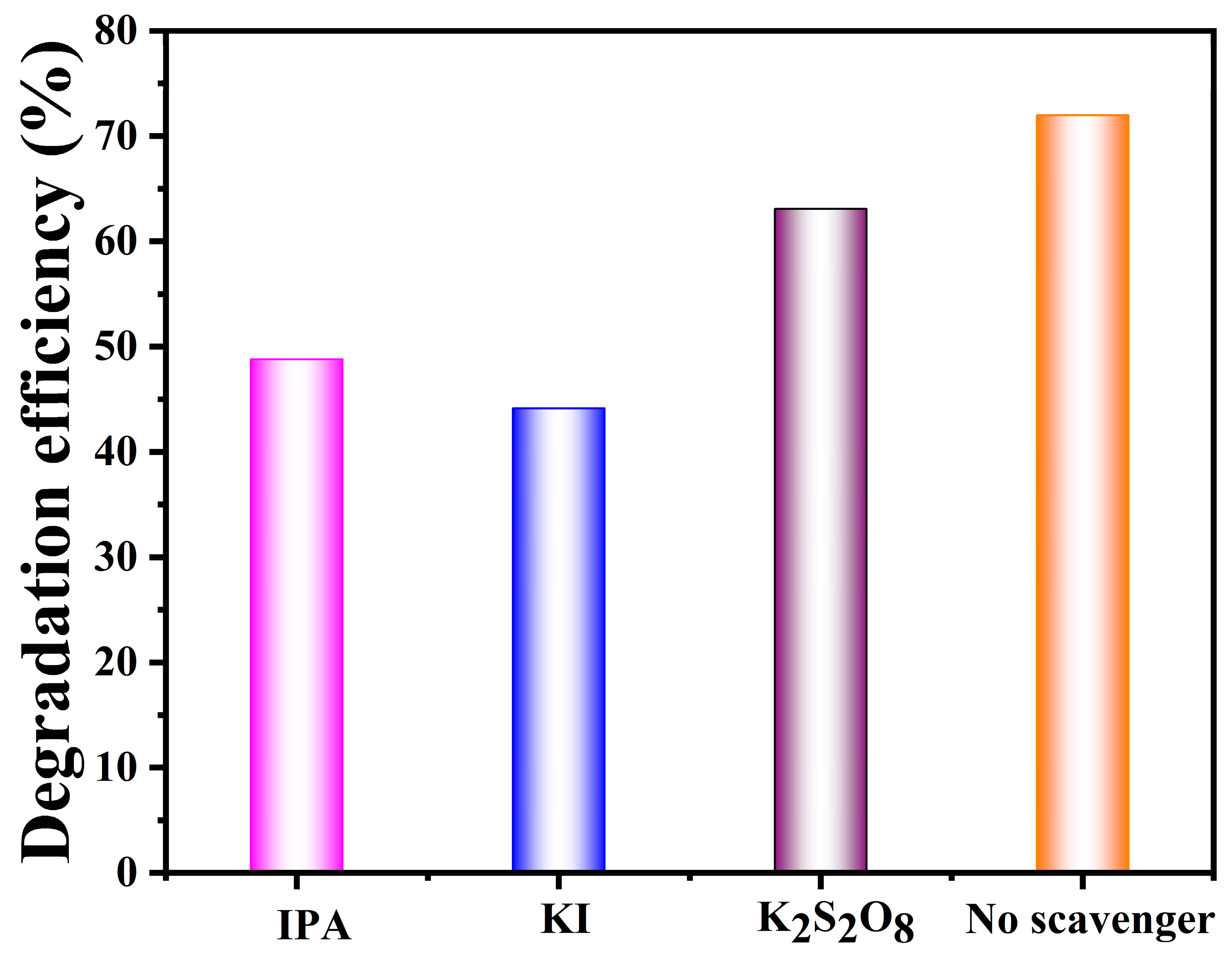
3.7. Charge Transfer Mechanism for Degradation of Tetracycline

A possible type-I heterojunction charge transfer mechanism was proposed for the ZIF-67/MoS2 hybrid, as shown in Figure 13. Under visible-light illumination, e in the VB of both ZIF-67 and MoS2 are excited from their respective VB into CB. At the same time, the e in the CB of ZIF-67 migrates into the CB of MoS2, due to the more negative potential of ZIF-67. Simultaneously, the h+ in the VB of ZIF-67 shifted towards the VB of MoS2. The accumulated e and h+ in the CB and VB of MoS2 facilitates the reduction and oxidation of surface-adsorbed oxygen and water molecules, converting them into harmless, biodegradable by-products [80,81]. In this way, the charge carriers are separated and their lifespan is extended, which improves the photocatalytic activity by reducing the rapid recombination of charges. The construction of an MoS2@ZIF-67 heterojunction accelerates the transfer and separation of charges through the heterojunction and uphold its redox ability for the degradation of TC under visible-light illumination. Combining some of the possible reactions participating in TC degradation of MoS2 with ZIF-67 enhances the photocatalytic degradation of tetracycline under visible light. The synergy between these materials facilitates efficient charge separation and increases the generation of reactive oxygen species, leading to an improved degradation efficiency. This approach offers a promising strategy for addressing antibiotic contamination in water sources [69,74,82].
MoS2 + hv → MoS2 (e) + MoS2 (h+)
ZIF67 + hv → ZIF-67 (e) + ZIF-67 (h+)
O2 + g-ZIF-67 (e) → ZIF-67+ O2
H2O + MoS2 (h+) → MoS2 + OH
OH, and O2 + TC → degradation products

4. Conclusions

Novel core–shell MoS2@ZIF-67 nanocomposites were synthesized using a simple in situ chemical method, and their characteristics were studied using XRD, FTIR, XPS, PL, and HRTEM. The Co 2p spectrum of MoS2@ZIF 67 displayed primary peaks at 781.8 eV and 797.4 eV, respectively, signifying a Co (II) oxidation state. HRTEM analysis suggested that the nanoscale MoS2 uniformly grown on the surface of ZIF-67 confirms the core–shell assembly. The band gap energy of MoS2@ZIF-67 was found to be 2.24 eV. Moreover, the PL emission intensity of the MoS2@ZIF-67 was lower than that of pure ZIF-67; this is due to the formation of a hybrid between ZIF-67 and MoS2, which signified the effective separation and migration of the charge with an increased life span. The MoS2@ZIF-67 hybrid revealed a substantial enhancement in the degradation of TC within 90 min of irradiation. The kinetic rate constant was evaluated, and the values of pure ZIF-67, MoS2, and the MoS2@ZIF-67 hybrid were found to be 0.0015, 0.00428, and 0.0164 min−1, respectively. These results revealed that the h+ and OH. radicals are majorly active radicals in the degradation of TC. An LSV study clearly showed the enhanced catalytic performance, with a lower overpotential of 373 mV at a current density of 10 mA/cm2 and 534 mV at a current density of 50 mA/cm2 and a scan rate of 5 mV/s.

Author Contributions

Conceptualization, V.S. and M.P.; methodology, V.S., A.M. and M.P.; investigation, A.S.S., M.P., T.B. and S.J.; data curation, S.S., I.H.S.A. and K.K.; writing—original draft preparation, M.P., V.S., A.M., A.S.S., T.B., S.J., S.S., I.H.S.A. and K.K.; writing—review and editing, M.P., V.S., A.M., A.S.S., T.B., S.J., S.S., I.H.S.A. and K.K.; supervision, V.S. All authors have read and agreed to the published version of the manuscript.

Funding

The author, V. Siva, is thankful to the management of Karpagam Academy of Higher Education, Coimbatore-641 021, Tamil Nadu, India, for providing financial support through a seed grant (No.: KAHE/R-Acad/A1/Seed Money/007, dt. 11 January 2023).

Data Availability Statement

Data are contained within the article.

Conflicts of Interest

The authors declare no conflicts of interest.

References

  1. Miethke, M.; Pieroni, M.; Weber, T.; Brönstrup, M.; Hammann, P.; Halby, L.; Arimondo, P.B.; Glaser, P.; Aigle, B.; Bode, H.B.; et al. Towards the sustainable discovery and development of new antibiotics. Nat. Rev. Chem. 2021, 5, 726–749. [Google Scholar] [CrossRef]
  2. Salam, M.A.; Al-Amin, M.Y.; Salam, M.T.; Pawar, J.S.; Akhter, N.; Rabaan, A.A.; Alqumber, M.A.A. Antimicrobial Resistance: A Growing Serious Threat for Global Public Health. Healthcare 2023, 11, 1946. [Google Scholar] [CrossRef]
  3. Samrot, A.V.; Wilson, S.; Sanjay Preeth, R.S.; Prakash, P.; Sathiyasree, M.; Saigeetha, S.; Shobana, N.; Pachiyappan, S.; Rajesh, V.V. Sources of Antibiotic Contamination in Wastewater and Approaches to Their Removal—An Overview. Sustainability 2023, 15, 12639. [Google Scholar] [CrossRef]
  4. Lu, S.; You, S.; Hu, J.; Li, X.; Li, L. Magnetic MnFe2O4/ZIF-67 nanocomposites with high activation of peroxymonosulfate for the degradation of tetracycline hydrochloride in wastewater. RSC Adv. 2024, 14, 7528–7539. [Google Scholar] [CrossRef]
  5. Madhogaria, B.; Banerjee, S.; Kundu, A.; Dhak, P. Efficacy of new generation biosorbents for the sustainable treatment of antibiotic residues and antibiotic resistance genes from polluted waste effluent. Infect. Med. 2024, 3, 100092. [Google Scholar] [CrossRef]
  6. Wang, F.; Wang, Z.; Zhao, Y.; Zhang, J. Performance of Traditional and Emerging Water-Treatment Technologies in the Removal of Tetracycline Antibiotics. Catalysts 2024, 14, 269. [Google Scholar] [CrossRef]
  7. Samal, K.; Mahapatra, S.; Ali, M.H. Pharmaceutical wastewater as Emerging Contaminants (EC): Treatment technologies, impact on environment and human health. Energy Nexus 2022, 6, 100076. [Google Scholar] [CrossRef]
  8. Zhao, X.; Zheng, M.; Gao, X.; Zhang, J.; Wang, E.; Gao, Z. The application of MOFs-based materials for antibacterials adsorption. Coord. Chem. Rev. 2021, 440, 213970. [Google Scholar] [CrossRef]
  9. Satyam, S.; Patra, S. Innovations and challenges in adsorption-based wastewater remediation: A comprehensive review. Heliyon 2024, 10, e29573. [Google Scholar] [CrossRef]
  10. Sabzehmeidani, M.M.; Mahnaee, S.; Ghaedi, M.; Heidari, H.; Roy, V.A. Carbon based materials: A review of adsorbents for inorganic and organic compounds. Mater. Adv. 2021, 2, 598–627. [Google Scholar] [CrossRef]
  11. Schumacher, L.; Marschall, R. Recent Advances in Semiconductor Heterojunctions and Z-Schemes for Photocatalytic Hydrogen Generation. Top. Curr. Chem. (Z) 2022, 380, 53. [Google Scholar] [CrossRef]
  12. Abhishek, B.; Jayarama, A.; Rao, A.S.; Nagarkar, S.S.; Dutta, A.; Duttagupta, S.P.; Prabhu, S.S.; Pinto, R. Challenges in photocatalytic hydrogen evolution: Importance of photocatalysts and photocatalytic reactors. Int. J. Hydrogen Energy 2024, 81, 1442–1466. [Google Scholar] [CrossRef]
  13. Yu, Z.; Qian, L.; Zhong, T.; Ran, Q.; Huang, J.; Hou, Y.; Li, F.; Li, M.; Sun, Q.; Zhang, H. Enhanced visible light photocatalytic activity of CdS through controllable self-assembly compositing with ZIF-67. Mol. Catal. 2020, 485, 110797. [Google Scholar] [CrossRef]
  14. Shameem, A.; Devendran, P.; Siva, V.; Raja, M.; Bahadur, S.A.; Manikandan, A. Preparation and Characterization Studies of Nanostructured CdO Thin Films by SILAR Method for Photocatalytic Applications. J. Inorg. Organomet. Polym. Mater. 2017, 27, 692–699. [Google Scholar] [CrossRef]
  15. Murugan, A.; Thirumal, V.; Kim, J.; Siva, V.; Kasinathan, P.; Alothman, A.A.; Mohammad, S.; Selvakumar, K. Effective visible light-driven binary ZnO catalyst decorated on layered double hydroxides for simultaneous photocatalytic degradation of Ciprofloxacin and Ampicillin. J. Taiwan Inst. Chem. Eng. 2024, 161, 105495. [Google Scholar] [CrossRef]
  16. Vijayakumar, P.; Vijayan, P.; Krishnan, P.S.; Raja, A.; Kumaravel, S.; Venthan, S.M.; Siva, V.; Palanisamy, G.; Lee, J.; Afzal, M. Design of bifunctional synergistic NiMoO4/g-C3N4 nanocomposite for the augmentation of electrochemical water splitting and photocatalytic antibiotic degradation performances. J. Alloys Compd. 2024, 987, 174128. [Google Scholar] [CrossRef]
  17. Chakravorty, A.; Roy, S. A review of photocatalysis, basic principles, processes, and materials. Sustain. Chem. Environ. 2024, 8, 100155. [Google Scholar] [CrossRef]
  18. Nadolska, M.; Szkoda, M.; Trzciński, K.; Ryl, J.; Lewkowicz, A.; Sadowska, K.; Smalc-Koziorowska, J.; Prześniak-Welenc, M. New light on the photocatalytic performance of NH4V4O10 and its composite with rGO. Sci. Rep. 2023, 13, 3946. [Google Scholar] [CrossRef]
  19. Vizza, M.; Giurlani, W.; Cerri, L.; Calisi, N.; Leonardi, A.A.; Faro, M.J.L.; Irrera, A.; Berretti, E.; Perales-Rondón, J.V.; Colina, A.; et al. Electrodeposition of Molybdenum Disulfide (MoS2) Nanoparticles on Monocrystalline Silicon. Molecules 2022, 27, 5416. [Google Scholar] [CrossRef]
  20. Rahman, A.; Jennings, J.R.; Tan, A.L.; Khan, M.M. Molybdenum disulfide-based nanomaterials for visible-light-induced photocatalysis. ACS Omega 2022, 7, 22089–22110. [Google Scholar] [CrossRef]
  21. Hasija, V.; Raizada, P.; Thakur, V.K.; Khan, A.A.P.; Asiri, A.M.; Singh, P. An overview of strategies for enhancement in photocatalytic oxidative ability of MoS2 for water purification. J. Environ. Chem. Eng. 2020, 8, 104307. [Google Scholar] [CrossRef]
  22. Thomas, N.; Mathew, S.; Nair, K.M.; O’Dowd, K.; Forouzandeh, P.; Goswami, A.; McGranaghan, G.; Pillai, S.C. 2D MoS2: Structure, mechanisms, and photocatalytic applications. Mater. Today Sustain. 2021, 13, 100073. [Google Scholar] [CrossRef]
  23. Luo, M.; Xu, J.; Xu, W.; Zheng, Y.; Wu, G.; Jeong, T. Photocatalytic Activity of MoS2 Nanoflower-Modified CaTiO3 Composites for Degradation of RhB under Visible Light. Nanomaterials 2023, 13, 636. [Google Scholar] [CrossRef]
  24. Kiteto, M.; Vidija, B.; Mecha, C.A.; Mrosso, R.; Chollom, M.N. Advances in metal–organic frameworks as adsorbents, photocatalysts and membranes: A new frontier in water purification. Discov. Water 2024, 4, 54. [Google Scholar] [CrossRef]
  25. Chen, J.; Abazari, R.; Adegoke, K.A.; Maxakato, N.W.; Bello, O.S.; Tahir, M.; Tasleem, S.; Sanati, S.; Kirillov, A.M.; Zhou, Y. Metal–organic frameworks and derived materials as photocatalysts for water splitting and carbon dioxide reduction. Coord. Chem. Rev. 2022, 469, 214664. [Google Scholar] [CrossRef]
  26. Naghdi, S.; Shahrestani, M.M.; Zendehbad, M.; Djahaniani, H.; Kazemian, H.; Eder, D. Recent advances in application of metal-organic frameworks (MOFs) as adsorbent and catalyst in removal of persistent organic pollutants (POPs). J. Hazard. Mater. 2023, 442, 130127. [Google Scholar] [CrossRef]
  27. Luo, T.; Gilmanova, L.; Kaskel, S. Advances of MOFs and COFs for photocatalytic CO2 reduction, H2 evolution and organic redox transformations. Coord. Chem. Rev. 2023, 490, 215210. [Google Scholar] [CrossRef]
  28. Santos, W.D.; Teixeira, M.M.; Campos, I.R.; de Lima, R.B.; Mantilla, A.; Osajima, J.A.; de Menezes, A.S.; Manzani, D.; Rojas, A.; Alcântara, A.C. Photocatalytic degradation of ciprofloxacin using semiconductor derived from heterostructured ZIF-8-based materials. Microporous Mesoporous Mater. 2023, 359, 112657. [Google Scholar] [CrossRef]
  29. Vadivel, S.; Muthuraj, A.; Anbazhagan, M.; Arumugam, R. A novel CoMoO4 enwrapped ZIF-8 nanocomposite with enhanced visible light photocatalytic activity. Environ. Pollut. 2023, 336, 122450. [Google Scholar] [CrossRef]
  30. Elaouni, A.; El Ouardi, M.; Zbair, M.; BaQais, A.; Saadi, M.; Ahsaine, H.A. ZIF-8 metal organic framework materials as a superb platform for the removal and photocatalytic degradation of organic pollutants, a review. RSC Adv. 2022, 12, 31801–31817. [Google Scholar] [CrossRef]
  31. Tang, Y.; Li, X.; Zhang, H.; Ouyang, T.; Jiang, Y.; Mu, M.; Yin, X. Cobalt-based ZIF coordinated hybrids with defective TiO2-x for boosting visible light-driven photo-Fenton-like degradation of bisphenol A. Chemosphere 2020, 259, 127431. [Google Scholar] [CrossRef] [PubMed]
  32. Mahmoodi, N.M.; Keshavarzi, S.; Oveisi, M.; Rahimi, S.; Hayati, B. Metal-organic framework (ZIF-8)/inorganic nanofiber (Fe2O3) nanocomposite: Green synthesis and photocatalytic degradation using LED irradiation. J. Mol. Liq. 2019, 291, 111333. [Google Scholar] [CrossRef]
  33. Sanjana, S.; Siva, V.; Murugan, A.; Shameem, A.S. Core-shell structured Zn3(VO4)2@ZIF-67 nanocrystallites with excellent cycling stability for energy storage systems. J. Energy Storage 2025, 106, 114882. [Google Scholar] [CrossRef]
  34. Zabihi, M.; Motavalizadehkakhky, A. PbS/ZIF-67 nanocomposite: Novel material for photocatalytic degradation of basic yellow 28 and direct blue 199 dyes. J. Taiwan Inst. Chem. Eng. 2022, 140, 104572. [Google Scholar] [CrossRef]
  35. Arabian, S.; Gordanshekan, A.; Farhadian, M.; Nazar, A.R.S.; Tangestaninejad, S.; Sabzyan, H. Adsorption/photocatalytic degradation of Cefixime by the green Bi2WO6/g-C3N4/ZIF-67 dual S-scheme heterojunction: Artificial neural network, genetic algorithm, density functional theory, and toxicity assessments. Chem. Eng. J. 2024, 488, 150686. [Google Scholar] [CrossRef]
  36. Wei, Y.; Chen, J.; Wang, M.; Zhang, X.; Xu, Y.; Wei, Q.; Lan, F.; Gao, P.; Liu, Y.; Wang, R. The double heterostructure photocatalyst MoS2@ ZIF-67/TiO2: Triumphant modification of TiO2 and efficient removal of Karenia mikimotoi. Chem. Eng. J. 2024, 479, 147954. [Google Scholar]
  37. Siva, V.; Sanjana, S.; Murugan, A.; Shameem, A.; Jauhar, R.M.; Babu, S. Facile synthesis and asymmetric device fabrication of zeolite like Co-MOF as a promising electrode material with improved cyclic stability. J. Mater. Sci. Mater. Electron. 2024, 35, 75. [Google Scholar] [CrossRef]
  38. Jubu, P.R.; Obaseki, O.S.; Ajayi, D.I.; Danladi, E.; Chahrour, K.M.; Muhammad, A.; Landi, S., Jr.; Igbawua, T.; Chahul, H.F.; Yam, F.K. Considerations about the determination of optical bandgap from diffuse reflectance spectroscopy using the Tauc plot. J. Opt. 2024, 53, 5054–5064. [Google Scholar] [CrossRef]
  39. Abdul-wahid, I.K.; Ammar, S.H.; Elaibi, A.I.; Jabbar, Z.H. Enhanced synergistic photocatalytic degradation of oxytetracycline antibiotic using novel Ag2MoO4/Co-zeolitic imidazolate framework (ZIF-67) Z-type heterojunction. Inorg. Chem. Commun. 2023, 156, 111277. [Google Scholar] [CrossRef]
  40. Teymourinia, H.; Alshamsi, H.A.; Al-nayili, A.; Sohouli, E.; Gholami, M. Synthesis of new photocatalyst based on g-C3N4/N, P CQD/ZIF-67 nanocomposite for ciprofloxacin degradation under visible light irradiation. J. Ind. Eng. Chem. 2023, 125, 259–268. [Google Scholar] [CrossRef]
  41. Davoodi, M.; Davar, F.; Rezayat, M.R.; Jafari, M.T.; Bazarganipour, M.; Shalan, A.E. Synthesis and characterization of a new ZIF-67@ MgAl2O4 nanocomposite and its adsorption behaviour. RSC Adv. 2021, 11, 13245–13255. [Google Scholar] [CrossRef] [PubMed]
  42. Kao, L.H.; Chuang, K.S.; Catherine, H.N.; Huang, J.H.; Hsu, H.J.; Shen, Y.C.; Hu, C. MoS2-coupled coniferous ZnO for photocatalytic degradation of dyes. J. Taiwan Inst. Chem. Eng. 2023, 142, 104638. [Google Scholar] [CrossRef]
  43. Kshirsagar, S.D.; Shelake, S.P.; Biswas, B.; Singh, A.; Pakhira, S.; Sainath, A.V.S.; Pal, U. In situ decoration of 2D-MoS2/ZIF-67 type II heterojunction for enhanced hydrogen production under simulated sunlight. Catal. Today 2024, 445, 115056. [Google Scholar] [CrossRef]
  44. Sanjana, S.; Siva, V.; Sharmila, S.; Murugan, A.; Shameem, A. Fabrication of V2O5@Co-MOF as a cathode material with excellent rate capability. Sustain. Mater. Technol. 2024, 41, e01072. [Google Scholar] [CrossRef]
  45. Lalithambika, K.C.; Shanmugapriya, K.; Sriram, S. Photocatalytic activity of MoS2 nanoparticles: An experimental and DFT analysis. Appl. Phys. A 2019, 125, 817. [Google Scholar] [CrossRef]
  46. Perkins, C.L.; Lee, S.H.; Li, X.; Asher, S.E.; Coutts, T.J. Identification of nitrogen chemical states in N-doped ZnO via x-ray photoelectron spectroscopy. J. Appl. Phys. 2005, 97, 034907. [Google Scholar] [CrossRef]
  47. Jin, Z.; Yang, M.; Hao, X.; Zhu, P. ZIF-67 dodecahedron coupled with CoAl-layered double hydroxide as S-scheme heterojunction for efficient visible-light-driven hydrogen evolution. Appl. Surf. Sci. 2022, 592, 153300. [Google Scholar] [CrossRef]
  48. Zhuang, S.; Lee, E.S.; Lei, L.; Nunna, B.B.; Kuang, L.; Zhang, W. Synthesis of nitrogen-doped graphene catalyst by high-energy wet ball milling for electrochemical systems. Int. J. Energy Res. 2016, 40, 2136–2149. [Google Scholar] [CrossRef]
  49. Dong, Z.; Zhao, J.; Tian, Y.; Zhang, B.; Wu, Y. Preparation and performances of ZIF-67-derived FeCo bimetallic catalysts for CO2 hydrogenation to light olefins. Catalysts 2020, 10, 455. [Google Scholar] [CrossRef]
  50. Devarayapalli, K.C.; Vattikuti, S.V.P.; Sreekanth, T.V.M.; Yoo, K.S.; Nagajyothi, P.C.; Shim, J. Hydrogen production and photocatalytic activity of g-C3N4/Co-MOF (ZIF-67) nanocomposite under visible light irradiation. Appl. Organomet. Chem. 2020, 34, e5376. [Google Scholar] [CrossRef]
  51. Ngo, C.M.; Nguyen, H.D.; Saito, N.; Do, T.M.D.; Nakayama, T.; Niihara, K.; Suematsu, H. Synthesis of β-MoO3 whiskers by the thermal evaporation method with flowing oxygen gas. J. Am. Ceram. Soc. 2022, 105, 1622–1628. [Google Scholar] [CrossRef]
  52. Ngo, M.T.T.; Ueyama, T.; Makabe, R.; Bui, X.T.; Nghiem, L.D.; Nga, T.T.V.; Fujioka, T. Fouling behavior and performance of a submerged flat-sheet nanofiltration membrane system for direct treatment of secondary wastewater effluent. J. Water Process Eng. 2021, 41, 101991. [Google Scholar] [CrossRef]
  53. Qiu, S.; Wang, T.; Fang, Y. High-efficient preparation of gasoline-ranged C5–C6 alkanes from biomass-derived sugar polyols of sorbitol over Ru-MoO3−x/C catalyst. Fuel Process. Technol. 2019, 183, 19–26. [Google Scholar] [CrossRef]
  54. Mei, P.; Yang, M.; Bai, Y.; Fei, S.; Dong, Y.; Cheng, H. Facile hydrothermal synthesis of nanorod-structured Mo0.6W0.4O3 catalyst for olefin hydrogenation with high activity. J. Catal. 2018, 360, 213–220. [Google Scholar] [CrossRef]
  55. Mao, Y.; Fang, Y.; Pan, J.; Wang, D.; Bu, K.; Che, X.; Zhao, W.; Huang, F. Synthesis, crystal structures and physical properties of Ax(H2O)yMoS2 (A = K, Rb, Cs). J. Solid State Chem. 2019, 279, 120937. [Google Scholar] [CrossRef]
  56. Amin, R.; Hossain, M.A.; Zakaria, Y. Interfacial kinetics and ionic diffusivity of the electrodeposited MoS2 film. ACS Appl. Mater. Interfaces 2018, 10, 13509–13518. [Google Scholar] [CrossRef]
  57. Veerasubramani, G.K.; Park, M.S.; Nagaraju, G.; Kim, D.W. Unraveling the Na-ion storage performance of a vertically aligned interlayer-expanded two-dimensional MoS2@C@MoS2 heterostructure. J. Mater. Chem. A Mater. 2019, 7, 24557–24568. [Google Scholar] [CrossRef]
  58. Luo, S.; Dong, S.; Lu, C.; Yu, C.; Ou, Y.; Luo, J.; Sun, J.; Sun, J. Rational and green synthesis of novel two-dimensional WS2/MoS2 heterojunction via direct exfoliation in ethanol-water targeting advanced visible-light-responsive photocatalytic performance. J. Colloid Interface Sci. 2018, 513, 389–399. [Google Scholar] [CrossRef]
  59. Lin, Y.; Yu, J.; Xing, Z.; Guo, X.; Yu, X.; Tang, B.; Zou, J. Enhanced generation of H2O2 and radicals on Co9S8/partly-graphitized carbon cathode for degradation of bio-refractory organic wastewater. Electrochim. Acta 2016, 213, 341–350. [Google Scholar] [CrossRef]
  60. Zhu, R.; Ding, J.; Yang, J.; Pang, H.; Xu, Q.; Zhang, D.; Braunstein, P. Quasi-ZIF-67 for boosted oxygen evolution reaction catalytic activity via a low temperature calcination. ACS Appl. Mater. Interfaces 2020, 12, 25037–25041. [Google Scholar] [CrossRef]
  61. Gong, H.; Zhang, X.; Wang, G.; Liu, Y.; Li, Y.; Jin, Z. Dodecahedron ZIF-67 anchoring ZnCdS particles for photocatalytic hydrogen evolution. Mol. Catal. 2020, 485, 110832. [Google Scholar] [CrossRef]
  62. Sahadevan, J.; Sivaprakash, P.; Esakki Muthu, S.; Kim, I.; Padmanathan, N.; Eswaramoorthi, V. Influence of Te-incorporated LaCoO3 on structural, morphology and magnetic properties for multifunctional device applications. Int. J. Mol. Sci. 2023, 24, 10107. [Google Scholar] [CrossRef] [PubMed]
  63. Pang, D.; Wang, C.C.; Wang, P.; Liu, W.; Fu, H.; Zhao, C. Superior removal of inorganic and organic arsenic pollutants from water with MIL-88A(Fe) decorated on cotton fibers. Chemosphere 2020, 254, 126829. [Google Scholar] [CrossRef]
  64. Ding, K.; Wang, W.; Yu, D.; Wang, W.; Gao, P.; Liu, B. Facile formation of flexible Ag/AgCl/polydopamine/cotton fabric composite photocatalysts as an efficient visi-ble-light photocatalysts. Appl. Surf. Sci. 2018, 454, 101–111. [Google Scholar] [CrossRef]
  65. Ao, K.; Daoud, W.A. Facile controlled formation of CoNi alloy and CoO em-bedded in N-doped carbon as advanced electrocatalysts for oxygen evolution and zinc-air battery. Electrochim. Acta 2021, 395, 139204. [Google Scholar] [CrossRef]
  66. Varodi, C.; Pogăcean, F.; Cioriță, A.; Pană, O.; Leoștean, C.; Cozar, B.; Radu, T.; Coroș, M.; Staden, R.I.Ș.-V.; Pruneanu, S.M. Nitrogen and sulfur co-doped graphene as efficient electrode material for l-cysteine detection. Chemosensors 2021, 9, 146. [Google Scholar] [CrossRef]
  67. Lin, S.; Luo, J.; Xiao, C.; Zheng, Y.; Gao, B.; Lin, B. Facile one-step synthesis of porous hybrid material fabricated by 2D nanosheets of molybdenum di-sulfide and reduced graphene oxide for efficient electrocatalytic hydrogen evolution. J. Porous Mater. 2020, 27, 123–131. [Google Scholar] [CrossRef]
  68. Sahadevan, J.; Radhakrishnan, M.; Padmanathan, N.; Muthu, S.E.; Sivaprakash, P.; Kadiresan, M. Effect of Mn substitution on magnetic behaviour of oxygen defective LaCoO3 perovskite oxide. Mater. Sci. Eng. B 2022, 284, 115875. [Google Scholar] [CrossRef]
  69. Aravinthkumar, K.; Das, B.; Chen, S.S.; Mohan, C.R. Synergistic effect in ZIF-67/MoS2 entrenched MWCNT photo (electro) catalyst for degradation of tetracycline and alternative to Pt counter electrode in dye-sensitized solar cells. J. Environ. Chem. Eng. 2024, 12, 113604. [Google Scholar] [CrossRef]
  70. Li, Y.; Niu, J.; Yin, L.; Wang, W.; Bao, Y.; Chen, J.; Duan, Y. Photocatalytic degradation kinetics and mechanism of pentachlorophenol based on superoxide radicals. J. Environ. Sci. 2011, 23, 1911–1918. [Google Scholar]
  71. Xia, S.; Zhang, L.; Pan, G.; Qian, P.; Ni, Z. Photocatalytic degradation of methylene blue with a nanocomposite system: Synthesis, photocatalysis and degradation pathways. Phys. Chem. Chem. Phys. 2015, 17, 5345–5351. [Google Scholar]
  72. Jin, C.; Kang, J.; Li, Z.; Wang, M.; Wu, Z.; Xie, Y. Enhanced visible light photocatalytic degradation of tetracycline by MoS2/Ag/g-C3N4 Z-scheme composites with peroxymonosulfate. Appl. Surf. Sci. 2020, 514, 146076. [Google Scholar] [CrossRef]
  73. Li, Y.; Lai, Z.; Huang, Z.; Wang, H.; Zhao, C.; Ruan, G.; Du, F. Fabrication of BiOBr/MoS2/graphene oxide composites for efficient adsorption and photocatalytic removal of tetracycline antibiotics. Appl. Surf. Sci. 2021, 550, 149342. [Google Scholar] [CrossRef]
  74. Liu, J.; Lin, H.; He, Y.; Dong, Y.; Menzembere, E.R.G.Y. Novel CoS2/MoS2@Zeolite with excellent adsorption and photocatalytic performance for tetracycline removal in simulated wastewater. J. Clean. Prod. 2020, 260, 121047. [Google Scholar] [CrossRef]
  75. Hunge, Y.M.; Yadav, A.A.; Kang, S.W.; Lim, S.J.; Kim, H. Visible light activated MoS2/ZnO composites for photocatalytic degradation of ciprofloxacin antibiotic and hydrogen production. J. Photochem. Photobiol. A Chem. 2023, 434, 114250. [Google Scholar] [CrossRef]
  76. Sundriyal, S.; Shrivastav, V.; Kaur, H.; Mishra, S.; Deep, A. High-performance symmetrical supercapacitor with a combination of a ZIF-67/rGO composite electrode and a redox additive electrolyte. ACS Omega 2018, 3, 17348–17358. [Google Scholar] [CrossRef] [PubMed]
  77. Lai, X.; Guo, R.; Cui, C.; Ren, E.; Xiao, H.; Qin, Q.; Jiang, S.; Zhang, Y. Co–N-codoped carbon/Co@ carbon cloth hybrid derived from ZIF-67 for the oxygen evolution reaction and supercapacitors. Energy Fuels 2020, 34, 13023–13031. [Google Scholar] [CrossRef]
  78. Wei, X.; Chai, Y.; Liu, N.; Qiao, S.; Fu, Y.; Chong, S. ZIF67@ MoO3 NSs@ NF composite electrocatalysts reinforced by chemical bonds and oxygen vacancy for efficient oxygen evolution reaction and overall water-splitting. Int. J. Hydrogen Energy 2022, 47, 9606–9615. [Google Scholar]
  79. Abdelkader-Fernández, V.K.; Fernandes, D.M.; Balula, S.S.; Cunha-Silva, L.; Freire, C. Advanced framework-modified POM@ ZIF-67 nanocomposites as enhanced oxygen evolution reaction electrocatalysts. J. Mater. Chem. A 2020, 8, 13509–13521. [Google Scholar] [CrossRef]
  80. Li, X.; You, S.; Du, J.; Dai, Y.; Chen, H.; Cai, Z.; Ren, N.; Zou, J. ZIF-67-derived Co3O4@ carbon protected by oxygen-buffering CeO2 as an efficient catalyst for boosting oxygen reduction/evolution reactions. J. Mater. Chem. A 2019, 7, 25853–25864. [Google Scholar] [CrossRef]
  81. Ângelo, J.; Magalhães, P.; Andrade, L.; Mendes, A. Characterization of TiO2-based semiconductors for photocatalysis by electrochemical impedance spectroscopy. Appl. Surf. Sci. 2016, 387, 183–189. [Google Scholar] [CrossRef]
  82. Liang, C.; Zhang, X.; Feng, P.; Chai, H.; Huang, Y. ZIF-67 derived hollow cobalt sulfide as superior adsorbent for effective adsorption removal of ciprofloxacin antibiotics. Chem. Eng. J. 2018, 344, 95–104. [Google Scholar] [CrossRef]
Figure 1. Schematic illustration for the synthesis of MoS2@ZIF-67 hybrid nanocomposite.
Nanomaterials 15 00545 g001
Figure 2. XRD patterns of pure and nanocomposites.
Nanomaterials 15 00545 g002
Figure 3. HR-TEM image of (a) ZIF-67; (bd) MoS2@ZIF-67 with different resolutions; and (e) lattice fringes with d-spacings of ZIF-67.
Nanomaterials 15 00545 g003
Figure 4. FT-IR spectra of pure MoS2 and ZIF-67, and nanocomposites.
Nanomaterials 15 00545 g004
Figure 5. (a) Survey spectrum of MoS2@ZIF-67, and core-level spectra of (b) N 1s, (c) C 1s, (d) Mo 3d, (e) S 2p, (f) Co 2p of MoS2@ZIF-67, and (g) O 1s of MoS2@ZIF-67.
Nanomaterials 15 00545 g005
Figure 6. DRS UV-Vis of ZIF-67, MoS2, and MoS2@ZIF-67 hybrid photocatalysts.
Nanomaterials 15 00545 g006
Figure 7. Kubelka–Munk plots of (a) MoS2, (b)ZIF-67, and (c) MoS2@ZIF-67 hybrid photocatalysts.
Nanomaterials 15 00545 g007
Figure 8. PL spectra of ZIF-67, MoS2, and MoS2@ZIF-67 hybrid photocatalysts.
Nanomaterials 15 00545 g008
Figure 9. (a) UV-vis spectra for the degradation of TC under visible light irradiation over MoS2@ZIF-67 hybrid. (b) Photocatalytic degradation, (c) Pseudo-first-order kinetics plots and (d) bar diagram for the rate constant values of pure ZIF-67 and MoS2, and MoS2@ZIF-67 hybrid.
Nanomaterials 15 00545 g009
Figure 10. Effect of radical quenchers in the degradation of TC over MoS2@ZIF-67 under visible-light irradiation.
Nanomaterials 15 00545 g010
Figure 11. Recycling runs of MoS2@ZIF-67 hybrid over the degradation of TC under visible light irradiation.
Nanomaterials 15 00545 g011
Figure 12. (a) CV curves, (b) LSV curves, (c) bar diagram of LSV potential, and (d) EIS plot.
Nanomaterials 15 00545 g012
Figure 13. Pictorial illustrations for the charge transfer mechanism of the MoS2@ZIF-67 hybrid towards the degradation of TC.
Nanomaterials 15 00545 g013
Table 1. Elements and their atomic percentages in composites.
S. NoElementAtomic %
1.S 2p5.67
2.Mo 3d2.47
3.C 1s62.32
4.N 1s21.04
5.Co 2p4.11
6.O 1s4.39
Table 2. Comparative analysis of MoS2 and ZIF-67 nanocomposites for antibiotic degradation.
S. NoPhotocatalystPollutantsPhotocatalytic Efficiency (%)Irradiation Time (min)Light SourceMorphologyRef.
1.MoS2/Ag/g-C3N4Tetracycline98.950300 W Xe lamp equipped with a UV cutoff filter (λ > 420 nm).Flower-like shape[72]
2.BiOBr/MoS2/GOTetracycline9840Visible 300 W Xe lamp with a 380 nm cut-off filter was used to simulate visible light source.Flower-like lamellar clusters[73]
3.CoS2/MoS2@ZeoliteTetracycline96.71160A 300 W Xe-lamp was used as the visible light source and the UV light was filtered by a filter (420 nm).Spherical hydrangea-like[74]
4.Ag2MoO4/ZIF-67Tetracycline98.275under a LED type visible light 50 W illumination with a cut-off filter (420 nm).Rhombic-like[39]
5.C3N4/N, P CQD/ZIF-67Ciprofloxacin9890Under Visible Light-[40]
6.ZIF-67/MoS2/MWCNTTetracycline96.180Visible light 25 W LED lamp.Rhombic dodecahedron shape[69]
7.MoS2/ZnOCiprofloxacin89120UV light 250 W metal halide lampRod-shaped ZnO microstructure and flakes of MoS2.[75]
8.MoS2@ZIF-67Tetracycline7290Visible 160 W
tungsten lamb
Polyhedral morphologyPresent work
Table 3. Comparison of radical’s activity experiments of MoS2 and ZIF-67 degradation of tetracycline.
S. NoPhotocatalystPollutantsTrapping AgentActive RadicalsIrradiation Time (min)Light SourceRef.
1.MoS2/Ag/g-C3N4TCIPA
BQ
EDTA-2Na
MeOH
BQ (O2)50300 W Xe lamp equipped with a UV cutoff filter (λ > 420 nm).[72]
2.BiOBr/MoS2/GOTCIPA
KI
Ascorbic acid
KI (h+)
Ascorbic acid (O2)
40Visible 300 W Xe lamp with a 380 nm cut-off filter was used to simulate visible light source.[73]
3.Ag2MoO4/ZIF-67TCIPA
Na2C2O4
BQ
AO
BQ (O2)
AO (h+)
75Under a LED type visible light 50 W illumination with a cut-off filter (420 nm).[39]
4.C3N4/N, P CQD/ZIF-67TCTEA
IPA
BQ
TEA (h+)
IPA (OH)
90Under Visible Light[40]
5.ZIF-67/MoS2/MWCNTTCIPA
EDTA-2Na
BQ
IPA (OH)
BQ (O2)
80Visible light 25 W LED lamp.[69]
6.MoS2@ZIF-67TCIPA
KI
K2S2O8
KI (h+)
IPA (OH)
90Visible 160 W
tungsten lamb
Present work
TC—Tetracycline; IPA—Isopropyl alcohol; BQ—benzenol; EDTA—Ethylenediamine; MeOH—Methyl alcohol; TEA—triethanolamine.
Disclaimer/Publisher’s Note: The statements, opinions and data contained in all publications are solely those of the individual author(s) and contributor(s) and not of MDPI and/or the editor(s). MDPI and/or the editor(s) disclaim responsibility for any injury to people or property resulting from any ideas, methods, instructions or products referred to in the content.

