Article

Visible-Light-Driven Degradation of Chloramphenicol Using CeO2 Nanoparticles Prepared by a Supercritical CO2 Route: A Proof of Concept

Department of Industrial Engineering, University of Salerno, Via Giovanni Paolo II 132, 84084 Fisciano, Salerno, Italy
*
Author to whom correspondence should be addressed.
These authors contributed equally to this work.
Nanomaterials 2025, 15(2), 102; https://doi.org/10.3390/nano15020102
Submission received: 29 November 2024 / Revised: 4 January 2025 / Accepted: 8 January 2025 / Published: 10 January 2025
(This article belongs to the Section Environmental Nanoscience and Nanotechnology)

Abstract

:
Recently, the extensive use of antibiotics has unavoidably resulted in the discharge of significant quantities of these drugs into the environment, causing contamination and fostering antibiotic resistance. Among various approaches employed to tackle this problem, heterogeneous photocatalysis has emerged as a technique for antibiotic degradation. This study explores the potential of CeO2 as a photocatalyst for the degradation of chloramphenicol. Supercritical antisolvent (SAS) processing was successfully employed to synthesize photocatalyst precursor nanoparticles. After thermal annealing, the CeO2 samples were characterized through UV–Vis diffuse reflectance spectroscopy to evaluate the band gap energy values. Raman and FT-IR spectroscopy confirmed the presence of oxygen vacancies in the CeO2 lattice. During photocatalytic experiments, the CeO2 derived from the SAS-processed precursor exhibited superior photocatalytic performance compared to the catalyst synthesized from the non-micronized precursor. Various annealing temperatures were employed to tune the oxygen vacancy of CeO2. Furthermore, the impact of catalyst dosage and chloramphenicol concentration was investigated. Under optimal reaction conditions (25 mg L−1 chloramphenicol and 2.25 g L−1 catalyst dosage), a degradation efficiency of 64% was achieved. Finally, to elucidate the degradation mechanism, different scavengers (EDTA, benzoquinone, and isopropyl alcohol) were utilized, revealing that the superoxide radical is the primary species responsible for chloramphenicol degradation.

1. Introduction

Since their discovery, antibiotics have been used extensively to treat various bacterial infections in humans and animals. However, most antibiotic molecules are only partially broken down in the body, leading to residual compounds or metabolites that can enter wastewater treatment plants (WWTPs) [1,2,3]. However, wastewater treatment processes cannot fully eliminate or degrade these compounds. It was shown that therapeutic activities are responsible for the presence of antibiotic residues in the effluents of wastewater treatment plants, surface waters, groundwater, and water used for aquaculture. Antibiotic accumulation in the environment has led to the development of antibiotic-resistant bacteria. In fact, due to their poor biodegradability, antibiotics can persist in the environment and exhibit toxic properties toward microorganisms even at trace levels [4,5,6].
Chloramphenicol (CAP) is a broad-spectrum antibiotic that inhibits bacterial protein synthesis [7]. It’s bacteriostatic at lower concentrations and bactericidal at higher doses [8]. CAP is a versatile antibiotic effective against a wide range of bacterial infections in both humans and animals, including Gram-positive, Gram-negative, and anaerobic bacteria. Its notable stability and resistance to degradation pose a significant challenge for wastewater treatment plants in eliminating it from the environment [9]. As a result, it is frequently released into water bodies. Moreover, if treated wastewater or WWTP sludge is applied to agricultural lands, extensive farmland and nearby water sources may become heavily contaminated with CAP. Several methods have been devised to diminish or eliminate CAP levels in water bodies. These techniques encompass traditional approaches like flocculation, membrane filtration, coagulation, and biodegradation, as well as innovative methods like advanced oxidation processes, adsorption, and zero-valent iron [10,11,12,13,14].
Among these, heterogeneous photocatalysis has garnered significant attention [15]. Indeed, photocatalysis is widely used to remove organic pollutants from both industrial and municipal wastewater [16,17,18,19,20,21]. The main advantage of photocatalysis lies in its ability to completely mineralize organic contaminants, driven by reactive oxygen species such as hydroxyl radicals (·OH) and superoxide radicals (O2.−) at room temperature and atmospheric pressure [22,23]. Unlike the adsorption process, where contaminants are transferred to another medium, photocatalysis leads to complete degradation [24,25,26,27,28]. This not only reduces the need for secondary disposal methods but also employs ambient oxygen and water, making it a cost-effective technique. An effective photocatalyst possesses several essential characteristics. First, it should be photoactive to utilize visible or near UV light; in addition, it must be chemically inert and photostable to ensure long-lasting performance.
Over the years, the possibility of using photocatalysis for the degradation of chloramphenicol has been studied primarily by employing TiO2-based materials as photocatalysts [29,30,31,32]. However, due to its band gap, TiO2 has limited practicality, since it requires ultraviolet light for activation, which is only 3–5% of solar radiation [33,34,35,36]. Ongoing research efforts are focused on the development of highly efficient photocatalysts active in the visible region for the degradation of CAP [37,38,39,40]. In this perspective, CeO2 is a rare-earth oxide that can absorb visible light because of the strong reducibility of Ce4+ ions to Ce3+, which creates significant oxygen vacancies [41,42]. In addition, this high reducibility prevents electron–hole recombination [43,44]. As a result, it is thought to be among the most promising semiconductors available for photocatalytic applications [45,46,47]. The photocatalytic properties of CeO2 strongly depend on the size and shape of the nanoparticles. In this regard, intensive research has focused on controlling the surface area and morphology of synthesized CeO2 nanomaterials to optimize their performance [48,49,50]. Traditional methods widely utilized for the synthesis of heterogeneous photocatalysts exhibit several limitations, primarily related to the lack of control over catalyst morphology, size, and particle size distribution, which affect the photocatalyst performance. Supercritical carbon dioxide (scCO2)-assisted techniques have emerged as an alternative to overcome these challenges [51,52]. The critical review by Franco et al. [53] provides an in-depth analysis of the potential and the advantages associated with the use of supercritical carbon dioxide in the synthesis process of nanometric photocatalysts. Specifically, these advanced methods enable the synthesis of photocatalysts while minimizing reliance on toxic and environmentally harmful solvents. It is essential to highlight that the number of studies specifically focused on utilizing pure CeO2, devoid of dopants or coupled with other semiconductors, for the photocatalytic degradation of CAP under visible light is still scarce.
Therefore, this study aims to investigate the photocatalytic degradation of chloramphenicol using cerium oxide (CeO2) as a photocatalyst and to clarify the potential of using a micronized precursor for the synthesis of CeO2. Indeed, the supercritical antisolvent (SAS) process has been employed to obtain CeO2 precursor particles at the nanoscale [54]. The SAS process exploits the properties of carbon dioxide under supercritical conditions, i.e., density comparable to that of liquids and diffusivity of the order of magnitude of those of gases [55]. By appropriately selecting the organic solvent, temperature, pressure, concentration of the solute in solution, and the ratio between the carbon dioxide and liquid solution flow rates, it is possible to obtain microparticles or nanoparticles of the compound of interest [56]. In particular, in this work, the effect of micronization on the photocatalyst performance was evaluated. Subsequently, the degradation kinetics of chloramphenicol was investigated by varying different operating parameters (photocatalyst dosage and antibiotic concentrations). The relevance of the calcination temperature was also examined. This study must be intended as a proof-of-concept investigation to determine the feasibility of utilizing the SAS method in future research to develop suitably modified CeO2-based photocatalysts capable of activating under visible light conditions and able to enhance the water pollutants’ photodegradation performance.

2. Materials and Methods

2.1. Materials

Cerium (III) acetylacetonate hydrate (Ce(acac)3, CAS 206996-61-4) was purchased by Thermo Scientific Chemicals (Segrate, Italy). Sigma Aldrich supplied Chloramphenicol (CAS 56-75-7). Ethylic alcohol (purity 99.9%) was purchased from Carlo Erba Reagenti (Cornaredo, Italy). CO2 at 99% purity was purchased by Morlando Group s.r.l. (Naples, Italy).

2.2. Micronization of Ce(acac)3

The SAS process was used to micronize the Ce(acac)3 powder. The SAS plant used for precipitations is schematically reported in Figure 1. The SAS experiment begins when CO2 is pumped inside the precipitator to obtain the appropriate pressure. Then, to obtain the quasi-steady state condition of the liquid solution, a certain amount of the liquid solvent is fed inside the chamber. Afterward, once steady-state conditions have been reached, the liquid solution is fed through a 100 µm diameter stainless-steel nozzle. Two high-pressure pumps were employed to feed the CO2 and the liquid phase. Once the precipitation is achieved, after a necessary washing step, essential to eliminate the residual solvent, the precipitated particles are collected with a cylindrical porous filter (pores diameter of 0.1 μm). Subsequently, the CO2 pump is switched off, and the precipitation chamber is slowly depressurized to atmospheric pressure. In particular, the flow rates of the CO2 and liquid used for the SAS experiments were equal to 30 g min−1 and 1 mL min−1, respectively. Further details regarding the precipitation mechanism have already been documented in previous studies [57].
In this work, for the micronization of Ce(acac)3, the conditions optimized in previous work [54] were used (i.e., a temperature equal to 40 °C, a pressure of 150 bar, and a solute total concentration of 5 mg mL−1.) Ethanol was used as the solvent. At the end of the experiments, the precipitated powder was collected, characterized, and, after an opportune calcination, used in the photocatalytic tests.

2.3. Characterization Methods

A field emission scanning electron microscope (FESEM, mod. LEO 1525, Carl Zeiss SMT AG, Oberkochen, Germany) was employed to examine the morphological characteristics of the samples. The powdered materials were dispersed on a carbon tab to be covered with a thin layer of gold (layer thickness 250 Å) to enhance the conductivity.
The average particle diameters and size distributions of the processed powder were determined by analyzing approximately 500 particles per sample, utilizing the Scan Pro image analysis software v5.0 (Aspire Software International, Ashburn, VA, USA).
Instead, PSDs of the photocatalysts were evaluated by dynamic light scattering analysis, using a Zetasizer Nano ZS (Malvern Instruments Ltd., Worcestershire, UK). Size evaluation analyses were performed in triplicate.
Infrared spectra were recorded using a Fourier Transform (FT-IR) spectrometer (Alpha model, Bruker Optics, Coventry, UK). To prepare the samples, 100 mg of KBr powder was mixed with 1 mg of the sample to obtain an infrared transparent matrix. The scan wavenumber range was 4000–400 cm−1.
Ultraviolet-visible diffuse reflectance spectra (UV–Vis DRS) of the samples were acquired using a Perkin Elmer Lambda 35 spectrophotometer (Waltham, MA, USA) equipped with an RSA-PE-20 reflectance spectroscopy accessory (Labsphere Inc., North Sutton, NH, USA). The band gap energies were determined through the Kubelka–Munk function (F(R∞)), by plotting [F(R∞) × hν]0.5 as a function of hυ (eV).
Raman spectra were collected at room temperature using a Dispersive MicroRaman spectrometer (Renishaw, Wotton-under-Edge Gloucestershire, UK), with a 514 nm laser, over a Raman shift range of 200–1000 cm−1.
X-ray diffraction (XRD) patterns were collected using a Bruker D8 Advance diffractometer equipped with a nickel-filtered Cu-Kα radiation source and a Bragg–Brentano θ-θ geometry. The crystallite sizes were calculated from the Scherrer equation applied to the (111) CeO2 crystalline plane.
The Brunauer, Emmett, and Teller (BET) specific surface area (SSA) was determined from dynamic nitrogen adsorption measurements at −196 °C, conducted on a Costech Sorptometer 1042 instrument (Milan, Italy). Before the measurements, the samples were pretreated at 150 °C for 180 min under a helium flow.

2.4. Photocatalysts Preparation

Since the SAS process does not lead to the formation of CeO2, a subsequent calcination step of the micronized precursor is necessary. A slow calcination process was employed for both samples, increasing the temperature by 2 °C min−1 up to the selected calcination temperature (450 °C, 600 °C, or 800 °C), then held at this temperature for two hours [58]. The photocatalytic performance of the catalyst obtained from the processed precursor (CeO2-SAS) was compared with that of the catalyst obtained after the calcination of the non-micronized precursor (CeO2-NM). The prepared photocatalysts were named:
  • CeO2 from non-micronized precursor calcined at 450 °C (CeO2-NM)
  • CeO2 from SAS-micronized precursor calcined at 450 °C (CeO2-SAS 450 °C)
  • CeO2 from SAS-micronized precursor calcined at 600 °C (CeO2-SAS 600 °C)
  • CeO2 from SAS-micronized precursor calcined at 800 °C (CeO2-SAS 800 °C)

2.5. Photocatalytic Experiments

In this study, CAP was used as the target antibiotic for photocatalytic tests under visible light irradiation. The suspension was contained within a cylindrical Pyrex reactor (ID = 3 cm and Volume = 200 mL, Microglass Heim Srl, Naples, Italy) and the irradiation was provided by a visible-LED strip (nominal power: 10 W; emission in the range of 400–800 nm) wrapped around the outer surface of the reactor. The reactor was also equipped with an air distribution system to ensure adequate mixing and avoid the sedimentation of the catalyst at the bottom of the reactor. The initial experiments were carried out to assess the effect of calcination temperature and micronization process on catalyst performance. In this case, 300 mg of catalyst was dispersed in 100 mL of chloramphenicol solution, with an antibiotic concentration of 25 mg L−1. The spontaneous pH of the solution was equal to 6.3 during the overall experiments. In subsequent photocatalytic activity tests, photocatalyst dosage and CAP initial concentration were also varied. For all the experiments, the total duration was 300 min, consisting of a 120-min dark phase, necessary to achieve the adsorption–desorption equilibrium on the photocatalyst surface, followed by 180 min under visible light. The samples were collected at regular intervals throughout the experiment, with the first sample representing time zero, taken before the addition of the photocatalyst. A high-performance liquid chromatography (HPLC) system was employed to monitor pollutant concentration over time. The analyses were performed utilizing C18 (Agilent Technologies, Santa Clara, CA, USA) as a stationary phase column, operated at isocratic elution. Following a previous study, a 65/35 v/v mixture of methanol and bidistilled water was used as a mobile phase, with a flow rate of 1 mL min−1, and detection was performed at a wavelength of 274 nm [59]. The system measured the area under the chromatographic peak corresponding to the retention time characteristic of the target compound which eluted, in this case, at 3.125 min. A calibration process was carried out in advance, generating a calibration curve that was subsequently employed to determine the CAP concentration in the solution.
The following formula was employed for the CAP removal efficiency:
CAP   removal   efficiency   ( % ) = C 0 C C 0 100
where C indicates the antibiotic concentration at generic irradiation time (mg L−1) while C0 is the chloramphenicol concentration after the dark phase (mg L−1).
To find a potential mechanism for CAP photocatalytic degradation, a series of experiments were conducted in the presence of scavenger molecules. The reactive substances used were ethylenediaminetetraacetic acid (EDTA), isopropyl alcohol (IPA), and p-benzoquinone (BZQ) for the trapping of holes, hydroxyl radical, and superoxide ions, respectively.

3. Results and Discussion

3.1. Characterization of Ce(acac)3 Micronized Particles Produced by SAS Process

The FESEM image of Ce(acac)3 produced by the SAS process is shown in Figure 2a, whereas the FESEM image of the unprocessed Ce(acac)3 is reported in Figure 2b.
FESEM images allow us to compare morphologies between the SAS-micronized and the unprocessed sample, revealing a significant change in particle morphology after the SAS process. The unprocessed precursor appears to be made up of large crystals, while the material processed with SAS is precipitated in the form of nanometric particles. Figure 3 shows the particle size distribution of the SAS-micronized sample, obtained through the analysis of the FESEM images with the aid of Sigma Scan Pro 5.0 software. The particles have dimensions lower than 100 nm, with an average size of approximately 65 nm.
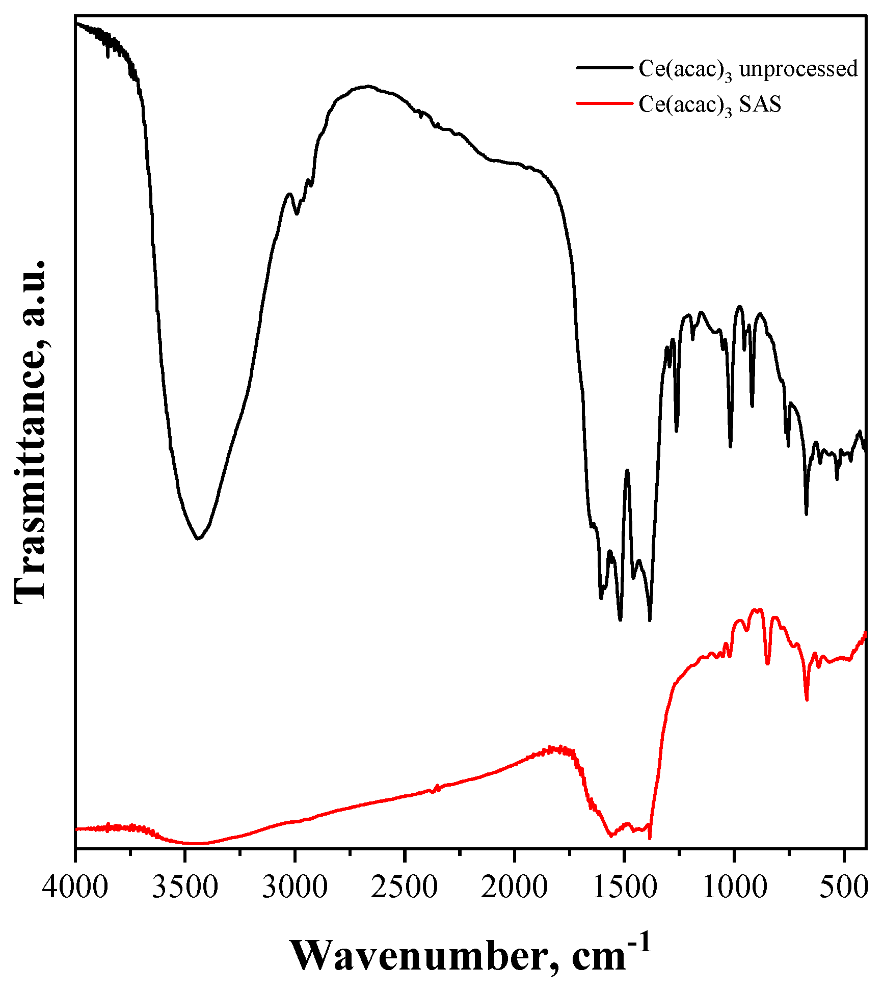
The FT-IR spectra of both samples (reported in Figure 4) exhibit an absorption band at 3500 cm−1 attributed to the stretching mode of the adsorbed water. In addition, the absorption band at around 1600 cm−1 can be attributed to the OH bending vibration of physically adsorbed water molecules [60]. As already reported in the literature, FT-IR spectroscopy shows that the Ce(acac)3 had been modified by the SAS process, because most of the bands of the FT-IR spectrum of the unprocessed Ce(acac)3 were different [61].

3.2. Characterization of CeO2 Particles from Thermal Annealing of Ce(acac)3 Micronized Particles

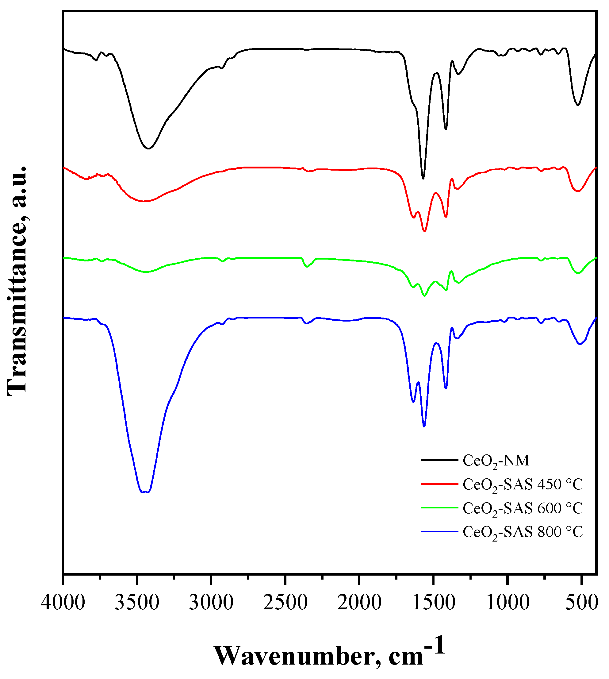
The photocatalysts obtained after thermal annealing of the non-micronized and SAS-processed precursors were characterized using different techniques (FT-IR spectroscopy, Raman spectroscopy, or UV–Vis diffuse reflectance spectroscopy). In the FT-IR spectra of all the CeO2 samples (Figure 5), the widened absorption band in the range of 3000–3750 cm−1 can be attributed to the O–H-stretching vibrations of physisorbed H2O or to surface Ce–OH groups [62]. The absorption features observed at 1410 cm−1 and 1560 cm−1 suggest the presence of carbonate-like species formed by the coordination of CO2 molecules on the CeO2 surface and physically surface-adsorbed CO2, respectively [62,63]. In addition, the FT-IR spectra show a band at about 510 cm−1 attributed to Ce–O stretching modes, corresponding to the vibrations of the oxygen atoms bonded to cerium in the crystal lattice [64]. Noticeably, the band located at about 1620 cm−1, assigned to the bending vibration of the hydroxyl group adsorbed on the surface of the CeO2 [64,65], is more prominent for the CeO2-SAS samples than for CeO2-NM. Since the intensity of this band is associated with the properties of the CeO2 surface, such as the presence of oxygen vacancies, which can enhance the adsorption of hydroxyl groups or water molecules [66,67], it can be inferred that all the CeO2-SAS samples exhibit a higher concentration of surface oxygen vacancies in comparison to the CeO2-NM sample, as corroborated by the Raman spectroscopy findings (vide infra).
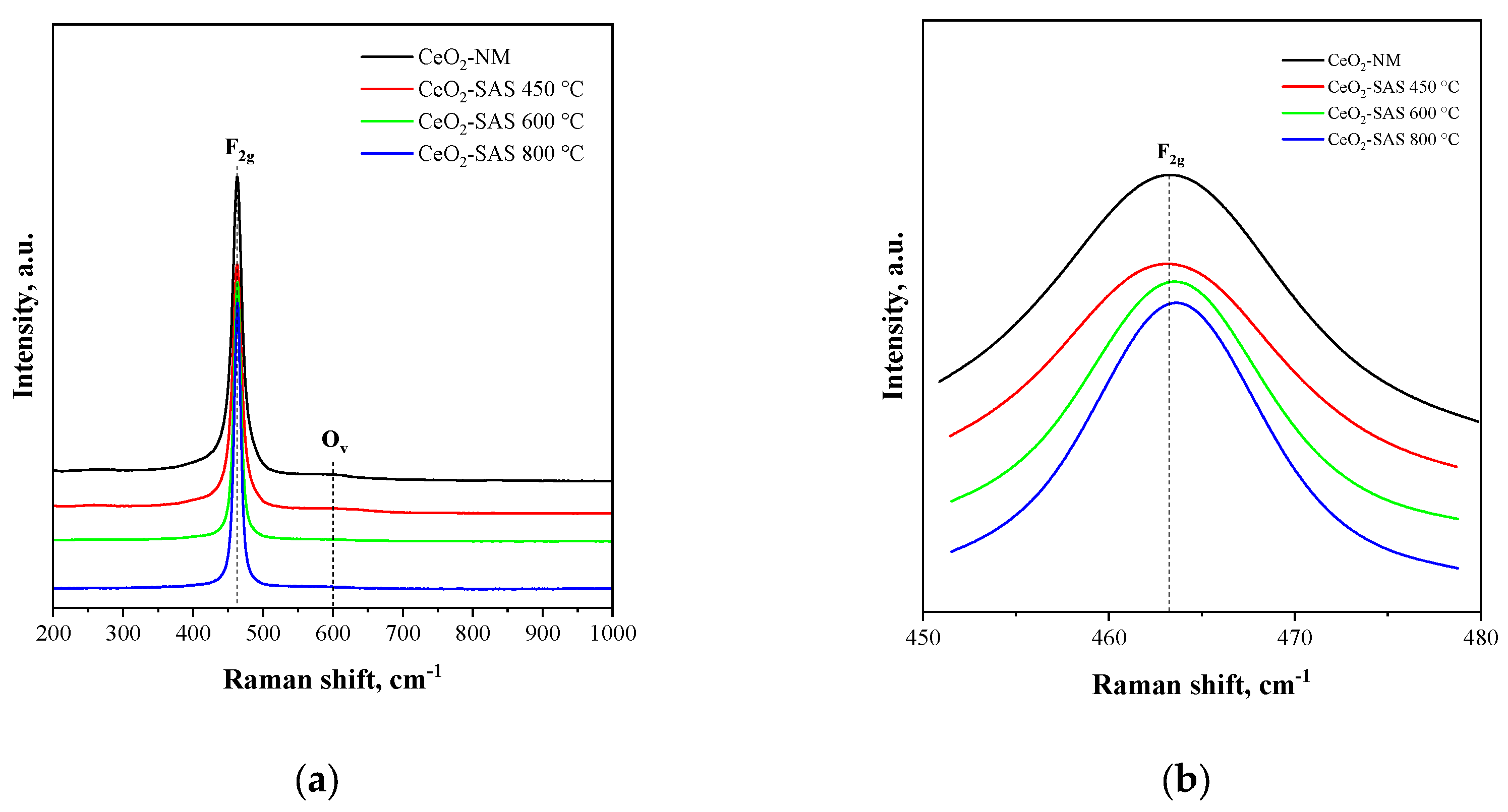
Figure 6a shows the Raman spectra for all the photocatalysts tested in the range of 200–1000 cm−1. The main band at 462 cm−1 corresponds to the F2g symmetric vibrational mode of the oxygen atoms surrounding the Ce4+ ions in the fluorite lattice of pure CeO2 [68], whereas, the weak band detected at about 607 cm−1 is generally attributed to the presence of oxygen vacancies (OV) in the CeO2 lattice [69].
In particular, the variation in the intensity of Ov bands at 607 cm−1 is useful to quantify the transformation from Ce4+ to Ce3+, and the concentration of oxygen vacancies in CeO2 is described using the Ov/F2g peak intensity ratio [70]. The larger the ratio, the higher the oxygen vacancy concentration is. From Figure 6b, it is possible to observe that with the increase of annealing temperature, the position of the F2g band shifts from 462.8 cm−1 at 450 °C to 463.4 cm−1 at 600 °C and 463.7 cm−1 at 800 °C. This is usually caused by the variation of the bond length, phonon relaxation, or stress in the CeO2 nanocrystals [71]. Concurrently, as shown in Table 1, the Ov/F2g ratio diminishes with increasing temperature, resulting in a lower oxygen vacancy concentration for the CeO2-SAS samples synthesized at 600 and 800 °C compared to CeO2-SAS 450 °C. However, no significant differences in the Ov/F2g ratio between CeO2 NM and CeO2-SAS 450 °C are evident.
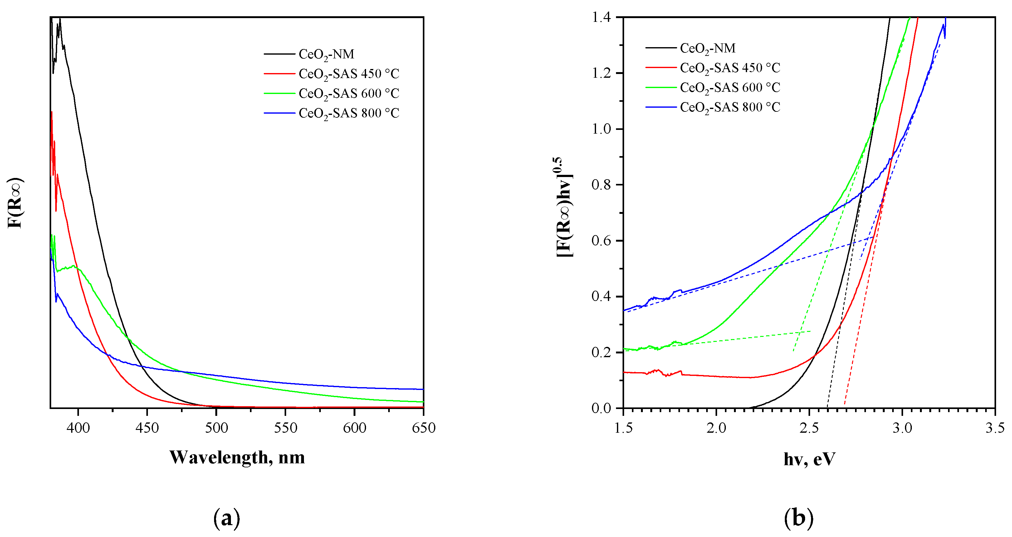
The data obtained from UV–Vis reflectance spectra (Figure 7a) were used for evaluating the optical band gap energy of the photocatalysts by plotting [F(R) × hν]0.5 vs. hν (Figure 7b). The obtained values are reported inTable 1. For the CeO2-SAS samples, at the lowest annealing temperature (450 °C), CeO2 remains mostly in its Ce3⁺ oxidation state, and oxygen vacancies are more abundant than the CeO2-SAS 600 °C and CeO2-SAS 800 °C samples (see Raman results). At the highest temperature (800 °C), the oxygen vacancy concentration becomes very low (see Raman results), meaning that CeO2 is largely in its Ce4⁺ oxidation state. As a result, the band gap energy further increases. However, CeO2-NM has a lower optical band gap than the CeO2-SAS 450 °C sample. This feature can be explained by considering that, in CeO2, the presence of oxygen vacancies facilitates the formation of mixed-valence states of cerium (Ce3⁺ and Ce4⁺), which contribute to the creation of defect states near the Fermi level [72,73,74]. The higher concentration of oxygen vacancies in the CeO2-NM and CeO2-SAS 450 °C samples (Table 1) may result in fewer defect states, leading to a relatively narrower gap for electron excitation. Additionally, it must be considered that oxygen vacancies can modify the crystal lattice and introduce strain, which also affects the band structure [75].
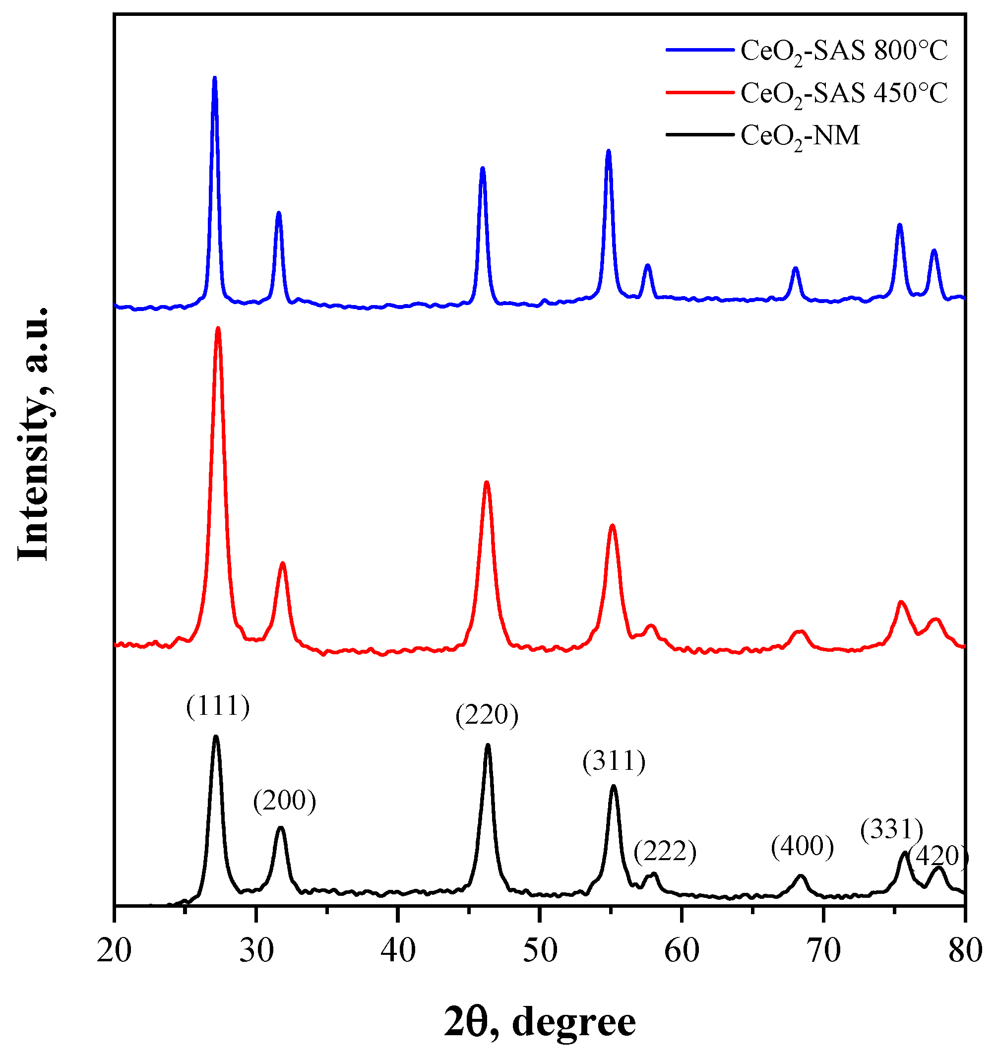
The structural properties of CeO2-NM, CeO2-SAS 450 °C, and CeO2-SAS 800 °C were analyzed through X-ray diffraction (XRD) patterns (Figure 8) and corroborated by crystallite size calculations using the Scherrer equation and specific surface area measurements (Table 2). The observed diffraction peaks can be indexed to the fluorite cubic structure of cerium dioxide (CeO2) [76,77], in agreement with the standard JCPDS for CeO2 (card n° 34-0394). Prominent peaks at 2θ values of approximately 28.5°, 33.0°, 47.5°, 56.5°, 59.1°, 69.5°, and 76.8° are assigned to the (111), (200), (220), (311), (222), (400), and (420) planes, respectively, confirming the cubic fluorite phase. Significant differences in peak intensity and width were observed among the samples, reflecting variations in crystallinity and crystallite size. CeO2-NM displays the broadest peaks, indicative of the smallest crystallite size (8 nm, as calculated using the Scherrer equation) and the highest specific surface area (43 m2·g⁻1). For CeO2-SAS 450 °C, the XRD peaks are slightly sharper, corresponding to a moderate increase in crystallite size to 9 nm and a decrease in specific surface area to 34 m2·g⁻1. This indicates partial grain growth during calcination at 450 °C while preserving a relatively high surface area. The CeO2-SAS 800 °C sample shows the sharpest XRD peaks, reflecting the largest crystallite size (17 nm) and the lowest specific surface area (8 m2·g⁻1). This behavior is attributed to significant grain growth and sintering during high-temperature calcination, which enhances crystallinity while substantially reducing surface area. The interplay between crystallite size and specific surface area highlights the critical role of thermal treatment in determining the structural and morphological characteristics of CeO2. Higher calcination temperatures promote crystallinity and reduce lattice strain, as evidenced by the sharper diffraction peaks, but also cause sintering and a corresponding decrease in surface area.

3.3. Photocatalytic Activity Results

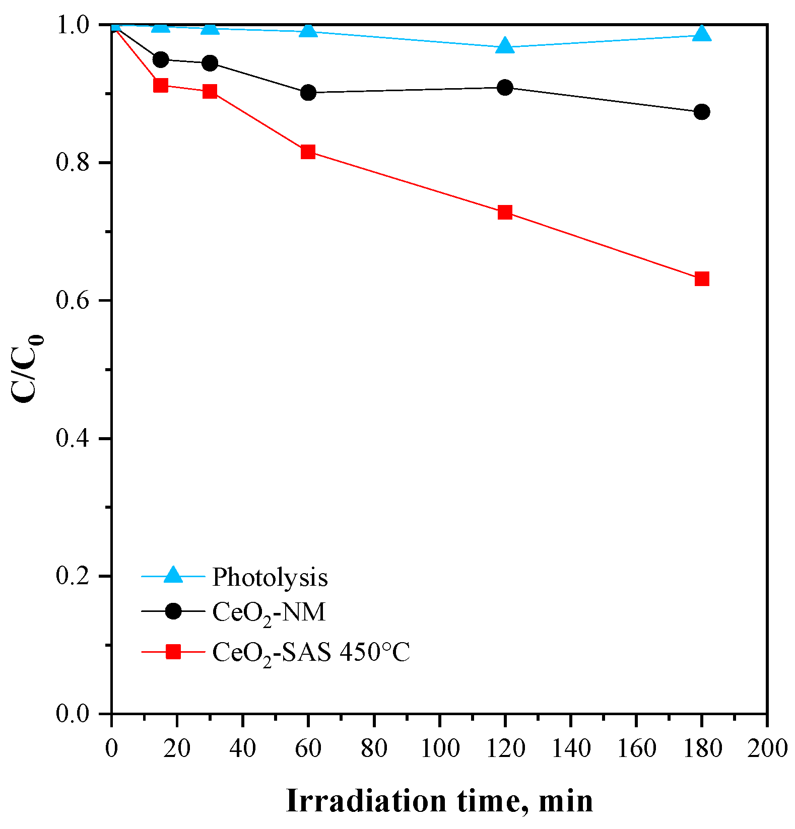
The initial experiments aimed to compare the performance of the catalyst obtained from the micronized precursor with that of the catalyst derived from the untreated precursor. In this case, 300 mg of catalyst was dispersed in 100 mL of chloramphenicol solution, with an antibiotic concentration of 25 mg L−1. The graph reported in Figure 9 illustrates the relative concentration of chloramphenicol (C/C0) as a function of irradiation time. The dataset compares photolysis, CeO2-NM, and CeO2-SAS 450 °C over a period of 180 min of treatment time. The photolysis process shows negligible degradation, as the relative concentration remained close to 1 throughout the experiment. Conversely, the CeO2-NM sample exhibits moderate photocatalytic activity, reaching approximately 10% of CAP degradation after 180 min of irradiation. Notably, the CeO2-SAS 450 °C catalyst demonstrates the highest photocatalytic activity since, at the end of the irradiation period, the CAP degradation was 37%, indicating a superior performance compared to the CeO2-NM photocatalyst.
This outcome may result from the difference in morphology and size between the two photocatalysts. For this reason, the particle size was analyzed and the results for hydrodynamic diameters in solution (deionized water) are presented in Table 3. For CeO2-SAS 450 °C, the hydrodynamic diameter was observed to be 313.8 nm while the hydrodynamic diameter increased to 621.6 nm for CeO2-NM.
From Figure 10, it is possible to observe that the CeO2-SAS 450 °C sample shows predominantly spherical nanoparticles that are non-uniform in size. In contrast, the CeO2-NM photocatalyst, obtained from the non-micronized precursor, shows a compact and non-micrometric structure.
A higher particle size value indicates that the CeO2-NM particles tend to agglomerate, forming larger clusters when dispersed in water. This aggregation limits the surface available for the contaminant, potentially affecting the efficiency of the catalytic process [78].

3.3.1. Effect of Annealing Temperature on CeO2-SAS Photocatalysts

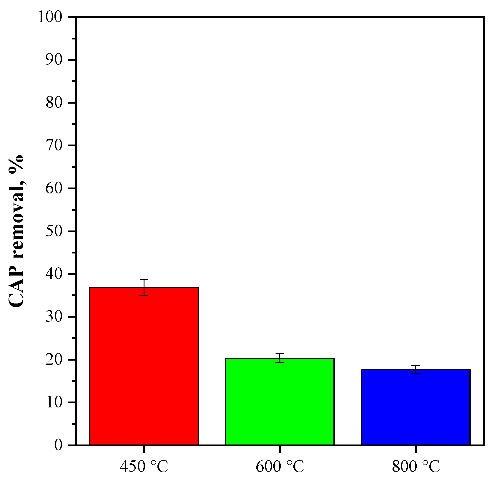
The effect of the annealing temperature on the photocatalytic activity was also evaluated in the CeO2-SAS samples since it can affect the CAP degradation performance. The results obtained are reported in Figure 11.
The CeO2-SAS 450 °C photocatalyst exhibited superior photocatalytic activity compared to the CeO2-SAS 600 °C and CeO2-SAS 800 °C photocatalysts. In detail, a CAP removal of 37% was obtained for the sample annealed at 450 °C, and it was reduced to 20 and 17% for the CeO2-SAS samples annealed at 600 and 800 °C, respectively. This result could be due to CeO2-SAS 450 °C’s higher concentration of oxygen vacancies (as evinced both from the FT-IR and Raman results), which significantly enhances the photocatalytic properties. More in detail, at a 450 °C annealing temperature, CeO2-SAS retained a higher concentration of oxygen vacancies because the formation of vacancies is favored at lower temperatures, where the oxygen loss from the lattice is less pronounced [79,80,81]. These oxygen vacancies act as shallow traps for electrons, creating a high density of reactive species, like superoxide radicals, that can degrade the target pollutant more effectively under light irradiation [82,83,84]. Indeed, the photocatalytic tests in the presence of scavenger molecules evidenced that the superoxide radical is the main reactive oxygen species involved in the CAP degradation mechanism (vide infra). In contrast, higher annealing temperatures (600 °C and 800 °C) led to a reduction in the number of oxygen vacancies, as the elevated temperatures promote the reordering of oxygen atoms in the CeO2 lattice, reducing the semiconductor defect density [81,85]. Consequently, the photocatalytic activity of CeO2-SAS 600 °C and CeO2-SAS 800 °C was lower because the reduced number of oxygen vacancies led to less efficient electron–hole separation and fewer active sites for the photocatalytic reaction.

3.3.2. Effect of CeO2-SAS 450 °C Dosage on CAP Photocatalytic Degradation

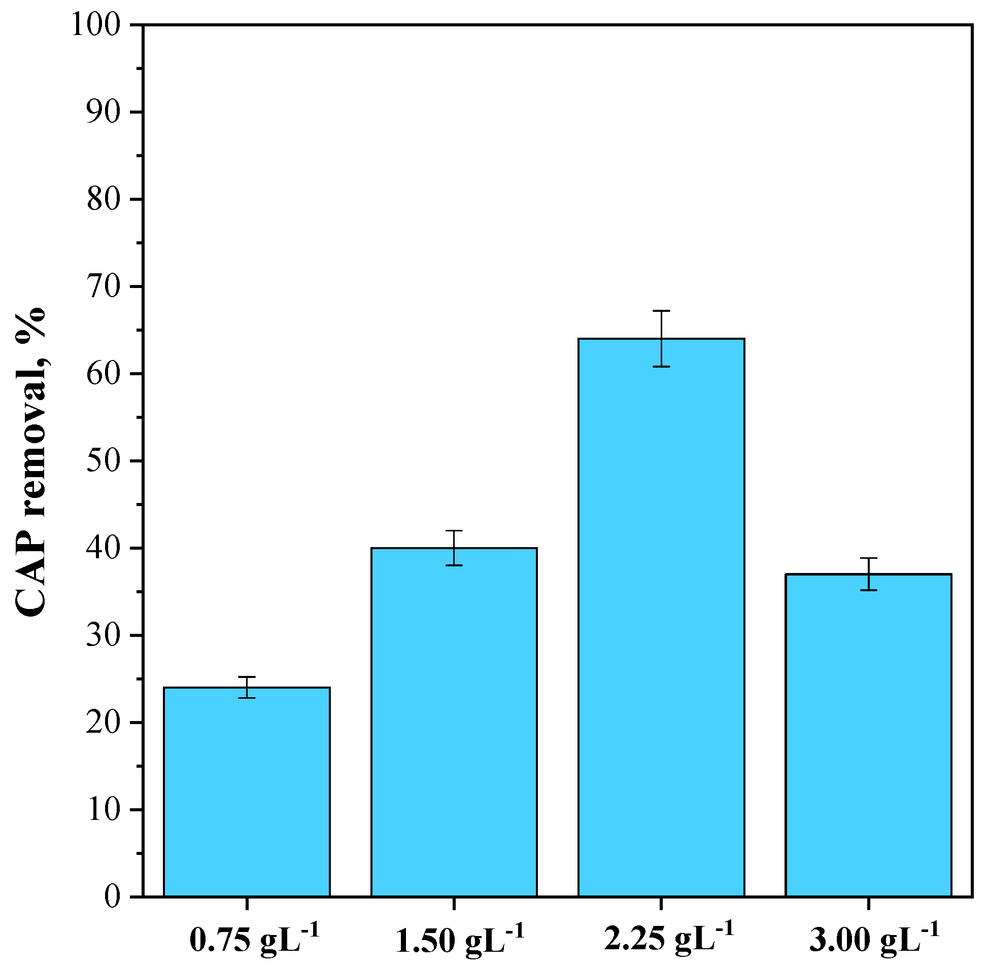
To investigate the impact of photocatalyst dosage, photocatalytic experiments were conducted using a fixed CAP concentration equal to 25 mg L−1 and varying the CeO2-SAS 450 °C dosage from 0.75 g L−1 to 3 g L−1. The results obtained are reported in Figure 12. It was noted that at the lowest investigated photocatalyst dosage (0.75 g L−1), the CAP degradation was only 24% after 180 min of visible light irradiation. However, by increasing the photocatalyst dosage to 1.50 g L−1, the CAP degradation increased to 40%, reaching the maximum value of 64% when the photocatalyst dosage was equal to 2.25 g L−1. Furthermore, at a higher dosage of 3 g L−1, the CAP degradation decreased to 37%.
At lower CeO2-SAS 450 °C dosages (in the range of 0.75–2.25 g L−1), the photocatalytic performance increased as more active sites on the photocatalyst surface became available to absorb visible light and interact with the CAP molecules, leading to an increase in the degradation efficiency [30]. However, when the CeO2-SAS 450 °C dosage was increased to 3 g L−1, the photocatalytic efficiency declined because the excess of photocatalyst particles turbid the suspension, reducing the effective exposure of each particle to light [86,87].

3.3.3. Influence of CAP Initial Concentration on CeO2-SAS 450 °C Photocatalytic Performance

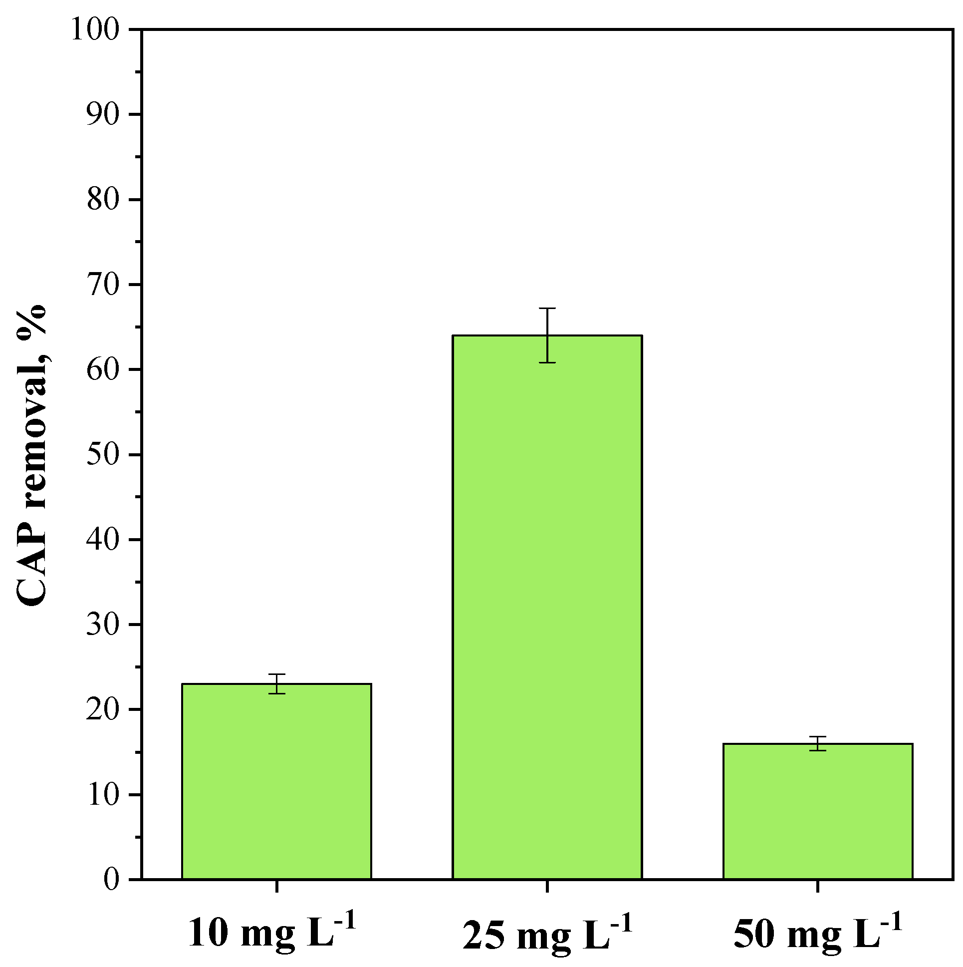
To investigate the impact of the CAP initial concentration on the photocatalytic degradation efficiency, experiments were carried out using the optimized CeO2-SAS 450 °C dosage (2.25 g L−1) and varying the antibiotic concentration from 10 mg L−1 to 50 mg L−1. Figure 13 illustrates that the CAP removal rose with an increasing initial concentration within the 10–25 mg L−1 range but declined at a higher CAP concentration (50 mg L−1). Previous studies have indicated that as pollutant concentration increases, the demand for reactive species to degrade the antibiotic also increases. Nevertheless, under fixed conditions of light intensity, photocatalyst loading, and irradiation time, the generation of radical species remains stable. Consequently, the available quantity of reactive species can become insufficient to degrade the pollutant effectively [88]. Moreover, at high initial CAP concentration, the possible accumulation of reaction intermediates can hinder the photocatalytic process. Indeed, these intermediates, which are generated during the partial degradation of CAP, may compete for active sites on the photocatalyst surface, diminishing the accessibility of these sites for further degradation of the parent compound [89]. This competition can result in decreased photocatalytic activity, as the generated intermediates may adsorb onto the photocatalyst surface, restricting the adsorption of additional CAP molecules from the liquid phase.

3.3.4. Role of Reactive Oxygen Species (ROS) on CAP Photodegradation Catalyzed by CeO2-SAS 450 °C

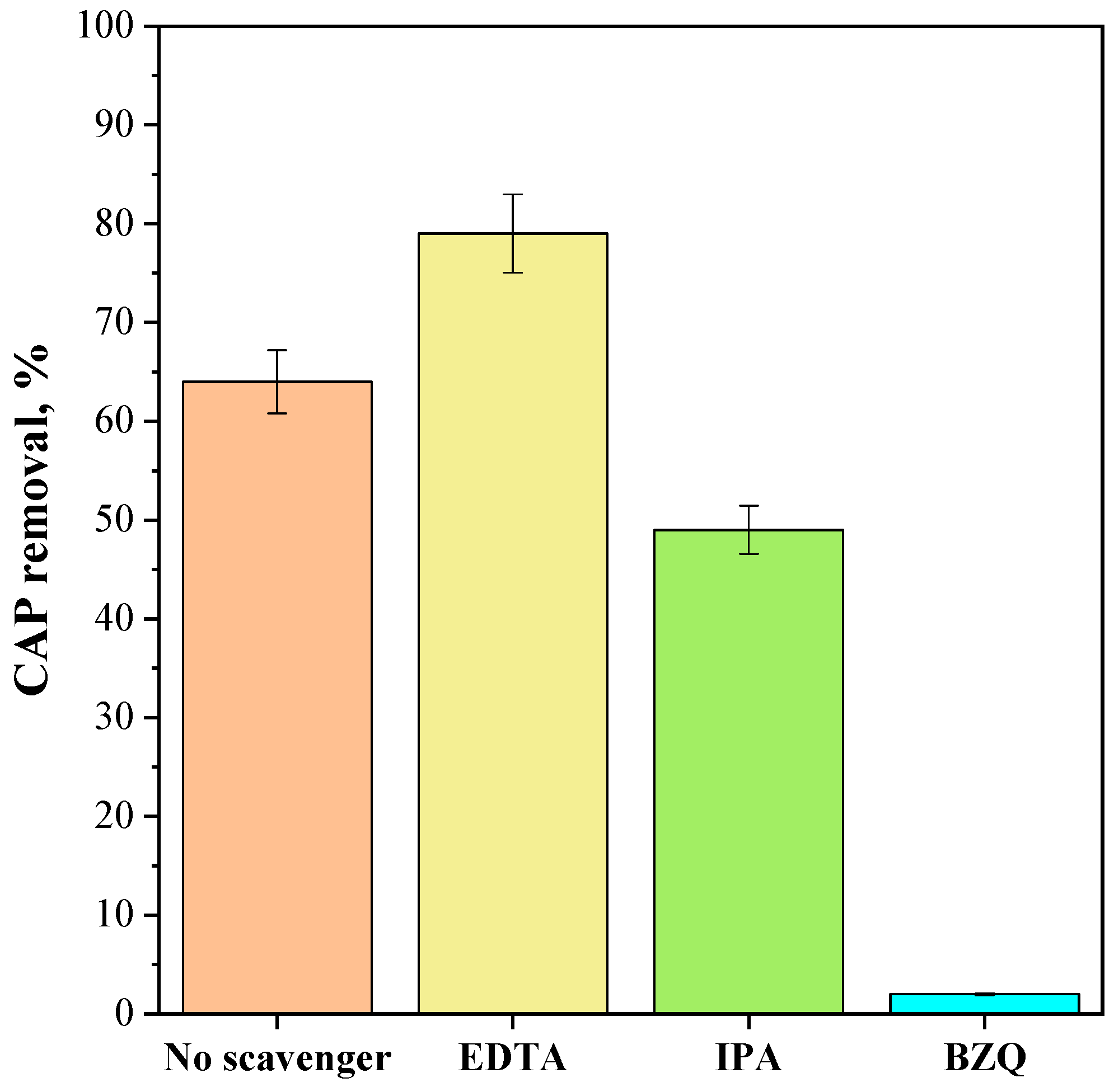
Further photocatalytic experiments were carried out to examine the potential impact of reactive oxygen species (ROS) on the degradation of CAP in the presence of the CeO2-SAS 450 °C photocatalyst. Specifically, photocatalytic degradation tests were performed using EDTA, benzoquinone (BZQ), and isopropyl alcohol (IPA) as scavengers for holes [90], superoxide [91,92], and hydroxyl radicals [93], respectively. To this end, under optimal reaction conditions (photocatalyst dosage: 2.25 g L−1; initial CAP concentration: 25 mg L−1), EDTA (at a concentration of 10 mmol L−1), BQ (at a concentration of 1 μmol L−1), and IPA (at a concentration of 10 mmol L−1) were added to the CAP solution. Figure 14 illustrates the effects of the scavenger’s presence on photocatalytic performances. The presence of IPA, BQ, and EDTA affected the photocatalytic degradation of CAP. In particular, IPA slightly decreased CAP removal, while BQ nearly completely suppressed the photocatalytic reaction. These results suggest that superoxide radicals are the primary reactive oxygen species responsible for CAP degradation, followed by hydroxyl radicals. On the other hand, in the presence of EDTA, the degradation ability of the catalyst increased. It could be argued that when EDTA traps holes in the semiconductor valence band, electron–hole pair separation is enhanced, leading to improved photocatalytic activity. This is because more electrons in the photocatalyst conduction band are available to reduce molecular oxygen into superoxide radicals. Overall, the test with EDTA further corroborates the crucial role of superoxide radicals in CAP degradation, aligning with the Raman results indicating the highest oxygen vacancy concentration in the CeO2-SAS 450 °C photocatalyst.

3.3.5. Stability Tests on CeO2-SAS 450 °C Photocatalyst

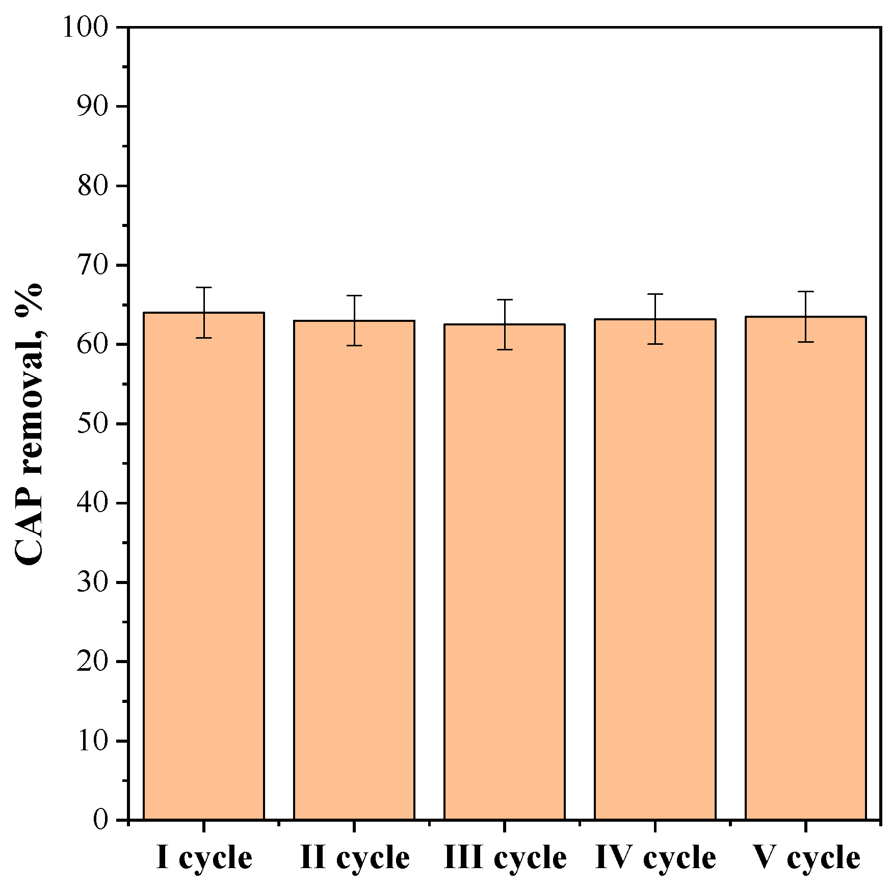
Additional photocatalytic tests were carried out to evaluate the stability of the CeO2-SAS 450 °C photocatalyst that displayed the highest photocatalytic activity. Specifically, the experiments were performed using a catalyst dosage of 2.25 g L−1, with an initial CAP concentration of 25 mg L−1. The photocatalyst was recovered from the suspension after each test, cleaned with distilled water, and reused again in a subsequent test with the same operating conditions. The results, after five reuse cycles, are reported in Figure 15, in terms of CAP removal after 180 min of visible light irradiation. The obtained results demonstrated the stability of the photocatalyst as its photocatalytic efficiency remained consistent throughout five consecutive reuse cycles.

4. Conclusions

In this study, CeO2 photocatalysts were synthesized via the supercritical antisolvent (SAS) technique and subsequently evaluated for their photocatalytic degradation of chloramphenicol. The SAS method enabled the production of CeO2 nanoparticles with enhanced photocatalytic activity compared to those derived from the unprocessed precursor. The influence of annealing temperature was also investigated, revealing that temperatures exceeding 450 °C led to a decrease in CeO2 oxygen vacancies, a widening of the band gap, and consequently, diminished photocatalytic performance. Further photocatalytic experiments were conducted to explore the impact of the chloramphenicol initial concentration and photocatalyst dosage. Specifically, employing a catalyst dosage of 2.25 g L−1 and an antibiotic concentration of 25 mg L−1, a degradation efficiency of 64% was achieved. Various scavengers were utilized to propose a plausible degradation mechanism, indicating that the superoxide radical is the primary species responsible for chloramphenicol degradation. Moreover, the optimized photocatalyst exhibited excellent recyclability and reusability over five consecutive reuse cycles. This preliminary study aimed to assess the feasibility of using the SAS method to develop novel CeO2-based photocatalysts with improved visible light absorption and catalytic activity for future research.

Author Contributions

Conceptualization, V.V. and I.D.M.; methodology, V.V. and I.D.M.; investigation, A.M., M.C.I. and A.P.; data curation, A.M., M.C.I. and S.M.; writing—original draft preparation, A.M., M.C.I., A.P. and S.M.; writing—review and editing, V.V. and I.D.M.; supervision, V.V. and I.D.M. All authors have read and agreed to the published version of the manuscript.

Funding

This work was partially realized in the framework of the project: Removal of Emerging Halogenated Pollutants by Advanced Oxidation Processes: A Multiapproach Assessment (REHAPOAP) with the technical and economic support of the Italian Ministero dell’Università e della Ricerca (Investimento 1.1 “Progetti di Ricerca di Rilevante Interesse Nazionale PRIN PNRR 2022”; codice del Progetto: P2022P3ENB).

Data Availability Statement

The data presented in this study are available on request from the corresponding authors.

Conflicts of Interest

The authors declare no conflicts of interest.

References

  1. Ribeiro da Cunha, B.; Fonseca, L.P.; Calado, C.R. Antibiotic discovery: Where have we come from, where do we go? Antibiotics 2019, 8, 45. [Google Scholar] [CrossRef] [PubMed]
  2. Tiwari, B.; Sellamuthu, B.; Ouarda, Y.; Drogui, P.; Tyagi, R.D.; Buelna, G. Review on fate and mechanism of removal of pharmaceutical pollutants from wastewater using biological approach. Bioresour. Technol. 2017, 224, 1–12. [Google Scholar] [CrossRef]
  3. Uluseker, C.; Kaster, K.M.; Thorsen, K.; Basiry, D.; Shobana, S.; Jain, M.; Kumar, G.; Kommedal, R.; Pala-Ozkok, I. A review on occurrence and spread of antibiotic resistance in wastewaters and in wastewater treatment plants: Mechanisms and perspectives. Front. Microbiol. 2021, 12, 717809. [Google Scholar] [CrossRef] [PubMed]
  4. Välitalo, P.; Kruglova, A.; Mikola, A.; Vahala, R. Toxicological impacts of antibiotics on aquatic micro-organisms: A mini-review. Int. J. Hyg. Environ. Health 2017, 220, 558–569. [Google Scholar] [CrossRef]
  5. Kovalakova, P.; Cizmas, L.; McDonald, T.J.; Marsalek, B.; Feng, M.; Sharma, V.K. Occurrence and toxicity of antibiotics in the aquatic environment: A review. Chemosphere 2020, 251, 126351. [Google Scholar] [CrossRef] [PubMed]
  6. Kumar, K.; Gupta, S.C.; Chander, Y.; Singh, A.K. Antibiotic use in agriculture and its impact on the terrestrial environment. Adv. Agron. 2005, 87, 1–54. [Google Scholar]
  7. Balbi, H.J. Chloramphenicol: A review. Pediatr. Rev. 2004, 25, 284–288. [Google Scholar] [CrossRef]
  8. Weisberger, A.S.; Wessler, S.; Avioli, L.V. Mechanisms of action of chloramphenicol. JAMA 1969, 209, 97–103. [Google Scholar] [CrossRef]
  9. Chu, W.; Krasner, S.W.; Gao, N.; Templeton, M.R.; Yin, D. Contribution of the antibiotic chloramphenicol and its analogues as precursors of dichloroacetamide and other disinfection byproducts in drinking water. Environ. Sci. Technol. 2016, 50, 388–396. [Google Scholar] [CrossRef]
  10. Falyouna, O.; Maamoun, I.; Ghosh, S.; Malloum, A.; Othmani, A.; Eljamal, O.; Amen, T.W.; Oroke, A.; Bornman, C.; Ahmadi, S. Sustainable technologies for the removal of Chloramphenicol from pharmaceutical industries Effluent: A critical review. J. Mol. Liq. 2022, 368, 120726. [Google Scholar] [CrossRef]
  11. Nguyen, L.M.; Nguyen, N.T.T.; Nguyen, T.T.T.; Nguyen, T.T.; Nguyen, D.T.C.; Tran, T.V. Occurrence, toxicity and adsorptive removal of the chloramphenicol antibiotic in water: A review. Environ. Chem. Lett. 2022, 20, 1929–1963. [Google Scholar] [CrossRef]
  12. Lin, J.; Zhang, K.; Jiang, L.; Hou, J.; Yu, X.; Feng, M.; Ye, C. Removal of chloramphenicol antibiotics in natural and engineered water systems: Review of reaction mechanisms and product toxicity. Sci. Total Environ. 2022, 850, 158059. [Google Scholar] [CrossRef] [PubMed]
  13. Liu, X.; Cao, Z.; Yuan, Z.; Zhang, J.; Guo, X.; Yang, Y.; He, F.; Zhao, Y.; Xu, J. Insight into the kinetics and mechanism of removal of aqueous chlorinated nitroaromatic antibiotic chloramphenicol by nanoscale zero-valent iron. Chem. Eng. J. 2018, 334, 508–518. [Google Scholar] [CrossRef]
  14. Singh, A.; Ramachandran, S.K.; Gumpu, M.B.; Zsuzsanna, L.; Veréb, G.; Kertész, S.; Gangasalam, A. Titanium dioxide doped hydroxyapatite incorporated photocatalytic membranes for the degradation of chloramphenicol antibiotic in water. J. Chem. Technol. Biotechnol. 2021, 96, 1057–1066. [Google Scholar] [CrossRef]
  15. Chen, Y.; Yang, J.; Zeng, L.; Zhu, M. Recent progress on the removal of antibiotic pollutants using photocatalytic oxidation process. Crit. Rev. Environ. Sci. Technol. 2022, 52, 1401–1448. [Google Scholar] [CrossRef]
  16. Kokkinos, P.; Venieri, D.; Mantzavinos, D. Advanced oxidation processes for water and wastewater viral disinfection. A systematic review. Food Environ. Virol. 2021, 13, 283–302. [Google Scholar] [CrossRef]
  17. Hodges, B.C.; Cates, E.L.; Kim, J.-H. Challenges and prospects of advanced oxidation water treatment processes using catalytic nanomaterials. Nat. Nanotechnol. 2018, 13, 642–650. [Google Scholar] [CrossRef]
  18. Mukherjee, J.; Lodh, B.K.; Sharma, R.; Mahata, N.; Shah, M.P.; Mandal, S.; Ghanta, S.; Bhunia, B. Advanced oxidation process for the treatment of industrial wastewater: A review on strategies, mechanisms, bottlenecks and prospects. Chemosphere 2023, 345, 140473. [Google Scholar] [CrossRef]
  19. Vaiano, V.; Sacco, O.; Sannino, D.; Ciambelli, P. Process intensification in the removal of organic pollutants from wastewater using innovative photocatalysts obtained coupling Zinc Sulfide based phosphors with nitrogen doped semiconductors. J. Clean. Prod. 2015, 100, 208–211. [Google Scholar] [CrossRef]
  20. Sacco, O.; Franco, P.; De Marco, I.; Vaiano, V.; Callone, E.; Ceccato, R.; Parrino, F. Photocatalytic activity of Eu-doped ZnO prepared by supercritical antisolvent precipitation route: When defects become virtues. J. Mater. Sci. Technol. 2022, 112, 49–58. [Google Scholar] [CrossRef]
  21. Pirilä, M.; Saouabe, M.; Ojala, S.; Rathnayake, B.; Drault, F.; Valtanen, A.; Huuhtanen, M.; Brahmi, R.; Keiski, R.L. Photocatalytic degradation of organic pollutants in wastewater. Top. Catal. 2015, 58, 1085–1099. [Google Scholar] [CrossRef]
  22. Dong, C.; Fang, W.; Yi, Q.; Zhang, J. A comprehensive review on reactive oxygen species (ROS) in advanced oxidation processes (AOPs). Chemosphere 2022, 308, 136205. [Google Scholar] [CrossRef]
  23. Zhang, J.; Tian, B.; Wang, L.; Xing, M.; Lei, J. Photocatalysis. In Lecture Notes in Chemistry; Springer: Singapore, 2018; pp. 1–17. [Google Scholar]
  24. Lu, Y.; Cai, Y.; Zhang, S.; Zhuang, L.; Hu, B.; Wang, S.; Chen, J.; Wang, X. Application of biochar-based photocatalysts for adsorption-(photo) degradation/reduction of environmental contaminants: Mechanism, challenges and perspective. Biochar 2022, 4, 45. [Google Scholar] [CrossRef]
  25. Abebe, B.; Murthy, H.A.; Amare, E. Summary on adsorption and photocatalysis for pollutant remediation: Mini review. J. Encapsul. Adsorpt. Sci. 2018, 8, 225–255. [Google Scholar] [CrossRef]
  26. Qumar, U.; Hassan, J.Z.; Bhatti, R.A.; Raza, A.; Nazir, G.; Nabgan, W.; Ikram, M. Photocatalysis vs adsorption by metal oxide nanoparticles. J. Mater. Sci. Technol. 2022, 131, 122–166. [Google Scholar] [CrossRef]
  27. Bruckmann, F.S.; Schnorr, C.; Oviedo, L.R.; Knani, S.; Silva, L.F.; Silva, W.L.; Dotto, G.L.; Bohn Rhoden, C.R. Adsorption and photocatalytic degradation of pesticides into nanocomposites: A review. Molecules 2022, 27, 6261. [Google Scholar] [CrossRef] [PubMed]
  28. Mancuso, A.; Kiani, A.; Sacco, O.; Lettieri, M.; Fittipaldi, R.; Vaiano, V.; Acocella, M.R.; Venditto, V. Ball–milled TiO2/biochar hybrid system as a heterogeneous photocatalyst for tannery dyes removal in aqueous solution. J. Mol. Liq. 2024, 399, 124357. [Google Scholar] [CrossRef]
  29. Chatzitakis, A.; Berberidou, C.; Paspaltsis, I.; Kyriakou, G.; Sklaviadis, T.; Poulios, I. Photocatalytic degradation and drug activity reduction of chloramphenicol. Water Res. 2008, 42, 386–394. [Google Scholar] [CrossRef]
  30. Lofrano, G.; Libralato, G.; Adinolfi, R.; Siciliano, A.; Iannece, P.; Guida, M.; Giugni, M.; Ghirardini, A.V.; Carotenuto, M. Photocatalytic degradation of the antibiotic chloramphenicol and effluent toxicity effects. Ecotoxicol. Environ. Saf. 2016, 123, 65–71. [Google Scholar] [CrossRef]
  31. Giri, A.S.; Golder, A.K. Mechanism and identification of reaction byproducts for the degradation of Chloramphenicol drug in heterogeneous photocatalytic process. Groundw. Sustain. Dev. 2018, 7, 343–347. [Google Scholar] [CrossRef]
  32. Zhang, Y.; Shao, Y.; Gao, N.; Gao, Y.; Chu, W.; Li, S.; Wang, Y.; Xu, S. Kinetics and by-products formation of chloramphenicol (CAP) using chlorination and photocatalytic oxidation. Chem. Eng. J. 2018, 333, 85–91. [Google Scholar] [CrossRef]
  33. Arora, I.; Chawla, H.; Chandra, A.; Sagadevan, S.; Garg, S. Advances in the strategies for enhancing the photocatalytic activity of TiO2: Conversion from UV-light active to visible-light active photocatalyst. Inorg. Chem. Commun. 2022, 143, 109700. [Google Scholar] [CrossRef]
  34. Kumar, S.G.; Devi, L.G. Review on modified TiO2 photocatalysis under UV/visible light: Selected results and related mechanisms on interfacial charge carrier transfer dynamics. J. Phys. Chem. A 2011, 115, 13211–13241. [Google Scholar] [CrossRef] [PubMed]
  35. Etacheri, V.; Di Valentin, C.; Schneider, J.; Bahnemann, D.; Pillai, S.C. Visible-light activation of TiO2 photocatalysts: Advances in theory and experiments. J. Photochem. Photobiol. C Photochem. Rev. 2015, 25, 1–29. [Google Scholar] [CrossRef]
  36. Magalhaes, P.; Andrade, L.; Nunes, O.C.; Mendes, A. Titanium dioxide photocatalysis: Fundamentals and application on photoinactivation. Rev. Adv. Mater. Sci. 2017, 51, 91–129. [Google Scholar]
  37. Chien, C.-W.; Dhenadhayalan, N.; Lin, K.-C. Implementation of visible light-driven photocatalytic degradation of antibiotic chloramphenicol using Bi2S3/ZrO2 and Bi2WO6/ZrO2 heteronanostructures. J. Environ. Chem. Eng. 2023, 11, 110613. [Google Scholar] [CrossRef]
  38. Leeladevi, K.; Kumar, J.V.; Arunpandian, M.; Thiruppathi, M.; Nagarajan, E. Investigation on photocatalytic degradation of hazardous chloramphenicol drug and amaranth dye by SmVO4 decorated g-C3N4 nanocomposites. Mater. Sci. Semicond. Process. 2021, 123, 105563. [Google Scholar] [CrossRef]
  39. Wang, T.; Shi, H.; Kumar, A.; Zhang, D.; Wang, H.; Wang, S.; Zheng, J. Efficient visible-light photocatalysis of chloramphenicol using novel engineered biochar-based Ti-doped Bi2WO6 composite: Mechanisms, degradation pathways, and applications. Sep. Purif. Technol. 2024, 332, 125780. [Google Scholar] [CrossRef]
  40. Wu, Y.; Mao, S.; Liu, C.; Pei, F.; Wang, F.; Hao, Q.; Xia, M.; Lei, W. Enhanced degradation of chloramphenicol through peroxymonosulfate and visible light over Z-scheme Photocatalysts: Synergetic performance and mechanism insights. J. Colloid Interface Sci. 2022, 608, 322–333. [Google Scholar] [CrossRef] [PubMed]
  41. Zhu, C.-Z.; Tian, Q.-H.; Wang, B.-H.; Xu, M.-T.; Jin, Q.-J.; Zhang, Z.-Y.; Le, S.-K.; Wu, Y.; Wei, Y.-C.; Xu, H.-T. Application of modified cerium dioxide for photocatalytic air pollution purification. Rare Met. 2024, 43, 5473–5486. [Google Scholar] [CrossRef]
  42. Huang, X.; Zhang, K.; Peng, B.; Wang, G.; Muhler, M.; Wang, F. Ceria-based materials for thermocatalytic and photocatalytic organic synthesis. ACS Catal. 2021, 11, 9618–9678. [Google Scholar] [CrossRef]
  43. Zhao, W.; Dong, Q.; Sun, C.; Xia, D.; Huang, H.; Yang, G.; Wang, G.; Leung, D.Y. A novel Au/g-C3N4 nanosheets/CeO2 hollow nanospheres plasmonic heterojunction photocatalysts for the photocatalytic reduction of hexavalent chromium and oxidation of oxytetracycline hydrochloride. Chem. Eng. J. 2021, 409, 128185. [Google Scholar] [CrossRef]
  44. Choudhury, B.; Chetri, P.; Choudhury, A. Oxygen defects and formation of Ce3+ affecting the photocatalytic performance of CeO2 nanoparticles. RSC Adv. 2014, 4, 4663–4671. [Google Scholar] [CrossRef]
  45. Tran, D.P.; Pham, M.-T.; Bui, X.-T.; Wang, Y.-F.; You, S.-J. CeO2 as a photocatalytic material for CO2 conversion: A review. Sol. Energy 2022, 240, 443–466. [Google Scholar] [CrossRef]
  46. Kusmierek, E. A CeO2 semiconductor as a photocatalytic and photoelectrocatalytic material for the remediation of pollutants in industrial wastewater: A review. Catalysts 2020, 10, 1435. [Google Scholar] [CrossRef]
  47. Yan, Y.-Q.; Wu, Y.; Wu, Y.-H.; Weng, Z.; Liu, S.-J.; Liu, Z.-G.; Lu, K.-Q.; Han, B. Recent Developments of CeO2-based Photocatalysts: Synthesis and Photocatalytic Applications. Chemsuschem 2024, 17, e202301778. [Google Scholar] [CrossRef]
  48. Roggenbuck, J.; Schäfer, H.; Tsoncheva, T.; Minchev, C.; Hanss, J.; Tiemann, M. Mesoporous CeO2: Synthesis by nanocasting, characterisation and catalytic properties. Microporous Mesoporous Mater. 2007, 101, 335–341. [Google Scholar] [CrossRef]
  49. Hu, C.; Zhang, Z.; Liu, H.; Gao, P.; Wang, Z.L. Direct synthesis and structure characterization of ultrafine CeO2 nanoparticles. Nanotechnology 2006, 17, 5983. [Google Scholar] [CrossRef]
  50. Pundir, S.; Priya, R.; Singh, K.; Kaur, H.; Choudhary, P. A systematic study on synthesis of CeO2 nanoparticles by various routes. In Proceedings of the IOP Conference Series: Earth and Environmental Science, Mohali, India, 23–24 June 2023; p. 012030. [Google Scholar]
  51. Tabernero, A.; Guastaferro, M.; Gonzalez-Garcinuno, A.; Misol, A.; Baldino, L.; Cardea, S.; del Valle, E.M.; Reverchon, E. The viscoelastic behavior of the precursor hydrogels can modify aerogel properties. J. Supercrit. Fluids 2022, 184, 105563. [Google Scholar] [CrossRef]
  52. Guastaferro, M.; Cardea, S.; Baldino, L.; Reverchon, E. Cellulose acetate nanocarrier production by supercritical assisted electrospray. Chem. Eng. Trans. 2021, 87, 391–396. [Google Scholar]
  53. Franco, P.; Sacco, O.; Vaiano, V.; De Marco, I. Supercritical carbon dioxide-based processes in photocatalytic applications. Molecules 2021, 26, 2640. [Google Scholar] [CrossRef] [PubMed]
  54. Iannaco, M.C.; Mottola, S.; Vaiano, V.; Iervolino, G.; De Marco, I. CeO2-CuO composites prepared via supercritical antisolvent precipitation for photocatalytic hydrogen production from lactic acid aqueous solution. J. CO2 Util. 2024, 85, 102878. [Google Scholar] [CrossRef]
  55. De Marco, I.; Reverchon, E. Supercritical antisolvent micronization of cyclodextrins. Powder Technol. 2008, 183, 239–246. [Google Scholar] [CrossRef]
  56. Franco, P.; Navarra, W.; Sacco, O.; De Marco, I.; Mancuso, A.; Vaiano, V.; Venditto, V. Photocatalytic degradation of atrazine under visible light using Gd-doped ZnO prepared by supercritical antisolvent precipitation route. Catal. Today 2022, 397, 240–248. [Google Scholar] [CrossRef]
  57. Prosapio, V.; Reverchon, E.; De Marco, I. Coprecipitation of polyvinylpyrrolidone/β-carotene by supercritical antisolvent processing. Ind. Eng. Chem. Res. 2015, 54, 11568–11575. [Google Scholar] [CrossRef]
  58. Franco, P.; Sacco, O.; De Marco, I.; Vaiano, V. Zinc oxide nanoparticles obtained by supercritical antisolvent precipitation for the photocatalytic degradation of crystal violet dye. Catalysts 2019, 9, 346. [Google Scholar] [CrossRef]
  59. Shen, H.-Y.; Jiang, H.-L. Screening, determination and confirmation of chloramphenicol in seafood, meat and honey using ELISA, HPLC–UVD, GC–ECD, GC–MS–EI–SIM and GCMS–NCI–SIM methods. Anal. Chim. Acta 2005, 535, 33–41. [Google Scholar] [CrossRef]
  60. Zagaynov, I.; Kutsev, S. Formation of mesoporous nanocrystalline ceria from cerium nitrate, acetate or acetylacetonate. Appl. Nanosci. 2014, 4, 339–345. [Google Scholar] [CrossRef]
  61. Tang, Z.-R.; Edwards, J.K.; Bartley, J.K.; Taylor, S.H.; Carley, A.F.; Herzing, A.A.; Kiely, C.J.; Hutchings, G.J. Nanocrystalline cerium oxide produced by supercritical antisolvent precipitation as a support for high-activity gold catalysts. J. Catal. 2007, 249, 208–219. [Google Scholar] [CrossRef]
  62. Chen, W.; Li, F.; Yu, J. Combustion synthesis and characterization of nanocrystalline CeO2-based powders via ethylene glycol–nitrate process. Mater. Lett. 2006, 60, 57–62. [Google Scholar] [CrossRef]
  63. Bolis, V.; Magnacca, G.; Cerrato, G.; Morterra, C. Microcalorimetric and IR-spectroscopic study of the room temperature adsorption of CO2 on pure and sulphated t-ZrO2. Thermochim. Acta 2001, 379, 147–161. [Google Scholar] [CrossRef]
  64. Heydari, H.; Gholivand, M.B. A novel high-performance supercapacitor based on high-quality CeO2/nitrogen-doped reduced graphene oxide nanocomposite. Appl. Phys. A 2017, 123, 187. [Google Scholar] [CrossRef]
  65. Kumar, E.; Selvarajan, P.; Balasubramanian, K. Preparation and studies of cerium dioxide (CeO2) nanoparticles by microwave-assisted solution method. Recent Res. Sci. Technol. 2010, 2, 37–41. [Google Scholar]
  66. Gupta, S.; Tomar, S.; Choi, J.H.; Jeong, H.; Lee, S.-C.; Bhattacharjee, S. Controlling Moisture for Enhanced Ozone Decomposition: A Study of Water Effects on CeO2 Surfaces and Catalytic Activity. J. Phys. Chem. C 2024, 128, 5889–5899. [Google Scholar] [CrossRef]
  67. Schaub, R.; Thostrup, P.; Lopez, N.; Lægsgaard, E.; Stensgaard, I.; Nørskov, J.K.; Besenbacher, F. Oxygen vacancies as active sites for water dissociation on rutile TiO2 (110). Phys. Rev. Lett. 2001, 87, 266104. [Google Scholar] [CrossRef] [PubMed]
  68. Daniel, M.; Loridant, S. Probing reoxidation sites by in situ Raman spectroscopy: Differences between reduced CeO2 and Pt/CeO2. J. Raman Spectrosc. 2012, 43, 1312–1319. [Google Scholar] [CrossRef]
  69. Sun, S.; Mao, D.; Yu, J. Enhanced CO oxidation activity of CuO/CeO2 catalyst prepared by surfactant-assisted impregnation method. J. Rare Earths 2015, 33, 1268–1274. [Google Scholar] [CrossRef]
  70. Wang, J.; Shi, X.; Chen, L.; Li, H.; Mao, M.; Zhang, G.; Yi, H.; Fu, M.; Ye, D.; Wu, J. Enhanced performance of low Pt loading amount on Pt-CeO2 catalysts prepared by adsorption method for catalytic ozonation of toluene. Appl. Catal. A Gen. 2021, 625, 118342. [Google Scholar] [CrossRef]
  71. Wang, X.; Fu, K.; Wen, X.; Qi, S.; Tong, G.; Wang, X.; Wu, W. Oxygen vacancy boosted microwave absorption in CeO2 hollow nanospheres. Appl. Surf. Sci. 2022, 598, 153826. [Google Scholar] [CrossRef]
  72. Wang, H.-F.; Li, H.-Y.; Gong, X.-Q.; Guo, Y.-L.; Lu, G.-Z.; Hu, P. Oxygen vacancy formation in CeO2 and Ce1−xZrxO2 solid solutions: Electron localization, electrostatic potential and structural relaxation. Phys. Chem. Chem. Phys. 2012, 14, 16521–16535. [Google Scholar] [CrossRef]
  73. Bagcivan, A.; Aktas, S.; Yildirim, Y.; Polat, O.; Sobola, D.; Sen, C.; Caglar, Y.; Caglar, M. Structural and Optical Evolution in CeO2 Films Induced by Aluminum Doping: A Comprehensive Study. Ceram. Int. 2024, in press. [Google Scholar] [CrossRef]
  74. Kamboj, V.; Raychowdhury, S. Unusual Hydrogen-mediated Oxidation in Doped Ceria Systems: Operando Insights. ChemRxiv 2024. [Google Scholar] [CrossRef]
  75. Xu, Y.; Zhou, Y.; Li, Y.; Liu, Y.; Ding, Z. Advances in Cerium Dioxide Nanomaterials: Synthesis Strategies, Property Modulation, and Multifunctional Applications. J. Environ. Chem. Eng. 2024, 12, 113719. [Google Scholar] [CrossRef]
  76. Fifere, N.; Airinei, A.; Dobromir, M.; Sacarescu, L.; Dunca, S.I. Revealing the effect of synthesis conditions on the structural, optical, and antibacterial properties of cerium oxide nanoparticles. Nanomaterials 2021, 11, 2596. [Google Scholar] [CrossRef]
  77. Ye, T.; Qu, G.; Song, Y.; Huang, D. Experimental study of the phase relationships in the CeO2–Gd2O3 system at 1200 °C to 1600 °C in air. Mater. Chem. Phys. 2023, 295, 127126. [Google Scholar] [CrossRef]
  78. Vaiano, V.; Sacco, O.; Sannino, D.; Navarra, W.; Daniel, C.; Venditto, V. Influence of aggregate size on photoactivity of N-doped TiO2 particles in aqueous suspensions under visible light irradiation. J. Photochem. Photobiol. A Chem. 2017, 336, 191–197. [Google Scholar] [CrossRef]
  79. Cheng, Z.; Zhang, L.; Jin, N.; Zhu, Y.; Chen, L.; Yang, Q.; Yan, M.; Ma, X.; Wang, X. Effect of calcination temperature on the performance of hexaaluminate supported CeO2 for chemical looping dry reforming. Fuel Process. Technol. 2021, 218, 106873. [Google Scholar] [CrossRef]
  80. Liu, Z.-G.; Chai, S.-H.; Binder, A.; Li, Y.-Y.; Ji, L.-T.; Dai, S. Influence of calcination temperature on the structure and catalytic performance of CuOxCoOyCeO2 ternary mixed oxide for CO oxidation. Appl. Catal. A Gen. 2013, 451, 282–288. [Google Scholar] [CrossRef]
  81. Choudhury, B.; Chetri, P.; Choudhury, A. Annealing temperature and oxygen-vacancy-dependent variation of lattice strain, band gap and luminescence properties of CeO2 nanoparticles. J. Exp. Nanosci. 2015, 10, 103–114. [Google Scholar] [CrossRef]
  82. Maksimchuk, P.; Seminko, V.; Bespalova, I.; Masalov, A. Role of shallow electronic traps formed by oxygen vacancies in formation of luminescent properties of CeO2−x nanocrystals. Funct. Mater. 2014, 21, 152–157. [Google Scholar] [CrossRef]
  83. Li, Z.; Yang, W.; Xie, L.; Li, Y.; Liu, Y.; Sun, Y.; Bu, Y.; Mi, X.; Zhan, S.; Hu, W. Prominent role of oxygen vacancy for superoxide radical and hydroxyl radical formation to promote electro-Fenton like reaction by W-doped CeO2 composites. Appl. Surf. Sci. 2021, 549, 149262. [Google Scholar] [CrossRef]
  84. Wei, X.; Li, K.; Zhang, X.; Tong, Q.; Ji, J.; Cai, Y.; Gao, B.; Zou, W.; Dong, L. CeO2 nanosheets with anion-induced oxygen vacancies for promoting photocatalytic toluene mineralization: Toluene adsorption and reactive oxygen species. Appl. Catal. B Environ. 2022, 317, 121694. [Google Scholar] [CrossRef]
  85. Wang, K.; Chang, Y.; Lv, L.; Long, Y. Effect of annealing temperature on oxygen vacancy concentrations of nanocrystalline CeO2 film. Appl. Surf. Sci. 2015, 351, 164–168. [Google Scholar] [CrossRef]
  86. Li, G.; Lv, L.; Fan, H.; Ma, J.; Li, Y.; Wan, Y.; Zhao, X. Effect of the agglomeration of TiO2 nanoparticles on their photocatalytic performance in the aqueous phase. J. Colloid Interface Sci. 2010, 348, 342–347. [Google Scholar] [CrossRef] [PubMed]
  87. Mancuso, A.; Sacco, O.; Vaiano, V.; Bonelli, B.; Esposito, S.; Freyria, F.S.; Blangetti, N.; Sannino, D. Visible light-driven photocatalytic activity and kinetics of Fe-doped TiO2 prepared by a three-block copolymer templating approach. Materials 2021, 14, 3105. [Google Scholar] [CrossRef]
  88. Bahnemann, W.; Muneer, M.; Haque, M. Titanium dioxide-mediated photocatalysed degradation of few selected organic pollutants in aqueous suspensions. Catal. Today 2007, 124, 133–148. [Google Scholar] [CrossRef]
  89. Pavel, M.; Anastasescu, C.; State, R.-N.; Vasile, A.; Papa, F.; Balint, I. Photocatalytic degradation of organic and inorganic pollutants to harmless end products: Assessment of practical application potential for water and air cleaning. Catalysts 2023, 13, 380. [Google Scholar] [CrossRef]
  90. Shah, B.R.; Patel, U.D. Mechanistic aspects of photocatalytic degradation of Lindane by TiO2 in the presence of Oxalic acid and EDTA as hole-scavengers. J. Environ. Chem. Eng. 2021, 9, 105458. [Google Scholar] [CrossRef]
  91. Martín-Gómez, J.; Pérez-Losada, M.; López-Tenllado, F.J.; Hidalgo-Carrillo, J.; Herrera-Beurnio, M.C.; Estévez, R.; Marinas, A.; Urbano, F.J. Insight into the reaction mechanism of photocatalytic production of solketal. Catal. Today 2024, 429, 114506. [Google Scholar] [CrossRef]
  92. Mancuso, A.; Sacco, O.; Mottola, S.; Pragliola, S.; Moretta, A.; Vaiano, V.; De Marco, I. Synthesis of Fe-doped ZnO by supercritical antisolvent precipitation for the degradation of azo dyes under visible light. Inorganica Chim. Acta 2023, 549, 121407. [Google Scholar] [CrossRef]
  93. Mottola, S.; Mancuso, A.; Sacco, O.; De Marco, I.; Vaiano, V. Photocatalytic performance assessment of Fe-N co-doped TiO2/β-cyclodextrin hybrid systems prepared by supercritical antisolvent micronization for organic dyes removal. J. Supercrit. Fluids 2023, 201, 106005. [Google Scholar] [CrossRef]
Figure 1. Schematic diagram of the supercritical antisolvent (SAS) laboratory plant, composed of LR: liquid reservoir; LS: liquid separator; M: manometer; MV: micrometric valve; P1, P2: pumps; PC: precipitation chamber, RB: refrigerating bath; and R: rotameter.
Nanomaterials 15 00102 g001
Figure 2. FESEM images of (a) Ce(acac)3 micronized by SAS process at 150 bar and 40 °C and (b) unprocessed Ce(acac)3.
Nanomaterials 15 00102 g002
Figure 3. PSD of SAS processed Ce(acac)3 (labeled in figure as Ce(acac)3SAS).
Nanomaterials 15 00102 g003
Figure 4. FT-IR spectra of unprocessed and SAS-processed Ce(acac)3.
Nanomaterials 15 00102 g004
Figure 5. FT-IR spectra of CeO2-SAS and CeO2-NM photocatalysts.
Nanomaterials 15 00102 g005
Figure 6. (a) Raman spectra for all tested photocatalysts and (b) Raman spectra for all tested photocatalysts from 450 cm−1 to 480 cm−1.
Nanomaterials 15 00102 g006
Figure 7. (a) F(R∞) vs. wavelength spectra of all the tested photocatalysts and (b) a Tauc plot of all the tested photocatalysts.
Nanomaterials 15 00102 g007
Figure 8. XRD patterns of CeO2-NM, CeO2-SAS 450 °C, and CeO2-SAS 800 °C.
Nanomaterials 15 00102 g008
Figure 9. Degradation of the chloramphenicol under visible light on CeO2-NM and CeO2-SAS 450 °C compared to the photolysis test.
Nanomaterials 15 00102 g009
Figure 10. FESEM images of (a) CeO2-NM and (b) CeO2-SAS 450 °C.
Nanomaterials 15 00102 g010
Figure 11. Percentage of CAP removal after 180 min of visible light irradiation in the presence of CeO2-SAS annealed at different temperatures. Photocatalyst dosage: 3 g L−1.
Nanomaterials 15 00102 g011
Figure 12. Effect of CeO2-SAS 450 °C dosage on CAP photocatalytic degradation.
Nanomaterials 15 00102 g012
Figure 13. Effect of the CAP initial concentration on the photocatalytic performance of CeO2-SAS 450 °C after 180 min of visible light irradiation. Photocatalyst dosage: 2.25 g L−1.
Nanomaterials 15 00102 g013
Figure 14. Effect of the scavengers on the photocatalytic degradation of CAP after 180 min of visible light irradiation in the presence of the CeO2-SAS 450 °C photocatalyst. Photocatalyst dosage: 2.25 g L−1; initial CAP concentration: 25 mg L−1.
Nanomaterials 15 00102 g014
Figure 15. Recyclability tests on CeO2-SAS 450 °C photocatalyst. Photocatalyst dosage: 2.25 g L−1; initial CAP concentration: 25 mg L−1.
Nanomaterials 15 00102 g015
Table 1. Ov/F2g intensity ratios and band gap of all tested photocatalysts.
PhotocatalystOv/F2g (-)Band Gap (eV)
CeO2-NM0.02682.60
CeO2-SAS 450 °C0.02802.70
CeO2-SAS 600 °C0.01202.45
CeO2-SAS 800 °C0.00962.80
Table 2. Crystallite size and specific surface area (SSA) of CeO2-NM, CeO2-SAS 450 °C, and CeO2-SAS 800 °C.
PhotocatalystCrystallite Size (nm)SSA (m2 g−1)
CeO2-NM843
CeO2-SAS 450 °C934
CeO2-SAS 800 °C178
Table 3. Hydrodynamic diameter and polydispersity index of CeO2-NM and CeO2-SAS 450 °C.
PhotocatalystDh (nm)PdI (-)
CeO2-NM621.60.463
CeO2-SAS 450 °C313.80.337
Disclaimer/Publisher’s Note: The statements, opinions and data contained in all publications are solely those of the individual author(s) and contributor(s) and not of MDPI and/or the editor(s). MDPI and/or the editor(s) disclaim responsibility for any injury to people or property resulting from any ideas, methods, instructions or products referred to in the content.