Article

Nanoparticles Enhance In Vitro Micropropagation and Secondary Metabolite Accumulation in Origanum petraeum

1
Hamdi Mango Center for Scientific Research, The University of Jordan, Amman P.O. Box 11942, Jordan
2
Department of Horticulture and Crop Sciences, Faculty of Agriculture, University of Jordan, Amman P.O. Box 11942, Jordan
3
Faculty of Pharmacy, The University of Jordan, Amman P.O. Box 11942, Jordan
*
Author to whom correspondence should be addressed.
Nanomaterials 2025, 15(19), 1496; https://doi.org/10.3390/nano15191496
Submission received: 9 September 2025 / Revised: 29 September 2025 / Accepted: 30 September 2025 / Published: 30 September 2025
(This article belongs to the Section Nanotechnology in Agriculture)

Abstract

Origanum petraeum Danin, an endemic medicinal shrub from Jordan, belongs to the Lamiaceae family and possesses significant pharmaceutical potential, yet its secondary metabolite profile remains largely unexplored. This study evaluated the effects of two types of nanoparticles, silver (Ag) and copper (Cu), on in vitro propagation and secondary metabolite composition in O. petraeum microshoots. Sterilized buds were used to initiate in vitro cultures on Murashige and Skoog (MS) medium supplemented with gibberellic acid (GA3) at 0.5 mg/L. Microshoots were treated with nanoparticles at concentrations of 0, 25, 50, 100, and 150 mg/L. AgNPs at 100 mg/L promoted growth, increasing the number of microshoots to 11.6 and shoot height to 9.22 cm. Transmission electron microscopy confirmed nanoparticle uptake and translocation, with AgNPs observed in root cells as small particles (≤24.63 nm), while CuNPs formed aggregates in leaves (47.71 nm). GC-MS analysis revealed that nanoparticles altered the volatile composition; 50 mg/L CuNPs enhanced monoterpenes, including α-terpinyl acetate (29.23%) and geranyl acetate (12.76%), whereas 50 mg/L AgNPs increased sesquiterpenes, such as caryophyllene oxide (28.45%). Control in vitro cultures without nanoparticles showed simpler profiles dominated by caryophyllene oxide, while wild plants contained both monoterpenes and sesquiterpenes, with eudesm-7(11)-en-4-ol (25.10%) as the major compound. Nutrient analysis indicated that nanoparticles influenced nutrient composition in microshoots. This study is the first to report nanoparticle-assisted growth and essential oil composition in O. petraeum, demonstrating their potential to enhance growth and secondary metabolite production for pharmacological and biotechnological applications.

Graphical Abstract

1. Introduction

Medicinal plants are herbs that contain various active therapeutic compounds; however, they currently face numerous threats in their habitats across different geographical regions [1]. Jordan possesses a rich diversity of medicinal plants and genetic resources, which are unique in their medicinal components; yet, many of these valuable species are now at risk of extinction in their natural environments [2,3]. The Origanum genus is known for its wide range of antioxidant, antibacterial, antifungal, antiparasitic, and antithrombin properties, primarily found in the Mediterranean region [4,5].
Origanum petraeum Danin is a threatened wild shrub endemic to limited areas of Petra, Jordan, making it a unique species within Jordan’s plant genetic resources [6]. It is valued as a medicinal, aromatic, and ornamental plant, particularly by the Bedouins of Petra. In vitro culture techniques provide a viable method for propagating plants that are difficult to propagate traditionally, while also serving as a tool for studying secondary metabolites [7,8,9]. Recent studies on plant tissue culture have shown the capability of nanoparticles in enhancing propagation and secondary metabolite production [10,11,12].
Metallic nanoparticles, such as Ag, Cu, Fe, and titanium, are employed in biological studies due to their active properties [13]. The exceptional properties of nanoparticles are attributed to their small size, typically ranging from 1 to 100 nm [14]. Nanoparticles have demonstrated multiple functions in the tissue culture of plants, such as enhancing the activity of anticancer and antimicrobial agents, promoting development, and increasing natural products in tissue-cultured medicinal plants [10,14,15]. Nanoparticles (NPs) have also been documented to assist plants in coping with stress, which can further enhance the production of secondary metabolites under adverse conditions [16]. Recently, the application of nanomaterials to increase the elicitation of natural products in plants has received significant attention [15]. Notably, an increase in secondary metabolite production has been observed when using silver nanoparticles (AgNPs) in in vitro plant cultures [17]. Additionally, copper nanoparticles (CuNPs) have been used to enhance natural products and their components, such as phenols, alkaloids, and flavonoids, in Bacopa monnieri L. [18], as well as to increase the accumulation of medicinal components in Hypericum perforatum L. in vitro cultures [19]. Furthermore, several studies have highlighted the benefits of incorporating nanoparticles into the medium of tissue-cultured plants to support growth and development [17,20].
Overall, the Origanum genus has been evaluated in prior research regarding propagation and conservation [21,22]. Some studies have investigated the impact of nanoparticles on growth, such as examining the effect of CuNPs on the growth and physiological characteristics of Origanum vulgare [23]. Other studies have utilized extracts from the Origanum genus for eco-friendly nanoparticle production [24,25]. However, there is a clear absence of studies addressing the micropropagation and secondary metabolite production of O. petraeum, particularly in relation to nanoparticle applications. Thus, the present research was designed to evaluate how silver and copper nanoparticles influence micropropagation and promote secondary metabolite formation in O. petraeum.

2. Materials and Methods

2.1. Plant Source and In Vitro Establishment

The plant material was originally established under a foreign-funded project by the Ministry of Higher Education in Jordan (Project No. AGR/1/13/2019), where in vitro shoots of O. petraeum were initiated from axillary buds collected from a wild shrub at the Royal Botanic Garden (RBG), Jordan. No voucher specimen of the plant from which the explants were taken is reported. The original plant occurs in the Dana Biosphere Reserve, Tafilah, Jordan (30°41′15″ N, 35°34′21″ E) [2], and the Royal Botanic Garden holds the authority to provide plant material. The in vitro shoots were subsequently established and subcultured on MS medium [26] supplemented with GA3 (0.5 mg/L) and sucrose (30 g/L) until sufficient material was obtained for further experiments. For more detailed information on establishing in vitro cultures from axillary buds, see the previous study [8].

2.2. Nanoparticles Applications

Silver nanoparticles (AgNPs, 50–60 nm, 99.9% purity; Product #0121XH, Lot #0121-010424) and copper nanoparticles (CuNPs, 40–60 nm, 99.9% purity; Product #0820XH, Lot #0820-100422) were purchased from SkySpring Nanomaterials, Inc. (Houston, TX, USA) for experimental use. Pre-manufactured nanoparticles were used instead of chemically synthesized nanoparticles to minimize size variability and ensure consistency. Silver (Ag) and copper (Cu) nanoparticles (NPs) were applied to MS media containing in vitro shoots. Each NP concentration (0.0, 25, 50, 100, and 150 mg/L) was incorporated into the MS medium before adding agar. After agar addition, the medium was boiled with continuous stirring and then autoclaved. Once autoclaved, 0.5 mg/L GA3 was added using filter sterilization (0.45 µm), followed by further stirring to ensure even NP distribution. The medium was then dispensed into flasks at 100 mL each. Microshoots of previously established in vitro Origanum petraeum shoots (1.0 cm each) were subsequently transferred to MS medium containing either AgNPs or CuNPs, and tissue cultured plants were maintained in growth room conditions with a 16 h light/8 h dark photoperiod, at an illumination intensity of 40–45 μmol m−2 s−1 and 24 ± 1 °C. The growth room was designed and constructed by Al-Maani Company, Amman, Jordan. For each treatment, five independent replicates were used, with each replicate containing three microshoots. Data on shoot height, shoot number, root number, and average leaf number were collected after four weeks of culture.

2.3. Transmission Electron Microscopy (TEM) Imaging of Roots and Leaves for Nanoparticle Tracking

Root and leaf samples were obtained from microshoots after one month of cultivation and grown on the previously mentioned MS media containing 50 mg/L of either Cu or Ag nanoparticles (NPs). The 50 mg/L nanoparticle concentration was selected for its balanced effect, avoiding toxicity and ensuring reliable results. This concentration also allows easy comparison across treatments, considering the high cost of TEM analysis. The samples were preserved in 3% glutaraldehyde for 24 h, followed by treatment with osmium tetroxide for one hour to enhance structural contrast. Subsequently, the samples underwent sequential dehydration using ethanol: 70% for 10 min, 95% for 10 min, and 100% ethanol for two cycles of 30 min each. After dehydration, the samples were cleared via propylene oxide treatment for 30 min, and infiltration was subsequently achieved using a 1:1 mixture of resin and propylene oxide for a minimum of three hours or left overnight, followed by pure resin infiltration overnight. The embedded samples were then sectioned, and ultrathin sections of approximately 70 nm were obtained, treated with uranyl acetate and lead citrate, and examined under a transmission electron microscope (TEM) to assess the localization of NPs. For analysis, a Morgagni FEI 268 transmission electron microscope, manufactured in Tilburg, The Netherlands, was used.

2.4. Phytochemical Analysis

2.4.1. Essential Oil Extraction Using Hydrodistillation

Air-dried plant materials were obtained from in vitro-derived microshoots of Origanum petraeum grown on the establishment MS media mentioned before and having sucrose at 30 g/L and GA3 at 0.5 mg/L, and treated with either Cu NPs or Ag NPs using 50 mg/L level for each. Two control groups were included: Control 1 consisted of tissue cultured plants on the same MS medium without nanoparticles, while Control 2 comprised aerial parts of wild O. petraeum plants. All samples were dried at 24 °C, and approximately 50 g from different samples underwent distillation via Clevenger-type apparatus. The extracted essential oils were recovered using n-hexane. The oils were put in dark glass vials at 4 °C until further analysis. For GC-MS profiling, each essential oil sample was diluted by mixing 5 μL of oil with 1 mL of GC-grade n-hexane, and the solutions were then injected into the GC-MS system for phytochemical characterization.

2.4.2. Analysis of Essential Oils Using Gas Chromatography with Flame Ionization Detection (GC-FID)

Essential oils were analyzed using a Focus GC system (Thermo Electron, Waltham, MA, USA) equipped with a split/splitless injector (1:50) and a flame ionization detector (FID). Separation was performed on a DB-5 column (30 m × 0.25 mm × 0.25 μm) using a linear temperature program from 60 °C to 250 °C at 3 °C/min, held at 250 °C for 5 min. Injector and detector temperatures were set at 250 °C and 300 °C, respectively. Hydrodistilled oils were directly injected, and relative peak areas were expressed as percentages of the total oil. Each sample was analyzed in triplicate to ensure accuracy.

2.4.3. Analysis of Essential Oils Using GC-MS

GC-MS analysis of essential oils was performed using a Varian Chrompack CP-3800 GC/MS/MS-200 system (Varian, Palo Alto, CA, USA) equipped with a split/splitless injector (1:10) and a DB-5 column (30 m × 0.25 mm × 0.25 µm). Injector, detector, and transfer line temperatures were set at 250 °C, 160 °C, and 230 °C, respectively. The oven temperature was programmed to separate the oil components. The mass spectrometer scanned 35–400 m/z using electron impact at 70 eV. A series of n-alkanes (C8–C20) was used to calculate linear retention indices (LRIs) [27], and compound identification was based on MS libraries (WILEY, NIST, ADAMS-2007) and literature LRIs [28]. Major compounds were verified by co-chromatography with authentic standards.

2.5. Effect of Nanoparticles on Nutrients Uptake in Microshoots

Samples of the tissue cultured O. petraeum microshoots after one month of growth on MS media with no nanoparticles (0.0 NPs, used as the control), or having 50 mg/L CuNPs, or 50 mg/L AgNPs. These samples were analyzed to show NPs influence on the following nutrient (P, K, and Ca) composition in the tissue cultured plants.
For each treatment (control, 50 mg/L AgNPs, and 50 mg/L CuNPs), 0.5 g of dried plant material was digested using a HNO3–HClO4 mixture following standard protocols [29,30]. After heating until clear, samples were cooled, treated with HCl, and diluted to 25 mL with deionized water. The resulting solutions were analyzed for nutrient content using ICP-OES at 40.68 MHz, with RF power of 1100 W, nebulizer flow of 0.5 L/min, auxiliary gas at 1.7 L/min, plasma gas at 11 L/min, and PMT voltage of 600 V. Mineral concentrations were quantified using calibration curves generated from ICP-OES standards.

2.6. Experiments Designations and Analysis of Data

The experiments were arranged in a completely randomized design (CRD), with five replications for the Section 2.2 experiments and three replications for Section 2.5. For Section 2.3 and Section 2.4, analyses were conducted according to the specific experimental procedures described above. Data were analyzed using SPSS software (PSWAD Statistics 18), and mean comparisons were performed at a 0.05 significance level using the Tukey HSD test. The standard error of each mean was also recorded to evaluate variability across treatments.

3. Results and Discussion

3.1. Effect of the NPs Application on O. petraeum Microshoots Growth

As shown in Table 1, AgNPs were applied at different concentrations in the medium to observe their effect on microshoot growth parameters. Interestingly, increasing the concentration of AgNPs up to 100 mg/L effectively boosted the microshoot number, achieving a four-fold increase (11.6 microshoots) and raising shoot height to 9.22 cm as shown in (Figure 1a) compared to the control (0.0 mg/L AgNPs), which produced 2.8 microshoots and a height of 5.2 cm (Figure 1b). This growth improvement was also observed at AgNP concentrations of 25 and 50 mg/L. However, increasing the level to 150 mg/L reduced the microshoot number to 7.6. Root number also increased at 100 mg/L AgNPs, while callus diameter at the base of the microshoots reached 1.8 cm. The highest callus formation occurred at the base of microshoots treated with the highest AgNP concentration, 150 mg/L, resulting in a callus diameter of 2.38 cm. This data suggests the enhancement effect of 100 mg/L AgNPs in the medium. In date palm (Phoenix dactylifera), Hayani species, AgNPs stimulated the number of shoots (up to 10.74) and roots (8.4), enhancing growth when applied to MS media at 3.0 mL/L AgNP in combination with 2.0 mg/L BA, 0.1 mg/L NAA, and 0.5 mg/L GA3 [31]. Furthermore, AgNPs at 4.0 mg/L improved in vitro propagation in Gaillardia pulchella, resulting in 16 shoots per explant [32].
The positive impact of AgNPs could be attributed to their mode of action, which enhances nutrient uptake and creates a healthier environment by eliminating active hydrogen peroxide (H2O2), leading to optimal growth parameters [33]. However, in some cases, AgNPs have a negative nature on plant development. For example, Tymoszuk and Kulus (2022) [34] recorded a suppressive effect of AgNPs at different levels when added to Chrysanthemum in vitro shoots. This may be due to toxicity at high concentrations, interference with nutrient uptake, or variation in sensitivity among species.
As shown in Table 2, Cu nanoparticles (CuNPs) were added to the media at varying concentrations. The microshoots responded positively to slight increases in CuNP concentration. At 25 mg/L and 50 mg/L, CuNPs gave in an average of 4.8 and 6.8 microshoots, respectively, in comparing with the control treatment (0 mg/L). Shoot height and root numbers showed a similar trend. The tallest shoots were obtained at 50 mg/L of CuNPs, reaching a height of 7.0 cm, with an average root number of 13.8 (Figure 1c). Conversely, higher concentrations of CuNPs negatively impacted plant growth, with plants turning brown at elevated Cu levels, as indicated in Table 2. For instance, a concentration of 150 mg/L produced only 1.6 microshoots, fewer roots, and increased callus diameter at the microshoot base, reaching 2.16 cm. The application of CuNPs in crystalline form at 5 µM enhanced shoot regeneration in Ocimum basilicum L. plants, producing 18.7 plantlets compared to CuSO4·5H2O at 5 µM, which yielded 7.4 shoots [35]. Furthermore, CuNPs improved the in vitro growth of Capsicum annuum L. by decreasing reactive oxygen species (ROS) and activation of antioxidant work [36].
Overall, Cu NPs and Ag NPs had distinct effects on the in vitro growth of O. petraeum microshoots, likely due to their different modes of action. Cu NPs can lead to increased copper accumulation within the plant, potentially causing toxicity in microshoots, especially at higher concentrations [37]. The use of CuSO4 at high concentrations has been reported to exert toxic effects on the regeneration of Mucuna pruriens L. [38]. However, such toxicity occurs primarily at elevated concentrations, as observed in Stevia rebaudiana, where significant toxicity was recorded with CuO NPs at a rate of 2000 mg/L [39]. The effects of CuNPs and AgNPs on the in vitro growth of O. petraeum microshoots highlight the complexity of nanoparticle interactions with plant systems. These findings emphasize the need to optimize nanoparticle concentrations to minimize toxicity while harnessing their potential benefits for plant growth and development. Additional research should be performed to specify the mechanisms of these responses and to determine safe and effective application levels of NPs that should be used with the tissue cultured plants.

3.2. Transmission Electron Microscopy (TEM) Imaging of Roots and Leaves for Nanoparticle Tracking in O. petraeum Plant Samples

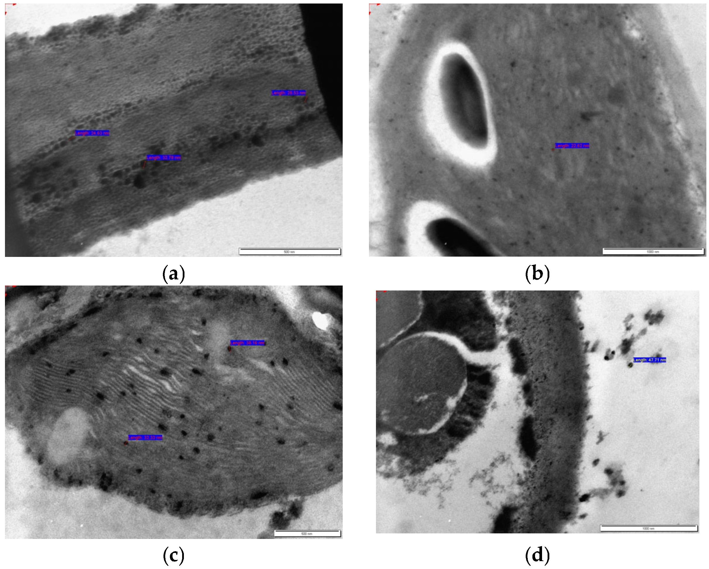
Transmission electron microscopy (TEM) images were obtained from microshoot samples treated with 50 mg/L of either AgNPs or CuNPs, focusing specifically on both root and leaf tissues. Each root and leaf sample was processed and imaged individually. As shown in Figure 2a, AgNPs were translocated within root cells, with particle sizes of 28.53, 32.78, and 24.63 nm. The smaller size of AgNPs in the plant samples, compared to those added to the MS medium, is attributed to the plant’s metabolic activity. Biomolecules such as phenolics, flavonoids, and proteins can reduce and stabilize the nanoparticles, preventing aggregation and limiting growth, resulting in smaller and more uniform particles [40,41]. Figure 2b shows AgNPs in stained leaf tissue, measuring 22.62 nm. CuNPs were observed inside root cells (Figure 2c) with a particle size of 32.33 nm, while translocated CuNPs in leaf tissue measured 47.71 nm (Figure 2d). The TEM images were captured after staining the tissues with uranyl acetate, which facilitated the visualization of nanoparticles.
The presence of nanoparticles in the roots and leaves following exposure indicates that they were absorbed by the plant and translocated internally. Nanoparticles are likely taken up by roots—the primary interface between the plant and its medium—through passive diffusion, active transport, or endocytosis [42]. Once inside, they are translocated via the vascular system (xylem or phloem) to other tissues, such as leaves [43]. This movement is affected by factors including the size of the nanoparticles, charge, surface coating, and the physiological state of the plant [44]. Earlier research has demonstrated that AgNPs smaller than 40 nm can pass through root cell walls [45]. TEM analyses have detected AgNPs in various parts of the root in Festuca rubra, including the cell wall and cortex of parenchymal cells [46]. Stomata in leaves of Arabidopsis thaliana have also been implicated in facilitating AgNP uptake and translocation [47], although it remains unclear whether AgNPs can move through the food chain [10,48]. In Zea mays (maize), TEM studies have demonstrated that CuNPs can be translocated from roots to shoots via the xylem, where they may aggregate or transform into other forms [49]. TEM imaging also revealed CuNPs (~25 nm) in soybean seeds, showing positive effects as they passed through the plasma membrane [50]. Additionally, CuNPs were observed to translocate from root to shoot in Elsholtzia splendens [51]. In summary, differences in the release rate and reactivity of Ag and Cu nanoparticles influence their uptake, translocation, and overall impact on plants [45,46,50].

3.3. Effect of Nanoparticles on the Phytochemical Composition of In Vitro O. petraeum

GC-MS analysis revealed that the volatile profiles of O. petraeum varied depending on nanoparticle treatment. Microshoots treated with 50 mg/L CuNPs (Table 3, Figure S1 (Supplementary Materials)) exhibited a profile dominated by α-Terpinyl Acetate (29.23%) and Geranyl Acetate (12.76%), along with Geraniol (7.18%), β-Caryophyllene (9.13%), and γ-Muurolene (7.16%), indicating enhanced geranyl derivative biosynthesis. Minor components such as Cyclopentadecanolide (3.81%) and Cedroxyde (1.74%) further emphasized the impact of CuNPs on terpenoid pathways. These findings align with Tabatabaee et al. (2021) [52], who reported that CuNPs can stimulate plant growth and secondary metabolism, while posing lower phytotoxicity than bulk copper. Similar effects of CuNPs on antioxidant activity and metabolite accumulation have been observed in Ocimum basilicum and Bacopa monnieri [18,53].
In contrast, treatment with 50 mg/L AgNPs (Table 4, Figure S2 (Supplementary File)) produced a sesquiterpene-rich profile, dominated by Caryophyllene Oxide (28.45%) and β-Caryophyllene (21.94%). Other notable compounds included α-Humulene (14.70%) and Humulene Epoxide II (11.40%), with minor constituents such as Geranial (2.94%) and Italicene Epoxide (2.93%). This indicates that AgNPs enhance sesquiterpene biosynthesis, consistent with previous findings in Carum carvi callus cultures, where AgNPs upregulated γ-terpinene synthase (CcTPS2) and increased secondary metabolites [54].
Under control conditions (30 g/L sucrose, no nanoparticles; Table 5, Figure S3 (Supplementary Materials)), microshoots showed a simpler profile, dominated by Caryophyllene Oxide (25.71%), n-Decanal (10.97%), and n-Nonanal (8.07%), with moderate amounts of α-Terpinyl Acetate (18.98%) and β-Caryophyllene (10.34%). This baseline volatile composition confirms that nanoparticles induce stress-related metabolic pathways, diversifying secondary metabolite production, as previously observed in Mentha pulegium and Thymus species [55,56].
The wild plant sample (Royal Botanic Garden, Jordan; Table 6, Figure S4 (Supplementary Materials)) displayed a profile dominated by monoterpenes and sesquiterpenes. Eudesm-7(11)-en-4-ol (25.10%) was the most abundant compound, followed by terpinen-4-ol (19.55%), β-Caryophyllene (12.20%), and abietatriene (7.13%), reflecting the plant’s natural chemotype. This has also been previously demonstrated in plants belonging to the Lamiaceae family, due to their content of the same bioactive compounds [57,58]. Environmental factors, such as stress and soil composition, likely contribute to this chemical diversity [59,60].
CuNPs and AgNPs can act as elicitors, specifically triggering stress responses in plants that enhance the production of secondary metabolites (e.g., flavonoids, phenolics, essential oils) [18,20,61]. In contrast, traditional chemical elicitors may be less selective or slower in inducing these pathways [40,62]. Additionally, nanoparticles can gradually release metal ions, providing a longer-lasting stimulus to the plant’s metabolism compared to the sudden burst from salts or other chemicals [63,64]. CuNPs and AgNPs can be taken up by plant tissues, especially in in vitro cultures [65]. However, studies show that most nanoparticles accumulate in roots and leaves, and their transfer into secondary metabolites (such as essential oils, flavonoids, or phenolics) is minimal or negligible [66]. Therefore, while nanoparticles stimulate metabolite production, they generally do not become part of the secondary metabolites [67]. This allows plants to respond at lower doses, reducing toxicity risks. Moreover, since lower doses are effective, using nanoparticles may result in less chemical runoff and lower environmental contamination compared to conventional metal salts or synthetic elicitors [68]. Proper extraction and purification further minimize any potential contamination, making health risks minimal when concentrations are controlled [66].
CuNPs and AgNPs were applied to culture media to evaluate their effects on microshoot growth and secondary metabolite production. Mechanistically, these nanoparticles act as elicitors, inducing mild oxidative stress that activates key enzymes involved in secondary metabolite biosynthesis, such as phenylalanine ammonia-lyase (PAL) [67,69]. Nanoparticles can enhance secondary metabolite production in in vitro cultures by inducing stress responses, modulating gene expression and enzyme activity, and acting as catalysts in biosynthetic pathways [40,70,71]. Their small size and high surface area allow efficient interactions with plant cells, enhancing nutrient uptake and influencing hormonal signaling pathways [69,72]. Recent studies [40,55,70,71,72] have reported similar effects of metal nanoparticles on plant tissue cultures, supporting the observed enhancement in metabolite production.
Collectively, these results provide a comprehensive understanding of how nanoparticles stimulate plant growth and secondary metabolism. They also highlight the potential of O. petraeum as a valuable source of bioactive compounds for pharmacological and biotechnological applications. Such effects underscore the promise of nanoparticles for enhancing the medicinal and industrial value of aromatic plants.

3.4. Effect of Nanoparticles on Nutrient Uptake in In Vitro O. petraeum Microshoots

As shown in Table 7, nanoparticle treatments significantly affected nutrient composition in O. petraeum microshoots. The control group (MS medium without nanoparticles) contained 3.15 mg/g P, 36.31 mg/g K, and 12.12 mg/g Ca, consistent with reported nutrient ranges in plant dry matter [73]. Microshoots treated with 50 mg/L CuNPs showed increased phosphorus (3.44 mg/g) but reduced potassium (24.98 mg/g) and calcium (3.75 mg/g). Similar effects of CuNPs on nutrient uptake have been reported in Glycine max, with phosphorus metabolism influenced by enzymatic regulation [37,50]. Conversely, 50 mg/L AgNPs increased calcium levels to 13.31 mg/g while decreasing phosphorus and potassium. AgNPs have been shown to enhance calcium uptake and other macronutrients in various plants, likely via stress-induced signaling, membrane stabilization, and increased nutrient bioavailability [11,74,75]. Overall, nanoparticle treatments influenced major element uptake, with CuNPs notably elevating phosphorus but reducing potassium and calcium, while AgNPs primarily increased calcium. These effects may result from enhanced root interaction, membrane permeability, and nutrient bioavailability induced by nanoparticles [72,76].

4. Conclusions

Nanoparticle treatments significantly enhanced growth, secondary metabolite production, and nutrient uptake in O. petraeum microshoots. CuNPs promoted phosphorus accumulation and geranyl derivatives, while AgNPs increased calcium levels and sesquiterpene biosynthesis. TEM analysis confirmed the absorption and movement of both nanoparticles within roots and leaves. This study is the first to report the influence of nanoparticles on the micropropagation of O. petraeum and its essential oil profile, highlighting the effect of culture conditions and nanoparticle type on chemical composition of plant metabolites. More studies are needed to improve nanoparticle use, enhance terpenoid yield, and assess long-term influence on plant development and genetic stability.

Supplementary Materials

The following supporting information can be downloaded at: https://www.mdpi.com/article/10.3390/nano15191496/s1. Figure S1: GC-MS chromatogram showing the volatile components in O. petraeum microshoots treated with copper nanoparticles (50 mg/L Cu NPs).; Figure S2: GC-MS chromatogram showing the volatile components in O. petraeum microshoots treated with silver nanoparticles (50 mg/L Ag NPs).; Figure S3: GC-MS chromatogram showing the volatile components in O. petraeum microshoots on MS medium with GA3 at 0.5 mg/L and sucrose at 30 g/L (Control 1).; Figure S4: GC-MS chromatogram showing the volatile components in wild O. petraeum aerial parts (Control 2).

Author Contributions

Conceptualization was carried out by T.S.A.Q., R.A.-Z. and R.A.S.; methodology was developed by T.S.A.Q.; investigation and data collection were performed by T.S.A.Q.; formal analysis was conducted by T.S.A.Q. and M.H.; supervision was provided by R.A.-Z. and R.A.S.; the original draft was written by T.S.A.Q.; review and editing of the manuscript were performed by R.A.-Z., R.A.S. and M.H.; resources were provided by R.A.S. and R.A.-Z.; project administration and funding acquisition were handled by R.A.-Z. All authors have read and agreed to the published version of the manuscript.

Funding

Financial support for this research was provided by the University of Jordan, Amman, Jordan.

Data Availability Statement

The data produced and examined in this study can be obtained from the corresponding authors upon request for research purposes.

Acknowledgments

The authors sincerely acknowledge the financial support provided by the University of Jordan—Deanship of Scientific Research at.

Conflicts of Interest

The authors declare no conflicts of interest.

References

  1. Chen, S.L.; Yu, H.; Luo, H.M.; Wu, Q.; Li, C.F.; Steinmetz, A. Conservation and sustainable use of medicinal plants: Problems, progress, and prospects. Chin. Med. 2016, 11, 37. [Google Scholar] [CrossRef]
  2. Taifour, H.; El-Oqlah, A. Jordan Plant Red List, 1st ed.; Royal Botanic Garden: Amman, Jordan, 2014; pp. 78, 572. [Google Scholar]
  3. Oran, S.A.; Al-Eisawi, D.M. Medicinal plants in the high mountains of northern Jordan. Int. J. Biodivers. Conserv. 2014, 6, 436–443. [Google Scholar] [CrossRef]
  4. Mechergui, K.; Jaouadi, W.; Coelho, J.A.; Serra, M.C.; Khouja, M.L. Biological activities and oil properties of Origanum glandulosum Desf: A review. Phytothérapie 2016, 14, 102–108. [Google Scholar] [CrossRef]
  5. Sakkas, H.; Papadopoulou, C. Antimicrobial activity of basil, oregano, and thyme essential oils. J. Microbiol. Biotechnol. 2017, 27, 429–438. [Google Scholar] [CrossRef] [PubMed]
  6. Danin, A. Two new species of Origanum (Labiatae) from Jordan. Willdenowia 1990, 19, 401–404. [Google Scholar]
  7. Debnath, M.; Malik, C.P. Micropropagation: A tool for production of high-quality plant-based medicines. Curr. Pharm. Biotechnol. 2006, 7, 33–49. [Google Scholar] [CrossRef]
  8. Al-Qudah, T.S.; Shibli, R.A.; Alali, F.Q. In vitro propagation and secondary metabolites production in wild germander (Teucrium polium L.). In Vitro Cell. Dev. Biol. Plant 2011, 47, 496–505. [Google Scholar] [CrossRef]
  9. Mherat, M.; Shatnawi, M.; Shibli, R.; Qudah, T.; Malloh, S.A.; Tamadour, S.Q. Clonal propagation of Tetragonolobus palaestinus Bioss: A Jordanian medicinal plant. Acta Agric. Slovenica 2022, 118, 1–9. [Google Scholar] [CrossRef]
  10. Al-Qudah, T.; Mahmood, S.H.; Abu-Zurayk, R.; Shibli, R.; Khalaf, A.; Lambat, T.L.; Chaudhary, R.G. Nanotechnology applications in plant tissue culture and molecular genetics: A holistic approach. Curr. Nanosci. 2022, 18, 442–464. [Google Scholar] [CrossRef]
  11. Shibli, R.; Mohusaien, R.; Abu-Zurayk, R.; Qudah, T.; Tahtamouni, R. Silver nanoparticles (Ag NPs) boost mitigation powers of Chenopodium quinoa (Q6 line) grown under in vitro salt-stressing conditions. Water 2022, 14, 3099. [Google Scholar] [CrossRef]
  12. Singh, R.; Ahamad, S. Integration of nanotechnology in plant tissue culture. Curr. Nanosci. 2022, 18, 604–610. [Google Scholar] [CrossRef]
  13. Yaqoob, A.A.; Ahmad, H.; Parveen, T.; Ahmad, A.; Oves, M.; Ismail, I.M.; Mohamad Ibrahim, M.N. Recent advances in metal decorated nanomaterials and their various biological applications: A review. Front. Chem. 2020, 8, 341. [Google Scholar] [CrossRef] [PubMed]
  14. Dubchak, S.; Ogar, A.; Mietelski, J.W.; Turnau, K. Influence of silver and titanium nanoparticles on arbuscular mycorrhiza colonization and accumulation of radiocaesium in Helianthus annuus. Span. J. Agric. Res. 2010, 8, 103–108. [Google Scholar] [CrossRef]
  15. Selvakesavan, R.K.; Kruszka, D.; Shakya, P.; Mondal, D.; Franklin, G. Impact of Nanomaterials on Plant Secondary Metabolism. In Nanomaterial Interactions with Plant Cellular Mechanisms and Macromolecules and Agricultural Implications; Springer: Berlin/Heidelberg, Germany, 2023; pp. 133–170. [Google Scholar]
  16. Khalid, M.F.; Khan, R.I.; Jawaid, M.Z.; Shafqat, W.; Hussain, S.; Ahmed, T.; Rizwan, M.; Ercisli, S.; Pop, O.L.; Marc, R.A. Nanoparticles: The plant saviour under abiotic stresses. Nanomaterials 2022, 12, 3915. [Google Scholar] [CrossRef] [PubMed]
  17. Al-Oubaidi, H.K.M.; Mohammed-Ameen, A.S. The effect of (AgNO3) NPs on increasing of secondary metabolites of Calendula officinalis L. in vitro. Int. J. Pharm. Pract. 2014, 5, 267–272. [Google Scholar]
  18. Lala, S. Enhancement of secondary metabolites in Bacopa monnieri (L.) Pennell plants treated with copper-based nanoparticles in vivo. IET Nanobiotechnol. 2020, 14, 78–85. [Google Scholar] [CrossRef]
  19. Kruszka, D.; Selvakesavan, R.K.; Kachlicki, P.; Franklin, G. Untargeted metabolomics analysis reveals the elicitation of important secondary metabolites upon treatment with various metal and metal oxide nanoparticles in Hypericum perforatum L. cell suspension cultures. Ind. Crops Prod. 2022, 178, 114561. [Google Scholar] [CrossRef]
  20. Pathak, A.; Haq, S.; Meena, N.; Dwivedi, P.; Kothari, S.L.; Kachhwaha, S. Multifaceted role of nanomaterials in modulating in vitro seed germination, plant morphogenesis, metabolism and genetic engineering. Plants 2023, 12, 3126. [Google Scholar] [CrossRef]
  21. Alexopoulos, A.A.; Kartsonas, E.; Karras, S.; Mavrommati, E.; Petropoulos, S.A.; Papafotiou, M. In vitro Propagation of Origanum scabrum (Boiss. & Heldr.): An Endemic Medicinal Plant of Greece. Plants 2023, 12, 2118. [Google Scholar] [CrossRef]
  22. Fokina, A.V.; Satarova, T.M.; Smetanin, V.T.; Kucenko, N.I. Optimization of microclonal propagation in vitro of oregano (Origanum vulgare). Biosyst. Divers. 2018, 26, 98–102. [Google Scholar] [CrossRef]
  23. Du, W.; Tan, W.; Yin, Y.; Ji, R.; Peralta-Videa, J.R.; Guo, H.; Gardea-Torresdey, J.L. Differential effects of copper nanoparticles/microparticles in agronomic and physiological parameters of oregano (Origanum vulgare). Sci. Total Environ. 2018, 618, 306–312. [Google Scholar] [CrossRef]
  24. Shaik, M.R.; Khan, M.; Kuniyil, M.; Al-Warthan, A.; Alkhathlan, H.Z.; Siddiqui, M.R.H.; Adil, S.F. Plant-extract-assisted green synthesis of silver nanoparticles using Origanum vulgare L. extract and their microbicidal activities. Sustainability 2018, 10, 913. [Google Scholar] [CrossRef]
  25. Erenler, R.; Dag, B. Biosynthesis of silver nanoparticles using Origanum majorana L. and evaluation of their antioxidant activity. Inorg. Nano-Met. Chem. 2022, 52, 485–492. [Google Scholar] [CrossRef]
  26. Murashige, T.; Skoog, F. A Revised Medium for Rapid Growth and Bio Assays with Tobacco Tissue Cultures. Physiol. Plant. 1962, 15, 473–497. [Google Scholar] [CrossRef]
  27. Van Den Dool, H.; Kratz, P.D. A Generalization of the Retention Index System Including Linear Temperature Programmed Gas–Liquid Partition Chromatography. J. Chromatogr. A 1963, 11, 463–471. [Google Scholar] [CrossRef] [PubMed]
  28. Adams, R.P. Identification of Essential Oil Components by Gas Chromatography/Mass Spectrometry; Allured Publishing Corporation: Carol Stream, IL, USA, 2007. [Google Scholar]
  29. Bonsucesso Santana, J.; Santos do Nascimento, A.; da Silva Conceição, A.L.; Lopes de Carvalho, C.A.; de Souza Dias, F. Efficiency of two digestion methods in determining the presence of metals (Cd, Cu, Cr, Pb and Zn) in geopropolis produced by Melipona scutellaris. Rev. Colomb. Química 2021, 50, 24–29. [Google Scholar] [CrossRef]
  30. Pequerul, A.; Pérez, C.; Madero, P.; Val, J.; Monge, E. A rapid wet digestion method for plant analysis. In Proceedings of the Optimization of Plant Nutrition: Refereed Papers from the Eighth International Colloquium for the Optimization of Plant Nutrition, Lisbon, Portugal, 31 August–8 September 1992; Springer: Dordrecht, The Netherlands, 1993; pp. 3–6. [Google Scholar]
  31. Elsayh, S.A.; Arafa, R.N.; Ali, G.A.; Abdelaal, W.B.; Sidky, R.A.; Ragab, T.I. Impact of silver nanoparticles on multiplication, rooting of shoots and biochemical analyses of date palm Hayani cv. by in vitro. Biocatal. Agric. Biotechnol. 2022, 43, 102400. [Google Scholar] [CrossRef]
  32. Manokari, M.; Raj, M.C.; Dey, A.; Faisal, M.; Alatar, A.A.; Joshee, N.; Shekhawat, M.S. Silver nanoparticles improved morphogenesis, biochemical profile and micro-morphology of Gaillardia pulchella Foug cv. ‘Torch Yellow’. Plant Cell Tissue Organ Cult. 2023, 155, 433–445. [Google Scholar] [CrossRef]
  33. Sreelekshmi, R.; Siril, E.A.; Muthukrishnan, S. Role of biogenic silver nanoparticles on hyperhydricity reversion in Dianthus chinensis L. an in vitro model culture. J. Plant Growth Regul. 2022, 41, 23–39. [Google Scholar] [CrossRef]
  34. Tymoszuk, A.; Kulus, D. Effect of silver nanoparticles on the in vitro regeneration, biochemical, genetic, and phenotype variation in adventitious shoots produced from leaf explants in Chrysanthemum. Int. J. Mol. Sci. 2022, 23, 7406. [Google Scholar] [CrossRef]
  35. Ibrahim, A.S.; Fahmy, A.H.; Ahmed, S.S. Copper nanoparticles elevate regeneration capacity of Ocimum basilicum L. plant via somatic embryogenesis. Plant Cell Tissue Organ Cult. 2019, 136, 41–50. [Google Scholar] [CrossRef]
  36. Mawale, K.S.; Praveen, A.; Giridhar, P. Efficacy of copper and silver nanoparticles on seedling growth, biochemical and antioxidant potential of Capsicum annuum L., in vitro and ex vitro. S. Afr. J. Bot. 2024, 175, 1–14. [Google Scholar] [CrossRef]
  37. Xiao, Y.; Ma, J.; Xian, J.; Peijnenburg, W.J.; Du, Y.; Tian, D.; Tu, L. Copper accumulation and physiological markers of soybean (Glycine max) grown in agricultural soil amended with copper nanoparticles. Ecotoxicol. Environ. Saf. 2022, 229, 113088. [Google Scholar] [CrossRef] [PubMed]
  38. Alam, N.; Anis, M.; Javed, S.B.; Alatar, A.A. Stimulatory effect of copper and zinc sulphate on plant regeneration, glutathione-S-transferase analysis and assessment of antioxidant activities in Mucuna pruriens L. (DC). Plant Cell Tissue Organ Cult. 2020, 141, 155–166. [Google Scholar] [CrossRef]
  39. Ahmad, M.A.; Javed, R.; Adeel, M.; Rizwan, M.; Ao, Q.; Yang, Y. Engineered ZnO and CuO nanoparticles ameliorate morphological and biochemical response in tissue culture regenerants of candyleaf (Stevia rebaudiana). Molecules 2020, 25, 1356. [Google Scholar] [CrossRef]
  40. Ali, A.; Mashwani, Z.U.R.; Raja, N.I.; Mohammad, S.; Luna-Arias, J.P.; Ahmad, A.; Kaushik, P. Phytomediated selenium nanoparticles and light regimes elicited in vitro callus cultures for biomass accumulation and secondary metabolite production in Caralluma tuberculata. Front. Plant Sci. 2023, 14, 1253193. [Google Scholar] [CrossRef]
  41. Alfarraj, N.S.; Tarroum, M.; Al-Qurainy, F.; Nadeem, M.; Khan, S.; Salih, A.M.; Perveen, K. Biosynthesis of Silver Nanoparticles and Exploring Their Potential of Reducing the Contamination of the In vitro Culture Media and Inducing the Callus Growth of Rumex nervosus Explants. Molecules 2023, 28, 3666. [Google Scholar] [CrossRef]
  42. Su, Y.; Ashworth, V.; Kim, C.; Adeleye, A.S.; Rolshausen, P.; Roper, C.; Jassby, D. Delivery, uptake, fate, and transport of engineered nanoparticles in plants: A critical review and data analysis. Environ. Sci. Nano 2019, 6, 2311–2331. [Google Scholar] [CrossRef]
  43. Rani, S.; Kumari, N.; Sharma, V. Uptake, translocation, transformation and physiological effects of nanoparticles in plants. Arch. Agron. Soil Sci. 2023, 69, 1579–1599. [Google Scholar] [CrossRef]
  44. Khan, I.; Saeed, K.; Khan, I. Nanoparticles: Properties, applications and toxicities. Arab. J. Chem. 2019, 12, 908–931. [Google Scholar] [CrossRef]
  45. Huang, D.; Dang, F.; Huang, Y.; Chen, N.; Zhou, D. Uptake, translocation, and transformation of silver nanoparticles in plants. Environ. Sci. Nano 2022, 9, 12–39. [Google Scholar] [CrossRef]
  46. Marchiol, L.; Mattiello, A.; Pošćić, F.; Giordano, C.; Musetti, R. In vivo synthesis of nanomaterials in plants: Location of silver nanoparticles and plant metabolism. Nanoscale Res. Lett. 2014, 9, 101. [Google Scholar] [CrossRef] [PubMed]
  47. He, J.; Zhang, L.; He, S.Y.; Ryser, E.T.; Li, H.; Zhang, W. Stomata facilitate foliar sorption of silver nanoparticles by Arabidopsis thaliana. Environ. Pollut. 2022, 292, 118448. [Google Scholar] [CrossRef] [PubMed]
  48. Dang, F.; Huang, Y.; Wang, Y.; Zhou, D.; Xing, B. Transfer and toxicity of silver nanoparticles in the food chain. Environ. Sci. Nano 2021, 8, 1519–1535. [Google Scholar] [CrossRef]
  49. Wang, Z.; Xie, X.; Zhao, J.; Liu, X.; Feng, W.; White, J.C.; Xing, B. Xylem- and phloem-based transport of CuO nanoparticles in maize (Zea mays L.). Environ. Sci. Technol. 2012, 46, 4434–4441. [Google Scholar] [CrossRef]
  50. Yusefi-Tanha, E.; Fallah, S.; Rostamnejadi, A.; Pokhrel, L.R. Root system architecture, copper uptake and tissue distribution in soybean (Glycine max (L.) Merr.) grown in copper oxide nanoparticle (CuONP)-amended soil and implications for human nutrition. Plants 2020, 9, 1326. [Google Scholar] [CrossRef]
  51. Shi, J.; Peng, C.; Yang, Y.; Yang, J.; Zhang, H.; Yuan, X.; Hu, T. Phytotoxicity and Accumulation of Copper Oxide Nanoparticles to the Cu-Tolerant Plant Elsholtzia splendens. Nanotoxicology 2014, 8, 179–188. [Google Scholar] [CrossRef]
  52. Tabatabaee, S.; Iranbakhsh, A.; Shamili, M.; Ardebili, Z.O. Copper nanoparticles mediated physiological changes and transcriptional variations in microRNA159 (miR159) and mevalonate kinase (MVK) in pepper; potential benefits and phytotoxicity assessment. J. Environ. Chem. Eng. 2021, 9, 106151. [Google Scholar] [CrossRef]
  53. Nazir, S.; Jan, H.; Zaman, G.; Khan, T.; Ashraf, H.; Meer, B.; Abbasi, B.H. Copper oxide (CuO) and manganese oxide (MnO) nanoparticles induced biomass accumulation, antioxidants biosynthesis and abiotic elicitation of bioactive compounds in callus cultures of Ocimum basilicum (Thai basil). Artif. Cells Nanomed. Biotechnol. 2021, 49, 625–633. [Google Scholar] [CrossRef]
  54. Dehghani-Aghchekohal, Z.; Omidi, M.; Azizinezhad, R.; Etminan, A. Stimulation of secondary metabolites and γ-terpinene synthase by silver nanoparticles in callus cultures of Carum carvi. Appl. Biochem. Biotechnol. 2022, 194, 3228–3241. [Google Scholar] [CrossRef]
  55. El-Naggar, H.M.; Osman, A.R. Enhancing growth and bioactive metabolites characteristics in Mentha pulegium L. via silicon nanoparticles during in vitro drought stress. BMC Plant Biol. 2024, 24, 657. [Google Scholar] [CrossRef] [PubMed]
  56. Mosavat, N.; Golkar, P.; Yousefifard, M.; Javed, R. Modulation of callus growth and secondary metabolites in different Thymus species and Zataria multiflora micropropagated under ZnO nanoparticles stress. Biotechnol. Appl. Biochem. 2019, 66, 316–322. [Google Scholar] [CrossRef] [PubMed]
  57. Cheminal, A.; Kokkoris, I.P.; Strid, A.; Dimopoulos, P. Medicinal and aromatic Lamiaceae plants in Greece: Linking diversity and distribution patterns with ecosystem services. Forests 2020, 11, 661. [Google Scholar] [CrossRef]
  58. Çelik, G.; Kılıç, G.; Kanbolat, Ş.; Özlem Şener, S.; Karaköse, M.; Yaylı, N.; Karaoğlu, Ş.A. Biological activity, and volatile and phenolic compounds from five Lamiaceae species. Flavour Fragr. J. 2021, 36, 223–232. [Google Scholar] [CrossRef]
  59. Felisberto, J.S.; Machado, D.B.; Assunção, J.A.; Massau, S.A.; Queiroz, G.A.D.; Guimarães, E.F.; Moreira, D.D.L. Spatio-Temporal Variations of Volatile Metabolites as an Eco-Physiological Response of a Native Species in the Tropical Forest. Plants 2024, 13, 2599. [Google Scholar] [CrossRef]
  60. Hudaib, M.M.; Abu Hajal, A.F.; Sakkal, M.M. Chemical composition of the volatile oil from aerial parts of Rosmarinus officinalis L. growing in UAE. J. Essent. Oil Bear. Plants 2022, 25, 282–289. [Google Scholar] [CrossRef]
  61. Darbahani, M.; Ghiyasi, M.R.; Rahaie, M. Nanoparticles as new elicitors for the production of bioactive and phytochemicals in vitro and in vivo plant culture. Phytochem. Rev. 2024, 24, 3179–3203. [Google Scholar] [CrossRef]
  62. Wen, Y.; Liao, Y.; Tang, Y.; Zhang, H.; Zhang, J.; Liao, Z. Metabolic effects of elicitors on the biosynthesis of tropane alkaloids in medicinal plants. Plants 2023, 12, 3050. [Google Scholar] [CrossRef]
  63. Yadav, A.; Yadav, K.; Abd-Elsalam, K.A. Nanofertilizers: Types, delivery and advantages in agricultural sustainability. Agrochemicals 2023, 2, 296–336. [Google Scholar] [CrossRef]
  64. Garg, D.; Sridhar, K.; Inbaraj, B.S.; Chawla, P.; Tripathi, M.; Sharma, M. Nano-biofertilizer formulations for agriculture: A systematic review on recent advances and prospective applications. Bioengineering 2023, 10, 1010. [Google Scholar] [CrossRef]
  65. Ochatt, S.; Abdollahi, M.R.; Akin, M.; Bello, J.B.; Eimert, K.; Faisal, M.; Nhut, D.T. Application of nanoparticles in plant tissue cultures: Minuscule size but huge effects. Plant Cell Tissue Organ Cult. 2023, 155, 323–326. [Google Scholar] [CrossRef]
  66. Antunes Filho, S.; Dos Santos, M.S.; Dos Santos, O.A.L.; Backx, B.P.; Soran, M.L.; Opriş, O.; Bououdina, M. Biosynthesis of nanoparticles using plant extracts and essential oils. Molecules 2023, 28, 3060. [Google Scholar] [CrossRef] [PubMed]
  67. Inam, M.; Attique, I.; Zahra, M.; Khan, A.K.; Hahim, M.; Hano, C.; Anjum, S. Metal oxide nanoparticles and plant secondary metabolism: Unraveling the game-changer nano-elicitors. Plant Cell Tissue Organ Cult. 2023, 155, 327–344. [Google Scholar] [CrossRef]
  68. Martinez-Chávez, L.A.; Hernández-Ramírez, M.Y.; Feregrino-Pérez, A.A.; Esquivel Escalante, K. Cutting-edge strategies to enhance bioactive compound production in plants: Potential value of integration of elicitation, metabolic engineering, and green nanotechnology. Agronomy 2024, 14, 2822. [Google Scholar] [CrossRef]
  69. Singh, R.; Choudhary, P.; Kumar, S.; Daima, H.K. Mechanistic approaches for crosstalk between nanomaterials and plants: Plant immunomodulation, defense mechanisms, stress resilience, toxicity, and perspectives. Environ. Sci. Nano 2024, 11, 2324–2351. [Google Scholar] [CrossRef]
  70. Javed, R.; Yucesan, B.; Zia, M.; Gurel, E. Nanoelicitation: A promising and emerging technology for triggering the sustainable in vitro production of secondary metabolites in medicinal plants. In Plant and Nanoparticles; Springer Nature: Singapore, 2022; pp. 265–280. [Google Scholar]
  71. Prasad, A.; Sidhic, J.; Sarbadhikary, P.; Narayanankutty, A.; George, S.; George, B.P.; Abrahamse, H. Role of metal nanoparticles in organogenesis, secondary metabolite production and genetic transformation of plants under in vitro condition: A comprehensive review. Plant Cell Tissue Organ Cult. 2024, 158, 33. [Google Scholar] [CrossRef]
  72. Wang, X.; Xie, H.; Wang, P.; Yin, H. Nanoparticles in plants: Uptake, transport and physiological activity in leaf and root. Materials 2023, 16, 3097. [Google Scholar] [CrossRef]
  73. Glew, R.S.; Vanderjagt, D.J.; Chuang, L.T.; Huang, Y.S.; Millson, M.; Glew, R.H. Nutrient content of four edible wild plants from West Africa. Plant Foods Hum. Nutr. 2005, 60, 187–193. [Google Scholar] [CrossRef]
  74. Salachna, P.; Byczyńska, A.; Zawadzińska, A.; Piechocki, R.; Mizielińska, M. Stimulatory effect of silver nanoparticles on the growth and flowering of potted oriental lilies. Agronomy 2019, 9, 610. [Google Scholar] [CrossRef]
  75. Guzmán-Báez, G.A.; Trejo-Téllez, L.I.; Ramírez-Olvera, S.M.; Salinas-Ruíz, J.; Bello-Bello, J.J.; Alcántar-González, G.; Gómez-Merino, F.C. Silver Nanoparticles Increase Nitrogen, Phosphorus, and Potassium Concentrations in Leaves and Stimulate Root Length and Number of Roots in Tomato Seedlings in a Hormetic Manner. Dose-Response 2021, 19, 15593258211044576. [Google Scholar] [CrossRef]
  76. Landa, P. Positive effects of metallic nanoparticles on plants: Overview of involved mechanisms. Plant Physiol. Biochem. 2021, 161, 12–24. [Google Scholar] [CrossRef]
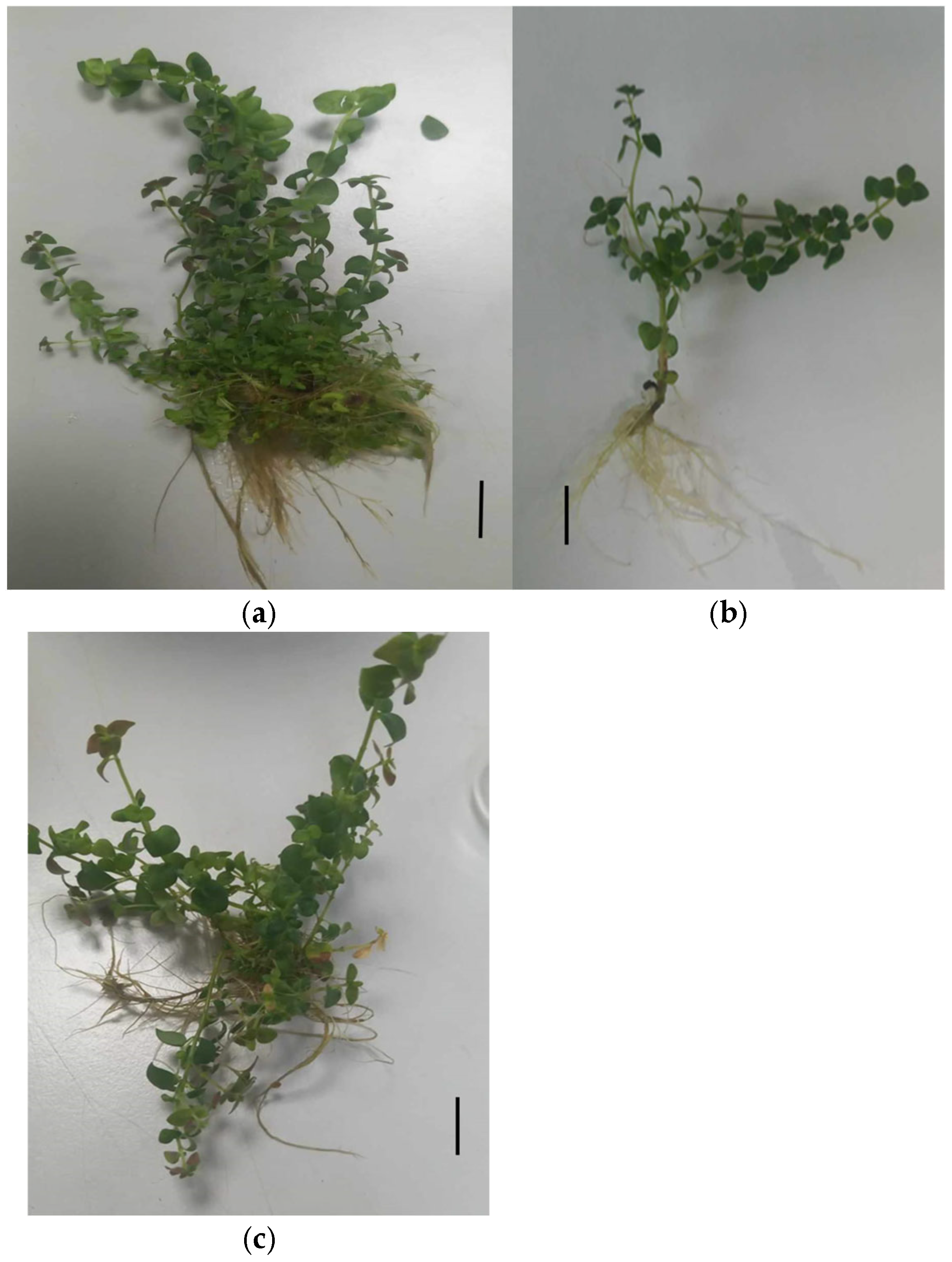
Figure 1. Influence of nanoparticles on growth characteristics of in vitro O. petraeum microshoots after 1 month of incubation on MS medium with GA3 at 0.5 mg/L and sucrose at 30 g/L. Treatments include (a) 100 mg/L AgNPs, (b) 0.0 mg/L NPs (control) and (c) 50 mg/L CuNPs. Scale bar = 1.0 cm.
Nanomaterials 15 01496 g001
Figure 2. TEM imaging showing the distribution of nanoparticles in O. petraeum tissues. (a) AgNPs localized within root cortical cells (particle sizes: 28.53, 32.78, and 24.63 nm). (b) AgNPs in mesophyll cells of stained leaf tissue (size: 22.62 nm). (c) CuNPs localized within root cortical cells (particle sizes: 32.33 and 38.16 nm). (d) CuNPs in mesophyll cells of stained leaf tissue (size: 47.71 nm).
Nanomaterials 15 01496 g002
Table 1. Influence of AgNPs on in vitro O. petraeum microshoot growth parameters after growing on MS medium enriched with various AgNPs concentrations and 0.5 mg/L GA3.
Ag NPs Concentration (mg/L)Microshoot
Number/Explant
Shoot Height
(cm)
Root
Numbers
Callus
Diameter (cm)
0 x2.80 ± 0.37 c *5.24 ± 0.12 c2.60 ± 0.51 c0.86 ± 0.50 d
253.80 ± 0.37 c4.54 ± 0.05 d3.80 ± 0.37 bc2.00 ± 0.71 b
506.80 ± 0.37 b5.94 ± 0.05 b4.80 ± 0.37 b1.54 ± 0.51 c
10011.60 ± 0.50 a9.22 ± 0.11 a8.20 ± 0.40 a1.84 ± 0.50 b
1507.60 ± 0.51 b6.18 ± 0.08 b5.2 ± 0.37 b2.38 ± 0.86 a
* Tukey’s HSD test shows that values with different letters within each column are significantly different at p ≤ 0.05. Data entries are presented as means ± standard error. x Control treatment.
Table 2. Influence of CuNPs on in vitro O. petraeum microshoot growth parameters after culturing on MS media enriched with various levels of CuNPs concentrations and 0.5 mg/L GA3.
Cu NPs Concentration (mg/L)Microshoot
Number/Explant
Shoot Height
(cm)
Root
Numbers
Callus
Diameter (cm)
0 x2.80 ± 0.37 cd *5.26 ± 0.06 b3.20 ± 037 c1.03 ± 0.37 c
254.80± 0.30 b3.84 ± 0.09 c5.60 ± 0.50 b1.08 ± 0.58 c
506.80 ± 0.37 a7.00 ± 0.31 a13.80 ± 0.58 a1.28 ± 0.37 c
1003.20 ± 0.30 c5.20 ± 0.37 b6.00 ± 0.44 b2.48 ± 0.86 a
1501.60 ± 0.24 d4.20 ± 0.30 bc4.80 ± 0.58 bc2.16 ± 0.92 b
* Tukey’s HSD test shows that values with different letters within each column are significantly different at p ≤ 0.05. Data entries are presented as means ± standard error. x Control treatment.
Table 3. Concentrations of chemical profile components in dried O. petraeum microshoots treated with copper nanoparticles (50 mg/L CuNPs).
SN *RT **RIexp xRIlit yCompound a%Content bClassification
Group z
19.83110281024Limonene3.60Monoterpene
Hydrocarbon
215.10011531155iso-Isopulegol4.94Monoterpenoid
Alcohol
319.05712421235Neral4.64Monoterpenoid
Aldehyde
420.42112731264Geraniol7.18Monoterpenoid
Alcohol
523.62013461346α-Terpinyl Acetate29.23Monoterpenoid
Ester
624.93013761379Geranyl Acetate12.76Monoterpenoid
Ester
726.51714131417β-Caryophyllene9.13Sesquiterpene
Hydrocarbon
828.05214501452α-Humulene4.91Sesquiterpene
Hydrocarbon
929.12914761478γ-Muurolene7.16Sesquiterpene
Hydrocarbon
1033.38415811582Caryophyllene oxide1.73Sesquiterpenoid
Oxide
1138.21717091713Cedroxyde1.74Sesquiterpenoid
Oxide
1242.54318341832Cyclopentadecanolide3.81Lactone
1344.93019081913(5E,9E)-Farnesyl acetone2.06Sesquiterpenoid
Ketone
1451.16121002087Abietadiene7.09Diterpene
Hydrocarbon
* SN: Compound serial number; ** RT: Retention time of the compound (minutes); x RI_exp: Linear retention index determined on a DB-5 equivalent column; y RI_lit: Reference retention index from literature (on a DB-5 equivalent column); a Compounds are presented according to their elution order; b Average of four measurements (two oil samples, each analyzed in duplicate) with standard deviation (SD) within ±5% of the mean. z Total Monoterpene Content: 62.35% (Monoterpene Hydrocarbons: 3.60%; Oxygenated Monoterpenes (have O group; (Alcohol; Aldehydes, Esters)): 58.75%). Total Sesquiterpene Content: 26.73% (Sesquiterpene Hydrocarbons: 21.20%; Oxygenated Sesquiterpenes: 5.53%). Miscellaneous Content: 10.90% (Lactone: 3.81%; Diterpene Hydrocarbon: 7.09%).
Table 4. Concentrations of chemical profile components in O. petraeum microshoots treated with silver nanoparticles (50 mg/L Ag NPs).
SN *RT **RIexp xRIlit yCompound a%Content bClassification Group z
16.475930932α-Pinene2.58Monoterpene Hydrocarbon
27.901975974β-Pinene1.83Monoterpene Hydrocarbon
319.06312421235Neral (z- citral)1.98Monoterpenoid Aldehyde
420.41312731265Geranial (E-citral)2.94Monoterpenoid Aldehyde
520.74612801283Isobornyl acetat1.00Monoterpenoid Ester
623.57413451346α-Terpinyl Acetat1.46Monoterpenoid Ester
726.51614131417β-Caryophyllene21.94Sesquiterpene Hydrocarbon
828.05714501452α-Humulene14.70Sesquiterpene Hydrocarbon
929.12514761478g-Muurolene4.76Sesquiterpene Hydrocarbon
1029.76014911500bicyclogermacrene1.46Sesquiterpene Hydrocarbon
1132.06015481547Italicene epoxide2.93Sesquiterpenoid Oxide
1233.36915801582Caryophyllene oxide28.45Sesquiterpenoid Oxide
1334.52816101608Humulene epoxide II11.40Sesquiterpenoid Oxide
1442.59618361832Cyclopentadecanone2.57Lactone
* SN: Compound serial number; ** RT: Retention time of the compound (minutes); x RI_exp: Linear retention index determined on a DB-5 equivalent column; y RI_lit: Reference retention index from literature (on a DB-5 equivalent column); a Compounds are arranged according to their elution sequence; b Mean values from four measurements (two oil samples, each analyzed in duplicate) with standard deviation (SD) within ±5% of the mean. z Total Monoterpene Content: 11.79% (Monoterpene Hydrocarbons: 4.41%; Oxygenated Monoterpenes: 7.38%) Total Sesquiterpene Content: 85.64% (Sesquiterpene Hydrocarbons: 42.86%; Oxygenated Sesquiterpenes: 42.78%) Miscellaneous Content: 2.57% (Lactone 2.57%).
Table 5. Concentrations of chemical profile components in dried O. petraeum microshoots cultured on MS medium containing 0.5 mg/L GA3 and 30 g/L sucrose (Control 1).
SN *RT **RIexp xRIlit yCompound a%Content bClassification Group z
112.94011031100n-Nonanal8.07Aldehyde
217.40312051201n-Decanal10.97Aldehyde
323.53413441346α-Terpinyl Acetat18.98Monoterpenoid Ester
426.48314121417β-Caryophyllene10.34Sesquiterpene Hydrocarbon
528.03514491452α-Humulene9.54Sesquiterpene Hydrocarbon
633.31615791582Caryophyllene oxide25.71Sesquiterpenoid Oxide
738.19417081713Cedroxyde16.39Sesquiterpenoid Oxide
* SN: Compound serial number; ** RT: Compound retention time (minutes); x RI_exp: Linear arithmetic retention index measured on a DB-5 equivalent column; y RI_lit: Reference arithmetic retention index from literature (measured on a DB-5 equivalent column); a Compounds are listed according to their elution order; b Mean of four determinations (two oil samples, each analyzed in duplicate) with standard deviation (SD) within ±5% of the mean.z Total Monoterpenoid Content: 18.98% (Oxygenated Monoterpenes: 18.98%; Hydrocarbon Monoterpenes: 0.00%). Total Sesquiterpene Content: 61.98% (Sesquiterpene Hydrocarbons: 19.88%; Oxygenated Sesquiterpenes: 42.10%). Miscellaneous Content: 19.04% (Aldehydes: 19.04%).
Table 6. Concentrations of chemical profile components in dried wild O. petraeum aerial (Control 2).
SN *RT **RIexp xRIlit yCompound a%Content bClassification
Group z
19.26510141014α-Terpinene2.56Monoterpene Hydrocarbon
210.95610551054γ-Terpinene4.70Monoterpene Hydrocarbon
316.80111911174Terpinen-4-ol (mix 1174)19.55Monoterpenoid
Alcohol
421.30812931299Terpinen-4-ol acetate4.22Monoterpenoid
Ester
526.52714131417β-Caryophyllene12.20Sesquiterpene Hydrocarbon
629.08814751478γ-Muurolene4.61Sesquiterpene Hydrocarbon
730.16315001505β-Bisabolene3.42Sesquiterpene Hydrocarbon
830.75115151522δ-Cadinene4.75Sesquiterpene Hydrocarbon
933.35115801582Caryophyllene oxide7.16Sesquiterpenoid
Oxide
1037.82716981700Eudesm-7(11)-en-4-ol25.10Sesquiterpenoid
Alcohol
1142.5601834NA m6,10,14-trimethyl-2-
Pentadecanone (Or Unk HC)
2.53Hydrocarbon
1243.32618591867(E)-β-Santalol acetate2.07Sesquiterpenoid
Ester
1349.66920532055Abietatriene7.13Diterpene
Hydrocarbon
* SN: Compound serial number; ** RT: Compound retention time (minutes); x RI_exp: Linear arithmetic retention index measured on a DB-5 equivalent column; y RI_lit: Reference arithmetic retention index from literature (measured on a DB-5 equivalent column); m: Not identified; a Compounds are listed according to their elution order; b Mean of four determinations (two oil samples, each analyzed in duplicate) with standard deviation (SD) within ±5% of the mean. z Total Monoterpene Content: 31.03% (Monoterpene Hydrocarbons: 7.26%; Oxygenated Monoterpenes: 23.77%). Total Sesquiterpene Content: 59.31% (Sesquiterpene Hydrocarbons: 25.98%; Oxygenated Sesquiterpenes: 34.33%). Miscellaneous Content: 9.66% (Hydrocarbon: 2.53%; Diterpene Hydrocarbon: 7.13%).
Table 7. Effect of Nanoparticles on Nutrients Uptake in Origanum petraeum Microshoots.
Sample Element (mg/g)
PKCa
Control x3.15133 ± 0.00491 b *36.31067± 0.00348 a12.11567 ± 0.00348 b
50 mg/L CuNPs3.43533 ± 0.00819 a24.97567± 0.00348 b3.75133 ± 0.0041 c
50 mg/L AgNPs2.95233 ± 0.01178 c24.41100± 0.00665 c13.310 ± 0.00577 a
* Tukey’s HSD test shows that values with different letters within each column are significantly different at p ≤ 0.05. Data entries are presented as means ± standard error. x Control treatment.
Disclaimer/Publisher’s Note: The statements, opinions and data contained in all publications are solely those of the individual author(s) and contributor(s) and not of MDPI and/or the editor(s). MDPI and/or the editor(s) disclaim responsibility for any injury to people or property resulting from any ideas, methods, instructions or products referred to in the content.