Article

Species-Specific Stress Responses to Selenium Nanoparticles in Pseudomonas aeruginosa and Proteus mirabilis

1
Division of Microbiology, National Center for Toxicological Research, U.S. Food and Drug Administration, Jefferson, AR 72079, USA
2
Nanotechnology Core Facility, Office of Scientific Coordination, National Center for Toxicological Research, U.S. Food and Drug Administration, Jefferson, AR 72079, USA
3
University of Arkansas, Little Rock, AR 72204, USA
4
Division of Biochemical Toxicology, National Center for Toxicological Research, U.S. Food and Drug Administration, Jefferson, AR 72079, USA
*
Author to whom correspondence should be addressed.
Nanomaterials 2025, 15(18), 1404; https://doi.org/10.3390/nano15181404
Submission received: 5 August 2025 / Revised: 29 August 2025 / Accepted: 8 September 2025 / Published: 12 September 2025

Abstract

Urinary tract infections (UTIs) remain a major global health concern, with rising antimicrobial resistance prompting the search for alternative therapies. Selenium nanoparticles (Se NPs) are promising antimicrobial agents due to their unique physicochemical properties and ability to disrupt bacterial physiology. This study evaluated the antibacterial efficacy of Se NPs against four uropathogens and conducted comparative proteomic analyses to elucidate stress responses. Enumeration assays showed that Se NPs effectively inhibited bacterial growth, with Pseudomonas aeruginosa being the most susceptible and Proteus mirabilis the most resistant. Microscopy revealed Se NP-induced membrane rupture and cellular deformation across all species. Proteomic and bioinformatic analyses showed more pronounced protein regulation in P. mirabilis than in P. aeruginosa. Cluster of Orthologous Groups (COG) analysis revealed both shared and species-specific responses, while Kyoto Encyclopedia of Genes and Genomes (KEGG) pathway analysis indicated activation of key stress pathways. Virulence-associated proteins were modulated in both species, with P. mirabilis uniquely upregulating stress survival and exotoxin-related proteins. Both regulated efflux pumps, suggesting active transport mitigates Se NP toxicity. P. aeruginosa showed mercury resistance, while P. mirabilis expressed tellurite resistance proteins. These findings highlight distinct yet overlapping strategies and support the potential of Se NPs in novel antimicrobial development.

1. Introduction

Urinary tract infections (UTIs) are a pervasive global health issue, affecting approximately 400 million individuals annually [1]. In the United States alone, UTIs impose an estimated $3.5 billion in healthcare costs each year [2]. These infections encompass a range of conditions, including urethritis, cystitis, prostatitis, ureteritis, and pyelonephritis, which can progress to severe complications such as bacteremia and septicemia [3]. UTIs account for 1–6% of all healthcare visits annually, including millions of medical consultations. Women are disproportionately affected, with at least 50% experiencing a UTI during their lifetime [4]. Additionally, UTIs are associated with significant morbidity, particularly among vulnerable populations such as the elderly. This widespread prevalence and associated economic burden highlight the urgent need for improved prevention, management, and treatment strategies to reduce the impact of UTIs on individuals and public health overall.
UTIs are commonly caused by a variety of pathogens, including Escherichia coli, Klebsiella pneumoniae, Proteus mirabilis, Citrobacter, Pseudomonas aeruginosa, Staphylococcus aureus, Enterococcus faecalis, Streptococcus bovis, and Candida albicans [5]. The widespread use of antibiotics to treat these infections has driven the alarming emergence of antibiotic resistance [6], with multidrug-resistant (MDR) E. coli posing a significant clinical challenge that often requires reliance on broad-spectrum antibiotics. Reliance on broad-spectrum treatments not only increases the risk of adverse effects but also contributes to prolonged hospital stays and higher treatment costs. The escalating threat of antibiotic resistance underscores the urgent need to develop alternative antimicrobial strategies for the treatment and prevention of UTIs. Developing and implementing these alternatives may offer viable solutions to reduce antibiotic dependence and combat the rise in resistant uropathogens.
Nanotechnology has emerged as a transformative approach in biomedicine, offering innovative solutions to address antimicrobial resistance [7]. Among emerging advancements, nanoparticle (NP)-based antibacterial strategies have gained prominence as a novel approach, offering distinct mechanisms of action that lower the likelihood of bacterial resistance compared to conventional antibiotics. Beyond their broad-spectrum antimicrobial activity, NPs exhibit unique physicochemical properties, including high surface area-to-volume ratios and tunable surface functionalities, which enhance their efficacy in biomedical applications [8]. These attributes enable NPs to disrupt bacterial membranes, induce reactive oxygen species generation, and facilitate targeted antimicrobial delivery, thereby improving therapeutic outcomes.
The integration of nanotechnology into antimicrobial strategies presents a promising avenue for overcoming MDR bacterial infections, providing an effective alternative to traditional antibiotics and expanding the arsenal against resistant pathogens. Selenium (Se) is an essential micronutrient with potent antioxidant properties that protect cellular membranes by neutralizing harmful free radicals [9]. Se plays a crucial role in immune system function, contributing to lymphocyte activation, proliferation, and differentiation [10]. Se NPs are gaining significant interest due to their exceptional biocompatibility and low toxicity profiles [11].
Recent studies have demonstrated that Se NPs possess broad-spectrum antimicrobial activity. Filipović et al. reported that Se NPs with different stabilizers showed size- and surface-dependent antibacterial effects against E. coli, P. aeruginosa, S. aureus, and E. faecalis [12]. Han et al. demonstrated that Se NPs and selenium nanowires exhibit potent activity against multidrug-resistant strains such as MRSA and VRE, with enhanced effects when combined with antibiotics like linezolid [13]. Ridha et al. further confirmed that Se NPs inhibit both planktonic growth and biofilm formation of S. aureus, S. epidermidis, and P. aeruginosa [14]. More recently, Salah et al. showed that polyvinylpyrrolidone (PVP)-stabilized Se NPs not only possess strong antibacterial activity at low MIC values but also induce structural damage visible by microscopy [15]. Collectively, these studies underscore the antimicrobial potential of Se NPs but also reveal gaps in understanding the species-specific stress responses and adaptive mechanisms triggered by Se NP exposure. To place these findings in the broader context of nanotechnology-enabled UTI therapeutics, a summary of recent nanoparticle-based strategies is provided in Table 1.
Building on these findings, the present study evaluates the antibacterial efficacy of Se NPs against four clinically significant uropathogens—E. coli, E. faecalis, P. mirabilis, and P. aeruginosa—while employing global quantitative proteomic analysis to elucidate molecular mechanisms underlying bacterial responses. By integrating bacterial enumeration assays, microscopy, and proteomics, this work provides new mechanistic insights into species-specific stress adaptations, resistance determinants, and virulence regulation under Se NP-induced stress. These results advance our understanding of nanoparticle–pathogen interactions and highlight the potential of Se NPs as alternative therapeutics against multidrug-resistant uropathogens.

2. Materials and Methods

2.1. Antibacterial Activity

Se beads (99.99%, <5 mm diameter, MilliporeSigma, Burlington, MA, USA) were used as targets for laser ablation synthesis in deionized (DI) water (~8 mm depth) using a Nd:YAG laser (Electro Scientific Industries, Portland, OR, USA) at a 45° angle (Figure S1). Laser parameters were set at repetition rates of 1–15 kHz with pulse energies of 16.5 mJ at 1 kHz. Se NPs was generously provided by Professor Gregory Guisbiers, School of Physical Sciences, University of Arkansas at Little Rock, AR. Detailed synthesis and characterization of Se NPs have been previously reported [31].
The bacterial strains used in this study included Escherichia coli (ATCC 700928), Proteus mirabilis (ATCC 7002), and Enterococcus faecalis (ATCC 29212), all purchased from the American Type Culture Collection (ATCC, Manassas, VA, USA). Additionally, Pseudomonas aeruginosa PA14 was generously provided by Professor Vincent Lee of the Department of Cell Biology and Molecular Genetics at the University of Maryland, College Park. Overnight cultures of these strains were prepared by inoculating single colonies into tryptic soy broth (TSB, Thermo Fisher Scientific, Waltham, MA, USA) and incubating at 37 °C with shaking. The optical density at 600 nm was adjusted to 0.01 before treating the bacterial suspension with 32 ppm Se NPs for 10, 30, and 60 min. A positive control (testing medium with bacteria) and a negative control (medium only) were included. Following treatment, 100 µL aliquots were serially diluted, plated on tryptic soy agar (TSA, Thermo Fisher Scientific), and incubated overnight at 37 °C. The number of viable bacterial colony-forming units (CFUs) was then quantified. All experiments were performed in triplicate. Figure S2 illustrates the methodology used to assess antibacterial activity. Statistical significance of bacterial CFU reductions over time was assessed using one-way analysis of variance followed by post hoc pairwise t-tests.

2.2. Field Emission Scanning Electron Microscopy (Fesem)

Bacterial suspensions were treated with 32 ppm Se NPs under the same conditions described in subchapter 2.1 (60 min of exposure). Following Se NP treatment, bacterial suspensions were centrifuged at 10,000 rpm for one minute, and the precipitates were washed three times with phosphate-buffered saline (PBS, Thermo Fisher Scientific). The samples were then fixed in 5 mL of 3% (v/v) glutaraldehyde (Electron Microscopy Sciences, Hatfield, PA, USA) prepared in 0.1 M sodium cacodylate buffer (pH 7.2, Electron Microscopy Sciences), and incubated for 24 h. Following primary fixation, samples were centrifuged at 8000 rpm for 2 min, and the fixative was carefully removed. The fixed bacterial pellets were washed three times with 0.1 M sodium cacodylate buffer (pH 7.2) for 10 min each wash to remove excess glutaraldehyde. Between each wash, samples were centrifuged at 8000 rpm for 2 min. Dehydration was performed using seven ethanol gradients (15%, 30%, 50%, 70%, 90%, 95%, 100%), with each step lasting 15 min. The 100% ethanol step was repeated twice to ensure complete water removal. After the removal of 100% ethanol, samples underwent further dehydration through sequential immersion in hexamethyldisilazane (HMDS, Electron Microscopy Sciences) and ethanol (100%) solutions. First, samples were incubated in a 1:1 solution of HMDS and ethanol for 15 min, followed by a 2:1 HMDS/ethanol solution for another 15 min. Next, the samples were immersed in HMDS for 20 min, a step that was repeated to ensure complete dehydration. Finally, the samples were left in HMDS and allowed to air dry overnight in a fume hood. The gradual evaporation of HMDS preserves the three-dimensional structure of bacterial cells without the surface tension effects that typically occur during air drying from aqueous solutions. Once completely dried, the bacterial samples were mounted onto specimen stubs using carbon adhesive discss. The samples were evenly distributed across the stub surface to ensure optimal imaging area. Care was taken to avoid sample contamination and to minimize electrostatic buildup during mounting. A thin conductive coating of gold–palladium alloy was then applied to the fixed samples using a sputter coater (Denton Vacuum, Moorestown, NJ, USA) to enhance conductivity for imaging. The coated samples were then examined in high vacuum mode using a Zeiss-Merlin Gemini2 FESEM (Carl Zeiss Microscopy, White Plains, NY, USA). Images were acquired at an accelerating voltage of 5 kV with a working distance of 3–5 mm using a secondary electron (SE) detector to achieve high-resolution surface topography. Multiple magnifications (5000× to 50,000×) were employed to visualize bacterial surface morphology, cell wall integrity, and potential structural alterations such as membrane disruption, pore formation, or cell lysis following Se NP treatment.

2.3. Protein Sample Preparation

Based on the antibacterial test results, proteomic analysis was performed on P. aeruginosa PA14, which exhibited the highest susceptibility to Se NPs, and P. mirabilis ATCC 7002, which showed the lowest susceptibility. Following Se NP treatment, bacterial cells were centrifuged at 14,000 rpm for one minute at 4 °C, washed with PBS, and lysed using the BugBuster Plus Lysonase kit (MilliporeSigma) in Lysing Matrix tubes via FastPrep-24 homogenization. The lysate was further disrupted by boiling and vortexing, followed by centrifugation at 14,000 rpm for 30 min to obtain the protein extract. Proteins were precipitated with trichloroacetic acid, solubilized in 8 M urea and 50 mM Tris-HCl (pH 8.0), and supplemented with protease inhibitors.
Reduction and alkylation were performed using dithiothreitol and iodoacetamide, respectively. Proteins were digested with trypsin at a 1:20 enzyme-to-substrate ratio, acidified with 0.3% trifluoroacetic acid (TFA), and subjected to solid-phase extraction (SPE) using μHLB cartridges. The resulting eluates were frozen, lyophilized, and reconstituted in 0.1% TFA for subsequent analysis. All chemicals required for protein sample preparation were obtained from MilliporeSigma.

2.4. Liquid Chromatography with Tandem Mass Spectrometry (Lc-Ms/MS) and Bioinformatic Analysis

One microgram of the peptide pool was analyzed by nano-liquid chromatography–mass spectrometry (LC-MS) using a Waters M-class high-performance liquid chromatography (HPLC) system coupled to an Orbitrap Exploris 480 Mass Spectrometer (Thermo Fisher Scientific). Peptides were separated on a 75 μm C18 analytical column at 55 °C and eluted with a 30 min gradient at 350 nL/min. The mass spectrometer operated in data-independent acquisition (DIA) mode, collecting six gas-phase fractionation (GPF) injections across defined mass ranges. Each GPF included a full MS scan (60,000 resolution) and 26 MS/MS scans with staggered isolation windows (30,000 resolution). MS parameters included an automated gain control (AGC) target of 1e6, a maximum ion injection time of 50 ms, and NCE set to 30. For sample analysis, the same LC-MS conditions were used, but with a DIA acquisition method featuring full MS scans (m/z 385–1015) at 60,000 resolution, followed by 61 MS/MS scans at 15,000 resolution. DIA data were processed using Scaffold DIA (Proteome Software, Portland, OR, USA), with RAW files converted to mzML format and aligned for retention time consistency. Peptide identification used the Prosit library, applying a 1% false discovery rate via Percolator [32].
Quantitation was performed using EncyclopeDIA [33], selecting the top five fragment ions. Protein expression was considered significant if the fold change was ≥2.0 (upregulated) or ≤0.5 (downregulated). Functional annotation was conducted using the Cluster of Orthologous Groups (COG) database, while Kyoto Encyclopedia of Genes and Genomes (KEGG) pathway analysis linked differentially expressed proteins to biological processes [34,35]. A custom Python script was employed to cluster protein expression (EPN) data across treatment time points [36].

3. Results and Discussion

3.1. Antibacterial Activity of SeNPs

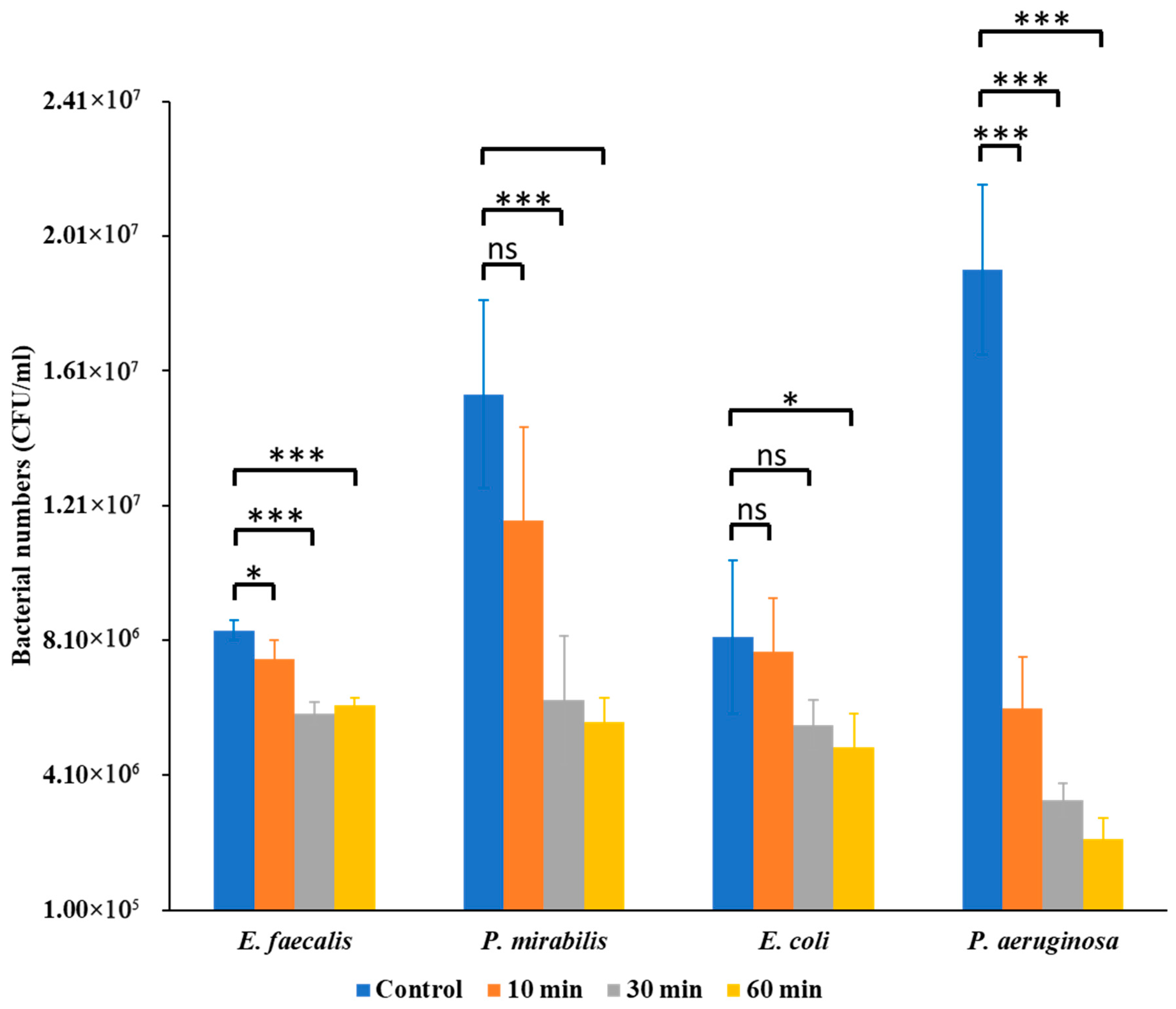
The antibacterial effects of Se NPs varied significantly among the tested uropathogens, revealing species-specific differences in susceptibility. Bacterial enumeration assays revealed that Se NPs exerted a notable inhibitory effect on bacterial CFU counts, but the magnitude of inhibition differed among species (Figure 1). E. faecalis showed moderate reductions at 30 min (p < 0.05) and stronger inhibition at 60 min (p < 0.01). P. mirabilis was more resilient, with no significant effect at 10 min but significant decreases at 30 min (p < 0.01) and 60 min (p < 0.001). E. coli exhibited progressive inhibition, significant at 30 min (p < 0.05) and 60 min (p < 0.01). P. aeruginosa was the most susceptible, showing highly significant reductions at 10, 30, and 60 min (p < 0.001 for all). In all cases, positive control samples showed significantly higher CFU counts than Se NP-treated samples.
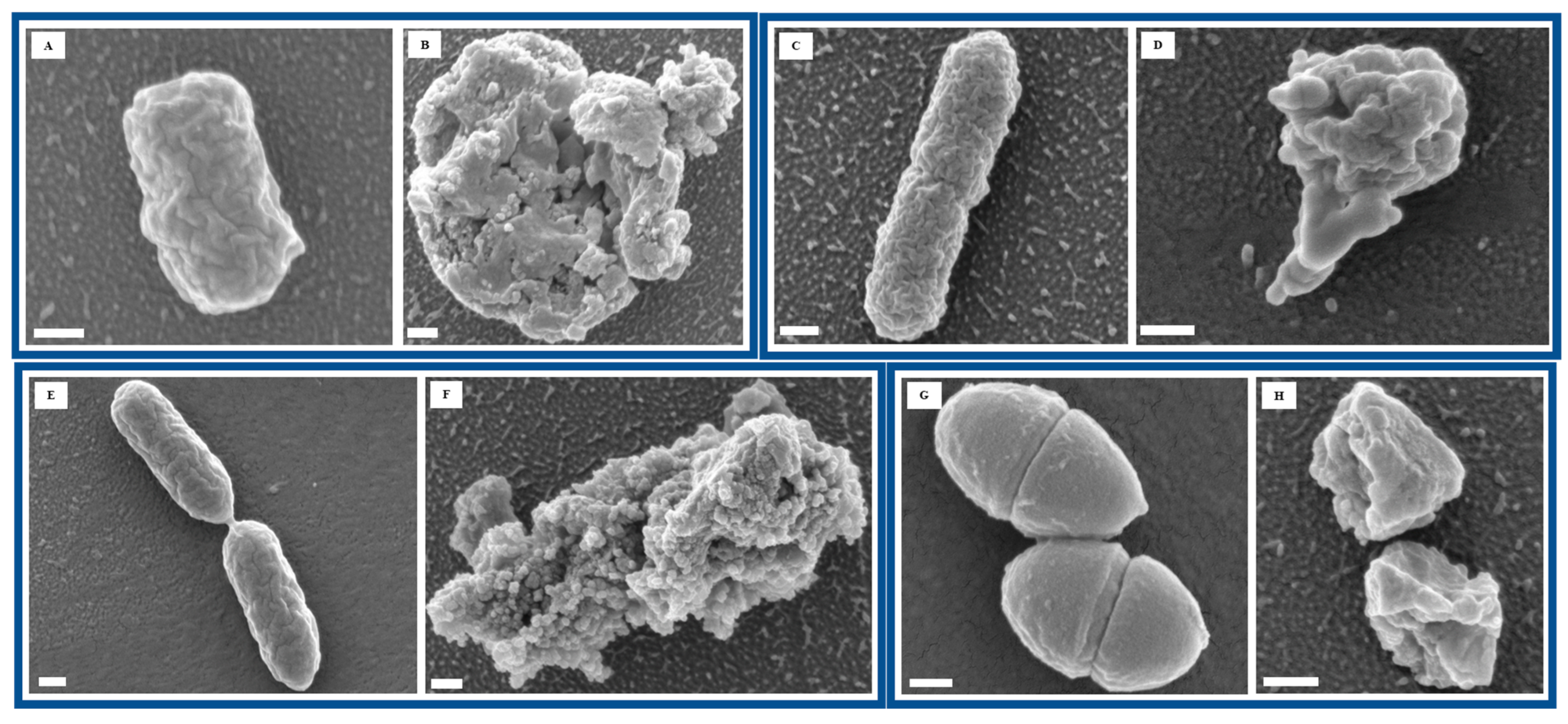
To further assess the impact of Se NP exposure on bacterial morphology, we performed FESEM to visualize structural changes at the cellular level. Imaging of untreated bacterial cells revealed smooth surfaces and intact cellular structures, indicative of healthy bacterial morphology (Figure 2A,C,E,G). However, after 60 min of Se NP exposure, all four bacterial species exhibited varying degrees of structural damage, suggesting significant morphological alterations in response to NP-induced stress (Figure 2B,D,F,H). Such morphological disruption points to bacterial membranes as a key target of Se NP action, leading to loss of integrity, leakage of intracellular contents, and eventual cell death.
These findings are consistent with prior reports that have demonstrated the broad-spectrum antibacterial potential of Se NPs. Shakibaie et al. showed that biogenic Se NPs inhibited P. aeruginosa and S. aureus growth, attributing their effects to oxidative stress and membrane damage [37]. Xu et al. reported that Se NPs penetrated bacterial membranes, triggering ROS-mediated toxicity [38]. More recently, Lin et al. confirmed Se NP-induced ROS generation and membrane disruption in S. aureus [39].
Taken together, these results reinforce the antibacterial potential of SeNPs while emphasizing that their efficacy is pathogen-dependent. The morphological and quantitative evidence provided here adds to the growing body of literature supporting SeNPs as promising antimicrobial candidates.

3.2. Comparative Proteome Profile

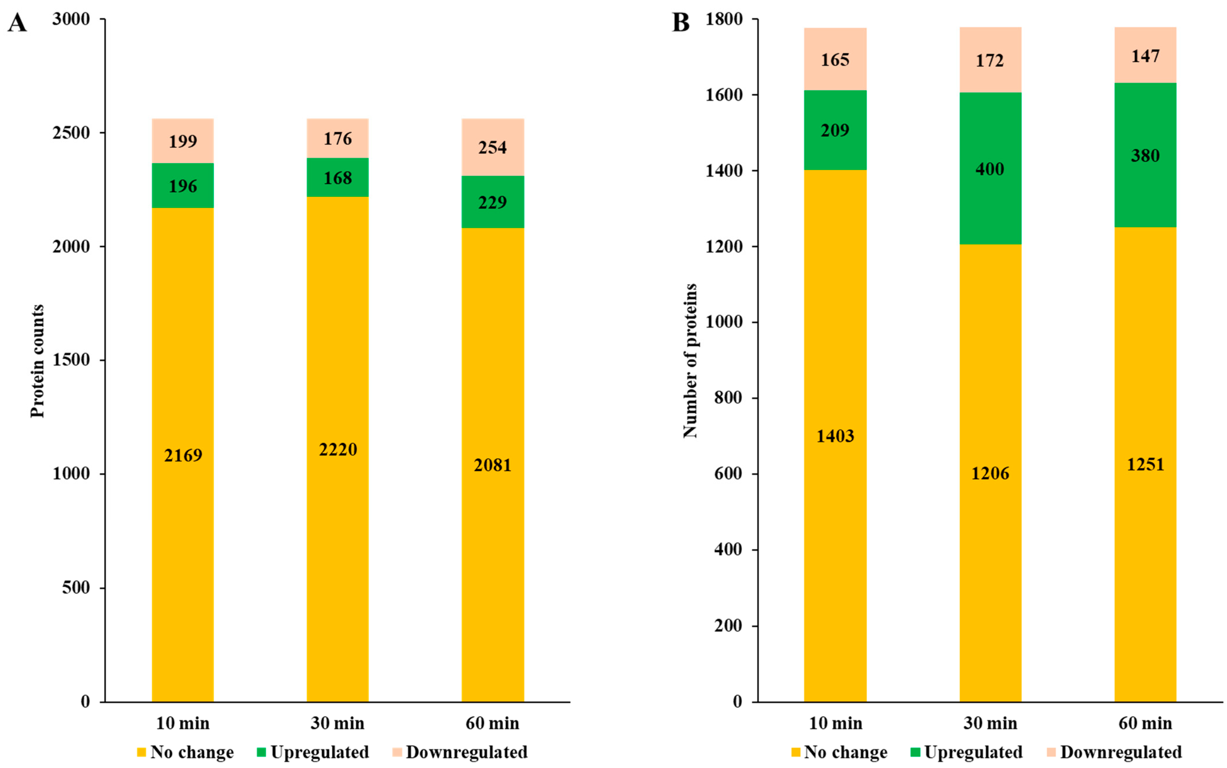
To gain a deeper understanding of bacterial adaptive mechanisms in response to Se NP exposure, we conducted a global proteomic analysis on P. aeruginosa and P. mirabilis, which exhibited the highest and lowest susceptibility, respectively. The proteomic profiling revealed distinct yet overlapping patterns of protein expression, demonstrating species-specific metabolic adjustments under NP-induced stress. Proteins showing no significant change in expression in P. aeruginosa ranged from 35.15% (2081 proteins) at 60 min to 37.50% (2220 proteins) at 30 min, while in P. mirabilis, these ranged from 33.21% (1206 proteins) at 30 min to 38.64% (1403 proteins) at 10 min (Figure 3A,B). The number of upregulated proteins was substantially higher in P. mirabilis, peaking at 30 min with 11.02% (400 proteins), compared to P. aeruginosa, which showed only 3.87% (229 proteins) at 60 min. This discrepancy suggests that P. mirabilis undergoes a more dynamic and rapid metabolic shift in response to Se NP-induced stress, likely reflecting an enhanced ability to modulate its metabolic pathways to counteract environmental stressors.

3.3. COG Functional Analysis

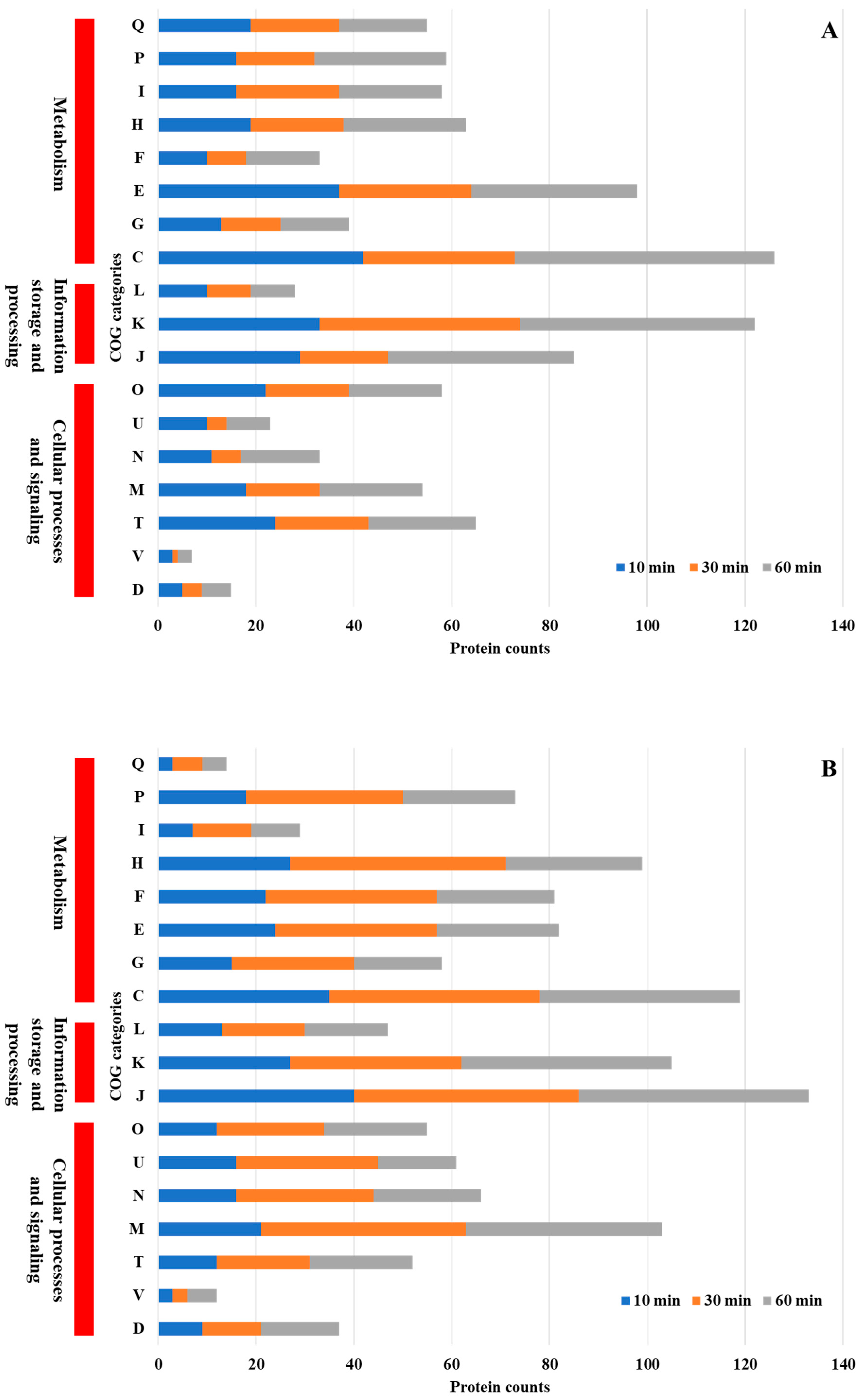
COG functional analysis of differentially expressed proteins revealed both shared and unique adaptive responses in P. aeruginosa and P. mirabilis following Se NP treatment (Figure 4A,B). Both P. aeruginosa and P. mirabilis exhibited significant alterations in protein expression related to core cellular functions, including energy production and conversion, transcriptional regulation, amino acid metabolism, and protein synthesis. These shared metabolic responses suggest that both species employ conserved strategies to maintain cellular homeostasis under NP-induced stress. Enhanced expression of proteins involved in energy metabolism and transcriptional regulation indicates that both bacteria activate stress response pathways that promote adenosine triphosphate (ATP) production and transcriptional reprogramming to sustain viability under adverse conditions.
Despite these similarities, species-specific adaptations were evident. P. mirabilis uniquely upregulated proteins associated with cell wall and membrane biogenesis. The enhanced expression of proteins involved in cell envelope synthesis suggests that P. mirabilis employs a protective strategy that strengthens its outer membrane and reduces NP permeability, thereby minimizing cellular disruption [40].
A comparison with prior research revealed both conserved and species-specific responses to NP-induced stress. For example, P. aeruginosa exposed to copper stress exhibited significant changes in inorganic ion transport and metabolism (P), energy production and conversion (C), amino acid transport and metabolism (E), cell wall/membrane/envelope biogenesis (M), and translation, ribosomal structure, and biogenesis (J) [41]. Similarly, when Streptococcus suis was exposed to silver NPs, key adaptive responses included translation, ribosomal structure, and biogenesis (J), cell wall/membrane/envelope biogenesis (M), amino acid transport and metabolism (E), transcription (K), and carbohydrate transport and metabolism (G) [42]. Likewise, Xanthomonas campestris pv. campestris under copper stress exhibited significant differential regulation of inorganic ion transport and metabolism (P), carbohydrate transport and metabolism (G), amino acid transport and metabolism (E), transcription (K), and replication, recombination, and repair (L) [43]. The observed similarities with bacterial responses to other NPs and heavy metals suggest that certain stress-adaptive mechanisms are conserved across species, while others are tailored to individual bacterial physiology.

3.4. KEGG Pathway Analysis

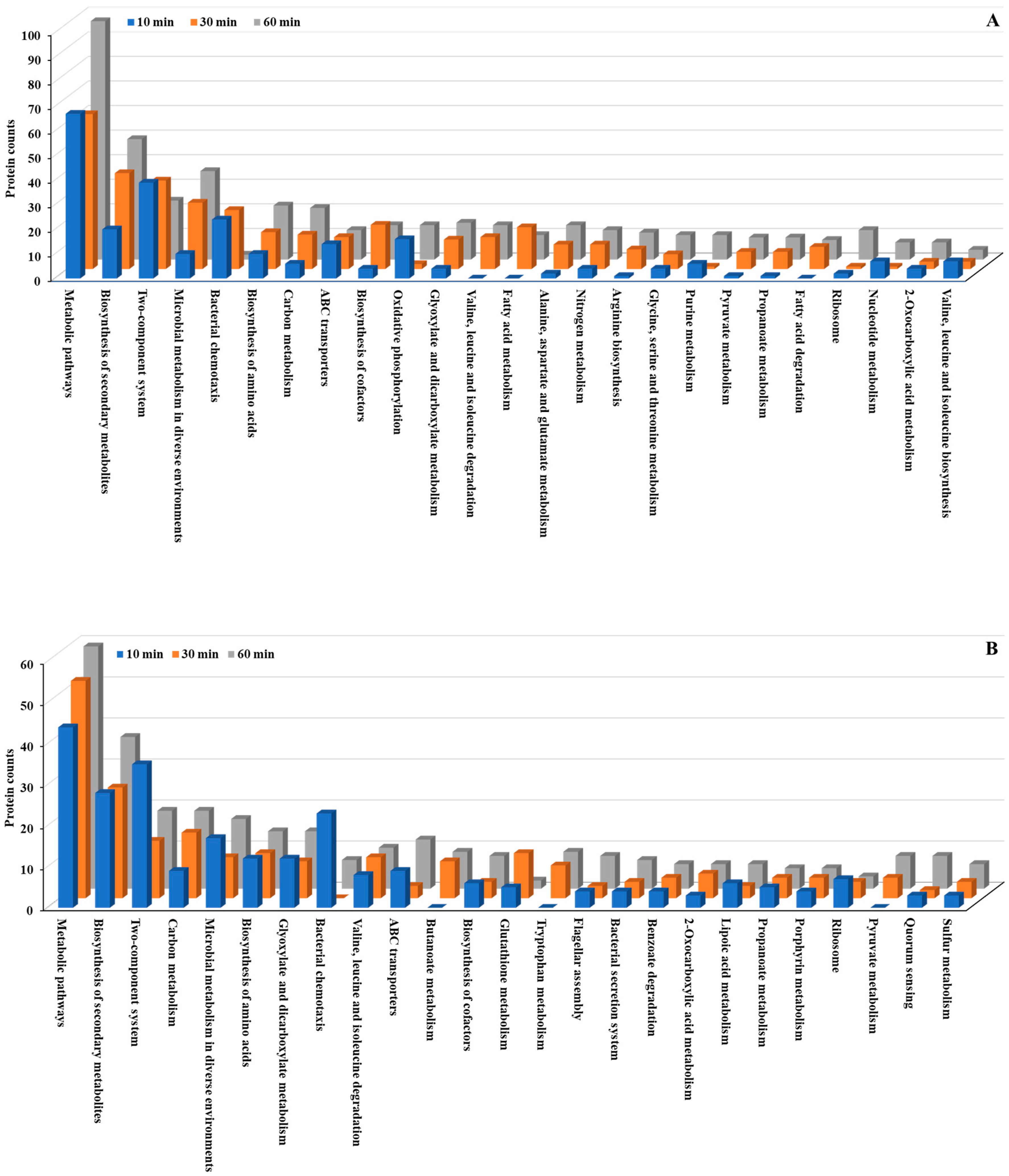
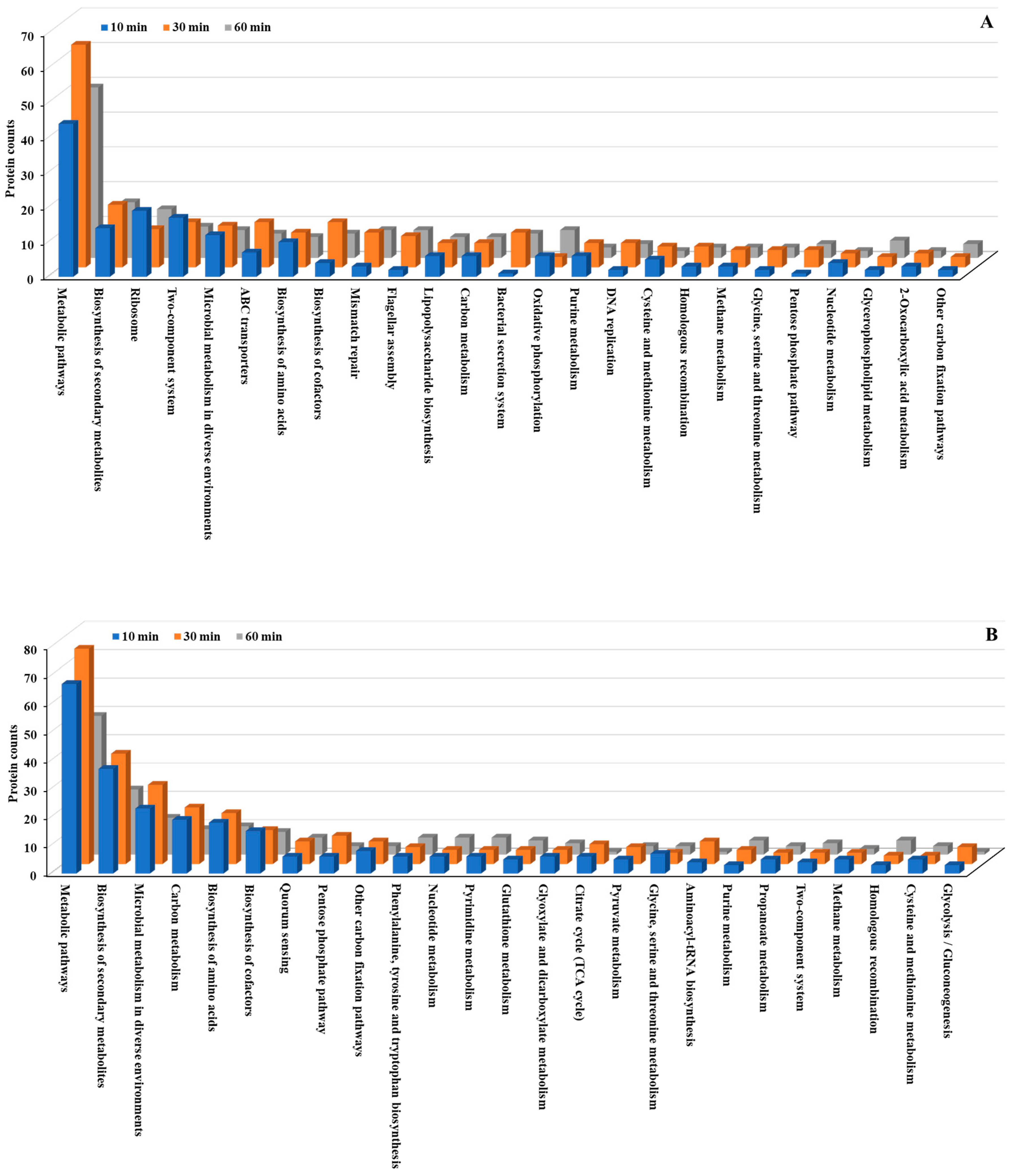
We conducted KEGG pathway analysis to further investigate the molecular mechanisms underlying the bacterial stress response to Se NP exposure. This analysis revealed significant alterations in metabolic pathways across both P. aeruginosa and P. mirabilis (Figure 5 and Figure 6; Tables S2, S3, S8 and S9). Metabolic pathways, including biosynthesis of secondary metabolites, microbial metabolism in diverse environments, and biosynthesis of amino acids, exhibited the most significant changes, with both upregulated and downregulated proteins in both bacteria. The extensive involvement of these pathways suggests that Se NP exposure induces a global metabolic shift in both species, requiring the reallocation of cellular resources to support survival under oxidative and structural stresses.
Beyond these fundamental cellular processes, several key pathways were markedly overexpressed in both species. Specifically, multiple two-component regulatory systems, along with pathways related to bacterial chemotaxis, flagellar assembly, and ATP-binding cassette (ABC) transporters, were significantly upregulated. These pathways are essential for bacterial sensing, mobility, and active transport [44], suggesting that Se NP exposure prompts both species to enhance environmental sensing and resource allocation mechanisms. This pattern underscores the bacteria’s ability to detect and respond to stress stimuli, regulate cellular motility, and manage transmembrane transport processes, all of which are crucial for mitigating NP-induced toxicity.
The two-component system is a highly conserved bacterial signaling mechanism that enables cells to sense environmental changes and rapidly trigger appropriate regulatory responses [45]. In both P. aeruginosa and P. mirabilis, Se NP treatment resulted in the upregulation of two-component system-associated proteins, including AlgB, AlgR, CcoN, CydA, CpxA, DctA, FlrC, GlnL, GlnR, NarG, PhoB, PhoR, and RegB. These proteins play essential roles in bacterial stress adaptation by modulating gene expression in response to such external stimuli as oxidative stress, metal ion toxicity, and membrane damage [46].
These findings align with previous findings. For example, Filipović et al. found that SeNPs with different surface chemistries induce distinct bacterial stress responses, including membrane-targeted effects, in E. coli and P. aeruginosa [12]. Moreover, Vahdati and Moghadam reported synergistic antibacterial effects when SeNPs were combined with lysozyme, among mechanisms that include disruption of bacterial signaling and cell envelope integrity [47].
The observed upregulation of these components suggests that Se NP exposure activates a broad stress-responsive signaling network, enabling bacteria to dynamically regulate gene expression and cellular functions to enhance survival. This response is consistent with findings from previous studies on bacterial adaptation to environmental stressors. For example, Salmonella Typhimurium exposed to high-intensity ultrasound exhibited significant upregulation of two-component system proteins [48]. Similarly, P. aeruginosa treated with antibiotics showed a comparable upregulation of two-component system components, suggesting that both antibiotic- and NP-induced stresses may elicit overlapping bacterial response mechanisms [49]. Additional evidence from studies on Staphylococcus epidermidis under tigecycline treatment and S. mutans under bacitracin stress further supports the critical role of this pathway in bacterial survival under diverse environmental challenges [50,51].
Chemotaxis and motility are crucial bacterial behaviors that facilitate movement toward favorable environments and away from harmful stressors [52]. In response to Se NP exposure, proteins associated with bacterial chemotaxis, including CheW, CheZ, FliG, FliM, Mcp, MotA, and RbsB, exhibited significant upregulation. Similarly, flagellar assembly proteins such as FlgB, FlgD, FlgF-H, FlgJ, FlgM, FliD, FliF-H, FliK, FliM, and FlrC were also overexpressed. This widespread upregulation suggests that enhanced bacterial motility is a key survival strategy for evading toxic NP interactions.
Previous studies have demonstrated that bacterial motility is a crucial adaptive mechanism under stress conditions. For instance, P. aeruginosa exposed to antibiotics exhibited a significant increase in bacterial chemotaxis gene expression, reinforcing the role of motility in bacterial adaptation [49]. Additionally, Natranaerobius thermophilus relied on bacterial chemotaxis to survive high-salt conditions, suggesting that motility plays a pivotal role in microbial stress responses across diverse bacterial species [53].
Bacterial cells also upregulated membrane transport mechanisms in response to Se NP-induced stress. ABC transporters, a superfamily of integral membrane proteins, play a vital role in nutrient uptake, efflux of toxic compounds, and overall bacterial homeostasis by utilizing ATP hydrolysis to transport a wide array of substrates [54]. After Se NP treatment, ABC transporter-associated proteins, including FtsX, LptB, MlaB, PotD, and ZnuA, were significantly upregulated, suggesting that bacteria prioritize transmembrane transport processes to mitigate NP-induced stress. Similarly, bacterial secretion system proteins, such as SecB, ShlA, TatA, TatB, VgrG, and YajC, were overexpressed, further supporting the hypothesis that Se NP exposure triggers an increase in transmembrane transport activity.
ABC transporters have been widely implicated in bacterial stress responses. In Brucella melitensis, ABC transporters were upregulated under salt stress to maintain osmotic balance, and in S. epidermidis, ABC transporter proteins were overexpressed in response to antibiotics, highlighting their importance in antimicrobial resistance mechanisms [50,55]. Furthermore, S. mutans exposed to theaflavins showed significant upregulation of membrane transport pathways, supporting the importance of transport mechanisms in bacterial adaptation to environmental challenges [56]. Interestingly, some studies have reported the downregulation of ABC transporters under acid stress, as observed in Lactiplantibacillus plantarum, where ABC transporter pathways were suppressed in response to pH changes [57], suggesting that bacterial responses to environmental stressors are context-dependent.

3.5. Expression Pattern (EPN) Analysis

Our EPN analysis revealed distinct yet overlapping adaptive responses. EPN clustering was used to categorize proteins based on their differential expression across time points, providing insights into metabolic and regulatory shifts triggered by NP exposure. P. aeruginosa exhibited 26 significant EPN clusters, whereas P. mirabilis had 23 clusters, indicating a comparable but distinct response to Se NP-induced stress (Tables S1 and S7).
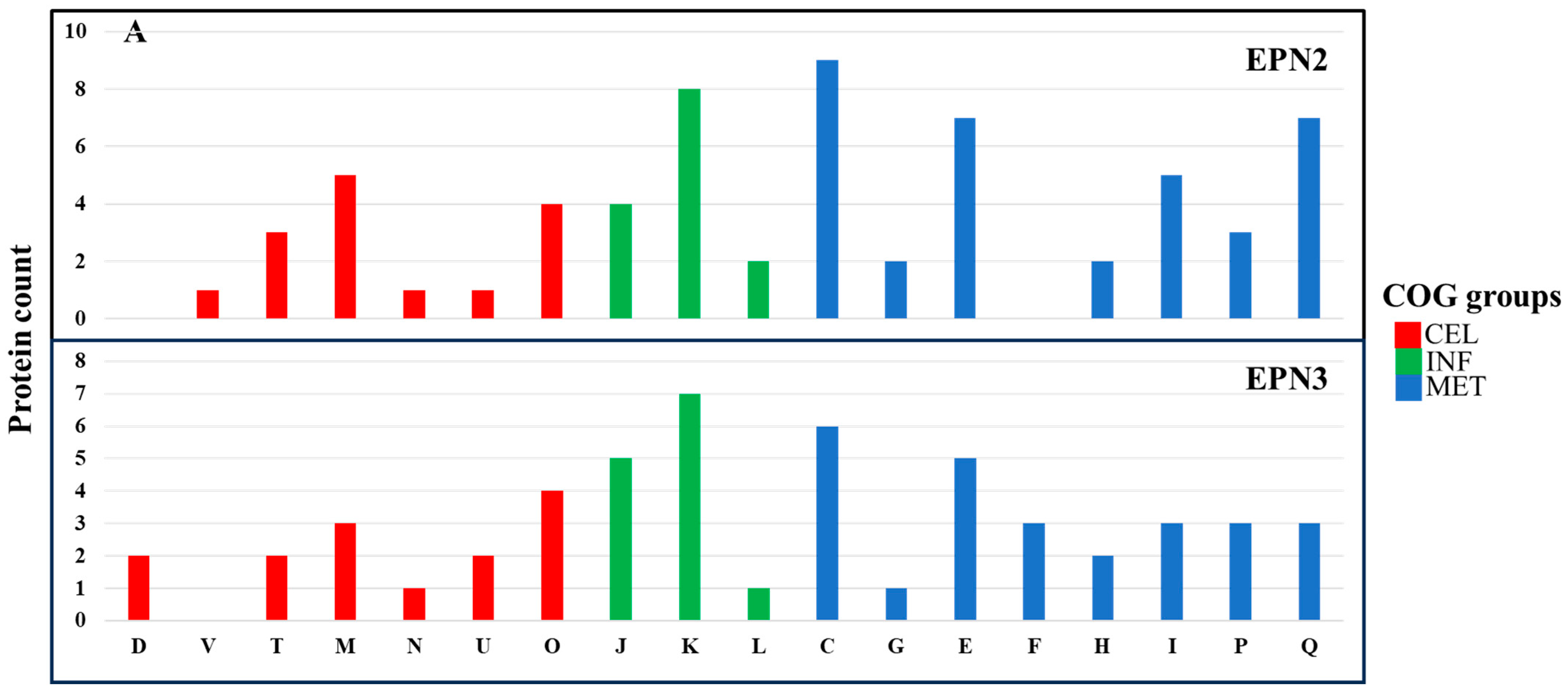
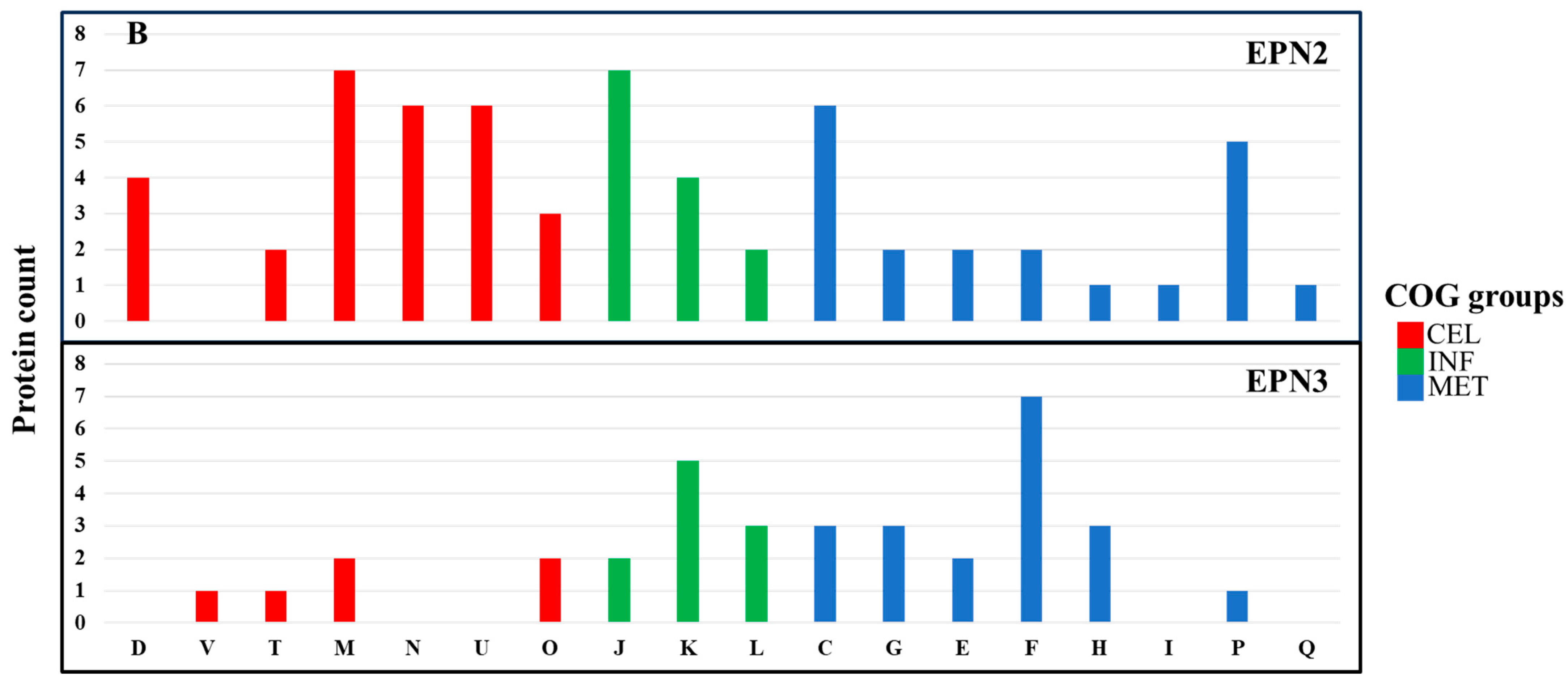
In the EPN2 dataset, which included proteins consistently overexpressed throughout the treatment, P. aeruginosa showed upregulation of 73 proteins involved primarily in energy production and conversion (C) (Figure 7A, Table S5). Among these, key components of the electron transport chain and ATP synthesis machinery, including AtpA, Fpr, GlcE, NapA, NuoM, Qor, RnfC, and RnfG, were markedly overexpressed [58]. The upregulation of these proteins suggests that P. aeruginosa responded to Se NP stress by increasing ATP production and optimizing oxidative phosphorylation. Additionally, the heightened expression of electron transport chain components, including NuoM and Qor, suggests a greater reliance on oxidative phosphorylation as a primary strategy to mitigate Se NP exposure-induced oxidative stress [59].
In contrast, P. mirabilis exhibited overexpression of 84 proteins in the EPN2 dataset, but with a different functional emphasis (Figure 7B, Table S11). The most significantly upregulated proteins were associated with cell wall/membrane/envelope biogenesis (M) and translation, ribosomal structure, and biogenesis (J). Proteins such as LptD, MipA, RfaB, RfaQ, Slp, TtgC, and YtfM, which are involved in cell envelope biogenesis [60], were significantly induced. This suggests that P. mirabilis reinforced its structural integrity in response to Se NP exposure [61]. Additionally, protein expression involved in translation, such as RpmF, RpmH, RpsS, RpsU, and TrmA, also had greatly increased, indicating that P. mirabilis prioritized sustaining ribosomal function to maintain protein synthesis under stress.
The EPN3 dataset, which included proteins consistently downregulated throughout the treatment, revealed further distinctions between the two species. In P. aeruginosa, 69 proteins were consistently downregulated, with transcription-related proteins (K) exhibiting the most pronounced decreases (Figure 7A, Table S6). Key transcriptional regulators such as Anr, ColR, Rnk, SpoT, and YebC showed significant downregulation, suggesting that Se NP exposure suppressed specific regulatory pathways governing stress responses. The suppression of transcriptional regulators such as Anr and ColR indicates a shift away from certain metabolic pathways, likely as an energy conservation strategy in response to stress [62].
In P. mirabilis, 40 proteins were consistently downregulated; the most affected category was nucleotide transport and metabolism (F) (Figure 7B, Table S12). Proteins such as Add, ChbG, Cmk, GpmB, KdsA, PneB, and Udk, which are involved in nucleotide biosynthesis and metabolism [63], exhibited significant decreases in expression.

3.6. Differentially Expressed Proteins Associated with Virulence Factors

Se NP treatment resulted in both shared and species-specific regulation of virulence-associated proteins. P. aeruginosa exhibited 47 differentially expressed virulence-related proteins, while P. mirabilis displayed a broader response, with 59 differentially expressed virulence-associated proteins (Table 2 and Table 3). Despite differences in the number of virulence factors affected, both species exhibited overlapping responses in key functional categories, including adherence, biofilm formation, secretion systems, immune modulation, motility, and nutrient acquisition. However, P. mirabilis displayed additional regulation of stress survival- and exotoxin-related proteins. Proteins are involved in capsular polysaccharide synthesis, lipid A modifications, and efflux-mediated immune resistance [64]. The broader immune modulation response in P. mirabilis suggests that this species may rely more extensively on modifying its cell surface structures to resist NP-induced stress. Previous research has shown that antimicrobial agents and NPs can induce modifications in outer membrane components, influencing bacterial immune evasion strategies [65,66].
In terms of adherence, P. aeruginosa regulated such proteins as ChpB, CheW, FimL, FimX, RpoN, RpoS, PilB, PilH, PilI, and PilU, all of which contribute to bacterial attachment and colonization [67]. Similarly, P. mirabilis exhibited differential expression of adherence-associated proteins, including Crp, MatB, MetQ, MrfD, Tuf, and Uca. The presence of multiple adherence-related proteins in both bacteria suggests that Se NP exposure may influence bacterial attachment properties, possibly altering surface interactions that impact colonization and biofilm formation. Biofilm-associated proteins were also significantly affected in both species. P. aeruginosa altered the expression of AlgB, AlgP, AlgR, LasR, and RhlR, which are key regulators of alginate biosynthesis and quorum sensing, crucial for biofilm maintenance [67]. In contrast, P. mirabilis differentially expressed such biofilm-related proteins as KGA92257.1 and RpoE. These findings suggest that Se NP exposure may disrupt bacterial aggregation and biofilm maturation, potentially weakening bacterial persistence mechanisms. Previous studies have demonstrated that silver NPs can interfere with bacterial adhesion and biofilm formation by disrupting cell surface structures and altering quorum-sensing pathways [68].
Secretion system proteins were altered in both species, indicating an increased emphasis on protein translocation and virulence factor secretion under stress. In P. aeruginosa, several type II and type III secretion system components, including GspE, XcpR-S, and YebC [67], were differentially expressed. Similarly, P. mirabilis exhibited differential expression of secretion system-related proteins, including Hcp, ImpB, Impl, PpiA, and TssQ_1, components associated with the type VI secretion system [64]. These findings suggest that both bacteria respond to Se NP stress by modulating secretion system components, possibly to facilitate the export of virulence factors or stress response proteins. Similar trends have been observed in Vibrio parahaemolyticus and S. aureus, where exposure to antimicrobial agents led to the modulation of secretion systems, aiding in bacterial persistence [69,70].
Immune modulation-related proteins exhibited distinct patterns between the two species. P. aeruginosa showed regulation of Gmd, LpxK, RfaG, RfbA, and RhlA, proteins involved in immune evasion and bacterial defense [67]. In contrast, P. mirabilis displayed a broader immune modulation response, with differentially expressed proteins including AcpP, GalE, GlmU, KdsA, KdtA, LpxB-C, LpxL, MsbA, RfaC, and WecC. Many of these motility-associated proteins were widely affected in both bacteria, indicating a potential impact on bacterial movement and environmental navigation under stress. In P. aeruginosa, proteins such as CheW, DctD, FlaG, FleR, FlgD-G, FlgL, FlgN, FliC, FliF, FliH, MotA, MotD, and NtrC were differentially expressed, while P. mirabilis exhibited regulation of motility-related proteins, including CheZ, FlgB, FlgF-H, FlgJ, FlgM, FliG-H, FliM, GlnG, Tap, and YfhA. These findings indicate that both species may be modulating their flagellar and chemotactic systems in response to Se NP exposure.
A key distinction between the two species was the differential regulation of stress survival proteins. P. mirabilis, but not P. aeruginosa, exhibited differential regulation of ClpB, KatA, SodC, and UreC, all of which are associated with bacterial stress survival and oxidative stress resistance [64]. This suggests that P. mirabilis may rely on oxidative stress resistance mechanisms to counteract Se NP toxicity, likely through increased antioxidant enzyme activity and proteolysis of damaged proteins. In contrast, the absence of similar stress survival proteins in P. aeruginosa may indicate that this species mitigates oxidative stress through alternative pathways, such as enhanced energy metabolism or efflux system activation.
Selenium nanoparticles are well known to catalyze the generation of reactive oxygen species, which impose oxidative stress and damage key biomolecules including DNA, proteins, and lipids. Han et al. demonstrated that Se NPs induce intracellular reactive oxygen species accumulation in methicillin-resistant S. aureus and vancomycin-resistant Enterococcus, ultimately triggering apoptosis-like bacterial death [13]. Consistent with this, our proteomic analysis revealed upregulation of stress response proteins, further supporting reactive oxygen species-mediated oxidative stress as a central mechanism of Se NP antibacterial activity.

3.7. Differentially Expressed Proteins Associated with Antimicrobial Resistance

The analysis of differentially expressed proteins associated with antimicrobial resistance in P. aeruginosa and P. mirabilis following Se NP treatment revealed distinct regulatory patterns, indicating species-specific strategies for mitigating NP-induced stress and potential antimicrobial effects (Table 4 and Table 5). In P. aeruginosa, 10 antimicrobial resistance-related proteins were differentially expressed, including efflux system components (AdeR, MexG, MexH, MexL, MexV, OpmD, RsmA) and antibiotic target replacement proteins (DfrA3, MyrA, RpoB). In P. mirabilis, 10 antimicrobial resistance-associated proteins were also differentially expressed, including efflux-related transporters (CpxA, CpxR, Crp, Hns, LptD, MsbA, RsmA, TolC), an antibiotic target alteration regulator (PhoB), and an antibiotic-modifying enzyme (CatA4). The expression patterns suggest that both bacteria employ efflux-mediated resistance and structural modifications to counteract Se NP-induced stress.
Efflux pump-associated proteins were regulated in both species, suggesting that active transport mechanisms play a central role in bacterial adaptation to Se NP exposure. In P. aeruginosa, MexG, MexH, MexV, and OpmD—components of the MexGHI-OpmD efflux system—were differentially expressed, indicating increased activity of the resistance-nodulation-cell division (RND) efflux system [71]. These efflux pumps are known to contribute to multidrug resistance by expelling toxic compounds, including antimicrobials and heavy metals, from Gram-negative bacteria. Additionally, the MexGHI-OpmD efflux system has been reported to be involved in stress responses in P. aeruginosa, further supporting its role in counteracting NP-induced toxicity [71].
Similarly, P. mirabilis displayed overexpression of TolC, MsbA, and LptD, components of tripartite efflux pumps and lipopolysaccharide-assembly pathways [72]. These proteins facilitate resistance against antimicrobial agents, metal ions, and environmental stressors. The upregulation of these efflux-related proteins suggests that P. mirabilis employs efflux-mediated detoxification to reduce intracellular accumulation of Se NPs, a common strategy observed in bacteria exposed to heavy metals and antibiotics [73,74].
Regulatory proteins governing antimicrobial resistance and stress responses were also differentially expressed in both species, indicating complex transcriptional and post-transcriptional regulation in response to Se NPs. In P. aeruginosa, RsmA, AdeR, and MexL were differentially expressed. RsmA plays a crucial role in secondary metabolism and antimicrobial resistance by modulating RNA stability and translation [75]. AdeR, a well-known regulator of efflux pump expression, facilitates adaptive resistance by modulating bacterial responses to environmental stress [76]. The observed downregulation of MexL, a negative regulator of the MexXY efflux system, suggests that Se NP exposure may trigger the derepression of certain efflux pumps, possibly leading to increased antimicrobial resistance [72].

3.8. Differentially Expressed Proteins Associated with Heavy Metal Resistance

The analysis of differentially expressed proteins associated with heavy metal resistance in P. aeruginosa and P. mirabilis following Se NP treatment revealed distinct regulatory patterns, reflecting species-specific strategies for coping with metal-induced stress (Table 6 and Table 7). P. aeruginosa exhibited differential expression of 17 heavy metal resistance-related proteins, including metal transporters (CorA, CorD, CueA), mercury detoxification proteins (MerA, MerP, MerR), regulatory transcription factors (CopR, MexT, ComR, CpxR, RpoS), and stress tolerance proteins (NirD/YgiW/YdeI family). In contrast, P. mirabilis displayed a broader response, with 25 differentially expressed heavy metal resistance proteins, including tellurite resistance proteins (TerB, TerD, TehB), phosphate transporters (PitA, PstB, PstS), copper homeostasis proteins (CutC, CuiD), metal-binding proteins (ZinT, ZntA), and regulatory proteins (ModE, PcoR, CpxR, IclR). These findings suggest that while both species employ metal transport, detoxification, and regulatory mechanisms, P. mirabilis activates a more extensive set of resistance pathways to counteract Se NP-induced toxicity.
Differential expression of metal transporters in both P. aeruginosa and P. mirabilis highlights the crucial role of metal ion homeostasis in their adaptive responses to Se NP stress. In P. aeruginosa, the expression of CorA (ion transporter) and CorD (a Co2+/Mg2+ efflux protein) was modulated, indicating a tightly controlled mechanism for metal ion homeostasis. Additionally, CueA, a copper-translocating P-type ATPase, was differentially expressed, suggesting that P. aeruginosa employs active copper transport mechanisms to mitigate metal toxicity. These findings are consistent with previous reports showing that CueA contributes to metal homeostasis and resistance in P. aeruginosa exposed to metal stress [77].
In P. mirabilis, multiple transport proteins were modulated, including CutC (copper homeostasis protein), CuiD (blue copper oxidase), ZinT (metal-binding protein), and ZntA (cadmium-translocating P-type ATPase). CutC and CuiD are known to contribute to bacterial copper tolerance, potentially by modulating copper oxidation and transport [78]. Meanwhile, ZinT and ZntA contribute to resistance against multiple heavy metals, including zinc, lead, cobalt, mercury, and cadmium, suggesting that P. mirabilis employs a wider range of transporters to maintain intracellular metal ion balance [79].
A notable difference between the two species was the activation of mercury resistance proteins in P. aeruginosa. Mer operon components MerA (Mercury(II) reductase), MerP (periplasmic binding protein), and MerR (mercury resistance transcriptional regulator) were differentially expressed. The MerA enzyme plays a crucial role in reducing toxic mercury (Hg2+) to volatile elemental mercury (Hg0), which is then expelled from the cell [80]. Presence of these detoxification proteins suggests that Se NP exposure may regulate heavy metal detoxification pathways in P. aeruginosa.
Conversely, P. mirabilis did not exhibit mercury resistance protein expression but instead displayed upregulation of tellurite resistance proteins (TerB, TerD, TehB). Tellurite resistance has been linked to oxidative stress protection, suggesting that P. mirabilis may rely on tellurite resistance mechanisms rather than mercury detoxification to mitigate NP-induced stress [81]. The upregulation of tellurite resistance proteins supports previous findings that tellurite resistance pathways enhance bacterial survival under oxidative and metal stress conditions [82].
Differential expression of several membrane-associated proteins suggests that both species undergo structural remodeling of their outer membranes to adapt to Se NP-induced stress. In P. aeruginosa, ModC (a heme ABC transporter ATP-binding protein) and VcaM (a lipid A export permease/ATP-binding protein) were differentially expressed, potentially indicating modifications in membrane permeability and lipid transport. Lipid A modifications have been shown to contribute to bacterial resistance against metal toxicity by reducing membrane permeability to toxic ions [83].
In P. mirabilis, LptD (lipopolysaccharide-assembly protein) and PitA (a low-affinity phosphate transporter) were differentially expressed, further supporting the hypothesis that membrane integrity and phosphate metabolism play crucial roles in stress adaptation. Increased expression of LptD suggests that P. mirabilis may reinforce its lipopolysaccharide layer to mitigate Se NP-induced damage, a strategy previously observed in bacteria exposed to metal stress [84].
A limitation of this study is that we conducted proteomic analysis only on P. aeruginosa and P. mirabilis, which exhibited the highest and lowest susceptibility to Se NPs, respectively. While this approach provided valuable insights into species-specific adaptive responses, it did not capture the full spectrum of proteomic changes across all tested uropathogens, including E. coli and E. faecalis. As a result, potential differences in stress adaptation mechanisms among other bacterial species remain unexplored. Future studies should expand proteomic analysis over a broader range of pathogens to provide a more comprehensive understanding of bacterial responses to Se NP-induced stress.
Another consideration is the potential loss of proteins during sample processing. Since Se NPs induce cell membrane damage, proteins—particularly secreted proteins or intracellular proteins released due to cell rupture—may leak into the surrounding medium and be lost during the centrifugation step post-treatment. This means that the detected proteomic profile primarily reflects proteins retained within partially intact or damaged cells and may not represent the full proteome. Furthermore, differences in proteomic profiles between P. aeruginosa and P. mirabilis could partly be attributed to differences in cellular integrity, as P. aeruginosa is more susceptible and prone to lysis, whereas P. mirabilis is more resistant. Despite this limitation, the detection of differentially regulated proteins provides meaningful insight into bacterial stress responses and adaptation strategies following Se NP exposure.
Finally, due to budgetary constraints, proteome analyses were not conducted in triplicates. Performing proteomic profiling for two bacterial species across three time points would have required substantial additional resources beyond the scope of the current funding. While this represents a limitation in statistical replication, we minimized variability by applying stringent data processing, normalization, and filtering criteria. The observed pathway-level trends were consistent with established bacterial stress responses, lending confidence to the robustness of the findings. Future studies, contingent on available funding, should aim to include triplicate analyses to further enhance the statistical power and reproducibility of the proteomic data.
4. Conclusions

This study provides comprehensive insights into the antibacterial mechanisms of Se NPs against uropathogenic bacteria. Antibacterial assays demonstrated that Se NPs effectively inhibited bacterial growth, though susceptibility varied among species. Our findings revealed that Se NPs induce significant morphological alterations and trigger distinct proteomic responses in uropathogens. While both P. aeruginosa and P. mirabilis modulated core cellular functions, including energy metabolism and transcriptional regulation, P. mirabilis displayed a more pronounced upregulation of proteins involved in cell wall biogenesis. The upregulation of two-component systems, chemotaxis, flagellar assembly, and ABC transporters in both species underscores the importance of environmental sensing, motility, and transmembrane transport in mitigating NP toxicity. Furthermore, analysis of virulence, antimicrobial resistance, and heavy metal resistance proteins revealed species-specific adaptations, including differential regulation of detoxification pathways. These results accentuate the complexity of bacterial responses to Se NP exposure and emphasize the importance of considering species-specific adaptations when evaluating their potential as antimicrobial agents.

Supplementary Materials

The following supporting information can be downloaded at https://www.mdpi.com/article/10.3390/nano15181404/s1, Table S1: Proteins identified from P. aeruginosa PA14 following selenium nanoparticle treatment; Table S2: KEGG pathways of upregulated proteins from P. aeruginosa PA14 following selenium nanoparticle treatment; Table S3: KEGG pathways of downregulated proteins from P. aeruginosa PA14 following selenium nanoparticle treatment; Table S4: Proteins that remained consistently unchanged throughout the time course of selenium nanoparticle treatment in P. aeruginosa PA14; Table S5: Proteins consistently upregulated throughout the time course of selenium nanoparticle treatment in P. aeruginosa PA14; Table S6: Proteins consistently downregulated throughout the time course of selenium nanoparticle treatment in P. aeruginosa PA14; Table S7: Proteins identified from P. mirabilis ATCC 7002 following selenium nanoparticle treatment; Table S8: KEGG pathways of upregulated proteins from P. mirabilis ATCC 7002 following selenium nanoparticle treatment; Table S9: KEGG pathways of downregulated proteins from P. mirabilis ATCC 7002 following selenium nanoparticle treatment; Table S10: Proteins that remained consistently unchanged throughout the time course of selenium nanoparticle treatment in P. mirabilis ATCC 7002; Table S11: Proteins consistently upregulated throughout the time course of selenium nanoparticle treatment in P. mirabilis ATCC 7002; Table S12: Proteins consistently downregulated throughout the time course of selenium nanoparticle treatment in P. mirabilis ATCC 7002. Figure S1: Pulsed laser ablation in liquids; Figure S2: Workflow of the antibacterial activity assay.

Author Contributions

Conceptualization, K.S.; methodology, K.S., M.P., O.K., A.S., A.P., M.S. and S.M.; software, K.S.; validation, K.S.; formal analysis, K.S.; investigation, K.S.; resources, K.S. and S.F.; data curation, K.S.; writing—original draft preparation, K.S.; writing—review and editing, K.S., M.P., A.S., A.P., S.K. and S.F.; visualization, K.S.; supervision, K.S.; project administration, K.S. and S.F.; funding acquisition, K.S. and S.F. All authors have read and agreed to the published version of the manuscript.

Funding

This work was funded by the U.S. Food and Drug Administration, grant number E0781501.

Data Availability Statement

Authors can provide raw data upon request.

Acknowledgments

We acknowledge Seongjae Kim and Seongwon Nho for their review of this manuscript, and Joanne Berger at the FDA Library for her editing contributions. We also extend our sincere gratitude to Gregory Guisbiers (School of Physical Sciences, University of Arkansas at Little Rock, AR) for generously providing the Se NPs, and to Vincent Lee (Department of Cell Biology and Molecular Genetics, University of Maryland, College Park) for providing the P. aeruginosa PA14 strain. The authors used GPT-4o (OpenAI) to assist with language editing and to enhance the clarity and readability of the manuscript. The AI tool was employed solely for linguistic refinement and did not contribute to the conceptual development, data analysis, or interpretation of results. All AI-assisted content was subsequently reviewed and edited by the authors to ensure accuracy and originality.

Conflicts of Interest

The authors declare no conflicts of interest.

Abbreviations

The following abbreviations are used in this manuscript:
UTIsUrinary tract infections
Se NPsSelenium nanoparticles
COGCluster of Orthologous Groups
KEGGKyoto Encyclopedia of Genes and Genomes
CFUColony-forming units
FESEMField emission scanning electron microscopy
LC-MS/MSLiquid chromatography with tandem mass spectrometry
EPNExpression pattern
MDRMultidrug-resistant

References

  1. Yang, X.; Chen, H.; Zheng, Y.; Qu, S.; Wang, H.; Yi, F. Disease burden and long-term trends of urinary tract infections: A worldwide report. Front. Public Heath 2022, 10, 888205. [Google Scholar] [CrossRef]
  2. Flores-Mireles, A.L.; Walker, J.N.; Caparon, M.; Hultgren, S.J. Urinary tract infections: Epidemiology, mechanisms of infection and treatment options. Nat. Rev. Microbiol. 2015, 13, 269–284. [Google Scholar] [CrossRef]
  3. Wagenlehner, F.M.E.; Bjerklund Johansen, T.E.; Cai, T.; Koves, B.; Kranz, J.; Pilatz, A.; Tandogdu, Z. Epidemiology, definition and treatment of complicated urinary tract infections. Nat. Rev. Urol. 2020, 17, 586–600. [Google Scholar] [CrossRef] [PubMed]
  4. Foxman, B.; Brown, P. Epidemiology of urinary tract infections. Infect. Dis. Clin. N. Am. 2003, 17, 227–241. [Google Scholar] [CrossRef] [PubMed]
  5. Bader, M.S.; Loeb, M.; A Brooks, A. An update on the management of urinary tract infections in the era of antimicrobial resistance. Postgrad. Med. 2016, 129, 242–258. [Google Scholar] [CrossRef]
  6. Foxman, B. The epidemiology of urinary tract infection. Nat. Rev. Urol. 2010, 7, 653–660. [Google Scholar] [CrossRef]
  7. Zhang, Q.; Zhou, H.; Jiang, P.; Xiao, X. Metal-based nanomaterials as antimicrobial agents: A novel driveway to accelerate the aggravation of antibiotic resistance. J. Hazard. Mater. 2023, 455, 131658. [Google Scholar] [CrossRef]
  8. Salmani-Zarchi, H.; Mousavi-Sagharchi, S.M.A.; Sepahdoost, N.; Ranjbar-Jamalabadi, M.; Gross, J.D.; Jooya, H.; Samadi, A. Antimicrobial Feature of Nanoparticles in the Antibiotic Resistance Era: From Mechanism to Application. Adv. Biomed. Res. 2024, 13, 113. [Google Scholar] [CrossRef]
  9. Hariharan, S.; Dharmaraj, S. Selenium and selenoproteins: It’s role in regulation of inflammation. Inflammopharmacology 2020, 28, 667–695. [Google Scholar] [CrossRef]
  10. Bai, S.; Zhang, M.; Tang, S.; Li, M.; Wu, R.; Wan, S.; Chen, L.; Wei, X.; Feng, S. Effects and Impact of Selenium on Human Health, A Review. Molecules 2024, 30, 50. [Google Scholar] [CrossRef] [PubMed]
  11. Zambonino, M.C.; Quizhpe, E.M.; Mouheb, L.; Rahman, A.; Agathos, S.N.; Dahoumane, S.A. Biogenic Selenium Nanoparticles in Biomedical Sciences: Properties, Current Trends, Novel Opportunities and Emerging Challenges in Theranostic Nanomedicine. Nanomaterials 2023, 13, 424. [Google Scholar] [CrossRef] [PubMed]
  12. Filipović, N.; Ušjak, D.; Milenković, M.T.; Zheng, K.; Liverani, L.; Boccaccini, A.R.; Stevanović, M.M. Comparative Study of the Antimicrobial Activity of Selenium Nanoparticles With Different Surface Chemistry and Structure. Front. Bioeng. Biotechnol. 2021, 8, 624621. [Google Scholar] [CrossRef]
  13. Han, H.-W.; Patel, K.D.; Kwak, J.-H.; Jun, S.-K.; Jang, T.-S.; Lee, S.-H.; Knowles, J.C.; Kim, H.-W.; Lee, H.-H.; Lee, J.-H. Selenium Nanoparticles as Candidates for Antibacterial Substitutes and Supplements against Multidrug-Resistant Bacteria. Biomolecules 2021, 11, 1028. [Google Scholar] [CrossRef] [PubMed]
  14. Ridha, D.M.; Al-Awady, M.J.; Al-Zwaid, A.J.A.; Balakit, A.A.; Al-Dahmoshi, H.O.; Alotaibi, M.H.; El-Hiti, G.A. Antibacterial and antibiofilm activities of selenium nanoparticles-antibiotic conjugates against anti-multidrug-resistant bacteria. Int. J. Pharm. 2024, 658, 124214. [Google Scholar] [CrossRef] [PubMed]
  15. Salah, M.; Elkabbany, N.A.S.; Partila, A.M. Evaluation of the cytotoxicity and antibacterial activity of nano-selenium prepared via gamma irradiation against cancer cell lines and bacterial species. Sci. Rep. 2024, 14, 20523. [Google Scholar] [CrossRef]
  16. Goda, R.M.; Maghrabi, I.; El-Badawy, M.F.; Kabel, A.M.; Omar, A.; El-Morsi, R.M.; Ramadan, H.; Shohayeb, M.M. Developing a Urinary Catheter with Anti-Biofilm Coated Surface Using Phyto-Assisted Synthesis of Zinc Oxide Nanoparticles. Infect. Drug Resist. 2025, 18, 1881–1893. [Google Scholar] [CrossRef]
  17. Sewid, A.H.; Sharaf, M.; El-Demerdash, A.S.; Ragab, S.M.; Al-Otibi, F.O.; Yassin, M.T.; Liu, C.-G. Hexagonal zinc oxide nanoparticles: A novel approach to combat multidrug-resistant Enterococcus faecalis biofilms in feline urinary tract infections. Front. Cell. Infect. Microbiol. 2025, 14, 1505469. [Google Scholar] [CrossRef]
  18. Francis, A.; Namasivayam, S.K.R.; Samrat, K. Potential of silver nanoparticles synthesized from Justicia adhatoda metabolites for inhibiting biofilm on urinary catheters. Microb. Pathog. 2024, 196, 106957. [Google Scholar] [CrossRef]
  19. NikAkhtar, A.; SamadiAfshar, S.; Farahmand, S. Antibacterial Property of Silver Nanoparticles Green Synthesized from Stachys schtschegleevii Plant Extract on Urinary Tract Infection Bacteria. Curr. Microbiol. 2024, 81, 135. [Google Scholar] [CrossRef]
  20. Saikawa, G.I.A.; Guidone, G.H.M.; Noriler, S.A.; Reis, G.F.; de Oliveira, A.G.; Nakazato, G.; Rocha, S.P.D. Green-Synthesized Silver Nanoparticles in the Prevention of Multidrug-Resistant Proteus mirabilis Infection and Incrustation of Urinary Catheters BioAgNPs Against P. mirabilis Infection. Curr. Microbiol. 2024, 81, 100. [Google Scholar] [CrossRef]
  21. Said, A.; Abu-Elghait, M.; Atta, H.M.; Salem, S.S. Antibacterial Activity of Green Synthesized Silver Nanoparticles Using Lawsonia inermis Against Common Pathogens from Urinary Tract Infection. Appl. Biochem. Biotechnol. 2023, 196, 85–98. [Google Scholar] [CrossRef] [PubMed]
  22. Diksha, D.; Gupta, S.K.; Gupta, P.; Banerjee, U.C.; Kalita, D. Antibacterial Potential of Gold Nanoparticles Synthesized From Leaf Extract of Syzygium cumini Against Multidrug-Resistant Urinary Tract Pathogens. Cureus 2023, 15, e34830. [Google Scholar] [CrossRef]
  23. Lethongkam, S.; Paosen, S.; Bilhman, S.; Dumjun, K.; Wunnoo, S.; Choojit, S.; Siri, R.; Daengngam, C.; Voravuthikunchai, S.P.; Bejrananda, T. Eucalyptus-Mediated Synthesized Silver Nanoparticles-Coated Urinary Catheter Inhibits Microbial Migration and Biofilm Formation. Nanomaterials 2022, 12, 4059. [Google Scholar] [CrossRef]
  24. Chakraborty, A.; Haque, S.M.; Dey, D.; Mukherjee, S.; Ghosh, B. Phytogenic silver nanoparticles from tissue-cultured Kaempferia angustifolia — an underutilized medicinal herb: A comparative antibacterial study on urinary pathogens. J. Genet. Eng. Biotechnol. 2022, 20, 131. [Google Scholar] [CrossRef]
  25. Swidan, N.S.; Hashem, Y.A.; Elkhatib, W.F.; Yassien, M.A. Antibiofilm activity of green synthesized silver nanoparticles against biofilm associated enterococcal urinary pathogens. Sci. Rep. 2022, 12, 3869. [Google Scholar] [CrossRef]
  26. Rahuman, H.B.H.; Dhandapani, R.; Palanivel, V.; Thangavelu, S.; Paramasivam, R.; Muthupandian, S.; Mukherjee, A. Bioengineered phytomolecules-capped silver nanoparticles using Carissa carandas leaf extract to embed on to urinary catheter to combat UTI pathogens. PLoS ONE 2021, 16, e0256748. [Google Scholar] [CrossRef] [PubMed]
  27. Wu, S.; Rajeshkumar, S.; Madasamy, M.; Mahendran, V. Green synthesis of copper nanoparticles using Cissus vitiginea and its antioxidant and antibacterial activity against urinary tract infection pathogens. Artif. Cells Nanomed. Biotechnol. 2020, 48, 1153–1158. [Google Scholar] [CrossRef] [PubMed]
  28. Srinivasan, R.; Vigneshwari, L.; Rajavel, T.; Durgadevi, R.; Kannappan, A.; Balamurugan, K.; Devi, K.P.; Ravi, A.V. Biogenic synthesis of silver nanoparticles using Piper betle aqueous extract and evaluation of its anti-quorum sensing and antibiofilm potential against uropathogens with cytotoxic effects: An in vitro and in vivo approach. Environ. Sci. Pollut. Res. 2017, 25, 10538–10554. [Google Scholar] [CrossRef]
  29. Hosseini, S.S.; Ghaemi, E.; Noroozi, A.; Niknejad, F. Zinc Oxide Nanoparticles Inhibition of Initial Adhesion and ALS1 and ALS3 Gene Expression in Candida albicans Strains from Urinary Tract Infections. Mycopathologia 2019, 184, 261–271. [Google Scholar] [CrossRef]
  30. Sivaraj, R.; Rahman, P.K.; Rajiv, P.; Salam, H.A.; Venckatesh, R. Biogenic copper oxide nanoparticles synthesis using Tabernaemontana divaricate leaf extract and its antibacterial activity against urinary tract pathogen. Spectrochim. Acta Part A Mol. Biomol. Spectrosc. 2014, 133, 178–181. [Google Scholar] [CrossRef]
  31. Guisbiers, G.; Wang, Q.; Khachatryan, E.; Mimun, L.; Mendoza-Cruz, R.; Larese-Casanova, P.; Webster, T.; Nash, K. Inhibition of E. coli and S. aureus with selenium nanoparticles synthesized by pulsed laser ablation in deionized water. Int. J. Nanomed. 2016, 11, 3731–3736. [Google Scholar] [CrossRef]
  32. Käll, L.; Canterbury, J.D.; Weston, J.; Noble, W.S.; MacCoss, M.J. Semi-supervised learning for peptide identification from shotgun proteomics datasets. Nat. Methods 2007, 4, 923–925. [Google Scholar] [CrossRef]
  33. Searle, B.C.; Pino, L.K.; Egertson, J.D.; Ting, Y.S.; Lawrence, R.T.; MacLean, B.X.; Villén, J.; MacCoss, M.J. Chromatogram libraries improve peptide detection and quantification by data independent acquisition mass spectrometry. Nat. Commun. 2018, 9, 1–12. [Google Scholar] [CrossRef]
  34. Tatusov, R.L.; Fedorova, N.D.; Jackson, J.D.; Jacobs, A.R.; Kiryutin, B.; Koonin, E.V.; Krylov, D.M.; Mazumder, R.; Mekhedov, S.L.; Nikolskaya, A.N.; et al. The COG database: An updated version includes eukaryotes. BMC Bioinform. 2003, 4, 41. [Google Scholar] [CrossRef]
  35. Kanehisa, M.; Goto, S. KEGG: Kyoto Encyclopedia of Genes and Genomes. Nucleic Acids Res. 2000, 28, 27–30. [Google Scholar] [CrossRef]
  36. Sung, K.; Park, M.; Chon, J.; Kweon, O.; Khan, S. Unraveling the molecular dynamics of Pseudomonas aeruginosa biofilms at the air–liquid interface. Futur. Microbiol. 2024, 19, 681–696. [Google Scholar] [CrossRef] [PubMed]
  37. Shakibaie, M.; Forootanfar, H.; Golkari, Y.; Mohammadi-Khorsand, T.; Shakibaie, M.R. Anti-biofilm activity of biogenic selenium nanoparticles and selenium dioxide against clinical isolates of Staphylococcus aureus, Pseudomonas aeruginosa, and Proteus mirabilis. J. Trace Elements Med. Biol. 2015, 29, 235–241. [Google Scholar] [CrossRef] [PubMed]
  38. Xu, Y.; Zhang, T.; Che, J.; Yi, J.; Wei, L.; Li, H. Evaluation of the antimicrobial mechanism of biogenic selenium nanoparticles against Pseudomonas fluorescens. Biofouling 2023, 39, 157–170. [Google Scholar] [CrossRef] [PubMed]
  39. Lin, A.; Liu, Y.; Zhu, X.; Chen, X.; Liu, J.; Zhou, Y.; Qin, X.; Liu, J. Bacteria-Responsive Biomimetic Selenium Nanosystem for Multidrug-Resistant Bacterial Infection Detection and Inhibition. ACS Nano 2019, 13, 13965–13984. [Google Scholar] [CrossRef]
  40. Silhavy, T.J.; Kahne, D.; Walker, S. The Bacterial Cell Envelope. Cold Spring Harb. Perspect. Biol. 2010, 2, a000414. [Google Scholar] [CrossRef]
  41. Wright, B.W.; Kamath, K.S.; Krisp, C.; Molloy, M.P. Proteome profiling of Pseudomonas aeruginosa PAO1 identifies novel responders to copper stress. BMC Microbiol. 2019, 19, 69. [Google Scholar] [CrossRef]
  42. Liu, B.; Liu, D.; Chen, T.; Wang, X.; Xiang, H.; Wang, G.; Cai, R. iTRAQ-based quantitative proteomic analysis of the antibacterial mechanism of silver nanoparticles against multidrug-resistant Streptococcus suis. Front. Microbiol. 2023, 14, 1293363. [Google Scholar] [CrossRef]
  43. Bai, K.; Xu, X.; Wang, X.; Li, Y.; Yu, C.; Jiang, N.; Li, J.; Luo, L. Transcriptional profiling of Xanthomonas campestris pv. campestris in viable but nonculturable state. BMC Genom. 2023, 24, 105. [Google Scholar] [CrossRef]
  44. De Gaetano, G.V.; Lentini, G.; Famà, A.; Coppolino, F.; Beninati, C. Antimicrobial Resistance: Two-Component Regulatory Systems and Multidrug Efflux Pumps. Antibiotics 2023, 12, 965. [Google Scholar] [CrossRef] [PubMed]
  45. Stock, A.M.; Robinson, V.L.; Goudreau, P.N. Two-Component Signal Transduction. Annu. Rev. Biochem. 2000, 69, 183–215. [Google Scholar] [CrossRef] [PubMed]
  46. Ducret, V.; Perron, K.; Valentini, M. Role of Two-Component System Networks in Pseudomonas aeruginosa Pathogenesis. Adv. Exp. Med. Biol. 2022, 1386, 371–395. [Google Scholar] [CrossRef]
  47. Vahdati, M.; Moghadam, T.T. Synthesis and Characterization of Selenium Nanoparticles-Lysozyme Nanohybrid System with Synergistic Antibacterial Properties. Sci. Rep. 2020, 10, 510. [Google Scholar] [CrossRef]
  48. Luo, W.; Wang, J.; Chen, Y.; Wang, Y.; Li, R.; Tang, J.; Geng, F. Quantitative proteomic analysis provides insight into the survival mechanism of Salmonella typhimurium under high-intensity ultrasound treatment. Curr. Res. Food Sci. 2022, 5, 1740–1749. [Google Scholar] [CrossRef] [PubMed]
  49. Sung, K.; Chon, J.; Kweon, O.; Nho, S.; Kim, S.; Park, M.; Paredes, A.; Lim, J.-H.; Khan, S.A.; Phillips, K.S.; et al. Dynamic Adaptive Response of Pseudomonas aeruginosa to Clindamycin/Rifampicin-Impregnated Catheters. Antibiotics 2021, 10, 752. [Google Scholar] [CrossRef]
  50. Sung, K.; Park, M.; Chon, J.; Kweon, O.; Khan, S.A.; Shen, A.; Paredes, A. Concentration-Dependent Global Quantitative Proteome Response of Staphylococcus epidermidis RP62A Biofilms to Subinhibitory Tigecycline. Cells 2022, 11, 3488. [Google Scholar] [CrossRef]
  51. Zaidi, S.; Bhardwaj, T.; Somvanshi, P.; Khan, A.U. Proteomic Characterization and Target Identification Against Streptococcus mutans Under Bacitracin Stress Conditions Using LC–MS and Subtractive Proteomics. Protein J. 2022, 41, 166–178. [Google Scholar] [CrossRef]
  52. Wadhams, G.H.; Armitage, J.P. Making sense of it all: Bacterial chemotaxis. Nat. Rev. Mol. Cell Biol. 2004, 5, 1024–1037. [Google Scholar] [CrossRef] [PubMed]
  53. Xing, Q.; Zhang, S.; Tao, X.; Mesbah, N.M.; Mao, X.; Wang, H.; Wiegel, J.; Zhao, B.; Atomi, H. The polyextremophile Natranaerobius thermophilus adopts a dual adaptive strategy to long-term salinity stress, simultaneously accumulating compatible solutes and K +. Appl. Environ. Microbiol. 2024, 90, e0014524. [Google Scholar] [CrossRef]
  54. Rees, D.C.; Johnson, E.; Lewinson, O. ABC transporters: The power to change. Nat. Rev. Mol. Cell Biol. 2009, 10, 218–227. [Google Scholar] [CrossRef] [PubMed]
  55. Guo, J.; Zhu, J.; Zhao, T.; Sun, Z.; Song, S.; Zhang, Y.; Zhu, D.; Cao, S.; Deng, X.; Chai, Y.; et al. Survival characteristics and transcriptome profiling reveal the adaptive response of the Brucella melitensis 16M biofilm to osmotic stress. Front. Microbiol. 2022, 13, 968592. [Google Scholar] [CrossRef]
  56. Kong, J.; Xia, K.; Su, X.; Zheng, X.; Diao, C.; Yang, X.; Zuo, X.; Xu, J.; Liang, X. Mechanistic insights into the inhibitory effect of theaflavins on virulence factors production in Streptococcus mutans. AMB Express 2021, 11, 102. [Google Scholar] [CrossRef]
  57. Peng, L.; Zhao, K.; Chen, S.; Ren, Z.; Wei, H.; Wan, C. Whole genome and acid stress comparative transcriptome analysis of Lactiplantibacillus plantarum ZDY2013. Arch. Microbiol. 2021, 203, 2795–2807. [Google Scholar] [CrossRef] [PubMed]
  58. Arai, H. Regulation and Function of Versatile Aerobic and Anaerobic Respiratory Metabolism in Pseudomonas aeruginosa. Front. Microbiol. 2011, 2, 103. [Google Scholar] [CrossRef]
  59. Liang, Y.; Plourde, A.; Bueler, S.A.; Liu, J.; Brzezinski, P.; Vahidi, S.; Rubinstein, J.L. Structure of mycobacterial respiratory complex I. Proc. Natl. Acad. Sci. USA 2023, 120, e2214949120. [Google Scholar] [CrossRef]
  60. Graham, C.L.B.; Newman, H.; Gillett, F.N.; Smart, K.; Briggs, N.; Banzhaf, M.; Roper, D.I. A Dynamic Network of Proteins Facilitate Cell Envelope Biogenesis in Gram-Negative Bacteria. Int. J. Mol. Sci. 2021, 22, 12831. [Google Scholar] [CrossRef]
  61. Mueller, E.A.; Levin, P.A.; Garsin, D.A. Bacterial Cell Wall Quality Control during Environmental Stress. mBio 2020, 11. [Google Scholar] [CrossRef]
  62. Gottesman, S. Trouble is coming: Signaling pathways that regulate general stress responses in bacteria. J. Biol. Chem. 2019, 294, 11685–11700. [Google Scholar] [CrossRef]
  63. Goncheva, M.I.; Chin, D.; Heinrichs, D.E. Nucleotide biosynthesis: The base of bacterial pathogenesis. Trends Microbiol. 2022, 30, 793–804. [Google Scholar] [CrossRef] [PubMed]
  64. Armbruster, C.E.; Mobley, H.L.T.; Pearson, M.M. Pathogenesis of Proteus mirabilis Infection. EcoSal. Plus 2018, 8, 8. [Google Scholar] [CrossRef]
  65. Gomes, L.P.; Anjo, S.I.; Manadas, B.; Coelho, A.V.; Paschoalin, V.M.F. Proteomic Analyses Reveal New Insights on the Antimicrobial Mechanisms of Chitosan Biopolymers and Their Nanosized Particles against Escherichia coli. Int. J. Mol. Sci. 2019, 21, 225. [Google Scholar] [CrossRef]
  66. Bian, X.; Li, M.; Liu, X.; Zhu, Y.; Li, J.; Bergen, P.J.; Li, W.; Li, X.; Feng, M.; Zhang, J. Transcriptomic investigations of polymyxins and colistin/sulbactam combination against carbapenem-resistant Acinetobacter baumannii. Comput. Struct. Biotechnol. J. 2024, 23, 2595–2605. [Google Scholar] [CrossRef]
  67. Gellatly, S.L.; Hancock, R.E. Pseudomonas aeruginosa: New insights into pathogenesis and host defenses. Pathog. Dis. 2013, 67, 159–173. [Google Scholar] [CrossRef]
  68. Singh, N.; Rajwade, J.; Paknikar, K. Transcriptome analysis of silver nanoparticles treated Staphylococcus aureus reveals potential targets for biofilm inhibition. Colloids Surfaces B Biointerfaces 2019, 175, 487–497. [Google Scholar] [CrossRef]
  69. Iqbal, Z.; Hussain, H.I.; Seleem, M.N.; Shabbir, M.A.B.; Sattar, A.; Aqib, A.I.; Kuang, X.; Ihsan, A.; Hao, H. RNA-seq-based transcriptome analysis of a cefquinome-treated, highly resistant, and virulent MRSA strain. Microb. Pathog. 2021, 160, 105201. [Google Scholar] [CrossRef] [PubMed]
  70. Zhang, M.; Cai, L.; Luo, X.; Li, X.; Zhang, T.; Wu, F.; Zhang, Y.; Lu, R. Effect of sublethal dose of chloramphenicol on biofilm formation and virulence in Vibrio parahaemolyticus. Front. Microbiol. 2023, 14, 1275441. [Google Scholar] [CrossRef] [PubMed]
  71. Sakhtah, H.; Koyama, L.; Zhang, Y.; Morales, D.K.; Fields, B.L.; Price-Whelan, A.; Hogan, D.A.; Shepard, K.; Dietrich, L.E.P. The Pseudomonas aeruginosa efflux pump MexGHI-OpmD transports a natural phenazine that controls gene expression and biofilm development. Proc. Natl. Acad. Sci. USA 2016, 113, E3538–E3547. [Google Scholar] [CrossRef]
  72. Poole, K. Efflux-mediated multiresistance in Gram-negative bacteria. Clin. Microbiol. Infect. 2004, 10, 12–26. [Google Scholar] [CrossRef]
  73. Mudde, S.E.; Schildkraut, J.A.; Ammerman, N.C.; de Vogel, C.P.; de Steenwinkel, J.E.; van Ingen, J.; Bax, H.I. Unraveling antibiotic resistance mechanisms in Mycobacterium abscessus: The potential role of efflux pumps. J. Glob. Antimicrob. Resist. 2022, 31, 345–352. [Google Scholar] [CrossRef]
  74. Bondarczuk, K.; Piotrowska-Seget, Z. Molecular basis of active copper resistance mechanisms in Gram-negative bacteria. Cell Biol. Toxicol. 2013, 29, 397–405. [Google Scholar] [CrossRef] [PubMed]
  75. Gooderham, W.J.; Hancock, R.E.W. Regulation of virulence and antibiotic resistance by two-component regulatory systems in Pseudomonas aeruginosa. FEMS Microbiol. Rev. 2009, 33, 279–294. [Google Scholar] [CrossRef]
  76. Ruzin, A.; Keeney, D.; Bradford, P.A. AdeABC multidrug efflux pump is associated with decreased susceptibility to tigecycline in Acinetobacter calcoaceticus–Acinetobacter baumannii complex. J. Antimicrob. Chemother. 2007, 59, 1001–1004. [Google Scholar] [CrossRef] [PubMed]
  77. Liang, J.; Zhang, M.; Lu, M.; Li, Z.; Shen, X.; Chou, M.; Wei, G. Functional characterization of a csoR-cueA divergon in Bradyrhizobium liaoningense CCNWSX0360, involved in copper, zinc and cadmium cotolerance. Sci. Rep. 2016, 6, 35155. [Google Scholar] [CrossRef] [PubMed]
  78. Giachino, A.; Waldron, K.J. Copper tolerance in bacteria requires the activation of multiple accessory pathways. Mol. Microbiol. 2020, 114, 377–390. [Google Scholar] [CrossRef]
  79. Colaço, H.G.; Santo, P.E.; Matias, P.M.; Bandeiras, T.M.; Vicente, J.B. Roles of Escherichia coli ZinT in cobalt, mercury and cadmium resistance and structural insights into the metal binding mechanism. Metallomics 2016, 8, 327–336. [Google Scholar] [CrossRef]
  80. Barkay, T.; Miller, S.M.; Summers, A.O. Bacterial mercury resistance from atoms to ecosystems. FEMS Microbiol. Rev. 2003, 27, 355–384. [Google Scholar] [CrossRef]
  81. Taylor, D.E. Bacterial tellurite resistance. Trends Microbiol. 1999, 7, 111–115. [Google Scholar] [CrossRef] [PubMed]
  82. Chasteen, T.G.; Fuentes, D.E.; Tantaleán, J.C.; Vásquez, C.C. Tellurite: History, oxidative stress, and molecular mechanisms of resistance. FEMS Microbiol. Rev. 2009, 33, 820–832. [Google Scholar] [CrossRef] [PubMed]
  83. Clark, M.M.; Paxhia, M.D.; Young, J.M.; Manzella, M.P.; Reguera, G.; Nojiri, H. Adaptive Synthesis of a Rough Lipopolysaccharide in Geobacter sulfurreducens for Metal Reduction and Detoxification. Appl. Environ. Microbiol. 2021, 87, e0096421. [Google Scholar] [CrossRef] [PubMed]
  84. Chng, S.-S.; Ruiz, N.; Chimalakonda, G.; Silhavy, T.J.; Kahne, D. Characterization of the two-protein complex in Escherichia coli responsible for lipopolysaccharide assembly at the outer membrane. Proc. Natl. Acad. Sci. USA 2010, 107, 5363–5368. [Google Scholar] [CrossRef]
Figure 1. Bactericidal activities of Se NPs on E. faecalis ATCC 29212, P. mirabilis ATCC 7002, E. coli ATCC 700928, and P. aeruginosa PA14. A single asterisk (*) indicates statistically significant differences between groups at p < 0.05, while three (***) asterisks denote increasingly stringent significance level (p < 0.001).
Nanomaterials 15 01404 g001
Figure 2. FESEM images of E. coli ATCC 700928 (A,B), P. mirabilis ATCC 7002 (C,D), P. aeruginosa PA14 (E,F), and E. faecalis ATCC 29212 (G,H). The scale bar in the images corresponds to 200 nm. (A,C,E,G) are untreated (control), and (B,D,F,H) are Se NP-treated for 60 min.
Nanomaterials 15 01404 g002
Figure 3. Number of differentially expressed proteins in P. aeruginosa PA14 (A) and P. mirabilis ATCC 7002 (B).
Nanomaterials 15 01404 g003
Figure 4. COG functional classification of differentially expressed proteins in P. aeruginosa PA14 (A) and P. mirabilis ATCC 7002 (B). COG functional categories: J, translation, ribosomal structure, and biogenesis; K, transcription; L, replication, recombination, and repair; D, cell cycle control, cell division, chromosome partitioning; V, defense mechanisms; T, signal transduction mechanisms; M, cell wall/membrane/envelope biogenesis; N, cell motility; U, intracellular trafficking, secretion, and vesicular transport; O, post-translational modification, protein turnover, chaperones; C, energy production and conversion; G, carbohydrate transport and metabolism; E, amino acid transport and metabolism; F, nucleotide transport and metabolism; H, coenzyme transport and metabolism; I, lipid transport and metabolism; P, inorganic ion transport and metabolism; Q, secondary metabolite biosynthesis, transport, and catabolism. Poorly characterized (S) was excluded.
Nanomaterials 15 01404 g004
Figure 5. Top 25 KEGG pathways in differentially expressed proteins identified from P. aeruginosa PA14. (A): upregulated proteins, (B): downregulated proteins.
Nanomaterials 15 01404 g005
Figure 6. Top 25 KEGG pathways in differentially expressed proteins identified from P. mirabilis ATCC 7002. (A): upregulated proteins, (B): downregulated proteins.
Nanomaterials 15 01404 g006
Figure 7. Protein expression patterns (EPNs) and functional distribution of proteins showing P. aeruginosa PA14 (A) and P. mirabilis ATCC 7002 (B). CEL (cellular process and signaling), INF (information storage and processing), and MET (metabolism). COG functional categories: J, translation, ribosomal structure, and biogenesis; K, transcription; L, replication, recombination, and repair; D, cell cycle control, cell division, chromosome partitioning; V, defense mechanisms; T, signal transduction mechanisms; M, cell wall/membrane/envelope biogenesis; N, cell motility; U, intracellular trafficking, secretion, and vesicular transport; O, post-translational modification, protein turnover, chaperones; C, energy production and conversion; G, carbohydrate transport and metabolism; E, amino acid transport and metabolism; F, nucleotide transport and metabolism; H, coenzyme transport and metabolism; I, lipid transport and metabolism; P, inorganic ion transport and metabolism; Q, secondary metabolite biosynthesis, transport, and catabolism.
Nanomaterials 15 01404 g007aNanomaterials 15 01404 g007b
Table 1. Nanotechnology-enabled therapeutic strategies for the treatment and prevention of UTIs.
NanoparticlesSizesUrinary BacteriaKey ActivityReference
Zinc Oxide70.9 ± 3.0 nmS. epidermidis, S. aureus, E. coli, K. pneumoniae, P. mirabilis, P. aeruginosaAntibiofilm [16]
Zinc Oxide60 nmE. faecalisAntibacterial and antibiofilm[17]
Silver 50–60 nmE. coli, P. aeruginosaAntibacterial and antibiofilm [18]
Silver 26–72 nmE. faecalis, E. coli, S. aureusAntibacterial[19]
Silver 126.3 nmP. mirabilisAntibacterial and antibiofilm [20]
Silver 28 to 60 nmE. coli, K. pneumoniae, A. baumannii, P. aeruginosa, P. mirabilis, E. faecalis, S. arlettaeAntibacterial[21]
Gold50–60 nmE. coli, K. pneumoniae, P. vulgaris, A. baumannii, S. aureus, E. faecalisAntibacterial[22]
Silver 20–120 nmE. faecalis, S. aureus, S. epidermidis, S. saprophyticus, E. coli, K. pneumoniae, P. mirabilis, P. aeruginosa, C. albicansAntibiofilm [23]
Silver 10–60 nmE. coli, K. pneumoniae, P. aeruginosa, S. saprophyticusAntibacterial[24]
Silver 55.7 nmE. faecalisAntibiofilm [25]
Silver 14 nmS. aureus, E. coli, P. aeruginosaAntibacterial and antibiofilm [26]
Copper5–20 nmE. coli, Enterococcus sp., Proteus sp., Klebsiella sp. Antibacterial[27]
Silver 20–400 nmS. marcescens, P. mirabilisAntibiofilm [28]
Zinc Oxide20–40 nmC. albicansAntibiofilm [29]
Copper48 ± 4 nmE. coliAntibacterial[30]
Table 2. Differentially expressed proteins associated with virulence in P. aeruginosa PA14.
Protein IDProtein NameGene NameCOGFold Ratio
10 min30 min60 min
WIV37158.1Flagellar hook assembly protein flgDN2.172.502.47
WIV38668.1Alginate biosynthesis regulator algRKT1.061.021.15
WIV37269.1Transcriptional regulatory proteinyebCK−1.11−1.21−1.73
WIV40961.1Tetraacyldisaccharide 4′-kinase lpxKF−1.08−1.19−1.31
WIV37818.1Type IV-A pilus assembly ATPase pilBNU1.030.931.11
WIV38362.1Cyclic di-GMP-binding protein fimXT1.040.941.37
WIV38414.1Glycosyltransferase family 4 protein rfaGM1.010.851.19
WIV36768.1Flagellar motor protein motBN−1.09−0.57−1.03
WIV40575.1Flagellar export chaperone flgNNOU−1.06−0.52−1.56
WIV36404.1Type IV pilus/biofilm regulator fimLT0.440.061.50
WIV36767.1ParA family protein -D0.030.331.02
WIV37136.1Flagellar basal-body MS-ring/collar protein fliFN0.770.521.24
WIV37138.1Sigma-54-dependent response regulator transcription factor fleRT−0.900.331.70
WIV37156.1Flagellar basal-body rod protein flgFN0.890.631.17
WIV38357.1Flagellar motor stator protein motAN0.780.581.15
WIV38857.1GDP-mannose 4,6-dehydratase gmdM0.690.761.02
WIV39375.1Type IV pilus ATPase pilUNU−0.29−0.561.11
WIV39389.1Chemotaxis protein pilINT0.760.701.14
WIV40448.1Transcriptional regulator sdiAK0.640.701.26
WIV38569.1Glucose-1-phosphate thymidylyltransferase rfbAM1.130.480.63
WIV38885.1Sigma-54-dependent response regulator transcription factor algBT1.060.24−0.73
WIV39703.1Pyocyanin biosynthetic protein mhbMCH1.32−0.03−0.37
WIV40032.1Nitrate reductase subunit beta narHC1.140.940.60
WIV40031.1Nitrate reductase subunit alpha narGC1.080.740.98
WIV36765.1Chemotaxis protein cheWNT1.031.330.99
WIV37144.1Flagellar protein flaGN−0.17−0.69−1.07
WIV37150.1Flagellar hook-associated protein flgLN0.20−0.46−1.32
WIV37155.1Flagellar basal-body rod protein flgGN−0.39−0.38−1.02
WIV37303.1TonB-dependent siderophore receptor fepAP−0.85−0.73−1.14
WIV37329.1Carbon storage regulator csrAJ−0.34−0.90−1.52
WIV38529.1Two-component system response regulatorglnGT−0.82−0.85−1.23
WIV38660.1Alginate regulatoralgPC−0.66−0.52−1.33
WIV39699.1Fe(3+)-pyochelin receptorfptAP−0.33−0.75−1.02
WIV40843.1GspF family T2SS innner membrane protein variant gspFU−0.29−0.19−1.76
WIV36799.1Transcriptional regulator lasRK−2.07−0.81−0.56
WIV37134.1Flagellar assembly protein fliHN−1.11−0.54−0.76
WIV39393.1Chemotaxis protein chpBNT−1.02−0.89−0.72
WIV40321.1RNA polymerase sigma factor rpoSK−1.15−0.190.12
WIV40446.13-(3-hydroxydecanoyloxy)decanoate synthase rhlAI−1.00−0.63−0.50
WIV40842.1Type II secretion system protein EgspENU−1.740.600.43
WIV36716.1Ureidoglycolate lyase allAF−0.47−1.17−1.07
WIV37145.1B-type flagellin fliCN−0.48−2.28−3.84
WIV37157.1Flagellar hook protein flgEN−0.47−1.63−2.51
WIV37754.1RNA polymerase factor sigma-54 rpoNK−0.96−1.75−1.62
WIV38911.1Sigma-54 dependent transcriptional regulator dctDT−0.79−1.02−1.04
WIV39388.1Twitching motility response regulatorpilHKT−0.79−1.02−1.19
Table 3. Differentially expressed proteins associated with virulence in P. mirabilis ATCC 7002.
Protein IDProtein NameGene NameCOGFold Ratio
10 min30 min60 min
KGA91932.1Hypothetical protein DR94_2402 -NT1.141.541.60
KGA89703.13-deoxy-8-phosphooctulonate synthase kdsAF−1.69−1.86−1.15
KGA90333.1Hypothetical protein DR94_3439 -S1.410.951.29
KGA91088.1Flagellar basal-body rod protein flgFN1.300.702.64
KGA89142.1Isocitrate lyase aceAC−1.54−1.24−0.82
KGA89261.1Translation elongation factor Tu tufJ−1.58−1.67−0.45
KGA90858.1UTP-glucose-1-phosphate uridylyltransferase -JM−1.48−1.38−0.93
KGA91915.1Bacterial regulatory s, crp family protein crpK−1.08−1.55−0.78
KGA92002.1Translation elongation factor Tu tufJ−1.46−2.50−0.52
KGA92112.1Nucleotide sugar dehydrogenase family protein wecCC−1.15−1.43−0.91
KGA90032.1FHA domain protein impIT0.201.360.88
KGA90576.1AAA domain family protein yfhAT0.771.060.77
KGA90599.1RNA polymerase sigma factor rpoEK−0.151.730.19
KGA90670.1Flagellar L-ring family protein flgHN0.661.100.61
KGA90924.1Catalase katAP0.571.11−0.22
KGA91197.1Flagellar M-ring protein fliFNU0.551.170.63
KGA91239.1Flagellar motor switch protein fliGN0.481.060.67
KGA92049.1Peptidyl-prolyl cis-trans isomerase A ppiAM0.221.020.72
KGA92300.1Aspartate carbamoyltransferase pyrBF0.271.080.14
KGA92373.1Glycosyl transferases group 1 family protein kdtAH−0.281.180.12
KGA88983.1Lipid-A-disaccharide synthase lpxBM−2.11−1.711.33
KGA92257.1Response regulator -KT1.740.35−1.42
KGA91588.1Carbamoyl-phosphate synthase large chain carBF−0.50−1.11−1.13
KGA91890.1Nitrogen regulation protein NR glnGT0.83−1.42−3.68
KGA92042.1Type VI secretion system effector, Hcp1 family protein hcpS−0.15−1.23−1.04
KGA89047.1D-methionine-binding lipoprotein metQM0.491.181.60
KGA90253.1Hypothetical protein DR94_2357 impBS0.681.301.36
KGA90581.1Lipid A biosynthesis lauroyl acyltransferase lpxLM−0.401.031.05
KGA90704.1Flagellar basal-body rod protein flgGN0.571.322.11
KGA90822.1Flagellar basal-body rod protein flgBN0.551.461.96
KGA90964.1Flagellar rod assembly protein/muramidase flgJMNOU-9.979.97
KGA91119.1Flagellar P-ring family protein -T0.541.322.11
KGA91216.1Flagellar assembly FliH family protein fliHN0.992.601.72
KGA91265.1Hypothetical protein DR94_351 tapNT0.681.251.60
KGA91966.1Fimbrillin matBS0.552.261.94
KGA92160.1dTDP-glucose 4,6-dehydratase -G−1.20−0.39−0.78
KGA90476.1Nitrate reductase, alpha subunit narGC1.630.70−0.30
KGA89485.1Acyl carrier protein acpPIQ0.630.811.61
KGA89838.1Superoxide dismutase Cu-Zn sodCP0.550.892.48
KGA90056.1Fimbrial family protein ucaNU0.511.002.29
KGA90208.1Lipid A export permease/ATP-binding protein msbAV0.050.871.44
KGA90291.1Carbon storage regulator csrAJ−0.110.921.41
KGA90582.1Flagellar motor switch protein fliMN−0.420.992.09
KGA90677.1Flagellar hook protein flgES0.530.922.13
KGA91171.1Protein phosphatase cheZNT−0.190.361.39
KGA91580.1Type VI secretion system effector, Hcp1 family protein tssQ_1S−0.83−0.461.91
KGA91834.1Hypothetical protein DR94_2403 -NT0.100.841.44
KGA92484.1Lipopolysaccharide heptosyltransferase I rfaCM0.080.521.34
KGA90317.1ATP-dependent chaperone protein clpBO−0.87−1.04−0.42
KGA90503.1UDP-glucose 4-epimerase galEM−0.73−1.180.00
KGA88936.1Beta-hydroxyacyl-(acyl-carrier-protein) dehydratase fabZI1.241.110.44
KGA90544.1UDP-3-O-[3-hydroxymyristoyl] N-acetylglucosamine deacetylase lpxCF1.463.74−0.26
KGA91159.1TonB-dependent hemoglobin/transferrin/lactoferrin receptor family protein hmuRP1.141.370.40
KGA91541.1Hypothetical protein DR94_1982 mrfDNU1.682.97−0.01
KGA91697.1Urease, alpha subunit ureCE1.121.060.20
KGA91802.1Hypothetical protein DR94_1992 mrfDNU1.192.080.85
KGA90440.1Nitrate reductase, beta subunit narHC0.840.78−1.30
KGA90968.1Capsular synthesis regulator component B rcsBK0.210.02−1.39
KGA92370.1UDP-N-acetylglucosamine diphosphorylase/glucosamine-1-phosphate N-acetyltransferase glmUM−0.59−0.87−1.33
Table 4. Differentially expressed proteins associated with antimicrobial resistance in P. aeruginosa PA14.
Protein IDProtein NameGene NameCOGFold Ratio
10 min30 min60 min
WIV37329.1Carbon storage regulator rsmAJ−0.34−0.90−1.52
WIV38764.1Phosphate regulon transcriptional regulator adeRK−0.96−0.92−1.56
WIV39713.1Multidrug efflux RND transporter inhibitory subunit mexGS1.18−0.22−0.03
WIV39712.1Multidrug efflux RND transporter periplasmic adaptor mexHM0.600.781.01
WIV37667.1Multidrug efflux RND transporter periplasmic adaptor subunit mexVM0.192.20−0.20
WIV39329.1Dihydrofolate reductase dfrA3H−0.511.020.46
WIV37075.1Methyltransferase domain-containing protein myrAQ−1.57−1.05−0.57
WIV39710.1Multidrug efflux transporter outer membrane subunit opmDMU1.36−0.43−1.20
WIV39651.1DNA-directed RNA polymerase subunit beta rpoBK1.090.631.05
WIV40266.1Efflux system transcriptional repressor mexLK−1.42−1.08−1.38
Table 5. Differentially expressed proteins associated with antimicrobial resistance in P. mirabilis ATCC 7002.
Protein IDProtein NameGene NameCOGFold Ratio
10 min30 min60 min
KGA90065.1Chloramphenicol acetyltransferase catA4H1.661.62−0.19
KGA90208.1Lipid A export permease/ATP-binding protein msbAV0.050.871.44
KGA90291.1Carbon storage regulator rsmAJ−0.110.921.41
KGA90545.1DNA-binding protein H-NS hnsK0.800.871.79
KGA91504.1Phosphate regulon transcriptional regulatory protein phoBK0.510.022.61
KGA92342.1Sensor protein cpxAT−0.75−0.393.75
KGA89035.1Outer membrane protein tolCS0.621.240.13
KGA92341.1Transcriptional regulatory protein cpxRK0.381.030.07
KGA91915.1Catabolite activator proteincrpK−1.08−1.55−0.78
KGA89026.1LPS-assembly protein lptDM1.401.551.18
Table 6. Differentially expressed proteins associated with heavy metal resistance in P. aeruginosa PA14.
Protein IDProtein NameGene NameCOGFold Ratio
10 min30 min60 min
WIV38676.1Ion transporter corAP0.53−1.19−1.51
WIV40321.1RNA polymerase sigma factorrpoSK−1.15−0.190.12
WIV41139.1Heavy metal response regulator transcription factor copRKT−1.29−0.81−0.69
WIV38112.1Heme ABC transporter ATP-binding protein modCP0.910.89−13.29
WIV39570.1Co2+/Mg2+ efflux protein corDP−0.88−0.58−1.01
WIV39699.1Fe(3+)-pyochelin receptor fptAP−0.33−0.75−1.02
WIV39983.1Copper-translocating P-type ATPase cueAP−0.60−0.85−1.15
WIV40152.1Mercury resistance system periplasmic binding protein merPP1.081.250.90
WIV40151.1Mercury(II) reductase merAH2.271.320.37
WIV39465.1TOBE domain-containing protein modEH−0.055.272.84
WIV38401.1Lipid A export permease/ATP-binding protein vcaMV1.060.64−0.03
WIV39298.1NirD/YgiW/YdeI family stress tolerance protein -S0.780.921.44
WIV41510.1Multidrug efflux system transcriptional regulator mexTK0.670.691.02
WIV37667.1Multidrug efflux RND transporter periplasmic adaptor subunit mexVM0.192.20−0.20
WIV41011.1TetR/AcrR family transcriptional regulator comRK0.23−1.64−0.57
WIV38749.1ATP-dependent DNA helicase recGL−1.29−1.66−0.67
WIV40154.1Mercury resistance transcriptional regulator merRK1.150.831.07
WIV37674.1Response regulator transcription factor cpxRK−2.36−1.78−1.45
Table 7. Differentially expressed proteins associated with heavy metal resistance in P. mirabilis ATCC 7002.
Protein IDProtein NameGene NameCOGFold Ratio
10 min30 min60 min
KGA90072.1Transcriptional regulator modEK−0.96−1.38−0.76
KGA90513.1Tellurite resistance protein tehBHP0.650.853.48
KGA91504.1Phosphate regulon transcriptional regulatory protein pcoRK0.510.022.61
KGA92336.1Protein YgiW ygiWS0.410.852.13
KGA92342.1Sensor protein cpxAT−0.75−0.393.75
KGA89141.1Acetate operon repressor iclRK1.090.170.85
KGA89884.1Hypothetical protein DR94_1398 fetAS−1.03−0.37−0.61
KGA91744.1Blue copper oxidase cuiDQ0.462.463.58
KGA92032.1Disulfide interchange protein dsbAO0.231.212.09
KGA88977.1terD domain protein terZT−2.75−1.561.20
KGA91967.1Low-affinity inorganic phosphate transporter 1 pitAP−1.10−0.51−2.31
KGA89008.1Tellurite resistance protein terBP0.941.600.04
KGA89027.1Tellurite resistance protein terDT0.491.290.61
KGA89035.1Outer membrane protein tolCS0.621.240.13
KGA89981.1Molybdate ABC transporter, periplasmic molybdate-binding protein modAP0.341.180.99
KGA90832.1Methionine gamma-lyase mdeAE0.991.96−0.54
KGA91860.1Phosphate ABC transporter, ATP-binding protein pstBP0.211.12−0.27
KGA91879.1Phosphate ABC transporter, phosphate-binding protein pstSP1.001.740.49
KGA92341.1Transcriptional regulatory protein cpxRK0.381.030.07
KGA91352.1Glutamate decarboxylase gadAE−1.04−1.21−0.92
KGA89707.1Copper homeostasis protein cutCP−3.04−3.45−1.68
KGA89877.1ABC transporter family protein yfeBV−1.74−1.71−1.08
KGA89026.1LPS-assembly protein lptDM1.401.551.18
KGA91809.1Cadmium-translocating P-type ATPase zntAP1.512.602.35
KGA92136.1Metal-binding protein zinTS1.061.061.78
Disclaimer/Publisher’s Note: The statements, opinions and data contained in all publications are solely those of the individual author(s) and contributor(s) and not of MDPI and/or the editor(s). MDPI and/or the editor(s) disclaim responsibility for any injury to people or property resulting from any ideas, methods, instructions or products referred to in the content.