Article

In Situ Synthesis of RMB6-TMB2 Composite Nanopowders via One-Step Solid-State Reduction

1
Center for High Pressure Science (CHiPS), State Key Laboratory of Metastable Materials Science and Technology, Yanshan University, Qinhuangdao 066004, China
2
State Key Laboratory of Advanced Space Propulsion, Space Engineering University, Beijing 101416, China
3
State Key Laboratory of Crane Technology, Yanshan University, Qinhuangdao 066004, China
4
Super Hard Material Industry Technology Research Institute, Zhengzhou 450000, China
*
Authors to whom correspondence should be addressed.
Nanomaterials 2025, 15(17), 1341; https://doi.org/10.3390/nano15171341
Submission received: 25 June 2025 / Revised: 30 July 2025 / Accepted: 9 August 2025 / Published: 1 September 2025
(This article belongs to the Special Issue Synthesis, Characterization and Upscaling of Nanomaterials)

Abstract

RMB6-TMB2 (RM = rare earth elements, TM = transition metal elements) composites retain superior field emission properties of RMB6 while addressing its inherent mechanical limitations by constructing a eutectic structure with TMB2. Herein, an in situ route for synthesizing RMB6-TMB2 composite nanopowders with homogeneous phase distribution using reduction reactions was proposed. The LaB6-ZrB2 composite nanopowders were synthesized in situ for the first time using sodium borohydride (NaBH4) as both a reducing agent and boron source, with lanthanum oxide (La2O3) and zirconium dioxide (ZrO2) serving as metal sources. The effects of the synthesis temperature on phase compositions and microstructure of the composites were systematically investigated. The LaB6-ZrB2 system with a eutectic weight ratio exhibited an accelerated reaction rate, achieving a complete reaction at 1000 °C, 300 °C lower than that of single-phase ZrB2 synthesis. The composite phases were uniformly distributed even at nanoscale. The composite powder displayed an average particle size of ~170 nm when synthesized at 1300 °C. With the benefit of the in situ synthesis method, LaB6-TiB2, CeB6-ZrB2, and CeB6-TiB2 composite powders were successfully synthesized. This process effectively addresses phase separation and contamination issues typically associated with traditional mixing methods, providing a scalable precursor for high-performance RMB6-TMB2 composites.

1. Introduction

Metal boride materials have great potential for various applications due to their diverse crystalline structures, stoichiometric compositions, and excellent physical and chemical features [1,2,3,4,5,6,7,8]. Among these, rare-earth hexaborides (RMB6, where RM = La, Ce, Pr, Nd, etc.) have drawn significant attention due to their excellent electron emission properties. In particular, lanthanum hexaboride (LaB6) and cerium hexaboride (CeB6), with their relatively low work function of 2.1–2.8 eV and 2.2–2.8 eV, respectively, have become ideal materials for advanced cathode applications [9,10,11,12,13,14,15,16,17,18,19]. Until now, LaB6 and CeB6 single crystals have been widely employed as electron emission cathodes and other vacuum electron devices [20]. Nevertheless, their practical applications are often constrained by inherent brittleness and size limitations, which pose significant challenges for their manufacture and application [21,22,23].
It has been observed that polycrystalline materials, particularly nanocrystals, can overcome the inherent defects of single crystals, resulting in enhanced mechanical properties. For example, the fracture toughness of LaB6 polycrystals (3.0–3.2 MPa·m1/2) is 67–78% larger than single crystals (1.8 MPa·m1/2) [9,24,25,26,27]. Additionally, LaB6 nanocrystalline ceramics exhibit a thermal field emission performance that is comparable to single crystals, with an emission current density of 34.8 A/cm2, which is 92% of the single crystal value (37.8 A/cm2) [28]. In addition, adding a secondary phase can further enhance the mechanical properties of LaB6 ceramics on the basis of fine-grained strengthening. Recent investigations have shown that the integration of transition metal diboride (TMB2, where TM = Zr, Ti, etc.) into LaB6 substrates can significantly improve mechanical properties while maintaining their superior thermal emission properties. Notably, the formation of a eutectic structure with a weight ratio of 79:21 (RMB6:TMB2) has shown remarkable improvements in both strength and ductility [29,30,31,32,33]. For example, LaB6-ZrB2 composites with a eutectic ratio exhibit enhanced fracture toughness (up to ~3.7 MPa·m1/2) compared to LaB6 polycrystals [34]. However, micron-scale precursors and blending techniques (e.g., mechanical mixing or ultrasonic agitation) have difficulty achieving sufficient uniformity or may introduce impurities, which directly impairs the performance of the final composites. It has been demonstrated that the in situ synthesis method achieves a nearly homogeneous phase distribution, effectively eliminates impurity introduction, and simplifies experimental procedures [35,36]. To our knowledge, no studies have reported on the synthesis method for RMB6-TMB2 composite powders. However, both individual boride nanoparticles (RMB6 and TMB2) can be successfully synthesized using NaBH4 as the reducing agent [13,30,37,38,39,40,41].
In this work, we introduce a novel in situ approach to fabricate RMB6-TMB2 composite powders via a one-step solid-state reduction process using metal oxides and sodium borohydride. We systematically investigated how synthesis temperature affects both the phase composition and particle morphology of the LaB6-ZrB2 composite powders. It has been observed that the complete reaction temperature for the eutectic ratio mixture is lower than that of the single-phase TMB2. Furthermore, the introduction of TMB2 partially suppresses the growth rate of RMB6-TMB2 grains, with abnormal grain growth occurring at 1300 °C. Then, LaB6-TiB2, CeB6-ZrB2, and CeB6-TiB2 composite powders were successfully prepared using NaBH4, confirming the universality of this method in the preparation of RMB6-TMB2. The resulting powders have a homogeneous phase distribution and nanosized particles, making them highly suitable applications in electronics, energy storage devices, and other high-performance technologies.

2. Experiment

The metal oxides included La2O3 powder (99.99% purity, 1–3 μm; Qinhuangdao ENO High-Tech Material Development Co., Ltd., Qinhuangdao, China), CeO2 powder (99.9% purity, 3–5 μm; InnoChem Science & Technology Co., Ltd., Beijing, China), and ZrO2 powder (99.99% purity, 50 nm; Shanghai Macklin Biochemical Co., Ltd., Shanghai, China); TiO2 powder (99.99% purity, 50 nm; Shanghai Macklin Biochemical Co., Ltd., Shanghai, China) acted as the metal source, whereas NaBH4 powder (99.99% purity; InnoChem Science & Technology Co., Ltd., Beijing, China) served as both the reducing agent and boron source. Metal oxides and NaBH4 were manually stirred in an agate mortar for 30 min to ensure intimate contact between reactants. The raw materials of the composite nanopowders were mixed according to the eutectic weight ratio of 79:21 (RMB6:TMB2). To ensure complete conversion of the raw materials, the molar ratio of NaBH4 was doubled compared with its theoretical requirement (La2O3:NaBH4 = 1:26, CeO2:NaBH4 = 3:40, and ZrO2/TiO2:NaBH4 = 1:6). The homogeneous mixtures were uniaxially pressed into cylindrical pellets (Φ30 mm × 5 mm) under 5 MPa of pressure and then covered with graphite foil. The wrapped pellets were treated in a tube furnace under argon (99.99% purity, 10 mL/min). The reactants were heated from room temperature to 120 °C over a constant heating rate of 10 °C/min, followed by a three-hour dwell to remove hydroxides and moisture. Then, further heating was carried out to target temperatures (800–1300 °C) at the same rate, holding for 1 h to ensure a complete reaction. After cooling, the reacted product was manually ground and pulverized into powders using an agate mortar. To remove byproducts (e.g., NaBO2, Na), the powders were magnetically stirred in deionized water for 2 h, followed by centrifugation at 3000 rpm for 10 min. The above washing operation was repeated 3–5 times, and the final product was obtained after drying at 80 °C for 6 h in a vacuum oven.
The phases in the synthesized nanopowders were detected via an X-ray diffractometer (XRD; DMAX2500/P, Rigaku, Akishima, Japan) working at an accelerating voltage of 40 kV and an emission current of 30 mA at a scan rate of 4°/s and a step size of 0.01° from 20–60° with Cu Kα radiation (λ = 1.5406 Å). The Rietveld refinement was conducted using the FullProf suit software package (v.5, GSAS). The phase compositions and microstructure of the powders were characterized using a scanning electron microscope (SEM; Verios G4 UC, Thermo Fisher Scientific, Waltham, MA, USA) equipped with an energy dispersive spectroscopy system (EDS; Ultim Extreme, Oxford Instruments, Abingdon, Oxfordshire, UK) at an accelerating voltage of 2–15 kV, and a transmission electron microscope (TEM; Talos F200X, Thermo Fisher Scientific, Waltham, MA, USA) paired with energy dispersive spectroscopy (EDS).

3. Results and Discussion

The SEM images and XRD patterns of the metal oxides are shown in Figure 1. The XRD patterns (Figure 1a) confirmed the absence of impurities in the CeO2 (PDF#97-016-7160), ZrO2 (PDF#97-009-6537), and TiO2 (Ti0.72O2, PDF#97-008-2084) powders, whereas La(OH)3 (PDF#97-024-5670) is detected in the La2O3 (PDF#97-005-6166) powder due to its hygroscopicity. Therefore, drying is necessary to remove hydroxides and the absorbed water from the powder prior to further processing. SEM images (Figure 1b–e) reveal that the particles of the RMB6 (La2O3 and CeO2) precursors have sizes within a range of several microns (1–5 μm), and the precursors of TMB2 (ZrO2 and TiO2) consist of agglomerated nanoparticles (~50 nm).
To synthesize composite powders, a single-phase system was employed to discuss the feasibility of this method. Single-phase LaB6 and ZrB2 nanopowders were synthesized via a reaction between their precursor (La2O3 or ZrO2) and NaBH4 from 800 °C to 1300 °C. The reaction between La2O3 and NaBH4 is depicted in Equation (1) [30,42]:
L a 2 O 3   +   13 N a B H 4 2 L a B 6   +   N a B O 3   +   12 N a   +   26 H 2  
N a B H 4 B H 3   +   N a H
L a 2 O 3   +   13 B H 3   +   13 N a H 2 L a B 6   +   N a B O 3   +   12 N a   +   26 H 2
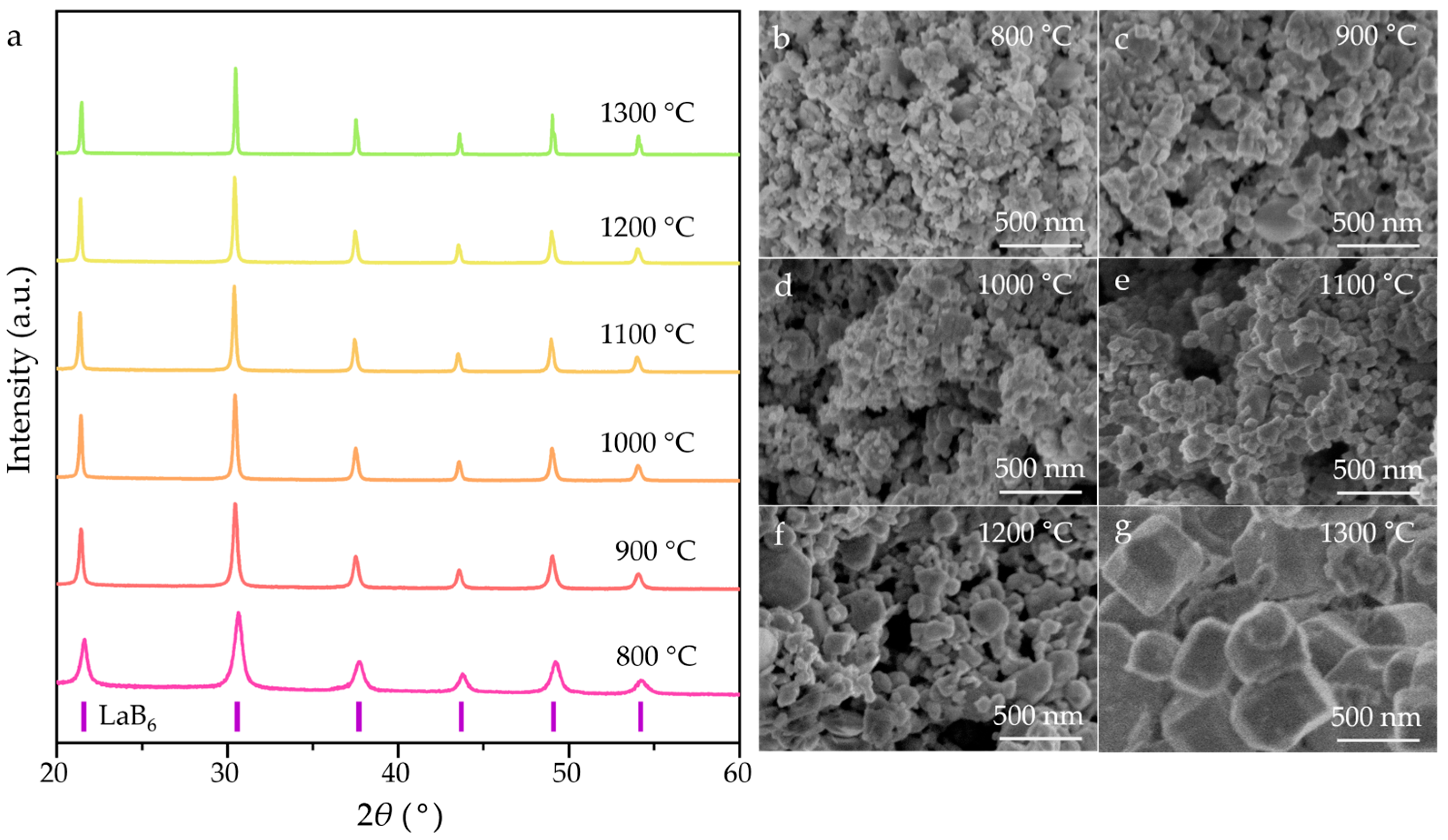
Initially, NaBH4 decomposes to BH3 and NaH when reaching 500 °C, according to Equation (2) [43]. Then, LaB6 was synthesized via a reaction between BH3, NaH, and La2O3 as a typical procedure following Equation (2) [40]. The XRD patterns and SEM images of the as-synthesized LaB6 nanopowders are shown in Figure 2. The XRD patterns (Figure 2a) confirm that only the LaB6 phase (PDF#97-060-2780) is detected in the powders synthesized at different temperatures, confirming a complete reaction. The full width at half maximum (FWHM) of the (110) peaks decreased as the synthesis temperature increased from 800 °C to 1300 °C, indicating a gradual growth in particle size. The SEM images (Figure 2b–g) show the evolution of LaB6 particle morphology and particle size with increasing temperature. As the synthesizing temperature increased from 800 °C to 900 °C, the as-produced LaB6 powder was a typical agglomeration formed by irregularly shaped particles, with an average particle size of less than ~50 nm. At 1000 °C, cubic particles began to emerge, and a well-crystallized cubic morphology dominated when the synthesizing temperature reached 1300 °C. In addition, the average particle size of the LaB6 powders synthesized at 1300 °C reached ~300 nm, indicating abnormal particle growth accompanied by a broad size distribution.
Similar to Equation (1), the complete reaction between ZrO2 and NaBH4 is displayed in Equation (4) [44]:
Z r O 2   +   3 N a B H 4 Z r B 2   +   N a B O 2   +   2 N a   +   6 H 2
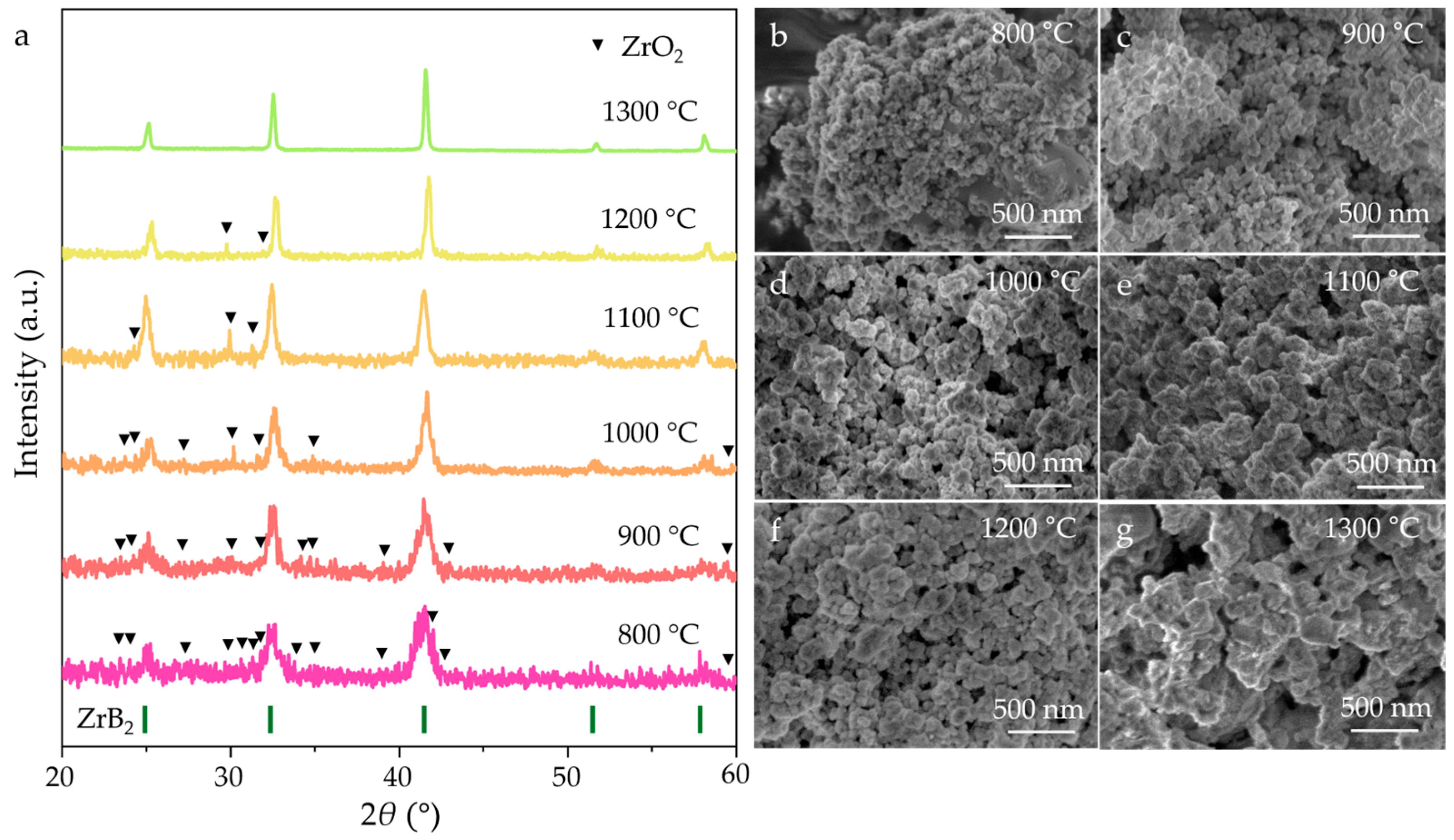
For the ZrB2 (PDF#97-061-5772) nanopowders synthesized at 800–1200 °C, the diffraction peaks of ZrO2 still existed (Figure 3a), which is due to nanoparticle agglomeration and the high melting point of ZrO2. The complete reaction between ZrO2 and NaBH4 occurred when the synthesizing temperature reached 1300 °C. The FWHM of the ZrB2 peaks gradually decrease with increasing synthesizing temperature, implying slow particle growth during synthesis. The SEM images (Figure 3b–g) revealed that all the particles in the ZrB2 powders synthesized at different temperatures exhibited an irregular morphology. Notably, the particle size remained at a relatively low value within the nanometer range (<70 nm) despite elevated sintering temperatures. These results demonstrate that the synthesizing temperature significantly influences the phase purity and morphological features of the final products.
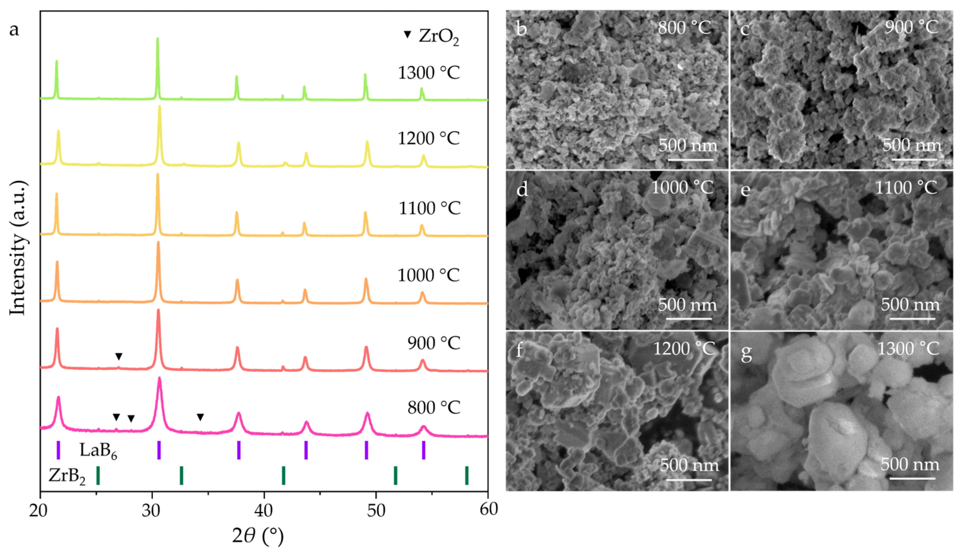
Based on the synthesis of single-phase LaB6 and ZrB2, LaB6-ZrB2 composite powders were successfully prepared via an in situ simultaneous reduction of La2O3, ZrO2, and NaBH4, with a weight ratio of LaB6:ZrB2 = 79:21. As shown in Figure 4a, the XRD analysis reveals that the LaB6, ZrB2, and residual ZrO2 peaks are detected at 800–900 °C. However, beyond 900 °C, the residual ZrO2 peaks become undetectable. This result indicates that the initial temperature for the complete reaction between ZrO2 and NaBH4 in the synthesis of LaB6-ZrB2 nanopowders is lower than that in synthesis of ZrB2 nanopowders. This reduction is attributed to the eutectic interaction between LaB6 and ZrB2, which enhances the reaction kinetics by lowering the activation energy. The in situ reaction can be summarized as follows:
L a 2 O 3   +   Z r O 2   +   16 N a B H 4 2 L a B 6   +   Z r B 2   +   N a B O 3   +   N a B O 2   +   14 N a   +   32 H 2
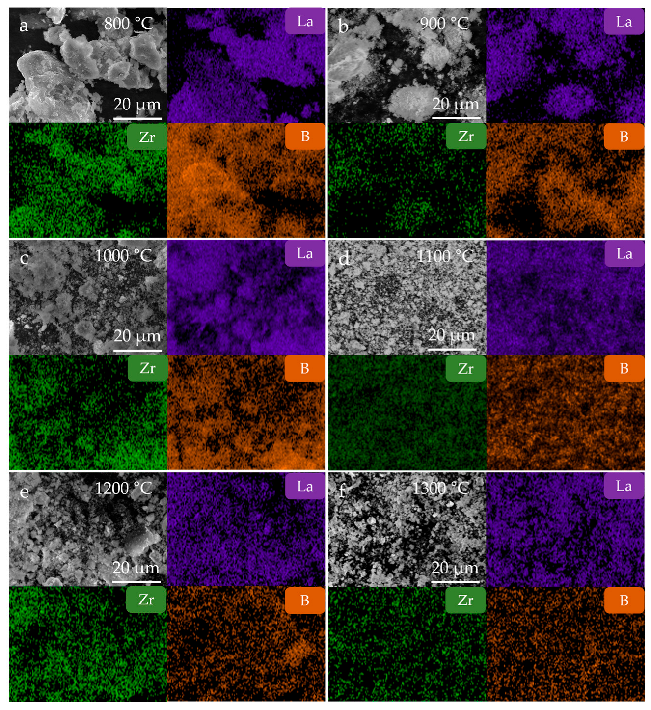
The SEM images (Figure 4b–g) of the LaB6-ZrB2 composite powders at various temperatures reveals particle growth behavior. At 800–1000 °C, the particles exhibited a polyhedral morphology, and their size gradually increased with rising temperature. At 1100 °C, cubic LaB6 particles form, and ZrB2 particles are distributed homogeneously around them. It should be noted that this attachment phenomenon is particularly evident when the synthesis temperature reaches 1200 °C, demonstrating uniform phase distribution. As displayed in EDS mappings of the LaB6-ZrB2 nanopowders synthesized at different temperatures (Figure 5), rare earth element La, transition metal element Zr, and element B are uniformly distributed in the composite boride nanopowders without obvious segregation, further confirming the advantages of the LaB6-ZrB2 composite nanopowders prepared using this method.
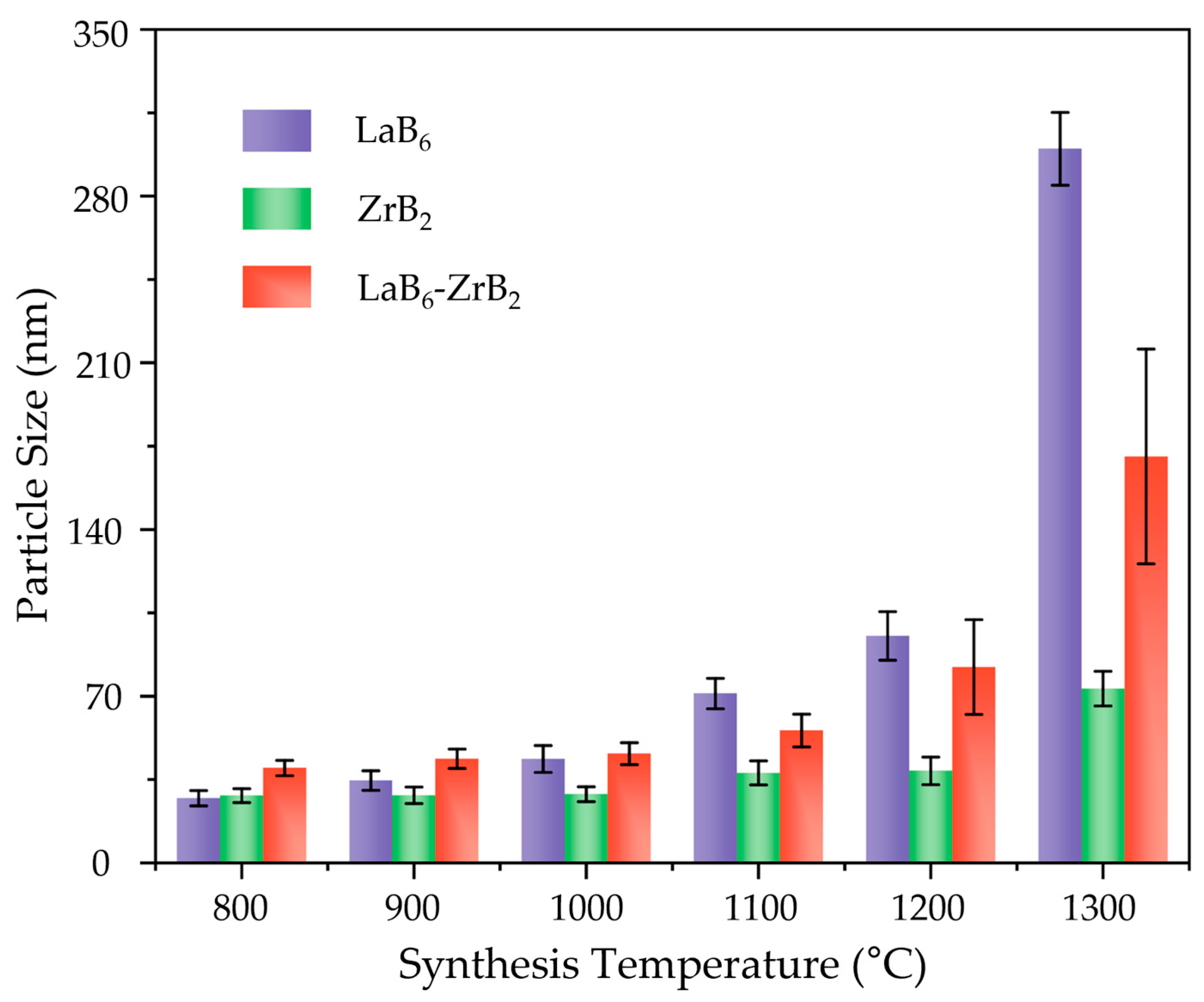
To comprehensively evaluate the effect of temperature on the particle sizes of single-phase and composite nanopowders, the average particle sizes of LaB6, ZrB2, and LaB6-ZrB2 synthesized at 800–1300 °C are summarized in Figure 6. The average particle sizes were obtained by randomly selecting 4–6 SEM images of each powder and measuring 100–200 particles in different areas of each image. The average particle sizes of these three nanopowders increased gradually with the increase in synthesizing temperatures. Below 1100 °C, the average particle size of the composite powders is higher than that of the single-phase powders, suggesting that the eutectic interaction facilitates the synthesis reaction while providing additional energy for grain growth. As the temperature reaches 1100 °C, the average particle size of the composite nanopowders is between the LaB6 and ZrB2 single phases due to abnormal particle growth of LaB6.
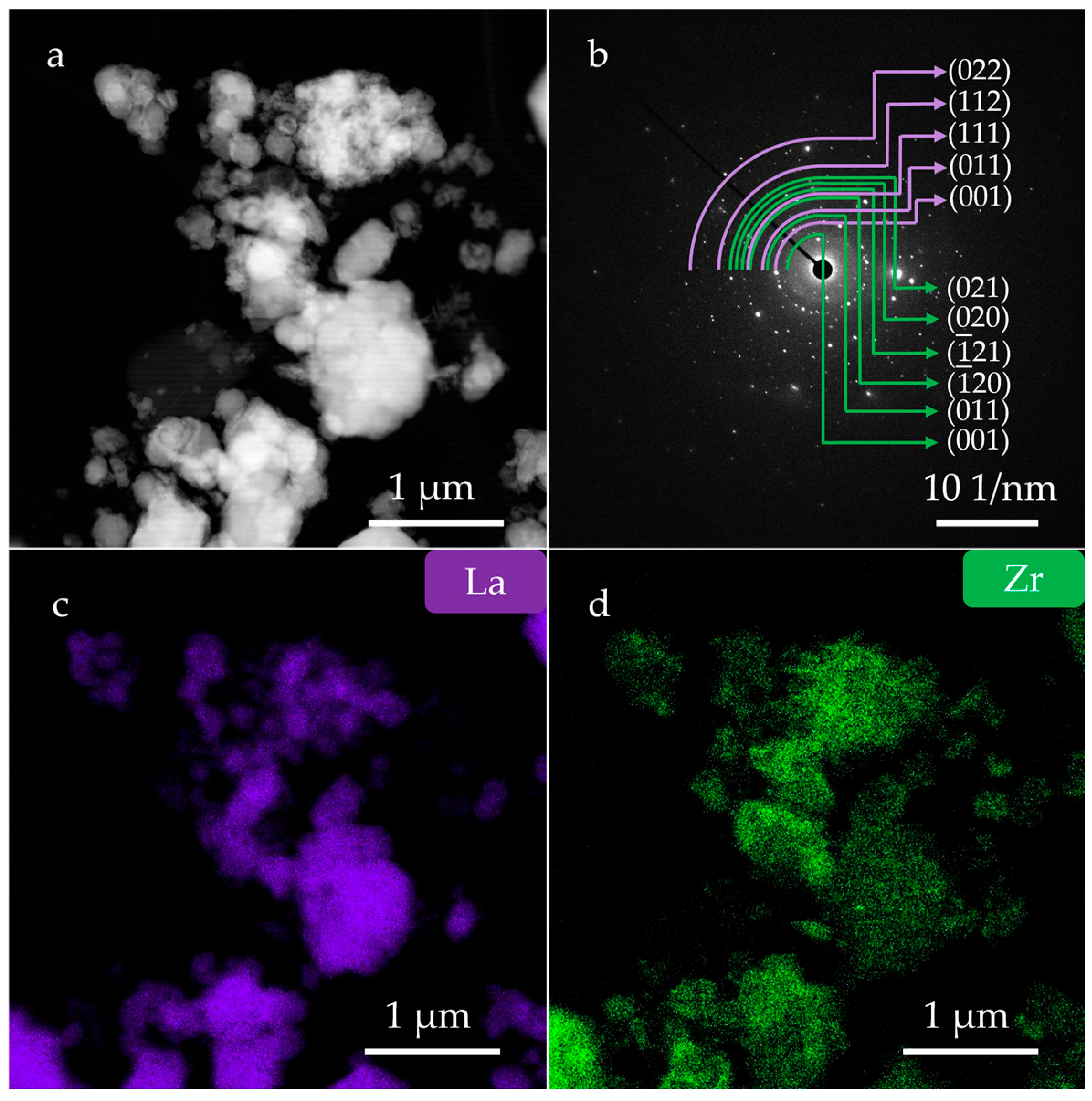
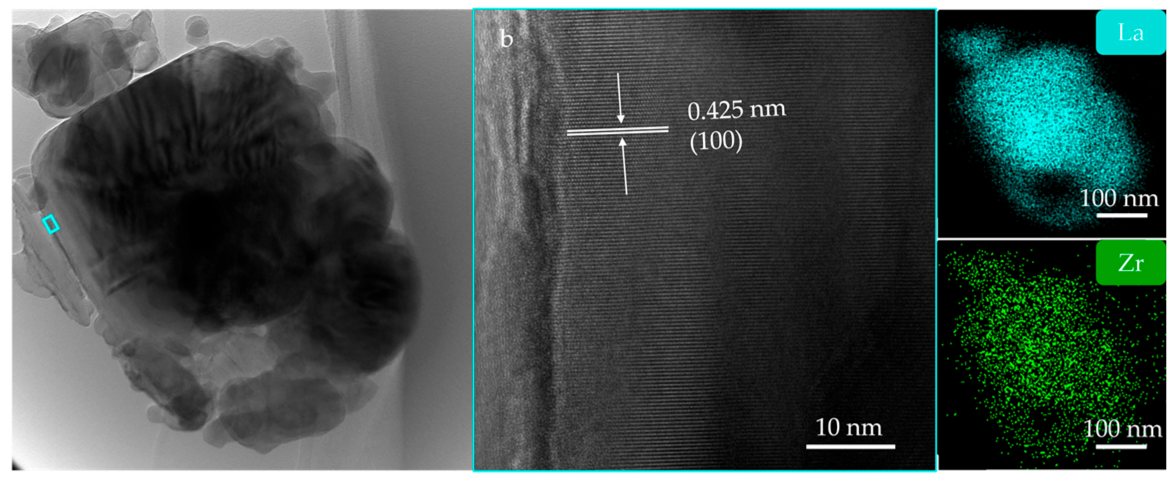
TEM analysis was performed to determine the microstructure and compositional uniformity of the LaB6-ZrB2 nanopowder obtained at 1100 °C. The dark field TEM micrograph was displayed in Figure 7a. Slightly agglomerated and nanoscaled particles were observed. As shown in Figure 7b, the corresponding SAED patterns confirm the coexistence of LaB6 and ZrB2, where the purple arcs indicate the diffraction rings of LaB6 and the green indicate ZrB2. The EDS mappings are shown in Figure 7c,d and demonstrate the uniform phase distribution of LaB6 and ZrB2. Figure 8a displays a high-resolution transmission electron microscopy (HRTEM) image. It can be observed that the cubic particles are tightly attached to irregular particles, resulting in a small aggregate. Figure 8b shows a partially enlarged image of the blue box in Figure 8a. It displays a periodic lattice structure with d-spacing of 0.425 nm, corresponding to the (100) plane of LaB6 (0.415 nm, PDF#97-060-2780). Figure 8c,d show the EDS mappings of the LaB6-ZrB2 nanopowders in Figure 8a, which further demonstrate the uniform phase distribution.
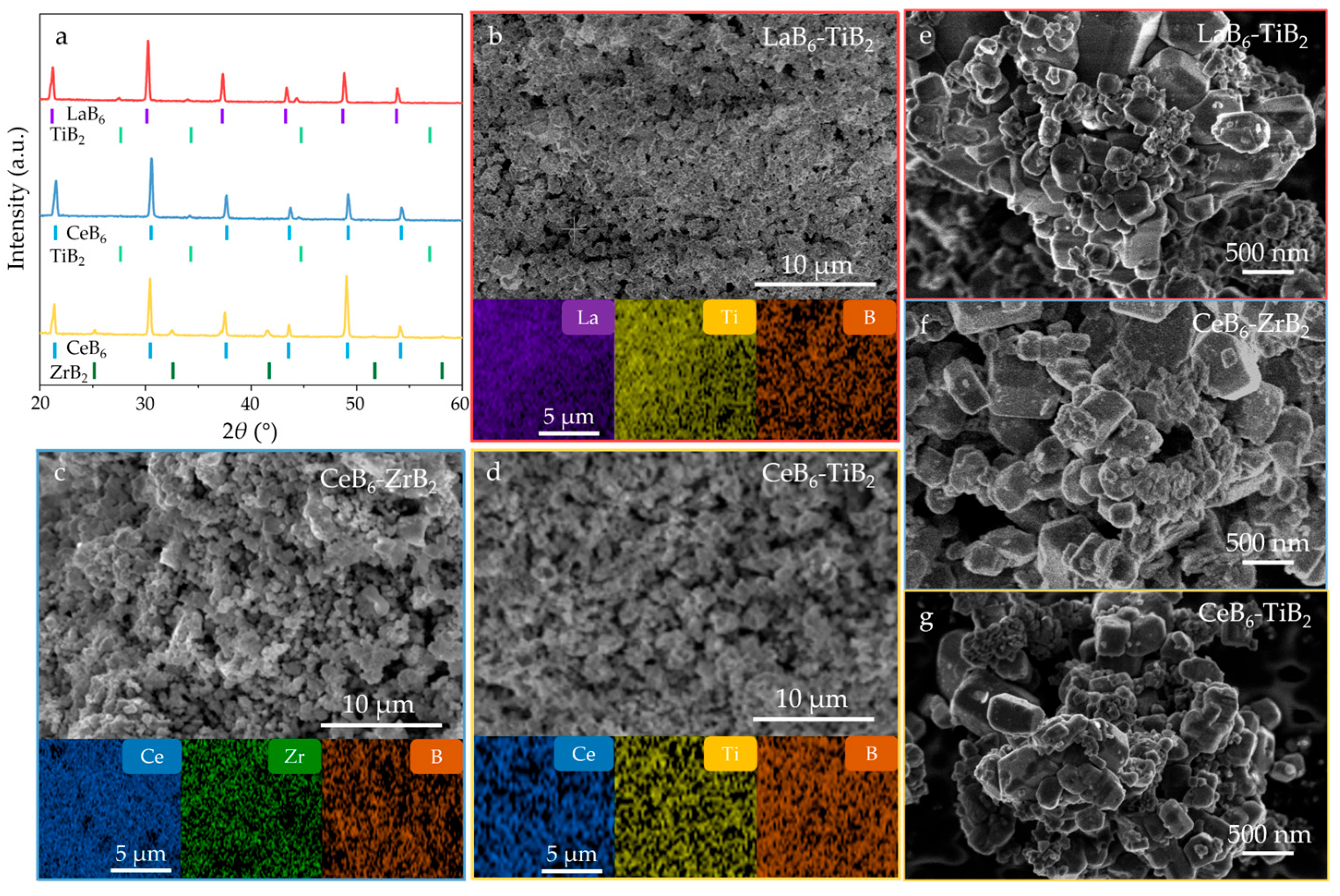
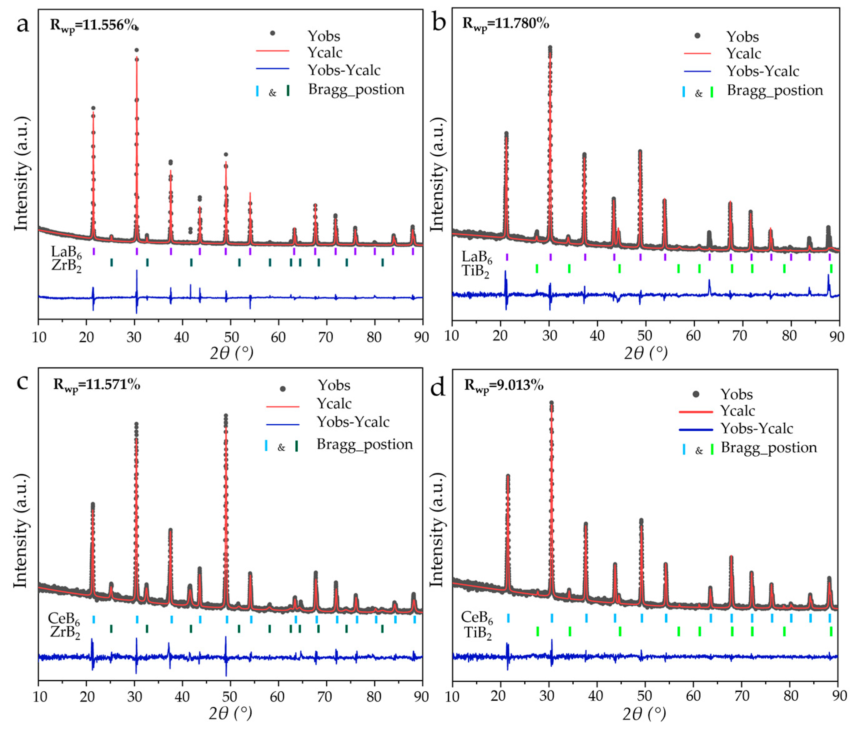
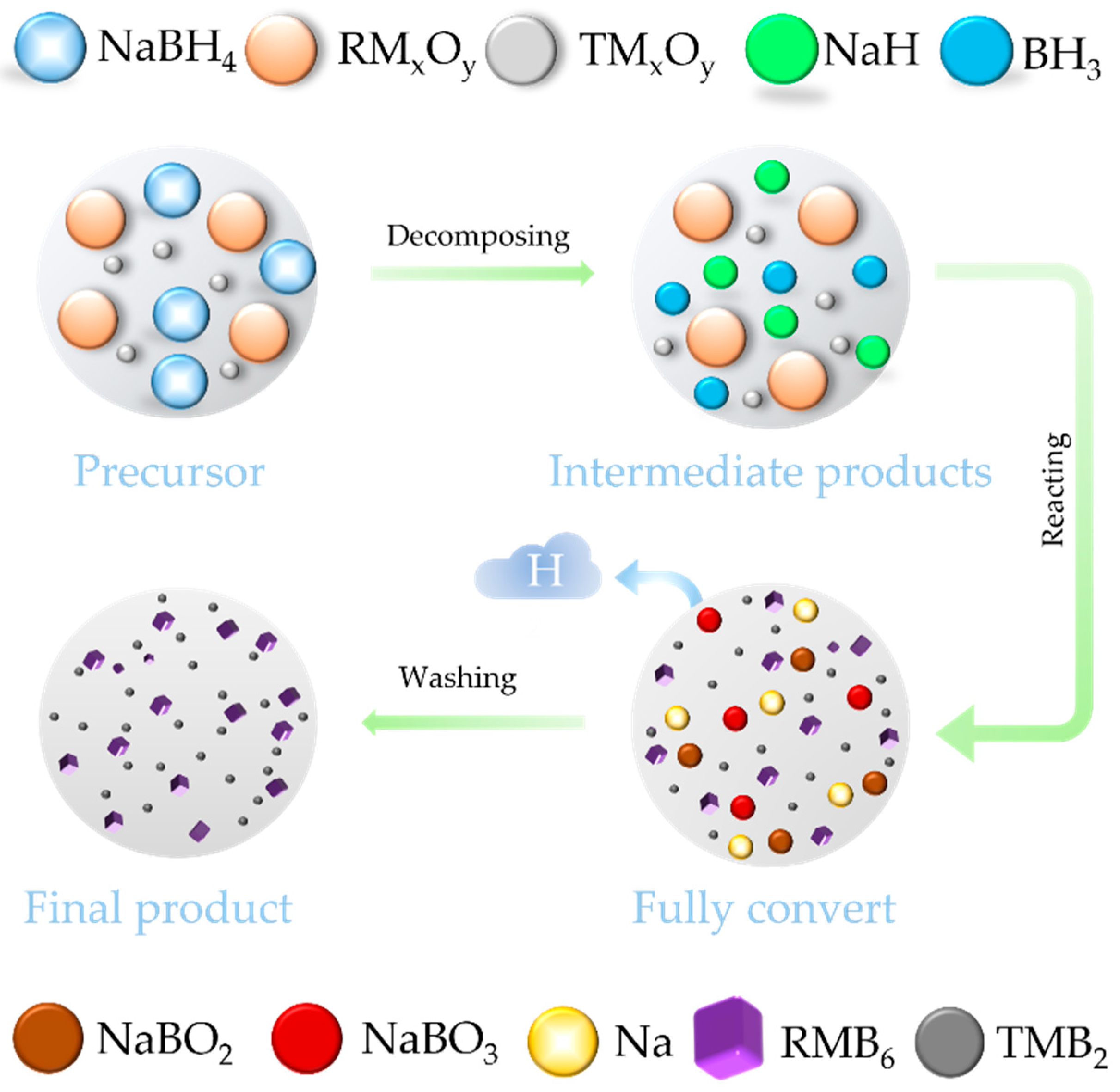
According to Equation (5), the LaB6-TiB2 (TiB2: PDF#97-003-0330), CeB6-ZrB2 (CeB6: PDF#97-019-4318), and CeB6-TiB2 composite nanopowders were successfully synthesized with a eutectic weight ratio (RMB6:TMB2 = 79:21). As exhibited in Figure 9a, XRD analysis confirms that all composite nanopowders synthesized at 1300 °C are composed of the target product without any impurities and by-products, indicating the feasibility of this synthesis pathway. The SEM and EDS mappings (Figure 9b–g) reveal uniform nanostructured morphologies without elemental agglomeration or segregation, which also demonstrates the effectiveness of the in situ method in achieving a homogeneous phase distribution. Figure 9e–g displays the high-magnification SEM images of the LaB6-TiB2, CeB6-ZrB2, and CeB6-TiB2 composite nanopowders, and it can be observed that these three products possess nanosized particles. The refinement results of the XRD patterns of the LaB6-ZrB2, LaB6-TiB2, CeB6-ZrB2, and CeB6-TiB2 nanopowders are shown in Figure 10. The Rwp values of the diffraction patterns obtained via calculations are 11.556, 11.780, 11.571, and 9.013, respectively, indicating the reliability of the refinement results. In addition, lattice constants and phase weight ratios were also calculated. The lattice constant of LaB6 is 0.420 nm in LaB6-ZrB2 and LaB6-TiB2 powders, which is close to the theoretical counterpart. The measured d-spacing of the (100) plane, as displayed in Figure 8, is larger than the theoretical value of 0.415 nm, which might be caused by the lattice distortion. The calculated weight ratios are LaB6:ZrB2 = 89.2:10.8, LaB6:TiB2 = 82.3:17.7, CeB6:ZrB2 = 81.8:18.2, and CeB6:TiB2 = 81.2:18.8, which are in agreement with the eutectic weight ratio of 79:21, further confirming the feasibility of this method. As depicted in Figure S1, the Raman spectra only detected the existence of LaB6, CeB6, and TiB2 without any other phase, and ZrB2 has no peaks because it is not Raman active. Based on the current study, the in situ formation mechanism of RMB6-TMB2 composite powders is summarized and presented in Figure 11. It is obvious that the decomposition of NaBH4 plays a critical role in this process by simultaneously providing both a boron source and reducing agents. The release of BH3 during NaBH4 decomposition enhances its accessibility to metal oxide surfaces, facilitating uniform boride nucleation. Therefore, it ensures that the synthesized composite powders remain at the nanoscale level while achieving uniform distribution of RMB6 and TMB2.

4. Conclusions

In this work, RMB6-TMB2 composite nanopowders were synthesized in situ via a one-step solid-state reduction method using metal oxides and NaBH4 as raw materials. The LaB6-ZrB2 composite nanopowders with a eutectic weight ratio exhibited accelerated reaction kinetics compared to their single-phase counterparts. The EDS mappings of the HRTEM images demonstrated that the phase distribution of the composite powders is uniform even at the nanoscale level. The refinement results prove that the lattice constant of the composite powder is close to the theoretical value, and the obtained mass ratio is in agreement with the designed eutectic ratio. This method eliminates the phase separation and contamination that exists with conventional manual mixing, which provides the feasibility for designing advanced boride composites of RMB6-TMB2 with excellent thermionic and mechanical properties.

Supplementary Materials

The following supporting information can be downloaded at https://www.mdpi.com/article/10.3390/nano15171341/s1: Figure S1: Raman spectras of (a) LaB6-ZrB2, (b)LaB6-TiB2, (c) CeB6-ZrB2 and (d) CeB6-TiB2. (▲: LaB6, ■: CeB6 and ●: TiB2).

Author Contributions

Conceptualization, L.S. and B.L. (Bing Liu); methodology, D.Y., L.S. and B.L. (Bing Liu); validation, H.Z. and A.L.; formal analysis, X.G., L.W., L.S. and B.L. (Bing Liu); investigation, H.Z., A.L., J.X. and W.Q.; resources, Y.G. and R.S.; data curation, X.G., L.S. and B.L. (Bing Liu); writing—original draft preparation, X.G. and L.W.; writing—review and editing, M.M., L.S. and B.L. (Bing Liu); visualization, X.G., L.W. and A.L.; project administration, M.M., L.S. and B.L. (Baozhong Li); funding acquisition, L.W., M.M., B.L. (Bing Liu) and B.L (Baozhong Li). All authors have read and agreed to the published version of the manuscript.

Funding

This work was supported by the National Natural Science Foundation of China (Grant Nos. 52302063 and 52302072), the State Key Laboratory of Advanced Space Propulsion, the Young Elite Scientist Sponsorship Program by CAST (Grant No. 2023QNRC001), the Postdoctoral Fellowship Program of CPSF (Grant No. GZC20231162), the China Postdoctoral Science Foundation (Grant No. 2023M741809), the Innovation Capacity Enhancement Program of Hebei Province (Grant No. 24461901D), the Hebei Natural Science Foundation (Grant No. E2023203126), the China Postdoctoral Science Foundation-Tianjin Joint Support Program (Grant No. 2023T019TJ), the Yanzhao Talent Program of Hebei Province (Grant No. B2024005011), and the S&T Program of Hebei (225A1102D).

Conflicts of Interest

The authors declare no conflicts of interest.

References

  1. Reich, S.; Suhr, H.; Hankó, K.; Szepes, L. Deposition of Thin Films of Zirconium and Hafnium Boride by Plasma Enhanced Chemical Vapor Deposition. Adv. Mater. 1992, 4, 650–653. [Google Scholar] [CrossRef]
  2. Çamurlu, H.E.; Maglia, F. Preparation of Nano-Size ZrB2 Powder by Self-Propagating High-Temperature Synthesis. J. Eur. Ceram. Soc. 2009, 29, 1501–1506. [Google Scholar] [CrossRef]
  3. Kelly, J.P.; Kanakala, R.; Graeve, O.A. A Solvothermal Approach for the Preparation of Nanostructured Carbide and Boride Ultra-High-Temperature Ceramics. J. Am. Ceram. Soc. 2010, 93, 3035–3038. [Google Scholar] [CrossRef]
  4. Barış, M.; Şimşek, T.; Gökmeşe, H.; Akkurt, A. Characterization of W2B Nanocrystals Synthesized by Mechanochemical Method. J. Boron 2016, 1, 45–51. [Google Scholar]
  5. Hwang, Y.; Lee, J.K. Preparation of TiB2 Powders by Mechanical Alloying. Mater. Lett. 2002, 54, 1–7. [Google Scholar] [CrossRef]
  6. Qiu, H.Y.; Guo, W.M.; Zou, J.; Zhang, G.J. ZrB2 Powders Prepared by Boro/Carbothermal Reduction of ZrO2: The Effects of Carbon Source and Reaction Atmosphere. Powder Technol. 2012, 217, 462–466. [Google Scholar] [CrossRef]
  7. Aksu, M.; Aydln, E. Nano and Microsized EuB6 via Magnesiothermic Reduction. J. Ceram. Process Res. 2013, 14, 4–7. [Google Scholar] [CrossRef]
  8. Liu, B.; Chen, S.L.; Ma, M.D.; Pan, Y.L.; Zhao, Z.S.; Zhou, H.; Sun, L.; Chen, S.; Guo, X.G.; Tong, K.; et al. Producing High Performance TiB2 Ceramics by High-Pressure Sintering Forging. Ceram. Int. 2025, 51, 20116–20123. [Google Scholar] [CrossRef]
  9. Wang, Y.; Yang, X.Y.; Ning, S.Y.; Wang, Z.H.; Xu, B.; Zhang, J.X. Preparation, Characterization and Properties of La0.6Ce0.3Pr0.1B6-ZrB2 Directionally Solidified Eutectic Grown via the Optical Floating Zone Technique. J. Alloys Compd. 2020, 818, 152924. [Google Scholar] [CrossRef]
  10. Wang, Y.; Bao, Y.; Luo, S.F.; Han, C.L.; Yang, X.Y.; Zhang, J.X. Spark Plasma Sintering of Polycrystalline La0.6Ce0.3Pr0.1B6-ZrB2 Composites. J. Am. Ceram. Soc. 2023, 106, 2118–2129. [Google Scholar] [CrossRef]
  11. Zhang, H.; Zhang, Q.; Zhao, G.; Tang, J.; Zhou, O.; Qin, L.C. Single-Crystalline GdB6 Nanowire Field Emitters. J. Am. Chem. Soc. 2005, 127, 13120–13121. [Google Scholar] [CrossRef]
  12. Sani, E.; Mercatelli, L.; Meucci, M.; Zoli, L.; Sciti, D. Lanthanum Hexaboride for Solar Energy Applications. Sci. Rep. 2017, 7, 718. [Google Scholar] [CrossRef]
  13. Cahill, J.T.; Graeve, O.A. Hexaborides: A Review of Structure, Synthesis and Processing. J. Mater. Res. Technol. 2019, 8, 6321–6335. [Google Scholar] [CrossRef]
  14. Plankovsky, S. Review of LaB6, Re-W Dispenser and BaHfO3-W Cathode Development. IEEE Trans. Electron. Devices 2009, 56, 760–765. [Google Scholar] [CrossRef]
  15. Lafferty, J. Boride Cathodes. J. Appl. Phys. 1951, 22, 299–309. [Google Scholar] [CrossRef]
  16. Bellucci, A.; Mastellone, M.; Girolami, M.; Serpente, V.; Generosi, A.; Paci, B.; Mezzi, A.; Kaciulis, S.; Carducci, R.; Polini, R.; et al. Nanocrystalline Lanthanum Boride Thin Films by Femtosecond Pulsed Laser Deposition as Efficient Emitters in Hybrid Thermionic-Photovoltaic Energy Converters. Appl. Surf. Sci. 2020, 513, 145829. [Google Scholar] [CrossRef]
  17. Bellucci, A.; Mastellone, M.; Girolami, M.; Serpente, V.; Sani, E.; Sciti, D.; Trucchi, D. Thermionic Emission Measurement of Sintered Lanthanum Hexaboride Discs and Modelling of Their Solar Energy Conversion Performance. Ceram. Int. 2021, 47, 20736–20739. [Google Scholar] [CrossRef]
  18. Aono, M.; Nishitani, R.; Oshima, C.; Tanaka, T.; Bannai, E.; Kawai, S. Direct Observation of LaB6 (001) Surface at High Temperatures by x-Ray and Ultraviolet Photoelectron Spectroscopy, Low-Energy Electron Diffraction, Auger Electron Spectroscopy, and Work-Function Measurements. J. Appl. Phys. 1979, 50, 4802–4807. [Google Scholar] [CrossRef]
  19. Ning, S.; Iitaka, T.; Yang, X.; Wang, Y.; Zhao, J.; Li, Z.; Zhang, J. Enhanced Thermionic Emission Performance of LaB6 by Ce Doping. J. Alloys Compd. 2018, 760, 1–5. [Google Scholar] [CrossRef]
  20. Ağaoğulları, D.; Duman, İ.; Öveçoğlu, M.L. Synthesis of LaB6 Powders from La2O3, B2O3 and Mg Blends via a Mechanochemical Route. Ceram. Int. 2012, 38, 6203–6214. [Google Scholar] [CrossRef]
  21. Fan, Q.; Zhao, Y.; Li, D. Synthesis of Single-Crystalline Lanthanum Hexaboride Nanowires by Au Catalyst. Ceram. Int. 2013, 39, 6271–6275. [Google Scholar] [CrossRef]
  22. Loboda, P. Structure and Properties of Reinforced Ceramic Materials Produced by Directional Solidification. Powder Metall. Met. Ceram. 2018, 57, 13–26. [Google Scholar] [CrossRef]
  23. Kusunoki, T.; Hashizume, T.; Kasuya, K.; Arai, N. Stabilization of Cold-Field-Emission Current from a CeB6 Single-Crystal Emitter by Using a Faceted (100) Plane. J. Vac. Sci. Technol. B 2020, 39, 013202. [Google Scholar] [CrossRef]
  24. Dub, S.; Kislaya, G.; Loboda, P. Study of Mechanical Properties of LaB6 Single Crystal by Nanoindentation. J. Superhard Mater. 2013, 35, 158–165. [Google Scholar] [CrossRef]
  25. Demirskyi, D.; Suzuki, T.S.; Yoshimi, K.; Vasylkiv, O.O. Consolidation and High-temperature Strength of Monolithic Lanthanum Hexaboride. J. Am. Ceram. Soc. 2022, 105, 4277–4290. [Google Scholar] [CrossRef]
  26. Sonber, J.K.; Sairam, K.; Murthy, T.S.R.C.; Nagaraj, A.; Subramanian, C.; Hubli, R.C. Synthesis, Densification and Oxidation Study of Lanthanum Hexaboride. J. Eur. Ceram. Soc. 2014, 34, 1155–1160. [Google Scholar] [CrossRef]
  27. Xu, B.; Yang, X.Y.; Cheng, H.F.; Zhao, J.J.; Wang, Y.; Zhu, E.; Zhang, J.X. Preparation, Characterization and Property of High-Quality LaB6 Single Crystal Grown by the Optical Floating Zone Melting Technique. Vacuum 2019, 168, 108845. [Google Scholar] [CrossRef]
  28. Zhou, S.L.; Zhang, J.X.; Liu, D.M.; Lin, Z.L.; Huang, Q.; Bao, L.H.; Ma, R.; Wei, Y. Synthesis and Properties of Nanostructured Dense LaB6 Cathodes by Arc Plasma and Reactive Spark Plasma Sintering. Acta Mate 2010, 58, 4978–4985. [Google Scholar] [CrossRef]
  29. Bogomol, I.; Nishimura, T.; Vasylkiv, O.; Sakka, Y.; Loboda, P. High-Temperature Strength of Directionally Reinforced LaB6-TiB2 Composite. J. Alloys Compd. 2010, 505, 130–134. [Google Scholar] [CrossRef]
  30. Guo, X.G.; Liu, A.; Hao, L.J.; Zhou, H.; Chen, S.; Ying, P.; Liu, B.; Li, B.Z.; Gao, Y.F.; Zhao, Z.S.; et al. Ultrafine-Grained LaB6–ZrB2 Composite Ceramics with Superior Mechanical and Thermionic Emission Properties Prepared by High-Pressure Sintering. J. Adv. Ceram. 2024, 13, 2021–2029. [Google Scholar] [CrossRef]
  31. Paderno, Y.; Paderno, V.; Filippov, V. Some Peculiarities of Structure Formation in Eutectic D-and f-Transition Metals Boride Alloys. In AIP Conference Proceedings; American Institute of Physics: Melville, NY, USA, 1991; Volume 231, pp. 561–569. [Google Scholar]
  32. Yang, X.Y.; Wang, P.; Wang, Z.H.; Hu, K.; Cheng, H.F.; Li, Z.; Zhang, J.X. Microstructure, Mechanical and Thermionic Emission Properties of a Directionally Solidified LaB6-VB2 Eutectic Composite. Mater. Des. 2017, 133, 299–306. [Google Scholar] [CrossRef]
  33. Paderno, Y.; Paderno, V.; Filippov, V. Some Peculiarities of Eutectic Crystallization of LaB6-(Ti, Zr)B2 Alloys. J. Solid. State Chem. 2000, 154, 165–167. [Google Scholar] [CrossRef]
  34. Min, G.H.; Gao, R.L.; Yu, H.S.; Han, J.D. Mechanical Properties of LaB6-ZrB2 Composites. Key Eng. Mater. 2005, 297–300, 1630–1638. [Google Scholar] [CrossRef]
  35. Chen, Y.; Fan, J.; Gong, H.; Zou, F.; Guo, Y.; Luo, H. Synthesis, Characterization and Formation Mechanism of Ta-W Nanocrystalline Composite Powders Fabricated by Nano-In Situ Composite Method. RSC Adv. 2016, 6, 106011–106018. [Google Scholar] [CrossRef]
  36. Deng, X.C.; Zhang, G.H.; Zhu, Z.G. In-Situ Preparation of WC-Co Composite Powders. Int. J. Refract. Met. Hard Mater. 2025, 128, 106995. [Google Scholar] [CrossRef]
  37. Naren, G.; Ren, T.Y.; Bao, L.H.; Chao, L.M.; Tegus, O. Preparation and Optical Absorption Properties of Ternary Rare Earth Boride LaxPr1-xB6 Submicron Powders. J. Nanosci. Nanotechnol. 2020, 20, 5064–5069. [Google Scholar] [CrossRef] [PubMed]
  38. Chen, L.; Gu, Y.; Yang, Z.; Shi, L.; Ma, J.; Qian, Y. Preparation and Some Properties of Nanocrystalline ZrB2 Powders. Scr. Mater. 2004, 50, 959–961. [Google Scholar] [CrossRef]
  39. Chen, L.; Gu, Y.; Qian, Y.; Shi, L.; Yang, Z.; Ma, J. A Facile One-Step Route to Nanocrystalline TiB2 Powders. Mater. Res. Bull. 2004, 39, 609–613. [Google Scholar] [CrossRef]
  40. Chen, L.; Gu, Y.; Shi, L.; Yang, Z.; Ma, J.; Qian, Y. Synthesis and Oxidation of Nanocrystalline HfB2. J. Alloys Compd. 2004, 368, 353–356. [Google Scholar] [CrossRef]
  41. Bates, S.E.; Buhro, W.E.; Frey, C.A.; Sastry, S.M.; Kelton, K. Synthesis of Titanium Boride (TiB)2 Nanocrystallites by Solution-Phase Processing. J. Mater. Res. 1995, 10, 2599–2612. [Google Scholar] [CrossRef]
  42. Ma, M.D.; Yang, X.Y.; Meng, H.; Zhao, Z.S.; He, J.L.; Chu, Y.H. Nanocrystalline High-Entropy Hexaboride Ceramics Enable Remarkable Performance as Thermionic Emission Cathodes. Fundam. Res. 2023, 3, 979–987. [Google Scholar] [CrossRef] [PubMed]
  43. Ostroff, A.; Sanderson, R. Oxidation and Thermal Decomposition of Sodium and Potassium Borohydrides. J. Inorg. Nucl. Chem. 1957, 4, 230–231. [Google Scholar] [CrossRef]
  44. Zoli, L.; Galizia, P.; Silvestroni, L.; Sciti, D. Synthesis of Group IV and V Metal Diboride Nanocrystals via Borothermal Reduction with Sodium Borohydride. J. Am. Ceram. Soc. 2018, 101, 2627–2637. [Google Scholar] [CrossRef]
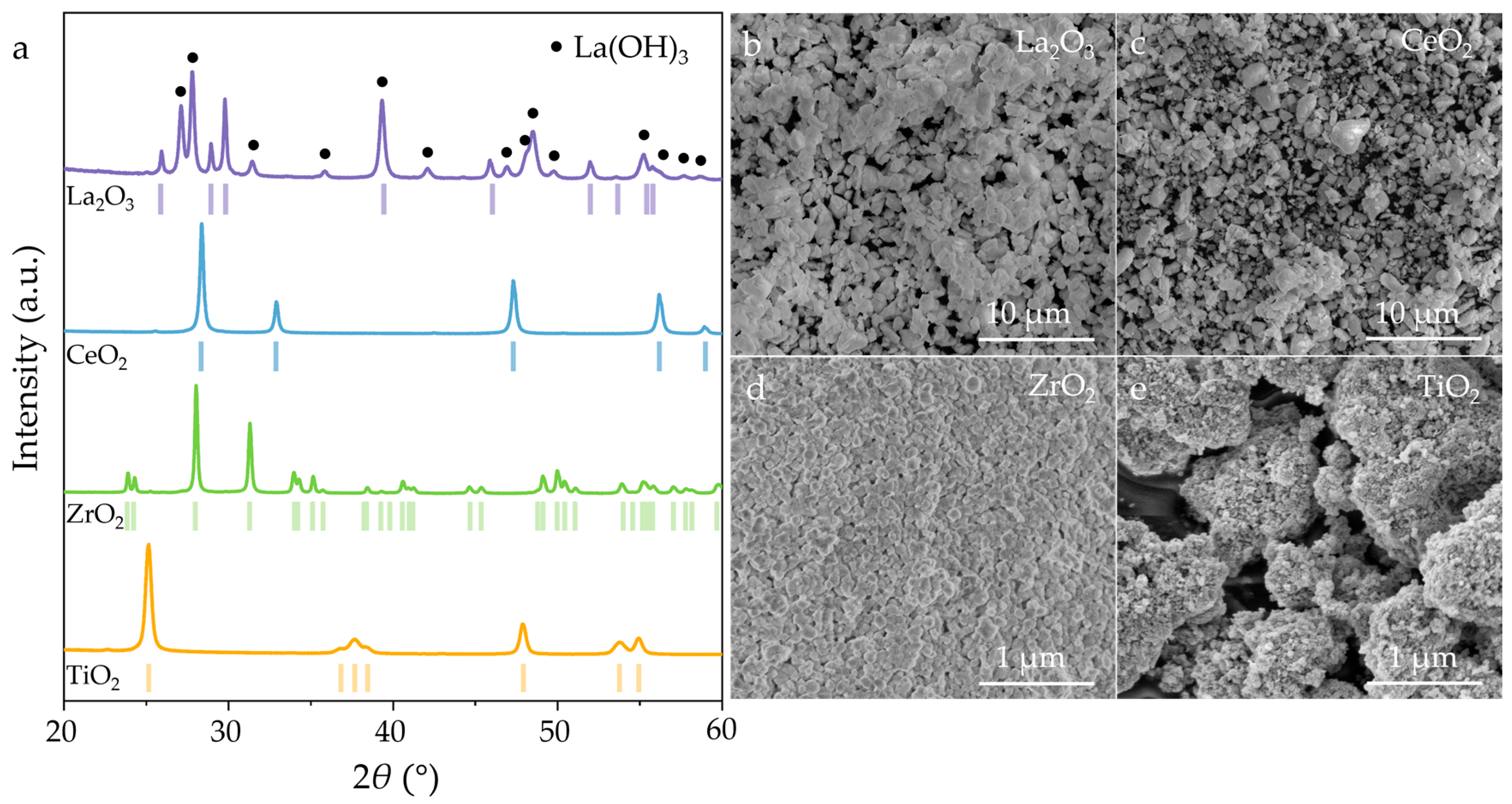
Figure 1. (a) XRD patterns and (be) SEM images of La2O3, CeO2, ZrO2, and TiO2 powders.
Nanomaterials 15 01341 g001
Figure 2. (a) XRD patterns and (bg) SEM images of LaB6 nanopowders synthesized at 800–1300 °C.
Nanomaterials 15 01341 g002
Figure 3. (a) XRD patterns and (bg) SEM images of ZrB2 nanopowders synthesized at 800–1300 °C.
Nanomaterials 15 01341 g003
Figure 4. (a) XRD patterns and (bg) SEM images of LaB6-ZrB2 nanopowders synthesized at 800–1300 °C.
Nanomaterials 15 01341 g004
Figure 5. The SEM images and the corresponding EDS mappings of LaB6-ZrB2 nanopowders synthesized at (a) 800 °C, (b) 900 °C, (c) 1000 °C, (d) 1100 °C, (e) 1200 °C and (f) 1300 °C.
Nanomaterials 15 01341 g005
Figure 6. The average particle sizes of LaB6, ZrB2 and LaB6-ZrB2 nanopowders synthesized from 800–1300 °C.
Nanomaterials 15 01341 g006
Figure 7. (a) TEM image of LaB6-ZrB2 nanopowders, (b) the corresponding SAED pattern of (a), and (c,d) the corresponding EDS maps of (a).
Nanomaterials 15 01341 g007
Figure 8. (a) HRTEM image of LaB6-ZrB2 nanopowders, (b) partial enlargement of the box in (a), and (c,d) corresponding EDS compositional maps of (a).
Nanomaterials 15 01341 g008
Figure 9. (a) XRD patterns, (bd) SEM images and EDS mappings, and (eg) the high magnifications of LaB6-TiB2, CeB6-ZrB2, and CeB6-TiB2 composite nanopowders obtained in situ at 1300 °C.
Nanomaterials 15 01341 g009
Figure 10. Rietveld refinement of synthesized composite nanopowders: (a) LaB6-ZrB2; (b) LaB6-TiB2; (c) CeB6-ZrB2; and (d) CeB6-TiB2.
Nanomaterials 15 01341 g010
Figure 11. Reaction mechanism schematic of the metal oxides and NaBH4.
Nanomaterials 15 01341 g011
Disclaimer/Publisher’s Note: The statements, opinions and data contained in all publications are solely those of the individual author(s) and contributor(s) and not of MDPI and/or the editor(s). MDPI and/or the editor(s) disclaim responsibility for any injury to people or property resulting from any ideas, methods, instructions or products referred to in the content.