Review

Nanocarriers for Combination Therapy in Pancreatic Ductal Adenocarcinoma: A Comprehensive Review

by
Iris Pontón
and
David Sánchez-García
*
Grup d’Enginyeria de Materials (GEMAT), Institut Químic de Sarrià (IQS), Universitat Ramon Llull (URL), Via Augusta 390, 08017 Barcelona, Spain
*
Author to whom correspondence should be addressed.
Nanomaterials 2025, 15(15), 1139; https://doi.org/10.3390/nano15151139
Submission received: 26 June 2025 / Revised: 21 July 2025 / Accepted: 21 July 2025 / Published: 22 July 2025
(This article belongs to the Special Issue Nanoparticles for Multiple Drug Release)

Abstract

Pancreatic ductal adenocarcinoma (PDAC) remains one of the deadliest cancers worldwide, characterized by late diagnosis, aggressive progression, and poor response to conventional monotherapies. Combination therapies have emerged as a promising approach to overcome multidrug resistance (MDR), enhance efficacy, and target the complex tumor microenvironment (TME). Nanoparticle-based drug delivery systems (DDSs) have gained significant attention for their ability to co-deliver multiple agents with controlled release profiles. This review comprehensively examines nanoparticle-based platforms developed for PDAC combination therapies, focusing on small-molecule drugs. The systems discussed are drawn from studies published between 2005 and 2025.

Graphical Abstract

1. Introduction

Cancer encompasses a diverse group of diseases arising from a multistage process that can originate in nearly any organ or tissue of the body, leading to the uncontrolled growth and spread of abnormal cells [1]. It is characterized by the accumulation of morphological and genetic alterations, caused by external (chemicals, radiation, infectious organisms) and internal factors (hormones, immune conditions, random mutations) [2]. Remarkably, it is a significant health issue in developed countries, where it is primarily associated with the aging population and their lifestyle [3]. According to the World Health Organization report from 2019, cancer is currently the second-leading cause of death worldwide [4,5]. In particular, pancreatic cancer is the fourth leading cause of cancer-related death in Europe and the United States and the most life-threatening cancer type worldwide. Despite representing only 3% of cancer incidence, it is responsible for 7% of all cancer-related deaths. This disproportionate impact is reflected in its dismal median overall survival, which is typically only 5 to 6 months [6,7]. This low survival is attributed to several factors, especially, the late diagnosis, as most patients remain asymptomatic until the disease progresses to advanced stages [8]. Pancreatic ductal adenocarcinoma (PDAC) is by far the most common pancreatic cancer, accounting for 95% of cases, and is one of the most aggressive solid malignancies, having become the third deadliest cancer in 2023 [9,10,11,12]. The 5-year survival rate is estimated to be less than 5%, and one-year survival is observed in less than 20% of cases [9,11].
PDAC originates in ductal cells, located at the exocrine part of the organ, which are responsible for transporting digestive enzymes. These cells, of epithelial origin, eventually undergo a process called epithelial-to-mesenchymal (EtM) transition. Briefly, the EtM transition involves a huge transformation where cells proliferate and exit the epithelial compartment, migrating and interacting with stromal cells producing the primary tumor [13].
The aggressiveness of pancreatic cancer is largely due to the dense and fibrotic stroma that surrounds the cancer cells, which not only acts as a physical barrier bur also promotes the malignancy of the tumor. Moreover, the leaky and abnormal vasculature, which tends to collapse, hinders drug accumulation and limits treatment efficacy [14].
From 1996 to 2011, Gemcitabine GEM (Gemzar®) was considered a gold-standard chemotherapy for treating pancreatic cancer [15]. However, chemo-resistance associated with this drug limits its effectivity [16]. To overcome this hurdle, combination therapies such as NALIRIFOX (liposomal irinotecan, oxaliplatin, fluorouracil, and leucovorin) have been proposed [12]. Unfortunately, despite significant medical advancements in the treatment of solid tumors over recent decades, the mortality-to-incidence ratio for PDAC remains stubbornly high [9].
However, in the last decade, nanomedicine has revolutionized PDAC therapy through nanoscale DDSs that enable controlled drug release. These systems surpass conventional therapies by offering sustained release, reduced systemic toxicity, enhanced stability, and increased drug concentration at tumor sites, improving efficacy and minimizing side effects. A key example is nab-paclitaxel (nab-PTX, Abraxane®), a ~130 nm nanoparticle conjugating paclitaxel with albumin, enhancing PTX solubility and tumor targeting via receptor-mediated uptake. Nab-PTX’s clinical success, with improved efficacy and lower toxicity, serves as proof of concept for PDAC nanomedicines [17]. Similarly, nal-IRI (Onivyde®), a ~111 nm liposomal nanoparticle encapsulating irinotecan, ensures prolonged circulation and tumor accumulation through the enhanced permeability and retention (EPR) effect, reducing toxicity while maintaining efficacy [18]. Nanomedicines like nab-PTX and nal-IRI highlight the potential of nanoscale DDSs to transform PDAC treatment, paving the way for selective and more effective PDAC therapies.
This review explores nanoparticle-based platforms developed for PDAC combination therapies, focusing on small-molecule drugs for monotherapy or in combination with other therapies. Nanocarriers designed for imaging applications [19], gene therapy, or stromal-targeted therapies [20] have been reviewed elsewhere in the literature.

2. Pancreatic Ductal Adenocarcinoma

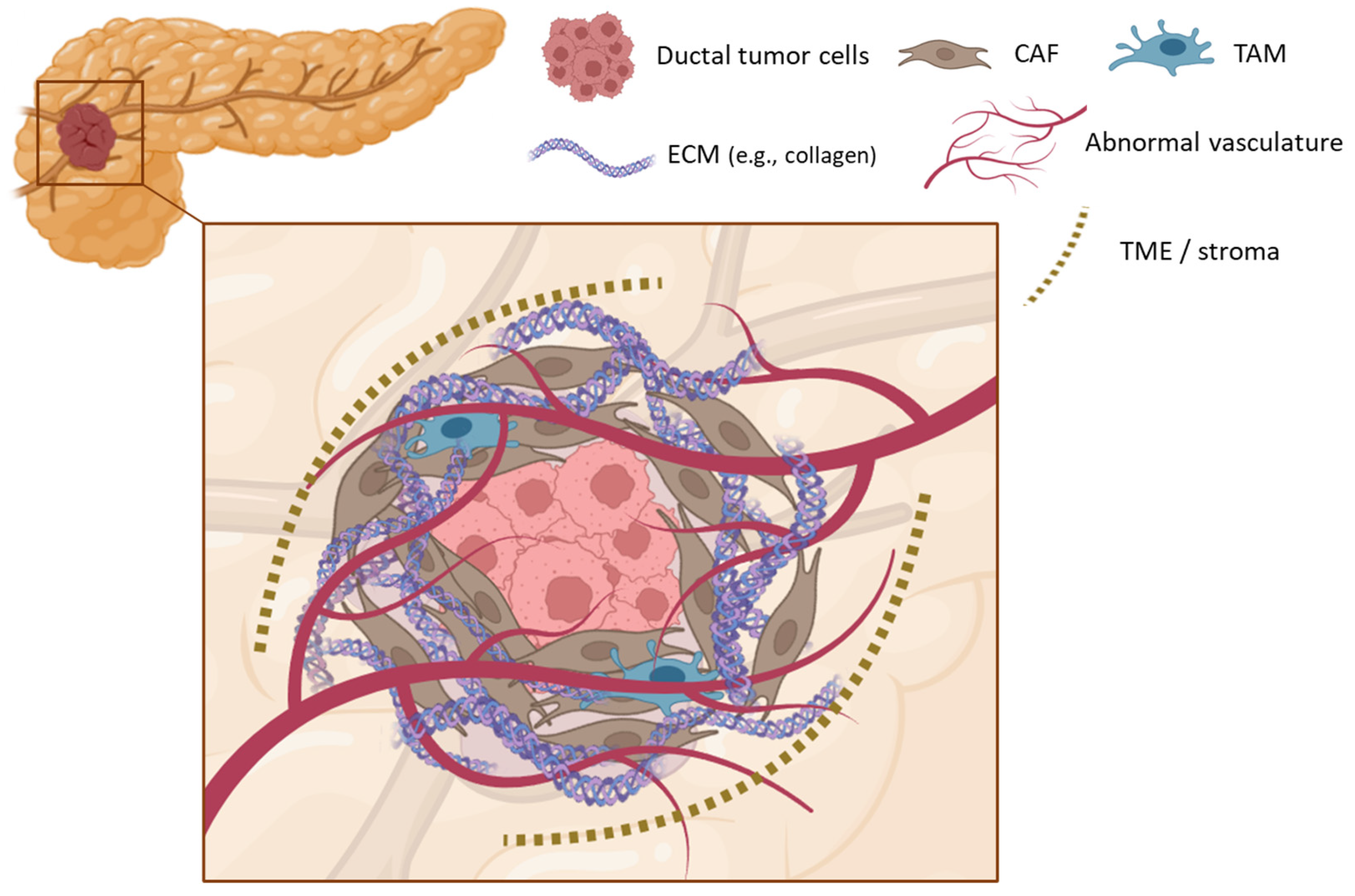
The pancreas is a vital organ of the digestive system, comprising both exocrine and endocrine components [21]. The exocrine pancreas, which constitutes the majority of the organ’s volume, produces digestive enzymes, while the endocrine pancreas, accounting for only 1–2% of the organ, consists of specialized cells that secrete hormones essential for regulating blood sugar and other functions [22]. Approximately 80% of PDAC cases originate in the exocrine pancreas, with about 75% located in the pancreatic head (Figure 1) [11]. Substantial evidence indicates that ductal cells are the primary origin of most PDAC cases [11,23]. Ductal cells are epithelial cells that comprise 10% of the exocrine pancreas and form small tubes known as ducts [23]. Their function consists of transporting digestive enzymes from the acinar cells to the duodenum [21].
Regarding the origin of PDAC, chronic pancreatitis is a major risk factor for promoting tumorigenesis, since chronic inflammatory processes often lead to cellular and tissue changes that favor malignant transformation [24]. In this context, ductal cells eventually can undergo the EtM transition event, changing their phenotype to mesenchymal [25]. The new phenotype is characterized by higher migratory capacity, invasiveness, and high resistance to apoptosis, features which form the basis of the high metastatic potential of pancreatic cancer cells [26,27]. One hallmark of pancreatic tumors is the exuberant stroma that dominates the tumor microenvironment (TME), where stromal cells promote malignant processes such as the EtM transition by the secretion of cytokines and growth factors [24,25]. A growing body of evidence confirmed that in PDAC, the TME plays a pivotal role in tumor development, invasion, and metastasis [20,28]. The neoplastic epithelium resides within a characteristic dense stroma, which is the cellular environment in which a tumor exists, localized between the tumor and the healthy tissue [29,30]. The TME encompasses the components that are in a constant dynamic interaction with tumoral cells, involving tumor blood vessels, lymphatic vessels, the extracellular matrix (ECM), non-tumor cells such as cancer-associated fibroblasts (CAFs), and secreted signaling molecules (Figure 2) [28,30,31].
Pancreatic stellate cells (PSC) are resident cells present in the exocrine portion of the pancreas, possessing a broad range of properties, including contractility, environmental sensing through specialized cell extensions, and the elaboration of ECM components [32]. During malignant processes, these PSCs can be activated into CAF [33]. This activation has been hypothesized to be driven by cross-talk between cancer cells and PSCs, mediated by signaling molecules including the sonic hedgehog protein, the cytokines TGF-β, TNF-α, and interleukins 1, 6, and 10 [34,35]. Additionally, evidence suggests that other signaling processes can reprogram stromal cells, such as direct cell-to-cell contact, specifically methylating the DNA of specific genes [36]. Once activated, CAFs, stimulated by pancreatic cancer cells, are the main contributors of desmoplasia through their excessive proliferation and the upregulation of ECM proteins, involving fibronectin, laminin, hyaluronic acid (HA), proteoglycans, tenascin C, and, especially, collagen [24,30]. The substantial increase in the deposition of these structural components results in a dense ECM, which forms the stroma and occupies the bulk of the tumor mass [24,30,37]. Intriguingly, other cells such as macrophages have been found to play a key role in promoting fibrosis and ECM remodeling, even regulating immune suppression. These cells are called tumor-associated macrophages (TAMs) and contribute to chronic inflammation, angiogenesis, metastasis, and drug-resistance [38,39]. Furthermore, enhanced angiogenesis is present in these tumors, leading to chaotic vasculature. This tumor-associated vasculature is composed of leaky vessels with blind ends, shunts, and a tendency to collapse. These abnormalities contribute to the low and unstable oxygenation within the TME [40]. Paradoxically, this hypoxic stromal environment promotes both tumor growth and metastatic spread while also inducing vascular collapse, thereby creating a barrier to the delivery of therapeutic agents [37]. Interestingly, although this process isolates malignant cells, it was discovered that the ECM still provides them with essential amino acids required for their invasive growth [41].

3. Conventional Treatments in PDAC

Traditionally, cancer treatments have focused on targeting neoplastic cells, employing methods such as surgery, radiation, chemotherapy, hormone therapy, and immunotherapy to address the rapidly proliferating tumor cells [42]. The first recommended therapeutic strategy, in solid tumors, involves their surgical resection, often followed by co-adjuvant treatments such as radiotherapy or chemotherapy. Although surgery is the most effective in early stages of cancer diseases, it is not always an option, especially in advanced cancers [42,43]. Indeed, curative surgery in PDAC is still inadequate, as the recurrence rate for resected cases is approximately 87% [44]. Under these circumstances, chemotherapy remains the most widely used treatment. It is currently considered one of the most effective approaches for cancer treatment, offering a diverse range of chemotherapeutic agents with varied mechanisms of action. However, despite advances in chemotherapy, the effectiveness of treatment in PDAC remains severely limited by multidrug resistance (MDR), driven by genetic heterogeneity and a fibrotic stromal environment, underscoring the need for a deeper insight into these complexities to enhance therapeutic outcomes [9,31].

3.1. Limitations of Conventional Treatments

MDR represents a major obstacle to effective therapeutic approaches against cancer. In this context, MDR refers to the ability of cancer cells to tolerate anticancer agents, significantly reducing the effectiveness of therapies and contributing to treatment failure in over 90% of patients with metastatic cancer [5,27,45]. Tumors can either be intrinsically resistant to specific drugs or develop resistance during treatment [16]. It has been found that the genetic diversity in human tumors leads to the rapid emergence of drug-resistant cells in response to the pressure exerted by toxic treatments [46].
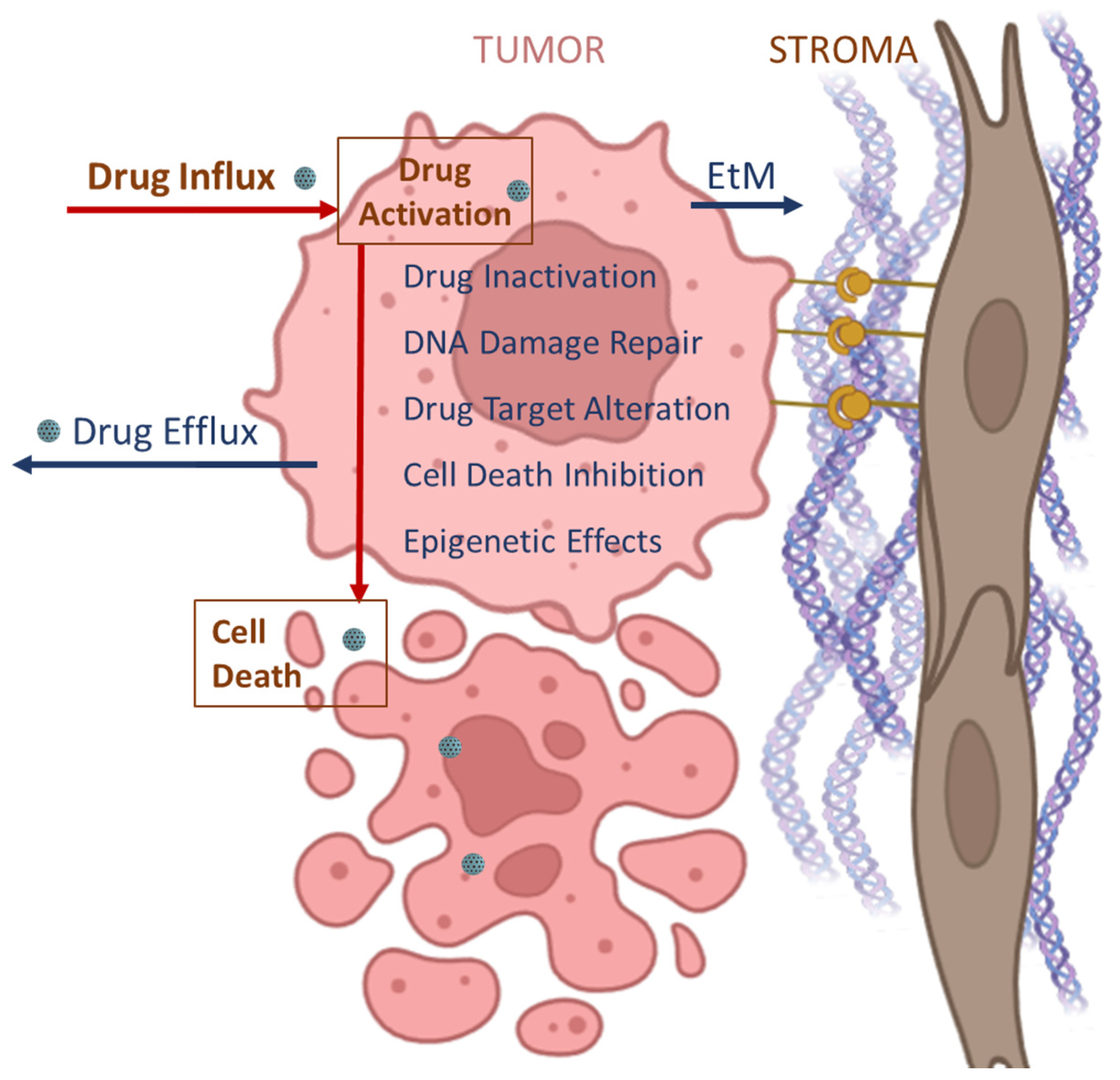
Drug efflux is one of the most extensively studied mechanisms of MDR, leading to the enhanced export of therapeutic agents from tumor cells (Figure 3) [27,45]. Regarding PDAC, both the EtM transition and the TME are critical factors that facilitate MDR. Although the role of the EtM transition in cancer resistance is still an emerging area of research, it may be influenced by processes involved in cell differentiation. The transition to a mesenchymal phenotype is a mechanism by which cancer cells from solid tumors become metastatic while enhancing their survival. This process is known to be regulated by both cancer and stromal cells. Moreover, the interaction of cancer cells with stromal cells and ECM elements are known to contribute the other forms of resistance, for example, by influencing epigenetic factors of cancer cells [27]. Furthermore, stromal cells not only hinder drug internalization but also secrete growth factors that stimulates cell proliferation and prevent the clearance of tumor cells [16,27,47].

3.2. Evolution of Chemotherapy in PDAC Treatment
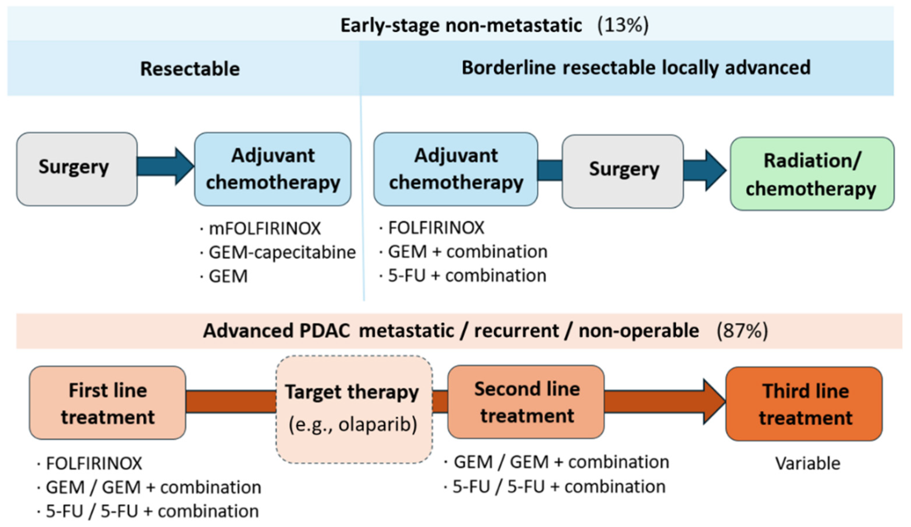
In 1996, gemcitabine (GEM) monotherapy was established as the gold standard for the treatment of PDAC across all disease stages. GEM demonstrated superior efficacy and a more favorable safety profile compared to the previously used chemotherapeutic agent, 5-fluorouracil (5-FU). Despite initial clinical benefits, the development of MDR, typically within a few months of treatment, significantly limited the long-term efficacy of GEM [15,48]. To overcome the limitations imposed by MDR, combination therapy, employing multiple mechanisms of action, has been recognized as a cornerstone of cancer treatment. Thus, GEM has been used in combination with other chemotherapeutic agents, including capecitabine, paclitaxel (PTX), and oxaliplatin [9]. However, these combinations have failed to demonstrated a significant improvement in the treatment efficacy [15,49]. A major breakthrough came in 2011 with the introduction of FOLFIRINOX, a combination of folinic acid, also known as leucovorin (LV), oxaliplatin, irinotecan (IRI), and 5-FU. This combination therapy exhibited a substantial improvement in clinical outcomes, doubling the median overall survival compared to GEM monotherapy, and it has been established as the new first-line chemotherapy for PDAC (Scheme 1) [8]. However, due to the toxic character of these treatments, modifications have been made to mitigate side effects in less tolerant patients. To address these concerns, modified regimens such as mFOLFIRINOX have been developed, typically involving dose reductions in IRI. Another alternative, FOLFOX, comprising only folinic acid, oxaliplatin, and 5-FU, excludes IRI and presents a more tolerable option for patients who may not withstand the FOLFIRINOX regimen, while exhibiting significant cytotoxic effects [49,50].
Scheme 1. General therapeutic approaches for PDAC categorized by disease stage. Percentages indicate the proportion of patients diagnosed at each stage. Clinical approaches to advanced PDAC are discussed in more detail in the following section. Adapted from the literature [51].
Nanomaterials 15 01139 sch001
Over the past decade, various therapies for PDAC have largely failed. For example, while numerous immune-based therapies have been approved for other cancers, none have been approved for pancreatic cancer due to its immunosuppressive TME, which shields tumor cells and hinders effective immunotherapy [15]. Targeted therapies, however, offer promise for specific mutations in PDAC genomes. Kinase inhibitors targeting growth factors have become prevalent due to their success in treating other cancers. In PDAC, only erlotinib has shown minimally significant improved efficacy over GEM monotherapy, earning FDA approval for metastatic, non-operable PDAC in combination with GEM [52]. Additionally, antibody-based targeted therapies, such as olaparib and pembrolizumab, have been approved for specific mutations affecting fewer than 10% of patients. Table 1 summarizes the main drugs approved for PDAC treatment in patients and their mechanisms of actions.

4. Nanomedicines as Emergent Treatment for Cancer

Despite advances in treating PDAC, conventional chemotherapeutics are limited by severe side effects, low accumulation in target tissues, rapid metabolism, and emerging drug resistance [42,59,60]. The alarming high mortality rate of PDAC highlights an urgent need for improved early-detection methods and more effective therapeutic strategies [49,59]. Addressing the challenging TME in PDAC would require the development of advanced, controlled drug release therapies that can precisely target the elusive pancreatic malignant cells while minimizing side effects [61].
Over the past few decades, nanomedicine has emerged as a promising discipline to address some of the limitations of conventional cancer treatments. Nanomedicine involves the application of nanoscale materials and devices to develop DDSs that enable controlled drug release. These nanoscale carriers, known as nanoparticles (NPs), possess unique properties, such as a high surface-to-mass ratio and the ability to adsorb and transport other substances [52,53,54]. Although nanomedicine is still in its early stages, nanoformulations have become a leading strategy for cancer imaging and therapy, with over 20 formulations approved for treating various cancers [31,62,63,64]. Integrating nanoscience into cancer treatments offers several advantages over conventional therapies, including controlled and sustained drug release, reduced systemic toxicity, enhanced stability, and increased drug concentration at the target site [62,65,66,67,68].
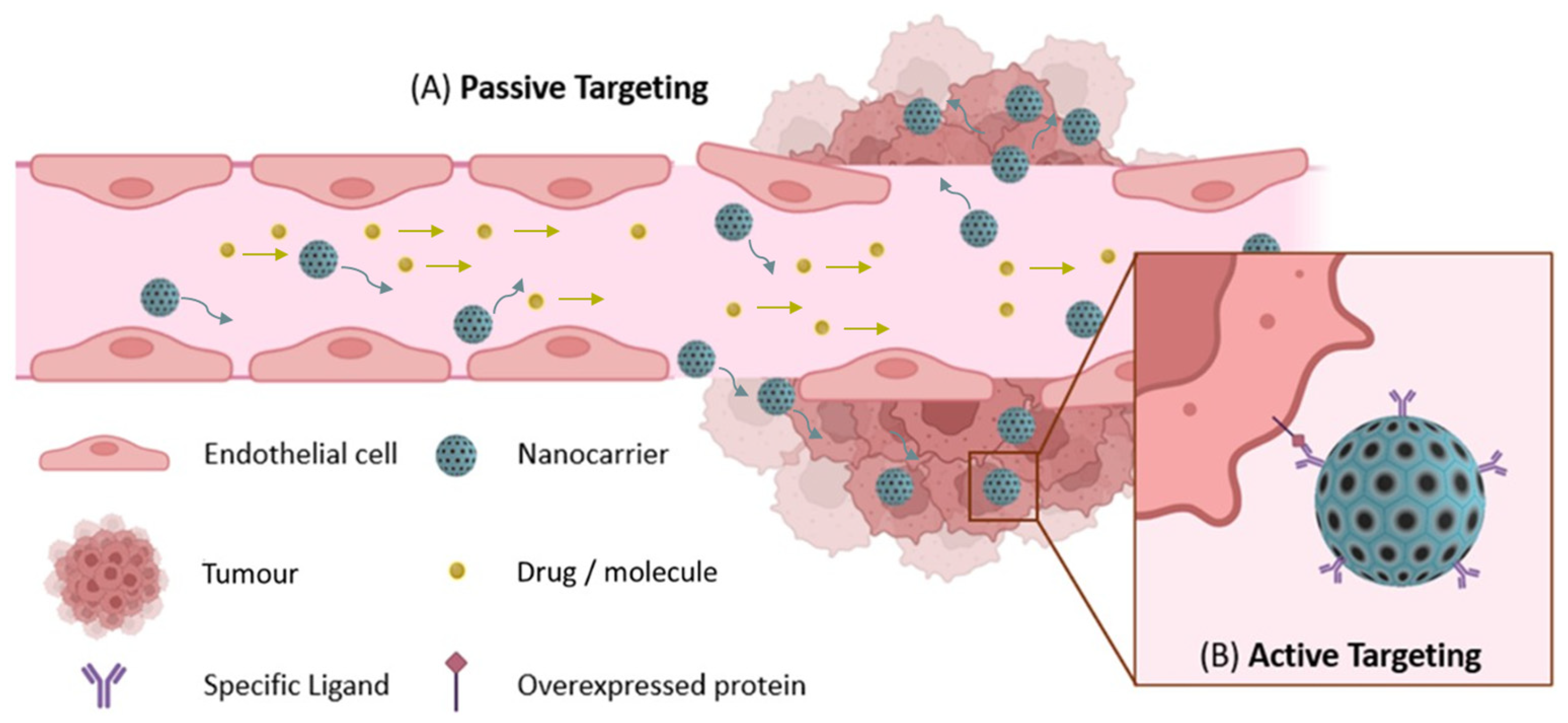
The abnormal TME in cancers exhibits distinct characteristics, such as irregular vasculature (Figure 4A), low pH levels, and elevated concentrations of the reductant glutathione (GSH) in the cytoplasm of cancer cells [20]. These key features can be leveraged to enhance the accumulation of DDSs and the release of drugs from nanocarriers within the TME through the design of selective stimuli-responsive systems [11,31,69]. To optimize their accumulation, it is well established that nanocarriers for cancer therapy should have hydrodynamic sizes ranging from 60 to 200 nm to exploit the enhanced permeability and retention (EPR) effect. This phenomenon, illustrated in Figure 4A, results from the abnormal, leaky vasculature of tumors. This size range enables preferential accumulation at the target site through the EPR effect, a mechanism not effective with conventional small-molecule drug treatments [70,71,72,73,74,75,76,77].

5. Emerging Nanosystems in PDAC Therapy

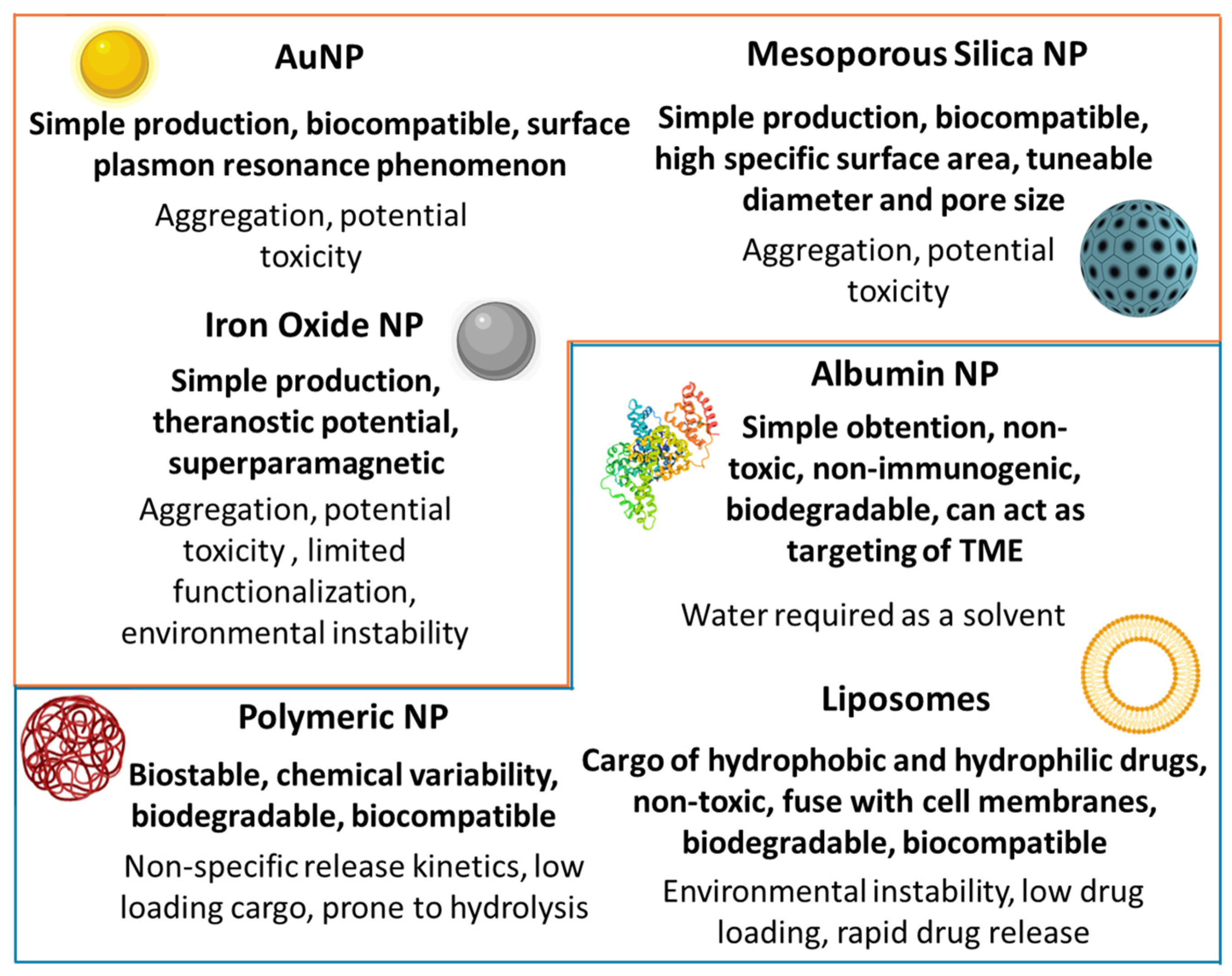
The highly metastatic and desmoplastic stroma of PDAC presents unique challenges that introduce additional hallmarks and treatment strategies to consider, such as the overexpression of specific proteins in either stromal or tumor cells, enabling specific active targeting strategies (Figure 4B) [69,78]. Various types of nanoparticles have been utilized to develop promising nanosystems to enhance the therapeutic efficacy of drugs for the treatment of PDAC (Figure 5). The present section provides a comprehensive overview of the nanosystems reported in the literature for PDAC. Specifically, Table 2, Table 3 and Table 4 summarize these systems and point out their main features.

5.1. Monotherapy Nanocarriers for PDAC Treatment

Monotherapy nanocarriers refer to nanosystems designed to deliver a single therapeutic agent with the aim of improving its pharmacological properties, bioavailability, and tumor-targeting ability of an individual anticancer agent. Table 2 compiles nanoconstructs developed for PDAC treatment that encapsulate one drug.
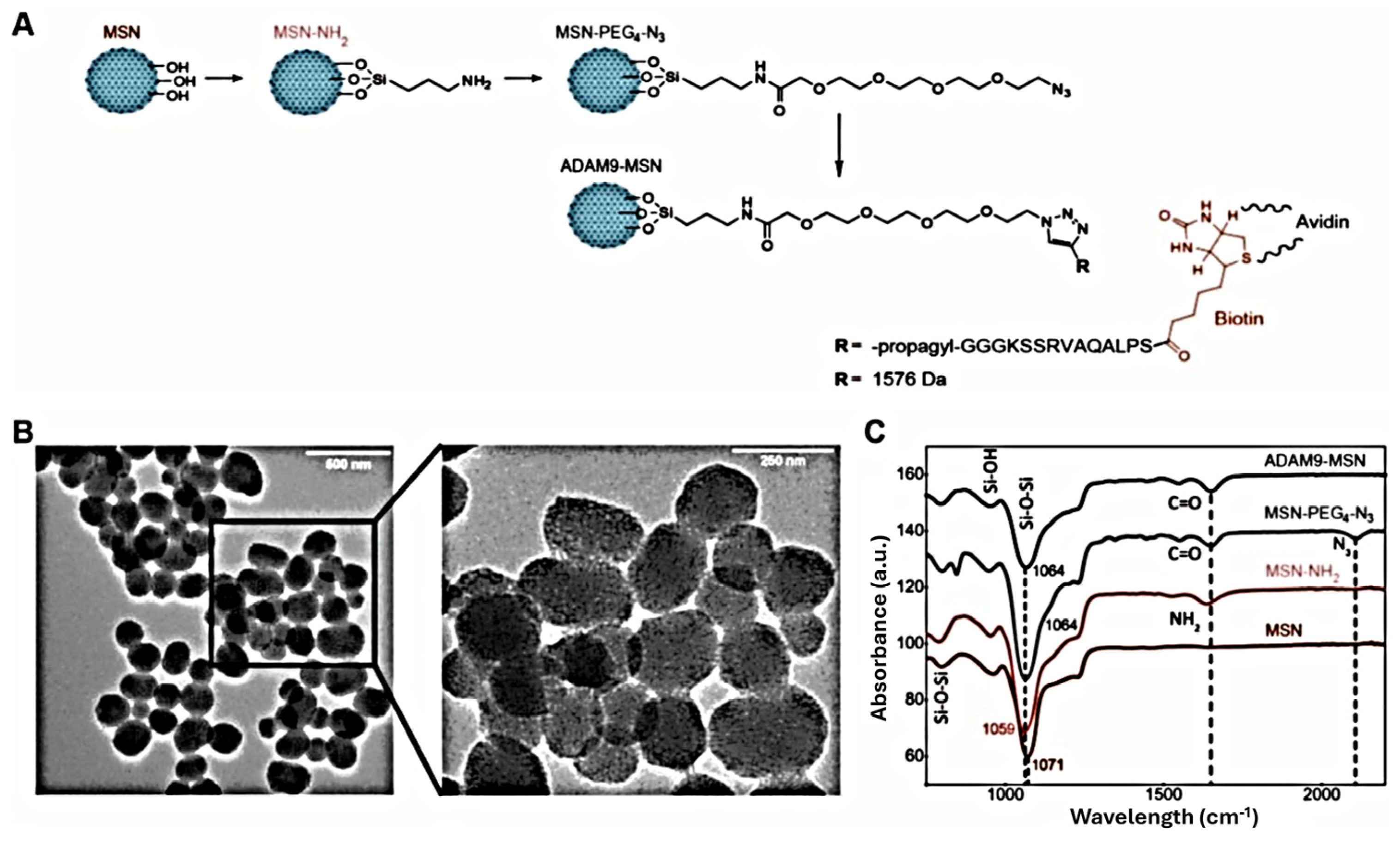
It is well-known that some chemotherapeutic agents such as paclitaxel (PTX), irinotecan (IRI) and cisplatin (CisPt) exhibit high water hydrophobicity, hindering their administration and delivery to target cells. This challenge can be exemplified by PTX, a key component of FOLFIRINOX gold standard therapy. Many authors have developed carriers to address paclitaxel hydrophobicity, involving albumin NP [11], polymeric NPs [79], and MSNs [80,81,82]. These systems have demonstrated controlled and sustained release, and some of them presented enhanced selectivity and cytotoxicity in in vivo assays, such as matrix metalloproteinase (MMP)-responsive systems (Figure 6) [80,81]. Particularly, the conjugation of PTX with albumin, known as nab-paclitaxel (nab-PTX, Abraxane®), has become the proof of concept for the development of nanomedicines for PDAC therapy [83]. This nanoformulation received FDA approval in 2013 for its use, in combination with GEM, as a frontline therapy for patients with advanced-stage metastatic PDAC [11]. Moreover, the outcomes suggested that albumin not only overcomes the poor solubility limitation of PTX, but it also acts as a targeting agent for the TME [15,78,84]. This is attributed to its affinity for the secreted protein acidic and rich in cysteine (SPARC), overexpressed by stromal fibroblast [78], and glycoprotein60 (GP60), an albumin receptor on endothelial cells that facilitates albumin transport across vascular cell layers [85].
Among the various types of nanoparticles (Figure 5), organic formulations, in particular liposomes, are the most commonly used DDSs in medicine. Liposomes are vesicles composed of phospholipid bilayers capable of encapsulating both hydrophilic and hydrophobic drugs. These types of formulations are the most FDA-approved platforms for cancer drug delivery due to their excellent biocompatibility [61,78,86].
In 2015, nanomedicine MM-398 (nal-IRI) was approved by FDA as a second-line therapy for metastatic PDAC in combination with 5-FU and LV [84]. Nal-IRI is a liposomal NP about 111 nm in diameter, encapsulating irinotecan. This formulation achieved high drug loading while optimizing retention and drug delivery in the tumor in comparison with the free drug IRI [18]. Furthermore, to enhance its therapeutic potential, this approach was improved by co-administering nal-IRI with 5-FU, leucovorin, and oxaliplatin, resulting in the formulation known as NALIRIFOX. The clinical evaluation of NALIRIFOX, using nab-PTX as the reference treatment, resulted in FDA approval in February 2024 as a new first-line treatment for advanced metastatic PDAC [12,87].
Over the past decade, research into inorganic nanoparticles for targeted PDAC nanocarriers has grown substantially. Among these, mesoporous silica nanoparticles (MSNs) have gained prominence due to their versatility and significant potential for surface functionalization [78,88]. Notably, hybrid systems, incorporating both organic and inorganic nanoparticles, have attracted significant attention due to their ability to combine the advantages of both types of materials [89,90,91,92]. Included in this group, lipid-hybrid MSNs are notable because they effectively integrate the biocompatibility of liposomes with the porosity and high loading capacity of MSNs [93,94]. For instance, Meng and colleagues demonstrated enhanced potency of IRI [93,94], oxaliplatin, and CisPt [95] by customizing lipid bilayer-modified MSNs capable of carrying high quantities of each drug while resulting in minimal leakage. Their DDSs demonstrated equivalent or superior biocompatibility and therapeutic efficacy compared to liposomal formulations and free drug assays in preclinical studies [93,94,95]. Moreover, the same authors used the lipid-film-coated procedure to rapidly seal GEM in a hybrid lipid MSN [96], improving the outcomes of their previous systems [97].
It should be noted that GEM is still a crucial drug in most available treatments for patients, either in combination or as a monotherapy (Scheme 1). Since GEM is highly water-soluble, its loading into certain carriers [89,98,99] and diffusion into cells are compromised [84]. However, this drawback has been addressed by other researchers through the attachment of GEM onto functionalized MSNs [100] or by utilizing more compatible carriers, such as albumin [101], liposomes [87,102,103], and polymeric NPs [72,73,104,105]. These strategies enhanced cellular internalization, improving the therapeutic efficacy of free GEM treatments.
In addition to the chemotherapeutic agents commonly used in PDAC treatment, other drugs with limited clinical application due to their hydrophobicity and poor biodistribution could become effective strategies when encapsulated in NPs. For instance, curcumin, loaded into liposomes [106], polymeric NPs [107], and MSNs [108], has been shown to inhibit tumor growth and reduce metastasis in PDAC xenograft models [78,106,108]. Similarly, the use of camptothecin (CPT) in clinics, the natural precursor of IRI, is also limited by its hydrophobicity and the significant side effects it causes. However, when encapsulated in nanocarriers, CPT has demonstrated substantial tumor regression in PDAC models using MSNs [109,110] and polymeric NPs [111], in comparison to the standard first-line drug GEM, for advanced pancreatic cancer [111].
Table 2. Summary of the main DDSs used for PDAC monotherapy. Glutathione (GSH); hydrodynamic size (HS); mesoporous silica NP (MSN); nanoparticle (NP); photodynamic therapy (PDT); polyethylene glycol (PEG); arginylglycylaspartic acid (RGD); reactive oxygen species (ROS); real size (RS); sonodynamic therapy (SDT). Targeting molecules are underscored for clarity.
DrugFormulationNanocarrierSize (nm)CharacteristicsRef.
PTX
paclitaxel
nab-PTXAlbumin NP130 RSTME Targeting[83]
PTX@MSN-responsive-ADAM9–biotyn–avidinMSN189 HSAvidin-capping
Protease-responsive linker
[80]
PTX@MSN-responsive-CAPN2–biotyn–avidinMSN235 HSAvidin-capping
Protease-responsive linker
[81]
PLGA-PTXPolymeric NP160 HSpH-sensitive polymeric coating[79]
PTX@MSNMSN100 RS-[82]
IRI
irinotecan
nal-IRI/MM-398Liposomal NP111 RS-[84]
Liposome conjugated to MSNHybrid lipid MSN80 RS
100–150 HS
Lipid bilayer[93]
Liposome conjugated to MSN with Au coreHybrid lipid MSN130 HSLipid bilayer[94]
GEM
gemcitabine
RGD peptide-conjugated magnetic MSNMagnetic modified MSN50 RSTumor-targeting[89]
Liposome–exosome fusionLiposome<200 HSTumor-targeting
Increased uptake
[87]
Liposome PEGylation-ligandLiposome<100 RSTumor-targeting[102]
Flow Focusing®Polymeric NP655 HSHigh payload
Narrow size distribution
[72]
MSNMSN42–64 RSPore-expanded[99]
HSA-GEM/IR780Albumin<10 HSCleavable peptide cathepsin B
Imaging with IR780
[101]
CurcuminCurcumin@PEGylated MSN-TransferrinMSN120 RS/167 HSPEGylation
Tumor-targeting
[108]
Liposomal curcuminLiposome-pH-responsive[106]
NanoCurc™Polymeric NP50 RSpH-responsive[107]
Methylene BlueGold-NP attached organically MSNAu-MSN hybrid NP30/55/80 RSPDT[90]
CisPt
cisplatin
Iron oxide NP covered silica shell -CisPtMagnetic- MSN54 RSpH-responsive[91]
Liposome conjugated to MSNHybrid lipid MSN82 RS
137 HS
pH-responsive[95]
FdUMP
5-FU metabolite
Aptamer (CCK-B)-PEG-FdUMP-CPNsCalcium phosphosilicate NPs (CPN)<100 RSTumor-targeting
pH-sensitive
[112]
CPT
camptothecin
CPT@MSN–Folate modificationMSN100–150 RSTumor-targeting[109]
Dendrimers–CPT
Charge switchable
Polymeric NP26 RSROS-responsive linker
Increased uptake
[111]
aptamer/cell-penetrating peptide–camptothecin prodrug NPscamptothecin prodrug NPs131 HSTumor-targeting[113]
DOX
doxorubicin
DOX liposome
Charge switchable
Liposome65 HSGSH-responsive
Increased uptake
[92]
Fluorinated amphiphilic dendrimerMicelle10 RSSelf-assembly micelles[114]
Ce6
chlorin e6
Polyphosphoester-based nanocarrierPolymeric NP40 RSpH-responsive
evasion immune clearance
[115]
Benzoporphyrin derivativeEutectic gallium–indium NPs, gallium oxide shell
Conjugated to hyaluronic acid
Gallium-indium NPs25–65 RSPDT[116]
IRT80 Encapsulated fluorocarbon chains and IRT80Hollow MSN<200 RSOxygen delivery, SDT[117]
Rose bengalSulfur hexafluoride PEG–biotin–avidinLipid microbubbles1–2 μmOxygen delivery, SDT[118]
Remarkably, photodynamic therapy (PDT) has emerged as a promising two-stage treatment based on the selective damage of tumoral cells, microorganisms, and blood vessels. This is achieved through the administration of a photosensitizer (PS), a non-toxic drug able to be selective while activated by light generating reactive oxygen species (ROS) [119]. Nanocarriers based on PDT have been developed for pancreatic cancer due to its high cytotoxic potential, achieving cell viabilities as low as 5%, thereby outperforming other therapies in targeting tumor cells [90,115,116,117,120,121]. Unfortunately, the fibrotic stroma and abnormal vasculature of PDAC contribute to severe hypoxia. Oxygen levels in PDAC tissue are estimated to be roughly 0.3%, compared to approximately 7.5% in healthy pancreatic tissue, constraining the PDT effectivity in pancreatic tumor cells [122]. In hypoxic environments the low oxygen levels hinders the formation of ROS. Nevertheless, the high cytotoxicity demonstrated by PDT in cancer cells has motivated researchers to design oxygen delivery strategies to overcome this challenge. To mitigate the hypoxia of PDAC tumors, some authors used AuNPs to increase the cytotoxic potential of 1O2. This effect is achieved by leveraging the localized surface plasmon resonance phenomenon of AuNPs, which facilitated surface energy transfer between the PS molecules and NPs. Such carriers are known to extend the lifetime of 1O2 through this mechanism, thereby improving the efficacy of PDT even under hypoxic conditions [90].
Other authors developed systems able to deliver oxygen to the target site by modification of carriers with fluorocarbon chains, increasing the efficacy of sonodynamic therapy (SDT), a variant of PDT, since it was demonstrated that several PS can also be activated by ultrasound irradiation. It has been demonstrated that several PSs can also be activated by ultrasound, a strategy that offers significant advantages over PDT, including cost-effectiveness, safety as a clinical imaging modality, and tunable penetration depending on the frequency used [117,118].

5.2. Combination Therapy Nanocarriers for PDAC Treatment

As depicted in Scheme 2, breakthroughs in nanotechnology are significantly redefining the therapeutic landscape for metastatic PDAC. Independent preclinical and clinical studies of nab-PTX and nal-IRI have proved that these nanoformulations offer superior pharmacokinetic profiles compared to their free drug counterparts. However, these formulations are not effective as standalone therapies; their combination with established antineoplastic agents is critical for achieving significant therapeutic efficacy and presenting notable improvements over conventional treatment strategies for PDAC [18,84,123]. These findings underscore the importance of combination therapy in the development of effective PDAC treatments; thereby, the co-delivery of multiple drugs in a single carrier emerges as a promising framework against this challenging disease.
Scheme 2. Overview of current approved treatments for metastatic PDAC patient therapy [12,51]. Treatments involving NPs are underlined. GEM: gemcitabine; 5-FU: 5-fluorouracil; LV: leucovorin, NALIRIFOX: nal-IRI + 5-FU + LV+ oxaliplatin; FOLFIRINOX: 5-FU + LV + irinotecan + paclitaxel; mFOLFIRINOX: modified-FOLFIRINOX.
Nanomaterials 15 01139 sch002
Nanocarriers for combination therapies are engineered to co-deliver two or more therapeutic agents within a single nanosystem, aiming to enhance treatment efficacy through combining mechanisms of action. This section explores the design, advantages, and therapeutic outcomes of combination nanocarrier systems developed for PDAC therapy (Table 3).
Table 3. Primary DDSs for the co-delivery of drugs for PDAC treatment. Chemo refers to chemotherapy. Cisplatin (CisPt); camptothecin (CPT); doxorubicin (DOX); gemcitabine (GEM); glypican 1 (GPC1); glutathione (GSH); hydrodynamic size (HS); mesoporous silica NP (MSN); glycoprotein mucin 1 (MUC1); nanoparticle (NP); photodynamic therapy (PDT); polyethylene glycol (PEG); polyethylenimine (PEI); poly(lactide-co-glycolide) (PGLA); paclitaxel (PTX); real size (RS); sonodynamic therapy (SDT) zinc-phthalocyanine (Zn-Pc).
DrugsTherapyNanocarrierCompositionSize (nm)FeaturesRef.
GEM/PTXChemoPolymeric NPTri-block co-polymer tumor-targeted peptide159 HSpH-responsive tumor-targeted Inhibition GEM deactivation[104]
ChemoHybrid lipid polymeric NPsLipid-bilayer
PGLA
70 HSpH-sensitive Drug conjugate Inhibition GEM deactivation[124]
ChemoHybrid lipid MSNsLipid bilayer65 RSpH-responsive[96]
GEM/CisPt ChemoAntibody (TAB004)-GEM-CisPt-MSNPEI/PEG150–200 HSRedox-responsive Tumor-targeting[100]
GEM/iron oxide NPs Chemo, ThermalPolymeric NPsPLGA
HER-2 Antibody
534 HSTumor-targeting[73]
GEM/Iron oxide NPs/CetuximabThermal, Chemo, TargetedMagnetic Albumin NPIron oxide NPs Cetuximab200 HSTumor-targeting Imaging[125]
GEM/Au-NPChemo, PDT, ThermalGold-nanoshell-coated MSNTransferrin Gold-nanoshell100–150 RSTumor-targeting[120]
GEM/Ferulic acidChemo, AntioxidantMSNanti-GPC1 antibodies206 HSTumor-targeting Low cargo[98]
GEM/ONC201Chemo, TargetedLiposomesONC201 (MUC1 peptide)113 HSTumor-targeting Apoptosis upregulation[103]
Oxaliplatin/IndoximodChemo, ImmuneHybrid lipid MSNLipid bilayer83 RSImmunoactivator[126]
Bortezomib/IR-820Chemo, PDT, thermalHybrid lipid MSNCiclosporin A160Increased uptake[121]
Zn-Pc/CetuximabPDT, TargetedMSNPEGylated Cetuximab303 HS
79 RS
Tumor-targeting[74]
[75]
CPT/DOXChemoMSNQuantum dot150–200 RSpH-responsive[110]
DOX/hydroxychloroquineChemoMesoporous silica nanorods-180 × 60 RSAutophagy inhibition
Macropinocytosis selectivity
[127]
DOX/iron oxide NPChemo, ThermalMagnetic-MSNShell of MSN55 RS
106 HS
Thermal-sensitive caps[128]
Numerous nanocarriers developed for PDAC have been based on combining chemotherapeutic agents, aiming to mitigate the common MDR associated with chemotherapeutic agents like GEM. For instance, PTX has been reported to function as an inhibitor of cytidine deaminase, the enzyme responsible for deactivating GEM, highlighting the significant therapeutic potential of combining PTX and GEM. Specifically, polymeric [104], hybrid lipid polymeric [124], and hybrid lipid MSN [96] formulations exhibited high stability and synergistic cytotoxic effect in mouse xenograft models of PDAC. As mentioned, Meng and colleagues developed a lipid film-coated method to rapidly encapsulate GEM in a hybrid lipid–MSN system. This lipid entrapment not only significantly increased GEM’s encapsulation efficiency but also enabled the co-loading of paclitaxel. As a result, the system showed enhanced efficacy compared to nab-paclitaxel combined with free GEM in preclinical studies [96]. Furthermore, Zhang and coworkers synthesized a GEM-PTX conjugate (1:1) via hydrolysable linker to encapsulate it in hybrid lipid polymeric NPs [124]. The conjugation through a sensitive linker endows the system with even more selectivity, minimizing size effects. The cytotoxicity of the resulting GEM-PTX against pancreatic cancer cells not only was comparable to that of the corresponding free drug mixtures, but also was significantly improved after its encapsulation into hybrid lipid polymeric NPs [124]. Interestingly, although drug conjugation offers superior control over drug stoichiometry and release kinetics, this strategy remains mainly underexplored in drug delivery approaches for PDAC, as observed in Table 3.
Other authors have taken advantage of the aforementioned high specific surface area of MSNs to design advanced drug delivery systems capable of co-loading multiple therapeutic agents within a single carrier [68,88,129]. Escoto and colleagues attached GEM and CisPt onto MSN surfaces thorugh a redox-sensitive linker and found cytotoxic synergistic effect with their system [100]. Moreover, the presence of polyethyleneimine (PEI) and polyethylene glycol (PEG) polymers on the NP surfaces increase its uptake and lifetime through the bloodstream, respectively. In addition, the conjugation of a specific antibody to the PEG chains endows the system with active targeting, further enhancing the internalization of the system to pancreatic tumoral cells [100].
To further improve the efficacy of anticancer drugs, chemotherapeutic agents have also been combined with other active molecules and therapies to explore other ways to tackle PDAC, including antioxidants [98], targeted therapy [103], thermal therapy [73], immunomodulation [126], autophagy inhibition approach [127], and photodynamic therapy [121].
In 2017, Meng and colleagues were the first to demonstrate an immune-based approach for drug delivery in PDAC. Indoximod was selected to activate the immune system by the suppression of an immunosuppressive pathway. Moreover, the co-delivery of this immune modulator with oxaliplatin resulted in a synergistic immune response in preclinical models [126].
Regarding targeted therapy, the co-delivery of GEM and cetuximab via magnetic-albumin NPs permits not only the triple therapy by magnetic-targeted thermo-chemotherapy but also the targeting of both stromal and tumor cells. While albumin facilitates the retention of the system at the TME, cetuximab targets the cancer cells, further enhancing the system’s uptake by PDAC cells (Figure 7) [125]. Another example is the combination of GEM with the novel targeted molecule ONC201. This combination therapy was encapsulated in a liposomal formulation, resulting in the upregulation of apoptosis and enhanced accumulation of the carrier within the TME. However, preclinical studies demonstrated no significant advantages beyond the inhibition of tumor progression [103].
PDT was also exploited in combination with chemotherapeutic agents. For instance, Nie and coworkers introduced a hybrid lipid MSN system for triple therapy, combining chemotherapy with bortezomib, photothermal therapy using IR-820, and enhanced cell penetration via cyclosporine A [120]. Another example is given by Yurt and colleagues. The authors prepared PEGylated MSNs loaded with zinc phthalocyanine and conjugated with cetuximab for combination therapy of PDT and targeted therapy against a growth factor receptor demonstrated limited effectiveness in preclinical experiments [75].
It should be noted that in recent examples, active targeting has played a major role in enhancing the uptake of nanocarriers by pancreatic cancer cells [103,125]. Many of the systems summarized in Table 2, Table 3 and Table 4 are functionalized with various targeting molecules, including liposomes [103], hybrid-NPs [89,120,124,125], polymeric NP [73], iron oxide NP [130], and MSN [74,98,108], which demonstrate improved selective internalization. It is also worth noting that alternative strategies, such as surface decoration with cationic or hydrophobic molecules, have been employed to increase cellular internalization. For instance, surface modification of MSNs with the cationic polymer PEI [100] and the use of charge-switchable approach in liposomes [92] and polymeric NPs [111] has proven effective in enhancing uptake in these systems [92,111]. Another example is provided by Kim and colleagues, who utilized cyclosporine A in a hybrid lipid-MSN system to improve cellular uptake in PDAC cells. This enhancement was achieved through the interaction of a hydrophobic peptide with cell membranes, promoting a more efficient internalization [121].

5.3. Nanocarriers for Combined Anticancer and Stromal Therapies in PDAC Treatment

The fibrotic stroma is rich in biopolymers such as hyaluronic acid and collagen that contribute to elevate the interstitial fluid pressure, which, in turn, hampers diffusion through the extracellular matrix, especially for larger-size nanocarriers [131]. Despite the advantages of combination therapies and targeting strategies to increase the intracellular internalization of nanoplatforms, this formidable barrier significantly hampers the selectivity of nano-formulations to be accumulated in the TME, contributing to the poor prognosis in PDAC treatment [37,132].
In light of this impediment, stroma modulation strategies have been emerging as attractive strategies (Table 4) [15,133]. These innovations aim to alleviate the stromal barrier, thereby enhancing drug penetration and improving the therapeutic efficacy of loaded chemotherapeutics in the treatment of PDAC [15,134].
Table 4. DDS for enhanced stromal penetration approaches against PDAC. Dendrigraft poly-l-lysine (DGL); docetaxel (DTX); extracellular matrix (ECM), hydrodynamic size (HS); gemcitabine (GEM); glutathione (GSH); doxorubicin (DOX); mesoporous silica NP (MSN); nanoparticle (NP); polyethylene glycol (PEG); polyethylenimine (PEI); poly(lactide-co-glycolide) (PGLA); poly(ethylene glycol)-poly(caprolactone) (PP); plectin-1 targeted peptide (PTP); real size (RS); transforming grow factor (TGF).
NanocarrierModificationsTumor
Therapy
Stromal ApproachSize (nm)FeaturesRef.
Hybrid liposome-MSNCo-polymer coating of MSN: PEG/PEIGEMTGF-β inhibitor143→50 RSpH-responsive
Low loading
[97]
MSNDOX@MSN-S-nitrosothiol/PEGDOXS-nitrosothiol107 HSCollagen depletion[134]
Mesoporous polydopamineCAF membrane, PTPIron carbonylLosartan126 nmECM degradation[135]
Lipid NPsaPD-L1 (antibody), perfluoropentane DTXPulsed ultrasound stimulation95 nmAntifibrotic, recover hypoxia[136]
HMONHollow organically MSNGEMpirfenidone115→33 RSpH/GSH-responsive[137]
Liposome-modified
Polymeric NP
Small NP:
PEG-PGLA
PTXTGF-β inhibitor
Shrinkable
155→40 RSpH-responsive[138]
Polymeric NPChloroquine phosphateGEMShrinkable125→30 HSpH-responsive
Inhibition autophagy
[105]
Dendrigraft to PP micelle (DGL/DOX@PP)DOXShrinkable100→30 RSMMP-responsive[139]
dendrigraft poly-l-lysine to PP micelleGEM18β-glycyrrhetinic acid
Shrinkable
151→30 RSMMP-responsive[28]
PLGA polymerGEMSimvastatin258 RSMitigation of stroma
pH-sensitive
[77]
Iron oxide NPpH low insertion peptides: pHLIPGEMMetformin23 HS
10 RS
Stromal depletion
pH-responsive
[130]
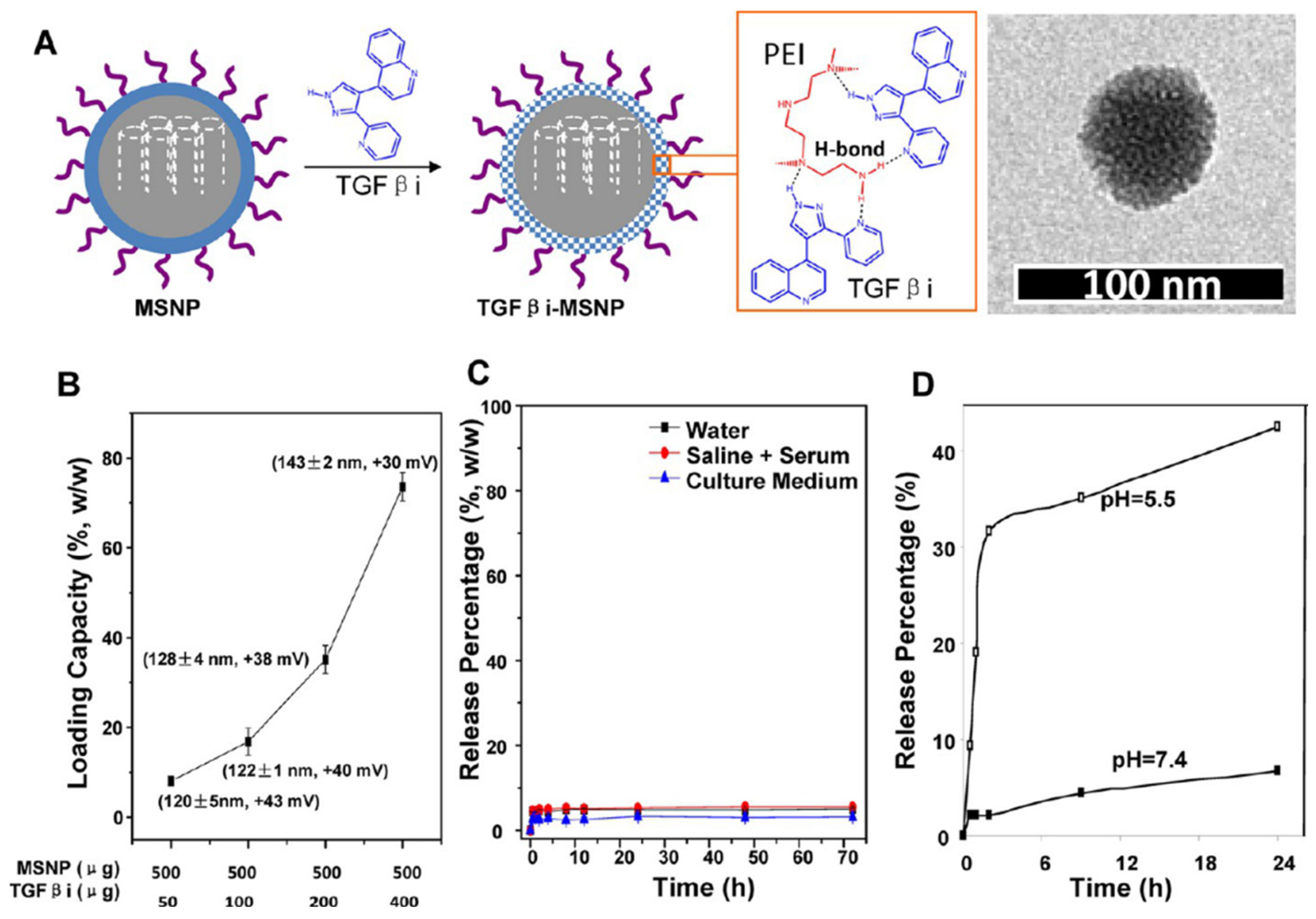
A growing number of stromal modulators have demonstrated significant potential for stromal depletion, including TGF-β inhibitors [97,138], pirfenidone [137], 18β-glycyrrhetinic acid [28], simvastatin [77], and metformin [130]. For instance, PEGylated MSNs have been employed to deliver S-nitrosothiol, a nitric oxide donor, aiming to activate specific proteases present in the TME that degrade collagen-composing ECM. This strategy facilitates deeper penetration of the carrier, allowing doxorubicin DOX to effectively reach the hindered pancreatic cancer cells [134]. It is worth highlighting that the two-wave nanocarrier strategy significantly enhanced tumor drug delivery (Figure 8). The first wave employed TGFβi-loaded MSNs to disrupt pericyte coverage and increase vascular permeability, priming the tumor for the second wave of PEGylated, drug-loaded liposomes. This sequential delivery enabled deeper TME penetration and superior therapeutic efficacy. By combining GEM loading with targeted TGF-β pathway inhibition, this approach achieved sustained tumor suppression beyond 25 days, outperforming both free-drug and conventional standalone liposome treatments [97].
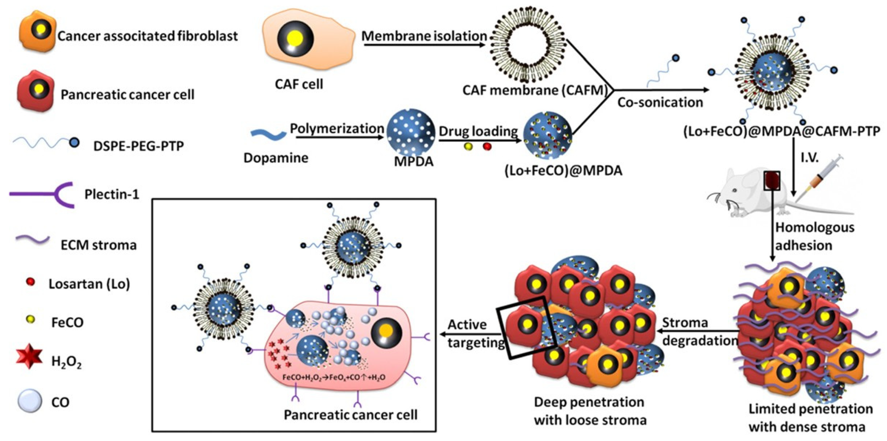
Recently, Cheng and colleagues engineered a nanocarrier for the dual targeting of pancreatic tumor. These researchers prepared mesoporous polydopamine nanoparticles loaded with an iron carbonyl and losartan, enabling gas therapy with CO against tumor cells and degradation of the ECM, respectively. The NPs were then coated with CAF cell membranes to facilitate the targeting of CAFs and the delivery of losartan. Additionally, functionalization of the CAF membranes with a plectin-1-targeting peptide (PTP) endowed the system with specific targeting of pancreatic cancer cells (Figure 9) [135].
The vasculature within the TME consists of tortuous, leaky, and non-uniform vessels [24]. The size of the pores or fenestrations in the tumor vascular walls range from 10 nm to 2 μm [63,131]. It is well-known that the transport of nanocarriers across tumor vessel walls and cell membranes is largely influenced by NP properties including, shape, charge, and size. For cancer targeting, NPs are typically recommended to be 20–100 nm in size [70,140]. Consequently, smaller NPs (<60 nm) are associated with greater infiltration efficiency. Paradoxically, NPs with larger hydrodynamic sizes (60–200 nm) exhibit longer blood circulation half-lives and an enhanced extravasation and retention (EPR) effect but reduced tumor permeability [70,71]. These conflicting size requirements have driven the development of size-shrinkable nanocarriers, which have emerged to balance NP accumulation and penetration in tumors simultaneously [138].
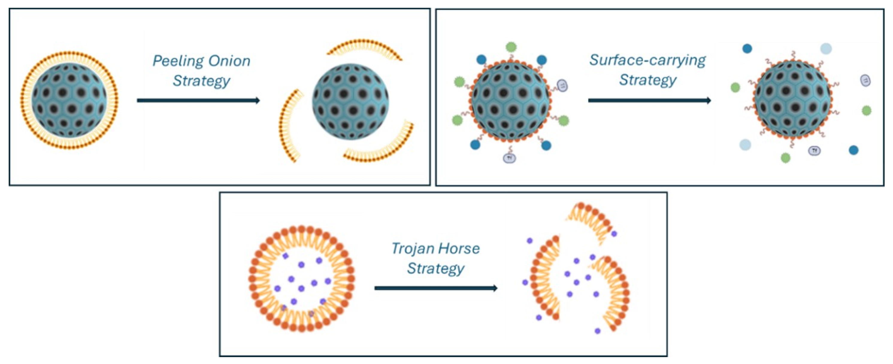
Shrinkable systems are engineered to reduce their size in response to specific stimuli, facilitating selective penetration through the stroma to target cancer cells (Figure 10). These systems can be summarized into the models according to their structural characteristics: a peeling onions strategy, a surface-carrying strategy, and a Trojan horse strategy [71]. In this context, polymeric NPs have predominantly been used to design size-switchable nanocarriers, largely due to the frequent presence of hydrolysable ester bonds [77,105]. Similarly, the incorporation of responsive linkers is a strategy commonly reported for other nanocarriers [96,113]. Furthermore, some authors have incorporated a stromal modulator into size-switchable NPs. The resulting systems exhibited enhanced drug accumulation in the TME, reduced stroma fibrosis, and deeper NP penetration, offering a promising strategy for future PDAC treatments [19,28,138].
The peeling onion strategy involves the construction of nanosystems with multilayered architecture. Each layer is designed to respond to a specific stimulus, allowing for a stepwise reduction in the overall size of the nanocarrier [71]. By that means the nanocarrier can shrink progressively through biological environments, enhancing its ability to penetrate deeper into the fibrotic stroma and target cancer cells. In this regard, NPs with versatile tunable surface and size, such as MSNs, are particularly well-suited for this strategy [88,141]. Nel and colleagues provided a notable example by developing a shrinkable co-polymer-modified MSNs, composed of PEI and PEG polymers. Once accumulated in the TME, the pH-sensitive polymer degradation reduced the size of the system from 143 nm to 50 nm while releasing LY364947, a TGF-β inhibitor [97].
In 2018, He and colleagues developed an innovative surface-carrying strategy. This approach relies on the construction of nanocomplexes formed by small NPs tethered to the surface of a larger NP, allowing the small NPs to be released from deeper stromal penetration (Figure 10) [71]. Firstly, they constructed a system based on dendrigraft poly-l-lysine (DGL) conjugated to poly(ethylene glycol)-poly(caprolactone) (PP) micelles, linked via an MMP-responsive peptide. This MMP-responsive system (100 nm) was designed to release DOX conjugated to DGL by action of MMP proteins in TME. As a consequence, DGL nanocarriers of 30 nm were able to deep penetration into the stroma (Figure 11) [139]. Building on this approach, the incorporation of a stroma modulator, 18β-glycyrrhetinic acid, and autophagy inhibitor, chloroquine, have been studied in other works to further enhance therapeutic efficacy [28,105].
Another strategy, called the Trojan horse, is based on encapsulating small NPs inside large NPs, thereby enabling the release of the small NPs under certain conditions (Figure 10) [71]. Recently, Nie and colleagues pioneered a novel approach based on the Trojan horse strategy for combination of a stromal modulation strategy with a size-switchable nanosystem. PEG−PLGA nanospheres of 40 nm were carried in liposomes of 155 nm. An antifibrotic agent, a TGF-β inhibitor, was attached onto liposomal surface, while containing smaller polymeric NPs carrying PTX. Upon liposome degradation, the system delivered the stromal-modulating therapy targeting CAFs, while facilitating the deeper penetration of the smaller polymeric NPs and enables the precise delivery of PTX to tumor cells [138].

6. Conclusions

This review highlights the significant advantages of using nanocarriers for combination therapy in PDAC. Specifically, DDSs for multi-drug delivery can substantially reduce the systemic toxicity of chemotherapeutics, simplify treatment regimens, and enhance efficacy through optimized drug combinations. By encapsulating drugs with distinct therapeutic mechanisms into delivery systems at optimal ratios, nanocarriers enable sequential delivery tailored to clinical needs.
While the EPR effect has been a cornerstone of nanomedicine, its limitations in achieving precise targeting are increasingly evident. Nevertheless, NPs are ideal platforms for developing advanced targeting strategies for PDAC, such as incorporating ligands like peptides, aptamers, or antibodies to improve treatment selectivity. Of particular significance in the context of PDAC is the development of size-switchable nanoparticles capable of penetrating the dense extracellular matrix surrounding the tumor.
Despite the immense promise of nanocarriers, challenges such as large-scale reproducibility and long-term safety must be addressed for widespread clinical adoption. Encouragingly, several nanocarrier-based formulations have already received FDA approval, marking significant progress toward clinical translation. However, challenges persist, as FDA-approved nanocarriers for PDAC often require co-administration with conventional chemotherapeutics to achieve therapeutic efficacy comparable to standard treatments. With continued innovation, nanomedicine is poised to revolutionize pancreatic cancer therapy, offering hope for more effective and targeted treatments in the near future.

Author Contributions

Writing—original draft preparation; I.P.; writing—review and editing funding acquisition, D.S.-G. All authors have read and agreed to the published version of the manuscript.

Funding

Spanish Ministerio de Ciencia e Innovación for the grant PID2021-126346OB-I00. The support of Agencia de Gestió d’Ajuts Universitaris i de Recerca (AGAUR) from Generalitat de Catalunya is also acknowledged (SGR 2021 00537).

Acknowledgments

The authors have reviewed and edited the output and take full responsibility for the content of this publication.

Conflicts of Interest

The authors declare no conflicts of interest.

References

  1. Garima, M.; Sumitra, N.; Pramod, K.S. Cancer: An Overview. Acad. J. Cancer Res. 2015, 8, 1–9. [Google Scholar]
  2. Senapati, S.; Mahanta, A.K.; Kumar, S.; Maiti, P. Controlled drug delivery vehicles for cancer treatment and their performance. Signal Transduct. Target. Ther. 2018, 3, 7. [Google Scholar] [CrossRef] [PubMed]
  3. Urruticoechea, A.; Alemany, R.; Balart, J.; Villanueva, A.; Vinals, F.; Capella, G. Recent Advances in Cancer Therapy: An Overview. Curr. Pharm. Des. 2010, 16, 3–10. [Google Scholar] [CrossRef] [PubMed]
  4. Bray, F.; Ferlay, J.; Soerjomataram, I.; Siegel, R.L.; Torre, L.A.; Jemal, A. Global Cancer Statistics 2018: GLOBOCAN Estimates of Incidence and Mortality Worldwide for 36 Cancers in 185 Countries. CA Cancer J. Clin. 2018, 68, 394–424. [Google Scholar] [CrossRef] [PubMed]
  5. Emran, T.B.; Shahriar, A.; Mahmud, A.R.; Rahman, T.; Abir, M.H.; Siddiquee, M.F.; Ahmed, H.; Rahman, N.; Nainu, F.; Wahyudin, E.; et al. Multidrug Resistance in Cancer: Understanding Molecular Mechanisms, Immunoprevention and Therapeutic Approaches. Front. Oncol. 2022, 12, 891652. [Google Scholar] [CrossRef]
  6. Samanta, K.; Setua, S.; Kumari, S.; Jaggi, M.; Yallapu, M.M.; Chauhan, S.C. Gemcitabine Combination Nano Therapies for Pancreatic Cancer. Pharmaceutics 2019, 11, 574. [Google Scholar] [CrossRef]
  7. Haeberle, L.; Esposito, I. Pathology of pancreatic cancer. Transl. Gastroenterol. Hepatol. 2019, 4, 50. [Google Scholar] [CrossRef]
  8. Kamisawa, T.; Wood, L.D.; Itoi, T.; Takaori, K. Pancreatic Cancer. Lancet 2016, 388, 73–85. [Google Scholar] [CrossRef]
  9. Adamska, A.; Domenichini, A.; Falasca, M. Pancreatic Ductal Adenocarcinoma: Current and Evolving Therapies. Int. J. Mol. Sci. 2017, 18, 1338. [Google Scholar] [CrossRef]
  10. Tempero, M.A.; Malafa, M.P.; Al-Hawary, M.; Behrman, S.W.; Benson, A.B.; Cardin, D.B.; Chiorean, E.G.; Chung, V.; Czito, B.; Del Chiaro, M.; et al. Pancreatic Adenocarcinoma, Version 2.2021, NCCN Clinical Practice Guidelines in Oncology. J. Natl. Compr. Cancer Netw. 2021, 19, 439–457. [Google Scholar] [CrossRef]
  11. Viegas, C.; Patrício, A.B.; Prata, J.; Fonseca, L.; Macedo, A.S.; Duarte, S.O.D.; Fonte, P. Advances in Pancreatic Cancer Treatment by Nano-Based Drug Delivery Systems. Pharmaceutics 2023, 15, 2363. [Google Scholar] [CrossRef]
  12. Imperial, R.; Mosalem, O.; Majeed, U.; Tran, N.H.; Borad, M.J.; Babiker, H. Second-Line Treatment of Pancreatic Adenocarcinoma: Shedding Light on New Opportunities and Key Talking Points from Clinical Trials. Clin. Exp. Gastroenterol. 2024, 17, 121–134. [Google Scholar] [CrossRef]
  13. Halbrook, C.J.; Lyssiotis, C.A.; Pasca di Magliano, M.; Maitra, A. Pancreatic cancer: Advances and challenges. Cell 2023, 186, 1729–1754. [Google Scholar] [CrossRef] [PubMed]
  14. Roscigno, G.; Jacobs, S.; Toledo, B.; Borea, R.; Russo, G.; Pepe, F.; Serrano, M.J.; Calabrò, V.; Troncone, G.; Giovannoni, R.; et al. The potential application of stroma modulation in targeting tumor cells: Focus on pancreatic cancer and breast cancer models. Semin. Cancer Biol. 2025, 113, 151–175. [Google Scholar] [CrossRef] [PubMed]
  15. Springfeld, C.; Jäger, D.; Büchler, M.W.; Strobel, O.; Hackert, T.; Palmer, D.H.; Neoptolemos, J.P. Chemotherapy for Pancreatic Cancer. Presse Medicale 2019, 48, e159–e174. [Google Scholar] [CrossRef] [PubMed]
  16. Vasan, N.; Baselga, J.; Hyman, D.M. A View on Drug Resistance in Cancer. Nature 2019, 575, 299–309. [Google Scholar] [CrossRef]
  17. Rajeshkumar, N.V.; Yabuuchi, S.; Pai, S.G.; Tong, Z.; Hou, S.; Bateman, S.; Pierce, D.W.; Heise, C.; Von Hoff, D.D.; Maitra, A.; et al. Superior therapeutic efficacy of nab-paclitaxel over cremophor-based paclitaxel in locally advanced and metastatic models of human pancreatic cancer. Br. J. Cancer 2016, 115, 442–453. [Google Scholar] [CrossRef]
  18. Carnevale, J.; Ko, A.H. MM-398 (Nanoliposomal Irinotecan): Emergence of a Novel Therapy for the Treatment of Advanced Pancreatic Cancer. Futur. Oncol. 2016, 12, 453–464. [Google Scholar] [CrossRef]
  19. Nagarajan, Y.; Chandrasekaran, N.; Deepa Parvathi, V. Functionalized Nanomaterials In Pancreatic Cancer Theranostics and Molecular Imaging. ChemistryOpen 2025, 14, e202400232. [Google Scholar] [CrossRef]
  20. Liu, X.; Shao, Y.; Li, Y.; Chen, Z.; Shi, T.; Tong, Q.; Zou, X.; Ju, L.; Pan, J.; Zhuang, R.; et al. Extensive Review of Nanomedicine Strategies Targeting the Tumor Microenvironment in PDAC. Int. J. Nanomed. 2025, 20, 3379–3406. [Google Scholar] [CrossRef]
  21. Cade, J.E.; Hanison, J. The Pancreas. Anaesth. Intensive Care Med. 2017, 18, 527–531. [Google Scholar] [CrossRef]
  22. Valente, R.; Coppola, A.; Scandavini, C.M.; Halimi, A.; Magnusson, A.; Lauro, A.; Sotirova, I.; Arnelo, U.; Franklin, O. Interactions between the Exocrine and the Endocrine Pancreas. J. Clin. Med. 2024, 13, 1179. [Google Scholar] [CrossRef]
  23. Pour, P.M.; Pandey, K.K.; Batra, S.K. What is the origin of pancreatic adenocarcinoma? Mol. Cancer 2003, 2, 13. [Google Scholar] [CrossRef] [PubMed]
  24. Chu, G.C.; Kimmelman, A.C.; Hezel, A.F.; DePinho, R.A. Stromal Biology of Pancreatic Cancer. J. Cell. Biochem. 2007, 101, 887–907. [Google Scholar] [CrossRef] [PubMed]
  25. Cano, C.; Motoo, Y.; Iovanna, J.L. Epithelial-to-Mesenchymal Transition in Pancreatic Adenocarcinoma. Sci. World J. 2010, 10, 1947–1957. [Google Scholar] [CrossRef]
  26. Beuran, M.; Negoi, I.; Paun, S.; Ion, A.D.; Bleotu, C.; Negoi, R.I.; Hostiuc, S. The Epithelial to Mesenchymal Transition in Pancreatic Cancer: A Systematic Review. Pancreatology 2015, 15, 217–225. [Google Scholar] [CrossRef]
  27. Housman, G.; Byler, S.; Heerboth, S.; Lapinska, K.; Longacre, M.; Snyder, N.; Sarkar, S. Drug Resistance in Cancer: An Overview. Cancers 2014, 6, 1769–1792. [Google Scholar] [CrossRef]
  28. Cun, X.; Chen, J.; Li, M.; He, X.; Tang, X.; Guo, R.; Deng, M.; Li, M.; Zhang, Z.; He, Q. Tumor-Associated Fibroblast-Targeted Regulation and Deep Tumor Delivery of Chemotherapeutic Drugs with a Multifunctional Size-Switchable Nanoparticle. ACS Appl. Mater. Interfaces 2019, 11, 39545–39559. [Google Scholar] [CrossRef]
  29. Hui, L.; Chen, Y. Tumor Microenvironment: Sanctuary of the Devil. Cancer Lett. 2015, 368, 7–13. [Google Scholar] [CrossRef]
  30. Hosein, A.N.; Brekken, R.A.; Maitra, A. Pancreatic Cancer Stroma: An Update on Therapeutic Targeting Strategies. Nat. Rev. Gastroenterol. Hepatol. 2020, 17, 487–505. [Google Scholar] [CrossRef]
  31. Zhou, Q.; Xiang, J.; Qiu, N.; Wang, Y.; Piao, Y.; Shao, S.; Tang, J.; Zhou, Z.; Shen, Y. Tumor Abnormality-Oriented Nanomedicine Design. Chem. Rev. 2023, 123, 10920–10989. [Google Scholar] [CrossRef]
  32. Omary, M.B.; Lugea, A.; Lowe, A.W.; Pandol, S.J. The Pancreatic Stellate Cell: A Star on the Rise in Pancreatic Diseases. J. Clin. Investig. 2007, 117, 50–59. [Google Scholar] [CrossRef]
  33. Pereira, B.A.; Vennin, C.; Papanicolaou, M.; Chambers, C.R.; Herrmann, D.; Morton, J.P.; Cox, T.R.; Timpson, P. CAF Subpopulations: A New Reservoir of Stromal Targets in Pancreatic Cancer. Trends Cancer 2019, 5, 724–741. [Google Scholar] [CrossRef]
  34. von Ahrens, D.; Bhagat, T.D.; Nagrath, D.; Maitra, A.; Verma, A. The Role of Stromal Cancer-Associated Fibroblasts in Pancreatic Cancer. J. Hematol. Oncol. 2017, 10, 76. [Google Scholar] [CrossRef]
  35. Nurmik, M.; Ullmann, P.; Rodriguez, F.; Haan, S.; Letellier, E. In Search of Definitions: Cancer-associated Fibroblasts and Their Markers. Int. J. Cancer 2020, 146, 895–905. [Google Scholar] [CrossRef] [PubMed]
  36. Norton, J.; Foster, D.; Chinta, M.; Titan, A.; Longaker, M. Pancreatic Cancer Associated Fibroblasts (CAF): Under-Explored Target for Pancreatic Cancer Treatment. Cancers 2020, 12, 1347. [Google Scholar] [CrossRef]
  37. Carapuça, E.F.; Gemenetzidis, E.; Feig, C.; Bapiro, T.E.; Williams, M.D.; Wilson, A.S.; Delvecchio, F.R.; Arumugam, P.; Grose, R.P.; Lemoine, N.R.; et al. Anti-Stromal Treatment Together With Chemotherapy Targets Multiple Signalling Pathways in Pancreatic Adenocarcinoma. J. Pathol. 2016, 239, 286–296. [Google Scholar] [CrossRef] [PubMed]
  38. Yang, S.; Liu, Q.; Liao, Q. Tumor-Associated Macrophages in Pancreatic Ductal Adenocarcinoma: Origin, Polarization, Function, and Reprogramming. Front. Cell. Dev. Biol. 2020, 8, 607209. [Google Scholar] [CrossRef] [PubMed]
  39. Poh, A.R.; Ernst, M. Tumor-Associated Macrophages in Pancreatic Ductal Adenocarcinoma: Therapeutic Opportunities and Clinical Challenges. Cancers 2021, 13, 2860. [Google Scholar] [CrossRef]
  40. McKeown, S.R. Defining Normoxia, Physoxia and Hypoxia in Tumours—Implications for Treatment Response. Br. J. Radiol. 2014, 87, 20130676. [Google Scholar] [CrossRef]
  41. Davidson, S.M.; Jonas, O.; Keibler, M.A.; Hou, H.W.; Luengo, A.; Mayers, J.R.; Wyckoff, J.; Del Rosario, A.M.; Whitman, M.; Chin, C.R.; et al. Direct Evidence for Cancer-Cell-Autonomous Extracellular Protein Catabolism in Pancreatic Tumors. Nat. Med. 2016, 23, 235–241. [Google Scholar] [CrossRef] [PubMed]
  42. Debela, D.T.; Muzazu, S.G.; Heraro, K.D.; Ndalama, M.T.; Mesele, B.W.; Haile, D.C.; Kitui, S.K.; Manyazewal, T. New Approaches and Procedures for Cancer Treatment: Current Perspectives. SAGE Open Med. 2021, 9, 20503121211034366. [Google Scholar] [CrossRef] [PubMed]
  43. Kolbeinsson, H.M.; Chandana, S.; Wright, G.P.; Chung, M. Pancreatic Cancer: A Review of Current Treatment and Novel Therapies. J. Investig. Surg. 2023, 36, 2129884. [Google Scholar] [CrossRef]
  44. Hu, X.; Xia, F.; Lee, J.; Li, F.; Lu, X.; Zhuo, X.; Nie, G.; Ling, D. Tailor-Made Nanomaterials for Diagnosis and Therapy of Pancreatic Ductal Adenocarcinoma. Adv. Sci. 2021, 8, 2002545. [Google Scholar] [CrossRef]
  45. Longley, D.; Johnston, P. Molecular Mechanisms of Drug Resistance. J. Pathol. 2005, 205, 275–292. [Google Scholar] [CrossRef]
  46. Chabner, B.A.; Roberts, T.G. Chemotherapy and the War on Cancer. Nat. Rev. Cancer 2005, 5, 65–72. [Google Scholar] [CrossRef]
  47. Minchinton, A.I.; Tannock, I.F. Drug Penetration in Solid Tumours. Nat. Rev. Cancer 2006, 6, 583–592. [Google Scholar] [CrossRef]
  48. Andersson, R.; Aho, U.; Nilsson, B.I.; Peters, G.J.; Pastor-Anglada, M.; Rasch, W.; Sandvold, M.L. Gemcitabine Chemoresistance in Pancreatic Cancer: Molecular Mechanisms and Potential Solutions. Scand. J. Gastroenterol. 2009, 44, 782–786. [Google Scholar] [CrossRef]
  49. Torphy, R.J.; Fujiwara, Y.; Schulick, R.D. Pancreatic Cancer Treatment: Better, but a Long Way to Go. Surg. Today 2020, 50, 1117–1125. [Google Scholar] [CrossRef]
  50. Singh, R.R.; O′Reilly, E.M. New Treatment Strategies for Metastatic Pancreatic Ductal Adenocarcinoma. Drugs 2020, 80, 647–669. [Google Scholar] [CrossRef]
  51. Casolino, R.; Biankin, A.V. Treatment of Pancreatic Cancer in 2022. Camb. Prism. 2023, 1, e14. [Google Scholar] [CrossRef]
  52. Hoyer, K.; Hablesreiter, R.; Inoue, Y.; Yoshida, K.; Briest, F.; Christen, F.; Kakiuchi, N.; Yoshizato, T.; Shiozawa, Y.; Shiraishi, Y.; et al. A Genetically Defined Signature of Responsiveness to Erlotinib in Early-Stage Pancreatic Cancer Patients: Results From the CONKO-005 Trial. EBioMedicine 2021, 66, 103327. [Google Scholar] [CrossRef]
  53. Raymond, E.; Chaney, S.G.; Taamma, A.; Cvitkovic, E. Oxaliplatin: A Review of Preclinical and Clinical Studies. Ann. Oncol. 1998, 9, 1053–1071. [Google Scholar] [CrossRef]
  54. Bailly, C. Irinotecan: 25 Years of Cancer Treatment. Pharmacol. Res. 2019, 148, 104398. [Google Scholar] [CrossRef]
  55. Siddiqui, N.S.; Godara, A.; Byrne, M.M.; Saif, M.W. Capecitabine for the Treatment of Pancreatic Cancer. Expert. Opin. Pharmacother. 2019, 20, 399–409. [Google Scholar] [CrossRef]
  56. Yardley, D.A. Nab-Paclitaxel Mechanisms of Action and Delivery. J. Control. Release 2013, 170, 365–372. [Google Scholar] [CrossRef] [PubMed]
  57. Longley, D.B.; Harkin, D.P.; Johnston, P.G. 5-Fluorouracil: Mechanisms of Action and Clinical Strategies. Nat. Rev. Cancer 2003, 3, 330–338. [Google Scholar] [CrossRef] [PubMed]
  58. Faller, B.A.; Burtness, B. Treatment of Pancreatic Cancer with Epidermal Growth Factor Receptor-Targeted Therapy. Biologics 2009, 3, 419–428. [Google Scholar] [PubMed]
  59. Keraliya, R.A.; Patel, C.; Patel, P.; Keraliya, V.; Soni, T.G.; Patel, R.C.; Patel, M.M. Osmotic Drug Delivery System as a Part of Modified Release Dosage Form. ISRN Pharm. 2012, 2012, 528079. [Google Scholar] [CrossRef]
  60. Pacheco, C.; Baião, A.; Ding, T.; Cui, W.; Sarmento, B. Recent Advances in Long-Acting Drug Delivery Systems for Anticancer Drug. Adv. Drug Deliv. Rev. 2023, 194, 114724. [Google Scholar] [CrossRef]
  61. Escalera-Anzola, S.; Rosado, M.; Yang, Y.; Parra-Sanchez, D.; Pedro-Liberal, C.S.; Acedo, P. Breakthroughs in nanoparticle-based strategies for pancreatic cancer therapy. Biochem. Pharmacol. 2025, 232, 116685. [Google Scholar] [CrossRef]
  62. Yusuf, A.; Almotairy, A.R.Z.; Henidi, H.; Alshehri, O.Y.; Aldughaim, M.S. Nanoparticles as Drug Delivery Systems: A Review of the Implication of Nanoparticles′ Physicochemical Properties on Responses in Biological Systems. Polymers 2023, 15, 1596. [Google Scholar] [CrossRef]
  63. Miao, L.; Lin, C.M.; Huang, L. Stromal Barriers and Strategies for the Delivery of Nanomedicine to Desmoplastic Tumors. J. Control. Release 2015, 219, 192–204. [Google Scholar] [CrossRef]
  64. Lopalco, A.; Denora, N. Nanoformulations for Drug Delivery: Safety, Toxicity, and Efficacy. Methods Mol. Biol. 2018, 1800, 347–365. [Google Scholar] [PubMed]
  65. Asefa, T.; Tao, Z. Biocompatibility of Mesoporous Silica Nanoparticles. Chem. Res. Toxicol. 2012, 25, 2265–2284. [Google Scholar] [CrossRef] [PubMed]
  66. Lérida-Viso, A.; Estepa-Fernández, A.; García-Fernández, A.; Martí-Centelles, V.; Martínez-Máñez, R. Biosafety of mesoporous silica nanoparticles; towards clinical translation. Adv. Drug Deliv. Rev. 2023, 201, 115049. [Google Scholar] [CrossRef] [PubMed]
  67. Sun, L.; Liu, H.; Ye, Y.; Lei, Y.; Islam, R.; Tan, S.; Tong, R.; Miao, Y.B.; Cai, L. Smart Nanoparticles for Cancer Therapy. Signal Transduct. Target. Ther. 2023, 8, 418. [Google Scholar] [CrossRef]
  68. Riehemann, K.; Schneider, S.; Luger, T.; Godin, B.; Ferrari, M.; Fuchs, H. Nanomedicine-Challenge and Perspectives. Angew. Chem. Int. Ed. 2009, 48, 872–897. [Google Scholar] [CrossRef]
  69. Alyassin, Y.; Sayed, E.G.; Mehta, P.; Ruparelia, K.; Arshad, M.S.; Rasekh, M.; Shepherd, J.; Kucuk, I.; Wilson, P.B.; Singh, N.; et al. Application of Mesoporous Silica Nanoparticles as Drug Delivery Carriers for Chemotherapeutic Agents. Drug Discov. Today 2020, 25, 1513–1520. [Google Scholar] [CrossRef]
  70. Feng, W.; Zong, M.; Wan, L.; Yu, X.; Yu, W. pH/redox Sequentially Responsive Nanoparticles With Size Shrinkage Properties Achieve Deep Tumor Penetration and Reversal of Multidrug Resistance. Biomater. Sci. 2020, 8, 4767–4778. [Google Scholar] [CrossRef]
  71. Niu, Y.; Zhu, J.; Li, Y.; Shi, H.; Gong, Y.; Li, R.; Huo, Q.; Ma, T.; Liu, Y. Size Shrinkable Drug Delivery Nanosystems and Priming the Tumor Microenvironment for Deep Intratumoral Penetration of Nanoparticles. J. Control. Release 2018, 277, 35–47. [Google Scholar] [CrossRef]
  72. Martín-Banderas, L.; Sáez-Fernández, E.; Holgado, M.Á.; Durán-Lobato, M.M.; Prados, J.C.; Melguizo, C.; Arias, J.L. Biocompatible Gemcitabine-Based Nanomedicine Engineered by Flow Focusing® for Efficient Antitumor Activity. Int. J. Pharm. 2013, 443, 103–109. [Google Scholar] [CrossRef]
  73. Jaidev, L.R.; Chellappan, D.R.; Bhavsar, D.V.; Ranganathan, R.; Sivanantham, B.; Subramanian, A.; Sharma, U.; Jagannathan, N.R.; Krishnan, U.M.; Sethuraman, S. Multi-Functional Nanoparticles as Theranostic Agents for the Treatment & Imaging of Pancreatic Cancer. Acta Biomater. 2017, 49, 422–433. [Google Scholar] [CrossRef]
  74. Er, Ö.; Colak, S.G.; Ocakoglu, K.; Ince, M.; Bresolí-Obach, R.; Mora, M.; Sagristá, M.L.; Yurt, F.; Nonell, S. Selective Photokilling of Human Pancreatic Cancer Cells Using Cetuximab-Targeted Mesoporous Silica Nanoparticles for Delivery of Zinc Phthalocyanine. Molecules 2018, 23, 2749. [Google Scholar] [CrossRef]
  75. Er, O.; Tuncel, A.; Ocakoglu, K.; Ince, M.; Kolatan, E.H.; Yilmaz, O.; Aktaş, S.; Yurt, F. Radiolabeling, in Vitro Cell Uptake, and in Vivo Photodynamic Therapy Potential of Targeted Mesoporous Silica Nanoparticles Containing Zinc Phthalocyanine. Mol. Pharm. 2020, 17, 2648–2659. [Google Scholar] [CrossRef] [PubMed]
  76. Li, Y.; Tang, Y.; Chen, S.; Liu, Y.; Wang, S.; Tian, Y.; Wang, C.; Teng, Z.; Lu, G. Sequential Therapy for Pancreatic Cancer by Losartan- And Gemcitabine-Loaded Magnetic Mesoporous Spheres. RSC Adv. 2019, 9, 19690–19698. [Google Scholar] [CrossRef] [PubMed]
  77. Jamil, A.; Aamir Mirza, M.; Anwer, M.K.; Thakur, P.S.; Alshahrani, S.M.; Alshetaili, A.S.; Telegaonkar, S.; Panda, A.K.; Iqbal, Z. Co-Delivery of Gemcitabine and Simvastatin Through PLGA Polymeric Nanoparticles for the Treatment of Pancreatic Cancer: In-Vitro Characterization, Cellular Uptake, and Pharmacokinetic Studies. Drug Dev. Ind. Pharm. 2019, 45, 745–753. [Google Scholar] [CrossRef] [PubMed]
  78. Slapak, E.J.; el Mandili, M.; Bijlsma, M.F.; Spek, C.A. Mesoporous Silica Nanoparticle-Based Drug Delivery Systems for the Treatment of Pancreatic Cancer: A Systematic Literature Overview. Pharmaceutics 2022, 14, 390. [Google Scholar] [CrossRef]
  79. Massey, A.E.; Sikander, M.; Chauhan, N.; Kumari, S.; Setua, S.; Shetty, A.B.; Mandil, H.; Kashyap, V.K.; Khan, S.; Jaggi, M.; et al. Next-Generation Paclitaxel-Nanoparticle Formulation for Pancreatic Cancer Treatment. Nanomedicine 2019, 20, 102027. [Google Scholar] [CrossRef]
  80. Slapak, E.J.; Kong, L.; el Mandili, M.; Nieuwland, R.; Kros, A.; Bijlsma, M.F.; Spek, C.A. ADAM9-Responsive Mesoporous Silica Nanoparticles for Targeted Drug Delivery in Pancreatic Cancer. Cancers 2021, 13, 3321. [Google Scholar] [CrossRef]
  81. Slapak, E.J.; el Mandili, M.; Ten Brink, M.S.; Kros, A.; Bijlsma, M.F.; Spek, C.A. CAPN2-responsive Mesoporous Silica Nanoparticles: A Promising Nanocarrier for Targeted Therapy of Pancreatic Cancer. Cancer Lett. 2024, 590, 216845. [Google Scholar] [CrossRef]
  82. Fu, Q.; Hargrove, D.; Lu, X. Improving Paclitaxel Pharmacokinetics by Using Tumor-Specific Mesoporous Silica Nanoparticles With Intraperitoneal Delivery. Nanomedicine 2016, 12, 1951–1959. [Google Scholar] [CrossRef] [PubMed]
  83. Gardner, E.R.; Dahut, W.L.; Scripture, C.D.; Jones, J.; Aragon-Ching, J.B.; Desai, N.; Hawkins, M.J.; Sparreboom, A.; Figg, W.D. Randomized crossover pharmacokinetic study of solvent-based paclitaxel and nab-paclitaxel. Clin. Cancer Res. 2008, 14, 4200–4205. [Google Scholar] [CrossRef] [PubMed]
  84. Adiseshaiah, P.P.; Crist, R.M.; Hook, S.S.; McNeil, S.E. Nanomedicine Strategies to Overcome the Pathophysiological Barriers of Pancreatic Cancer. Nat. Rev. Clin. Oncol. 2016, 13, 750–765. [Google Scholar] [CrossRef] [PubMed]
  85. Ji, Q.; Zhu, H.; Qin, Y.; Zhang, R.; Wang, L.; Zhang, E.; Zhou, X.; Meng, R. GP60 and SPARC as Albumin Receptors: Key Targeted Sites for the Delivery of Antitumor Drugs. Front. Pharmacol. 2024, 15, 1329636. [Google Scholar] [CrossRef]
  86. Raza, F.; Evans, L.; Motallebi, M.; Zafar, H.; Pereira-Silva, M.; Saleem, K.; Peixoto, D.; Rahdar, A.; Sharifi, E.; Veiga, F.; et al. Liposome-Based Diagnostic and Therapeutic Applications for Pancreatic Cancer. Acta Biomater. 2023, 157, 1–23. [Google Scholar] [CrossRef]
  87. Kim, B.; Park, H.; Liu, H.; Kim, S.; Lee, Y.-K.; Kim, Y.-C. Hybrid Nanoparticles of Extracellular Vesicles and Gemcitabine Prodrug-Loaded Liposomes with Enhanced Targeting Ability for Effective PDAC Treatment. ACS Appl. Bio Mater. 2024, 7, 6025–6033. [Google Scholar] [CrossRef]
  88. Baeza, A.; Colilla, M.; Vallet-Regí, M. Advances in Mesoporous Silica Nanoparticles for Targeted Stimuli-Responsive Drug Delivery. Expert. Opin. Drug Deliv. 2014, 12, 319–337. [Google Scholar] [CrossRef] [PubMed]
  89. Sun, J.; Kim, D.-H.; Guo, Y.; Teng, Z.; Li, Y.; Zheng, L.; Zhang, Z.; Larson, A.C.; Lu, G. A c(RGDfE) conjugated multi-functional nanomedicine delivery system for targeted pancreatic cancer therapy. J. Mater. Chem. B 2015, 3, 1049–1058. [Google Scholar] [CrossRef]
  90. Roy, I. Gold Nanoparticle-Enhanced Photodynamic Therapy from Photosensitiser-Entrapped Ormosil Nanoparticles. J. Nanosci. Nanotechnol. 2019, 19, 6942–6948. [Google Scholar]
  91. Ferreira, L.; Martel, F.; Silva, C.; Santos, T.M.; Daniel-da-Silva, A.L. Nanostructured Functionalized Magnetic Platforms for the Sustained Delivery of Cisplatin: Synthesis, Characterization and in Vitro Cytotoxicity Evaluation. J. Inorg. Biochem. 2020, 213, 111258. [Google Scholar] [CrossRef]
  92. Wang, G.; Wu, B.; Li, Q.; Chen, S.; Jin, X.; Liu, Y.; Zhou, Z.; Shen, Y.; Huang, P. Active Transportation of Liposome Enhances Tumor Accumulation, Penetration, and Therapeutic Efficacy. Small 2020, 16, 2004172. [Google Scholar] [CrossRef] [PubMed]
  93. Liu, X.; Situ, A.; Kang, Y.; Villabroza, K.R.; Liao, Y.; Chang, C.H.; Donahue, T.; Nel, A.E.; Meng, H. Irinotecan Delivery by Lipid-Coated Mesoporous Silica Nanoparticles Shows Improved Efficacy and Safety over Liposomes for Pancreatic Cancer. ACS Nano 2016, 10, 2702–2715. [Google Scholar] [CrossRef] [PubMed]
  94. Liu, X.; Lin, P.; Perrett, I.; Lin, J.; Liao, Y.-P.; Chang, C.H.; Jiang, J.; Wu, N.; Donahue, T.; Wainberg, Z.; et al. Tumor-Penetrating Peptide Enhances Transcytosis of Silicasome-Based Chemotherapy for Pancreatic Cancer. J. Clin. Investig. 2017, 127, 2007–2018. [Google Scholar] [CrossRef] [PubMed]
  95. Liu, X.; Jiang, J.; Chang, C.H.; Liao, Y.; Lodico, J.J.; Tang, I.; Zheng, E.; Qiu, W.; Lin, M.; Wang, X.; et al. Development of Facile and Versatile Platinum Drug Delivering Silicasome Nanocarriers for Efficient Pancreatic Cancer Chemo-Immunotherapy. Small 2021, 17, 2005993. [Google Scholar] [CrossRef]
  96. Meng, H.; Wang, M.; Liu, H.; Liu, X.; Situ, A.; Wu, B.; Ji, Z.; Chang, C.H.; Nel, A.E. Use of a lipid-coated mesoporous silica nanoparticle platform for synergistic gemcitabine and paclitaxel delivery to human pancreatic cancer in mice. ACS Nano 2015, 9, 3540–3557. [Google Scholar] [CrossRef]
  97. Meng, H.; Zhao, Y.; Dong, J.; Xue, M.; Lin, Y.-S.; Ji, Z.; Mai, W.X.; Zhang, H.; Chang, C.H.; Brinker, C.J.; et al. Two-wave nanotherapy to target the stroma and optimize gemcitabine delivery to a human pancreatic cancer model in mice. ACS Nano 2013, 7, 10048–10065. [Google Scholar] [CrossRef]
  98. Estevão, B.M.; Comparetti, E.J.; Rissi, N.C.; Zucolotto, V. Anti-Gpc1-Modified Mesoporous Silica Nanoparticles as Nanocarriers for Combination Therapy and Targeting of PANC-1 Cells. Mater. Adv. 2021, 2, 5224–5235. [Google Scholar] [CrossRef]
  99. Saini, K.; Prabhuraj, R.S.; Bandyopadhyaya, R.; Bandyopadhyaya, R. Development of Mesoporous Silica Nanoparticles of Tunable Pore Diameter for Superior Gemcitabine Drug Delivery in Pancreatic Cancer Cells. J. Nanosci. Nanotechnol. 2020, 20, 3084–3096. [Google Scholar] [CrossRef]
  100. Tarannum, M.; Hossain, M.A.; Holmes, B.; Yan, S.; Mukherjee, P.; Vivero-Escoto, J.L. Advanced Nanoengineering Approach for Target-Specific, Spatiotemporal, and Ratiometric Delivery of Gemcitabine-Cisplatin Combination for Improved Therapeutic Outcome in Pancreatic Cancer. Small 2021, 18, 2104449. [Google Scholar] [CrossRef]
  101. Han, H.; Wang, J.; Chen, T.; Yin, L.; Jin, Q.; Ji, J. Enzyme-sensitive gemcitabine conjugated albumin nanoparticles as a versatile theranostic nanoplatform for pancreatic cancer treatment. J. Colloid Interface Sci. 2017, 507, 217–224. [Google Scholar] [CrossRef]
  102. Wu, L.; Zhang, F.; Chen, X.; Wan, J.; Wang, Y.; Li, T.; Wang, H. Self-Assembled Gemcitabine Prodrug Nanoparticles Show Enhanced Efficacy against Patient-Derived Pancreatic Ductal Adenocarcinoma. ACS Appl. Mater. Interfaces 2020, 12, 3327–3340. [Google Scholar] [CrossRef]
  103. Shrestha, P.; Ghanwatkar, Y.; Mahto, S.; Pramanik, N.; Mahato, R.I. Gemcitabine–Lipid Conjugate and ONC201 Combination Therapy Effectively Treats Orthotopic Pancreatic Tumor-Bearing Mice. ACS Appl. Mater. Interfaces 2024, 16, 29686–29698. [Google Scholar] [CrossRef] [PubMed]
  104. Nakka, N.M.R.; Rachamala, H.K.; Angom, R.S.; Indla, N.R.; Dutta, S.K.; Wang, E.; Bhattacharya, S.; Sesha Sainath, A.V.; Babiker, H.; Pal, K.; et al. Dual Drug-Loaded Tumor-Targeted Polymeric Nanoparticles for Enhancing Therapeutic Response in Pancreatic Ductal Adenocarcinoma. Mater. Today Bio 2024, 28, 101199. [Google Scholar] [CrossRef] [PubMed]
  105. Chen, X.; Tao, Y.; He, M.; Deng, M.; Guo, R.; Sheng, Q.; Wang, X.; Ren, K.; Li, T.; He, X.; et al. Co-Delivery of Autophagy Inhibitor and Gemcitabine Using a pH-activatable Core-Shell Nanobomb Inhibits Pancreatic Cancer Progression and Metastasis. Theranostics 2021, 11, 8692–8705. [Google Scholar] [CrossRef] [PubMed]
  106. Li, L.; Braiteh, F.S.; Kurzrock, R. Liposome-Encapsulated Curcumin: In Vitro and in Vivo Effects on Proliferation, Apoptosis, Signaling, and Angiogenesis. Cancer 2005, 104, 1322–1331. [Google Scholar] [CrossRef]
  107. Bisht, S.; Mizuma, M.; Feldmann, G.; Ottenhof, N.A.; Hong, S.-M.; Pramanik, D.; Chenna, V.; Karikari, C.; Sharma, R.; Goggins, M.G.; et al. Systemic Administration of Polymeric Nanoparticle-Encapsulated Curcumin (NanoCurc) Blocks Tumor Growth and Metastases in Preclinical Models of Pancreatic Cancer. Mol. Cancer Ther. 2010, 9, 2255–2264. [Google Scholar] [CrossRef]
  108. Prabhuraj, R.S.; Mal, A.; Valvi, S.K.; Srivastava, R.; De, A.; Bandyopadhyaya, R. Noninvasive Preclinical Evaluation of Targeted Nanoparticles for the Delivery of Curcumin in Treating Pancreatic Cancer. ACS Appl. Bio Mater. 2020, 3, 4643–4654. [Google Scholar] [CrossRef]
  109. Lu, J.; Li, Z.; Zink, J.I.; Tamanoi, F. In Vivo Tumor Suppression Efficacy of Mesoporous Silica Nanoparticles-Based Drug-Delivery System: Enhanced Efficacy by Folate Modification. Nanomedicine 2012, 8, 212–220. [Google Scholar] [CrossRef]
  110. Muhammad, F.; Guo, M.; Wang, A.; Zhao, J.; Qi, W.; Guo, Y.; Zhu, G. Responsive Delivery of Drug Cocktail via Mesoporous Silica Nanolamps. J. Colloid. Interface Sci. 2014, 434, 1–8. [Google Scholar] [CrossRef]
  111. Wang, G.; Zhou, Z.; Zhao, Z.; Li, Q.; Wu, Y.; Yan, S.; Shen, Y.; Huang, P. Enzyme-Triggered Transcytosis of Dendrimer-Drug Conjugate for Deep Penetration into Pancreatic Tumors. ACS Nano 2020, 14, 4890–4904. [Google Scholar] [CrossRef] [PubMed]
  112. Loc, W.S.; Linton, S.S.; Wilczynski, Z.R.; Matters, G.L.; McGovern, C.O.; Abraham, T.; Fox, T.; Gigliotti, C.M.; Tang, X.; Tabakovic, A.; et al. Effective Encapsulation and Biological Activity of Phosphorylated Chemotherapeutics in Calcium Phosphosilicate Nanoparticles for the Treatment of Pancreatic Cancer. Nanomed. Nanotechnol. Biol. Med. 2017, 13, 2313–2324. [Google Scholar] [CrossRef] [PubMed]
  113. He, X.; Chen, X.; Liu, L.; Zhang, Y.; Lu, Y.; Zhang, Y.; Chen, Q.; Ruan, C.; Guo, Q.; Li, C.; et al. Sequentially Triggered Nanoparticles with Tumor Penetration and Intelligent Drug Release for Pancreatic Cancer Therapy. Adv. Sci. 2018, 5, 1701070. [Google Scholar] [CrossRef]
  114. Liu, J.; Chen, C.; Wei, T.; Gayet, O.; Loncle, C.; Borge, L.; Dusetti, N.; Ma, X.; Marson, D.; Laurini, E.; et al. Dendrimeric nanosystem consistently circumvents heterogeneous drug response and resistance in pancreatic cancer. Exploration 2021, 1, 21–34. [Google Scholar] [CrossRef]
  115. Ding, F.; Li, H.-J.; Wang, J.-X.; Tao, W.; Zhu, Y.-H.; Yu, Y.; Yang, X.-Z. Chlorin e6-Encapsulated Polyphosphoester Based Nanocarriers with Viscous Flow Core for Effective Treatment of Pancreatic Cancer. ACS Appl. Mater. Interfaces 2015, 7, 18856–18865. [Google Scholar] [CrossRef]
  116. Hafiz, S.S.; Xavierselvan, M.; Gokalp, S.; Labadini, D.; Barros, S.; Duong, J.; Foster, M.; Mallidi, S. Eutectic Gallium-Indium Nanoparticles for Photodynamic Therapy of Pancreatic Cancer. ACS Appl. Nano Mater. 2022, 5, 6125–6139. [Google Scholar] [CrossRef]
  117. Chen, J.; Luo, H.; Liu, Y.; Zhang, W.; Li, H.; Luo, T.; Zhang, K.; Zhao, Y.; Liu, J. Oxygen-Self-Produced Nanoplatform for Relieving Hypoxia and Breaking Resistance to Sonodynamic Treatment of Pancreatic Cancer. ACS Nano 2017, 11, 12849–12862. [Google Scholar] [CrossRef]
  118. McEwan, C.; Owen, J.; Stride, E.; Fowley, C.; Nesbitt, H.; Cochrane, D.; Coussios, C.; Borden, M.; Nomikou, N.; McHale, A.P.; et al. Oxygen Carrying Microbubbles for Enhanced Sonodynamic Therapy of Hypoxic Tumours. J. Control. Release 2015, 203, 51–56. [Google Scholar] [CrossRef]
  119. Dougherty, T.J.; Gomer, C.J.; Henderson, B.W.; Jori, G.; Kessel, D.; Korbelik, M.; Moan, J.; Peng, Q. Photodynamic Therapy. JNCI J. Natl. Cancer Inst. 1998, 90, 889–905. [Google Scholar] [CrossRef]
  120. Zhao, R.; Han, X.; Li, Y.; Wang, H.; Ji, T.; Zhao, Y.; Nie, G. Photothermal Effect Enhanced Cascade-Targeting Strategy for Improved Pancreatic Cancer Therapy by Gold Nanoshell@Mesoporous Silica Nanorod. ACS Nano 2017, 11, 8103–8113. [Google Scholar] [CrossRef]
  121. Thapa, R.K.; Nguyen, H.T.; Gautam, M.; Shrestha, A.; Lee, E.S.; Ku, S.K.; Choi, H.-G.; Yong, C.S.; Kim, J.O. Hydrophobic binding peptide-conjugated hybrid lipid-mesoporous silica nanoparticles for effective chemo-photothermal therapy of pancreatic cancer. Drug Deliv. 2017, 24, 1690–1702. [Google Scholar] [CrossRef] [PubMed]
  122. Muz, B.; de la Puente, P.; Azab, F.; Azab, A.K. The Role of Hypoxia in Cancer Progression, Angiogenesis, Metastasis, and Resistance to Therapy. Hypoxia 2015, 3, 83–92. [Google Scholar] [CrossRef] [PubMed]
  123. Philip, P.A.; Lacy, J.; Portales, F.; Sobrero, A.; Pazo-Cid, R.; Manzano Mozo, J.L.; Kim, E.J.; Dowden, S.; Zakari, A.; Borg, C.; et al. Nab-Paclitaxel Plus Gemcitabine in Patients With Locally Advanced Pancreatic Cancer (LAPACT): A Multicentre, Open-Label Phase 2 Study. Lancet Gastroenterol. Hepatol. 2020, 5, 285–294. [Google Scholar] [CrossRef]
  124. Aryal, S.; Hu, C.-M.J.; Zhang, L. Combinatorial drug conjugation enables nanoparticle dual-drug delivery. Small 2010, 6, 1442–1448. [Google Scholar] [CrossRef]
  125. Wang, L.; An, Y.; Yuan, C.; Zhang, H.; Liang, C.; Ding, F.; Gao, Q.; Zhang, D. GEM-loaded Magnetic Albumin Nanospheres Modified With Cetuximab for Simultaneous Targeting, Magnetic Resonance Imaging, and Double-Targeted Thermochemotherapy of Pancreatic Cancer Cells. Int. J. Nanomed. 2015, 10, 2507–2519. [Google Scholar] [CrossRef]
  126. Lu, J.; Liu, X.; Liao, Y.-P.; Salazar, F.; Sun, B.; Jiang, W.; Chang, C.H.; Jiang, J.; Wang, X.; Wu, A.M.; et al. Nano-Enabled Pancreas Cancer Immunotherapy Using Immunogenic Cell Death and Reversing Immunosuppression. Nat. Commun. 2017, 8, 1811. [Google Scholar] [CrossRef]
  127. Cong, V.T.; Tilley, R.D.; Sharbeen, G.; Phillips, P.A.; Gaus, K.; Gooding, J.J. How to Exploit Different Endocytosis Pathways to Allow Selective Delivery of Anticancer Drugs to Cancer Cells Over Healthy Cells. Chem. Sci. 2021, 12, 15407–15417. [Google Scholar] [CrossRef]
  128. Chen, W.; Cheng, C.-A.; Zink, J.I. Spatial, Temporal, and Dose Control of Drug Delivery using Noninvasive Magnetic Stimulation. ACS Nano 2019, 13, 1292–1308. [Google Scholar] [CrossRef]
  129. Ghaferi, M.; Koohi Moftakhari Esfahani, M.; Raza, A.; Al Harthi, S.; Ebrahimi Shahmabadi, H.; Alavi, S.E. Mesoporous silica nanoparticles: Synthesis methods and their therapeutic use-recent advances. J. Drug Target. 2021, 29, 131–154. [Google Scholar] [CrossRef]
  130. Han, H.; Hou, Y.; Chen, X.; Zhang, P.; Kang, M.; Jin, Q.; Ji, J.; Gao, M. Metformin-Induced Stromal Depletion to Enhance the Penetration of Gemcitabine-Loaded Magnetic Nanoparticles for Pancreatic Cancer Targeted Therapy. J. Am. Chem. Soc. 2020, 142, 4944–4954. [Google Scholar] [CrossRef]
  131. Ruan, S.; Cao, X.; Cun, X.; Hu, G.; Zhou, Y.; Zhang, Y.; Lu, L.; He, Q.; Gao, H. Matrix Metalloproteinase-Sensitive Size-Shrinkable Nanoparticles for Deep Tumor Penetration and pH Triggered Doxorubicin Release. Biomaterials 2015, 60, 100–110. [Google Scholar] [CrossRef]
  132. Gu, X.; Minko, T. Targeted Nanoparticle-Based Diagnostic and Treatment Options for Pancreatic Cancer. Cancers 2024, 16, 1589. [Google Scholar] [CrossRef]
  133. He, X.; Yang, Y.; Li, L.; Zhang, P.; Guo, H.; Liu, N.; Yang, X.; Xu, F. Engineering Extracellular Matrix to Improve Drug Delivery for Cancer Therapy. Drug Discov. Today 2020, 25, 1727–1734. [Google Scholar] [CrossRef]
  134. Dong, X.; Liu, H.-J.; Feng, H.-Y.; Yang, S.-C.; Liu, X.-L.; Lai, X.; Lu, Q.; Lovell, J.F.; Chen, H.-Z.; Fang, C. Enhanced Drug Delivery by Nanoscale Integration of a Nitric Oxide Donor To Induce Tumor Collagen Depletion. Nano Lett. 2019, 19, 997–1008. [Google Scholar] [CrossRef]
  135. Zhou, G.; Zhang, Y.; Cai, Z.; Yao, H.; Liu, M.; Jiang, C.; Cheng, Z. A biomimetic dual-targeting nanomedicine for pancreatic cancer therapy. J. Mater. Chem. B 2025, 13, 3716–3729. [Google Scholar] [CrossRef]
  136. Tang, Y.; Shen, Q.; Lin, P.; Chen, Z.; Fan, D.; Zhuo, M.; Gan, Y.; Su, Y.; Qian, Q.; Lin, L.; et al. aPD-L1-facilitated theranostic and tumor microenvironment remodeling of pancreatic cancer via docetaxel-loaded phase-transformation nanoparticles triggered by low-intensity pulsed ultrasound. J. Nanobiotechnol. 2025, 23, 48. [Google Scholar] [CrossRef]
  137. Gao, F.; Wu, J.; Niu, S.; Sun, T.; Li, F.; Bai, Y.; Jin, L.; Lin, L.; Shi, Q.; Zhu, L.-M.; et al. Biodegradable, pH-Sensitive Hollow Mesoporous Organosilica Nanoparticle (HMON) with Controlled Release of Pirfenidone and Ultrasound-Target-Microbubble-Destruction (UTMD) for Pancreatic Cancer Treatment. Theranostics 2019, 9, 6002–6018. [Google Scholar] [CrossRef] [PubMed]
  138. Zhao, X.; Yang, X.; Wang, X.; Zhao, X.; Zhang, Y.; Liu, S.; Anderson, G.J.; Kim, S.-J.; Li, Y.; Nie, G. Penetration Cascade of Size Switchable Nanosystem in Desmoplastic Stroma for Improved Pancreatic Cancer Therapy. ACS Nano 2021, 15, 14149–14161. [Google Scholar] [CrossRef] [PubMed]
  139. Cun, X.; Li, M.; Wang, S.; Wang, Y.; Wang, J.; Lu, Z.; Yang, R.; Tang, X.; Zhang, Z.; He, Q. A Size Switchable Nanoplatform for Targeting the Tumor Microenvironment and Deep Tumor Penetration. Nanoscale 2018, 10, 9935–9948. [Google Scholar] [CrossRef]
  140. Shi, Z.; Li, Q.; Mei, L. pH-Sensitive Nanoscale Materials as Robust Drug Delivery Systems for Cancer Therapy. Chin. Chem. Lett. 2020, 31, 1345–1356. [Google Scholar] [CrossRef]
  141. Watermann, A.; Brieger, J. Mesoporous Silica Nanoparticles as Drug Delivery Vehicles in Cancer. Nanomaterials 2017, 7, 189. [Google Scholar] [CrossRef]
Figure 1. Graphic representation of the digestive tract, with the pancreas and its anatomical regions (head, body, and tail) highlighted. The exocrine portion of the pancreas is emphasized for clarity. Image created using BioRender.com.
Nanomaterials 15 01139 g001
Figure 2. Illustration of the characteristic desmoplastic stroma of PDAC. The legend highlights ductal tumor cells, cancer-associated fibroblasts (CAFs), tumor-associated macrophages (TAMs), the extracellular matrix (ECM), abnormal vasculature, and the boundaries of the tumor microenvironment (TME). Image created using BioRender.com.
Nanomaterials 15 01139 g002
Figure 3. Depiction of categories that promote direct or indirect MDR in human cancer cells within a TME, highlighted in blue. The pathway leading to cancer cell death is shown in red. TME: tumor microenvironment; EtM: epithelial-to-mesenchymal transition. Adapted from the literature [27]. Image created using BioRender.com.
Nanomaterials 15 01139 g003
Figure 4. Graphic representation of an abnormal blood vessel and the transport of both small molecules and NPs through the bloodstream. (A) EPR effect; (B) selective targeting using specific ligands. Image created using Biorender.com.
Nanomaterials 15 01139 g004
Figure 5. Overview of the principal nanoparticles (NPs) used in the development of DDSs for chemotherapy in PDAC. In orange: inorganic NPs; in blue: organic NPs. Advantageous properties are highlighted in bold, while negative features are presented in regular text. Image created using Biorender.com.
Nanomaterials 15 01139 g005
Figure 6. Synthesis and characterization of ADAM9-MSNs. (A) Schematic overview of the stepwise synthesis of peptide–biotin-coupled MSNs. (B) Transmission electron microscopy images of peptide–biotin-coupled MSNs. (C) Fourier-transform infrared spectra. Reproduced from https://doi.org/10.3390/cancers13133321. Copyright © 2021 by Arnold Spek et al. [80].
Nanomaterials 15 01139 g006
Figure 7. Schematic procedure for synthesis of C225-GEM/MANs. BSA: bovine serum albumin; C225: cetuximab; GEM: gemcitabine; MANs: magnetic albumin nanospheres; NPs: nanoparticles. Reproduced from reference https://doi.org/10.2147/IJN.S77642. Copyright © 2015 Taylor & Francis Group [125].
Nanomaterials 15 01139 g007
Figure 8. Development of an efficient TGF-β inhibitor (TGF-βi) carrier, using molecular complexation to attach TGF-βi to PEI-PEG-coated MSNP. (A) Graphical presentation of the particle showing H-bonding of TGF-βi to the PEI polymer. (B) Assessment of the maximum loading for TGF-βi to the particle. (C) Stability of TGF-βi attachment in different solutions. (D) TGF-βi release was studied in pH 5.5 aqueous solution for 24 h and compared with the release profile in PBS (pH 7.4). Reproduced from reference https://doi.org/10.1021/nn404083m. Copyright © 2013 American Chemical Society [97].
Nanomaterials 15 01139 g008
Figure 9. Schematic illustration of the (Lo + FeCO)@MPDA@CAFM-PTP biomimetic dual-targeting nanomedicine preparation and its therapeutic effects on pancreatic cancer through targeting CAF and pancreatic cancer cells, ECM degradation, and CO gas therapy. Losartan (Lo), mesoporous polydopamine (MPDA), cancer-associated fibroblast cell membrane (CAFM), plectin-1 targeted peptide (PTP), extracellular matrix (ECM). Reproduced from reference https://doi.org/10.1039/D4TB02206H. Copyright © 2025 Royal Society of Chemistry [135].
Nanomaterials 15 01139 g009
Figure 10. Illustration of the size-shrinkable strategies. Peeling onion strategy, surface-carrying strategy, and Trojan horse strategy. Adapted from the literature [71]. Image created using Biorender.com.
Nanomaterials 15 01139 g010
Figure 11. (A) Illustration of the preparation of MMP-2-sensitive, multistage NPs (DGL/DOX@PP). (B) Schematic illustration of DGL/DOX@PP accumulation at the tumor site and shrinkage to overcome the biological barriers to reach deep within the tumor. (C) Size reduction in response to MMP-2 in the TME and drug release in response to intracellular acidic pH. Metalloprotease (MMP), dendrigraft poly-l-lysine (DGL), doxorubicin (DOX), poly(ethylene glycol)-poly(caprolactone) micelles (PP), polyethylene glycol (PEG), poly(caprolactone) (PCL). Reproduced from reference https://doi.org/10.1039/C8NR00640G. Copyright © 2018 The Royal Society of Chemistry [139].
Nanomaterials 15 01139 g011
Table 1. Summary of the main drugs approved for the treatment of PDAC in patients, along with their primary modes of action [15], their use (as monotherapy or combination therapy), and therapy type. Gemcitabine (GEM) [48], oxaliplatin [53], irinotecan (IRI) [54], capecitabine [55], erlotinib [52], paclitaxel (PTX) [56], 5-fluorouracil (5-FU) [57] are among the commonly used agents. Epidermal growth factor receptor (EGFR) is overexpressed in up to 89% of patients and its expression is correlated with worse outcomes [58].
Drug/TreatmentMechanism of ActionUseTherapy
CapecitabineOral prodrug of 5-FU; inhibits DNA synthesis by blocking thymidylate synthase.BothChemotherapy
ErlotinibEGFR tyrosine kinase inhibitor; blocks signaling pathways involved in cell proliferation.Combination Targeted therapy
GemcitabineAntimetabolite; inhibits DNA synthesis by incorporation into DNA and inhibition of ribonucleotide reductase.BothChemotherapy
IrinotecanTopoisomerase I inhibitor; prevents DNA unwinding, leading to DNA damage.CombinationChemotherapy
OxaliplatinPlatinum-based drug; causes DNA crosslinking and subsequent apoptosis.CombinationChemotherapy
PaclitaxelBinds and stabilizes microtubules, preventing cell division.CombinationChemotherapy
5-FUPyrimidine analog; inhibits thymidylate synthase, blocking DNA synthesis.BothChemotherapy
FOLFOXCombination of 5-FU (DNA synthesis inhibition), leucovorin (enhances 5-FU effect), and oxaliplatin (DNA crosslinking).CombinationChemotherapy
FOLFIRINOXCombination of 5-FU (DNA synthesis inhibition), leucovorin (enhances 5-FU effect), irinotecan (topoisomerase I inhibition), and oxaliplatin (DNA crosslinking).CombinationChemotherapy
mFOLFIRINOXModified doses of FOLFIRINOX with reduced irinotecan to reduce toxicity; same mechanisms as FOLFIRINOX.CombinationChemotherapy
Disclaimer/Publisher’s Note: The statements, opinions and data contained in all publications are solely those of the individual author(s) and contributor(s) and not of MDPI and/or the editor(s). MDPI and/or the editor(s) disclaim responsibility for any injury to people or property resulting from any ideas, methods, instructions or products referred to in the content.