Article

The Role of the Surface Functionalities in the Electrocatalytic Activity of Cytochrome C on Graphene-Based Materials

by
Andrés Felipe Quintero-Jaime
1,2,*,
Diego Cazorla-Amorós
3,* and
Emilia Morallón
1
1
Departamento de Química Física and Instituto Universitario de Materiales de Alicante (IUMA), University of Alicante, Ap. 99, 03080 Alicante, Spain
2
Bernal Institute and Department of Chemical Sciences, School of Natural Sciences, University of Limerick (UL), V94 T9PX Limerick, Ireland
3
Departamento de Química Inorgánica and Instituto Universitario de Materiales de Alicante (IUMA), University of Alicante, Ap. 99, 03080 Alicante, Spain
*
Authors to whom correspondence should be addressed.
Nanomaterials 2025, 15(10), 722; https://doi.org/10.3390/nano15100722 (registering DOI)
Submission received: 28 March 2025 / Revised: 24 April 2025 / Accepted: 5 May 2025 / Published: 11 May 2025
(This article belongs to the Section Energy and Catalysis)

Abstract

:
The development of efficient electron transfer between enzymatic elements and the electrode is considered an important issue in the synthesis and design of bioelectrochemical devices. In this regard, the modification of the surface properties is an effective route to obtain a high-performance electrode using enzymatic elements. As we present here, understanding the role of surface functional groups generated by the electrochemical functionalization of graphene-based materials facilitates the design and optimization of effective electroactive bioelectrodes. In this sense, the surface chemistry directly influences the inherent electrocatalytic activity of cytochrome c (Cyt C) toward the electrochemical reduction of H2O2. Although the surface oxygen groups provide an immobilization matrix for the Cyt C in the pristine graphene oxide, the electrochemical functionalization with N and P species in one step significantly improves the electrocatalytic activity, since they may facilitate an optimal electrostatic interaction and orientation between the electrode material and the redox heme cofactor in the Cyt C, enhancing the electron transfer process. On the other hand, the lack of surface functional groups in the reduced graphene oxide does not favor the electron transfer with the Cyt C immobilized on the surface being completely inactive. Thus, the incorporation of surface groups using electrochemical functionalization with N and P species provokes a remarkable enhancement of the electrocatalytic activity of cytochrome c, up to four times more than the H2O2 reduction reaction. This demonstrated the effectiveness of the functionalization process and the impact in the electrochemical performance of Cyt C immobilized in graphene-based electrodes.

1. Introduction

The implementation of redox enzymes as efficient electrocatalysts has allowed for the design of new disruptive technologies in new applications and fields, including electrochemical biofuel cells (EBFCs) [1] and artificial photosynthesis [2] for energy harvesting devices, electrochemical biosensors, and active catalysts for the synthesis of industrial compounds, such as the production of enzymatic ammonia [3] or the reduction of CO2 [4]. One of the key challenges in the development of high-performance bioelectrochemical devices lies in the establishment of an effective electron transfer (ET) pathway between the electrode and the active site of the redox enzyme [5,6], providing a high turnover number, sensitivity, and outstanding electrocatalytic activity of the enzymatic element [1]. In this sense, the arrangement on top of the electrode surface provides an adequate orientation and bonding to the electrode surface, which is extensively studied in order to promote a direct electrode/enzyme communication [7,8]. Unfortunately, direct ET is considerably limited to a few redox enzymes in which the redox cofactor is either close to the surface or more exposed, such as some pyrroloquinoline (PQQ)-based dehydrogenases [9,10], multicopper oxidases, and heme-based enzymes [11], requiring the use of redox mediator species as electron acceptor species to promote the shuttling of electrons to the electrode, such as Os-polymers [1,12]. However, new approaches focus on more efficient enzymatic elements by engineering the redox cofactor by tuning the redox potential or immobilizing redox-like cofactors on the electrode surface [13,14].
Undoubtedly, the interfacial properties of the electrode surface play an important role in the assembly of the biomolecule in a specific orientation in which the electrons can be shuttled from the redox cofactor to the electrode [15]. Initial approaches that employ self-assembled monolayers (SAMs) on gold electrodes have demonstrated that the terminal motifs of the SAM can favor or hinder the ET process of the redox enzyme [16,17,18], as a result of the electrostatic interactions generated. Xu et al. have demonstrated that the optimal orientation of PQQ-aldehyde dehydrogenase is achieved by a covalent attachment through a histidine-tag (His-tag) and the R-COO terminal groups, generating a fast ET and a short tunneling distance [19]. Following the same principle, functional groups or dopant agents of conjugated polymers or copolymers facilitate the immobilization, even mediating the ET process with biocatalysts [20,21,22]. For example, a higher concentration of cellobiose dehydrogenase is electrically connected to a screen-printed electrode (SPE) modified with PANI by the backbone groups of the polymer, improving the stability and sensitivity 10 times compared to the unmodified SPE [23]. Furthermore, negatively charged poly-(styrenesulfonate) in PEDOT (PEDOT:PSS) produces binding sites with the positively charged lysine residues in cytochrome c (Cyt C), which facilitates the direct ET mechanism with the electrode surface [24,25]. Another remarkable strategy to promote controlled orientation in multicopper oxidase, such as bilirubin oxidase or laccase, involves the use of surface moieties that mimic the enzyme substrate, such as pyrene [26] and napthaquinones [27,28]. Tahar et al. functionalized multiwalled carbon nanotubes (MWCNTs) with naphtylated-like structures using electrochemical grafting that preferentially orients the active sites of the copper cluster T1 in the laccase, increasing the electrocatalytic activity for the oxygen reduction reaction (ORR) [29]. Following same approach, Gandhi et al. functionalized carboxylic hydroquinone (HQ-COOH) species on MWCNTs’ surfaces, generating an optimal immobilization electrode surface for Cyt C and facilitating the direct electron transfer with Cyt C as well as the redox species of the HQ-COOH, which mediated the H2O2 detection [30]. By the in situ encapsulation of redox enzymes using an oligomer-like matrix [31] or a Cu/Co-MOF [32], researchers have been able to produce both an immobilization matrix as well as a mediated system to promote direct electron transfer with the superior electrocatalytic performance of the enzymatic element toward glucose oxidation or H2O2 reduction.
Nowadays, nanometer scale interactions between the electrode and the biocatalyst have been considered a crucial aspect to build up highly efficient ET kinetics in bioelectrodes [33,34]. In this regard, nanostructured carbon-based materials rapidly grow as building blocks in bioelectrode synthesis due to their outstanding electrical conductivity, high electron density, and the presence of different active sites, providing facile electron transfer mobility [35]. Regarding this, the use of carbon nanotubes (CNTs) and graphene-based and other carbon-based materials (i.e., activated carbon, carbon nitride, etc.) has been extensively explored for the design of high-performance electrocatalysts for the oxygen reduction reaction [36], alcohol oxidation [37], and the electrosynthesis of valuable products [38,39,40]. Nonetheless, the use of carbon materials has gained great interest in recent decades for designing noninvasive electrochemical biosensor platforms for the detection of lactate, glucose, and alcohol, as well as EBFCs [41,42,43,44].
In this regard, tailoring the surface properties of carbon materials throughout the surface functionalization (non- and covalently attached) is considered a promising alternative to improve the electrocatalytic activity of the biomolecules and the functionalized carbon material electrodes [45,46,47,48]. Among the different methods of functionalization, electrochemical procedures generate a local and controlled surface functionalization without affecting the bulk properties of the carbon materials by the generation of active species that favor the grafting or deposition of a wide variety of functional species, including nitrogen, oxygen, and phosphorus among others [49,50]. The resulting surface functional groups generate binding sites with the glycosylation shell of the redox enzyme, improving the immobilization process [51,52]. In addition, the functionalities grafted on the surface might present an electroactive behavior that can even work as an effective electron mediator between the redox enzyme and the electrode, as has been observed with CNTs modified with poly-phenothiazine and FAD-dependent glucose dehydrogenase [45]. Interestingly, recent studies have proven that surface functionalities in carbon materials constitute an effective electrical wiring that facilitates the electron transfer kinetics [9,53,54].
Hitherto, typical surface modification procedures of graphene-based electrode materials have involved the formation of surface oxygen groups followed by their substitution with amino-terminated groups in a step-by-step methodology to promote a better ET process during the assembling of the redox enzymatic element. Here, we explore the impact of the different surface chemistry of graphene-based materials using a single-step electrochemical functionalization with 4-aminophenyl phosphonic acid (4-APPA) on the electrocatalytic activity of Cyt C, used as a model biomolecule, towards the electrochemical reduction of H2O2. By understanding the role of surface chemistry in the electrocatalytic activity of Cyt C, we were able to identify the suitable surface functionalities as well as their impact to improve the activity of Cyt C towards H2O2. In both cases, the removal of oxygen surface groups by the electrochemical reduction of graphene oxide and the surface functionalization with N and P species considerably improve the electrochemical active surface area of the electrode material and electrocatalytic activity for the H2O2 of the electrode without enzymes. Interestingly, the immobilization of Cyt C demonstrated that the redox molecule requires the presence of some specific surface functionalities to maintain the activity for the electrochemical reduction of H2O2 due to an unfavored redox cofactor orientation. In fact, the redox activity of Cyt C and the electrocatalytic performance for the H2O2 reduction reaction is remarkably enhanced with the presence of N and P species instead of single oxygen species as in GO, suggesting an improved interaction between Cyt C and the nitrogen and phosphorus functional groups on the electrode material. The superior electrocatalytic activity observed in the electrochemical functionalization with N and P species proves the viability of this functionalization process in the synthesis of bioelectrodes.

2. Experimental Section

All reagents and methodologies for the electrode preparation and electrochemical surface modification of the graphene-based materials are detailed and described in Section S1 in the Supporting Information.

3. Results and Discussion

3.1. Electrochemical Reduction of GO Under Neutral Conditions

Figure 1 shows the voltammogram performed during the electrochemical reduction of GO in an extended potential window (−1.63 to 0.36 V vs. Ag/AgCl (3 M KCl)) at neutral pH conditions. During the first cycle to less positive potentials an irreversible reduction wave (R) at −1.13 V and a high reduction current at the lower potential limit are observed due to the partial reduction of the surface oxygen groups in the GO. The latter behavior is concomitant with a continuous decrease in the current density at lower potentials with potential sweeping. Electrochemical reduction occurs preferentially in the C=O species, decreasing from about 35% to 9% after the electrochemical treatment, as can be observed in the signal of XPS C1s after the electrochemical treatment (see Figure S1 in supporting information) in agreement with Marrani et al. [55]. Furthermore, two different phenomena take place during the electrochemical reduction of GO. The first one involves the increase in the voltammetric charge associated with the formation of the double layer and the second includes the appearance of one redox process at −0.49 V during the positive scan, in which the intensity increases with cycling, which is associated with the redox activity of remaining oxygen functionalities. After 20 cycles under these conditions, the reduced graphene oxide is obtained (rGO).
The electrochemical characterization for both GO and rGO in 0.5 M H2SO4 between −0.2 and 0.8V (see Figure S1A,C in Supporting Information) shows a tilted voltammogram in GO electrodes, corresponding to a carbon material with low conductivity, as well as a low value of the double layer capacitance. However, the voltammogram of rGO shows important differences with respect to the GO. First, an important increase in the current associated with the double layer is observed, which is related to a higher surface area after the electrochemical reduction. Furthermore, a redox process is clearly observed at around 0.5 V in the positive scan, with a separation of peak potential of 0.4 V, which can be related to the remaining electroactive surface oxygen groups in the graphene layer [56,57].

3.2. Electrochemical Surface Functionalization with 4-APPA of GO and rGO

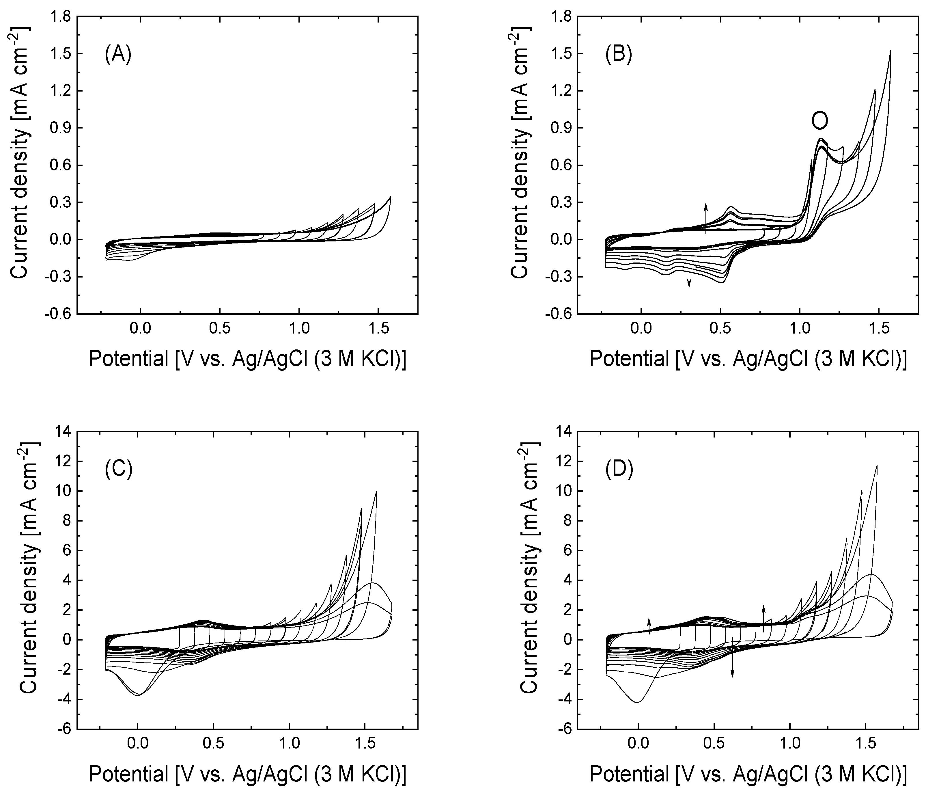
Figure 2 shows the cyclic voltammograms at the upper potential limit stepwise opened from 0.78 to 1.58 V in the absence of 4-APPA (Figure 2A,C) and in the presence of 4-APPA (Figure 2B,D) for GO and rGO, respectively.
In the presence of 4-APPA, GO and rGO present different voltammetric behavior. In the case of GO, the oxidation of 4-APPA shows a clear irreversible oxidation peak at approximately 1.13 V. After this oxidation peak, several reduction processes at 0.53, 0.13, and −0.10 V appear in the voltammograms of GO during the reverse scan associated with the redox activity of the surface functionalities on the surface, with a lower reversibility in comparison with previous results in carbon nanotubes [50]. In contrast, the voltammetric profiles of the rGO in the presence of 4-APPA are very similar to those obtained with rGO in the absence of 4-APPA, suggesting that the contribution of the oxidation of 4-APPA is less important compared to the oxidation of the carbon material. Indeed, rGO does not show significant redox processes compared to those observed during the rGO oxidation in the absence of 4-APPA (see Figure 2C), suggesting the growth and deposition of oligomer layers occur to a lesser extent than in the case of GO, being more important for the oxidation of the rGO than the oxidation of 4-APPA, as has been observed in oxidized carbon nanotubes [58].
Based on the study of the stepwise upper potential limit, two upper potential limits (1.28 and 1.58 V) were chosen for the functionalization of the materials (see Figure S2 in Supporting Information). Once the electrochemical modification with 4-APPA is carried out, a clear oxidation peak is observed in GO; however, this oxidation overlaps with the oxidation of the carbon material in the case of rGO. Interestingly, the clear irreversible oxidation current observed in the case of GO at 1.13 V has not been observed in previous studies with oxidized CNTs [58]. The electrochemical characterization by cyclic voltammetry in 0.5 M H2SO4 for GO after the electrochemical modification (see Figure S2 in the Supporting Information) shows the appearance of different redox processes at potentials of 0.44, 0.28, and −0.09 V. The increase in the upper potential limit during the 4-APPA oxidation does not produce an important change in the voltammetric profile; only a decrease in the voltammetric charge can be observed, which might be related to the degradation of the oligomers produced during the oxidation of 4-APPA [59,60,61,62]. For rGO, no significant differences can be observed in the voltammograms with respect to those obtained after oxidation in the absence of 4-APPA (see inset plot in Figure S2 in Supporting Information), and only a small increase in the voltammetric charge can be observed when the oxidation is produced in the presence of 4-APPA. In this case, a small and reversible redox process is observed at −0.10 V after the electrochemical treatment in the presence of 4-APPA at both upper potential limits. However, the functionalization of rGO at 1.8 V tends to produce an important oxidation of the carbon material to a greater extent than the oxidation of 4-APPA, generating a tilted voltammogram due to an electrical resistive behavior, as a result of the oxidation of the graphene-like structure.
The XPS analysis for the GO and rGO-modified electrodes shows the amount of N and P species incorporated after the electrochemical functionalization and their chemical nature. Table 1 summarizes the amount of O, N, and P incorporated in graphene-based materials after electrochemical modification.
It can be observed that for GO the amount of nitrogen species increases with the upper potential limit from 1.28 to 1.58 V (Table 1). Nevertheless, a high polarization above 1.28 V causes a decrease in the phosphorus content, due to the overoxidation of the polymeric species formed during the oxidation and the oxidation of the phosphonic groups to phosphoric groups that can be hydrolyzed and released to the electrolyte solution. The XPS data of N and P for rGO show a less important functionalization of the graphene-based material with both species (N and P). Nevertheless, the functionalization of rGO shows an unclear dependence with the upper potential limit, suggesting the saturation of the available sites in the rGO. This behavior is in agreement with the electrochemical response of these electrodes (see Figure S2).
The XPS analysis of both functionalized graphene-based materials demonstrates differences in the chemical nature of the incorporated functional groups. The XPS N1s spectra for GO-4APPA (see Figure S3 in the Supporting Information) can be deconvoluted into two peaks with a binding energy of around 400 and 402 eV. The fist peak at 400 eV can be assigned to the presence of amine-like groups, as a consequence of the formation of oligomer/polymeric chains [63,64,65,66]. The second contribution at 402 eV increases with the increasing of the upper potential limit due to oxidation of N species into oxidized nitrogen species, specifically positively charged nitrogen species (N+) due to protonated nitrogen functionalities (-NH2+/-NH+=) during the oxidative electrochemical functionalization [61,63]. On the other hand, the XPS N1s spectrum of rGO-4APPA shows a small additional contribution at a lower energy binding at around ~398.5 eV related to the presence of imines as well as a non-variation in the distribution of the nitrogen species after the functionalization (see Table S1 in Supporting Information).
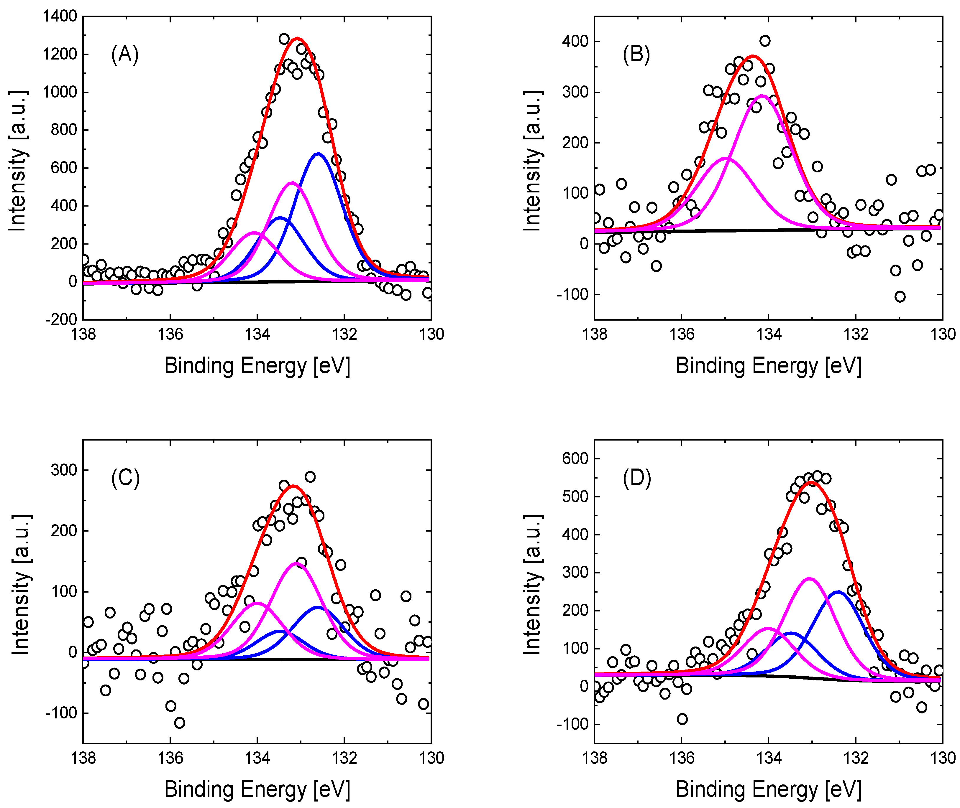
Regarding the XPS P2p spectra, the deconvolution of the phosphorus species can be performed in one or two doublets, as can be observed in Figure 3. For GO-4APPA and rGO-4APPA electrodes modified at a potential of 1.28 V, the spectra show a first contribution at 132.6 eV, which is associated in the literature with the binding energy of C-P species [67,68], in agreement with the presence of the phosphonic group of 4-APPA. Furthermore, a second contribution at 133.3 eV, related to the P-O species, increases with the upper potential limit, as a consequence of the oxidation of the phosphonic group to phosphate or phosphoric groups: (-O-P(OH)2). However, when GO is electrochemically modified at a potential of 1.58 V, the phosphorus species shows an important shift to a higher binding energy at 134.1 eV, which is related to the oxidized phosphorus species (see Table S1 in supporting information) in which a greater oxygen interaction is present, in agreement with values reported in the literature for this species in carbon materials [63,66,69]. Interestingly, the presence of the P species on the electrode surface might suggest a negatively charged surface of the graphene-based material after the functionalization process.
The electrochemical and physicochemical characterization of pristine and electrochemically functionalized graphene-based materials have demonstrated that the functionalization with N and P species at 1.58 V tends to produce a partial desorption of the surface functionalities, especially the phosphorus groups. Additionally, the degradation process of the electrode surface by the electrooxidation generates important damage to the pristine GO and rGO structures and their properties. Thus, in the following sections, pristine and functionalized graphene-based materials at 1.28V have been evaluated as electrodes for the immobilization of cytochrome C and for the evaluation of their electrochemical activity.

3.3. Electrochemical Behavior at Neutral pH

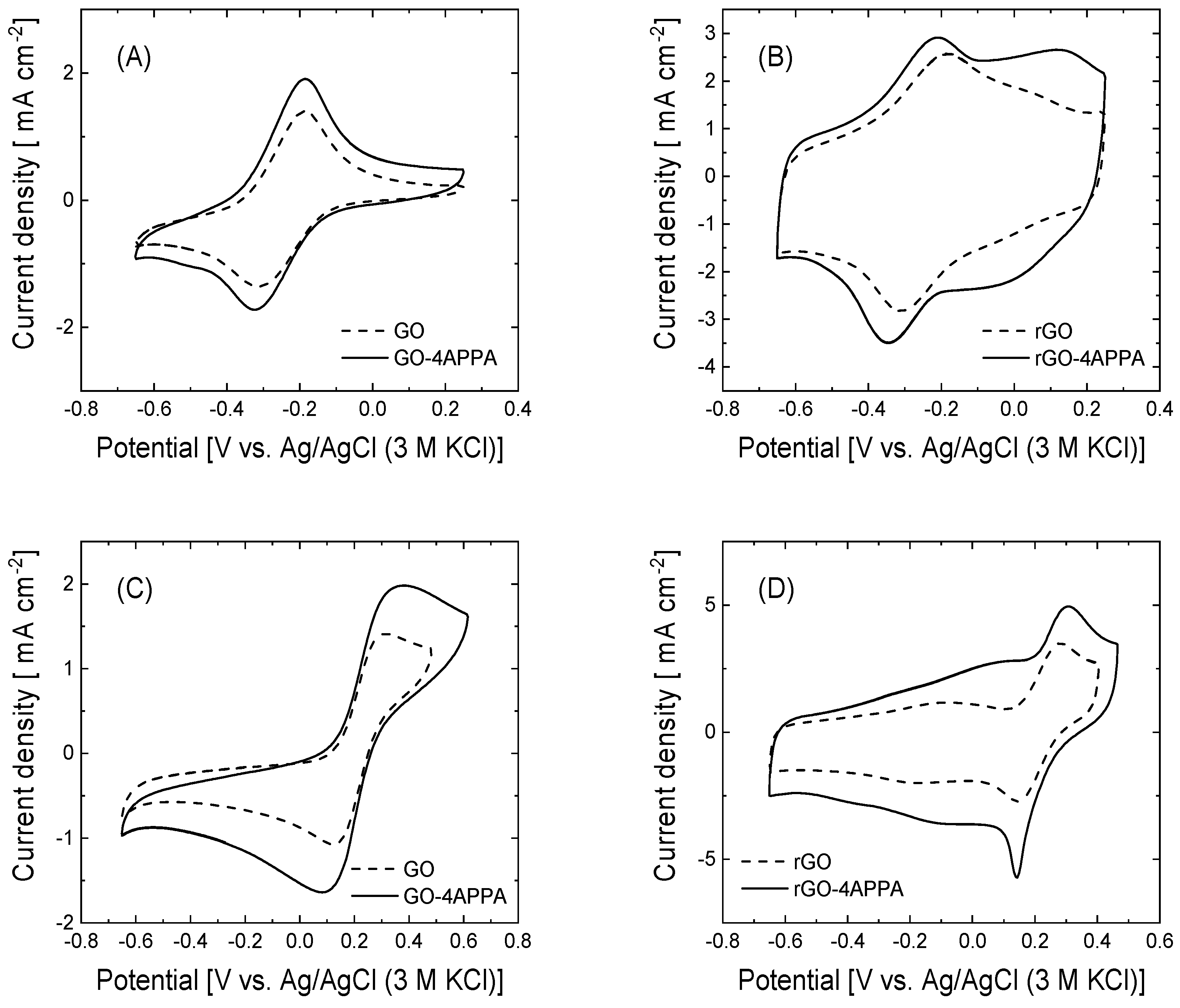
The properties of the electrode/electrolyte interface are strongly influenced by the chemical nature of the electrode surface, which can favorably affect the electrochemical activity of the electrode material. In this context, the influence of the electrode surface on pristine and electrochemically modified graphene-based materials has been evaluated by cyclic voltammetry in the presence of a positive ([Ru(NH3)6]3/4+) and negative ([Fe(CN)6]3/4−) redox probe. Figure 4 shows the cyclic voltammograms of GO-4APPA and rGO-APPA (solid line) and the pristine graphene-based materials (dotted line) in the presence of each redox probe.
In all the cyclic voltammograms of graphene-based materials, the quasi-reversible redox couple corresponding to the one electron transfer processes of [Ru(NH3)6]3/4+ and [Fe(CN)6]3/4− are clearly observed in the cyclic voltammograms. The analysis of both the cathodic and anodic peak currents of the processes shows a linear dependence with the square root of the scan rate (see Figures S4 and S5 in Supporting Information), corresponding with a diffusion-controlled process. The linear fitting of the Ipeak vs. vscan1/2 can be expressed as a function of the active surface area of the electrode available for the electron transfer of the redox species following the Randles–Sevcik equation [70] (Equation (1)):
Ip = 2.69 × 10⁻⁵ · n3/2 · A · D01/2 · C0* · vscan1/2
where Ip (A) is the peak current of the process; n is the number of exchanged electrons; A is the electroactive surface area (cm2); D0 is the diffusion coefficient of the electroactive species (cm2 s−1), which has been reported to be about 6.70 × 10⁻⁶ and 7.65 × 10⁻⁶ cm s−1 for [Ru(NH3)6]3/4+ and [Fe(CN)6]3/4− [71,72], respectively; C0* is the concentration of the electroactive species (mol cm−3); and vscan is the scan rate (V s−1). The estimation of the electroactive surface area employing the Randles–Sevcik relation, as can be observed in Table 2, shows an important ca. 3.8 times increase once the electrochemical reduction of GO takes place. In addition, an increase in the active surface area is also observed in both graphene-based materials after the electrochemical functionalization with the 4-APPA, reaching up to 37 and 60% of the pristine active surface area for the [Ru(NH3)6]3/4+ and [Fe(CN)6]3/4−, respectively, which is in agreement with the increase in the double layer charge in the cyclic voltammograms after the electrochemical functionalization (see Figure S2). These results can be interpreted taking into account that the mechanism of the functionalization process includes the formation of both covalently attached molecules of 4-APPA and non-covalently adsorbed oligomer chains of 4-APPA. Then, the deposited functional groups and oligomer structures can provide a greater number of active sites in which the electron transfer process might take place at higher rates.
The [Ru(NH3)6]3/4+ and [Fe(CN)6]3/4− are two typical outer-sphere redox probes vastly employed in the study of the electrocatalytic activity of electrode materials, which have a high dependence on the nature of the electrode material. Therefore, the kinetics of the electron transfer process can be influenced by the chemical surface of the electrode material. From the voltammogram profiles in Figure 4, in the peak separation (∆E) of both the redox probe and the values of the heterogeneous rate constant (k0), determined following the Nicholson and Mahe methods [73,74], different electrochemical behaviors, depending on the redox probe and the graphene-based material, can be observed (Table 2).
The peak separation of the [Ru(NH3)6]3/4+ redox couple presents a negligible variation of about 10 mV for all the electrode materials evaluated, suggesting that the electron transfer process is not significantly affected by the incorporation of the 4-APPA functionalities. In contrast, the peak separation values for the [Fe(CN)6]3/4− increase to about 80 and 17 mV once the GO and rGO are functionalized with the 4-APPA. Taking into account the different functional groups generated, specifically the N and P species, a large part of the nitrogen groups in the functional species are in their neutral state at neutral pH conditions [75]. On the other hand, the phosphonic and phosphate groups are fully deprotonated, increasing the net negative charge of the surface of the graphene-based materials. Then, electrostatic repulsion between the [Fe(CN)6]3/4− and the modified electrode surface is more important, affecting the concentration of the redox species near the electrode surface. This behavior is more notable on the GO-4APPA electrodes, where the highest amounts of phosphorus groups are incorporated on the surface, causing an important increase in the potential peak separation (Table 2). The values of k0 are shown in Table 2. It can be observed that the constant rate values for [Fe(CN)6]3/4− decrease in the functionalized graphene-based materials with 4-APPA. For instance, the value of k0 in GO-4APPA decreases more by than four times with respect to GO. In the case of [Ru(NH3)6]3/4+, the effect of the functionalization is lower than the values in [Fe(CN)6]3/4−. Therefore, the insertion of different functionalities that modify the net surface charge can promote the immobilization of biocatalytic elements, specifically positively charged enzymes, as well as the effective electrochemical activity of the biocatalyst.

3.4. Electrocatalytic Activity of Cyt C Immobilized on Functionalized GO and rGO

The electrocatalytic activity of the Cyt C immobilized on the graphene-based electrodes has been evaluated regarding the electrochemical reduction of H2O2 (Figure 5). For the analysis of the electrocatalytic activity, the electrochemical characterization of the different electrodes in absence of H2O2 has been performed (see Figure S6 in supporting information), showing modifications in the double layer contribution after the immobilization of Cyt C. On the other hand, the pseudocapacitive contribution of the surface functionalities (oxygen, nitrogen, and phosphorus species) tends to produce a hindrance of the redox process associated with the heme redox cofactor; thus, the electrocatalytic activity toward H2O2 has been used to evaluate the electrocatalytic activity.
The cyclic voltammograms of the pristine GO and rGO show that both electrode materials have a catalytic activity toward the electrochemical reduction of H2O2, which reaches variations in the current density of 6.5 × 10⁻³ mA cm−2 and 1.14 mA cm−2 for GO and rGO at −0.43 V, respectively. A notable improvement in the electrocatalytic activity towards the H2O2 reduction for rGO can be understood considering the higher electrochemical active surface area (Table 2) [55,76,77]. Once both materials are electrochemically functionalized with 4-APPA, the electrocatalytic activity toward the H2O2 reduction is dramatically improved, reaching a current density variation value of about 0.18 mA cm−2 and 4.2 mA cm−2 for GO and rGO, respectively, which is approximately six and three times the current density variation in the pristine electrodes. According to our previous observation (see Table 2), the incorporation of nitrogen and phosphorus surface functionalities provides an increase in the electroactive surface area as well as the presence of active sites for the electrocatalytic reaction of hydrogen peroxide.
Depending on the electrode employed as the support for the immobilization of the Cyt C, the electrocatalytic activity is enhanced or almost unchanged following the reaction pathway indicated in Equations (2) and (3). In the case of the GO-Cyt C electrode, the variation in the current density in the presence of hydrogen peroxide starts at an onset potential (Eonset) of about −0.2 V, which is 100 mV more positive than electrodes without the biomolecule. The maximum current value was 16.5 × 10⁻³ mA cm−2 at −0.43 V, which is up to 2.5 times higher than the catalytic activity of the pristine GO without the immobilized biomolecule. Then, Cyt C maintains its catalytic activity on this electrode surface after the immobilization process. Interestingly, after the immobilization of Cyt C in 4-APPA functionalized GO electrodes, the variation in the current density related to the H2O2 electroreduction has an additional contribution of 0.15 mA cm−2 to the current density values, compared to the same electrode without Cyt C, suggesting an improvement in the catalytic activity of around 83%. This increase in the current is directly associated with the current generated by the biomolecule on the electrode. In comparison, the current density contribution associated with Cyt C in the unmodified GO (about 0.01 mA cm−2) is 15 times lower than the activity of Cyt C in the GO-4APPA electrode. In this sense, the improvement in the electrocatalytic activity of Cyt C in the functionalized GO is mainly caused by the interactions between the surface functional groups and the amino acid residues in the structure of Cyt C which produces its adequate orientation towards the electrode.
Chemical pathway: ½ H2O2 + H+ + Cyt C [Heme Fe(II)]→ H2O + Cyt C [Heme Fe(III)]
Electrochemical pathway: Cyt C [Heme Fe(III)] + 1e→ Cyt C [Heme Fe(II)]
Surprisingly, rGO and rGO-Cyt C show the same current density variation in the presence of H2O2 in the solution, which indicates that the Cyt C does not contribute to the electrochemical reduction of H2O2. This behavior might indicate that the interaction between immobilized Cyt C and the surface of rGO generates a biomolecule assemblage in which the electrical communication between the redox cofactor of Cyt C and the electrode is not favored. The incorporation of the surface functionalities of the N and P species in rGO provokes a dramatic improvement in the electrocatalytic activity in the presence of H2O2 due to a larger electrochemical surface area, as has been observed for GO. Following the immobilization of Cyt C in the rGO-4APPA electrodes (see Figure 5D), the cyclic voltammogram in the presence of the H2O2 displays two important features: a positive shifting at around 100 mV in the onset potential for the electrochemical reduction of the H2O2 as well as an increase of 58% in the current density variation in the presence of H2O2. The latter can be associated with the current generated due to the immobilized Cyt C. The current contribution of the Cyt C in the functionalized rGO electrode material demonstrates an effective improvement in the electrical communication between the electrode and the biomolecule through the surface functional groups. Previous works have demonstrated that hydrophilic and negatively charged groups can interact directly with the lysine residues, which might favor the electron transfer with the heme cofactor in the Cyt C [25,78]. In this sense, the deprotonated phosphonic/phosphoric groups under the pH conditions of the immobilization of Cyt C (isoelectric point ≈ 9.5) [79] produces a net negative charge of the electrode surface, which results in a strong electrostatic interaction with the positively charged Cyt C. In this sense, the arrangement of the biomolecule redox cofactor can be close to the electrode surface, as has been observed with other redox biomolecules [9,15].
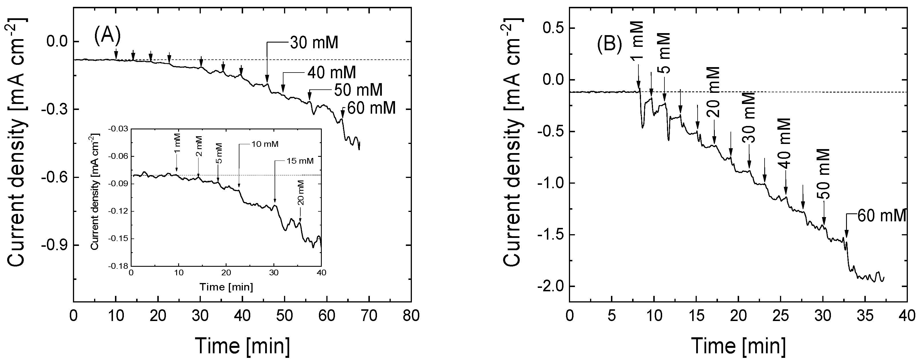
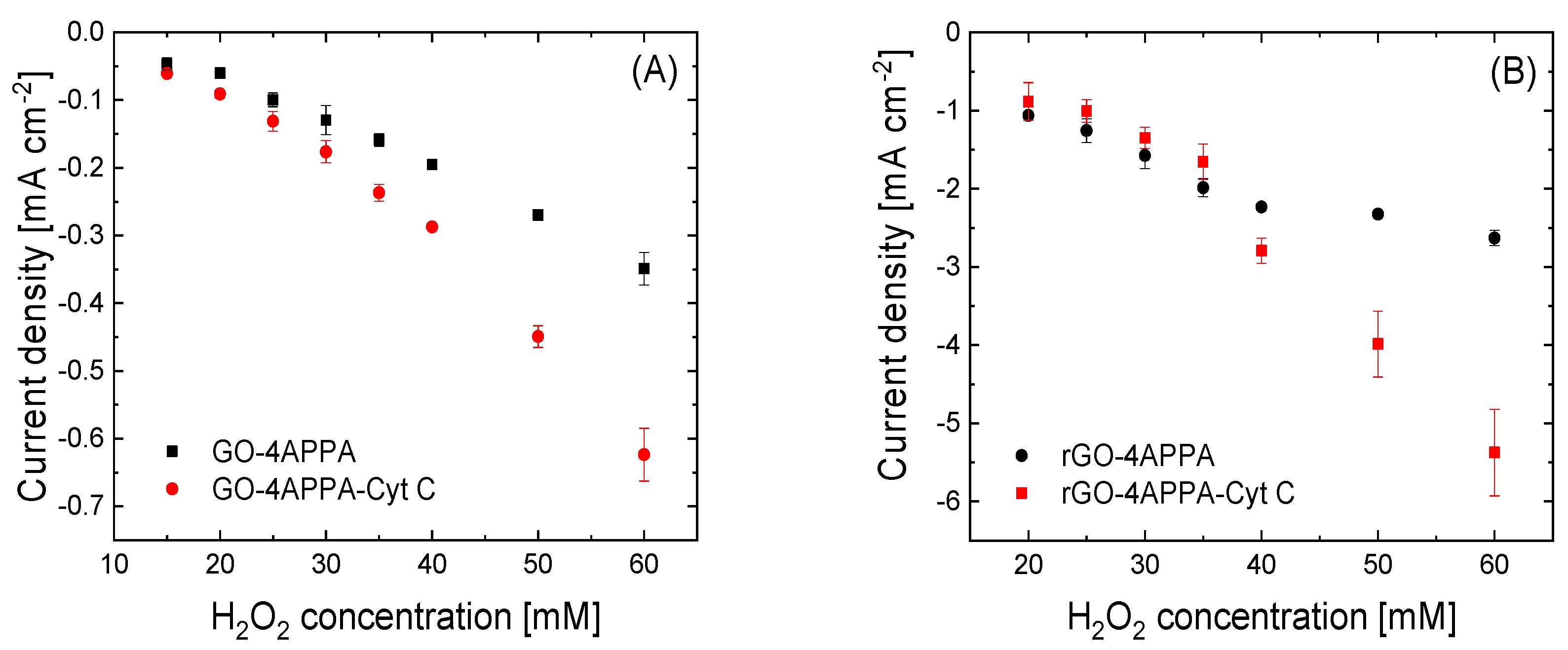
Figure 6 displays the chronoamperometric profiles of the different functionalized electrode materials during the continuous addition of H2O2 aliquots. As can be observed in all electrode materials, the addition of H2O2 produces a rapid increase in the negative current only 10 s after the addition, which is associated with the electrochemical reduction of H2O2. These changes are more important for the electrodes in which Cyt C has been immobilized. In this sense, the increase in the current density values shows an enhancement in the electrocatalytic activity of the electrodes GO-4APPA and rGO-4APPA of two and five times the maximum current density value at 60 mM H2O2, respectively. Figure 7 shows the calibration curves for the different electrodes. Table 3 contains the most relevant parameters for the detection of H2O2, in which the sensitivity, LOD, and LOQ are presented. Furthermore, a comparison of the electrochemical parameters with similar bioelectrodes, as reported in the literature, based on Cyt C, is presented in Table S2 in the Supporting Information.
As expected, the sensitivity and reduction current density of H2O2 increase significantly with the electrochemical reduction of GO in rGO and with its further electrochemical functionalization with 4-APPA (see Figure 7B). In this regard, the decrease in the surface oxygen groups after the electrochemical reduction and the incorporation of negatively charged functionalities (phosphonic/phosphoric groups) reduce the hindrance effect, promoting a better electrical communication with the heme cofactor in the Cyt C, as well as the electron mobility over the electrode due to the decrease in the concentration of defects in the graphene layer of the rGO.

4. Conclusions

The presence and nature of surface functional groups in graphene-based electrode materials have demonstrated an important influence in the immobilization and redox activity of Cyt C, promoting an optimal interaction of the biomolecule and promoting a more efficient electrical communication between the electrode surface and the heme redox cofactor. In the case of graphene oxide, the incorporation of negatively charged phosphonic/phosphoric groups at a neutral pH facilitates the interaction with positively charged residues in the Cyt C during the immobilization process, maintaining the activity of the biomolecule. In this sense, the good compatibility of the functionalized graphene-based materials provides a suitable platform for the immobilization of the Cyt C. At the same time, this interaction produces an enhancement in the electrocatalytic activity toward the H2O2 reduction, which is observed as an increase in the catalytic current density associated with the electrochemical reduction of H2O2. Furthermore, the partial elimination of surface oxygen groups in pristine GO electrochemically induces an enhancement in the electroactive surface area and the electrocatalytic activity of the electrode. Nevertheless, a low concentration of functional groups hinders the redox activity of the Cyt C, avoiding the electron transfer between the rGO and the Cyt C. The functionalization of the rGO electrode with 4-APPA modulates the surface chemistry and improves the immobilization and electrical communication of the biomolecule with the electrode, reaching current density values of up to 5.5 mA cm−2, which are 10 times higher than the GO modified with 4-APPA. Therefore, depending on the surface chemistry in graphene-based materials the catalytic performance of bioelectrodes can promote an effective interconversion of the redox element, a higher catalytic current density, and low overpotentials, which are ideal for the fabrication of bioelectrodes as was observed with the modified graphene-based electrode with P and N species. In this regard, the electrochemical functionalization process with 4-APPA represents an alternative to the conventional processes of multi-step bioelectrode fabrication, due to the remarkable compatibility with the biomolecule elements and the great performance achieved.

Supplementary Materials

The following supporting information can be downloaded at: https://www.mdpi.com/article/10.3390/nano15100722/s1. Figure S1. (A) Cyclic voltammogram for GO in 0.5 M H2SO4 at 50 mV s−1 under N2 atmosphere. (B) XPS spectra of C1s signal for GO. (C) Cyclic voltammogram for rGO in 0.5 M H2SO4 at 50 mV s−1 under N2 atmosphere. (D) XPS spectra of C1s signal for rGO after the electrochemical reduction; Figure S2. Cyclic voltammograms obtained during 10 cycles for a GO and rGO electrodes in 0.5 M H2SO4 (solid lines) and 0.5 M H2SO4 + 1 mM 4-APPA (dash lines) at 10 mV s−1 under N2 atmosphere at different positive potential limits: (A) GO-1.28 V, (B) GO-1.58 V, (C) rGO-1.28 V, and (D) rGO-1.58 V. Inset: Steady state CVs in 0.5 M H2SO4, for graphene-based materials electrochemically modified in absence (dash line) and in presence (solid line) at vscan = 50 mV s−1; Figure S3. N1s XPS spectra deconvoluted for GO and rGO electrochemical modified with 4-APPA at different oxidation potential: (A) GO modified at 1.28 V, (B) GO modified at 1.58 V, (C) rGO modified at 1.28 V and (D) rGO modified at 1.58 V; Table S1. Chemical distribution of the N and P species obtained from XPS of electrochemical modified graphene-based materials with 4-APPA; Figure S4. Cyclic voltammograms at different scan rates (10, 20, 50, 100, 200, and 500 mV s−1) of the pristine graphene-based materials in 0.1 M PBS (pH = 7.2): (A,B) 1 mM [Ru(NH3)6]3/4+ and (C,D) 5 mM ([Fe(CN)6]3/4); Figure S5. Cyclic voltammograms at different scan rates (10, 20, 50, 100, 200, and 500 mV s−1) of the graphene-based materials electrochemically modified with 4-APPA in 0.1 M PBS (pH = 7.2): (A,B) 1 mM [Ru(NH3)6]3/4+ and (C,D) 5 mM ([Fe(CN)6]3/4−); Figure S6. Cyclic voltammograms of electrochemical modified GO and rGO before and after the immobilization of Cyt C in 0.1 M PBS (pH = 7.2) at 20 mV s−1; Table S2. Comparative of the electrochemical parameters of bioelectrodes reported in literature. See Refs. [28,30,50,80,81,82,83,84,85,86,87].

Author Contributions

Conceptualization, D.C.-A. and E.M.; Methodology, A.F.Q.-J. and D.C.-A.; Validation, A.F.Q.-J.; Formal analysis, A.F.Q.-J.; Investigation, A.F.Q.-J., D.C.-A. and E.M.; Resources, E.M.; Data curation, A.F.Q.-J.; Writing—original draft, A.F.Q.-J.; Writing—review & editing, D.C.-A. and E.M.; Supervision, D.C.-A.; Funding acquisition, E.M. All authors have read and agreed to the published version of the manuscript.

Funding

This research was funded by Generalitat Valenciana (Prometeo Project CIPROM/2021/062).

Data Availability Statement

The raw data supporting the conclusions of this article will be made available by the authors on request.

Acknowledgments

The authors are grateful to the Generalitat Valenciana (Prometeo Project CIPROM/2021/062).

Conflicts of Interest

The authors declare no conflict of interest.

References

  1. Ruff, A.; Conzuelo, F.; Schuhmann, W. Bioelectrocatalysis as the basis for the design of enzyme-based biofuel cells and semi-artificial biophotoelectrodes. Nat. Catal. 2020, 3, 214–224. [Google Scholar] [CrossRef]
  2. Riedel, M.; Höfs, S.; Ruff, A.; Schuhmann, W.; Lisdat, F. A Tandem Solar Biofuel Cell: Harnessing Energy from Light and Biofuels. Angew. Chem. Int. Ed. 2021, 60, 2078–2083. [Google Scholar] [CrossRef] [PubMed]
  3. Milton, R.D.; Abdellaoui, S.; Khadka, N.; Dean, D.R.; Leech, D.; Seefeldt, L.C.; Minteer, S.D. Nitrogenase bioelectrocatalysis: Heterogeneous ammonia and hydrogen production by MoFe protein. Energy Environ. Sci. 2016, 9, 2550–2554. [Google Scholar] [CrossRef]
  4. Lee, Y.S.; Ruff, A.; Cai, R.; Lim, K.; Schuhmann, W.; Minteer, S.D. Electroenzymatic Nitrogen Fixation Using a MoFe Protein System Immobilized in an Organic Redox Polymer. Angew. Chem. Int. Ed. 2020, 59, 16511–16516. [Google Scholar] [CrossRef]
  5. Schuhmann, W. Amperometric enzyme biosensors based on optimised electron-transfer pathways and non-manual immobilisation procedures. Rev. Mol. Biotechnol. 2002, 82, 425–441. [Google Scholar] [CrossRef]
  6. Bartlett, P.N. Bioelectrochemistry: Fundamentals, Experimental Techniques and Applications; John Wiley & Sons: Chichester, UK, 2008. [Google Scholar]
  7. Page, C.C.; Moser, C.C.; Chen, X.; Dutton, P.L. Natural engineering principles of electron tunnelling in biological oxidation–reduction. Nature 1999, 402, 47–52. [Google Scholar] [CrossRef]
  8. de Poulpiquet, A.; Ciaccafava, A.; Lojou, E. New trends in enzyme immobilization at nanostructured interfaces for efficient electrocatalysis in biofuel cells. Electrochim. Acta 2014, 126, 104–114. [Google Scholar] [CrossRef]
  9. Quintero-Jaime, A.F.; Conzuelo, F.; Schuhmann, W.; Cazorla-Amorós, D.; Morallón, E. Multi-wall carbon nanotubes electrochemically modified with phosphorus and nitrogen functionalities as a basis for bioelectrodes with improved performance. Electrochim. Acta 2021, 387, 138530. [Google Scholar] [CrossRef]
  10. Smutok, O.; Ngounou, B.; Pavlishko, H.; Gayda, G.; Gonchar, M.; Schuhmann, W. A reagentless bienzyme amperometric biosensor based on alcohol oxidase/peroxidase and an Os-complex modified electrodeposition paint. Sens. Actuators B: Chem. 2006, 113, 590–598. [Google Scholar] [CrossRef]
  11. Ferapontova, E.E.; Shleev, S.; Ruzgas, T.; Stoica, L.; Christenson, A.; Tkac, J.; Yaropolov, A.I.; Gorton, L. Direct Electrochemistry of Proteins and Enzymes. In Electrochemistry of Nucleic Acids and Proteins—Towards Electrochemical Sensors for Genomics and Proteomics; Paleček, E., Scheller, F., Wang, J.B.T.-P., Eds.; Elsevier: Amsterdam, The Netherlands, 2005; Volume 1, pp. 517–598. ISBN 1871-0069. [Google Scholar]
  12. Demurtas, D.; Alvarez-Malmagro, J.; Rathore, A.; Mandal, T.; Quintero-Jaime, A.F.; Belochapkine, S.; Lielpetere, A.; Jayakumar, K.; Leech, D.; Schuhmann, W.; et al. Development of flexible nanoporous gold electrodes for the detection of glucose. Bioelectrochemistry 2025, 165, 108949. [Google Scholar] [CrossRef]
  13. Reuillard, B.; Ly, K.H.; Hildebrandt, P.; Jeuken, L.J.C.; Butt, J.N.; Reisner, E. High Performance Reduction of H2O2 with an Electron Transport Decaheme Cytochrome on a Porous ITO Electrode. J. Am. Chem. Soc. 2017, 139, 3324–3327. [Google Scholar] [CrossRef] [PubMed]
  14. Cziegler, C.; Csarman, F.; Breslmeyer, E.; Ma, S.; Meinert, H.; Ludwig, R.; Rudroff, F.; Mihovilovic, M.D. Design and Synthesis of Artificial FAD Cofactors for the Light-Triggered Covalent Flavinylation of Flavoenzymes. ACS Catal. 2024, 14, 15988–15996. [Google Scholar] [CrossRef]
  15. Milton, R.D.; Minteer, S.D. Direct enzymatic bioelectrocatalysis: Differentiating between myth and reality. J. R. Soc. Interface 2017, 14, 20170253. [Google Scholar] [CrossRef] [PubMed]
  16. Mendes, R.K.; Carvalhal, R.F.; Kubota, L.T. Effects of different self-assembled monolayers on enzyme immobilization procedures in peroxidase-based biosensor development. J. Electroanal. Chem. 2008, 612, 164–172. [Google Scholar] [CrossRef]
  17. Allen, P.M.; Allen, H.; Hill, O.; Walton, N.J. Surface modifiers for the promotion of direct electrochemistry of cytochrome c. J. Electroanal. Chem. Interfacial Electrochem. 1984, 178, 69–86. [Google Scholar] [CrossRef]
  18. Takeda, K.; Kusuoka, R.; Birrell, J.A.; Yoshida, M.; Igarashi, K.; Nakamura, N. Bioelectrocatalysis based on direct electron transfer of fungal pyrroloquinoline quinone-dependent dehydrogenase lacking the cytochrome domain. Electrochim. Acta 2020, 359, 136982. [Google Scholar] [CrossRef]
  19. Xu, S.; Minteer, S.D. Investigating the Impact of Multi-Heme Pyrroloquinoline Quinone-Aldehyde Dehydrogenase Orientation on Direct Bioelectrocatalysis via Site Specific Enzyme Immobilization. ACS Catal. 2013, 3, 1756–1763. [Google Scholar] [CrossRef]
  20. Xu, S.; Minteer, S.D. Pyrroloquinoline Quinone-Dependent Enzymatic Bioanode: Incorporation of the Substituted Polyaniline Conducting Polymer as a Mediator. ACS Catal. 2014, 4, 2241–2248. [Google Scholar] [CrossRef]
  21. Ramanavičius, A.; Ramanavičienė, A.; Malinauskas, A. Electrochemical sensors based on conducting polymer—Polypyrrole. Electrochim. Acta 2006, 51, 6025–6037. [Google Scholar] [CrossRef]
  22. Schuhmann, W.; Lammert, R.; Hämmerle, M.; Schmidt, H.-L. Electrocatalytic properties of polypyrrole in amperometric electrodes. Biosens. Bioelectron. 1991, 6, 689–697. [Google Scholar] [CrossRef]
  23. Trashin, S.A.; Haltrich, D.; Ludwig, R.; Gorton, L.; Karyakin, A.A. Improvement of direct bioelectrocatalysis by cellobiose dehydrogenase on screen printed graphite electrodes using polyaniline modification. Bioelectrochemistry 2009, 76, 87–92. [Google Scholar] [CrossRef] [PubMed]
  24. López-Bernabeu, S.; Huerta, F.; Morallón, E.; Montilla, F. Direct Electron Transfer to Cytochrome c Induced by a Conducting Polymer. J. Phys. Chem. C 2017, 121, 15870–15879. [Google Scholar] [CrossRef]
  25. López-Bernabeu, S.; Gamero-Quijano, A.; Huerta, F.; Morallón, E.; Montilla, F. Enhancement of the direct electron transfer to encapsulated cytochrome c by electrochemical functionalization with a conducting polymer. J. Electroanal. Chem. 2017, 793, 34–40. [Google Scholar] [CrossRef]
  26. Patel, J.; Cai, R.; Milton, R.; Chen, H.; Minteer, S.D. Pyrene-Based Noncovalent Immobilization of Nitrogenase on Carbon Surfaces. ChemBioChem 2020, 21, 1729–1732. [Google Scholar] [CrossRef]
  27. Hasan, K.; Grattieri, M.; Wang, T.; Milton, R.D.; Minteer, S.D. Enhanced Bioelectrocatalysis of Shewanella oneidensis MR-1 by a Naphthoquinone Redox Polymer. ACS Energy Lett. 2017, 2, 1947–1951. [Google Scholar] [CrossRef]
  28. Lavanya, V.; Pavithra, D.; Mohanapriya, A.; Santhakumar, K.; Senthil Kumar, A. A π–π Bonding-Assisted Molecular-Wiring of Folded-Cytochrome c and Naphthoquinone and Its Electron-Relay-Based Bioelectrocatalytic H2O2 Reduction Reaction Visualized by Redox-Competitive Scanning Electrochemical Microscopy. Langmuir 2023, 39, 11556–11570. [Google Scholar] [CrossRef]
  29. Ben Tahar, A.; Żelechowska, K.; Biernat, J.F.; Paluszkiewicz, E.; Cinquin, P.; Martin, D.; Zebda, A. High catalytic performance of laccase wired to naphthylated multiwall carbon nanotubes. Biosens. Bioelectron. 2020, 151, 111961. [Google Scholar] [CrossRef]
  30. Gandhi, M.; Rajagopal, D.; Senthil Kumar, A. Molecularly wiring of Cytochrome c with carboxylic acid functionalized hydroquinone on MWCNT surface and its bioelectrocatalytic reduction of H2O2 relevance to biomimetic electron-transport and redox signalling. Electrochim. Acta 2021, 368, 137596. [Google Scholar] [CrossRef]
  31. Felipe Quintero-Jaime, A.; Conzuelo, F.; Cazorla-Amorós, D.; Morallón, E. Pyrroloquinoline quinone-dependent glucose dehydrogenase bioelectrodes based on one-step electrochemical entrapment over single-wall carbon nanotubes. Talanta 2021, 232, 122386. [Google Scholar] [CrossRef]
  32. Niu, J.; Jin, X.; Wang, X.; Ren, Z.; Li, B.; Liu, X.; Wong, D.K.Y. An Enzyme-Encapsulated MOF@MOF Nanocomposite for Detecting H2O2 Derived From Superoxide Anion Released by Mitochondria of HeLa Cells. Small Methods 2025, 9, 2401070. [Google Scholar] [CrossRef]
  33. Cosnier, S.; Holzinger, M.; Le Goff, A. Recent Advances in Carbon Nanotube-Based Enzymatic Fuel Cells. Front. Bioeng. Biotechnol. 2014, 2, 45. [Google Scholar] [CrossRef] [PubMed]
  34. Kitazumi, Y.; Shirai, O.; Kano, K. Significance of Nanostructures of an Electrode Surface in Direct Electron Transfer-Type Bioelectrocatalysis of Redox Enzymes. In Novel Catalyst Materials for Bioelectrochemical Systems: Fundamentals and Applications; ACS Symposium Series; American Chemical Society: Washington, DC, USA, 2020; Volume 1342, pp. 147–163. ISBN 978-0-8412-3668-4. [Google Scholar]
  35. Oliveira, T.M.B.F.; Ribeiro, F.W.P.; Sousa, C.P.; Salazar-Banda, G.R.; de Lima-Neto, P.; Correia, A.N.; Morais, S. Current overview and perspectives on carbon-based (bio)sensors for carbamate pesticides electroanalysis. TrAC Trends Anal. Chem. 2020, 124, 115779. [Google Scholar] [CrossRef]
  36. Quílez-Bermejo, J.; Melle-Franco, M.; San-Fabián, E.; Morallón, E.; Cazorla-Amorós, D. Towards understanding the active sites for the ORR in N-doped carbon materials through fine-tuning of nitrogen functionalities: An experimental and computational approach. J. Mater. Chem. A 2019, 7, 24239–24250. [Google Scholar] [CrossRef]
  37. Veizaga, N.S.; Mendow, G.; Quintero-Jaime, A.F.; Berenguer-Murcia, Á.; de Miguel, S.; Morallón, E.; Cazorla-Amorós, D. Controlled synthesis of mono- and bimetallic Pt-based catalysts for electrochemical ethanol oxidation. Mater. Chem. Phys. 2022, 275, 125282. [Google Scholar] [CrossRef]
  38. Wang, Z.; Ding, G.; Huang, H.; Zhang, J.; Lv, Q.; Shuai, L.; Ni, Y.; Liao, G. Unraveling the dipole field in ultrathin, porous, and defective carbon nitride nanosheets for record-high piezo-photocatalytic H2O2 production. eScience 2024, 27, 100370. [Google Scholar] [CrossRef]
  39. Zhou, J.; Shan, T.; Zhang, F.; Boury, B.; Huang, L.; Yang, Y.; Liao, G.; Xiao, H.; Chen, L. A Novel Dual-Channel Carbon Nitride Homojunction with Nanofibrous Carbon for Significantly Boosting Photocatalytic Hydrogen Peroxide Production. Adv. Fiber Mater. 2024, 6, 387–400. [Google Scholar] [CrossRef]
  40. Xiao, H.; Shan, Y.; Wu, S.; Shan, T.; Luo, H.; Zhang, F.; Huang, L.; Chen, L.; Liao, G. Enlarging the π-conjugation system of carbon nitride for boosting hydrogen peroxide generation. Chem. Eng. J. 2024, 492, 152228. [Google Scholar] [CrossRef]
  41. Miyake, T.; Haneda, K.; Yoshino, S.; Nishizawa, M. Flexible, layered biofuel cells. Biosens. Bioelectron. 2013, 40, 45–49. [Google Scholar] [CrossRef]
  42. Abellán-Llobregat, A.; Jeerapan, I.; Bandodkar, A.; Vidal, L.; Canals, A.; Wang, J.; Morallón, E. A stretchable and screen-printed electrochemical sensor for glucose determination in human perspiration. Biosens. Bioelectron. 2017, 91, 885–891. [Google Scholar] [CrossRef]
  43. Zebda, A.; Gondran, C.; Le Goff, A.; Holzinger, M.; Cinquin, P.; Cosnier, S. Mediatorless high-power glucose biofuel cells based on compressed carbon nanotube-enzyme electrodes. Nat. Commun. 2011, 2, 370. [Google Scholar] [CrossRef]
  44. Abreu, C.; Nedellec, Y.; Gross, A.J.; Ondel, O.; Buret, F.; Goff, A.L.; Holzinger, M.; Cosnier, S. Assembly and Stacking of Flow-through Enzymatic Bioelectrodes for High Power Glucose Fuel Cells. ACS Appl. Mater. Interfaces 2017, 9, 23836–23842. [Google Scholar] [CrossRef] [PubMed]
  45. Gross, A.J.; Tanaka, S.; Colomies, C.; Giroud, F.; Nishina, Y.; Cosnier, S.; Tsujimura, S.; Holzinger, M. Diazonium Electrografting vs. Physical Adsorption of Azure A at Carbon Nanotubes for Mediated Glucose Oxidation with FAD-GDH. ChemElectroChem 2020, 7, 4543–4549. [Google Scholar] [CrossRef]
  46. Haddad, R.; Cosnier, S.; Maaref, A.; Holzinger, M. Non-covalent biofunctionalization of single-walled carbon nanotubesviabiotin attachment by π-stacking interactions and pyrrole polymerization. Analyst 2009, 134, 2412–2418. [Google Scholar] [CrossRef]
  47. Vashist, S.K.; Zheng, D.; Al-Rubeaan, K.; Luong, J.H.T.; Sheu, F.-S. Advances in carbon nanotube based electrochemical sensors for bioanalytical applications. Biotechnol. Adv. 2011, 29, 169–188. [Google Scholar] [CrossRef] [PubMed]
  48. Meredith, M.T.; Minson, M.; Hickey, D.; Artyushkova, K.; Glatzhofer, D.T.; Minteer, S.D. Anthracene-Modified Multi-Walled Carbon Nanotubes as Direct Electron Transfer Scaffolds for Enzymatic Oxygen Reduction. ACS Catal. 2011, 1, 1683–1690. [Google Scholar] [CrossRef]
  49. González-Gaitán, C.; Ruiz-Rosas, R.; Morallón, E.; Cazorla-Amorós, D. Effects of the surface chemistry and structure of carbon nanotubes on the coating of glucose oxidase and electrochemical biosensors performance. RSC Adv. 2017, 7, 26867–26878. [Google Scholar] [CrossRef]
  50. Quintero-Jaime, A.F.; Cazorla-Amorós, D.; Morallón, E. Electrochemical functionalization of single wall carbon nanotubes with phosphorus and nitrogen species. Electrochim. Acta 2020, 340, 135935. [Google Scholar] [CrossRef]
  51. Gentil, S.; Che Mansor, S.M.; Jamet, H.; Cosnier, S.; Cavazza, C.; Le Goff, A. Oriented Immobilization of [NiFeSe] Hydrogenases on Covalently and Noncovalently Functionalized Carbon Nanotubes for H2/Air Enzymatic Fuel Cells. ACS Catal. 2018, 8, 3957–3964. [Google Scholar] [CrossRef]
  52. Yang, J.; Xu, Y.; Zhang, R.; Wang, Y.; He, P.; Fang, Y. Direct Electrochemistry and Electrocatalysis of the Hemoglobin Immobilized on Diazonium-Functionalized Aligned Carbon Nanotubes Electrode. Electroanalysis 2009, 21, 1672–1677. [Google Scholar] [CrossRef]
  53. Cosnier, S. Biomolecule immobilization on electrode surfaces by entrapment or attachment to electrochemically polymerized films. A review. Biosens. Bioelectron. 1999, 14, 443–456. [Google Scholar] [CrossRef]
  54. Yon-Hin, B.F.Y.; Lowe, C.R. An investigation of 3-functionalized pyrrole-modified glucose oxidase for the covalent electropolymerization of enzyme films. J. Electroanal. Chem. 1994, 374, 167–172. [Google Scholar] [CrossRef]
  55. Marrani, A.G.; Motta, A.; Schrebler, R.; Zanoni, R.; Dalchiele, E.A. Insights from experiment and theory into the electrochemical reduction mechanism of graphene oxide. Electrochim. Acta 2019, 304, 231–238. [Google Scholar] [CrossRef]
  56. Toh, S.Y.; Loh, K.S.; Kamarudin, S.K.; Daud, W.R.W. Graphene production via electrochemical reduction of graphene oxide: Synthesis and characterisation. Chem. Eng. J. 2014, 251, 422–434. [Google Scholar] [CrossRef]
  57. Salinas-Torres, D.; Huerta, F.; Montilla, F.; Morallón, E. Study on electroactive and electrocatalytic surfaces of single walled carbon nanotube-modified electrodes. Electrochim. Acta 2011, 56, 2464–2470. [Google Scholar] [CrossRef]
  58. Quintero-Jaime, A.F.; Cazorla-Amorós, D.; Morallón, E. Effect of surface oxygen groups in the electrochemical modification of multi-walled carbon nanotubes by 4-amino phenyl phosphonic acid. Carbon 2020, 165, 328–339. [Google Scholar] [CrossRef]
  59. Probst, M.; Holze, R. A systematic spectroelectrochemical investigation of alkyl-substituted anilines and their polymers. Macromol. Chem. Phys. 1997, 198, 1499–1509. [Google Scholar] [CrossRef]
  60. Nateghi, M.R.; Zahedi, M.; Mosslemin, M.H.; Hashemian, S.; Behzad, S.; Minnai, A. Autoacceleration/degradation of electrochemical polymerization of substituted anilines. Polymer 2005, 46, 11476–11483. [Google Scholar] [CrossRef]
  61. Benyoucef, A.; Huerta, F.; Ferrahi, M.I.; Morallon, E. Voltammetric and in situ FT-IRS study of the electropolymerization of o-aminobenzoic acid at gold and graphite carbon electrodes: Influence of pH on the electrochemical behaviour of polymer films. J. Electroanal. Chem. 2008, 624, 245–250. [Google Scholar] [CrossRef]
  62. Wang, B.; Tang, J.; Wang, F. Electrochemical polymerization of aniline. Synth. Met. 1987, 18, 323–328. [Google Scholar] [CrossRef]
  63. Quílez-Bermejo, J.; Ghisolfi, A.; Grau-Marín, D.; San-Fabián, E.; Morallón, E.; Cazorla-Amorós, D. Post-synthetic efficient functionalization of polyaniline with phosphorus-containing groups. Effect of phosphorus on electrochemical properties. Eur. Polym. J. 2019, 119, 272–280. [Google Scholar] [CrossRef]
  64. Abidi, M.; López-Bernabeu, S.; Huerta, F.; Montilla, F.; Besbes-Hentati, S.; Morallón, E. Spectroelectrochemical study on the copolymerization of o-aminophenol and aminoterephthalic acid. Eur. Polym. J. 2017, 91, 386–395. [Google Scholar] [CrossRef]
  65. Wang, Z.; Dong, Y.; Li, H.; Zhao, Z.; Bin Wu, H.; Hao, C.; Liu, S.; Qiu, J.; Lou, X.W. (David) Enhancing lithium–sulphur battery performance by strongly binding the discharge products on amino-functionalized reduced graphene oxide. Nat. Commun. 2014, 5, 5002. [Google Scholar] [CrossRef] [PubMed]
  66. Valero-Romero, M.J.; García-Mateos, F.J.; Rodríguez-Mirasol, J.; Cordero, T. Role of surface phosphorus complexes on the oxidation of porous carbons. Fuel Process. Technol. 2017, 157, 116–126. [Google Scholar] [CrossRef]
  67. Quesada-Plata, F.; Ruiz-Rosas, R.; Morallón, E.; Cazorla-Amorós, D. Activated Carbons Prepared through H3PO4-Assisted Hydrothermal Carbonisation from Biomass Wastes: Porous Texture and Electrochemical Performance. ChemPlusChem 2016, 81, 1349–1359. [Google Scholar] [CrossRef]
  68. Berenguer, R.; Ruiz-Rosas, R.; Gallardo, A.; Cazorla-Amorós, D.; Morallón, E.; Nishihara, H.; Kyotani, T.; Rodríguez-Mirasol, J.; Cordero, T. Enhanced electro-oxidation resistance of carbon electrodes induced by phosphorus surface groups. Carbon 2015, 95, 681–689. [Google Scholar] [CrossRef]
  69. Ruiz-Soria, G.; Susi, T.; Sauer, M.; Yanagi, K.; Pichler, T.; Ayala, P. On the bonding environment of phosphorus in purified doped single-walled carbon nanotubes. Carbon 2015, 81, 91–95. [Google Scholar] [CrossRef]
  70. Bard, A.; Faulkner, L. Electrochemical Methods: Fundamental and Applications; Harris, D., Swain, E., Aiello, E., Eds.; John Wiley & Sons, Ltd.: Hoboken, NJ, USA, 2000; ISBN 0-471-04372-9. [Google Scholar]
  71. Wang, J.; Zhang, S.; Zhang, Y. Fabrication of chronocoulometric DNA sensor based on gold nanoparticles/poly(l-lysine) modified glassy carbon electrode. Anal. Biochem. 2010, 396, 304–309. [Google Scholar] [CrossRef]
  72. Wang, Y.; Limon-Petersen, J.G.; Compton, R.G. Measurement of the diffusion coefficients of [Ru(NH3)6]3+ and [Ru(NH3)6]2+ in aqueous solution using microelectrode double potential step chronoamperometry. J. Electroanal. Chem. 2011, 652, 13–17. [Google Scholar] [CrossRef]
  73. Mahé, E.; Devilliers, D.; Comninellis, C. Electrochemical reactivity at graphitic micro-domains on polycrystalline boron doped diamond thin-films electrodes. Electrochim. Acta 2005, 50, 2263–2277. [Google Scholar] [CrossRef]
  74. Nicholson, R.S. Theory and Application of Cyclic Voltammetry for Measurement of Electrode Reaction Kinetics. Anal. Chem. 1965, 37, 1351–1355. [Google Scholar] [CrossRef]
  75. Sezer, M.; Spricigo, R.; Utesch, T.; Millo, D.; Leimkuehler, S.; Mroginski, M.A.; Wollenberger, U.; Hildebrandt, P.; Weidinger, I.M. Redox properties and catalytic activity of surface-bound human sulfite oxidase studied by a combined surface enhanced resonance Raman spectroscopic and electrochemical approach. Phys. Chem. Chem. Phys. 2010, 12, 7894–7903. [Google Scholar] [CrossRef] [PubMed]
  76. Gabe, A.; Ruiz-Rosas, R.; Morallón, E.; Cazorla-Amorós, D. Understanding of oxygen reduction reaction by examining carbon-oxygen gasification reaction and carbon active sites on metal and heteroatoms free carbon materials of different porosities and structures. Carbon 2019, 148, 430–440. [Google Scholar] [CrossRef]
  77. Han, G.-F.; Li, F.; Zou, W.; Karamad, M.; Jeon, J.-P.; Kim, S.-W.; Kim, S.-J.; Bu, Y.; Fu, Z.; Lu, Y.; et al. Building and identifying highly active oxygenated groups in carbon materials for oxygen reduction to H2O2. Nat. Commun. 2020, 11, 2209. [Google Scholar] [CrossRef] [PubMed]
  78. Gorton, L.; Lindgren, A.; Larsson, T.; Munteanu, F.D.; Ruzgas, T.; Gazaryan, I. Direct electron transfer between heme-containing enzymes and electrodes as basis for third generation biosensors. Anal. Chim. Acta 1999, 400, 91–108. [Google Scholar] [CrossRef]
  79. Luo, Y.; Liu, H.; Rui, Q.; Tian, Y. Detection of Extracellular H2O2 Released from Human Liver Cancer Cells Based on TiO2 Nanoneedles with Enhanced Electron Transfer of Cytochrome c. Anal. Chem. 2009, 81, 3035–3041. [Google Scholar] [CrossRef]
  80. Marcano, D.C.; Kosynkin, D.V.; Berlin, J.M.; Sinitskii, A.; Sun, Z.; Slesarev, A.; Alemany, L.B.; Lu, W.; Tour, J.M. Improved Synthesis of Graphene Oxide. ACS Nano 2010, 4, 4806–4814. [Google Scholar] [CrossRef]
  81. Shao, Y.; Wang, J.; Engelhard, M.; Wang, C.; Lin, Y. Facile and controllable electrochemical reduction of graphene oxide and its applications. J. Mater. Chem. 2010, 20, 743–748. [Google Scholar] [CrossRef]
  82. Thermo Scientific XPS Simplified [Online], Thermo Scientific XPS, Phosphorus (2020). Available online: https://xpssimplified.com/elements/phosphorus.php#appnotes (accessed on 3 March 2018).
  83. Zhang, L. Direct electrochemistry of cytochrome c at ordered macroporous active carbon electrode. Biosens. Bioelectron. 2008, 23, 1610–1615. [Google Scholar] [CrossRef]
  84. Zhou, Y.; Zhi, J.; Zou, Y.; Zhang, W.; Lee, S.T. Direct Electrochemistry and Electrocatalytic Activity of Cytochrome c Covalently Immobilized on a Boron-Doped Nanocrystalline Diamond Electrode. Anal. Chem. 2008, 80, 4141–4146. [Google Scholar] [CrossRef]
  85. Lee, K.; Gopalan, A.; Komathi, S. Direct electrochemistry of cytochrome c and biosensing for hydrogen peroxide on polyaniline grafted multi-walled carbon nanotube electrode. Sens. Actuators B Chem. 2009, 141, 518–525. [Google Scholar] [CrossRef]
  86. Seo, H.; Kim, C.; Sohn, B.; Yeow, T.; Son, M.; Haskard, M. ISFET glucose sensor based on a new principle using the electrolysis of hydrogen peroxide. Sens. Actuators B Chem. 1997, 40, 1–5. [Google Scholar] [CrossRef]
  87. Zhao, G.; Yin, Z.; Zhang, L.; Wei, X. Direct electrochemistry of cytochrome c on a multi-walled carbon nanotubes modified electrode and its electrocatalytic activity for the reduction of H2O2. Electrochem. Commun. 2005, 7, 256–260. [Google Scholar] [CrossRef]
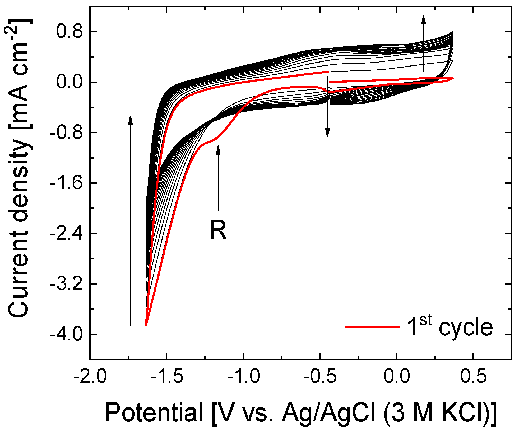
Figure 1. Cyclic voltammograms of the electrochemical reduction of the GO electrode in 0.1 M Na2SO4 at 50 mV s−1 for 20 consecutive cycles. The arrows indicate the evolution with the number of cycles.
Nanomaterials 15 00722 g001
Figure 2. Cyclic voltammograms during the first cycle of the open stepwise potential window for (A) GO in 0.5 M H2SO4, (B) GO in 0.5 M H2SO4+ 1 mM 4-APPA, (C) rGO in 0.5 M H2SO4, and (D) rGO in 0.5 M H2SO4+ 1 mM 4-APPA. 50 mV s−1. The arrows indicate the evolution with the number of cycles.
Nanomaterials 15 00722 g002
Figure 3. P2p XPS spectra for GO and rGO electrochemically modified with 4-APPA at different oxidation potentials: (A) GO-4APPA modified at 1.28 V, (B) GO-4APPA modified at 1.58 V, (C) rGO-4APPA modified at 1.28 V, and (D) rGO-4APPA modified at 1.58 V.
Nanomaterials 15 00722 g003
Figure 4. Cyclic voltammograms at 50 mV s−1 of graphene-based materials electrochemically modified with 4-APPA in 0.1 M PBS (pH = 7.2): (A,B) 1 mM [Ru(NH3)6]3/4+ and (C,D) 5 mM ([Fe(CN)6]3/4−).
Nanomaterials 15 00722 g004
Figure 5. Cyclic voltammograms in 0.1 M PBS (pH = 7.2) + 100 mM H2O2 for (A) GO, (B) rGO, (C) GO-4-APPA, and (D) rGO-4-APPA before (dash line) and after (solid line) immobilization of Cyt C at 20 mV s−1 under room atmosphere.
Nanomaterials 15 00722 g005
Figure 6. Chronoamperometric profiles under stirring conditions at −0.4 V vs. Ag/AgCl (3 M KCl) in room atmosphere for graphene-based electrode materials electrochemically functionalized with 4-APPA with and without immobilized Cyt C: (A) GO-4APPA, (B) rGO-4APPA, (C) GO-4APPA-Cyt C, and (D) rGO-4APPA-Cyt C. Inset: enlargement of low concentration region.
Nanomaterials 15 00722 g006aNanomaterials 15 00722 g006b
Figure 7. Calibration curves toward H2O2 reduction obtained for all graphene-based electrode materials electrochemically functionalized with 4-APPA for (A) graphene oxide electrodes and (B) electrochemically reduced graphene oxide electrodes. Measurements were carried out in triplicate.
Nanomaterials 15 00722 g007
Table 1. Chemical composition obtained from XPS of electrochemically modified graphene-based materials with 4-APPA.
Graphene-Based MaterialUpper Potential Limit
[V vs. RHE]
O (at%)N (at%)P (at%)C (at%)
GOPristine34.80.28--64.92
1.2822.20.711.1475.95
1.5829.71.540.6168.15
rGOPristine18.70.39--80.91
1.2823.01.370.5275.11
1.5821.51.030.6576.82
Table 2. Electrochemical parameters of peak separation (∆E), formal potential (E0), electroactive surface area (A), and heterogeneous rate constant (k0) for [Ru(NH3)6]3/4+ and 5 mM [Fe(CN)6]3/4− redox probe performed with electrochemically modified graphene-based materials with 4-APPA.
Graphene-Based Electrode Material A R u 2 + / 3 +  [cm2] A F e 2 + / 3 +  [cm2] E R u 2 + / 3 +  [mV] * E F e 2 + / 3 +  [mV] * E R u 2 + / 3 + 0  [V] * E F e 2 + / 3 + 0  [V] * k 0 R u 2 + / 3 +  
[×10−3 cm s−1]
k 0 F e 2 + / 3 +  
[×10−3 cm s−1]
GO0.4440.045126150−0.2500.2162.701.38
GO-4APPA0.6090.075136230−0.2540.2531.820.28
rGO1.6800.228129111−0.2470.2121.162.12
rGO-4APPA2.0030.399137128−0.2790.2210.981.34
* Parameters have been determined at 100 mV s−1.
Table 3. The figures of merit for the electrocatalytic activity of the graphene-based electrode material functionalized with 4APPA and Cyt C.
Graphene-Based
Electrode Material
LOD [mM] *LOQ [mM] **Sensitivity
[μA mM−1 cm−2]
GO-4APPA0.10.336.65
GO-4APPACyt C9.20
rGO-4APPA44.30
rGO-4APPA-Cyt C111.68
* The LOD was determined experimentally as the H2O2 concentration that produces a notable current variation in the background current. For comparative purposes, 1 mM was selected as the initial concentration. ** LOQ = 3.3LOD.
Disclaimer/Publisher’s Note: The statements, opinions and data contained in all publications are solely those of the individual author(s) and contributor(s) and not of MDPI and/or the editor(s). MDPI and/or the editor(s) disclaim responsibility for any injury to people or property resulting from any ideas, methods, instructions or products referred to in the content.