Article

Optimizing the Structure and Optical Properties of Lanthanum Aluminate Perovskite through Nb5+ Doping

1
Key Laboratory of Inorganic Coating Materials CAS, Shanghai Institute of Ceramics, Chinese Academy of Sciences, Shanghai 200050, China
2
School of Chemistry and Materials Science, Shanghai Normal University (SNU), Shanghai 200234, China
3
School of Physical Science and Technology, ShanghaiTech University (STU), Shanghai 201210, China
4
School of Materials Science and Engineering, Shanghai University (SHU), Shanghai 200444, China
*
Author to whom correspondence should be addressed.
Nanomaterials 2024, 14(7), 608; https://doi.org/10.3390/nano14070608
Submission received: 25 February 2024 / Revised: 27 March 2024 / Accepted: 28 March 2024 / Published: 29 March 2024

Abstract

:
This work involves the introduction of niobium oxide into lanthanum aluminate (LaAlO3) via a conventional solid-state reaction technique to yield LaAlO3:Nb (LaNbxAl1−xO3+δ) samples with Nb5+ doping levels ranging from 0.00 to 0.25 mol%. This study presents a comprehensive investigation of the effects of niobium doping on the phase evolution, defect control, and reflectance of LaNbxAl1−xO3+δ powder. Powder X-ray diffraction (XRD) analysis confirms the perovskite structure in all powders, and XRD and transmission electron microscopy (TEM) reveal successful doping of Nb5+ into LaNbxAl1−xO3+δ. The surface morphology was analyzed by scanning electron microscopy (SEM), and the results show that increasing the doping concentration of niobium leads to fewer microstructural defects. Oxygen vacancy defects in different compositions are analyzed at 300 K, and as the doping level increases, a clear trend of defect reduction is observed. Notably, LaNbxAl1−xO3+δ with 0.15 mol% Nb5+ exhibits excellent reflectance properties, with a maximum infrared reflectance of 99.7%. This study shows that LaNbxAl1−xO3+δ powder materials have wide application potential in the field of high reflectivity coating materials due to their extremely low microstructural defects and oxygen vacancy defects.

1. Introduction

Rare earth LaAlO3 with an ABO3 crystal structure is a material that has attracted a lot of attention in recent years due to its unique properties. Its high melting point [1,2,3], chemical stability [4,5], and large band gap (3.8 eV) [6,7,8] make it a promising material for various applications. If the radius of the doping ion is close to La3+ or Al3+, LaAlO3, an essential cubic perovskite oxide, can be doped with significant concentrations of ions through substitution [9,10,11,12]. Due to its strong optoelectronic capabilities, the B site held by Al3+ can be replaced by transition metal ions to produce a variety of applications, including photodetector [13,14] and luminescent phosphor [15,16], which have been reported in numerous papers. Furthermore, due to its distinctively extended fluorescence lifespan and significant Stokes shift, LaAlO3 is widely used in the field of luminous materials [17,18,19,20]. In addition, numerous researchers have reported on its additional characteristics, such as poor heat conductivity [21,22].
Nevertheless, there is little research focusing on the optical characteristics of LaAlO3, particularly with regard to the materials used for reflective coatings materials. In order to consistently maintain a high and stable reflectivity coating material, it must retain its original crystal structure and not undergo a phase transition as the conditions change. The high symmetry of the crystal structure is advantageous for boosting the inter-band transition and has a significant impact on the optical characteristics of materials, according to the findings of earlier studies. Additionally, the intrinsic qualities of materials are also impacted by particle size homogeneity. Therefore, based on its high symmetry structure and thermal stability, LaAlO3 may be able to replace other inorganic compounds currently used as high-reflectance materials.
Unfortunately, the present research disregards the effects of the LaAlO3 perovskite preparation procedure [23,24,25]. For instance, a high sintering temperature results in irregular particles and rough grains, which restricts the optical characteristics [26,27]. Additionally, the LaAlO3 lattice will release oxygen atoms and create oxygen vacancies [8,28,29,30,31] at high temperatures, leading to a dramatic decrease in reflectivity, according to the phase transition mechanism. The band gap of LaAlO3 narrows as a result of the creation of oxygen vacancies and defect energy levels within the band gap [32,33,34,35,36]. Additionally, the oxygen vacancies will distort the perfect cubic perovskite lattice of LaAlO3 to some extent, lowering the material’s reflectivity. Exploring a technique to lessen the oxygen vacancy flaws in LaAlO3 is, therefore, significant [37,38,39,40]. The structure can also be tuned, and high-temperature flaws can be prevented by optimizing the preparation conditions based on the current preparation process.
Nb5+ doping has recently been discovered to be advantageous for enhancing the reflectivity of LaSrTiO3 materials [41]. The role and impact of Nb5+ doping in LaAlO3 perovskite, however, have not been explored. In this study, the high-temperature solid-state method was used to synthesize Nb5+-doped LaAlO3 perovskite material. This paper investigates the potential changes in microstructure, oxygen vacancy defects, and optical properties resulting from the partial replacement of Al3+ with Nb5+ in the LaAlO3 perovskite structure through a high-temperature solid-state reaction. Additionally, this paper examines the influence of oxygen vacancy on the band gap and reflectivity of materials. To the best of our knowledge, no previous research has been conducted on the impact of Nb5+-doped LaAlO3 on reflectivity.

2. Experimental

The high-temperature solid-state method was used to create LaNbxAl1−xO3+δ perovskite powders. LaNbxAl1−xO3+δ (x = 0, 0.05, 0.10, 0.15, 0.20, 0.20) was made from La2O3, Al2O3, and Nb2O5 for the high-temperature solid-state method. All raw materials are of analytical grade and sourced from Aladdin Reagent Company Limited. The high-temperature solid-state preparation technique is used conventionally. Raw materials are weighed according to the stoichiometric ratio, and anhydrous ethanol is added. The raw materials are then placed into a nylon ball milling tank. First, wet grinding and mixing are conducted in a ball mill, uniformly mixing the powders, drying, sieving with a 40-mesh sieve, and sintering at 1250 °C for 3 h in an air atmosphere to make samples. LaNbxAl1−xO3+δ powder with a high reflectivity was produced in this manner.
X-ray diffractometry (XRD, D/max 2550V, RIGAKU, Tokyo, Japan) using Cu-K radiation (λ = 0.1542 nm) was used to observe the phase composition of the samples and the degree of lattice variation, and transmission electron microscopy (TEM, JEM-2100F, JEOL, Tokyo, Japan) was used to analyze the variation in the crystal plane d spacing and microscopic morphology. Scanning electron microscopy (SEM, Magellan 400, FEI, Hillsboro, OR, USA) was used to examine the microscopic morphology. Additionally, measurements of electron paramagnetic resonance (EPR, Bruker EMXplus-6/1, Herborn, Germany) were conducted to ascertain the number of oxygen vacancies. The photoluminescence spectra were recorded by a steady-state transient fluorescence spectrometer (PL, Fluorolog-3, HORIBA, Tokyo, Japan). An ultraviolet-visible-near-infrared spectrophotometer (UV-Vis-NIR, Lambda 1050, PerkinElmer, Waltham, MA, USA) was used to measure the reflectivity from 250 nm to 2500 nm. Samples were mixed proportionally with LaNbxAl1−xO3+δ perovskite powder and 6% wt Polyvinyl Alcohol (PVA) binder, mixed evenly, and then transferred to a tablet mold. Under a pressure of 4 MPa for 2 min, a small disk with a diameter of 30 mm and a thickness of 2 mm was formed. The plate was placed in a muffle furnace and heated at 650 °C for 3 h to discharge the PVA binder.

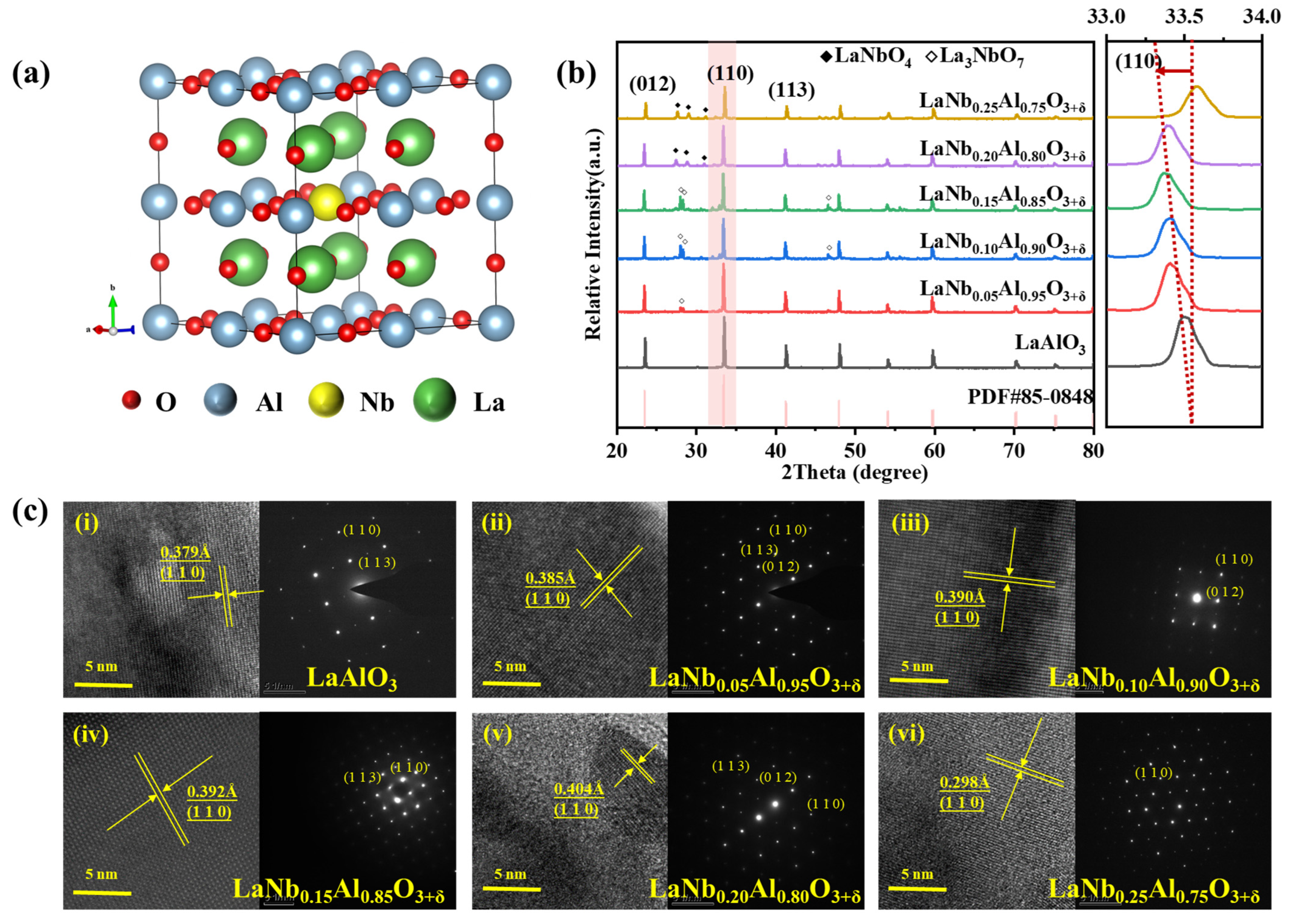
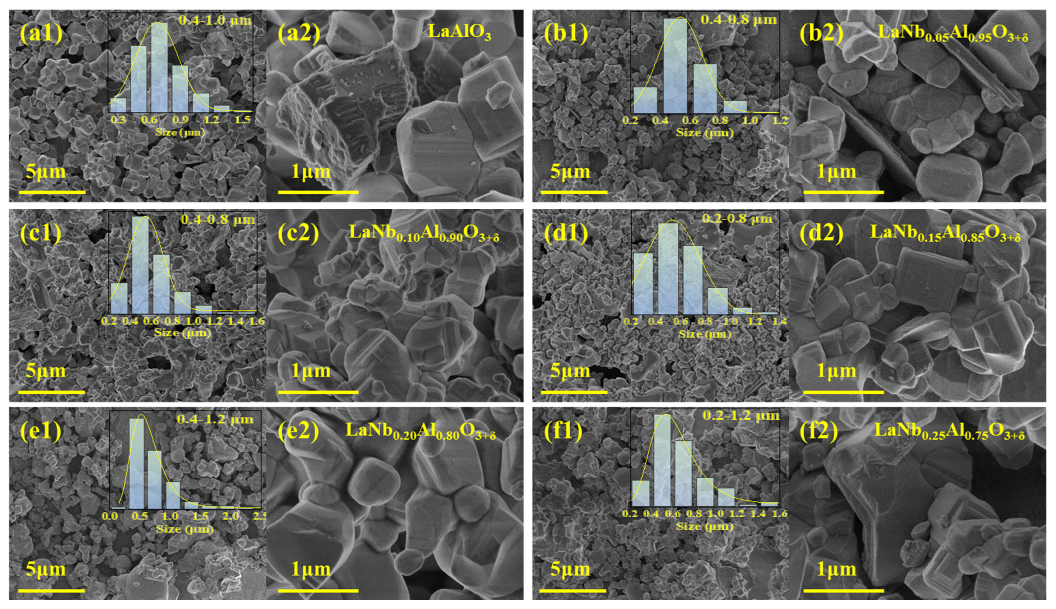
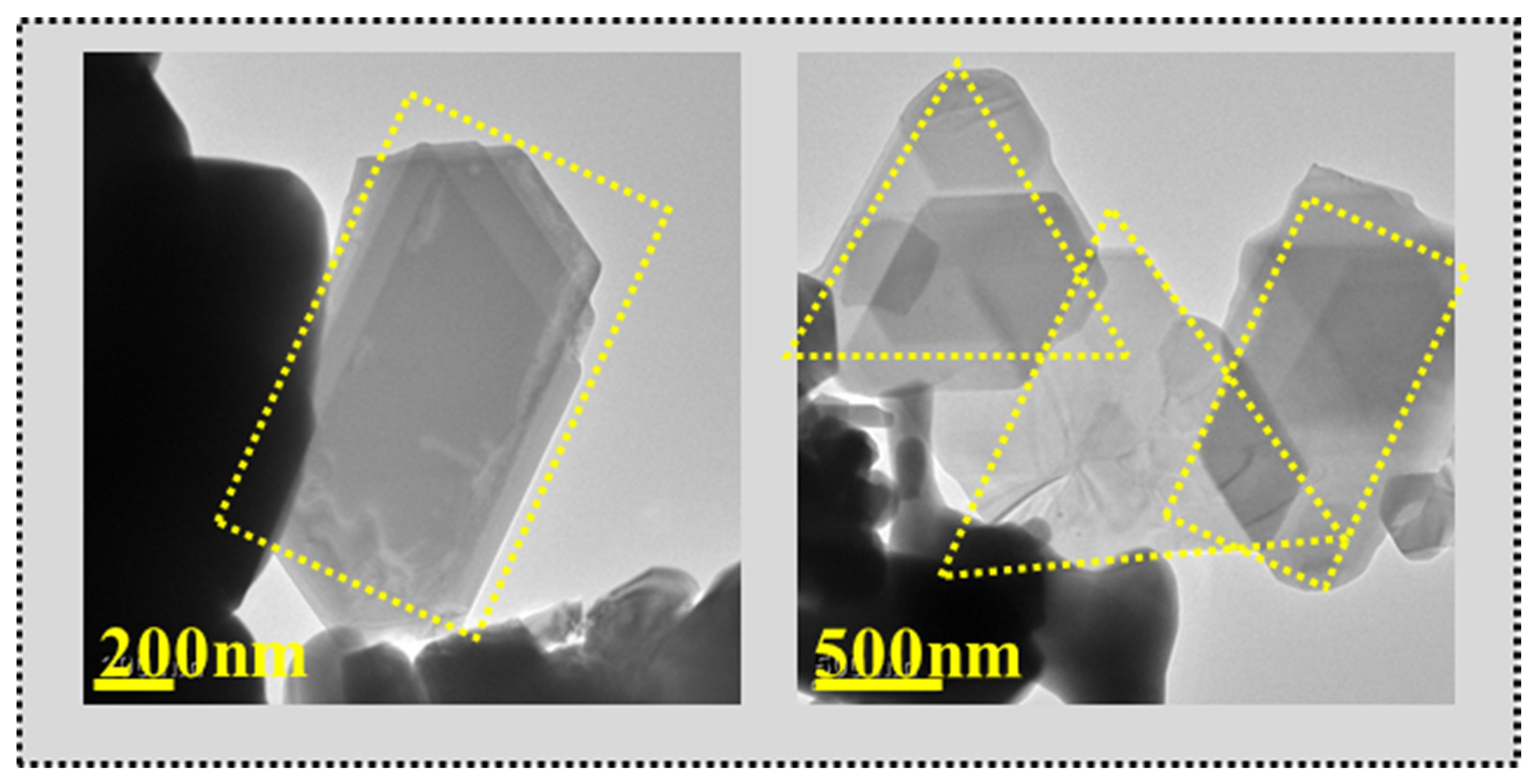
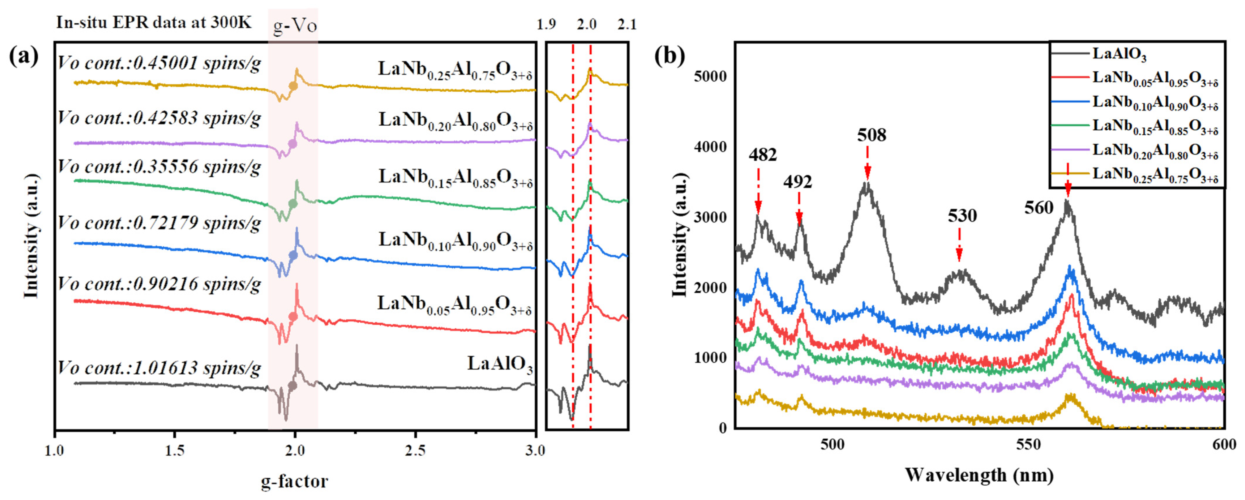
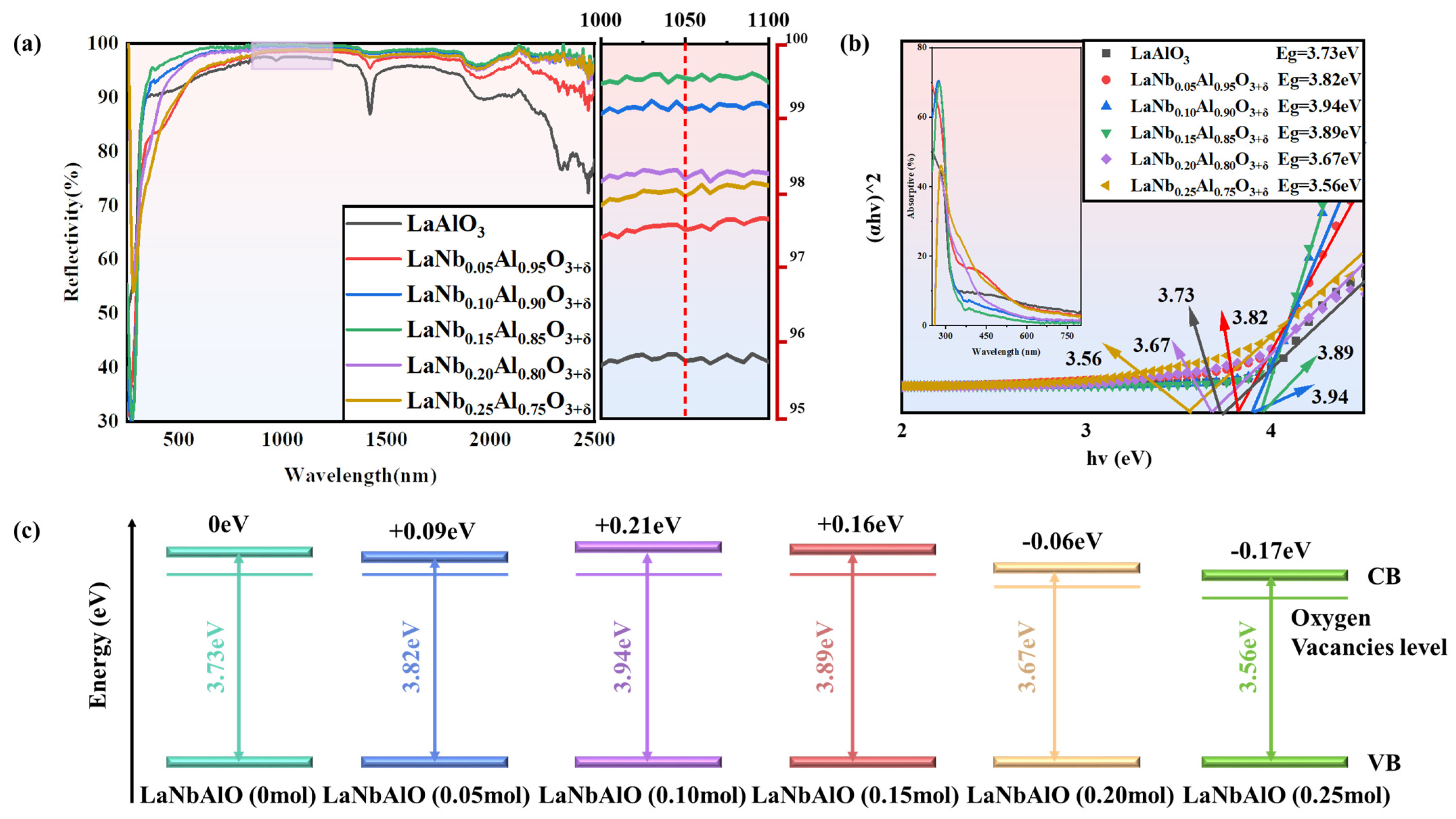
3. Results and Discussion

The crystal structure of LaAlO3 is shown in Figure 1. The Vesta software (Ver:3.0.1) was used to expand the cell of the LaAlO3 structure by 2 × 2 × 2. The lowest formation energy Nb atom was then selected to replace the Al atom, resulting in the schematic diagram shown in Figure 1a. Figure 1b shows the XRD patterns of the LaNbxAl1−xO3+δ (x = 0, 0.05, 0.10, 0.15, 0.20, 0.25) samples. The main diffraction peaks of LaNbxAl1−xO3+δ were indexed to LaAlO3 with structure in accordance with the PDF card no. 85-0548. The diffraction peaks shifted slightly to a lower angle with an increase in the Nb5+ doping content from 0.05 mol% to 0.20 mol%, which could be attributed to the successful incorporation of Nb5+ into the LaAlO3 lattice. To obtain the accurate lattice parameters, the XRD patterns of LaNbxAl1−xO3+δ were refined, and the refined results are shown in Table 1. Based on the structure, the lattice volumes of LaNbxAl1−xO3+δ (x = 0, 0.05, 0.10, 0.15, 0.20, 0.25) were calculated to be 54.13, 54.16, 54.28, 54.49, 54.54, and 54.58 Å3, respectively. Obviously, the lattice volume of LaAlO3 increased from 54.13 to 54.58 Å3 with Nb5+ doping. The increase in lattice volume can be attributed to the partial substitution of Al3+ by Nb5+, whose Nb5+ has a slightly larger radius (0.78 Å) than Al3+ (0.675 Å) [42].
After doping, Nb5+ ions are dispersed into the lattice point of LaAlO3 perovskite structure, occupying Al3+ position, accompanied by a small number of heterogeneous diffraction peaks, which are consistent with the diffraction peaks of LaNbO4 (PDF#71-1405) and La3NbO7 (PDF#71-1345), indicating that most Nb5+ has been doped into the lattice of LaAlO3. It can be inferred that the reaction of synthesizing LaNbxAl1−xO3+δ powder is shown in Formula (1):
La 2 O 3 + 1 x Al 2 O 3 + x Nb 2 O 5 heat 2 La Nb x Al 1 x O 3 + δ .
Under this reaction’s conditions, La2O3 and Nb2O5 can also react in a solid state at a high temperature, and the chemical reaction equation is shown in Formula (2):
2 La 2 O 3 + Nb 2 O 5 heat LaNbO 4 + La 3 NbO 7 .
Moreover, the detailed microstructure of the LaNbxAl1−xO3+δ samples was investigated by high-resolution transmission electron microscopy (HR-TEM). As shown in Figure 1c, the grains of LaNbxAl1−xO3+δ exhibit clear lattice fringes, and the d spacing of the Nb5+ doping sample is a little bigger than that of the (110) plane of LaAlO3. With the increase in doping content, the interplanar spacing of 110 crystal faces increased from the initial 0.379 Å to 0.404 Å. The lattice spacing change observed by HR-TEM is consistent with a shift toward lower angles in the XRD patterns. In the case of 0.25 mol% doping, the XRD diffraction peaks shift toward higher angles, most likely due to the significant growth trend of LaNbO4 under 0.25 mol% Nb5+ doping, causing changes in atomic positions within the crystal, leading to lattice non-uniformity and alterations in the crystal structure, thus resulting in the shift of XRD diffraction peaks toward higher angles. At this point, the interplanar spacing observed in TEM also shows irregularities.
The SEM surface image of LaNbxAl1−xO3+δ is shown in Figure 2. Based on the SEM image, it can be concluded that the growth of the LaAlO3 perovskite is of high quality, and the growth steps are clearly visible. This suggests that the LaAlO3 material prepared has excellent crystallinity and lattice integrity, meeting the requirements for a perovskite structure. This is of great significance for the performance and application of the material. At high magnification (30,000×), for the Nb5+ doping level between 0.10 mol% and 0.20 mol%, the structure of the crystal particles shows the complete morphology. Perovskite structure growth stages show the clearest crystal structure at a doping concentration of 0.15 mol%, the rough bulk structure disappears, and the perovskite structure becomes more pronounced due to the role played by Nb5+ as a sintering aid. The lamellar structure appears when the doping content exceeds 0.25 mol%. As can be seen, this sample has a distinct grain boundary.
Drawing the particle size distribution curve of 100 particles in the image reveals that (a) the particle size of Nb5+ undoped LaAlO3 is mostly in the range of 0.4–1.0 μm; (b) the particle size of 0.05 mol% Nb5+ doped LaAlO3 is in the range of 0.4–0.8 μm; (c) the particle size of 0.10 mol% doped LaAlO3 is in the range of 0.4–0.8 μm; (d) the particle size 0.15 mol% doped LaAlO3 is in the range of 0.2–0.8 μm; (e) the particle size of 0.20 mol% doped LaAlO3 is in the range of 0.4–1.2 μm; and (f) the particle size of 0.25 mol% doped LaAlO3 is in the range of 0.2–1.2 μm.
Figure 3 shows the lamellar structures in LaNb0.25Al0.75O3+δ. The appearance of lamellar structures resulting from 0.25 mol% Nb5+ doping leads to the material’s unevenness and changes in the crystal structure. This result is consistent with the conclusions obtained by SEM.
The EPR spectrum of the complex form of LaNbxAl1−xO3+δ is provided by the anisotropy of the EPR signal and the superposition of concentrated EPR signals from distinct defect types, as shown in Figure 4a. EPR technology is a common means to quantitatively characterize the concentration of oxygen vacancy defects. By measuring and analyzing the standard sample with known concentration, the quantitative relationship between the concentration of oxygen vacancy defects and the EPR signal can be established so as to realize the quantitative characterization of the concentration of oxygen vacancy defects in the sample. When unpaired electrons interact with an external magnetic field, energy level transition, and resonance absorption will occur. In solid materials, oxygen vacancies usually contain unpaired electrons, which will participate in the EPR process. Due to the influence of the electronic environment around oxygen vacancy, the EPR signal that leads to oxygen vacancy defect usually has a characteristic peak near g ≈ 2.000. This characteristic peak corresponds to the resonance absorption of unpaired electrons. In order to reduce the experimental error, the same test conditions and atmosphere are used for the test. It is evident that there is a large EPR signal peak at the peak position of g ≈ 2.000, where hv = gβB predicts that the known sample’s oxygen vacancy concentration is g ≈ 2.000. Where h is Planck constant, ν is microwave frequency; β is Bohr magneton; B is magnetic induction intensity, and g is the g-factor. Through testing the EPR signal peaks of oxygen vacancies at 300 K (Figure 4a), it can be observed that with an increasing Nb5+ doping concentration, the oxygen vFacancy concentration decreases. Figure 4a shows the specific quantitative data. It is evident that the oxygen vacancy concentration decreases significantly with increasing doping content. The vacancy of undoped oxygen is reduced from 1.01613 spins/g to 0.35556 spins/g. But when the doping content reached 0.20 mol% and 0.25 mol%, the oxygen vacancy concentration began to increase. This may be due to the structural change caused by high-concentration doping and the growth of the second phase. To further confirm the defect concentration of LaNbxAl1−xO3+δ material, photoluminescence (PL) spectroscopy was performed in Figure 4b. As can be seen from this Figure, there are five luminescence peaks, which are 482 nm double-ionized oxygen ( V O * * ), 530 nm monoionized oxygen vacancy ( V O * ) and oxygen vacancy at 565 nm, respectively [43]. With the appearance of a large number of carriers, some monoionized oxygen vacancies and double-ionized oxygen vacancies are transformed into oxygen vacancies. In addition, the luminescence peaks at 492 nm and 508 nm are mainly caused by the change in energy band structure caused by lattice defects, and the reason for this needs further study. Therefore, the main optical absorption centers in LaNbxAl1−xO3+δ materials are oxygen vacancy defects and lattice defects.
The reflectance spectra are used to characterize the optical characteristics of LaNbxAl1−xO3+δ samples. The reflectance spectrum of the samples prepared with various Nb5+ doping levels by the high-temperature solid-state method is shown in Figure 5a. Reflectivity increases initially and then decreases as Nb5+ content increases. The reflectivity is the greatest at 0.15 mol% of doping. The greatest reflectivity reaches 99.7% at 1050 nm in the near-infrared light band with LaNb0.15Al0.85O3+δ, while LaAlO3 is only 95.8%.
The improvement in reflectivity is significantly influenced by the changes in phase composition and material structure. According to the SEM image in Figure 2, it is known that when the Nb5+-doped concentration rises to 0.15 mol% and the reflection path and interface number expand after doping. Combining the XRD and SEM analysis of the materials, it is found that with the doping content of 0.20 mol% and 0.25 mol%, the impurity peaks are obviously increased, and other phase structure particles appear, so there are more components affecting the energy band structure, and reflectivity changes greatly.
According to the UV-Vis diffuse reflectance spectroscopy in Figure 5a. All of the samples exhibit a distinct absorption edge, and the absorption edges are all located near the wavelength of 310 nm. Based on the UV-Vis DRS spectra, the band gap energy (Eg) of the samples was estimated via Kubelka–Munk as follows [44,45]:
( α h ν ) n = A ( h ν Eg )
where h is the Planck constant; ν is the photon frequency; α is the absorption coefficient, and A is the proportional constant. The value of the index n is determined by the properties of the sample transition: for direct band gap semiconductors, n = 0.5; for indirect band gap semiconductors, n = 2 [46]. Considering the nature of optical transition in LaAlO3 as an indirect transition [47], the band gaps for the respective LaNbxAl1−xO3+δ composites were evaluated, as shown in Figure 5b,c. Obviously, with the increase in Nb5+ content, the band gap of LaNbxAl1−xO3+δ prepared by the solid-state method first increases and then decreases.
The effect of oxygen vacancy on the optical properties of LaAlO3 materials is significant. Burstein–Moss effect proposed by the Pauling incompatibility principle emphasizes that when additional energy levels are added between VB and CB, the band gap of the material narrows, allowing for electrons to absorb lower energy, thus realizing the transition behavior. The more electrons in the energy band, the stronger the ability to move from low energy level to high energy level. As shown in Figure 6, when light passes through the crystal, oxygen vacancies act as absorption centers in the crystal, resulting in a decrease in reflectivity because oxygen vacancies generate oxygen vacancy levels in the band gap interval. Electrons absorb energy and make it jump from the valence band to the conduction band. In addition, the generation of oxygen vacancies will also lead to changes in the electronic structure of materials, which will lead to changes in their optical properties. When Nb5+ is added into the LaAlO3 lattice, the defect equation can be used to describe the doping mechanism, which proves that Nb5+ inhibits oxygen vacancies, neutralizes oxygen vacancy defects, and, therefore, widens the band gap, decreases the ability of electrons to absorb energy and transition to the conduction band, and boosts reflection while decreasing absorption. Combined with Figure 4, it is not difficult to see that with the increase in Nb5+ doping content, the oxygen vacancy concentration decreases. The reflectivity is influenced by multiple factors, such as microstructure defects and oxygen vacancy defects. Because there are more microstructure defects in 0.20 mol% and 0.25 mol% doping, the reflectivity and band gap are obviously lower than that in 0.15 mol% doping.

4. Conclusions

In this study, powders of LaNbxAl1−xO3+δ with high reflectivity were prepared by a high-temperature solid phase reaction method. Evidence shows that Nb5+ successfully entered the LaAlO3 lattice, inducing changes in the lattice constants and crystal plane spacing. The lattice volume increases with increasing Nb5+ doping, which is due to the fact that the radius of Nb5+ is slightly larger than that of Al3+. The sintering-assisted effect of Nb5+ leads to a gradual reduction in microstructural defects in the particles; the powder of LaNb0.15Al0.85O3+δ tends to range in size from 0.2 to 0.8 μm. Based on the quantitative study of EPR and PL emission peaks, it is clearly seen that the concentration of oxygen vacancies decreases with increasing doping, which proves the important role of Nb5+ in neutralizing the charge during high-temperature sintering.
Fewer microstructural defects contribute to the enhancement of the light reflection path, and lower oxygen vacancy concentration contributes to fewer oxygen vacancy defect energy levels and increases the band gap, which reduces the ability of the material to absorb photon jumps. The superposition of the two effects contributes to further enhancing the reflectivity of the material. For LaNbxAl1−xO3+δ, the maximum reflectivity reaches 99.7%, which appears at 1050 nm doped with 0.15 mol% Nb5+, while the pristine LaAlO3 sample can only reach 95.8%. This study presents a feasible method to reduce sintering defects and improve the optical properties of LaAlO3 material. This has the potential to create a new category of highly reflective materials.

Author Contributions

Conceptualization, L.Z. and L.S.; Methodology, W.L., Y.Z. and L.Z.; Formal analysis, W.L.; Investigation, W.L.; Resources, W.L.; Data curation, W.L., Y.Z. and L.Z.; Writing—original draft, W.L.; Writing—review & editing, W.L., Y.Z., Y.C., Z.L. and L.Z.; Visualization, W.L. and Y.C. All authors have read and agreed to the published version of the manuscript.

Funding

This research received no external funding.

Data Availability Statement

Data are contained within the article.

Conflicts of Interest

The authors declare no conflict of interest.

References

  1. Dereń, P.J.; Mahiou, R.; Goldner, P. Multiphonon transitions in LaAlO3 doped with rare earth ions. Opt. Mater. 2009, 31, 465–469. [Google Scholar] [CrossRef]
  2. Dhahri, A.; Horchani-Naifer, K.; Benedetti, A.; Enrichi, F.; Ferid, M. Combustion synthesis and photoluminescence of Eu3+ doped LaAlO3 nanophosphors. Opt. Mater. 2012, 34, 1742–1746. [Google Scholar] [CrossRef]
  3. He, X.; Li, Y.; Wang, L.; Sun, Y.; Zhang, S. High emissivity coatings for high temperature application: Progress and prospect. Thin Solid Film. 2009, 517, 5120–5129. [Google Scholar] [CrossRef]
  4. Muñoz, H.J.; Korili, S.A.; Gil, A. Progress and Recent Strategies in the Synthesis and Catalytic Applications of Perovskites Based on Lanthanum and Aluminum. Materials 2022, 15, 3288. [Google Scholar] [CrossRef] [PubMed]
  5. Choi, M.; Janotti, A.; Van de Walle, C.G. Native point defects in LaAlO3: A hybrid functional study. Phys. Rev. B 2013, 88, 214117. [Google Scholar] [CrossRef]
  6. Han, Z.; Li, X.; Ye, J.; Kang, L.; Chen, Y.; Li, J.; Lin, Z.; Rohnke, M. Significantly Enhanced Infrared Emissivity of LaAlO3 by Co-Doping with Ca2+ and Cr3+ for Energy-Saving Applications. J. Am. Ceram. Soc. 2015, 98, 2336–2339. [Google Scholar] [CrossRef]
  7. Ankoji, P.; Rudramadevi, B.H. Structural and luminescence properties of Eu3+ doped LaAlO3 nanophosphors by hydrothermal method. J. Mater. Sci. Mater. Electron. 2018, 30, 2750–2762. [Google Scholar] [CrossRef]
  8. Wang, Z.; Zhai, X.; Fu, Z.; Lu, Y. Tuning LaAlO3 lattice structure by growth rate at the picometer scale in LaAlO3 SrTiO3 heterostructures. J. Appl. Phys. 2018, 124, 125305. [Google Scholar] [CrossRef]
  9. Silveira, I.S.; Ferreira, N.S.; Souza, D.N. Structural, morphological and vibrational properties of LaAlO3 nanocrystals produced by four different methods. Ceram. Int. 2021, 47, 27748–27758. [Google Scholar] [CrossRef]
  10. Lu, L.; Zhang, C.-L.; Mi, S.-B. Probing interface structure and cation segregation in (In, Nb) co-doped TiO2 thin films. Mater. Charact. 2022, 191, 112164. [Google Scholar] [CrossRef]
  11. Wang, Q.; Yan, S.; Dong, B.; Zhang, Y.; Zhang, Q.; Du, P.; Wang, G. Preparation of environmentally friendly high-emissivity Ca2+-Fe3+ co-doped LaAlO3 ceramic. Int. J. Appl. Ceram. Technol. 2023, 20, 1785–1792. [Google Scholar] [CrossRef]
  12. Liu, H.; Sun, H.; Xie, R.; Zhang, X.; Zheng, K.; Peng, T.; Wu, X.; Zhang, Y. Substrate-dependent structural and CO sensing properties of LaCoO3 epitaxial films. Appl. Surf. Sci. 2018, 442, 742–749. [Google Scholar] [CrossRef]
  13. Du, J.-Y.; Ge, C.; Xing, J.; Li, J.-K.; Jin, K.-J.; Yang, J.-T.; Guo, H.-Z.; He, M.; Wang, C.; Lu, H.-B.; et al. Solar-blind ultraviolet photodetector based on (LaAlO3)0.3-(SrAl0.5Ta0.5O3)0.7 single crystal. AIP Adv. 2017, 7, 035302. [Google Scholar] [CrossRef]
  14. Sun, X.; Wang, D.; Memon, M.H.; Zhu, S.; Yu, H.; Wang, H.; Fang, S.; Kang, Y.; Liu, X.; Luo, Y.; et al. Anisotropic photoresponse behavior of a LaAlO3 single-crystal-based vacuum-ultraviolet photodetector. Nanoscale 2022, 14, 16829–16836. [Google Scholar] [CrossRef] [PubMed]
  15. Chen, Y.; Yang, C.; Deng, M.; He, J.; Xu, Y.; Liu, Z.Q. A highly luminescent Mn4+ activated LaAlO3 far-red-emitting phosphor for plant growth LEDs: Charge compensation induced Mn4+ incorporation. Dalton Trans. 2019, 48, 6738–6745. [Google Scholar] [CrossRef] [PubMed]
  16. Visweswara Rao, T.K.; Satya Kamal, C.; Samuel, T.; Srinivasa Rao, V.; Srinivasa Rao, V.; Reddy, P.V.S.S.S.N.; Ramachandra Rao, K. Color tunable luminescence from LaAlO3:Bi3+, Ho3+ doped phosphors for field emission displays. J. Mater. Sci. Mater. Electron. 2017, 29, 1011–1017. [Google Scholar] [CrossRef]
  17. Dhahri, A.; Horchani-Naifer, K.; Benedetti, A.; Enrichi, F.; Férid, M.; Riello, P. Combustion synthesis and photoluminescence of Tb3+ doped LaAlO3 nanophosphors. Opt. Mater. 2013, 35, 1184–1188. [Google Scholar] [CrossRef]
  18. Huang, C.; Li, S.; Gong, Q.; Fang, Q.; Xu, M.; Tao, S.; Zhao, C.; Hang, Y. Optical properties of Nd,Th:LaAlO3 demonstrates its potential in high-energy pulsed laser. Opt. Laser Technol. 2022, 156, 108495. [Google Scholar] [CrossRef]
  19. Liu, G.; Fu, L.; Gao, Z.; Yang, X.; Fu, Z.; Wang, Z.; Yang, Y. Investigation into the temperature sensing behavior of Yb3+ sensitized Er3+ doped Y2O3, YAG and LaAlO3 phosphors. RSC Adv. 2015, 5, 51820–51827. [Google Scholar] [CrossRef]
  20. Shaik, E.B.; Pindiprolu, S.K.S.S.; Phanikumar, C.S.; Samuel, T.; Kumar, B.V.N.; Santhoshi, P.M.; Reddy, P.V.S.S.S.N.; Kumar, B.P.; Ramachandra, R.K. Optical emissions of chitosan modified LaAlO3: Bi3+, Tb3+ nanoparticles for bio labeling and drug delivery to breast cancer cells. Opt. Mater. 2020, 107, 110162. [Google Scholar] [CrossRef]
  21. Breckenfeld, E.; Wilson, R.B.; Martin, L.W. Effect of growth induced (non)stoichiometry on the thermal conductivity, permittivity, and dielectric loss of LaAlO3 films. Appl. Phys. Lett. 2013, 103, 082901. [Google Scholar] [CrossRef]
  22. Zahoor, A.; Isa, M.; Mahmood, T. Computational study of Be doped LaAlO3 perovskite. Phys. B Condens. Matter 2023, 652, 414631. [Google Scholar] [CrossRef]
  23. Xue, Y.; Deng, W.; Liu, Y.; Bai, X.; Chen, X.; Zhao, P.; Pan, Y.; Zhang, H.; Chang, A.; Xie, Y. Effect of Ce-doping on microstructure and electrical properties of LaAlO3 ceramics. Ceram. Int. 2023, 49, 5884–5892. [Google Scholar] [CrossRef]
  24. Wang, T.; de Oliveira, R.B.; Andreeta, M.R.B.; Wang, H.; Jia, Z.; Tao, X. Oriented Crystal Growth of La0.557Li0.330TiO3 in Bulk Ceramics Induced by LaAlO3 Single-Crystal Fibers. Cryst. Growth Des. 2021, 21, 2093–2100. [Google Scholar] [CrossRef]
  25. Ji, Y.; Zhang, P.; He, L.; Wang, D.; Luo, H.; Otsuka, K.; Wang, Y.; Ren, X. Tilt strain glass in Sr and Nb co-doped LaAlO3 ceramics. Acta Mater. 2019, 168, 250–260. [Google Scholar] [CrossRef]
  26. Gu, X.; Jin, S.; Guan, X.; Yu, X.; Yu, Z.; Yan, Y.; Wu, K.; Zhao, L.; Liu, X. Comparative study of La0.7Ca0.18Sr0.12MnO3 films with room-temperature TCR grown on SrTiO3, La0.3Sr0.7Al0.65Ta0.35O3 and LaAlO3 substrates. Ceram. Int. 2023, 49, 22952–22960. [Google Scholar] [CrossRef]
  27. Van Thiet, D.; Dung, D.D.; Nguyen, V.Q.; Duong, A.T.; Chung, N.X.; Hong, N.T.; Cho, S. Thermoelectric, Magnetic Properties and Re-entrant Spin-glass State in MBE Grown FeAs Film on LaAlO3(100) Substrate. ECS J. Solid State Sci. Technol. 2023, 12, 023005. [Google Scholar] [CrossRef]
  28. Choi, M. Hydrogen passivation of oxygen vacancies in LaAlO3. Curr. Appl. Phys. 2022, 39, 154–157. [Google Scholar] [CrossRef]
  29. Gupta, M.; Rambadey, O.V.; Shirbhate, S.C.; Acharya, S.; Sagdeo, A.; Sagdeo, P.R. Probing the Signature of Disordering and Delocalization of Oxygen Vacancies and Anti-site Defects in Doped LaAlO3 Solid Electrolytes. J. Phys. Chem. C 2022, 126, 20251–20262. [Google Scholar] [CrossRef]
  30. Hu, X.; Ren, R.; Xu, Y.; Maroof, Z. Effects of strain and oxygen vacancy on the electronic structure of (SrMnO3)2/(LaAlO3)2.5 (001) heterostructure. Phys. B Condens. Matter 2022, 624, 413310. [Google Scholar] [CrossRef]
  31. Wang, B.; Wu, Y.; Wei, H.; Chen, X.; Zhang, X.; Cao, B. Tunable the kondo effect at LaAlO3/SrTiO3 interface by oxygen vacancies. Vacuum 2022, 204, 111372. [Google Scholar] [CrossRef]
  32. Ferrari, V.; Weissmann, M. Tuning the insulator-metal transition in oxide interfaces: An ab initio study exploring the role of oxygen vacancies and cation interdiffusion. Phys. Status Solidi (b) 2014, 251, 1601–1607. [Google Scholar] [CrossRef]
  33. Hossain, R.; Billah, A.; Ishizaki, M.; Kubota, S.; Hirose, F.; Ahmmad, B. Oxygen vacancy mediated room-temperature ferromagnetism and band gap narrowing in DyFe0.5Cr0.5O3 nanoparticles. Dalton Trans. 2021, 50, 9519–9528. [Google Scholar] [CrossRef] [PubMed]
  34. Linderalv, C.; Lindman, A.; Erhart, P. A Unifying Perspective on Oxygen Vacancies in Wide Band Gap Oxides. J. Phys. Chem. Lett. 2018, 9, 222–228. [Google Scholar] [CrossRef] [PubMed]
  35. Xiong, K.; Robertson, J.; Clark, S.J. Electronic defects in LaAlO3. Microelectron. Eng. 2008, 85, 65–69. [Google Scholar] [CrossRef]
  36. Zhang, Y.; Hao, F.; Liu, C.; Chen, X. Band gap and oxygen vacancy diffusion of anatase (101) surface: The effect of strain. Theor. Chem. Acc. 2016, 135, 171. [Google Scholar] [CrossRef]
  37. Sarkar, S.; Saha, S.; Motapothula, M.R.; Patra, A.; Cao, B.C.; Prakash, S.; Cong, C.X.; Mathew, S.; Ghosh, S.; Yu, T.; et al. Magneto-Optical Study of Defect Induced Sharp Photoluminescence in LaAlO3 and SrTiO3. Sci. Rep. 2016, 6, 33145. [Google Scholar] [CrossRef]
  38. Kumar, P.; Singh, S.; Gupta, I.; Kumar, V.; Singh, D. Preparation and luminescence behaviour of perovskite LaAlO3:Tb3+ nanophosphors for innovative displays. Optik 2022, 267, 169709. [Google Scholar] [CrossRef]
  39. Chen, B.; Li, C.; Deng, D.; Ruan, F.; Wu, M.; Wang, L.; Zhu, Y.; Xu, S. Temperature sensitive properties of Eu2+/Eu3+ dual-emitting LaAlO3 phosphors. J. Alloys Compd. 2019, 792, 702–712. [Google Scholar] [CrossRef]
  40. Jusza, A.; Lipińska, L.; Baran, M.; Polis, P.; Olszyna, A.; Piramidowicz, R. Short wavelength emission properties of Tm3+ and Tm3++Yb3+ doped LaAlO3 nanocrystals and polymer composites. Opt. Mater. 2019, 97, 109365. [Google Scholar] [CrossRef]
  41. Zhao, Y.; Zhu, J.; Wang, H.; Ma, Z.; Gao, L.; Liu, Y.; Liu, Y.; Shu, Y.; He, J. Enhanced optical reflectivity and electrical properties in perovskite functional ceramics by inhibiting oxygen vacancy formation. Ceram. Int. 2021, 47, 5549–5558. [Google Scholar] [CrossRef]
  42. Zhang, B.; Li, L.; Luo, W. Oxygen vacancy regulation and its high frequency response mechanism in microwave ceramics. J. Mater. Chem. C 2018, 6, 11023–11034. [Google Scholar] [CrossRef]
  43. Mikhailov, M. Possibilities of replacing electromagnetic radiation of the Sun by accelerated electrons in testing space technology materials. J. Adv. Mater.-Covina 1996, 6, 465–470. [Google Scholar]
  44. Chen, X.; Liu, L.; Yu, P.Y.; Mao, S.S.J.S. Increasing solar absorption for photocatalysis with black hydrogenated titanium dioxide nanocrystals. Science 2011, 331, 746–750. [Google Scholar] [CrossRef] [PubMed]
  45. Soleymani, M.; Moheb, A.; Joudaki, E.J.O.C. High surface area nano-sized La0.6Ca0.4MnO3 perovskite powder prepared by low temperature pyrolysis of a modified citrate gel. Open Chem. 2009, 7, 809–817. [Google Scholar] [CrossRef]
  46. Bao, W.; Ma, F.; Zhang, Y.; Hao, X.; Deng, Z.; Zou, X.; Gao, W. Synthesis and characterization of Fe3+ doped Co0.5Mg0.5Al2O4 inorganic pigments with high near-infrared reflectance. Powder Technol. 2016, 292, 7–13. [Google Scholar] [CrossRef]
  47. Nepomniashchaia, N.; Vetokhina, V.; Chvostova, D.; Bryknar, Z.; Dejneka, A.; Tyunina, M. Low-temperature NIR-VUV optical constants of (001) LaAlO3 crystal. Opt. Mater. Express 2022, 12, 3081–3089. [Google Scholar] [CrossRef]
Figure 1. (a) Schematic illustration of 2 × 2 × 2 supercell of LaNbxAl1−xO3+δ perovskite crystal. (b) XRD patterns for pure (LaAlO3) and Nb5+ doped LaAlO3 with various Nb: LaNbxAl1−xO3+δ by high-temperature of solid-state method. Enlarge image of LaNbxAl1−xO3+δ (x = 0.05–0.25) ceramics at 2θ range of 33~34°. The red dotted line represents the shift trend of the diffraction peak of the (110) crystal plane. (c) TEM images of LaNbxAl1−xO3+δ synthesized by high-temperature solid-state method. (i) A total of 0 mol% Nb5+-doped LaAlO3. (ii) A total of 0.05 mol% Nb5+-doped LaAlO3. (iii) A total of 0.10 mol% Nb5+-doped LaAlO3. (iv) A total of 0.15 mol% Nb5+-doped LaAlO3. (v) A total of 0.20 mol% Nb5+-doped LaAlO3. (vi) A total of 0.25 mol% Nb5+-doped LaAlO3.
Nanomaterials 14 00608 g001
Figure 2. Grain growth observed by SEM on LaNbxAl1−xO3+δ specimens formed by doping with different Nb5+ contents and particle size distribution curves. (a) A total of 0 mol% Nb5+-doped LaAlO3. (b) A total of 0.05 mol% Nb5+-doped LaAlO3. (c) A total of 0.10 mol% Nb5+-doped LaAlO3. (d) A total of 0.15 mol% Nb5+-doped LaAlO3. (e) A total 0.20 mol% Nb5+-doped LaAlO3. (f) A total 0.25 mol% Nb5+-doped LaAlO3. Within each content group, the magnifications of the images were 5000× and 30,000×, respectively.
Nanomaterials 14 00608 g002
Figure 3. LaNb0.25Al0.75O3+δ sample lamellar structure in TEM images.
Nanomaterials 14 00608 g003
Figure 4. (a) In situ EPR data at 300 K. (b) PL spectra of LaNbxAl1−xO3+δ at different doping ratios. The red arrow points defect luminescence peak.
Nanomaterials 14 00608 g004
Figure 5. (a) Reflectance spectra of LaAlO3 samples doped with different contents of Nb5+. The enlarged image has a wavelength of 1000–1100 nm. (b) Optical band gap obtained by diffuse reflection spectrum using Kubelka–Munk equation. (c) Schematic illustration of band structure of LaNbxAl1−xO3+δ. (From left to right, the doping content is 0, 0.05, 0.10, 0.15, 0.20, 0.25 mol%).
Nanomaterials 14 00608 g005
Figure 6. Schematic illustration of vacancy migration of LaNbxAl1−xO3+δ under light conditions.
Nanomaterials 14 00608 g006
Table 1. Refined results for the LaNbxAl1−xO3+δ (x = 0, 0.05, 0.10, 0.15, 0.20, 0.25) samples.
Lattice Parameters (Refined)
a (Å)b (Å)c (Å)α (deg)β (deg)γ (deg)V (Å3)
x = 03.782743.782743.7827489.962989.962989.962954.13
x = 0.053.783383.783383.7833889.956889.956889.956854.16
x = 0.103.786283.786283.7862889.959789.959789.959754.28
x = 0.153.791183.791183.7911889.978889.978889.978854.49
x = 0.203.792393.792393.7923989.956689.956689.956654.54
x = 0.253.793213.793213.7932189.981489.981489.981454.58
Disclaimer/Publisher’s Note: The statements, opinions and data contained in all publications are solely those of the individual author(s) and contributor(s) and not of MDPI and/or the editor(s). MDPI and/or the editor(s) disclaim responsibility for any injury to people or property resulting from any ideas, methods, instructions or products referred to in the content.

