

Dentine Remineralisation Induced by “Bioactive” Materials through Mineral Deposition: An In Vitro Study
 ,
,  ,
,  ,
,  ,
,  ,
,  and
and 
Abstract
1. Introduction
2. Materials and Methods
2.1. Sample Preparation and Incubation in DPBS
2.2. SEM Imaging
2.3. EDX Analysis
2.4. Statistical Analysis
3. Results
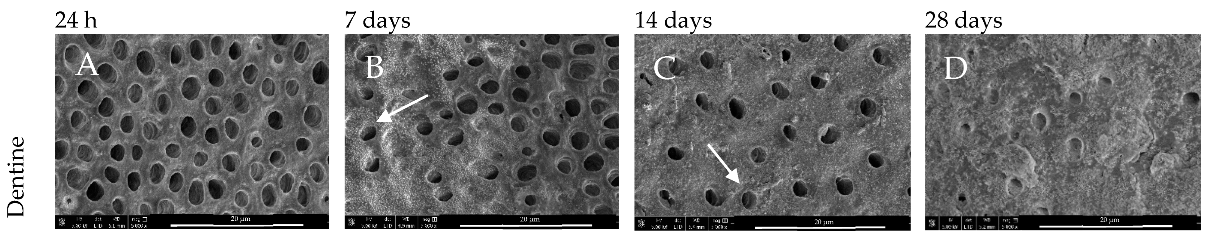
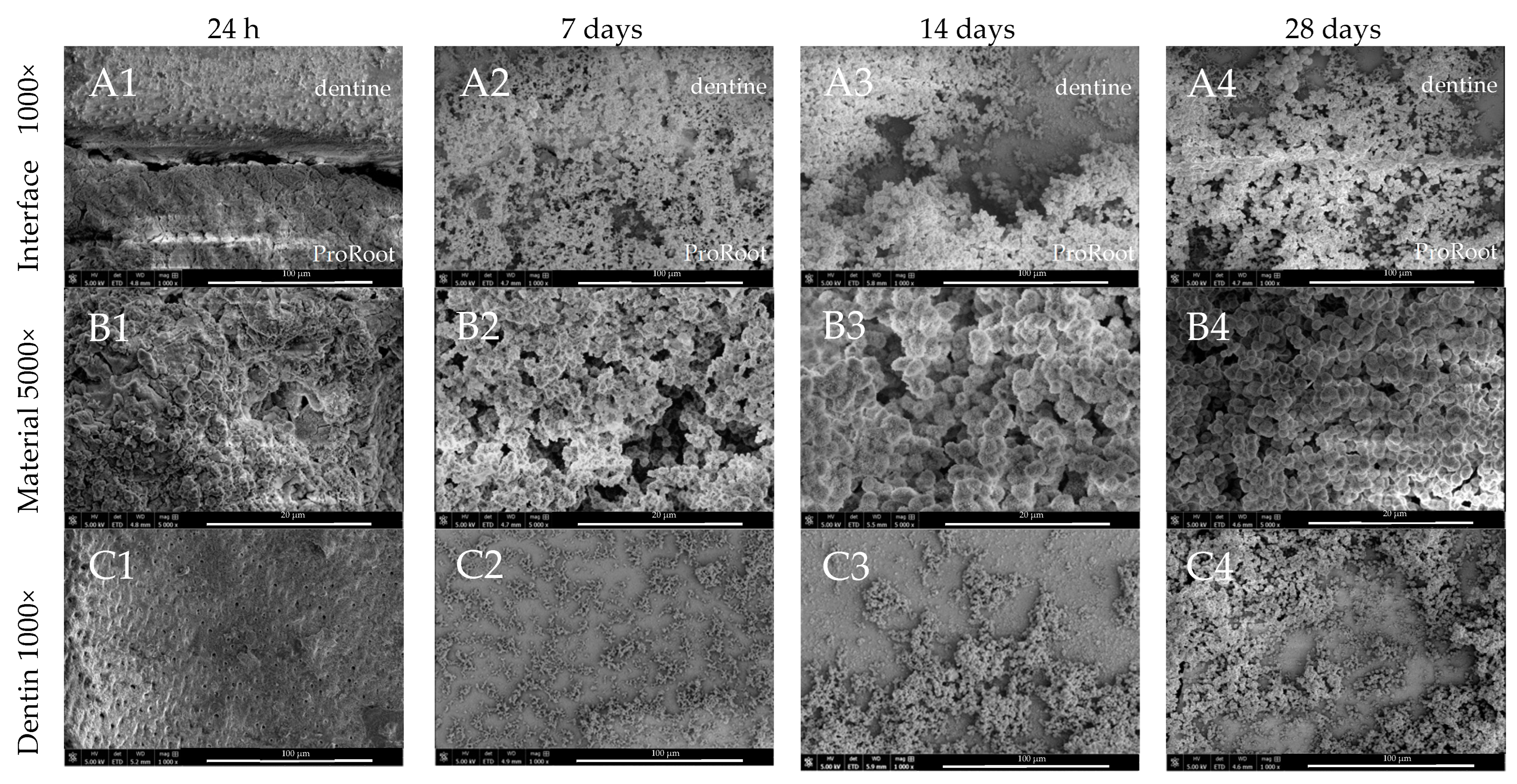
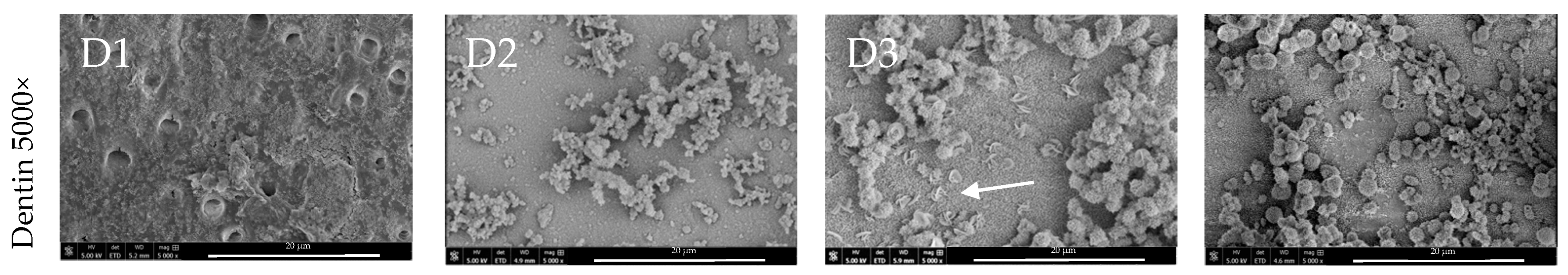
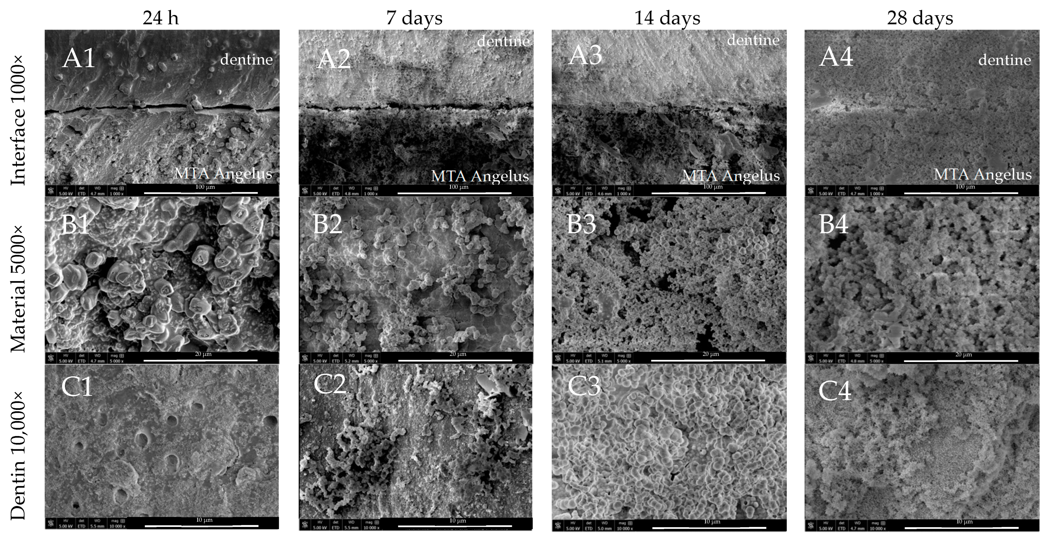
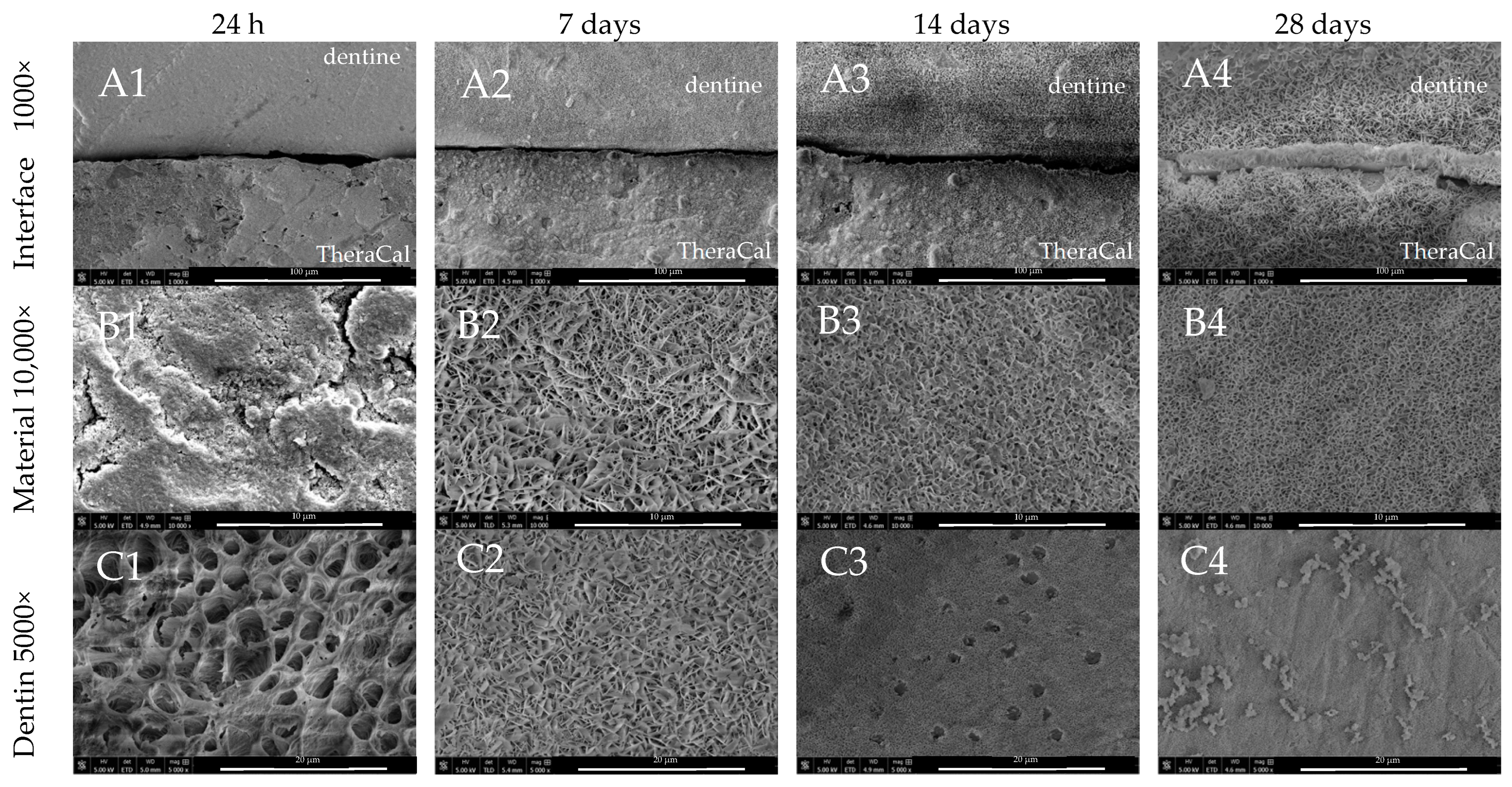
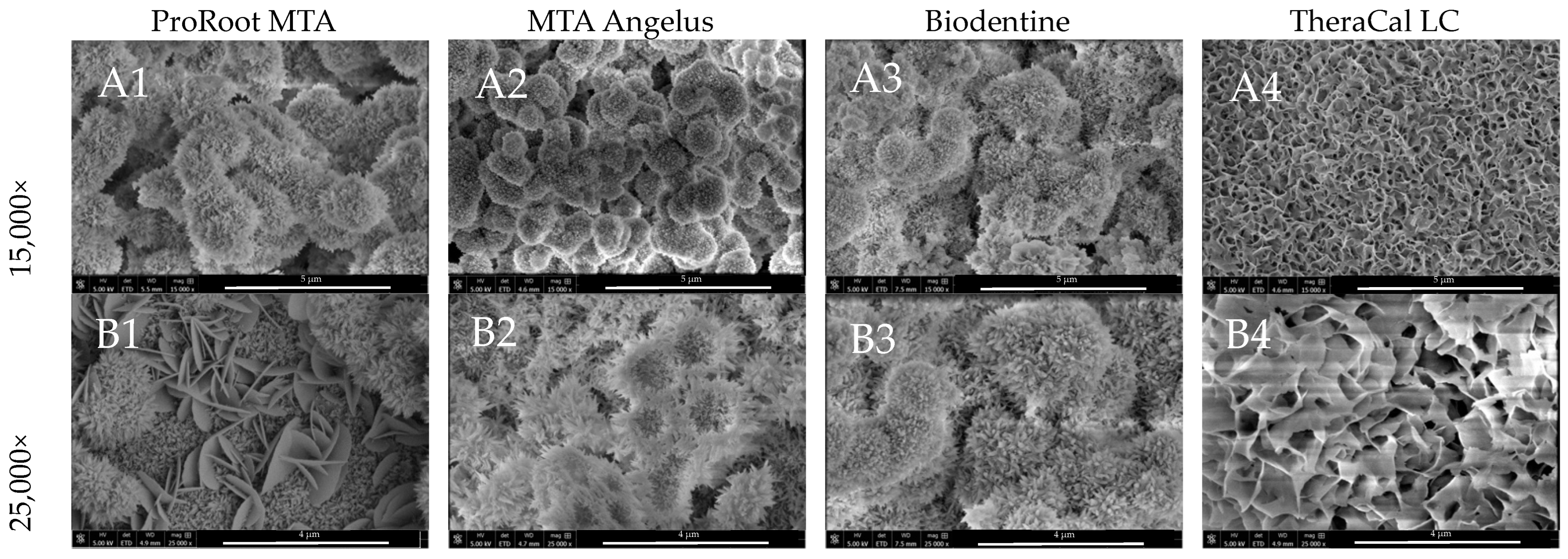
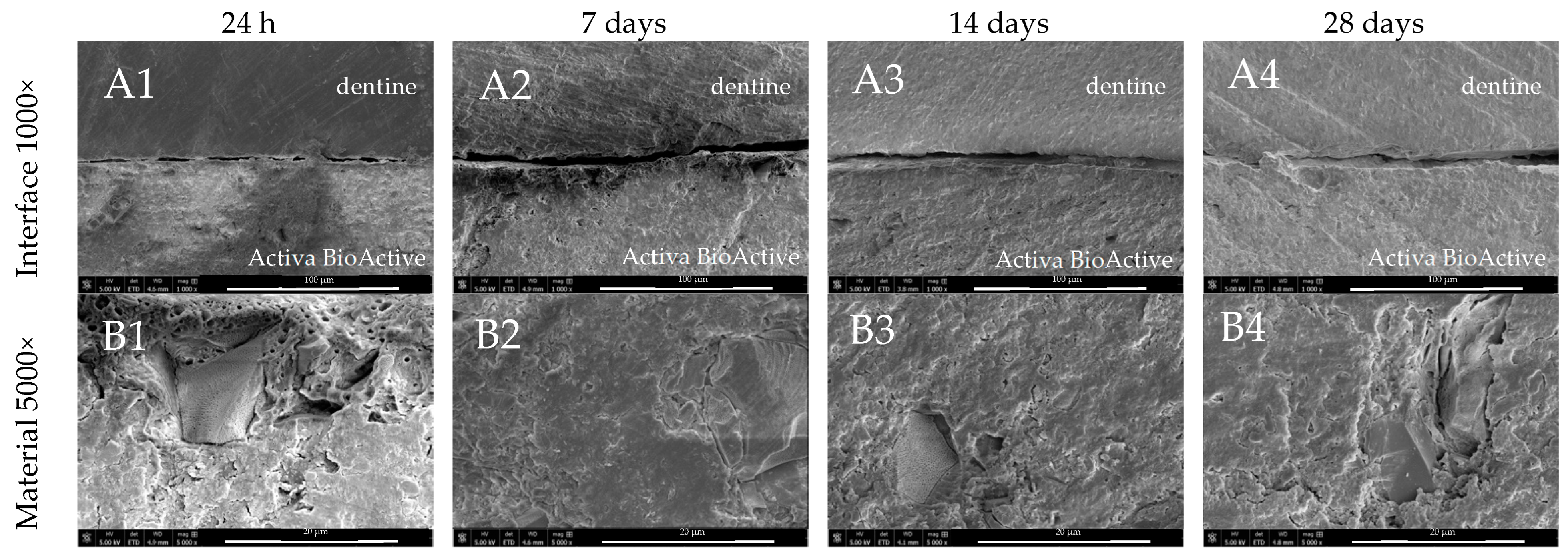
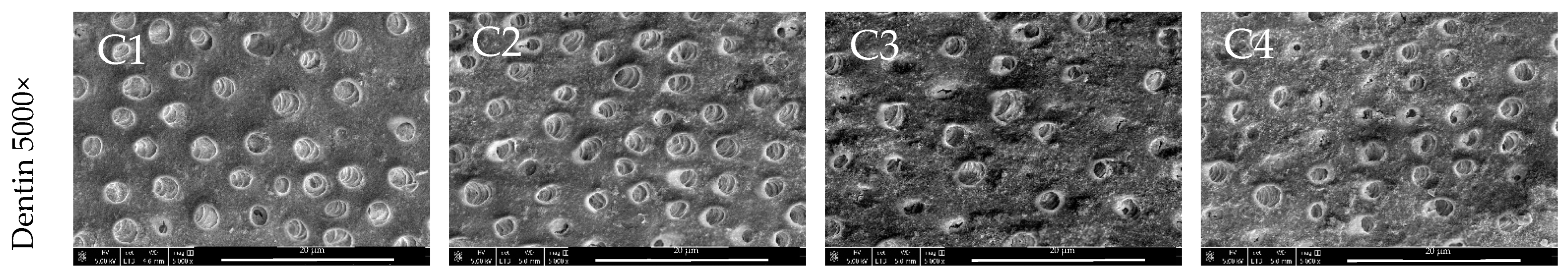
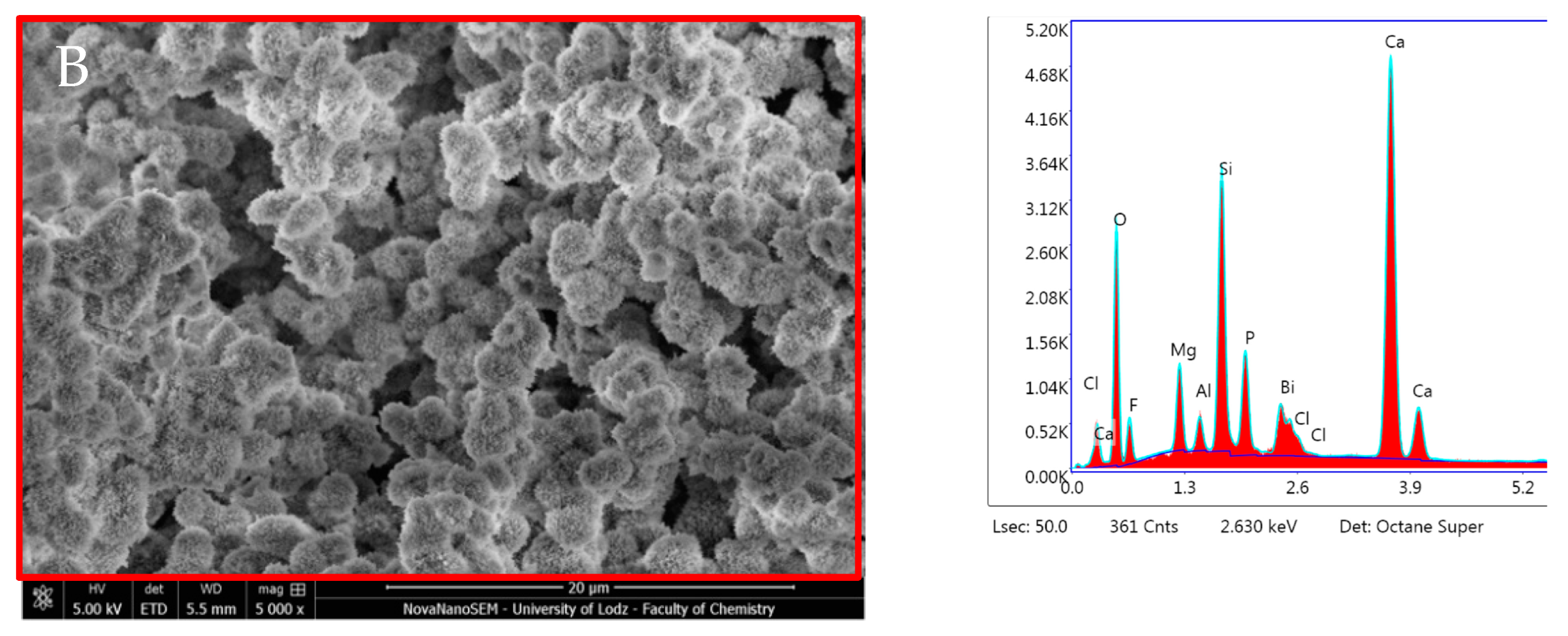
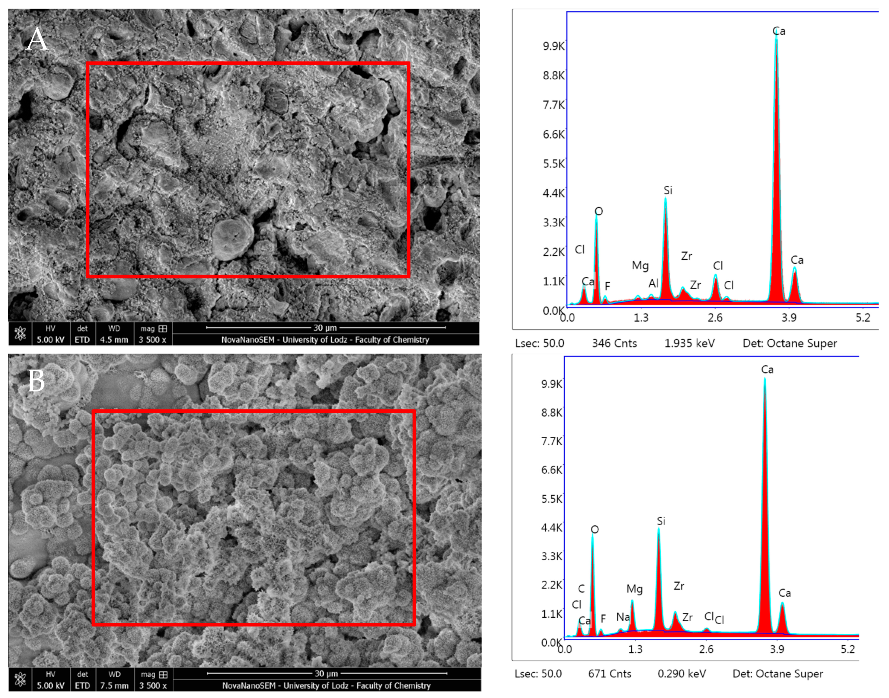
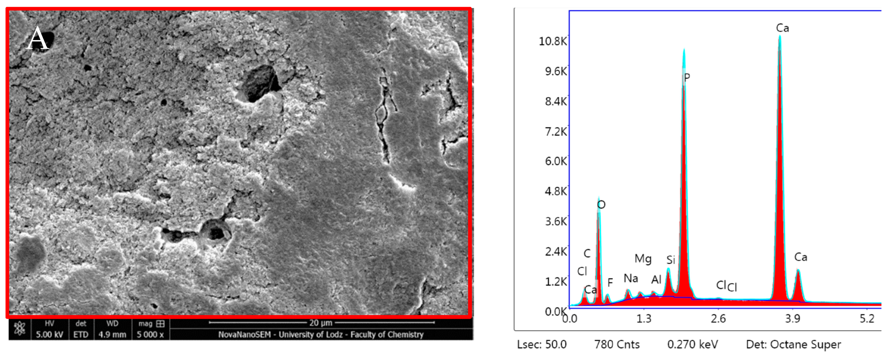
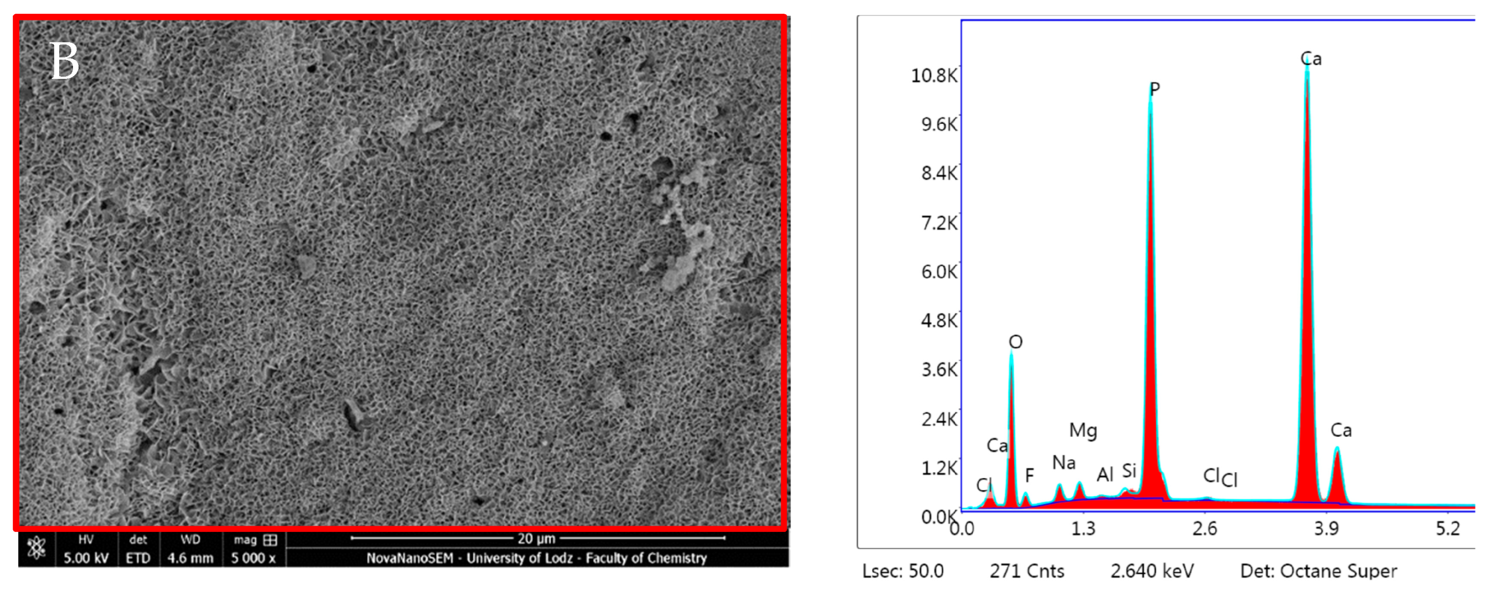
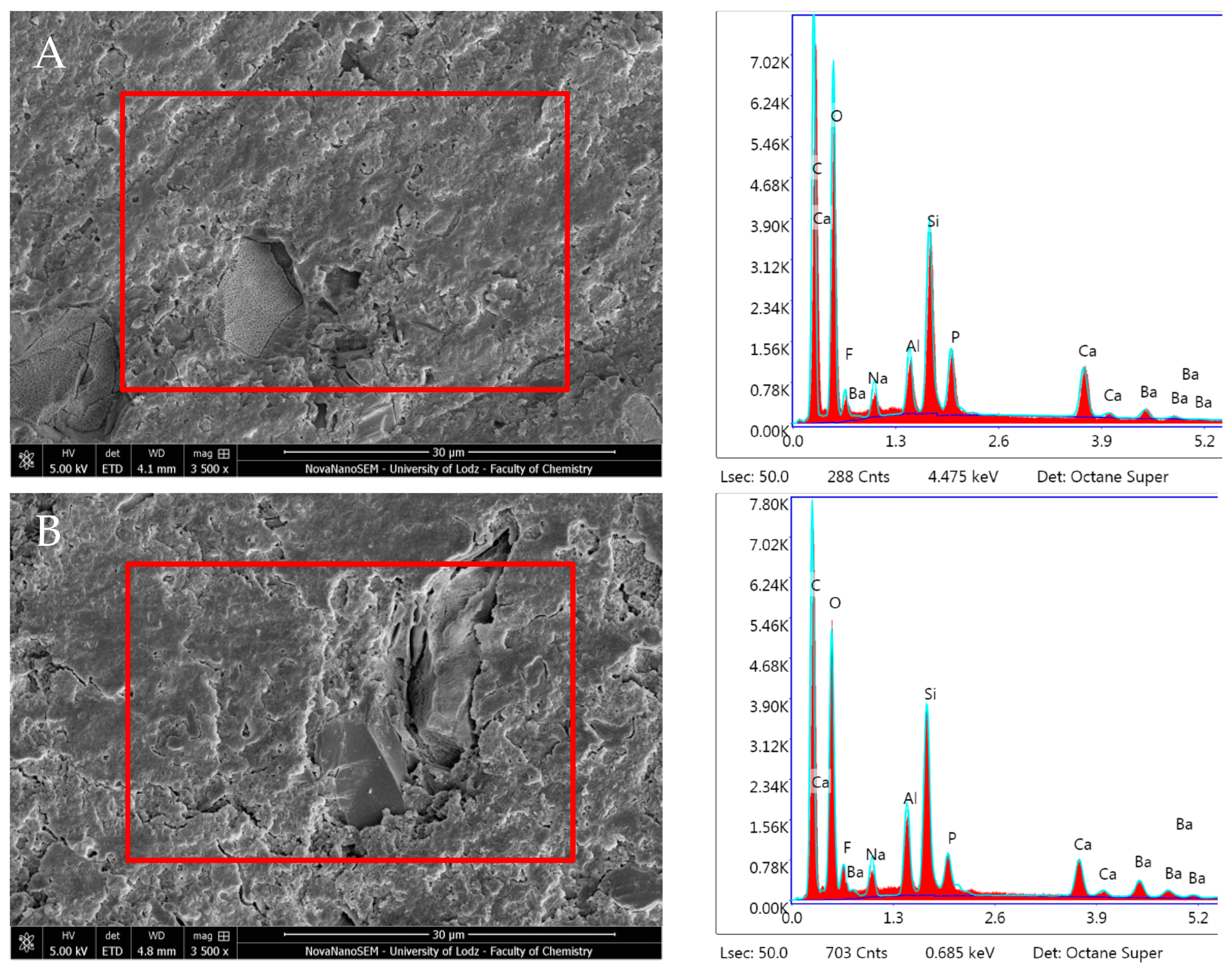
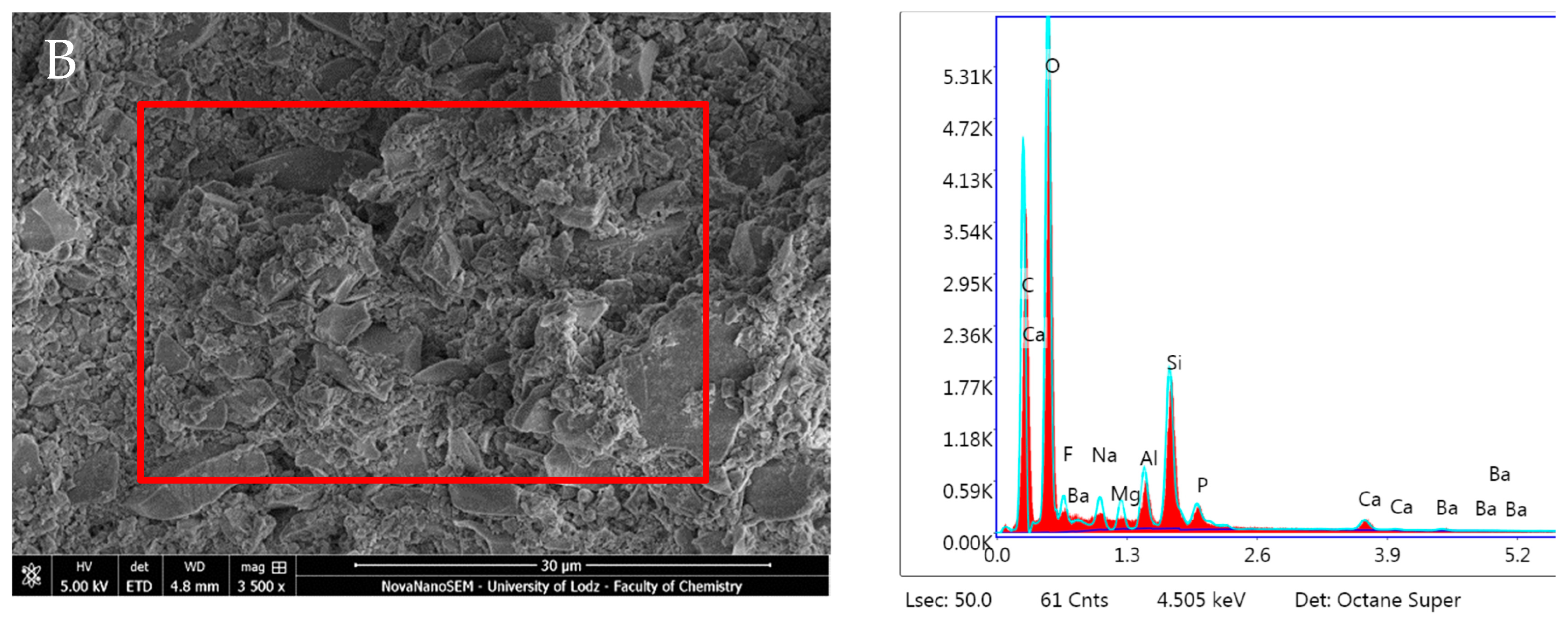
3.1. SEM Imaging
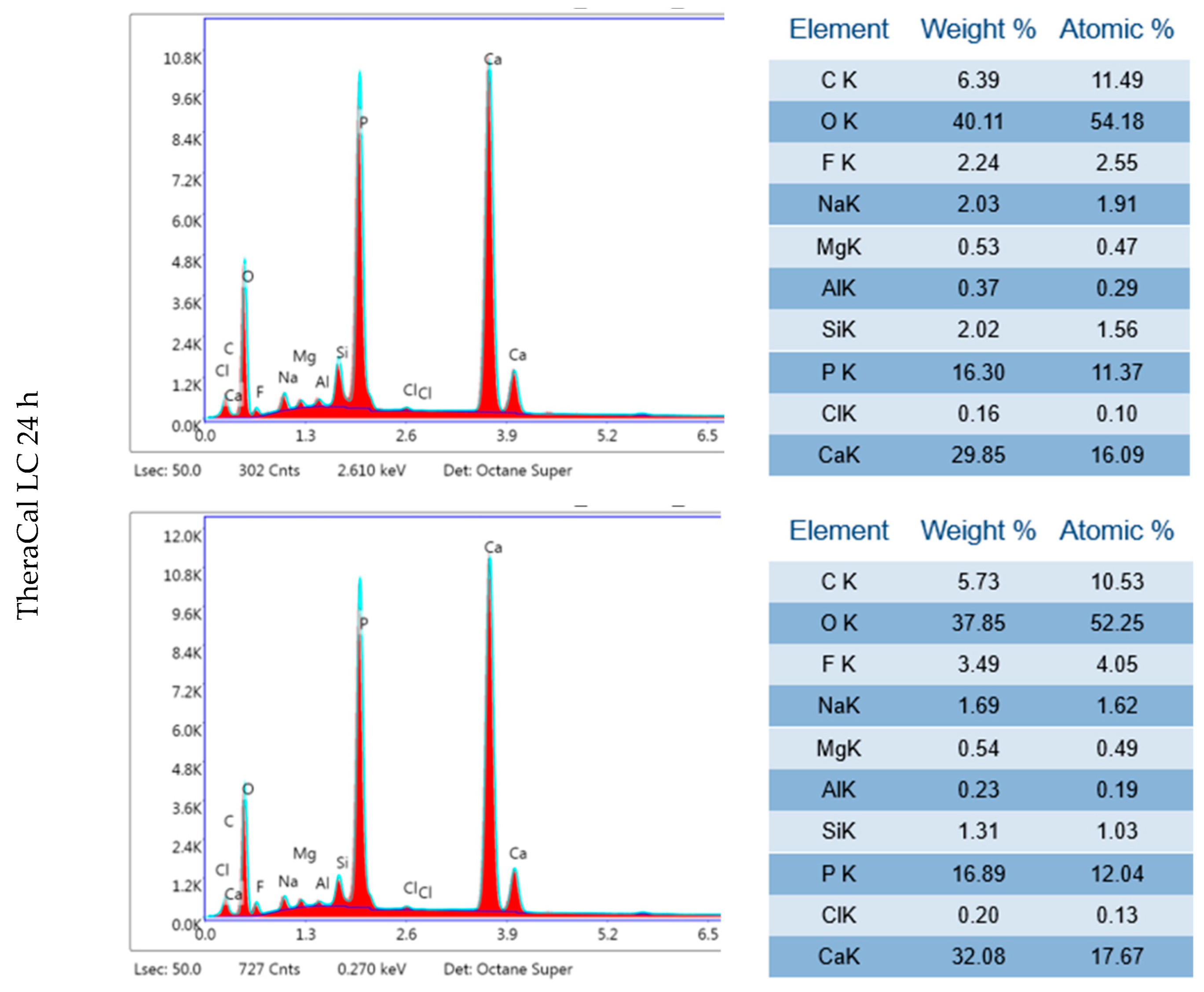
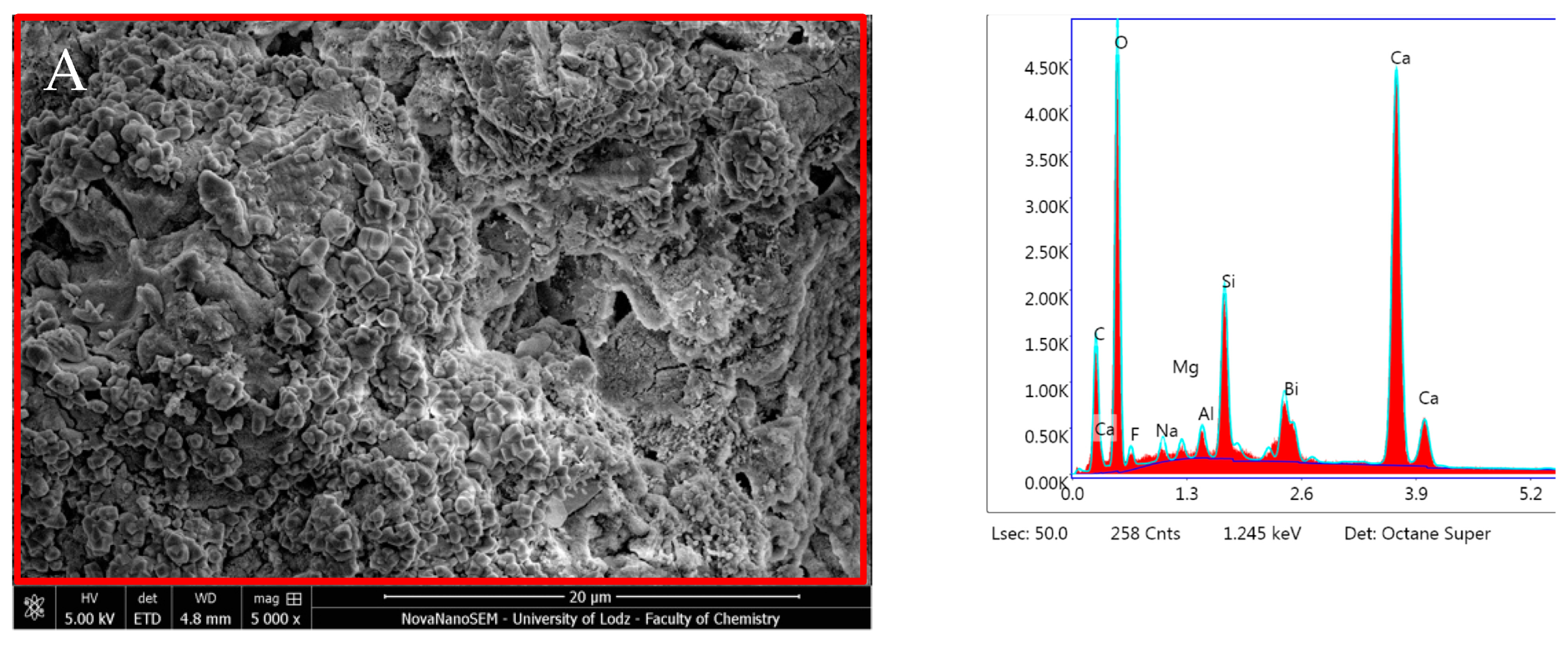
3.2. EDX Analysis
4. Discussion
5. Conclusions
- ProRoot MTA, MTA Angelus, Biodentine, and TheraCal LC showed significant surface precipitation. Consequently, these materials also formed an interfacial layer between the material and the dentin, filling the gap with precipitates, and demonstrated a higher concentration of Ca within the material. However, Biodentine exhibited the most evident sealing effect at the interfacial site.
- Thanks to an evident bioactivity, ProRoot MTA, MTA Angelus, Biodentine, and TheraCal LC may be suitable for remineralisation of caries dentine and pulp capping in vital pulp therapies.
- ACTIVA BioACTIVE, ACTIVA Presto, and Predicta Bulk exhibited inferior mineral precipitation compared to the CSMs so they should be used only for indirect pulp capping and/or restorative procedures.
Author Contributions
Funding
Data Availability Statement
Conflicts of Interest
Appendix A

| Group | Name | Composition | Application and Setting Mechanism |
|---|---|---|---|
| Control | Dentinal Discs | ||
| CSM | ProRoot MTA (Dentsply Sirona) (PR) | Powder: tricalcium silicate, dicalcium silicate, calcium dialuminate, calcium sulfate dehydrated calcium phosphate, calcium oxide, silica, bismuth oxide. Liquid: distilled water. | Left for setting for 4 h. |
| MTA Angelus (Angelus) (MA) | Powder: potassium oxide, aluminum oxide, sodium oxide, iron oxide, sulfur trioxide, calcium oxide, bismuth oxide, magnesium oxide, potassium sulfate, sodium sulfate, silica. Liquid: distilled water. | Left for setting for 15 min. | |
| Biodentine (Septodont) (BD) | Powder: tricalcium silicate, dicalcium silicate, calcium oxide, calcium carbonate, zirconium oxide, iron oxide. Liquid: calcium chloride, water-soluble polymer, water. | Left for setting for 12 min. | |
| LCSM | TheraCal LC (Bisco) (THC) | Liner single paste containing calcium oxide, calcium silicate particles (type III Portland cement), strontium glass, fumed silica, barium sulphate, barium zirconate and resin containing Bis-GMA and PEGDMA. | Light-cured for 20 s. |
| RMGIC | ACTIVA BioACTIVE Liner (Pulpdent) (AB) | Diurethane dimethacrylate, bis (2-(methacryloyloxy) ethyl) phosphate, barium glass, ionomer glass, polyacrylic acid/maleic acid copolymer, dual-cure chemistry, sodium fluoride, colorants Tooth-shade. | Self-cured for 20 s. Light-cured for 20 s. |
| BC | ACTIVA Presto (Pulpdent) (AP) | Blend of diurethane and other methacrylate resins, amorphous silica. | Etching Bonding agent—3M Single Bond Universal and light-cured for 10 s. BC light cured for 20 s/increment. |
| Predicta Bioactive Bulk (Parkell) (PB) | Poly(oxy-1,2-ethanediyl),.alpha.′-[(1- methylethylidene)di-4,1-phenylene]bis[.omega.-[(2- methyl-1-oxo-2-propenyl)oxy], 2-Propenoic acid, 2-methyl-, 1,6-hexanediyl ester, 2-Propenoic acid, 2-methyl-, (1- methylethylidene)bis[4,1-phenyleneoxy(2- hydroxy-3,1-propanediyl)] ester, 2,6-Di-tert-butyl-4-methylphenol, Dibenzoyl peroxide, 2-Propenoic acid, 2-methyl-, 1,6-hexanediyl ester | Etching Bonding agent—3M Single Bond Universal and light-cured for 10 s. Self-cured for 30 s. Light-cured for 20 s. PB self-cured for 5 min. |
Appendix B

References
- Duncan, H.F.; Galler, K.M.; Tomson, P.L.; Simon, S.; El-Karim, I.; Kundzina, R.; Krastl, G.; Dammaschke, T.; Fransson, H.; Markvart, M.; et al. European Society of Endodontology position statement: Management of deep caries and the exposed pulp. Int. Endod. J. 2019, 52, 923–934. [Google Scholar] [CrossRef] [PubMed]
- Innes, N.P.T.; Frencken, J.E.; Bjørndal, L.; Maltz, M.; Manton, D.J.; Ricketts, D.; Van Landuyt, K.; Banerjee, A.; Campus, G.; Doméjean, S.; et al. Managing Carious Lesions: Consensus Recommendations on Terminology. Adv. Dent. Res. 2016, 28, 49–57. [Google Scholar] [CrossRef] [PubMed]
- Maciel Pires, P.; Ionescu, A.C.; Pérez-Gracia, M.T.; Vezzoli, E.; Soares, I.P.M.; Brambilla, E.; de Almeida Neves, A.; Sauro, S. Assessment of the remineralisation induced by contemporary ion-releasing materials in mineral-depleted dentine. Clin. Oral Investig. 2022, 26, 6195–6207. [Google Scholar] [CrossRef] [PubMed]
- Islam, R.; Islam, M.R.R.; Tanaka, T.; Alam, M.K.; Ahmed, H.M.A.; Sano, H. Direct pulp capping procedures—Evidence and practice. Jpn. Dent. Sci. Rev. 2023, 59, 48–61. [Google Scholar] [CrossRef] [PubMed]
- Tan, S.Y.; Yu, V.S.H.; Lim, K.C.; Tan, B.C.K.; Neo, C.L.J.; Shen, L.; Messer, H.H. Long-term Pulpal and Restorative Outcomes of Pulpotomy in Mature Permanent Teeth. J. Endod. 2020, 46, 383–390. [Google Scholar] [CrossRef] [PubMed]
- Giannini, M.; Sauro, S. “Bioactivity” in Restorative Dentistry: Standing for the Use of Innovative Materials to Improve the Longevity of Restorations in Routine Dental Practice. J. Adhes. Dent. 2021, 23, 176–178. [Google Scholar] [CrossRef] [PubMed]
- Hench, L.L.; Paschall, H.A. Direct chemical bond of bioactive glass-ceramic materials to bone and muscle. J. Biomed. Mater. Res. 1973, 7, 25–42. [Google Scholar] [CrossRef] [PubMed]
- Jefferies, S.R. Bioactive and Biomimetic Restorative Materials: A Comprehensive Review. Part I. J. Esthet. Restor. Dent. 2014, 26, 14–26. [Google Scholar] [CrossRef]
- Cao, C.Y.; Mei, M.L.; Li, Q.; Chin, E.; Lo, M.; Chu, C.H. Methods for Biomimetic Remineralization of Human Dentine: A Systematic Review. Int. J. Mol. Sci. 2015, 16, 4615–4627. [Google Scholar] [CrossRef]
- Pires, P.M.; Rosa, T.d.C.; Ribeiro-Lages, M.B.; Duarte, M.L.; Cople Maia, L.; Neves, A.d.A.; Sauro, S. Bioactive Restorative Materials Applied over Coronal Dentine—A Bibliometric and Critical Review. Bioengineering 2023, 10, 731. [Google Scholar] [CrossRef]
- Prati, C.; Gandolfi, M.G. Calcium silicate bioactive cements: Biological perspectives and clinical applications. Dent. Mater. 2015, 31, 351–370. [Google Scholar] [CrossRef] [PubMed]
- Mickenautsch, S.; Yengopal, V.; Banerjee, A. Pulp response to resin-modified glass ionomer and calcium hydroxide cements in deep cavities: A quantitative systematic review. Dent. Mater. 2010, 26, 761–770. [Google Scholar] [CrossRef]
- Paula, A.; Laranjo, M.; Marto, C.M.; Abrantes, A.M.; Casalta-Lopes, J.; Gonçalves, A.C.; Sarmento-Ribeiro, A.B.; Ferreira, M.M.; Botelho, M.F.; Carrilho, E. BiodentineTM boosts, WhiteProRoot®MTA increases and Life® suppresses odontoblast activity. Materials 2019, 12, 1184. [Google Scholar] [CrossRef]
- Birant, S.; Gokalp, M.; Duran, Y.; Koruyucu, M.; Akkoc, T.; Seymen, F. Cytotoxicity of NeoMTA Plus, ProRoot MTA and Biodentine on human dental pulp stem cells. J. Dent. Sci. 2021, 16, 971–979. [Google Scholar] [CrossRef]
- Che, J.-L.; Kim, J.-H.; Kim, S.-M.; Choi, N.; Moon, H.-J.; Hwang, M.-J.; Song, H.-J.; Park, Y.-J. Comparison of Setting Time, Compressive Strength, Solubility, and pH of Four Kinds of MTA. Korean J. Dent. Mater. 2016, 43, 61–72. [Google Scholar] [CrossRef]
- Vivan, R.R.; Zapata, R.O.; Zeferino, M.A.; Bramante, C.M.; Bernardineli, N.; Garcia, R.B.; Hungaro Duarte, M.A.; Tanomaru Filho, M.; Gomes De Moraes, I. Evaluation of the physical and chemical properties of two commercial and three experimental root-end filling materials. Oral Surg. Oral Med. Oral Pathol. Oral Radiol. Endodontol. 2010, 110, 250–256. [Google Scholar] [CrossRef] [PubMed]
- Pornamazeh, T.; Yadegari, Z.; Ghasemi, A.; Sheykh-al-Eslamian, S.M.; Shojaeian, S.H. In Vitro Cytotoxicity and Setting Time Assessment of Calcium-Enriched Mixture Cement, Retro Mineral Trioxide Aggregate and Mineral Trioxide Aggregate. Iran. Endod. J. 2017, 12, 488–492. [Google Scholar] [CrossRef] [PubMed]
- Camilleri, J.; Damidot, D. Investigation of the hydration and bioactivity of radiopacified tricalcium silicate cement, Biodentine and MTA Angelus. Dent. Mater. 2013, 9, 580–593. [Google Scholar] [CrossRef]
- Sarkar, N.K.; Caicedo, R.; Ritwik, P.; Moiseyeva, R.; Kawashima, I. Physicochemical Basis of the Biologic Properties of Mineral Trioxide Aggregate. J. Endod. 2005, 31, 97–100. [Google Scholar] [CrossRef]
- Han, L.; Okiji, T.; Okawa, S. Morphological and chemical analysis of different precipitates on mineral trioxide aggregate immersed in different fluids. Dent. Mater. J. 2010, 29, 512–517. [Google Scholar] [CrossRef]
- Palma, P.J.; Marques, J.A.; Falacho, R.I.; Vinagre, A.; Santos, J.M.; Ramos, J.C. Does delayed restoration improve shear bond strength of different restorative protocols to calcium silicate-based cements? Materials 2018, 11, 2216. [Google Scholar] [CrossRef] [PubMed]
- Grech, L.; Mallia, B.; Camilleri, J. Investigation of the physical properties of tricalcium silicate cement-based root-end filling materials. Dent. Mater. 2013, 29, e20–e28. [Google Scholar] [CrossRef] [PubMed]
- Ha, H.-T. The effect of the maturation time of calcium silicate-based cement (BiodentineTM) on resin bonding: An in vitro study. Appl. Adhes. Sci. 2019, 7, 1. [Google Scholar] [CrossRef]
- Hashem, D.F.; Foxton, R.; Manoharan, A.; Watson, T.F.; Banerjee, A. The physical characteristics of resin composite–calcium silicate interface as part of a layered/laminate adhesive restoration. Dent. Mater. 2014, 30, 343–349. [Google Scholar] [CrossRef]
- Kim, J.; Song, Y.-S.; Min, K.-S.; Kim, S.-H.; Koh, J.-T.; Lee, B.-N.; Chang, H.-S.; Hwang, I.-N.; Oh, W.-M.; Hwang, Y.-C. Evaluation of reparative dentin formation of ProRoot MTA, Biodentine and BioAggregate using micro-CT and immunohistochemistry. Restor. Dent. Endod. 2016, 41, 29–36. [Google Scholar] [CrossRef]
- Mahmoud, S.; El-Negoly, S.; Zaen El-Din, A.; El-Zekrid, M.; Grawish, L.; Grawish, H.; Grawish, M. Biodentine versus mineral trioxide aggregate as a direct pulp capping material for human mature permanent teeth—A systematic review. J. Conserv. Dent. 2018, 21, 466–473. [Google Scholar] [CrossRef]
- Kunert, M.; Rozpedek-Kaminska, W.; Galita, G.; Sauro, S.; Bourgi, R.; Hardan, L.; Majsterek, I.; Lukomska-Szymanska, M. The Cytotoxicity and Genotoxicity of Bioactive Dental Materials. Cells 2022, 11, 3238. [Google Scholar] [CrossRef]
- Kunert, M.; Lukomska-Szymanska, M. Bio-Inductive Materials in Direct and Indirect Pulp Capping—A Review Article. Materials 2020, 13, 1204. [Google Scholar] [CrossRef]
- Hardan, L.; Mancino, D.; Bourgi, R.; Alvarado-Orozco, A.; Rodríguez-Vilchis, L.E.; Flores-Ledesma, A.; Cuevas-Suárez, C.E.; Lukomska-Szymanska, M.; Eid, A.; Danhache, M.-L.; et al. Bond Strength of Adhesive Systems to Calcium Silicate-Based Materials: A Systematic Review and Meta-Analysis of In Vitro Studies. Gels 2022, 8, 311. [Google Scholar] [CrossRef]
- Deepa, V.; Dhamaraju, B.; Bollu, I.; Balaji, T. Shear bond strength evaluation of resin composite bonded to three different liners: TheraCal LC, Biodentine, and resin-modified glass ionomer cement using universal adhesive: An in vitro study. J. Conserv. Dent. 2016, 19, 166–170. [Google Scholar] [CrossRef] [PubMed]
- Kamal, E.; Nabih, S.; Obeid, R.; Abdelhameed, M. The reparative capacity of different bioactive dental materials for direct pulp capping. Dent. Med. Probl. 2018, 55, 147–152. [Google Scholar] [CrossRef] [PubMed]
- Bakhtiar, H.; Nekoofar, M.H.; Aminishakib, P.; Abedi, F.; Naghi Moosavi, F.; Esnaashari, E.; Azizi, A.; Esmailian, S.; Ellini, M.R.; Mesgarzadeh, V.; et al. Human Pulp Responses to Partial Pulpotomy Treatment with TheraCal as Compared with Biodentine and ProRoot MTA: A Clinical Trial. J. Endod. 2017, 43, 1786–1791. [Google Scholar] [CrossRef]
- Tabari, K.; Rahbar, M.; Safyari, L.; Safarvand, H. Comparison of compressive strength and setting time of four experimental nanohybrid mineral trioxide aggregates and angelus mineral trioxide aggregate. World J. Dent. 2017, 8, 386–392. [Google Scholar] [CrossRef]
- Mosallam, S.M.; Abdel-Gawad, R.; El-Shehaby, F.; Elchaghaby, M. Evaluation of Remineralization Potential of ACTIVA Bioactive Restorative Material Versus Resin Modified Glass Ionomer in Restoration of Premolars: In Vitro Study. Acta Sci. Dent. Sci. 2021, 5, 2581–4893. [Google Scholar] [CrossRef]
- Garoushi, S.; Vallittu, P.K.; Lassila, L. Characterization of fluoride releasing restorative dental materials. Dent. Mater. J. 2018, 37, 293–300. [Google Scholar] [CrossRef]
- Safety Data Sheet, Predicta ™. Flow Dual Cure Bulk-fill Composite; Parkell Inc.: Brentwood, NY, USA, 2018; Volume 77, pp. 1–25. [Google Scholar]
- Suprastiwi, E.; Putranto, A.W.; Maharti, I.D. The ability of biodentineTM of guided tissue remineralization (GTR): Analysis using SEM, EDX and TEM. Pesqui. Bras. Odontopediatria Clin. Integr. 2019, 19, 1–8. [Google Scholar] [CrossRef]
- Gandolfi, M.G.; Taddei, P.; Tinti, A.; De Dorigo, E.S.; Rossi, P.L.; Prati, C. Kinetics of apatite formation on a calcium-silicate cement for root-end filling during ageing in physiological-like phosphate solutions. Clin. Oral Investig. 2010, 14, 659–668. [Google Scholar] [CrossRef]
- Ashofteh Yazdi, K.; Ghabraei, S.; Bolhari, B.; Kafili, M.; Meraji, N.; Nekoofar, M.H.; Dummer, P.M.H. Microstructure and chemical analysis of four calcium silicate-based cements in different environmental conditions. Clin. Oral Investig. 2019, 23, 43–52. [Google Scholar] [CrossRef]
- Ghilotti, J.; Sanz, J.L.; López-García, S.; Guerrero-Gironés, J.; Pecci-Lloret, M.P.; Lozano, A.; Llena, C.; Rodríguez-Lozano, F.J.; Forner, L.; Spagnuolo, G. Comparative surface morphology, chemical composition, and cytocompatibility of Bio-C repair, biodentine, and proroot MTA on hDPCs. Materials 2020, 13, 2189. [Google Scholar] [CrossRef]
- Gandolfi, M.G.; Taddei, P.; Tinti, A.; Prati, C. Apatite-forming ability (bioactivity) of ProRoot MTA. Int. Endod. J. 2010, 43, 917–929. [Google Scholar] [CrossRef]
- Kaur, M.; Singh, H.; Dhillon, J.S.; Batra, M.; Saini, M. MTA versus Biodentine: Review of Literature with a Comparative Analysis. J. Clin. Diagn. Res. 2017, 11, ZG01–ZG05. [Google Scholar] [CrossRef]
- Gandolfi, M.G.; Ciapetti, G.; Taddei, P.; Perut, F.; Tinti, A.; Cardoso, M.V.; Van Meerbeek, B.; Prati, C. Apatite formation on bioactive calcium-silicate cements for dentistry affects surface topography and human marrow stromal cells proliferation. Dent. Mater. 2010, 26, 974–992. [Google Scholar] [CrossRef]
- Martin, R.L.; Monticelli, F.; Brackett, W.W.; Loushine, R.J.; Rockman, R.A.; Ferrari, M.; Pashley, D.H.; Tay, F.R. Sealing properties of mineral trioxide aggregate orthograde apical plugs and root fillings in an in vitro apexification model. J. Endod. 2007, 33, 272–275. [Google Scholar] [CrossRef]
- García, A.J.; Ducheyne, P.; Boettiger, D. Effect of surface reaction stage on fibronectin-mediated adhesion of osteoblast-like cells to bioactive glass. J. Biomed. Mater. Res. 1998, 40, 48–56. Available online: https://onlinelibrary.wiley.com/doi/10.1002/(SICI)1097-4636(199804)40:1%3C48::AID-JBM6%3E3.0.CO;2-R (accessed on 16 November 2023). [CrossRef]
- Garoushi, S.; Vallittu, P.; Lassila, L. Development and characterization of ion-releasing fiber-reinforced flowable composite. Dent. Mater. 2022, 38, 1598–1609. [Google Scholar] [CrossRef] [PubMed]
- Akbulut, M.B.; Mutlu, Ş.N.; Soylu, M.A.; Şimşek, E. Interfacial characteristics of BIOfactor MTA and Biodentine with dentin. Microsc. Res. Tech. 2023, 86, 258–267. [Google Scholar] [CrossRef] [PubMed]
- Maciel Pires, P.; Dávila-Sánchez, A.; Faus-Matoses, V.; Nuñez Martí, J.M.; Lo Muzio, L.; Sauro, S. Bonding performance and ultramorphology of the resin-dentine interface of contemporary universal adhesives. Clin. Oral Investig. 2022, 26, 4391–4405. [Google Scholar] [CrossRef]
- About, I. Biodentine: From biochemical and bioactive properties to clinical applications. G. Ital. Endod. 2016, 30, 81–88. [Google Scholar] [CrossRef]
- Tomás-Catalá, C.J.; Collado-González, M.; García-Bernal, D.; Oñate-Sánchez, R.E.; Forner, L.; Llena, C.; Lozano, A.; Moraleda, J.M.; Rodríguez-Lozano, F.J. Biocompatibility of New Pulp-capping Materials NeoMTA Plus, MTA Repair HP, and Biodentine on Human Dental Pulp Stem Cells. J. Endod. 2018, 44, 126–132. [Google Scholar] [CrossRef] [PubMed]
- Tomás-Catalá, C.J.; Collado-González, M.; García-Bernal, D.; Oñate-Sánchez, R.E.; Forner, L.; Llena, C.; Lozano, A.; Castelo-Baz, P.; Moraleda, J.M.; Rodríguez-Lozano, F.J. Comparative analysis of the biological effects of the endodontic bioactive cements MTA-Angelus, MTA Repair HP and NeoMTA Plus on human dental pulp stem cells. Int. Endod. J. 2017, 50, e63–e72. [Google Scholar] [CrossRef] [PubMed]
- Aksoy, M.K.; Oz, F.T.; Orhan, K. Evaluation of calcium (Ca2+) and hydroxide (OH−) ion diffusion rates of indirect pulp capping materials. Int. J. Artif. Organs 2017, 40, 641–646. [Google Scholar] [CrossRef]
- Gandolfi, M.; Siboni, F.; Polimeni, A.; Bossù, M.; Riccitiello, F.; Rengo, S.; Prati, C. In Vitro Screening of the Apatite-Forming Ability, Biointeractivity and Physical Properties of a Tricalcium Silicate Material for Endodontics and Restorative Dentistry. Dent. J. 2013, 1, 41–60. [Google Scholar] [CrossRef]
- Poggio, C.; Lombardini, M.; Colombo, M.; Beltrami, R.; Rindi, S. Solubility and pH of direct pulp capping materials: A comparative study. J. Appl. Biomater. Funct. Mater. 2015, 13, e181–e185. [Google Scholar] [CrossRef] [PubMed]
- Kot, K.; Kucharski, Ł.; Marek, E.; Safranow, K.; Lipski, M. Alkalizing Properties of Six Calcium-Silicate Endodontic Biomaterials. Materials 2022, 15, 6482. [Google Scholar] [CrossRef] [PubMed]
- Montoya, C.; Roldan, L.; Yu, M.; Valliani, S.; Ta, C.; Yang, M.; Orrego, S. Smart dental materials for antimicrobial applications. Bioact. Mater. 2022, 24, 1–19. [Google Scholar] [CrossRef] [PubMed]
- Al-Sanabani, J.S.; Madfa, A.A.; Al-Sanabani, F.A. Application of Calcium Phosphate Materials in Dentistry. Int. J. Biomater. 2013, 2013, 876132. [Google Scholar] [CrossRef] [PubMed]
- Daneshpoor, N.; Pishevar, L. Comparative evaluation of bioactive cements on biomimetic remineralization of dentin. J. Clin. Exp. Dent. 2020, 12, e291. [Google Scholar] [CrossRef] [PubMed]
- Sajini, S.I.; Alshawi, B.A.; Alharbi, L.M. Assessment of remineralisation potentials of bioactive dental composite using an in-vitro demineralised dentine model. J. Taibah Univ. Med. Sci. 2022, 17, 640–647. [Google Scholar] [CrossRef] [PubMed]
- Yamamoto, S.; Han, L.; Noiri, Y.; Okiji, T. Evaluation of the Ca ion release, pH and surface apatite formation of a prototype tricalcium silicate cement. Int. Endod. J. 2017, 50 (Suppl. 2), e73–e82. [Google Scholar] [CrossRef]
- Mahmoud, O.; Al-Afifi, N.A.; Salihu Farook, M.; Ibrahim, M.A.; Al Shehadat, S.; Alsaegh, M.A. Morphological and Chemical Analysis of Different Types of Calcium Silicate-Based Cements. Int. J. Dent. 2022, 2022, 6480047. [Google Scholar] [CrossRef]
- Guimarães, B.M.; Prati, C.; Duarte, M.A.H.; Bramante, C.M.; Gandolfi, M.G. Physicochemical properties of calcium silicate-based formulations MTA repair HP and MTA vitalcem. J. Appl. Oral Sci. 2018, 26, e2017115. [Google Scholar] [CrossRef]
- De Caluwé, T.; Vercruysse, C.W.J.; Fraeyman, S.; Verbeeck, R.M.H. The influence of particle size and fluorine content of aluminosilicate glass on the glass ionomer cement properties. Dent. Mater. 2014, 30, 1029–1038. [Google Scholar] [CrossRef]
- Tian, K.V.; Chass, G.A.; Tommaso, D. Di Simulations reveal the role of composition into the atomic-level flexibility of bioactive glass cements. Phys. Chem. Chem. Phys. 2015, 18, 837–845. [Google Scholar] [CrossRef]
- Vouzara, T.; Roussou, K.; Nikolaidis, A.K.; Tolidis, K.; Koulaouzidou, E.A. Organic Eluates Derived from Intermediate Restorative Dental Materials. Molecules 2020, 25, 1593. [Google Scholar] [CrossRef]
- Tay, F.R.; Pashley, D.H.; Rueggeberg, F.A.; Loushine, R.J.; Weller, R.N. Calcium Phosphate Phase Transformation Produced by the Interaction of the Portland Cement Component of White Mineral Trioxide Aggregate with a Phosphate-containing Fluid. J. Endod. 2007, 33, 1347–1351. [Google Scholar] [CrossRef]
- Kokubo, T.; Takadama, H. How useful is SBF in predicting in vivo bone bioactivity? Biomaterials 2006, 27, 2907–2915. [Google Scholar] [CrossRef] [PubMed]
- Jevnikar, A.P.; Malgaj, T.; Radan, K.; Özden, I.; Kušter, M.; Kocjan, A. Rheological Properties and Setting Kinetics of Bioceramic Hydraulic Cements: ProRoot MTA versus RS+. Materials 2023, 16, 3174. [Google Scholar] [CrossRef] [PubMed]
- Gandolfi, M.G.; Taddei, P.; Siboni, F.; Modena, E.; Ciapetti, G.; Prati, C. Development of the foremost light-curable calcium-silicate MTA cement as root-end in oral surgery. Chemical-physical properties, bioactivity and biological behavior. Dent. Mater. 2011, 27, e134–e157. [Google Scholar] [CrossRef] [PubMed]
- Lipski, M.; Nowicka, A.; Kot, K.; Postek-Stefańska, L.; Wysoczańska-Jankowicz, I.; Borkowski, L.; Andersz, P.; Jarząbek, A.; Grocholewicz, K.; Sobolewska, E.; et al. Factors affecting the outcomes of direct pulp capping using Biodentine. Clin. Oral Investig. 2018, 22, 2021–2029. [Google Scholar] [CrossRef] [PubMed]


| ProRoot MTA | MTA Angelus | Biodentine | TheraCal | Activa BioActive Base Liner | Activa Presto | Predicta Bulk | ||||||||
|---|---|---|---|---|---|---|---|---|---|---|---|---|---|---|
| 24 h | 28 d | 24 h | 28 d | 24 h | 28 d | 24 h | 28 d | 24 h | 28 d | 24 h | 28 d | 24 h | 28 d | |
| C | 11.30 | 3.42 ↓ | 4.82 | 6.42 | 0.98 | 4.99 ↑ | 6.06 | 1.69 ↓ | 47.77 | 49.28 | 33.44 | 33.04 | 36.74 | 38.43 |
| O | 50.49 | 39.86 ↓ | 41.96 | 37.61 ↓ | 38.72 | 42.43 | 38.58 | 37.22 | 37.28 | 32.90 ↓ | 35.60 | 33.24 | 46.44 | 48.95 |
| F | 2.97 | 3.48 | 3.85 | 1.93 ↓ | 3.07 | 3.27 | 3.93 | 3.51 | 2.92 | 3.25 | 1.01 | 0.67 | 3.42 | 2.78 |
| Na | 2.18 | 0.30 ↓ | 0.40 | 1.83 ↑ | 0.56 | 0.86 | 1.68 | 1.78 | 1.37 | 1.67 | 1.27 | 1.58 | 1.58 | 1.22 |
| Mg | 1.09 | 1.80 | 0.13 | 1.06 ↑ | 0.23 | 3.09 ↑ | 0.47 | 0.92 | 0.30 | 0.46 | 0.43 | 0.34 | 0.69 | 0.88 |
| Al | 1.18 | 0.86 | 2.03 | 0.05 ↓ | 0.16 | 0.04 | 0.27 | 0.06 | 1.32 | 2.12 ↑ | 3.66 | 3.81 | 1.99 | 1.64 |
| Si | 5.57 | 7.98 ↑ | 11.81 | 1.23 ↓ | 7.21 | 7.08 | 1.57 | 0.22 | 3.87 | 4.28 | 14.52 | 15.63 | 4.81 | 3.87 |
| Zr | 0.26 | 0.28 | 0.00 | 1.05 ↑ | 1.88 | 2.20 | 0.00 | 1.91 ↑ | 0.33 | 0.35 | 0.19 | 0.19 | 0.25 | 0.47 |
| P | 0.14 | 5.96 ↑ | 0.14 | 14.81 ↑ | 0.19 | 0.49 | 16.39 | 17.00 | 1.61 | 0.70 | 0.40 | 0.42 | 1.32 | 0.16 |
| Bi | 4.86 | 3.50 ↓ | 0.01 | 0.21 | 0.07 | 0.02 | 0.00 | 0.00 | 0.00 | 0.31 ↑ | 0.23 | 0.22 | 0.15 | 0.33 |
| Cl | 0.14 | 0.66 | 0.00 | 0.16 | 2.14 | 0.36 ↓ | 0.09 | 0.09 | 0.01 | 0.10 | 0.22 | 0.47 | 0.05 | 0.10 |
| Ca | 19.70 | 31.78 ↑ | 34.64 | 33.51 ↓ | 44.73 | 35.09 ↓ | 30.78 | 35.48 ↑ | 2.14 | 1.72 | 0.55 | 0.58 | 1.58 | 0.62 |
| Ba | 0.12 | 0.11 | 0.20 | 0.13 | 0.07 | 0.07 | 0.19 | 0.12 | 1.36 | 2.86 ↑ | 8.49 | 9.83 ↑ | 0.98 | 0.54 |
| Ca/P ratio | 140.71 | 5.33 ↓ | 247.43 | 2.26 ↓ | 235.42 | 71.61 ↓ | 1.87 | 2.08 | 3.50 | 2.45 | 1.36 | 1.38 | 1.20 | 3.38 |
| Control | ProRoot MTA | MTA Angelus | Biodentine | TheraCal LC | Activa BioActive Base Liner | Activa Presto | Predicta Bulk | |
|---|---|---|---|---|---|---|---|---|
| C | 8.69 | 5.01 | 2.34 | 5.87 | 5.49 | 8.32 | 1.82 | 4.03 |
| O | 45.32 | 39.07 | 37.81 | 39.64 | 38.45 | 51.00 | 45.61 | 39.93 |
| F | 3.18 | 2.27 | 1.60 | 2.52 | 3.76 | 5.02 | 4.21 | 2.79 |
| Na | 3.59 | 1.00 | 3.14 | 1.10 | 2.07 | 4.40 | 2.74 | 1.88 |
| Mg | 1.76 | 0.42 | 1.40 | 2.76 | 1.34 | 1.89 | 1.71 | 1.42 |
| Al | 0.56 | 0.08 | 0.01 | 0.00 | 0.27 | 0.82 | 0.51 | 0.33 |
| Si | 1.33 | 0.37 | 0.96 | 1.23 | 0.17 | 0.63 | 0.26 | 0.24 |
| Zr | 0.16 | 1.57 | 0.57 | 0.21 | 2.32 | 6.28 | 7.67 | 4.51 |
| P | 12.73 | 12.43 | 14.31 | 10.25 | 14.90 | 6.86 | 9.64 | 12.85 |
| Bi | 0.28 | 0.80 | 0.00 | 0.08 | 0.18 | 0.36 | 0.32 | 0.37 |
| Cl | 0.18 | 0.74 | 1.04 | 0.38 | 0.16 | 0.23 | 0.25 | 0.28 |
| Ca | 20.49 | 36.13 | 36.69 | 35.85 | 30.73 | 14.03 | 25.13 | 31.01 |
| Ba | 0.15 | 0.11 | 0.14 | 0.12 | 0.16 | 0.14 | 0.12 | 0.36 |
| Ca/P ratio | 1.61 | 2.91 | 2.56 | 3.50 | 2.06 | 2.04 | 2.60 | 2.41 |
Disclaimer/Publisher’s Note: The statements, opinions and data contained in all publications are solely those of the individual author(s) and contributor(s) and not of MDPI and/or the editor(s). MDPI and/or the editor(s) disclaim responsibility for any injury to people or property resulting from any ideas, methods, instructions or products referred to in the content. |
© 2024 by the authors. Licensee MDPI, Basel, Switzerland. This article is an open access article distributed under the terms and conditions of the Creative Commons Attribution (CC BY) license (https://creativecommons.org/licenses/by/4.0/).
Share and Cite
Kunert, M.; Piwonski, I.; Hardan, L.; Bourgi, R.; Sauro, S.; Inchingolo, F.; Lukomska-Szymanska, M. Dentine Remineralisation Induced by “Bioactive” Materials through Mineral Deposition: An In Vitro Study. Nanomaterials 2024, 14, 274. https://doi.org/10.3390/nano14030274
Kunert M, Piwonski I, Hardan L, Bourgi R, Sauro S, Inchingolo F, Lukomska-Szymanska M. Dentine Remineralisation Induced by “Bioactive” Materials through Mineral Deposition: An In Vitro Study. Nanomaterials. 2024; 14(3):274. https://doi.org/10.3390/nano14030274
Chicago/Turabian StyleKunert, Marta, Ireneusz Piwonski, Louis Hardan, Rim Bourgi, Salvatore Sauro, Francesco Inchingolo, and Monika Lukomska-Szymanska. 2024. "Dentine Remineralisation Induced by “Bioactive” Materials through Mineral Deposition: An In Vitro Study" Nanomaterials 14, no. 3: 274. https://doi.org/10.3390/nano14030274
APA StyleKunert, M., Piwonski, I., Hardan, L., Bourgi, R., Sauro, S., Inchingolo, F., & Lukomska-Szymanska, M. (2024). Dentine Remineralisation Induced by “Bioactive” Materials through Mineral Deposition: An In Vitro Study. Nanomaterials, 14(3), 274. https://doi.org/10.3390/nano14030274