
Enhanced Photoelectrochemical Performance Using Cobalt-Catalyst-Loaded PVD/RF-Engineered WO3 Photoelectrodes


Abstract
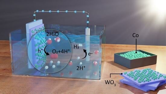
1. Introduction
2. Experimental Section
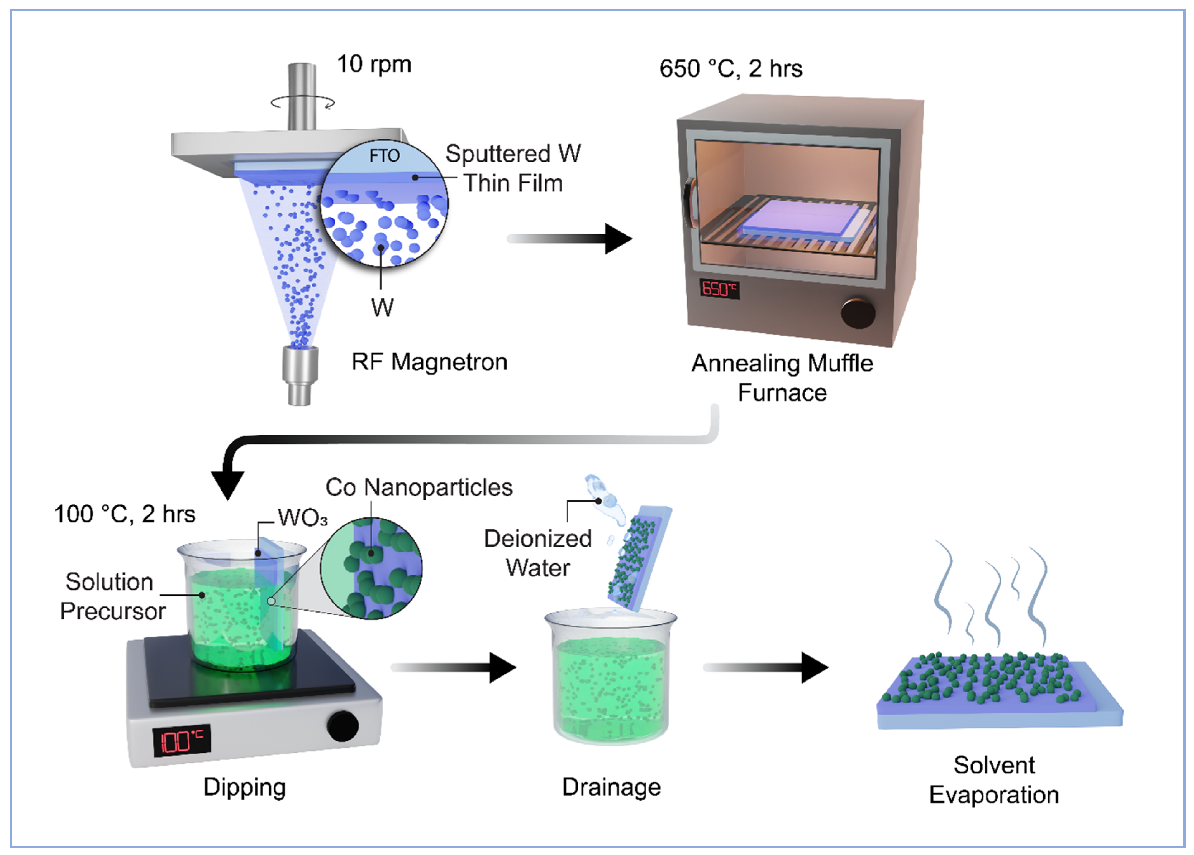
2.1. Fabrication of RF-Sputtered Nanostructured WO3 Thin Films
2.2. Synthesis of the WO3 Photoanode with Cobalt Nanoparticles
2.3. Characterisations
2.4. Electrochemical Measurements
3. Result and Discussion
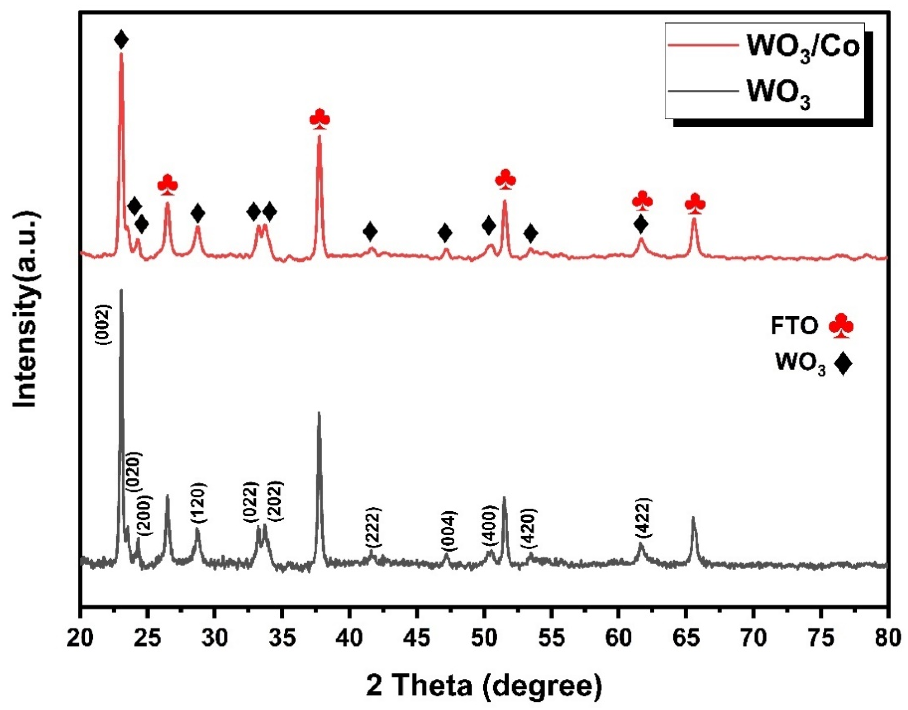
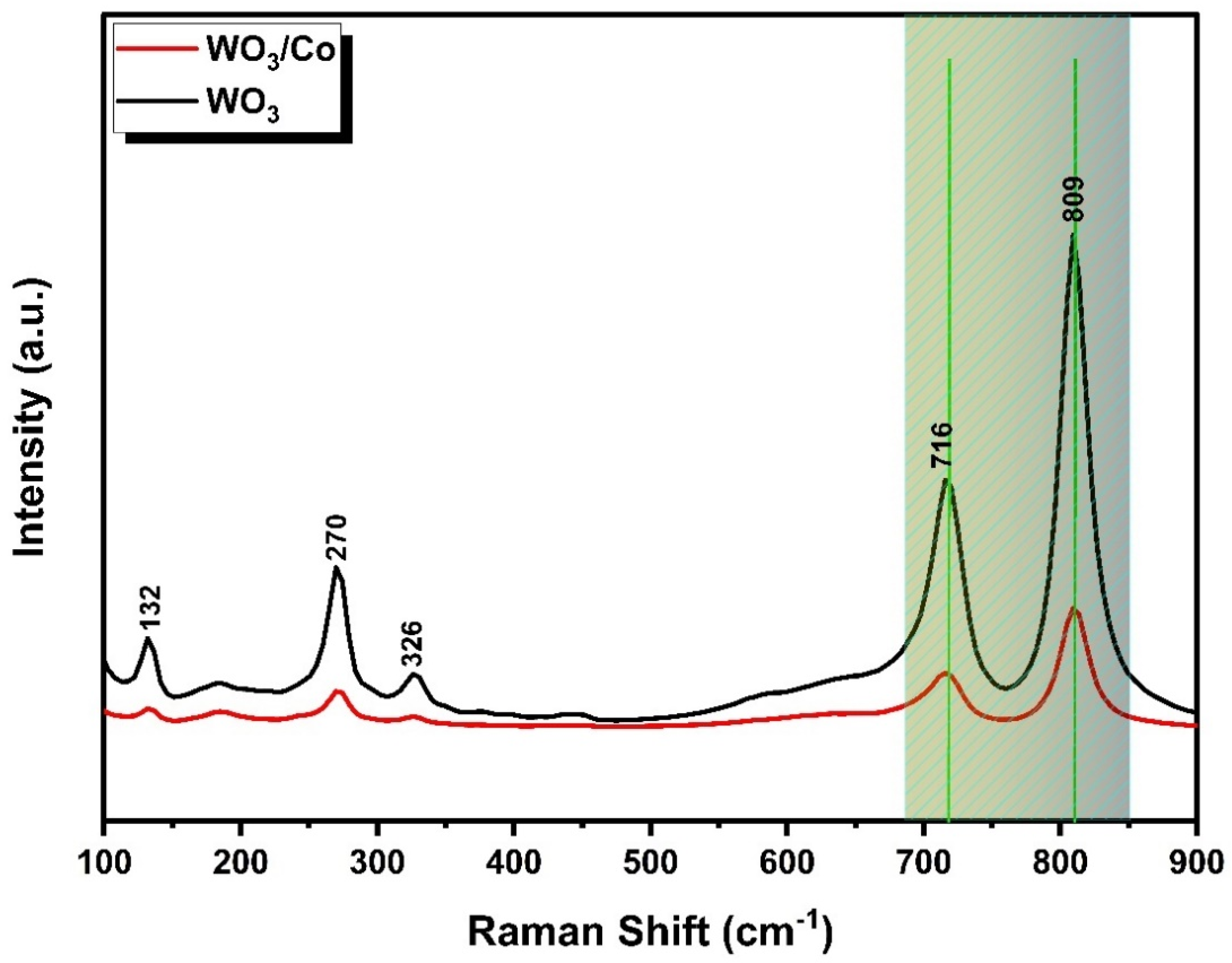
3.1. XRD Structural and Raman Characterisation
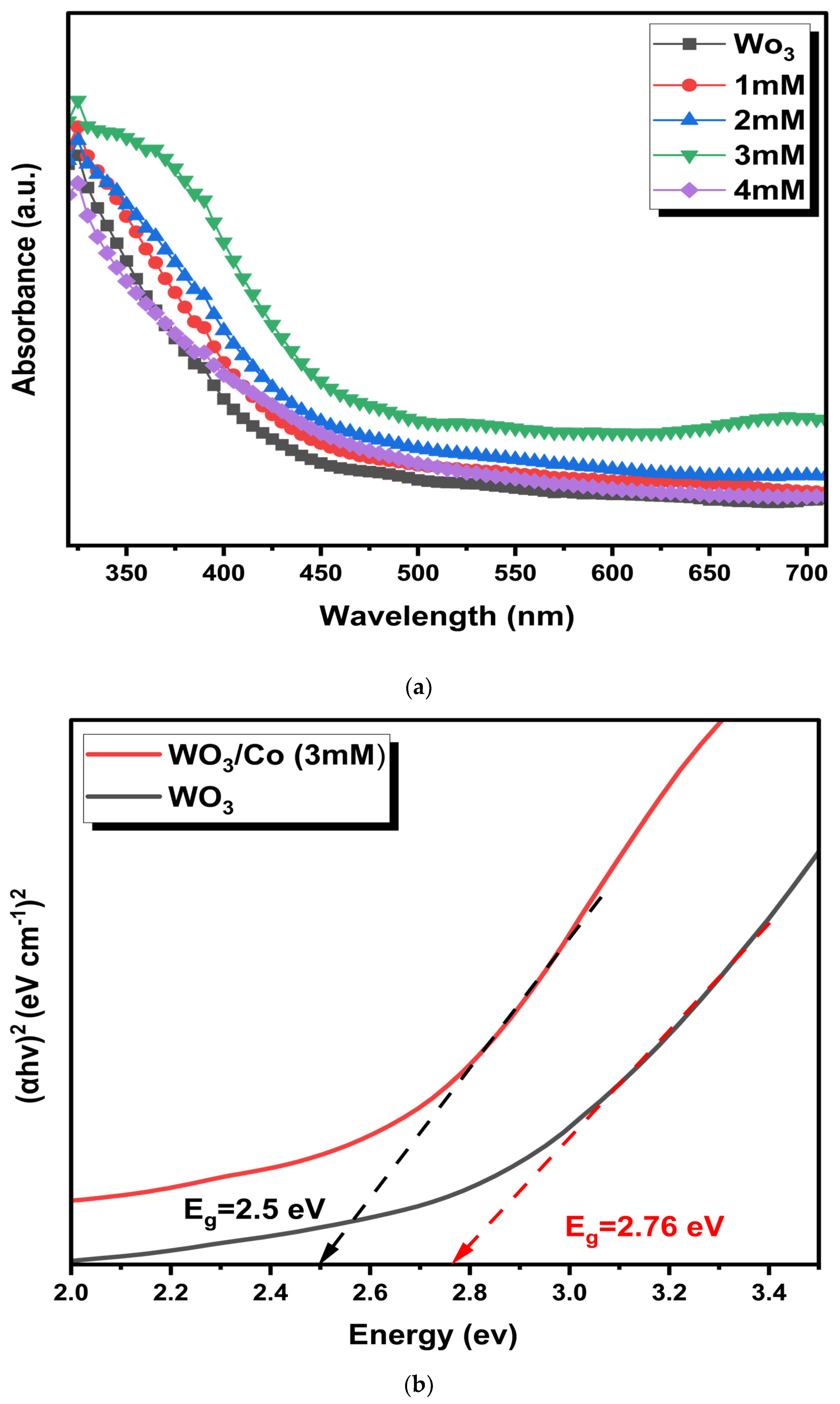
3.2. UV-DRS Spectroscopic Analysis
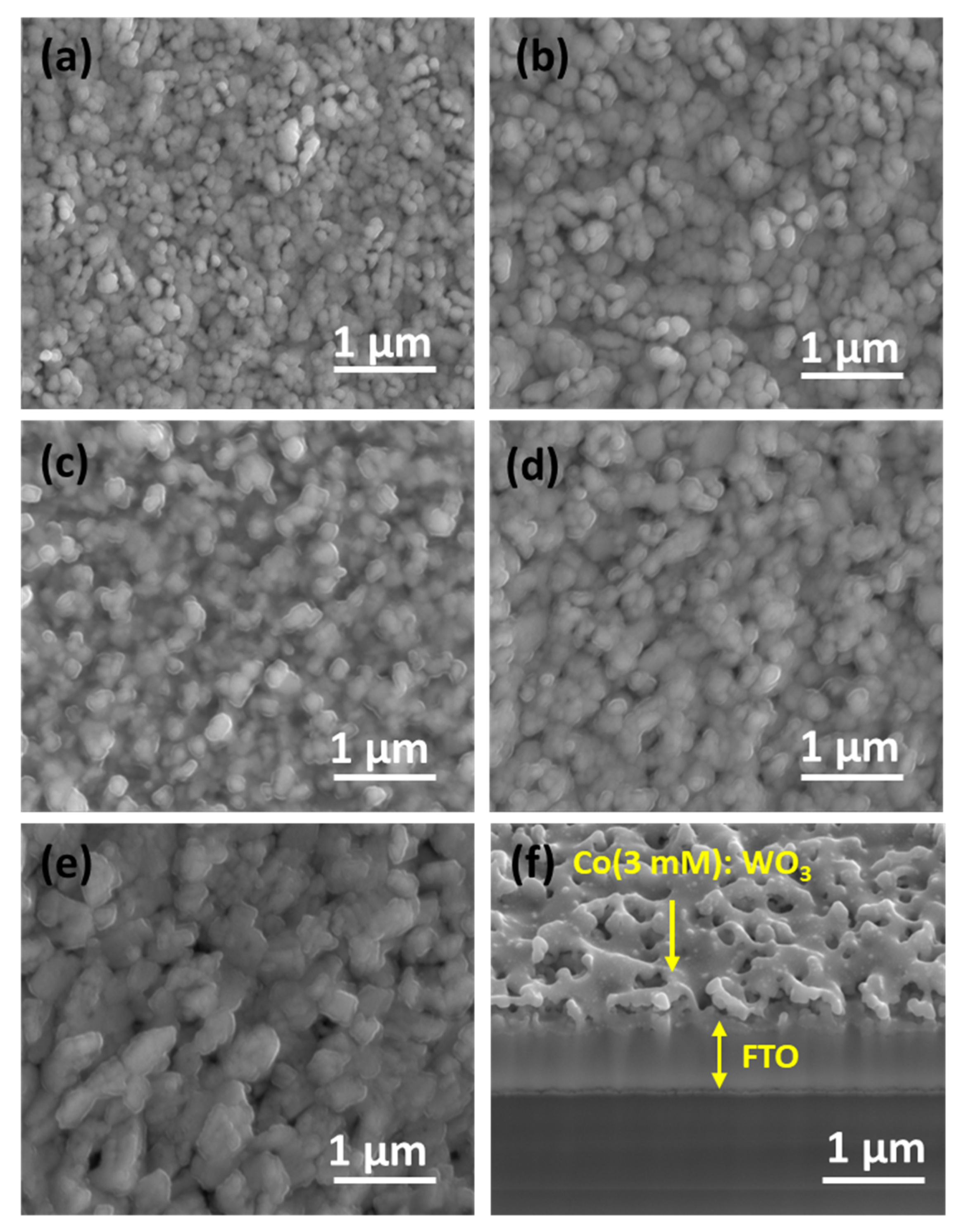
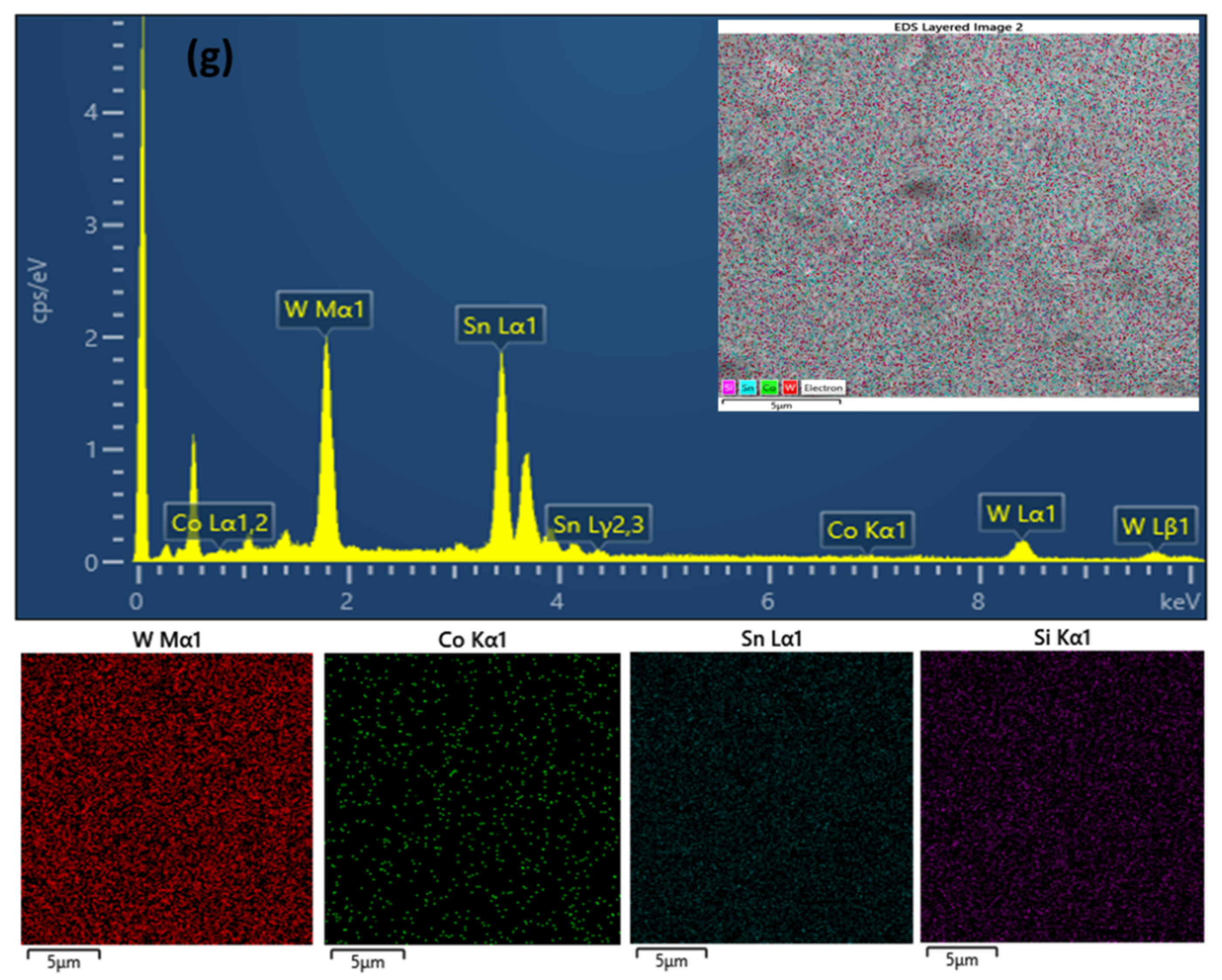
3.3. Surface Morphology and Elemental Analysis
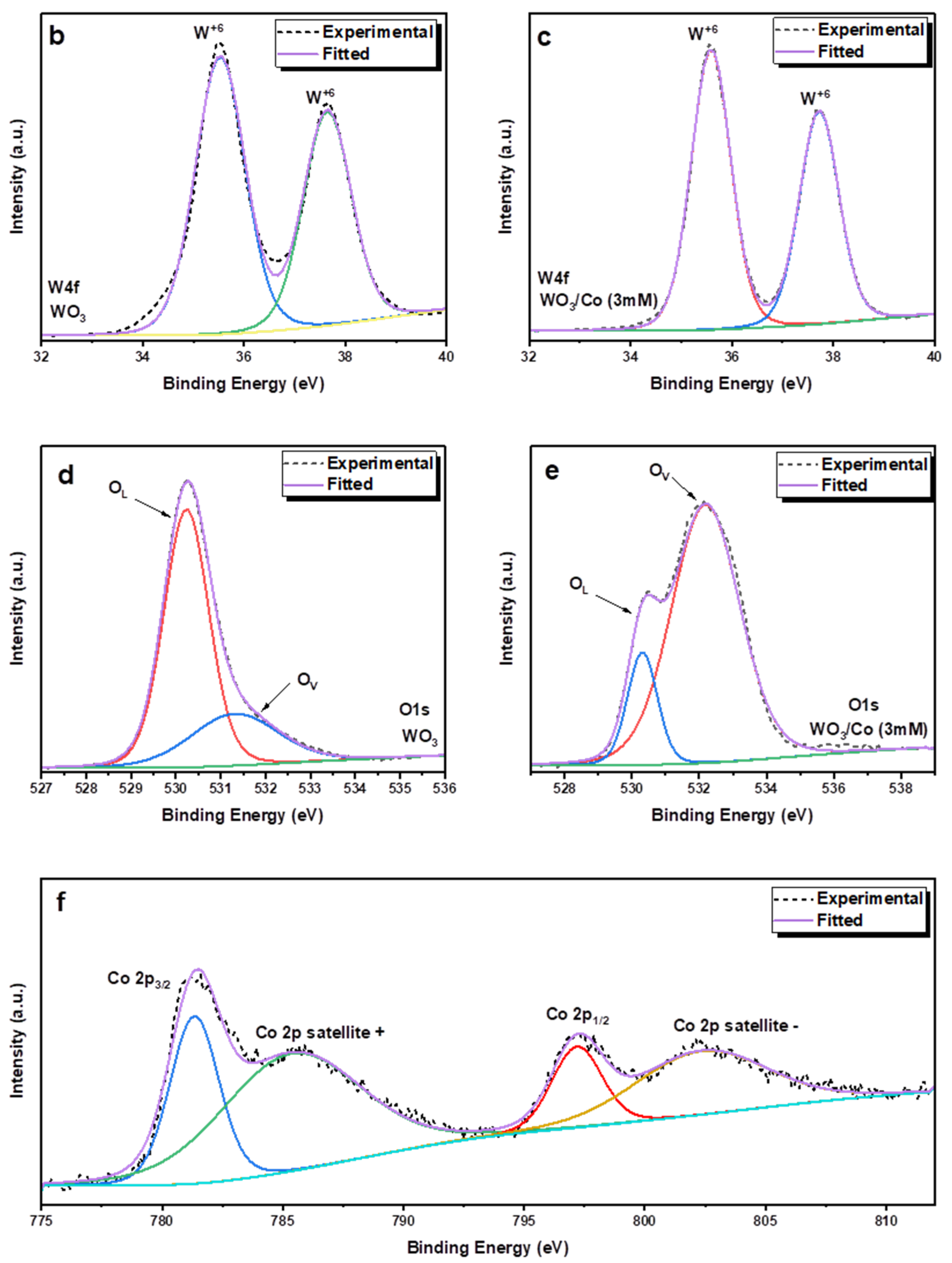
3.4. X-ray Photoelectron Spectroscopic (XPS) Analysis
3.5. Photoelectrochemical Measurements
4. Conclusions
Author Contributions
Funding
Data Availability Statement
Acknowledgments
Conflicts of Interest
References
- Bhatt, M.D.; Lee, J.S. Recent theoretical progress in the development of photoanode materials for solar water splitting photoelectrochemical cells. J. Mater. Chem. A 2015, 3, 10632–10659. [Google Scholar] [CrossRef]
- Walter, M.G.; Warren, E.L.; McKone, J.R.; Boettcher, S.W.; Mi, Q.; Santori, E.A.; Lewis, N.S. Solar water splitting cells. Chem. Rev. 2010, 110, 6446–6473. [Google Scholar] [CrossRef] [PubMed]
- Kalanur, S.S.; Duy, L.T.; Seo, H. Recent Progress in Photoelectrochemical Water Splitting Activity of WO3 Photoanodes. Top. Catal. 2018, 61, 1043–1076. [Google Scholar] [CrossRef]
- Fujishima, A.; Honda, K. Electrochemical photolysis of water at a semiconductor electrode. Nature 1972, 238, 37–38. [Google Scholar] [CrossRef]
- Tahir, A.A.; Wijayantha, K.G.U. Photoelectrochemical Water Splitting at Nanostructured ZnFe2O4 Electrodes. J. Photochem. Photobiol. A Chem. 2010, 216, 119–125. [Google Scholar] [CrossRef]
- Tahir, A.A.; Burch, H.A.; Wijayantha, K.G.U.; Pollet, B.G. A new route to control texture of materials: Nanostructured ZnFe2O4 photoelectrodes. Int. J. Hydrogen Energy 2013, 38, 4315–4323. [Google Scholar] [CrossRef]
- Alruwaili, M.; Roy, A.; Nundy, S.; Tahir, A.A. Fabrication of TiVO4 photoelectrode for photoelectrochemical application. RSC Adv. 2022, 12, 34640–34651. [Google Scholar] [CrossRef] [PubMed]
- Costa, M.B.; de Araújo, M.A.; Tinoco, M.V.d.L.; de Brito, J.F.; Mascaro, L.H. Current Trending and Beyond for Solar-Driven Water Splitting Reaction on WO3 Photoanodes. J. Energy Chem. 2022, 73, 88–113. [Google Scholar] [CrossRef]
- Hodes, G.; Cahen, D.; Manassen, J. Tungsten trioxide as a photoanode for a photoelectrochemical cell (PEC). Nature 1976, 260, 312–313. [Google Scholar] [CrossRef]
- Kalanur, S.S.; Seo, H. Influence of molybdenum doping on the structural, optical and electronic properties of WO3 for improved solar water splitting. J. Colloid Interface Sci. 2018, 509, 440–447. [Google Scholar] [CrossRef] [PubMed]
- Kalanur, S.S.; Noh, Y.-G.; Seo, H. Engineering band edge properties of WO3 with respect to photoelectrochemical water splitting potentials via a generalized doping protocol of first-row transition metal ions. Appl. Surf. Sci. 2020, 509, 145253. [Google Scholar] [CrossRef]
- Dong, P.; Hou, G.; Xi, X.; Shao, R.; Dong, F. WO3-based photocatalysts: Morphology control, activity enhancement and multifunctional applications. Environ. Sci. Nano 2017, 4, 539–557. [Google Scholar] [CrossRef]
- Al-Aisaee, N.; Alhabradi, M.; Yang, X.; Alruwaili, M.; Rasul, S.; Tahir, A.A. Fabrication of WO3/Fe2O3 heterostructure photoanode by PVD for photoelectrochemical applications. Sol. Energy Mater. Sol. Cells 2023, 263, 112561. [Google Scholar] [CrossRef]
- Alruwaili, M.; Roy, A.; Alhabradi, M.; Yang, X.; Tahir, A.A. Synergistic Photoelectrochemical and Photocatalytic Properties of the Cobalt Nanoparticles-Embedded TiVO(4) Thin Film. ACS Omega 2023, 8, 27067–27078. [Google Scholar] [CrossRef] [PubMed]
- Wang, F.; Di Valentin, C.; Pacchioni, G. Doping of WO3 for Photocatalytic Water Splitting: Hints from Density Functional Theory. J. Phys. Chem. C 2012, 116, 8901–8909. [Google Scholar] [CrossRef]
- Erbs, W.; Desilvestro, J.; Borgarello, E.; Graetzel, M. Visible-light-induced oxygen generation from aqueous dispersions of tungsten(VI) oxide. J. Phys. Chem. 1984, 88, 4001–4006. [Google Scholar] [CrossRef]
- Maruthamuthu, P.; Ashokkumar, M.; Gurunathan, K.; Subramanian, E.; Sastri, M.V.C. Hydrogen evolution from water with visible radiation in presence of Cu(II)/WO3 and electron relay. Int. J. Hydrogen Energy 1989, 14, 525–528. [Google Scholar] [CrossRef]
- Hwang, D.W.; Kim, J.; Park, T.J.; Lee, J.S. Mg-doped WO3 as a novel photocatalyst for visible light-induced water splitting. Catal. Lett. 2002, 80, 53–57. [Google Scholar] [CrossRef]
- Hameed, A.; Gondal, M.A.; Yamani, Z.H. Effect of transition metal doping on photocatalytic activity of WO3 for water splitting under laser illumination: Role of 3d-orbitals. Catal. Commun. 2004, 5, 715–719. [Google Scholar] [CrossRef]
- Radecka, M.; Sobas, P.; Wierzbicka, M.; Rekas, M. Photoelectrochemical properties of undoped and Ti-doped WO3. Phys. B Condens. Matter 2005, 364, 85–92. [Google Scholar] [CrossRef]
- Cheng, X.F.; Leng, W.H.; Liu, D.P.; Zhang, J.Q.; Cao, C.N. Enhanced photoelectrocatalytic performance of Zn-doped WO3 photocatalysts for nitrite ions degradation under visible light. Chemosphere 2007, 68, 1976–1984. [Google Scholar] [CrossRef] [PubMed]
- Liu, H.; Peng, T.; Ke, D.; Peng, Z.; Yan, C. Preparation and photocatalytic activity of dysprosium doped tungsten trioxide nanoparticles. Mater. Chem. Phys. 2007, 104, 377–383. [Google Scholar] [CrossRef]
- Enesca, A.; Duta, A.; Schoonman, J. Influence of tantalum dopant ions (Ta5+) on the efficiency of the tungsten trioxide photoelectrode. Phys. Status Solidi A 2008, 205, 2038–2041. [Google Scholar] [CrossRef]
- Karuppasamy, K.M.; Subrahmanyam, A. Results on the electrochromic and photocatalytic properties of vanadium doped tungsten oxide thin films prepared by reactive dc magnetron sputtering technique. J. Phys. D Appl. Phys. 2008, 41, 035302. [Google Scholar] [CrossRef]
- Yang, B.; Luca, V. Enhanced long-wavelength transient photoresponsiveness of WO3 induced by tellurium doping. Chem. Commun. 2008, 37, 4454–4456. [Google Scholar] [CrossRef] [PubMed]
- Zhou, L.; Zhu, J.; Yu, M.; Huang, X.; Li, Z.; Wang, Y.; Yu, C. MoxW1−xO3·0.33H2O Solid Solutions with Tunable Band Gaps. J. Phys. Chem. C 2010, 114, 20947–20954. [Google Scholar] [CrossRef]
- Chang, X.; Sun, S.; Zhou, Y.; Dong, L.; Yin, Y. Solvothermal synthesis of Ce-doped tungsten oxide nanostructures as visible-light-driven photocatalysts. Nanotechnology 2011, 22, 265603. [Google Scholar] [CrossRef]
- Wang, C.-C.; Chou, P.-H.; Yu, Y.-H.; Kei, C.-C. Deposition of Ni nanoparticles on black TiO2 nanowire arrays for photoelectrochemical water splitting by atomic layer deposition. Electrochim. Acta 2018, 284, 211–219. [Google Scholar] [CrossRef]
- Pawar, G.S.; Elikkottil, A.; Seetha, S.; Reddy, K.S.; Pesala, B.; Tahir, A.A.; Mallick, T.K. Enhanced photoactivity and hydrogen generation of LaFeO3 photocathode by plasmonic silver nanoparticle incorporation. ACS Appl. Energy Mater. 2018, 1, 3449–3456. [Google Scholar] [CrossRef]
- Pawar, G.S.; Elikkottil, A.; Pesala, B.; Tahir, A.A.; Mallick, T.K. Plasmonic nickel nanoparticles decorated on to LaFeO3 photocathode for enhanced solar hydrogen generation. Int. J. Hydrogen Energy 2019, 44, 578–586. [Google Scholar] [CrossRef]
- Hsiao, P.-T.; Chen, L.-C.; Li, T.-L.; Teng, H. Vapor treatment of nanocrystalline WO3 photoanodes for enhanced photoelectrochemical performance in the decomposition of water. J. Mater. Chem. 2011, 21, 19402–19409. [Google Scholar] [CrossRef]
- Zhang, X.; Lu, X.; Shen, Y.; Han, J.; Yuan, L.; Gong, L.; Xu, Z.; Bai, X.; Wei, M.; Tong, Y. Three-dimensional WO3 nanostructures on carbon paper: Photoelectrochemical property and visible light driven photocatalysis. Chem. Commun. 2011, 47, 5804–5806. [Google Scholar] [CrossRef] [PubMed]
- Marsen, B.; Miller, E.L.; Paluselli, D.; Rocheleau, R.E. Progress in sputtered tungsten trioxide for photoelectrode applications. Int. J. Hydrogen Energy 2007, 32, 3110–3115. [Google Scholar] [CrossRef]
- Li, W.; Li, J.; Wang, X.; Luo, S.; Xiao, J.; Chen, Q. Visible light photoelectrochemical responsiveness of self-organized nanoporous WO3 films. Electrochim. Acta 2010, 56, 620–625. [Google Scholar] [CrossRef]
- Hong, T.; Liu, Z.; Zheng, X.; Zhang, J.; Yan, L. Efficient photoelectrochemical water splitting over Co3O4 and Co3O4/Ag composite structure. Appl. Catal. B Environ. 2017, 202, 454–459. [Google Scholar] [CrossRef]
- Mahadik, M.A.; Lee, H.H.; Chae, W.-S.; Cho, M.; Jang, J.S. Energy-efficient photoelectrochemical water splitting and degradation of organic dyes over microwave-assisted WO3 nanosheets/W foil with rapid charge transport. Sol. Energy Mater. Sol. Cells 2022, 246, 111939. [Google Scholar] [CrossRef]
- Zheng, G.; Wang, J.; Zu, G.; Che, H.; Lai, C.; Li, H.; Murugadoss, V.; Yan, C.; Fan, J.; Guo, Z. Sandwich structured WO3 nanoplatelets for highly efficient photoelectrochemical water splitting. J. Mater. Chem. A 2019, 7, 26077–26088. [Google Scholar] [CrossRef]
- Kwak, J.H.; Han, G.Y.; Yoon, K.J. Zirconia-supported tungsten oxides for cyclic production of syngas and hydrogen by methane reforming and water splitting. Int. J. Hydrogen Energy 2013, 38, 8293–8305. [Google Scholar] [CrossRef]
- Zhang, X.; Chandra, D.; Kajita, M.; Takahashi, H.; Dong, L.; Shoji, A.; Saito, K.; Yui, T.; Yagi, M. Facile and simple fabrication of an efficient nanoporous WO3 photoanode for visible-light-driven water splitting. Int. J. Hydrogen Energy 2014, 39, 20736–20743. [Google Scholar] [CrossRef]
- Nagaraj, D.; Rajagopal, S.; Lonappan, D.; Veluswamy, P.; Paulraj, S.; Venugopal, K. Microwave-assisted synthesis of cobalt doped WO3 nanostructure as an electrode material for supercapacitor. J. Mater. Sci. Mater. Electron. 2023, 34, 846. [Google Scholar] [CrossRef]
- Reddy, C.V.; Reddy, I.N.; Sreedhar, A.; Shim, J. Investigation of dopant and Ag plasmonic effect on α-Fe2O3 photoelectrode for photoelectrochemical water splitting activity. Appl. Surf. Sci. 2019, 488, 629–638. [Google Scholar] [CrossRef]
- Lin, H.-Y.; Yang, H.-C.; Wang, W.-L. Synthesis of mesoporous Nb2O5 photocatalysts with Pt, Au, Cu and NiO cocatalyst for water splitting. Catal. Today 2011, 174, 106–113. [Google Scholar] [CrossRef]
- Han, X.; Xu, D.; An, L.; Hou, C.; Li, Y.; Zhang, Q.; Wang, H. Ni-Mo nanoparticles as co-catalyst for drastically enhanced photocatalytic hydrogen production activity over g-C3N4. Appl. Catal. B Environ. 2019, 243, 136–144. [Google Scholar] [CrossRef]
- Ayala, P.; Giesriegl, A.; Nandan, S.P.; Myakala, S.N.; Wobrauschek, P.; Cherevan, A. Isolation strategy towards earth-abundant single-site co-catalysts for photocatalytic hydrogen evolution reaction. Catalysts 2021, 11, 417. [Google Scholar] [CrossRef]
- Liu, C.; Wang, F.; Zhu, S.; Xu, Y.; Liang, Q.; Chen, Z. Controlled charge-dynamics in cobalt-doped TiO2 nanowire photoanodes for enhanced photoelectrochemical water splitting. J. Colloid Interface Sci. 2018, 530, 403–411. [Google Scholar] [CrossRef]
- Mehmood, F.; Iqbal, J.; Jan, T.; Mansoor, Q. Structural, Raman and photoluminescence properties of Fe doped WO3 nanoplates with anti cancer and visible light driven photocatalytic activities. J. Alloys Compd. 2017, 728, 1329–1337. [Google Scholar] [CrossRef]
- Pastrana, E.C.; Zamora, V.; Wang, D.; Alarcón, H. Fabrication and characterization of α-Fe2O3/CuO heterostructure thin films via dip-coating technique for improved photoelectrochemical performance. Adv. Nat. Sci. Nanosci. Nanotechnol. 2019, 10, 035012. [Google Scholar] [CrossRef]
- Srinivasan, P.; Kulandaisamy, A.J.; Mani, G.K.; Babu, K.J.; Tsuchiya, K.; Rayappan, J.B.B. Development of an acetone sensor using nanostructured Co3 O4 thin films for exhaled breath analysis. RSC Adv. 2019, 9, 30226–30239. [Google Scholar] [CrossRef]
- Mohamedkhair, A.K.; Drmosh, Q.A.; Qamar, M.; Yamani, Z.H. Tuning Structural Properties of WO3 Thin Films for Photoelectrocatalytic Water Oxidation. Catalysts 2021, 11, 381. [Google Scholar] [CrossRef]
- Miao, Y.; Liu, J.; Chen, L.; Sun, H.; Zhang, R.; Guo, J.; Shao, M. Single-atomic-Co cocatalyst on (040) facet of BiVO4 toward efficient photoelectrochemical water splitting. Chem. Eng. J. 2022, 427, 131011. [Google Scholar] [CrossRef]
- Lan, Y.; Liu, Z.; Liu, G.; Guo, Z.; Ruan, M.; Rong, H.; Li, X. 1D ZnFe2O4 nanorods coupled with plasmonic Ag, Ag2S nanoparticles and Co-Pi cocatalysts for efficient photoelectrochemical water splitting. Int. J. Hydrogen Energy 2019, 44, 19841–19854. [Google Scholar] [CrossRef]
- Chen, K.; Feng, X.; Hu, R.; Li, Y.; Xie, K.; Li, Y.; Gu, H. Effect of Ag nanoparticle size on the photoelectrochemical properties of Ag decorated TiO2 nanotube arrays. J. Alloys Compd. 2013, 554, 72–79. [Google Scholar] [CrossRef]
- Kalanur, S.S.; Yoo, I.-H.; Eom, K.; Seo, H. Enhancement of photoelectrochemical water splitting response of WO3 by Means of Bi doping. J. Catal. 2018, 357, 127–137. [Google Scholar] [CrossRef]
- Xiao, N.; Li, S.; Li, X.; Ge, L.; Gao, Y.; Li, N. The roles and mechanism of cocatalysts in photocatalytic water splitting to produce hydrogen. Chin. J. Catal. 2020, 41, 642–671. [Google Scholar] [CrossRef]
- Soni, V.; Xia, C.; Cheng, C.K.; Nguyen, V.-H.; Nguyen, D.L.T.; Bajpai, A.; Kim, S.Y.; Le, Q.V.; Khan, A.A.P.; Singh, P.; et al. Advances and recent trends in cobalt-based cocatalysts for solar-to-fuel conversion. Appl. Mater. Today 2021, 24, 101074. [Google Scholar] [CrossRef]
- Wang, G.; Wang, H.; Ling, Y.; Tang, Y.; Yang, X.; Fitzmorris, R.C.; Wang, C.; Zhang, J.Z.; Li, Y. Hydrogen-treated TiO2 nanowire arrays for photoelectrochemical water splitting. Nano Lett. 2011, 11, 3026–3033. [Google Scholar] [CrossRef]


| Photoanode | Synthesis Method | Electrolyte | Photocurrent Density | Reference Electrode | Reference |
|---|---|---|---|---|---|
| Ag–LaFeO3 | spin coating | 0.1 M NaOH | 0.074 mA/cm2 | 0.6 V vs. RHE | [29] |
| Ni–LaFeO3 | spin coating | 0.1 M NaOH | 0.066 mA/cm2 | 0.6 V vs. RHE | [30] |
| Co–BiVO4 | electrochemical synthesis | 0.1 M PBS | 0.69 mA/cm2 | 1.23 V vs. RHE | [50] |
| Ag–ZnFe2O4 | chemical water bath | 0.5 M Na2SO4 | 0.91 mA/cm2 | 1.23 V vs. RHE | [51] |
| Mo–WO3 | hydrothermal method | 0.1 M Na2SO4 | 1.15 mA/cm2 | 0.8 V vs. Ag/AgCl | [10] |
| WO3 | PVD/DC | 0.1 M Na2SO4 | 0.9 mA/cm2 | 0.6 V vs. SCE | [49] |
| WO3 | PVD/RF | 1 M NaOH | 1.020 mA/cm2 | 1.23 V vs. RHE | present work |
| Co-WO3 | PVD/RF and thermal oxidation | 1 M NaOH | 1.485 mA/cm2 | 1.23 V vs. RHE | present work |
Disclaimer/Publisher’s Note: The statements, opinions and data contained in all publications are solely those of the individual author(s) and contributor(s) and not of MDPI and/or the editor(s). MDPI and/or the editor(s) disclaim responsibility for any injury to people or property resulting from any ideas, methods, instructions or products referred to in the content. |
© 2024 by the authors. Licensee MDPI, Basel, Switzerland. This article is an open access article distributed under the terms and conditions of the Creative Commons Attribution (CC BY) license (https://creativecommons.org/licenses/by/4.0/).
Share and Cite
Alhabradi, M.; Yang, X.; Alruwaili, M.; Chang, H.; Tahir, A.A. Enhanced Photoelectrochemical Performance Using Cobalt-Catalyst-Loaded PVD/RF-Engineered WO3 Photoelectrodes. Nanomaterials 2024, 14, 259. https://doi.org/10.3390/nano14030259
Alhabradi M, Yang X, Alruwaili M, Chang H, Tahir AA. Enhanced Photoelectrochemical Performance Using Cobalt-Catalyst-Loaded PVD/RF-Engineered WO3 Photoelectrodes. Nanomaterials. 2024; 14(3):259. https://doi.org/10.3390/nano14030259
Chicago/Turabian StyleAlhabradi, Mansour, Xiuru Yang, Manal Alruwaili, Hong Chang, and Asif Ali Tahir. 2024. "Enhanced Photoelectrochemical Performance Using Cobalt-Catalyst-Loaded PVD/RF-Engineered WO3 Photoelectrodes" Nanomaterials 14, no. 3: 259. https://doi.org/10.3390/nano14030259
APA StyleAlhabradi, M., Yang, X., Alruwaili, M., Chang, H., & Tahir, A. A. (2024). Enhanced Photoelectrochemical Performance Using Cobalt-Catalyst-Loaded PVD/RF-Engineered WO3 Photoelectrodes. Nanomaterials, 14(3), 259. https://doi.org/10.3390/nano14030259