Article

Single-Step Synthesis of Graphitic Carbon Nitride Nanomaterials by Directly Calcining the Mixture of Urea and Thiourea: Application for Rhodamine B (RhB) Dye Degradation

1
Faculty of Materials Science and Engineering, Jimma University, Jimma P.O. Box 378, Ethiopia
2
Department of Physics, College of Natural and Computational Science, Bonga University, Bonga P.O. Box 334, Ethiopia
3
Faculty of Civil and Environmental Engineering, Jimma University, Jimma P.O. Box 378, Ethiopia
4
School of Chemical Sciences, Mahatma Gandhi University, Kottayam 686560, India
5
School of Pure and Applied Physics, Mahatma Gandhi University, Kottayam 686560, India
6
Faculty of Agricultural and Environmental Sciences, University of Rostock, Justus-Von-Liebig-Weg 6, 18059 Rostock, Germany
*
Authors to whom correspondence should be addressed.
Nanomaterials 2023, 13(4), 762; https://doi.org/10.3390/nano13040762
Submission received: 27 January 2023 / Revised: 11 February 2023 / Accepted: 14 February 2023 / Published: 17 February 2023

Abstract

:
Recently, polymeric graphitic carbon nitride (g-C3N4) has been explored as a potential catalytic material for the removal of organic pollutants in wastewater. In this work, graphitic carbon nitride (g-C3N4) photocatalysts were synthesized using mixtures of low-cost, environment-friendly urea and thiourea as precursors by varying calcination temperatures ranging from 500 to 650 °C for 3 h in an air medium. Different analytical methods were used to characterize prepared g-C3N4 samples. The effects of different calcination temperatures on the structural, morphological, optical, and physiochemical properties of g-C3N4 photocatalysts were investigated. The results showed that rhodamine B (RhB) dye removal efficiency of g-C3N4 prepared at a calcination temperature of 600 °C exhibited 94.83% within 180 min visible LED light irradiation. Photocatalytic activity of g-C3N4 was enhanced by calcination at higher temperatures, possibly by increasing crystallinity that ameliorated the separation of photoinduced charge carriers. Thus, controlling the type of precursors and calcination temperatures has a great impact on the photocatalytic performance of g-C3N4 towards the photodegradation of RhB dye. This investigation provides useful information about the synthesis of novel polymeric g-C3N4 photocatalysts using a mixture of two different environmentally benign precursors at high calcination temperatures for the photodegradation of organic pollutants.

1. Introduction

Nowadays, growing industrial activities have resulted in the generation of huge amounts of hazardous pollutants that are released into environmental water bodies [1,2]. The major causes of water pollution are the release of untreated industrial effluents and uncontrolled anthropogenic activities [3,4,5]. Meanwhile, various hazardous pollutants including antibiotics, phenols, pesticides, pharmaceuticals, and dyes from textile industries are causes of major water pollution [6]. Among them, dyes from textile industries generate a large amount of industrial wastewater due to the huge water demand and frequent discharge into the environment [6,7]. In addition, approximately 10–20% of total dyes from textile industries in the world are dissipated as effluent into the environment throughout the production process [2,8,9]. Therefore, the random and untreated discharge of these hazardous pollutants causes significant water pollution and has a direct impact on human health [2]. As a result, water pollution remediation is one of the imperative issues that the scientific community has given significant attention to with the primary goal of safeguarding and conserving natural water resources [10]. To date, wastewater has been treated using a variety of physical, chemical, and biological techniques, but the scientific community has yet to find a method that is renewable and sustainable [11,12]. Furthermore, these conventional methods have drawbacks, such as requiring a large capital investment, low effectiveness, expensive maintenance costs, and requiring large operating areas, and also cannot fully eliminate hazardous organic compounds beyond changing their phase. Therefore, it is essential to design efficient, sustainable, and renewable wastewater treatment technologies for effective removal of hazardous organic pollutants [2,11,12,13]. Among the several known advanced oxidation technologies, semiconductor-based heterogeneous photocatalysis is the most efficient advanced oxidation process to degrade pollutants at ambient pressure and temperature without generating harmful intermediates [11]. In the photocatalytic process, light is absorbed by the semiconductor material and initiates the photocatalytic reaction. The rate of photocatalytic reaction depends on the ability of the photocatalyst to generate electron-hole pairs, the specific surface area, the crystallinity, the spectral response, and the light absorption capacity [14]. An efficient photocatalyst should meet a variety of requirements, including excellent optical response, tunable band gap energy, superior photochemical stability, and high affinity for light [15]. Therefore, photocatalytic degradation of organic pollutants has become one of the most promisingly sustainable, cost-effective, green, and clean technology [13,16,17]. In the past few decades, metal oxide-based photocatalysts such as TiO2, ZnO, and SnO2 have been exploited in the remediation of environmental pollution. Nevertheless, the substantial electron-hole pair recombination in these photocatalysts significantly decreases their photocatalytic efficiency. Furthermore, ZnO, TiO2, and SnO2 can absorb solar energy exclusively in the UV region because of their wide energy bandgap, which accounts for just 4% of the total solar energy irradiated. Nowadays, because of its fascinating physicochemical properties, g-C3N4 is a superior alternative for visible light-assisted photocatalytic wastewater treatment than other photocatalysts such as TiO2, ZnO, SnO2, and so on [18].
The majority of photocatalysis research has used xenon lamps [19,20], halogen lamps [21], UV lamps [22], and mercury vapor lamps [23] as the light source, which has some advantages but is dangerous, has a short lifetime, overheats, is expensive and difficult to handle [24]. Recently, in the field of photocatalysis technology, less toxic, longer life, less expensive, and environmentally friendly light-emitting diodes have been employed as visible light sources for organic pollutant degradation [24,25].
Since its discovery by Wang’s group in 2009, g-C3N4 has piqued the interest of researchers as a visible-light-driven semiconductor photocatalyst [26]. Polymeric g-C3N4 is an extensively explored n-type semiconductor for the removal of organic pollutants due to its inexpensiveness, non-toxicity, eco-friendliness, simple synthesis, mid-energy bandgap, and robust physicochemical stability [2,27]. In general, g-C3N4 can be synthesized by thermal calcination of various nitrogen-rich precursors, such as cyanamide [28], melamine [29], dicyandiamide [30], thiourea [31], and urea [20,32].
Nevertheless, pristine g-C3N4 suffers from a low photocatalytic performance due to the rapid recombination of photogenerated charge carriers, limited range absorption of visible light, and low specific surface area [33]. Therefore, researchers have made numerous attempts to overcome the limitations of pristine g-C3N4, such as elemental doping [27,34,35,36], controlling morphology [37], and constructing g-C3N4-based heterojunctions [38,39,40]. Moreover, recently various studies have been conducted on g-C3N4 for photocatalytic elimination of pollutants [19,24,41], photoreduction of carbon dioxide [42,43,44], water splitting for hydrogen production [28,45], and so on.
Recently, many researchers have reported the impact of different types of precursors on the structural, optical, morphological, and physicochemical properties as well as the efficiency of g-C3N4 photocatalysts. Enumerating some of the recent outcomes, Zhang et al. fabricated g-C3N4 by pyrolysis using an aggregate of urea and thiourea as precursors to enhance the specific surface area and separation of photo-induced electron-hole pairs, consequently improving photocatalytic activity of g-C3N4 photocatalysts [27]. Similarly, Kadam et al. successfully synthesized porous g-C3N4 nanosheets by heating low-cost urea and thiourea precursors at 520 °C for 2 h, and g-C3N4 showed the best photocatalytic efficiency for the elimination of cationic MB dye when exposed to visible light [23]. In addition, An et al. synthesized g-C3N4 materials by simply calcining mixtures of urea and thiourea precursors at 550 °C for photocatalytic degradation of RhB upon irradiation of visible light [46]. Guo et al. prepared g-C3N4 by polycondensation of melamine and urea at 550 °C for 3 h. For a melamine-to-urea molar ratio of 1:1, the photocatalytic degradation rate for MB was about 75% after 4 h of light exposure [47].
Similarly, calcination temperature control is one of the most common reaction parameters affecting the performance of the g-C3N4 photocatalyst. As a result, the synthesis of g-C3N4 by varying calcination temperature allows improvements in the surface area, crystallinity, extended absorption range of visible light, and overall enhanced photocatalytic activity [19,20,32]. Therefore, some recent studies have been carried out on the influence of calcination temperature on g-C3N4 photocatalytic performance. Enumerating a few of the outcomes, Fang et al. prepared porous g-C3N4 through direct pyrolysis of low-cost urea by controlling the condensation temperature in the range between 400 and 650 °C. In addition, the synthesized U-g-C3N4 had better photoactivity and long-term photocatalytic stability [20]. Similarly, Paul et al. prepared g-C3N4 by calcining urea at temperatures between 350 and 750 °C. It was discovered that increasing the calcination temperature to 550 °C enhanced the photocatalytic activity of g-C3N4 for the degradation of MB dye, whereas increasing the temperature beyond the optimum value decreased the photoactivity [32]. Moreover, Ke et al. synthesized g-C3N4 for the decomposition of rhodamine B (RhB) and methyl orange (MO) via thermal polycondensation of melamine at different calcination temperatures ranging from 450–700 °C. The g-C3N4 prepared at a calcination temperature of 700 °C revealed robust photocatalytic activity and stability against RhB and MO decomposition [19]. In addition, according to a review of the published literature in the field of visible-light-driven photocatalysis, the calcination temperatures for the fabrication of g-C3N4 using urea and thiourea were generally considered to be in the range of 520–550 °C and 450–650 °C, respectively [48].
Synthesis of the g-C3N4 photocatalyst using mixtures of stochiometric amounts of urea and thiourea at different calcination temperatures, especially in wastewater treatment using visible LED light irradiation, has not been reported in the previous literatures. Herein, in this work, we designed a novel single-step synthesis route to fabricate g-C3N4 nanomaterials by controlling various calcination temperatures of a mixture of stochiometric amounts of low-cost, environment-friendly urea and thiourea for complete photocatalytic degradation of RhB dye. Furthermore, the influence of varying calcination temperatures on the structural, optical, morphological, and physicochemical properties of g-C3N4 photocatalysts was carefully examined. Subsequently, the properties of the prepared g-C3N4 samples were characterized using several analytical techniques, and the photocatalytic activities of g-C3N4 samples were assessed by measuring the removal of rhodamine B (RhB) after irradiation of visible LED light.

2. Materials and Methods

2.1. Materials

Urea (CH4N2O, 99.5%), and thiourea (CH4N2S, 99%) were bought from Merck, Mumbai, India. Rhodamine B (99.9%) was obtained from Loba Chemie Pvt. Ltd., Mumbai, India. All reagents were used without further purification as received.

2.2. Methods

2.2.1. Synthesis of g-C3N4 Samples

The preparation of g-C3N4 nanosheets was achieved by direct one-step calcination of mixtures of urea and thiourea in a muffle furnace under an air medium. Typically, weighed stoichiometric amounts of urea and thiourea were transferred to a 50 mL covered ceramic crucible with lid, and tightly wrapped in aluminum foil paper, then subjected to various calcination temperatures ranging from 500 to 650 °C in a muffle furnace for 3 h. Moreover, when the temperature is raised to 700 °C, nothing remains in the crucible, showing that the mixture of urea and thiourea has completely decomposed at calcination temperatures above 650 °C. Thus, the resultant powders were yellow. The resulting products were finely ground in a mortar and pestle after being cooled to room temperature. Finally, a series of light-yellow powders labeled g-C3N4-500 °C, g-C3N4-550 °C, g-C3N4-600 °C, and g-C3N4-650 °C were obtained and characterized using various analytical tools.

2.2.2. Characterization Techniques

X-ray diffraction patterns of g-C3N4 samples were studied using X-ray powder diffraction (PanAlyticals, Almelo, The Netherlands) with Cu-Kα radiation (λ = 1.5406 Å), operating at 40 kV, and 15 mA. Diffraction intensity was continuously recorded over a 2θ range from 10° to 70° with a sweep step width of 0.01° and a scan rate of 10°/min. Fourier transform infrared (FTIR) spectroscopy (IR Tracer-100, Shimadzu, Kyoto, Japan) was used to probe functional groups in the range 400–4000 cm−1. The optical properties of the materials were evaluated with a UV-Vis diffuse reflectance spectrometer (UV-2600, Shimadzu, Kyoto, Japan) in the range 200–800 nm using barium sulfate (BaSO4) as background. Photoluminescence (PL) spectral data were obtained using a PL spectrophotometer (RF-6000, Shimadzu, Kyoto, Japan) at an excitation wavelength of 360 nm. The width of the excitation and emission slits was 10 nm, and the thermal stability of each g-C3N4 sample was obtained by TGA (Q600, TA Instruments, Hüllhorst, Germany). The morphology of the g-C3N4-600 °C sample was investigated by FE-SEM and HR-TEM.

2.2.3. Photocatalytic Degradation Experiments

The photocatalytic activity of g-C3N4 photocatalysts with different calcination temperatures was evaluated by decomposing RhB as a model pollutant under visible-LED-light irradiation. In a typical experiment, 50 mg of g-C3N4 photocatalyst was dispersed in 100 mL of RhB solution (10 mg/L of RhB in 100 mL of distilled water). The suspension was stirred vigorously for 60 min in the dark before irradiation to ensure proper dispersion and establish adsorption–desorption equilibrium between the photocatalysts and RhB dye. The initial concentration (C0) of suspensions was obtained at adsorption–desorption equilibrium. A visible light source of (50 W, 5000 lm, 6500 k daylight, 220–240 V) LED lamp (Phillips, Kolkata, India) was used to initiate the photocatalytic reaction. At predetermined intervals, 3 mL of the solution was taken and centrifuged to separate the photocatalyst and to obtain the supernatant. Subsequently, the concentration of RhB in the supernatant was measured with a UV-Vis NIR spectrophotometer (Cary5000, Agilent, Santa Clara, California, USA) at its maximum absorption wavelength of λ max = 554 nm .
The intensity change of the major absorption peak was used to assess dye degradation. The following equation was used to calculate photocatalytic degradation efficiency [32]:
  Degradation   Efficiency = 1 C C 0 × 100 % = 1 A A 0 × 100 %
where C0 represents the concentration of RhB dye at adsorption–desorption equilibrium (mg L−1), and C is the concentration of RhB at reaction time t; similarly, A and A0 represent corresponding values.
To solve the apparent degradation rate constant (k) of the RhB dye, a simplified Langmuir–Hinshelwood pseudo-first-order kinetic model was used [49]:
  ln C 0 C = kt  
where C0 is the concentration of RhB dye at adsorption–desorption equilibrium (mgL−1), C is the concentration of RhB at reaction time t, k is the apparent reaction rate constant (min−1), and t is the irradiation time (min). Furthermore, the photolysis experiment was carried out following the same procedure without the addition of a catalyst.

3. Results and Discussion

3.1. XRD Analysis

X-ray diffraction patterns were used to identify the crystal structure of g-C3N4 prepared at various temperatures, as shown in Figure 1. The XRD peaks of g-C3N4 samples generated at calcination temperatures of 500, 550, 600, and 650 °C revealed two main diffraction peaks at around 13.1° and 27.2°. The characteristic peak at 27.2° represents interplanar graphitic stacking, while the peak at 13.1° represents inplanar structural packing, demonstrating g-C3N4 formation in all prepared g-C3N4 samples [42,50]. As the calcination temperature increases, the peak of g-C3N4 samples becomes sharper, while its width becomes narrower. As a result, an increase in peak sharpness indicated a higher degree of crystallinity of a photocatalyst [51]. Therefore, a higher degree of crystallinity of the photocatalyst accelerated the transfer of charge carriers to the surface, thereby inhibiting photoinduced electron-hole recombination by promoting the separation of charge carriers [19].
No solid sample was produced when the calcination temperature reached 700 °C during calcination, indicating that g-C3N4 completely decomposed into gases including CO2, H2S, NH3, and water. Therefore, at calcination temperatures above 650 °C, g-C3N4-prepared samples become thermally unstable, and optimal calcination could be between 600–650 °C [32]. In addition, Figure 1 revealed that the 2θ position for typical (002) peak of g-C3N4 samples prepared at calcination temperatures of 500, 550, 600, and 650 °C is 27.25, 27.08, 27.30, 27.19, and 27.25°, respectively.
Moreover, the crystallite size of synthesized g-C3N4 samples was determined using the Debye–Scherrer formula [52]:
  D = k λ β   cos θ
where D is the crystallite size, λ is the wavelength of X-ray radiation (λ = 1.5406 Å), k is the Scherrer’s constant (0.94), θ is the diffraction angle, and β is the full-width half-maximum of the diffraction peak relating to the (002) plane. In this study, k was rounded to 0.9. Similarly, the interplanar spacing (dhkl) can be calculated as follows:
  d hkl = λ 2 sin θ
Table 1 shows the crystallite size and interplanar spacing of all synthesized g-C3N4 samples at various temperatures corresponding to the (002) plane.
Table 1 demonstrates that as the calcination temperature rises, so does the intensity of the diffraction peak and the crystal size of the prepared g-C3N4 samples. A sharp intense peak at high calcination temperatures implies that the s-triazine layer arrangement is more regular and that there is a significant degree of aggregation [53].

3.2. Optical Properties Studies

Photoluminescence (PL), UV-Vis diffuse reflectance spectra (DRS), and FTIR spectra were used to investigate the optical properties of the prepared g-C3N4 samples.

3.2.1. PL Analysis

Photoluminescence spectra are useful for understanding the separation and recombination of electron-hole pairs, which is an important mechanism in photocatalysis [54]. Lowering the PL intensity indicates a decrease in the rate of recombination of electron-hole pairs and that increases photocatalytic activity [19,55]. The PL spectra of g-C3N4 samples prepared at various calcination temperatures were obtained at 360 nm excitation wavelength. As shown in Figure 2, all prepared g-C3N4 samples exhibited an intense and broad photoluminescence peak between 437–440 nm and showed a slight red-shift at a temperature of 550 °C. A slight shift of PL peak was attributable to differences in the bandgap energy of each sample [47]. Except for the sample prepared at 550 °C, the PL intensity of all prepared g-C3N4 samples increased as the calcination temperature increased. This indicated that g-C3N4 prepared at 550 °C could have the lowest recombination rate of photoinduced electron-hole pairs [56]. In addition, an increase in the amount of tri-s-triazine content at a calcination temperature of 550 °C may lead to higher π-states and cause orbital overlap and reduce the PL intensity [48].

3.2.2. UV-Vis DRS Analysis

Diffuse reflectance spectroscopy was used to study the optical properties of all prepared photocatalysts at different calcination temperatures, as shown in Figure 3a. The variation in the absorption edges of g-C3N4 samples was observed with changing calcination temperatures, as shown in Figure 3a. In addition, the variation of energy bandgap influences photocatalytic activity by affecting visible light absorption and the separation of e/h+ pairs [32]. The bandgap energy (Eg) of the samples was calculated by using a Kubelka–Munk transformation, which can be expressed by the equation [8]:
  F R hv n = A   h ν Eg  
where F R   is Kubelka–Munk function, h is the Planck constant, ν is the light frequency, A is a constant, and n is equal to ½ for direct bandgap and 2 for indirect bandgap materials. Generally, g-C3N4 is a well-known indirect bandgap semiconductor material [37]. Therefore, the energy bandgaps of g-C3N4 samples were calculated by extrapolating the linear portion of the F R h ν 1/2 versus h ν curve (as shown in Figure 3b). The energy bandgap of g-C3N4 samples prepared at different calcination temperatures of 500, 550, 600, and 650 °C was estimated to be 2.78, 2.75, 2.82, and 2.89 eV, respectively [37,47]. In comparison with calcination temperatures of 500, 600, and 650 °C, the bandgap at 550 °C was narrower. When the temperature further increased to 650 °C, this caused a blue shift in the absorption edge and an increase in band gap energy to 2.89 eV. The blue shift may be a result of a significant quantum confinement effect and the possible reason for better photoactivity of a sample obtained at 650 °C than at 500 and 550 °C [32].

3.2.3. FTIR Analysis

FTIR spectroscopy is used to analyze the chemical compositions and functional groups of g-C3N4 samples synthesized at various calcination temperatures. As shown in Figure 4a, there were no significant differences in FTIR peaks at different calcination temperatures. All synthesized samples displayed prominent peaks in the wavenumber range of 1230–1740 cm−1, which corresponds to the stretching mode of conjugated heterocycles [1]. The stretching mode of C=N heterocycles is responsible for peaks of g-C3N4 samples seen at wavenumber about 1458, 1560, and 1630 cm−1. However, the C=O stretching mode at wavenumber 1740 cm−1 was observed in the g-C3N4 samples prepared at calcination temperatures of 500, 600, and 650 °C. FTIR peaks at wavenumber around 1230, 1319, and 1400 cm−1 belong to the aromatic C-N stretching mode, and also the peaks at wavenumber of 805 and 885 cm−1 resulted from the distinctive breathing mode of tri-s-triazine ring units. Moreover, the absorbed H2O molecules cause the occurrence of N-H and O-H stretching modes at broad bands situated in the wavenumber range of 3074–3320 cm−1 [57,58,59]. In Figure 4a, all peaks seen are the typical pattern of g-C3N4.

3.3. TGA Analysis

Thermal analysis of g-C3N4 samples was performed by heating them from ambient temperature to 700 °C in an air atmosphere. Figure 4b shows the thermal stability of g-C3N4 photocatalysts synthesized at various calcination temperatures. The weight loss is less at temperatures below 450 °C but increases as calcination temperatures decrease from 650 to 500 °C, which may be caused by the evaporation of water molecules that have been adsorbed on the surface of catalysts [60]. Meanwhile, the g-C3N4-650 °C sample did not lose weight significantly at a temperature below 450 °C, which is an indication of its great thermal stability. In addition, the significant weight loss of all prepared samples between temperatures 450 °C and 700 °C is attributed to the decomposition and condensation of g-C3N4 photocatalysts [42,61]. A thermogravimetric study shows that as-prepared g-C3N4 is non-volatile up to 600 °C and will be practically entirely degraded when the temperature climbs to 700 °C [36].

3.4. FE-SEM and HR-TEM Analysis

Figure 5a,b demonstrated low-magnification SEM and high-magnification TEM images of g-C3N4 synthesized at a calcination temperature of 600 °C. Figure 5a revealed that g-C3N4 prepared at 600 °C consists of large nanoflakes [46]. In addition, the formation of these nanoflakes could result in increased surface area which could lead to a large number of active sites, and a higher adsorption of pollutants over its surface [62]. Figure 5b showed a high magnification TEM image of g-C3N4 prepared at 600 °C, and it was almost transparent, which was due to its thin structure. The results indicated that g-C3N4 prepared at 600 °C was fluffy [56]. Therefore, we conclude that the enhanced photocatalytic activity is caused by the formation of nanoflake structures in g-C3N4 photocatalysts calcined at 600 °C.

3.5. Photocatalytic Activity Evaluation

The photocatalytic activity of rhodamine B for all the synthesized g-C3N4 samples was shown by plots of C/C0 vs. irradiation time (min), as shown in Figure 6. Without g-C3N4, the degradation rate of RhB was found to be almost insignificant, showing that the photolysis of RhB itself was negligible. Therefore, in the absence of a g-C3N4 photocatalyst, the rhodamine B dye solution was stable under visible LED light irradiation. The degradation of RhB was greatly improved when photocatalysts were introduced. As a result, the degradation of rhodamine B dye was caused by the presence of g-C3N4 in the solution.
The adsorption efficiencies of g-C3N4 photocatalysts prepared at 500, 550, 600, and 650 °C were about 1.85%, 0.66%, 3.5%, and 1.55% for RhB dye solution, respectively. The g-C3N4 sample prepared at a calcination temperature of 600 °C displayed the highest adsorption ability. Meanwhile, the photodegradation efficiency of g-C3N4-500 °C, g-C3N4-550 °C, g-C3N4-600 °C, and g-C3N4-650 °C was 18.34%, 22.2%, 94.83%, and 94.80%, after 180 min irradiation, as shown in Figure 6. The degradation efficiency and reaction rate constant of all prepared samples of g-C3N4 at various temperatures towards RhB dye removal are shown in Figure 6a,b, respectively, and are represented in Table 2.
Generally, Table 2 showed that boosted photodegradation efficiency of g-C3N4 photocatalysts was obtained as calcination temperatures vary between 600 and 650 °C. Furthermore, the crystallinity of the as-prepared samples increased at higher calcination temperatures.
Table 3 summarizes the findings of a comparison of the photocatalytic activities of g-C3N4-600 °C with other previously reported photocatalysts. As shown, g-C3N4-600 °C was a very potential photocatalyst and was extremely attractive for the actual treatment of industrial wastewater containing organic pollutants.

4. Conclusions

A visible LED light-driven graphitic carbon nitride photocatalyst was fabricated by single-step calcination of mixtures of urea and thiourea at different temperatures. Meanwhile, various analytical tools were used to study the structural, morphological, optical, and physicochemical properties of all prepared g-C3N4 samples. The presence of a typical g-C3N4 pattern was demonstrated by the XRD and FTIR spectra of all prepared g-C3N4 samples at various calcination temperatures. The influence of calcining temperatures on photocatalytic activities of g-C3N4 samples was investigated by photodegradation of RhB when exposed to visible LED light. The best photocatalytic activity of g-C3N4 photocatalyst was achieved at a temperature of 600 °C, due to its efficient separation of photoinduced charge carriers, high crystallinity, and enhanced visible light absorption range. Therefore, single-step synthesis of g-C3N4 using a mixture of urea and thiourea at high calcination temperature leads to the highest photocatalytic activity for the degradation of RhB dyes. This work can give a good insight into the synthesis of a cost-effective and efficient g-C3N4 photocatalyst at high calcination temperature for the treatment of industrial wastewater containing organic pollutants.

Author Contributions

Conceptualization, A.S.; methodology, A.S.; experiments, A.S.; writing-original draft, A.S. and E.A.; data curation, A.S.; supervision, E.A., M.A., D.M., S.T., N.K. and B.L.; writing- review, and editing, E.A., M.A., D.M., S.T., N.K. and B.L. All authors have read and agreed to the published version of the manuscript.

Funding

This research received no external funding.

Institutional Review Board Statement

Not applicable.

Informed Consent Statement

Not applicable.

Data Availability Statement

The data that support the findings of this study are available upon reasonable request from the authors.

Acknowledgments

We gratefully acknowledge Sabu Thomas and Nandakumar Kalarikkal from Mahatma Gandhi University for granting access to the laboratory and characterization facilities in their research groups.

Conflicts of Interest

The authors declared no conflict of interest.

References

  1. Hu, F.; Sun, S.; Xu, H.; Li, M.; Hao, X.; Shao, G.; Wang, H.; Chen, D.; Lu, H.; Zhang, R. Investigation on g-C3N4/rGO/TiO2 nanocomposite with enhanced photocatalytic degradation performance. J. Phys. Chem. Solids 2021, 156, 110181. [Google Scholar] [CrossRef]
  2. Pham, T.H.; Myung, Y.; Van Le, Q.; Kim, T. Visible-light photocatalysis of Ag-doped graphitic carbon nitride for photodegradation of micropollutants in wastewater. Chemosphere 2022, 301, 134626. [Google Scholar] [CrossRef]
  3. Thines, R.K.; Mubarak, N.M.; Nizamuddin, S.; Sahu, J.N.; Abdullah, E.C.; Ganesan, P. Application potential of carbon nanomaterials in water and wastewater treatment: A review. J. Taiwan Inst. Chem. Eng. 2017, 72, 116–133. [Google Scholar] [CrossRef]
  4. Zhang, S.; Gu, P.; Ma, R.; Luo, C.; Wen, T.; Zhao, G.; Cheng, W.; Wang, X. Recent developments in fabrication and structure regulation of visible-light-driven g-C3N4-based photocatalysts towards water purification: A critical review. Catal. Today 2018, 335, 65–77. [Google Scholar] [CrossRef]
  5. Gusain, R.; Gupta, K.; Joshi, P.; Khatri, O.P. Adsorptive removal and photocatalytic degradation of organic pollutants using metal oxides and their composites: A comprehensive review. Adv. Colloid Interface Sci. 2019, 272, 102009. [Google Scholar] [CrossRef]
  6. Sewnet, A.; Abebe, M.; Asaithambi, P.; Alemayehu, E. Visible-Light-Driven g-C3N4/TiO2 Based Heterojunction Nanocomposites for Photocatalytic Degradation of Organic Dyes in Wastewater: A Review. Air Soil Water Res. 2022, 15, 11786221221117266. [Google Scholar] [CrossRef]
  7. Katheresan, V.; Kansedo, J.; Lau, S.Y. Efficiency of various recent wastewater dye removal methods: A review. J. Environ. Chem. Eng. 2018, 6, 4676–4697. [Google Scholar] [CrossRef]
  8. Singh, R.; Dutta, S. Synthesis and characterization of solar photoactive TiO2 nanoparticles with enhanced structural and optical properties. Adv. Powder Technol. 2017, 29, 211–219. [Google Scholar] [CrossRef]
  9. Saravanan, R.; Garcia, F.; Stephen, A. Basic principles, mechanism, and challenges of photocatalysis. In Nanocomposites for Visible Light-Induced Photocatalysis; Springer: Cham, Switzerland, 2017; pp. 19–40. [Google Scholar] [CrossRef]
  10. Sudhaik, A.; Raizada, P.; Shandilya, P.; Jeong, D.Y.; Lim, J.H.; Singh, P. Review on fabrication of graphitic carbon nitride based efficient nanocomposites for photodegradation of aqueous phase organic pollutants. J. Ind. Eng. Chem. 2018, 67, 28–51. [Google Scholar] [CrossRef]
  11. Basavarajappa, P.S.; Patil, S.B.; Ganganagappa, N.; Reddy, K.R.; Raghu, A.; Reddy, C.V. Recent progress in metal-doped TiO2, non-metal-doped/codoped TiO2, and TiO2 nanostructured hybrids for enhanced photocatalysis. Int. J. Hydrog. Energy 2019, 45, 7764–7778. [Google Scholar] [CrossRef]
  12. Hwang, K.J.; Lee, J.W.; Shim, W.G.; Jang, H.D.; Lee, S.I.; Yoo, S.J. Adsorption and photocatalysis of nanocrystalline TiO2 particles prepared by sol-gel method for methylene blue degradation. Adv. Powder Technol. 2012, 23, 414–418. [Google Scholar] [CrossRef]
  13. Ameta, R.; Solanki, M.S.; Benjamin, S.; Ameta, S.C. Photocatalysis. In Advanced Oxidation Processes for Wastewater Treatment: Emerging Green Chemical Technology; Academic Press: Cambridge, MA, USA, 2018; pp. 135–175. [Google Scholar] [CrossRef]
  14. Hunge, Y.M.; Yadav, A.A.; Kang, S.-W.; Mohite, B.M. Role of Nanotechnology in Photocatalysis Application. Recent Pat. Nanotechnol. 2023, 17, 5–7. [Google Scholar] [CrossRef] [PubMed]
  15. Yadav, A.A.; Hunge, Y.M.; Kang, S.W.; Fujishima, A.; Terashima, C. Enhanced Photocatalytic Degradation Activity Using the V2O5/RGO Composite. Nanomaterials 2023, 13, 338. [Google Scholar] [CrossRef] [PubMed]
  16. Jiang, L.; Yuan, X.; Pan, Y.; Liang, J.; Zeng, G.; Wu, Z.; Wang, H. Doping of graphitic carbon nitride for photocatalysis: A review. Appl. Catal. B 2017, 217, 388–406. [Google Scholar] [CrossRef]
  17. Iqbal, S.; Ahmad, N.; Javed, M.; Qamar, M.A.; Bahadur, A.; Ali, S.; Ahmad, Z.; Irfan, R.M.; Liu, G.; Akbar, M.B.; et al. Designing highly potential photocatalytic comprising silver deposited ZnO NPs with sulfurized graphitic carbon nitride (Ag/ZnO/S-g-C3N4) ternary composite. J. Environ. Chem. Eng. 2020, 9, 104919. [Google Scholar] [CrossRef]
  18. Yadav, A.A.; Kang, S.W.; Hunge, Y.M. Photocatalytic degradation of Rhodamine B using graphitic carbon nitride photocatalyst. J. Mater. Sci. Mater. Electron. 2021, 32, 15577–15585. [Google Scholar] [CrossRef]
  19. Ke, P.; Zeng, D.; Cui, J.; Li, X.; Chen, Y. Improvement in Structure and Visible Light Catalytic Performance of g-C3N4 Fabricated at a Higher Temperature. Catalysts 2022, 12, 247. [Google Scholar] [CrossRef]
  20. Fang, H.B.; Luo, Y.; Zheng, Y.Z.; Ma, W.; Tao, X. Facile Large-Scale Synthesis of Urea-Derived Porous Graphitic Carbon Nitride with Extraordinary Visible-Light Spectrum Photodegradation. Ind. Eng. Chem. Res. 2016, 55, 4506–4514. [Google Scholar] [CrossRef]
  21. Jourshabani, M.; Shariatinia, Z.; Badiei, A. Controllable Synthesis of Mesoporous Sulfur-Doped Carbon Nitride Materials for Enhanced Visible Light Photocatalytic Degradation. Langmuir 2017, 33, 7062–7078. [Google Scholar] [CrossRef]
  22. Papailias, I.; Giannakopoulou, T.; Todorova, N.; Demotikali, D.; Vaimakis, T.; Trapalis, C. Effect of processing temperature on structure and photocatalytic properties of g-C3N4. Appl. Surf. Sci. 2015, 358, 278–286. [Google Scholar] [CrossRef]
  23. Kadam, A.N.; Moniruzzaman, M.; Lee, S.W. Dual functional S-doped g-C3N4 pinhole porous nanosheets for selective fluorescence sensing of Ag+ and visible-light photocatalysis of dyes. Molecules 2019, 24, 450. [Google Scholar] [CrossRef] [PubMed] [Green Version]
  24. Svoboda, L.; Praus, P.; Lima, M.J.; Sampaio, M.J.; Matýsek, D.; Ritz, M.; Dvorský, R.; Faria, J.L.; Silva, C.G. Graphitic carbon nitride nanosheets as highly efficient photocatalysts for phenol degradation under high-power visible LED irradiation. Mater. Res. Bull. 2018, 100, 322–332. [Google Scholar] [CrossRef]
  25. Yang, C.C.; Doong, R.A.; Chen, K.F.; Chen, G.S.; Tsai, Y.P. The photocatalytic degradation of methylene blue by green semiconductor films that is induced by irradiation by a light-emitting diode and visible light. J. Air Waste Manag. Assoc. 2018, 68, 29–38. [Google Scholar] [CrossRef] [PubMed] [Green Version]
  26. Wang, X.; Maeda, K.; Thomas, A.; Takanabe, K.; Xin, G.; Carlsson, J.M.; Domen, K.; Antonietti, M. A metal-free polymeric photocatalyst for hydrogen production from water under visible light. Nat. Mater. 2009, 8, 76–80. [Google Scholar] [CrossRef] [PubMed]
  27. Zhang, Y.; Gong, H.; Li, G.; Zeng, H.; Zhong, L.; Liu, K.; Cao, H.; Yan, H. Synthesis of graphitic carbon nitride by heating mixture of urea and thiourea for enhanced photocatalytic H2 production from water under visible light. Int. J. Hydrog. Energy 2016, 42, 143–151. [Google Scholar] [CrossRef]
  28. Ge, L.; Han, C.; Xiao, X.; Guo, L.; Li, Y. Enhanced visible light photocatalytic hydrogen evolution of sulfur-doped polymeric g-C3N4 photocatalysts. Mater. Res. Bull. 2013, 48, 3919–3925. [Google Scholar] [CrossRef]
  29. Wang, L.; Hou, Y.; Xiao, S.; Bi, F.; Zhao, L.; Li, Y.; Zhang, X.; Gai, G.; Dong, X. One-step, high-yield synthesis of g-C3N4 nanosheets for enhanced visible light photocatalytic activity. RSC Adv. 2019, 9, 39304–39314. [Google Scholar] [CrossRef] [Green Version]
  30. Yang, L.; Liu, X.; Liu, Z.; Wang, C.; Liu, G.; Li, Q.; Feng, X. Enhanced photocatalytic activity of g-C3N4 2D nanosheets through thermal exfoliation using dicyandiamide as precursor. Ceram Int. 2018, 44, 20613–20619. [Google Scholar] [CrossRef]
  31. Hong, Y.; Liu, E.; Shi, J.; Lin, X.; Sheng, L.; Zhang, M.; Wang, L.; Chen, J. A direct one-step synthesis of ultrathin g-C3N4 nanosheets from thiourea for boosting solar photocatalytic H2 evolution. Int. J. Hydrog. Energy 2019, 44, 7194–7204. [Google Scholar] [CrossRef]
  32. Paul, D.R.; Sharma, R.; Nehra, S.P.; Sharma, A. Effect of calcination temperature, pH and catalyst loading on photodegradation efficiency of urea derived graphitic carbon nitride towards methylene blue dye solution. RSC Adv. 2019, 9, 15381–15391. [Google Scholar] [CrossRef] [Green Version]
  33. Qin, H.; Lv, W.; Bai, J.; Zhou, Y.; Wen, Y.; He, Q.; Tang, J.; Wang, L.; Zhou, Q. Sulfur-doped porous graphitic carbon nitride heterojunction hybrids for enhanced photocatalytic H2 evolution. J. Mater. Sci. 2019, 54, 4811–4820. [Google Scholar] [CrossRef]
  34. Wang, Y.; Tian, Y.; Yan, L.; Su, Z. DFT Study on Sulfur-Doped g-C3N4 Nanosheets as a Photocatalyst for CO2 Reduction Reaction. J. Phys. Chem. C 2018, 122, 7712–7719. [Google Scholar] [CrossRef]
  35. Li, D.-F.; Huang, W.-Q.; Zou, L.-R.; Pan, A.; Huang, G.-F. Mesoporous g-C3N4 Nanosheets: Synthesis, Superior Adsorption Capacity, and Photocatalytic Activity. J. Nanosci. Nanotechnol. 2018, 18, 5502–5510. [Google Scholar] [CrossRef]
  36. Dong, G.; Zhang, Y.; Pan, Q.; Qiu, J. A fantastic graphitic carbon nitride (g-C3N4) material: Electronic structure, photocatalytic and photoelectronic properties. J. Photochem. Photobiol. C Photochem. Rev. 2014, 20, 33–50. [Google Scholar] [CrossRef]
  37. Linh, P.H.; Do Chung, P.; Van Khien, N.; Thu, V.T.; Bach, T.N.; Hang, L.T.; Hung, N.M.; Lam, V.D. A simple approach for controlling the morphology of g-C3N4 nanosheets with enhanced photocatalytic properties. Diam. Relat. Mater. 2021, 111, 108214. [Google Scholar] [CrossRef]
  38. Lu, Z.; Zeng, L.; Song, W.; Qin, Z.; Zeng, D.; Xie, C. In situ synthesis of C-TiO2/g-C3N4 heterojunction nanocomposite as highly visible light active photocatalyst originated from effective interfacial charge transfer. Appl. Catal. B 2017, 202, 489–499. [Google Scholar] [CrossRef]
  39. Liu, R.; Bie, Y.; Qiao, Y.; Liu, T.; Song, Y. Design of g-C3N4/TiO2 nanotubes heterojunction for enhanced organic pollutants degradation in wastewater. Mater. Lett. 2019, 251, 126–130. [Google Scholar] [CrossRef]
  40. Hao, R.; Wang, G.; Jiang, C.; Tang, H.; Xu, Q. In situ hydrothermal synthesis of g-C3N4/TiO2 heterojunction photocatalysts with high specific surface area for Rhodamine B degradation. Appl. Surf. Sci. 2017, 411, 400–410. [Google Scholar] [CrossRef]
  41. Lan, Y.; Li, Z.; Li, D.; Yan, G.; Yang, Z.; Guo, S. Graphitic carbon nitride synthesized at different temperatures for enhanced visible-light photodegradation of 2-naphthol. Appl. Surf. Sci. 2019, 467–468, 411–422. [Google Scholar] [CrossRef]
  42. Su, Q.; Sun, J.; Wang, J.; Yang, Z.; Cheng, W.; Zhang, S. Urea-derived graphitic carbon nitride as an efficient heterogeneous catalyst for CO2 conversion into cyclic carbonates. Catal. Sci. Technol. 2013, 4, 1556–1562. [Google Scholar] [CrossRef]
  43. Wang, K.; Li, Q.; Liu, B.; Cheng, B.; Ho, W.; Yu, J. Sulfur-doped g-C3N4 with enhanced photocatalytic CO2-reduction performance. Appl. Catal. B 2015, 176–177, 44–52. [Google Scholar] [CrossRef]
  44. Sun, Z.; Wang, H.; Wu, Z.; Wang, L. g-C3N4 based composite photocatalysts for photocatalytic CO2 reduction. Catal. Today 2018, 300, 160–172. [Google Scholar] [CrossRef]
  45. Zhou, Y.; Lv, W.; Zhu, B.; Tong, F.; Pan, J.; Bai, J.; Zhou, Q.; Qin, H. Template-Free One-Step Synthesis of g-C3N4 Nanosheets with Simultaneous Porous Network and S-Doping for Remarkable Visible-Light-Driven Hydrogen Evolution. ACS Sustain. Chem. Eng. 2019, 7, 5801–5807. [Google Scholar] [CrossRef]
  46. An, T.D.; Phuc N van Tri, N.N.; Phu, H.T.; Hung, N.P.; Vo, V. Sulfur-Doped g-C3N4 with Enhanced Visible-Light Photocatalytic Activity. Appl. Mech. Mater. 2019, 889, 43–50. [Google Scholar] [CrossRef]
  47. Guo, X.; Duan, J.; Wang, W.; Zhang, Z. Modified graphitic carbon nitride as the photocatalyst for wastewater treatment under visible light irradiation. Fuel 2020, 280, 118544. [Google Scholar] [CrossRef]
  48. Ong, W.; Tan, L.; Ng, Y.H.; Yong, S.; Chai, S. Graphitic Carbon Nitride (g-C3N4)-Based Photocatalysts for Artificial Photosynthesis and Environmental Remediation: Are We a Step Closer to Achieving Sustainability? Chem. Rev. 2016, 116, 7159–7329. [Google Scholar] [CrossRef] [PubMed]
  49. Paul, D.R.; Sharma, R.; Panchal, P.; Malik, R.; Sharma, A.; Tomer, V.K.; Nehra, S.P. Silver Doped Graphitic Carbon Nitride for the Enhanced Photocatalytic Activity Towards Organic Dyes. J. Nanosci. Nanotechnol. 2019, 19, 5241–5248. [Google Scholar] [CrossRef]
  50. Zhang, W.; Zhang, Q.; Dong, F.; Zhao, Z. The multiple effects of precursors on the properties of polymeric carbon nitride. Int. J. Photoenergy 2013, 2013, 685038. [Google Scholar] [CrossRef] [Green Version]
  51. Ohtani, B. Photocatalysis A to Z-What we know and what we do not know in a scientific sense. J. Photochem. Photobiol. C Photochem. Rev. 2010, 11, 157–178. [Google Scholar] [CrossRef] [Green Version]
  52. Padhiari, S.; Tripathy, M.; Hota, G. Nitrogen-Doped Reduced Graphene Oxide Covalently Coupled with Graphitic Carbon Nitride/Sulfur-Doped Graphitic Carbon Nitride Heterojunction Nanocatalysts for Photoreduction and Degradation of 4-Nitrophenol. ACS Appl. Nano Mater. 2021, 4, 7145–7161. [Google Scholar] [CrossRef]
  53. Liao, G.; Yao, W. Facile synthesis of porous isotype heterojunction g-C3N4 for enhanced photocatalytic degradation of RhB under visible light. Diam. Relat. Mater. 2022, 128, 109–227. [Google Scholar] [CrossRef]
  54. Lei, X.F.; Xue, X.X.; Yang, H. Preparation and characterization of Ag-doped TiO2 nanomaterials and their photocatalytic reduction of Cr(VI) under visible light. Appl. Surf. Sci. 2014, 321, 396–403. [Google Scholar] [CrossRef]
  55. Chen, Y.; Huang, W.; He, D.; Situ, Y.; Huang, H. Construction of heterostructured g-C3N4/Ag/TiO2 microspheres with enhanced photocatalysis performance under visible-light irradiation. ACS Appl. Mater. Interfaces 2014, 6, 14405–14414. [Google Scholar] [CrossRef]
  56. Mo, Z.; She, X.; Li, Y.; Liu, L.; Huang, L.; Chen, Z.; Zhang, Q.; Xu, H.; Li, H. Synthesis of g-C3N4 at different temperatures for superior visible/UV photocatalytic performance and photoelectrochemical sensing of MB solution. RSC Adv. 2015, 5, 101552–101562. [Google Scholar] [CrossRef]
  57. Narkbuakaew, T.; Sattayaporn, S.; Saito, N.; Sujaridworakun, P. Investigation of the Ag species and synergy of Ag-TiO2 and g-C3N4 for the enhancement of photocatalytic activity under UV-Visible light irradiation. Appl. Surf. Sci. 2022, 573, 151617. [Google Scholar] [CrossRef]
  58. Narkbuakaew, T.; Sujaridworakun, P. Synthesis of Tri-S-Triazine Based g-C3N4 Photocatalyst for Cationic Rhodamine B Degradation under Visible Light. Top. Catal. 2020, 63, 1086–1096. [Google Scholar] [CrossRef]
  59. Kocijan, M.; Ćurković, L.; Radošević, T.; Podlogar, M. Enhanced Photocatalytic Activity of Hybrid rGO@TiO2/CN Nanocomposite for Organic Pollutant Degradation under Solar Light Irradiation. Catalysts 2021, 11, 1023. [Google Scholar] [CrossRef]
  60. Madima, N.; Kefeni, K.K.; Mishra, S.B.; Mishra, A.K. TiO2-modified g-C3N4 nanocomposite for photocatalytic degradation of organic dyes in aqueous solution. Heliyon 2022, 8, e10683. [Google Scholar] [CrossRef] [PubMed]
  61. Xu, J.; Li, Y.; Peng, S.; Lu, G.; Li, S. Eosin Y-sensitized graphitic carbon nitride fabricated by heating urea for visible light photocatalytic hydrogen evolution: The effect of the pyrolysis temperature of urea. Phys. Chem. Chem. Phys. 2013, 15, 7657–7665. [Google Scholar] [CrossRef]
  62. Rattan, P.D.; Nehra, S.P. Graphitic carbon nitride: A sustainable photocatalyst for organic pollutant degradation and antibacterial applications. Environ. Sci. Pollut. Res. 2021, 28, 3888–3896. [Google Scholar] [CrossRef]
  63. Kadam, A.N.; Kim, H.; Lee, S.W. Low-temperature in situ fabrication of porous S-doped g-C3N4 nanosheets using gaseous-bubble template for enhanced visible-light photocatalysis. Ceram. Int. 2020, 46, 28481–28489. [Google Scholar] [CrossRef]
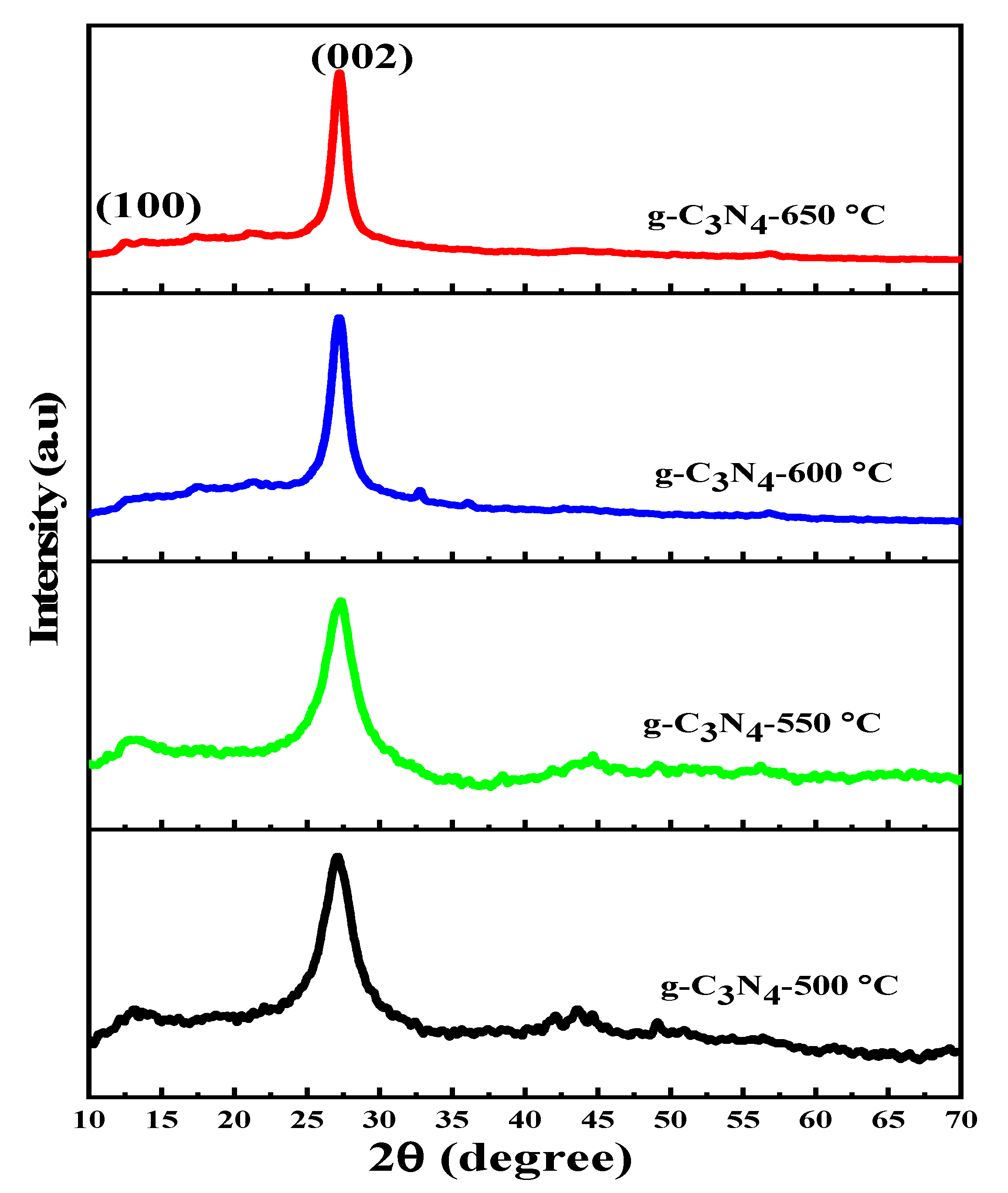
Figure 1. Calcination temperature-dependent XRD patterns of g-C3N4.
Nanomaterials 13 00762 g001
Figure 2. Photoluminescence spectra of g-C3N4 samples prepared at different calcination temperatures.
Nanomaterials 13 00762 g002
Figure 3. (a) UV-Vis DRS spectra and (b) corresponding Kubelka–Munk function plot of all prepared g-C3N4 samples.
Nanomaterials 13 00762 g003
Figure 4. (a) FTIR spectra and (b) TGA curves of g-C3N4 photocatalysts prepared at different calcination temperatures.
Nanomaterials 13 00762 g004
Figure 5. FE-SEM (a,b) HR-TEM images of g-C3N4 samples obtained at 600 °C.
Nanomaterials 13 00762 g005
Figure 6. (a) Photoactivity of RhB of all prepared g-C3N4 photocatalysts under visible-LED light irradiation (b) kinetics of RhB degradation as function of ln(C0/C) vs. irradiation time of all prepared g-C3N4 photocatalysts and (c) UV-Vis spectra of RhB at different irradiation times over g-C3N4-600 °C (with inset image at t = 0 and t = 180 min).
Nanomaterials 13 00762 g006
Table 1. Summary of structural properties and bandgap energy (eV) of all prepared g-C3N4 photocatalysts.
Samples2θ (°)β (FWHM) (Rad)Crystalline Size (nm)dhkl-Spacing (nm)Eg (eV)
g-C3N4-500 °C27.080.0642.230.3292.78
g-C3N4-550 °C27.300.0562.550.3202.75
g-C3N4-600 °C27.190.0334.320.3282.82
g-C3N4-650 °C27.250.0265.490.3272.89
Table 2. Photodegradation efficiency and reaction rate constant of all prepared g-C3N4 samples at different calcination temperatures.
S. No.PhotocatalystDegradation
Efficiency
Adsorption EfficiencyReaction Rate Constant (k) (min−1)
1g-C3N4-500 °C18.34%1.85%0.00113
2g-C3N4-550 °C22.20%0.66%0.00139
3g-C3N4-600 °C94.83%3.5%0.01646
4g-C3N4-650 °C94.80%1.55%0.01643
Table 3. Comparison of photocatalytic activities of some selected photocatalysts with the present work.
PhotocatalystCatalyst DosageLight SourceIrradiation TimePollutant LoadDegradation EfficiencyRef.
U-g-C3N410 mg300 W xenon lamp25 minRhB
(0.25 μmol/L)
100%[20]
CN-700 °C100 mg1000 W xenon lamp30 minRhB
(100 mg/L)
99.11%[19]
p–SCN–250 mg1000 W Xe lamp90 minRhB
(10 ppm)
97.5%[63]
75:25SCN0.12 g100 W tungsten lamp420 minRhB
(30 mg/L)
84.25%[46]
g-C3N4-600 °C50 mg50 W LED lamp180 minRhB
(10 mg/L)
94.83% This work
Disclaimer/Publisher’s Note: The statements, opinions and data contained in all publications are solely those of the individual author(s) and contributor(s) and not of MDPI and/or the editor(s). MDPI and/or the editor(s) disclaim responsibility for any injury to people or property resulting from any ideas, methods, instructions or products referred to in the content.