Article

X-ray Absorption Spectroscopy Study of Thickness Effects on the Structural and Magnetic Properties of Pr2−δNi1−xMn1+xO6−y Double Perovskite Thin Films

by
Mónica Bernal-Salamanca
1,*,
Javier Herrero-Martín
2,
Zorica Konstantinović
3,
Lluis Balcells
1,
Alberto Pomar
1,
Benjamín Martínez
1 and
Carlos Frontera
1,*
1
Institut de Ciència de Materials de Barcelona, ICMAB-CSIC, Campus UAB, 08193 Cerdanyola del Vallès, Spain
2
ALBA Synchrotron Light Source, 08920 Cerdanyola del Vallès, Spain
3
Center for Solid State Physics and New Materials, Institute of Physics Belgrade, University of Belgrade, Pregrevica 118, 11080 Belgrade, Serbia
*
Authors to whom correspondence should be addressed.
Nanomaterials 2022, 12(23), 4337; https://doi.org/10.3390/nano12234337
Submission received: 17 November 2022 / Revised: 1 December 2022 / Accepted: 2 December 2022 / Published: 6 December 2022

Abstract

:
In this work, we report a systematic study of the influence of film thickness on the structural and magnetic properties of epitaxial thin films of Pr2−δNi1−xMn1+xO6−y (PNMO) double perovskite grown on top of two different (001)-SrTiO3 and (001)-LaAlO3 substrates by RF magnetron sputtering. A strong dependence of the structural and magnetic properties on the film thickness is found. The ferromagnetic transition temperature (TC) and saturation magnetization (Ms) are found to decrease when reducing the film thickness. In our case, the thinnest films show a loss of ferromagnetism at the film-substrate interface. In addition, the electronic structure of some characteristic PNMO samples is deeply analyzed using X-ray absorption spectroscopy (XAS) and X-ray magnetic circular dichroism (XMCD) measurements and compared with theoretical simulations. Our results show that the oxidation states of Ni and Mn ions are stabilized as Ni2+ and Mn4+, thus the ferromagnetism is mainly due to Ni2+-O-Mn4+ superexchange interactions, even in samples with poor ferromagnetic properties. XMCD results also make evident large variations on the spin and orbital contributions to the magnetic moment as the film’s thickness decreases.

1. Introduction

Double perovskite (DP) oxides of the R2NiMnO6 family (RNMO, where R is a rare earth element) have attracted much attention from the scientific community due to their potential interest for future technological applications. Particularly, these materials are attractive because, being ferromagnetic insulators (FM-Is), they are promising candidates for applications in spintronic devices, such as multiple state logic devices, magnetodielectric capacitors, and spin filters tunnel junctions [1,2,3,4,5]. Since FM-Is are very scarce, DPs of the RNMO family may play a relevant role in the future development of spintronics because they are among the few known FM-Is [6,7,8]. Magnetic tunnel junctions (MTJs), one of the most important spintronic devices, require high spin-polarized materials to enhance the performance of tunnel magnetoresistance (TMR), and FM-Is have the potential to increase the magnitude of TMR as spin-filtering barriers [9]. The spin filtering effect of FM-I barriers is caused by the spin-sensitive conductance induced by spin-dependent potentials in FM-Is [10,11].
Previous reports on this class of compounds (RNMO) have been mainly focused on La2NiMnO6 (LNMO) due to its stable ferromagnetic insulating phase, high Curie temperature (TC ≈ 280 K), magneto-dielectric properties, spin-phonon coupling, and even catalytic properties [2,12,13,14]. Nevertheless, there are few studies available in the literature of other members of this family, either in bulk or thin film form, such as Pr2NiMnO6 (PNMO). According to the Goodenough-Kanamori rules, the magnetic ground states of RNMO systems are often expected to be ferromagnetic because of the superexchange interaction between the half-filled eg orbitals of Ni2+ ( d 8 ,   S = 1 ) and empty eg orbitals of Mn+4  ( d 3 ,   S = 3 / 2 ) , which are ferromagnetic via 180° TM( e g 2 )–O–TM ( e g 0 ) geometry [15,16,17,18]. However, the presence of anti-site disorder with the interchange between Ni and Mn atom positions, as well as oxygen vacancies, may result in antiferromagnetic (AF) coupling due to superexchange interactions between Mn4+−O−Mn4+ and Ni2+−O−Ni2 ions [6,19,20,21]. Consequently, the physical properties of these materials are found to be very sensitive to the Ni/Mn ordering in the B-site of the DP structure.
The properties of the RNMO family show a gradual structural change as the A-site is occupied by rare-earth elements R3+ with a smaller ionic radius. As a result, the magnetic transition temperature, TC, decreases monotonously with decreasing the rR3+ radius and the octahedral tilting of MnO6 and NiO6 octahedra increases [8,18,22]. Therefore, the superexchange interaction between the Ni2+ and Mn4+ ions is affected due to the larger deviation of the Ni–O–Mn bond angle from 180°. Therefore, the crystal structure and the magnetic behavior of these materials are correlated not only to the Ni−O−Mn bond angle but also to the variation of covalency/ionicity of the Ni/Mn−O bond length [8,18,23,24]. On the other hand, it has been found that for RNMO double perovskites, one of the greatest challenges is to control the ordering of the B-site cations (Ni/Mn), which strongly affects the microstructure and physical properties of RNMO thin films. Particularly, B-site ordering can be influenced by several factors, such as the growth conditions (i.e., growth temperature, oxygen pressure, and annealing), the epitaxial strain induced by the film-substrate lattice mismatch, and film thickness. In this regard, it is noted that in comparison with the studies on the synthesis conditions and properties of LNMO compounds, investigations on PNMO thin films related to these growth parameters are still very scarce [2,6,19].
Our studies are focused on PNMO thin films, which are a less explored member of the RNMO family. The parent compounds of PNMO are PrNiO3 and PrMnO3 single perovskites, which have an orthorhombic Pbnm structure with Pr3+ occupying the A-site and Ni3+ and Mn+3 occupying the B-sites [25,26]. PrMnO3 and PrNiO3 are A-type and G-type antiferromagnetic insulators, while Pr2NiMnO6 double perovskite, with Pr3+ (~1.06 Å) occupying the A-site and Mn4+ (~0.53 Å), Ni2+ (~0.69 Å) occupying the B and B’ sites in A2BB’O6 with rock-salt type order, is a ferromagnetic insulator [27,28]. Studies have demonstrated that well-ordered Pr2NiMnO6 in bulk or film form may be arranged in a monoclinic P21/n structure with Ni2+ and Mn4+ cations alternatively arranged at the B sites [8,27].
In a previous work [29], we have carried out a detailed optimization of the growth conditions (such as oxygen partial pressure and growth/annealing temperature) of PNMO thin films on top of (001)-oriented SrTiO3 (STO) substrates by RF magnetron sputtering technique. In our results, we have obtained high-quality double perovskite PNMO thin films with good ferromagnetic properties (i.e., TC ≈ 210 K and Ms ≈ 4.5 µB/f.u. at 10 K, very close to the bulk value) and insulating behavior using high oxygen pressures (350 mTorr) and high growth/annealing temperatures (800–850 °C). In order to obtain additional information on the composition of the films, we have analyzed the stoichiometry by electron probe microanalysis (EPMA). Particularly, the stoichiometry of the PNMO films has revealed that samples grown under high oxygen pressures (PO2 ≥ 350 mTorr) show a certain degree of Pr deficiency (not related to Pr migration as in, e.g., [30]). Cationic vacancies can have an impact on the properties of perovskite oxides [31,32]. Nevertheless, in spite of this Pr deficiency, the stoichiometry of the samples has little impact on the ferromagnetic properties. In this regard, the stoichiometry of our PNMO films has been expressed as Pr2−δNi1−xMn1+xO6−y.
Taking into account these previous results, in the first part of this work, we study the dependence of the structural and magnetic properties on the film thickness of PNMO samples deposited on top of two different substrates, namely (001)-oriented SrTiO3 (STO) and (001)-oriented LaAlO3 (LAO). The purpose of selecting two types of substrates, which impose different structural strains, is to evaluate the effect of structural strain (induced by lattice mismatch with the underlying substrate) on the crystal structure of the films and its impact on the ferromagnetic properties. At the same time, the structural strain is also expected to be strongly dependent on the film thickness. Strain effects often modify both in-plane and out-of-plane lattice parameters when varying the film thickness. In this regard, examining the physical properties of ultrathin PNMO films (~3 nm thick) could be useful for applications such as spin filters in tunnel barriers. In our case, both the structural and ferromagnetic properties of PNMO films have shown strong dependence on film thickness. In particular, the thinnest films showed a loss of ferromagnetism at the interface. On the other hand, the selection of the substrates also plays an important role in controlling the nature of magnetic anisotropy. Additionally, in the second part of this paper, we have focused our attention on exploring the local electronic structure of some representative PNMO samples deposited on STO and LAO substrates (both for high and low-TC), using X-ray absorption spectroscopy (XAS) and X-ray magnetic circular dichroism (XMCD) measurements. Finally, experimental data have been compared with theoretical simulations of the XAS and XMCD spectra.

2. Materials and Methods

Pr2−δNi1−xMn1+xO6−y (PNMO) films were deposited on top of (001)-oriented SrTiO3 (STO) and (001)-oriented LaAlO3 (LAO) substrates by RF magnetron sputtering technique, using a stoichiometric Pr2NiMnO6 target prepared by the solid-state reaction method [29]. Before deposition, substrates were cleaned in an ultrasonic bath with Milli-Q water and then annealed at 1000 °C in air for 2 h to obtain a clean and smooth step-terrace morphology [33,34].
Films were grown under an oxygen pressure of 350 mTorr and a growth temperature of 800 °C. The optimization of the growth conditions and the stoichiometry of the samples (obtained by EPMA) have been reported elsewhere [29]. The film thickness (t) determined by X-ray reflectivity was modulated by varying the deposition time (i.e., 60, 30, 15, 7, 5, and 3 min). After thin film growth, samples were annealed in-situ at the same growth temperature (800 °C) for 1 h under high oxygen pressure (420 Torr) and then slowly cooled down to room temperature at 10 °C/min. All PNMO films were prepared at a fixed RF power of 40 W and a fixed target-to-substrate distance of 5 cm, respectively.
The surface morphology of the samples was characterized by atomic force microscopy (AFM, MFP-3D AFM Asylum Research, Goleta, CA, USA) in tapping mode. The crystallinity quality of the samples was studied by X-ray diffraction (XRD), and the film thickness was determined by X-ray reflectivity (XRR) using a Bruker D8-Advance and a Siemens D5000 diffractometer (Cu-Kα1 and Cu-Kα1,2 radiation, respectively, both from ICMAB’s scientific and technical services). Synchrotron X-ray diffraction measurements were performed using the KMC-II beamline of BESSY (Berliner Elektronen-Speicherring Gesellschaft für Synchrotronstrahlung, Hemholtz Zentrum Berlin). Magnetization measurements were done using a superconducting quantum interferometer device (SQUID, Quantum Design, from ICMAB’s scientific and technical services).
X-ray absorption spectroscopy (XAS) and X-ray magnetic circular dichroism (XMCD) were investigated at the Pr M4,5, Ni L2,3, Mn L2,3 and O K edges in the BL29-BOREAS beamline [35] at the ALBA Synchrotron Light Source (Barcelona, Spain). The spectra were measured in total electron yield (TEY) mode at T = 100 K under ultrahigh vacuum conditions (2 × 10⁻¹⁰ mbar). The applied magnetic field (parallel to the X-ray beam) was 2 T. These experiments were also supported by theoretical simulations. The degree of circular polarization of the beam in the energy range used is higher than 99% [35].

3. Results and Discussion

3.1. Structural Properties

Figure 1a,c show the XRD diffraction patterns of the PNMO films deposited on STO (001) and LAO (001) substrates. Accordingly, the PNMO films (apPNMO bulk 3.871 Å [29], where ap is the bulk pseudocubic cell parameter) grown on STO (ap STO ≈ 3.905 Å) and LAO (ap LAO ≈ 3.789 Å) substrates are under tensile strain and compressive strain, respectively. The highest intensity peak in each diffractogram corresponds to the STO and LAO substrate reflections (see dashed vertical black lines). Furthermore, all XRD patterns show a clear thickness dependence on structural properties in the PNMO films.
For more details, Figure 1b,d show the (002) reflection of both the STO and LAO substrates and the PNMO film, respectively. As expected, the (002) peak of the film is placed at a 2θ position larger (smaller) than that of the bulk PNMO (see dashed vertical red line) for the PNMO/STO (PNMO/LAO) substrate. This observation indicates that the out-of-plane c lattice parameter shrinks when the film is under in-plane tensile strain and expands when it is under in-plane compressive strain, in agreement with the lattice mismatch imposing a tensile and a compressive in-plane strain. At the same time, in Figure 1b,d, it can be appreciated that the position of the (002) peak shows a slight shift towards lower 2θ angles (higher 2θ angles) when increasing the film thickness for PNMO/STO (PNMO/LAO) substrate (see arrow). This indicates that the out-of-plane c lattice parameter of the film increases with increasing film thickness for STO, while it decreases for LAO, approaching the bulk value in both cases (see Figure 3a,b).
Finally, additional reflections denoted by (*), located at 2θ ≈ 43.9° (PNMO/STO films) and 2θ ≈ 43.5° (PNMO/LAO films), correspond to the parasitic NiO phase, as similarly observed in samples deposited at different pressures and temperatures [29]. The presence of the secondary NiO phase in the PNMO compound is not well understood. As the film thickness increases, the parasitic NiO peak increases in intensity. This fact points out the possibility of an increasing Pr-deficiency upon increasing the thickness, as revealed by EPMA for thick samples [29].
In order to discern the orientation of the film cell axes (monoclinic or orthorhombic) with respect to the substrate, we have explored, in reciprocal space (using a four circle diffractometer at the KMC-II beamline of the BESSY synchrotron), the appearance of the (021)m reflection of PNMO (the subscript “m” stands for indexation using the monoclinic √2apx√2apx2ap cell). This reflection is equivalent to (11½) of STO when c of PNMO is oriented along (001) of the substrate and is equivalent to (1½1) or (½11) of STO when c of PNMO lies along (010) or (100) of the substrate, respectively. As can be seen in Figure 2a, the two types of orientation are present for the 47.6-nm-thick PNMO film on STO (001) substrate, with a strong predominance of the orientation with c in-plane. On the contrary, in Figure 2b, for the 43.4-nm-thick PNMO film on LAO (001) substrate, the relative orientation of the monoclinic cell of the PNMO film is only oriented with c in-plane, and no domains with c out-of-plane can be detected.
In order to determine the values of the in-plane (a) and out-of-plane (c) lattice parameters, reciprocal space maps (RSMs) around (−103) reflection were performed on PNMO/STO and PNMO/LAO samples. In the RSMs shown in Figure 2c–f, the x axis corresponds to the in-plane qx [100] direction, and the y axis corresponds to the out-of-plane qz [001] direction. RSMs around the (−103) STO and (−103) PNMO reflections of the thinner (5.2 nm) and thicker (47.6 nm) PNMO/STO samples are shown in Figure 2c,e, respectively. Analog RSMs around the (−103) LAO and (−103) PNMO reflections of the thinner (4.7 nm) and thicker (43.4 nm) PNMO/LAO samples are shown in Figure 2d,f respectively. In Figure 2c,d, the RSMs for thinner films reveal both film and substrate (−103) diffraction spots are placed at the same position in qx, so the estimated in-plane (a) pseudocubic cell parameters of the film coincide with those of the STO substrate (aSTO = 3.905 Å) and LAO substrate (aLAO = 3.789 Å), showing that the films grow in-plane fully strained. On the contrary, the out-of-plane (c) lattice parameters were found to be c = 3.831 Å for PNMO/STO film and c = 3.890 Å for PNMO/LAO film, respectively.
Concerning the thicker films in Figure 2e,f, the RSMs reveal that the peak position qx of (−103) PNMO film reflections is slightly shifted along the in-plane direction with respect to the position of the corresponding substrate, indicating a partial relaxation of the cell. This shift, in accordance with the strain induced, is towards larger absolute values of qx for PNMO/STO and towards smaller absolute values for PNMO/LAO. From the positions of the peaks, the estimated cell parameters of a 47.6-nm thick PNMO/STO film are a = 3.877 Å and c = 3.848 Å, and those of a 43.4-nm thick PNMO/LAO film are a = 3.842 Å and c = 3.874 Å, respectively. These values are found to be similar to those of the La2NiMnO6 [6,36] and Pr2NiMnO6 systems [7].
The variation of the cell parameters (in pseudo-cubic notation) for both substrates is depicted in Figure 3a,b, respectively. It can be observed that when the film thickness increases, the in-plane (a) lattice parameter decreases (increases) for STO (LAO) towards the bulk value (see the red and blue dashed lines). Furthermore, the out-of-plane (c) lattice parameter progressively increases (decreases) for STO (LAO) with increasing thickness (see the black dashed line). In this regard, strain effects modify both in-plane and out-of-plane parameters by varying the film thickness. Both the tensile and compressive strains have a dominant effect in PNMO films with low thickness, affecting the lattice parameters strongly. Thus, the in-plane lattice parameters of the film tend to acquire the same value as that of the substrate. Therefore, from these observations, a (partial) relaxation of the in-plane (a) tensile strain and compressive strain takes place when film thickness increases, and consequently, the lattice parameters tend to acquire the bulk value (abulk = 3.871 Å) [37,38].

3.2. Magnetic Properties

In order to explore the thickness dependence on the magnetic properties, Figure 4 shows the in-plane magnetization of PNMO/STO and PNMO/LAO samples of different thicknesses (t) as a function of temperature under an applied magnetic field of 5 kOe. Temperature-dependent magnetization M(T) of PNMO films grown on STO and LAO substrates with different thicknesses is depicted in Figure 4a,d, respectively. The TC value (estimated from the inflection point) was extracted and plotted in Figure 4b,e. From the results, it can be appreciated that the magnetization and the Curie temperature TC (onset of the ferromagnetic behavior) reach lower values as the film thickness decreases. Therefore, a notable degradation of the magnetic properties takes place as the samples become thinner.
In fact, the absence of a ferromagnetic ordering has been reported in ultrathin films (t < 4 nm) [39,40,41]. This could be attributed, as a first approximation, to the existence of an interfacial dead layer that modifies the magnetic and structural properties. Some factors that contribute to the formation of a dead layer effect on very thin films could be a chemically and/or structurally altered film-substrate interface as well as a discontinuous film coverage over the substrate surface during the initial film growth [42]. The insets in Figure 4b,e depict the magnetization (emu/cm2)*103 at 10 K as a function of thickness. Therefore, by extrapolating to zero, the thickness of the dead layer for PNMO films was estimated to be around ~3 nm (on both substrates).
At the same time, it should also be noticed that the M(T) curve, for the thickest (47.6 nm) PNMO/STO film, displays a FM transition at TC ≈ 210 K and a saturation magnetization of Ms ≈ 4.5 µB/f.u. at 10 K (see Figure 4c), which is very similar to that reported in the literature and close to the bulk value [7]. The thickest (43.4 nm) PNMO/LAO film displays TC ≈ 216 K and Ms ≈ 4.85 µB/f.u. at 10 K (see Figure 4f), also very close to the bulk value Ms = 5 µB/f.u. [6]. For comparison, the Curie temperature (TC), saturation magnetization (Ms), coercive field (HC), and remanence magnetization (Mr) data are listed in Table 1 for thicker samples (on both substrates). In this regard, the M(H) curves reveal that the easy magnetization axis prevails in the IP orientation for both substrates. The HC and Mr reinforce that the easy axis lies in the IP orientation. On the other hand, a coercive field HC of about 565 Oe (IP field) and 264 Oe (OP field) is found for the 47.6-nm-thick PNMO/STO film, while a coercive field HC of about 538 Oe (IP field) and 631 Oe (OP field) is found for the 43.4-nm-thick PNMO/LAO film. This could indicate that the IP anisotropy is higher in the PNMO/LAO film, in agreement with the larger coercive field (OP), than in the PNMO/STO film.

3.3. XAS and XMCD

To evaluate the spin and orbital moments, the valence state of ions, and the nature of the ferromagnetic (FM) interactions in the PNMO compounds, XAS and XMCD measurements were carried out at the Ni L2,3, Mn L2,3, Pr M4,5, and O K edges. At the same time, in order to qualitatively analyze the ferromagnetic character of the PNMO system, we performed X-ray spectroscopic calculations using CTM4XAS [43] and Crispy software [44,45]. For this study, we have chosen three PNMO samples of different thicknesses and different Curie temperatures (high-TC and low-TC) deposited on STO and LAO substrates. The first two samples with good FM properties were a 47.6-nm-thick PNMO/STO sample (TC ≈ 210 K and Ms ≈ 4.5 µB/f.u at 10 K) and another 43.4-nm-thick PNMO/LAO sample (TC ≈ 216 K and Ms ≈ 4.7 µB/f.u at 10 K) with a Pr:(Ni + Mn) ratio of ~0.86 (Pr1.7Ni0.9Mn1.1O6-y) [29]. The third was a 4.7-nm-thick PNMO/LAO sample with poor FM properties (TC ≈ 95 K). In the following, the PNMO samples grown on STO substrate (high-TC) were labeled as PNMO/STO-(A), and the thicker (high-TC) and thinner (low-TC) samples grown on LAO substrates were labeled as PNMO/LAO-(B) and PNMO/LAO-(C), respectively.

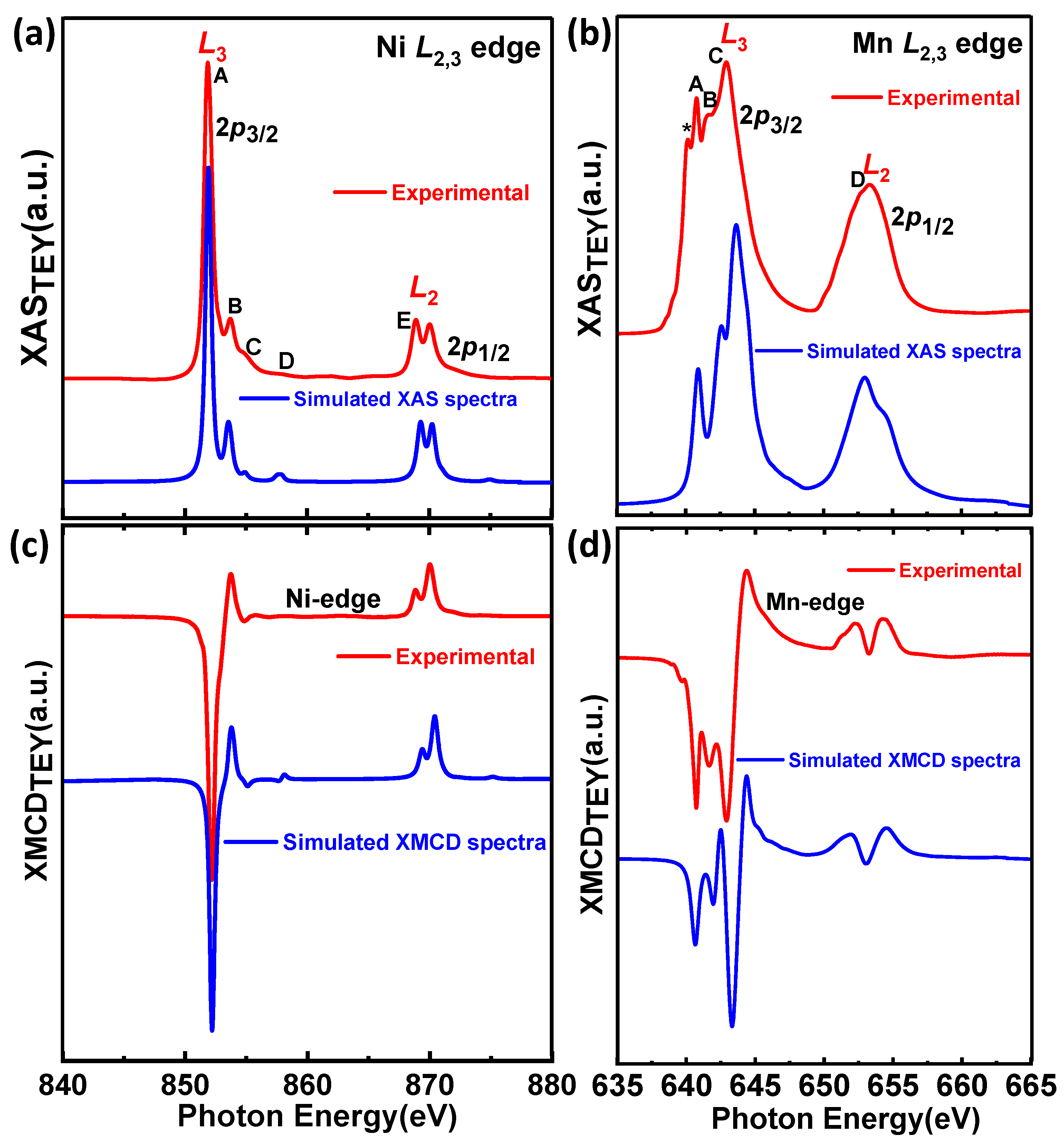
3.3.1. Ni and Mn L2,3 Edges

Considering that the ferromagnetic character of Pr2NiMnO6 is explained in terms of the superexchange interactions between Ni2+ and Mn4+ according to the Goodenough-Kanamori rules, it is of major interest to determine the valence states and orbital occupancies of these two ions. For this purpose, we recorded the XAS and XMCD spectra across the Ni-L2,3 edges (2p→3d transitions) for the three samples mentioned earlier (see Figure 5a–c).
We found that the strong Ni L3 edge peak and the Ni L2 edge double-peak structure of (a) PNMO/STO-(A), (b) PNMO/LAO-(B), and (c) PNMO/LAO-(C) samples were very similar to those already reported for La2NiMnO6 [46,47], Pr2NiMnO6 [28], and Nd2NiMnO6 [48] double perovskites. For the thinner PNMO/LAO-(C) sample (low-TC), a sharp peak appears at around 849.5 eV, which is due to the La M4 (3d3/2→4f) XAS from the substrate. Beyond this particularity, we can say that both the Ni L3 (ħυ ≈ 850 − 855 eV) and L2 edge (ℏυ ≈ 865 − 875 eV) XAS line shapes are quite similar and can be easily compared with the corresponding spectra of other divalent Ni2+ compounds, such as isoelectronic NiO (also shown in the inset) or Ni dihalides [49].
According to the electric dipole selection rules, Ni 2p electrons may be excited into empty states either with 3d or 4s symmetry. The 2p→3d transitions are about 30 times stronger in intensity than 2p→4s ones due to the large overlap of the 3d wave functions with the 2p ones (Fermi’s golden rule) [50]. The presence of this double-peak structure (labeled as E and shown in the inset) in Ni L2 edges is nevertheless well understood in terms of a covalent ground state of mainly Ni2+ (3d8) character, which in Oh symmetry can be written as 3A2g ( t 2 g 6   e g 2 ) plus an anion-dependent fraction of the 3d9L and 3d10L2 configurations, where L corresponds to a ligand hole in the O 2p state [49]. This double-peak at the Ni L2 edge was previously observed in a nonstoichiometric sample of the La2Ni1-xMn1+xO6 series [51]. In a similar way, this double peak was also found in stoichiometric samples of R2NiMnO6 (R = La, Pr, and Nd) with almost full cationic ordering, yet very different from the L2 peak of PrNiO3 and NdNiO3 corresponding to Ni3+ [28,46,48]. Therefore, we can conclude that Ni ions in our three PNMO samples are in a divalent state with a high-spin (HS) electronic configuration (Ni2+: t 2 g 3 t 2 g 3 e g 2 ) [52,53]. We further examined this point by means of XAS and XMCD simulations (see Section 3.3.2).
The Mn-L2,3 edge XAS spectra as collected by TEY at T = 100 K for the three PNMO samples investigated are shown in Figure 6 and Figure 7. They all look very similar to the XAS previously reported for La2NiMnO6 [46,47], Pr2NiMnO6 [28], and Nd2NiMnO6 [48] double perovskites. Additionally, in order to qualitatively evaluate the valence state of Mn ions in our three PNMO samples, we have also recorded the Mn L2,3 XAS of some reference samples: LaMnO3 (Mn3+), La2Ni0.6Mn1.4O6 (mixed-valence, Mn3.6+) and SrMnO3 (Mn4+) (see Figure 6). The energy position (particularly at the L3-edge) and the overall spectral shape of our PNMO samples are quite similar to those of SrMnO3 and to other nominal Mn4+ references with Oh crystal field symmetry, like LaMn0.5Ni0.5O3 [54], LaMn0.5Co0.5O3 [55], and Ca3CoMnO6 [56], but clearly different from those in LaMnO3. Nevertheless, a small feature at approximately 640.2 eV, labeled with (*), and identified as due to Mn2+ is also present in our case. This could be related to surface contamination in the films. In any case, the overall Mn-L2,3 XAS spectra of PNMO and SrMnO3 are quite similar, which indicates that Mn in our PNMO samples is very likely, mostly in a tetravalent state (Mn4+: t 2 g 3 ) [52,53]. In order to confirm this hypothesis, as in the case of the Ni-L2,3 edges, we performed XAS and XMCD calculations, which are shown in Section 3.3.2.
In Figure 7a–c, the Mn L3-edge main x-ray spectroscopic structures have been labeled as A, B, and C, while D corresponds to the L2-edge. In addition, the presence of Mn2+ is denoted by (*). All three samples show similar Mn-L2,3 XAS (see Figure 6), but the D feature in the PNMO/LAO-(C) sample is slightly more prominent than in the other two samples.
At the same time, it also shows slight differences in the intensity of peaks A, B, and C. In this case, the branching ratio (defined as I(L2)/I(L3), where I(L3) and I(L2) are the XAS maximum amplitudes at the L3 and L2 peaks, respectively) is larger for the PNMO/LAO-(C) sample (=0.581). This might be associated with an electron-yield saturation effect at the Mn L3-edge due to the thickness of the samples [57,58,59,60]. With PNMO/LAO-(C) being the thinnest sample (4.7 nm), a saturation effect would enhance the intensity of the spectroscopic features at low energy within a given absorption edge as compared to those in the higher energy part. Saturation effects result in a recorded signal that is not proportional to the photoabsorption cross-section as the photon energy is varied. In this case, the intensities of prominent absorption peaks get reduced or “saturated” [61]. Though being stronger at more grazing photon incidence, in sufficiently thin films saturation can also affect spectra recorded at normal incidence [62].
X-ray magnetic circular dichroism (XMCD) was used to investigate the specific magnetic ordering, namely the nature of exchange couplings between the different magnetic sites (Ni, Mn, and Pr) and O in the PNMO compounds. Panels (d), (e), and (f) of Figure 5 and Figure 7 show the Ni and Mn L2,3 edge XMCD spectra for PNMO/STO-(A), PNMO/LAO-(B), and PNMO/LAO-(C) films, as recorded at T = 100 K, under an applied field of 2 T. This temperature value was chosen to lie well below the high-temperature magnetic transition at 216 K. We note that all XMCD spectra were normalized to the integrated area of the corresponding XAS spectra to ease their comparison [53,54]. Looking at the large negative XMCD signal in both the Mn and Ni-L3 regions, we can extract that the Mn and Ni spin moments are ferromagnetically coupled to each other, as also proposed in La2NiMnO6 [46].
In order to extract quantitative information about the orbital angular µorb and spin magnetic moment µspin contributions to Mn 3d and Ni 3d state magnetization, we applied the sum-rules to the XMCD spectra. For this, we took the threshold between the 2p3/2 and 2p1/2 regions at 650 eV for the Mn L2,3 edges and at 865 eV for the Ni L2,3 edges and neglected the contribution of the magnetic dipole operator TZ [46,63,64]. We have that:
μ L = 4 ( L 3 Δ I ( E ) d E + L 2 Δ I ( E ) d E ) 3 ( L 3 I ( E ) d E + L 2 I ( E ) d E ) ( 10 N 3 d )
μ S + 7 μ T = 2 L 3 Δ I ( E ) d E 4 L 2 Δ I ( E ) d E L 3 I ( E ) d E + L 2 I ( E ) d E ( 10 N 3 d )
where ΔI = I+I; I = I+ + I; N3d is the 3d electron occupation number; and µT is the magnetic dipole moment (usually negligible for transition metals in a local octahedral environment). The corresponding integral of the XMCD signal is also depicted in panels (d), (e), and (f) of Figure 5 and Figure 7.
Therefore, using Equations (1) and (2), at 100 K, we obtained μ L N i / μ S N i = 0.24 1 and μ L M n / μ S M n = 0.099 for PNMO/STO-(A), μ L N i / μ S N i = 0.167   and μ L M n / μ S M n = 0.055 for the PNMO/LAO-(B) sample, and for the thinner PNMO/LAO-(C) sample (low-TC), we obtained μ L N i / μ S N i = 0.077 and μ L M n / μ S M n = 0.018 . These values are similar to those in previous reports [46,54]. The orbital moment values we obtained are in all cases compatible with Mn4+ ions, where they are expected to be quenched. Regarding the spin moment, we must note that the difficulty in separating the L3 from the L2 edges of Mn4+ introduces a large degree of uncertainty. Following [65,66], μS could be underestimated by a factor of 0.59. In the case of Ni ions, the spin-orbit coupling is larger, and the spin momentum calculated value using the corresponding XMCD-derived sum rule is estimated not to deviate more than 10% from the actual value [67].
Then, based on the XMCD data, we can say that (i) the Mn orbital to spin moment ratio is directly proportional to thickness, being nearly quenched in the thinnest sample investigated, and (ii) the Ni XMCD signal being in general very small at the L2 edge while still largely negative at the L3 edge indicates a very large orbital contribution to the Ni magnetic moment. This allows us to conclude that the magnetic anisotropy observed in the magnetic measurements (M(H) loops of the PNMO/STO-(A) and PNMO/LAO-(B) samples) is of magnetocrystalline origin and induced by Ni (see Figure 4c,f).

3.3.2. Numerical Simulation of XAS and XMCD Spectra of Ni2+ and Mn4+ Edges

In order to obtain more detailed information on the local electronic structure of Ni and Mn edges of our PNMO compounds, theoretical simulations of the XAS and XMCD spectra were performed to fit the experimental data using CTM4XAS [43]. Figure 8 displays the best calculated XAS and the corresponding XMCD spectra for Ni2+ and Mn4+ ions. For the sake of comparison, the experimental spectra of the PNMO/STO-(A) sample are also plotted.
The Ni and Mn-L2,3 edges spectra are calculated from the sum of all possible transitions for an electron excited from the 2p core level to an unoccupied 3d level. The ground state is approximated by the electronic configuration 3dn. For a transition-metal ion in octahedral symmetry, the crystal field multiplet calculation uses an empirical value of the crystal field splitting 10 Dq (energy between the t2g and eg states). In the ground state, both the 3d spin−orbit coupling and the crystal field 10 Dq affect the 3dn configuration. The 3dn ground state and the 2p53dn+1 final state are affected by 3d3d and 2p3d intra-atomic Coulomb interactions (Udd, Upd). The 2p and 3d spin−orbit couplings and local crystal field, which are described with empirical parameters (10 Dq, Ds, Dt, and M) in appropriate symmetry, are also included [68,69,70]. In addition, the charge-transfer energy ∆ (needed to transfer one electron from the ligand band to the transition-metal site) is strongly anion dependent, being given roughly by the electronegativity difference between anion and cation. For high covalency, ∆ may be in the negative regime due to the strong hybridization with the oxygen band [71].
In the PNMO compound, Ni2+ ions are surrounded by oxygen octahedra, and their ground state ionic configuration (as a first approach) in Oh symmetry can be written as 3 A 2 g ( t 2 g 6 e g 2 ) . On the other hand, Mn4+ ion valence band filling can be written as 4 A 2 g ( t 2 g 3 ) , also in Oh symmetry [28]. For Ni2+ XAS calculations (see Figure 8a,c), d9L and d10L2 had to be actually considered, where L corresponds to a ligand hole in the O 2p state. As in the case of Mn4+ ions (see Figure 8b,d), this is due to the large covalency of metal-oxygen bonds, which renders the ionic approximation very inaccurate. In this latter case, the ground state could be well described using d3 and d4L configurations [72].
In Figure 8a,c, when dealing with Ni-L2,3XAS and XMCD calculations, we observed that the double peak feature at both the L3 and L2 edges gets strongly affected by the charge transfer energy parameter (∆). So, for small (<3 eV) or even negative ∆ values, calculations do not properly fit the experimental data at the L2 edge, which leads to the formation of weak satellites and to changes in the multiplet structure. A good fit requires using a moderately positive ∆ value (∆ = 3.0 eV) and a crystal-field energy of 10Dq = 1.2 eV (see more details in Table 2). On the other hand, the number of holes for the Ni-L2,3 XAS and XMCD calculations is 1.82, which is in good agreement with the ionic expected value of Ni2+ (3d8 configuration plus some contribution of 3d9L and 3d10L2).
Figure 8b,d display the calculated XAS and XMCD spectra for Mn4+ ions for the PNMO compound. By comparing the XAS and XMCD spectra (experimental and simulated), it can be observed that the multiplet structure (spectrum shape) and the peaks marked as A, B, C, and D at the Mn-L3 and Mn-L2 edges fit well with the experimental data. For that, we used 10Dq = 2.5 eV and ∆ = 2.5 eV (see more details in Table 2). The number of holes that follows from these calculations is 6.69 per Mn atom.
Therefore, according to these results, we can conclude that Ni and Mn cations, respectively, adopt dominant divalent and tetravalent oxidation states in Oh local symmetry and HS configuration. On the other hand, considering that FM ordering in the PNMO double perovskite structure is due to the Ni2+–O– Mn4+ superexchange interactions according to the Goodenough-Kanamori rules, XAS measurements at the Ni and Mn L2,3 edges in our PNMO compound allow us to confirm this statement even in samples with low-TC. Similar results were observed on LNMO compounds [73].

3.3.3. Pr M4,5 Edges

The Pr M4,5 XAS spectra for the three samples under study with very different TC values, which probe the unoccupied density of 4f states, are shown in Figure 9a. The spectra of PrCoO3 (Pr3+) and PrO2 (Pr4+) at T = 300 K are used as references [74,75,76] in the same plot. In the literature, the experimental XAS spectra of formally tetravalent 4f oxides (namely PrO2, CeO2, and LaO2) show a main broad peak at both the M5 and M4 absorption edges [75,76]. Calculations for PrO2 in ref [75] finely reproduced its spectroscopic structure. In contrast, the richer multiplet structure of Pr3+-based bands is determined by a strong Coulomb interaction between the two 4f electrons as well as by a covalent mixing with oxygen 2p states. This has been earlier shown by XAS calculations for La3+, Ce3+ and Pr3+-based compounds [77]. In the case of our Pr M4,5 XAS spectra taken from PNMO samples, we can see in Figure 9a that it strongly resembles that of PrNiO3 [77] and PrCoO3, pointing to a trivalent state of Pr cations.
Nevertheless, in order to confirm this statement, we performed XAS and XMCD calculations using the Crispy interface, based on the Quanty code [44,45]. We considered both Pr3+ and Pr4+ ions, where the dipolar electronic transitions are 3d104f2→3d94f3 and 3d104f1→3d94f2, respectively, and Oh crystal field symmetry (see Figure 9c,d). Only the Pr3+ calculations were able to properly reproduce all XAS features (spectral shape and amplitude). We can thus conclude that the A-site deficiency in our Pr2−δNi1−xMn1+xO6−y compound, as analyzed by EPMA [29], has no evident impact on the oxidation state of Pr.
Regarding the XMCD, the experimental results obtained at Pr M4,5 edges in our three samples (see Figure 9b) show a dichroic signal with the opposite sign as compared to that found at the Ni and Mn L2,3 edges. In the case of rare earths, the final state multiplet 3d104fN → 3d94fN+1 is split in two parts by the 3d spin-orbit interaction, and, depending on N, the description involves the discrete energy levels of the initial and final state N-particle wavefunctions (multiplets) [78]. Each multiplet state has a definite atomic angular momentum quantum number J. In this description, the dipole selection rules are ∆J = 0, ±1, and only a part of these lines can be reached from the initial state [77,79].
At the same time, XMCD involves the contributions of dipole transitions with the effects of interatomic hybridization between the 4f states of Pr and the 3d states of Mn and Ni, and intra-atomic exchange interaction between the 4f–5d states of Pr. This implies that the inverted XMCD signal could be related to the coupling of the 4f electrons with the valence band as well as 3d–4f electron-electron interactions [78]. Moreover, there is a strong correlation between 3d→4f transitions (∆J = −1) of the Pr M4,5 edge. Hence, the ∆J = −1 terms could also be dominating in the XMCD spectrum, giving rise to an inverted dichroic signal. Otherwise said, this is not due to an antiparallel alignment of the Pr moments to the externally applied magnetic field or to Ni and Mn spins.
Concerning the calculated XMCD spectra, the Pr3+ case fits pretty well with the experimental data, which further corroborates that the valence state of Pr in our samples is 3+.

3.3.4. O K Edge

Figure 10a shows the O K XAS edge spectra of the same three samples with high-TC and low-Tc measured at T = 100 K in TEY mode. Focusing on the pre-edge zone, we can analyze the hybridization of oxygen valence states with unoccupied Ni/Mn 3d and Pr 5d bands [80]. The first peak found, located around 529.8 eV (in the three samples), corresponds to available O 2p–Ni/Mn 3d states. At higher energies, the broad structure around 535 eV corresponds to O 2p mixing with Pr 5d states, and the bumps around 540–545 eV are due to the hybridization of O 2p with Ni and Mn 4sp bands, which are consistent with earlier results reported on LaMnO3, LaFeO3, and LaCoO3 [81,82,83].
The XMCD results obtained at the O K-edge in our three samples are shown in Figure 10b. These are particularly relevant since the magnetic interaction is mediated by O ions. In Figure 10b, one can observe a strong negative peak that is more prominent for samples PNMO/STO-(A) and PNMO/LAO-(B), with an intensity equivalent to several percent of the total XAS intensity of the t2g region. This O K-edge XMCD signal is attributed to the 3d orbital moment on the neighboring sites of the Ni or Mn ions interacting through the p-d hybridization [84]. The intensity of this peak becomes much lower for the PNMO/LAO-(C) sample, which corroborates the loss of ferromagnetism in this last sample. On the other hand, because of the absence of spin-orbit splitting for the 1s core level, the O K-edge XMCD spectra show the orbital moment simply but are insensitive to the spin moment. Therefore, the integral area of the O K-edge XMCD is directly proportional to the orbital moment, and, since the negative XMCD signal indicates a positive magnetic moment (µL > 0), the orbital magnetic moment of O 2p is parallel to that of Ni/Mn 3d [85,86].

4. Conclusions

In summary, epitaxial PNMO thin films with varying thickness have been prepared on (001) STO and (001) LAO substrates under optimized growth conditions (grown/annealed at 800 °C under 350 mTorr O2) by the RF sputtering technique. PNMO films show a strong dependence of structural and magnetic properties on film thickness. Particularly, reciprocal space maps (RSMs) around (−103) reflection (on both substrates) reveal a (partial) relaxation of the in-plane tensile strain and compressive strain when film thickness increases; therefore, in-plane lattice parameters approach the bulk value. As structural strain decreases with increasing film thickness, the ferromagnetic behavior (on both substrates) improves and is optimal for the thicker PNMO films. In this regard, the Curie temperature (TC) and saturation magnetization (Ms) (i.e., for 47.6-nm-thick PNMO/STO film, TC ≈ 210 K and Ms ≈ 4.5 µB/f.u. and, for 43.4-nm-thick PNMO/LAO film, TC ≈ 216 K and Ms ≈ 4.85 µB/f.u., at 10 K) display values very close to the bulk value. In fact, M(H) curves reveal that the IP orientation of the easy magnetization axis prevails. Consequently, for thinner films, lattice distortion, oxygen deficiency, and sample inhomogeneity could induce a loss of ferromagnetism at the film-substrate interface. XAS and XMCD measurements on some characteristic samples (high and low-TC) deposited on STO and LAO substrates reveal that irrespective of the structural strain state (tensile or compressive) and the film thickness, the oxidation states of Ni and Mn ions are stabilized as Ni2+ and Mn4+, even in samples with poor magnetic properties. In addition, based on the XMCD data, the very large orbital moment contribution to the magnetic moment of Ni ions (on both substrates) allows us to conclude that the magnetic anisotropy observed in the magnetic measurements (M(H) loops) is of magnetocrystalline origin induced by Ni. At the same time, the Pr M4,5 edge XAS spectra of the rare earth element reveal that the valence state of the Pr ions is 3+, indicating that the Pr deficiency in our Pr2−δNi1−xMn1+ xO6−y (PNMO) compound (as analyzed by EPMA) had no evident impact on the oxidation state of Pr. Theoretical simulations based on a charge transfer multiplet model of XAS and XMCD data at Ni L2,3 and Mn L2,3 edges allow us to conclude that the experimental spectra are in good agreement with the calculated spectra of Ni2+ and Mn4+ in Oh symmetry and high-spin configuration.

Author Contributions

Conceptualization, M.B.-S., Z.K., L.B., C.F. and B.M.; methodology, M.B.-S., J.H.-M. and A.P.; validation, B.M., Z.K. and L.B.; formal analysis, C.F., J.H.-M. and M.B.-S.; investigation, M.B.-S., C.F., Z.K. and L.B.; resources, B.M. and L.B.; data curation, C.F., M.B.-S. and J.H.-M.; writing—original draft preparation, M.B.-S.; writing—review and editing, C.F., M.B.-S. and J.H.-M.; supervision, Z.K. and L.B.; funding acquisition, B.M. and L.B. All authors have read and agreed to the published version of the manuscript.

Funding

This research was founded by the Spanish Ministry of Science and Innovation through the Severo Ochoa (CEX2019-000917-S), SPINCURIOX (RTI2018-099960-BI00), and OXISOT (PID2021-128410OB-I00) projects, which were co-financed by the European Regional Development Funds. ZK acknowledges the support of the Institute of Physics Belgrade through a grant from the Serbian Ministry of Education, Science, and Technological Development.

Data Availability Statement

Not applicable.

Acknowledgments

The authors thank ALBA and BESSY synchrotron radiation facilities for the provision of beamtime, the kind help of D. Többens during data collection at BESSY, and the assistance from ICMAB-CSIC Scientific & Technological Services: X-ray Diffraction (J. Esquius and X. Campos), Electron Microscopy, and Low Temperatures and Magnetometry (B. Bozzo).

Conflicts of Interest

The authors declare no conflict of interest.

References

  1. Rogado, N.S.; Li, J.; Sleight, A.W.; Subramanian, M.A. Magnetocapacitance and magnetoresistance near room temperature in a ferromagnetic semiconductor: La2NiMnO6. Adv. Mater. 2005, 17, 2225–2227. [Google Scholar] [CrossRef]
  2. Hashisaka, M.; Kan, D.; Masuno, A.; Takano, M.; Shimakawa, Y. Epitaxial growth of ferromagnetic with ordered double-perovskite structure. Appl. Phys. Lett. 2006, 89, 032504. [Google Scholar] [CrossRef]
  3. Serrate, D.; de Teresa, J.M.; Ibarra, M.R. Double perovskites with ferromagnetism above room temperature. J. Phys. Condens. Matter 2007, 19, 023201. [Google Scholar] [CrossRef]
  4. Guo, Y.; Shi, L.; Zhou, S.; Zhao, J.; Liu, W. Near room-temperature magnetoresistance effect in double perovskite La2NiMnO6. Appl. Phys. Lett. 2013, 102, 222401. [Google Scholar] [CrossRef]
  5. Hashisaka, M.; Kan, D.; Masuno, A.; Terashima, T.; Takano, M.; Mibu, K. Spin-filtering effect of ferromagnetic semiconductor La2NiMnO6. J. Magn. Magn. Mater. 2007, 310, 1975–1977. [Google Scholar] [CrossRef]
  6. Dass, R.I.; Yan, J.-Q.; Goodenough, J.B. Oxygen stoichiometry, ferromagnetism and transport properties of La2−xNiMnO6+δ. Phys. Rev. B 2003, 68, 064415. [Google Scholar] [CrossRef]
  7. Singh, M.P.; Truong, K.D.; Jandl, S.; Fournier, P. Magnetic properties and phonon behavior of Pr2NiMnO6 thin films. Appl. Phys. Lett. 2011, 98, 162506. [Google Scholar] [CrossRef]
  8. Booth, R.J.; Fillman, R.; Whitaker, H.; Nag, A.; Tiwari, A.M.; Ramanujachary, K.V.; Gopalakrishnan, J.; Lofland, S.E. An investigation of structural, magnetic and dielectric properties of R2NiMnO6 (R = rare earth, Y). Mater. Res. Bull. 2009, 44, 1559. [Google Scholar] [CrossRef]
  9. Moodera, J.S.; Hao, X.; Gibson, G.A.; Meservey, R. Electron-Spin Polarization in Tunnel Junctions in Zero Applied Field with Ferromagnetic EuS Barriers. Phys. Rev. Lett. 1988, 61, 637–640. [Google Scholar] [CrossRef]
  10. LeClair, P.; Ha, J.K.; Swagten, H.J.M.; van de Vin, C.H.; Kohlhepp, J.T.; de Jonge, W.J.M. Large magnetoresistance using hybrid spin filter devices. Appl. Phys. Lett. 2002, 80, 625–627. [Google Scholar] [CrossRef]
  11. Fuh, H.R.; Liu, Y.P.; Xiao, Z.R.; Wang, Y.K. New type of ferromagnetic insulator: Double perovskite La2NiMO6 (M= Mn, Tc, Re, Ti, Zr, and Hf). J. Magn. Magn. Mater. 2014, 357, 7–12. [Google Scholar] [CrossRef]
  12. Iliev, M.N.; Guo, H.; Gupta, A. Raman spectroscopy evidence of strong spin-phonon coupling in epitaxial thin films of the double perovskite La2NiMnO6. Appl. Phys. Lett. 2007, 90, 151914. [Google Scholar] [CrossRef]
  13. Chandrasekhar, K.D.; Das, A.K.; Mitra, C.; Venimadhav, A. The extrinsic origin of the magnetodielectric effect in the double perovskite La2NiMnO6. J. Phys. Cond. Matter 2012, 24, 495901. [Google Scholar] [CrossRef]
  14. Sun, H.; Song, S.; Xu, X.; Dai, J.; Yu, J.; Zhou, W.; Shao, Z.; Jung, W. Recent Progress on Structurally Ordered Materials for Electrocatalysis. Adv. Energy Mater. 2021, 11, 2101937. [Google Scholar] [CrossRef]
  15. Goodenough, J.B. Theory of the role of covalence in the perovskite-type manganites [La,M(II)]MnO3. Phys. Rev. 1955, 100, 564–573. [Google Scholar] [CrossRef] [Green Version]
  16. Kanamori, J. Superexchange interaction and symmetry properties of electron orbitals. J. Phys. Chem. Solids 1959, 10, 87–98. [Google Scholar] [CrossRef]
  17. Majumder, S.; Tripathi, M.; Raghunathan, R.; Rajput, P.; Jha, S.N.; de Souza, D.O.; Olivi, L.; Choudhary, R.J.; Phase, D.M. Tailoring the magnetic states by controlling octahedral cation arrangement in double perovskite. arXiv 2020, arXiv:2009.06030v2. [Google Scholar] [CrossRef]
  18. Nasir, M.; Kumar, S.; Patra, N.; Bhattacharya, D.; Jha, S.N.; Basaula, D.R.; Bhatt, S.; Khan, M.; Liu, S.-W.; Biring, S.; et al. Role of Antisite Disorder, Rare-Earth Size, and Superexchange Angle on Band Gap, Curie Temperature and Magnetization of R2NiMnO6 Double Perovskites. ACS Appl. Electron. Mater. 2019, 1, 141–153. [Google Scholar] [CrossRef]
  19. Guo, H.Z.; Burgess, J.; Ada, E.; Street, S.; Gupta, A.; Iliev, M.N.; Kellock, A.J.; Magen, C.; Varela, M.; Pennycook, S.J. Influence of defects on structural and magnetic properties of multifunctional La2NiMnO6 thin films. Phys. Rev. B 2008, 77, 174423. [Google Scholar] [CrossRef]
  20. Truong, K.D.; Singh, M.P.; Jandl, S.; Fournier, P. Influence of Ni/Mn cation order on the spin-phonon coupling in multifunctional La2NiMnO6 epitaxial films by polarized Raman spectroscopy. Phys. Rev. B 2009, 80, 134424. [Google Scholar] [CrossRef]
  21. Zhao, S.; Shi, L.; Zhou, S.; Zhao, J.; Yang, H.; Guo, Y. Size-dependent magnetic properties and Raman spectra of La2NiMnO6 nanoparticles. J. Appl. Phys. 2009, 106, 123901. [Google Scholar] [CrossRef]
  22. Zhao, H.J.; Liu, X.Q.; Chen, X.M.; Bellaiche, L. Effects of chemical and hydrostatic pressures on structural, magnetic, and electronic properties of R2NiMnO6 (R = rare−earth ion) double perovskites. Phys. Rev. B 2014, 90, 195147. [Google Scholar] [CrossRef]
  23. Retuerto, M.; Muñoz, A.; Martínetz-Lope, M.J.; Alonso, J.A.; Mompeán, F.J.; Fernández-Díaz, M.T.; Sánchez-Benítez, J. Magnetic Interactions in the Double Perovskites R2NiMnO6 (R = Tb, Ho, Er, Tm) Investigated by Neutron Diffraction. Inorg. Chem. 2015, 54, 10890–10900. [Google Scholar] [CrossRef] [PubMed]
  24. Hossain, A.; Bandyopadhyay, P.; Roy, S. An overview of double perovskites A2BB’O6 with small ions at A site: Synthesis, structure and magnetic properties. J. Alloys Compd. 2018, 740, 414–427. [Google Scholar] [CrossRef]
  25. Lacorre, P.; Torrance, J.B.; Pannetier, J.S.A.I.; Nazzal, A.I.; Wang, P.W.; Huang, T.C. Synthesis, crystal structure, and properties of metallic PrNiO3: Comparison with metallic NdNiO3 and semiconducting SmNiO3. J. Solid State Chem. 1991, 91, 225–237. [Google Scholar] [CrossRef]
  26. Toulemonde, O.; Millange, F.; Studer, F.; Raveau, B.; Park, J.H.; Chen, C.T. Changes in the Jahn-Teller distortion at the metal-insulator transition in CMR manganites. J. Phys. Cond. Matter 1999, 11, 109–120. [Google Scholar] [CrossRef]
  27. Truong, K.D.; Singh, M.P.; Jandl, S.; Fournier, P. Investigation of phonon behavior in Pr2NiMnO6 by micro-Raman spectroscopy. J. Phys. Condens. Matter 2011, 23, 052202. [Google Scholar] [CrossRef] [Green Version]
  28. Balasubramanian, P.; Joshi, S.R.; Yadav, R.; de Groot, F.M.F.; Singh, A.K.; Ray, A.; Gupta, M.; Singh, A.; Maurya, S.; Elizabeth, S.; et al. Electronic structure of Pr2MnNiO6 from X-ray photoemission, absorption and density functional theory. J. Phys. Condens. Matter 2018, 30, 435603. [Google Scholar] [CrossRef] [Green Version]
  29. Bernal-Salamanca, M.; Balcells, L.; Konstantinović, Z.; Pomar, A.; Martínez, B.; Frontera, C. Optimization of the Growth Process of Double Perovskite Pr2−δNi1−xMn1+xO6−y Epitaxial Thin Films by RF Sputtering. Materials 2022, 15, 5046. [Google Scholar] [CrossRef]
  30. Liu, J.; Kim, J.K.; Wang, Y.; Kim, H.; Belotti, A.; Koo, B.; Wang, Z.; Jung, W.C.; Ciucci, F. Understanding and mitigating A-site surface enrichment in Ba-containing perovskites: A combined computational and experimental study of BaFeO3. Energy Environ. Sci. 2022, 15, 4069. [Google Scholar] [CrossRef]
  31. Wei, Z.; Liedienov, N.A.; Li, Q.; Pashchenko, A.V.; Xu, W.; Turchenko, V.A.; Yuan, M.; Fesych, I.V.; Levchenko, G.G. Influence of post-annealing, defect chemistry and high pressure on the magnetocaloric effect of non-stoichiometric La0.8−xK0.2Mn1+xO3 compounds. Ceram. Int. 2021, 47, 24553–24563. [Google Scholar] [CrossRef]
  32. Pashchenko, A.V.; Liedienov, N.A.; Pashchenko, V.P.; Prokopenko, V.K.; Burhovetskii, V.V.; Voznyak, A.V.; Fesych, I.V.; Tatarchuk, D.D.; Didenko, Y.V.; Gudymenko, A.I.; et al. Modification of multifunctional properties of the magnetoresistive La0.6Sr0.15Bi0.15Mn1.1−xBxO3−δ ceramics when replacing manganese with 3d-ions of Cr, Fe, Co, Ni. J. Alloys Compd. 2018, 767, 1117. [Google Scholar] [CrossRef]
  33. Iwahori, K.; Watanabe, S.; Kawaia, M.; Mizuno, K.; Sasaki, K.; Yoshimoto, M. Nanoscale composition analysis of atomically flat SrTiO3 (001) by friction force microscopy. J. Appl. Phys. 2000, 88, 7099–7103. [Google Scholar] [CrossRef]
  34. van der Torren, A.J.H.; van der Molen, S.J.; Aarts, J. Formation of a mixed ordered termination on the surface of LaAlO3 (001). Phys. Rev. B 2015, 91, 245426. [Google Scholar] [CrossRef]
  35. Barla, A.; Nicolás, J.; Cocco, D.; Valvidares, S.M.; Herrero-Martín, J.; Gargiani, P.; Moldes, J.; Ruget, C.; Pellegrin, E.; Ferrer, S. Design and performance of BOREAS, the Beamline fOr REsonant X-ray Absorption and Scattering experiments at the ALBA Synchrotron Light Source. J. Synchrotron Radiat. 2016, 23, 1507–1517. [Google Scholar] [CrossRef] [PubMed]
  36. Mouallem-Bahout, M.; Roisnel, T.; Bourée, F.; André, G.; Moure, C.; Peña, O. Neutron diffraction evidence for a cationic order in the REMn0.5Ni0.5O3 (RE = La, Nd) and YMn0.5Co0.5O3 perovskites. Prog. Solid State Chem. 2007, 35, 257–264. [Google Scholar] [CrossRef]
  37. Chen, X.J.; Habermeier, H.-U.; Zhang, H.; Gu, G.; Varela, M.; Santamaria, J.; Almasan, C.C. Metal-insulator transition above room temperature in maximum colossal magnetoresistance manganite thin films. Phys. Rev. B 2005, 72, 104403. [Google Scholar] [CrossRef] [Green Version]
  38. Sakurai, Y.; Ohkubo, I.; Matsumoto, Y.; Koinuma, H.; Oshima, M. Influence of substrates on epitaxial growth of B-site-ordered perovskite La2NiMnO6 thin films. J. Appl. Phys. 2011, 110, 063913. [Google Scholar] [CrossRef]
  39. Liao, Z.; Li, F.; Gao, P.; Li, L.; Guo, J.; Pan, X.; Jin, R.; Plummer, E.W.; Zhalng, J. Origin of the metal-insulator transition in ultrathin films of La2/3Sr1/3MnO3. Phys. Rev. B 2015, 92, 125123. [Google Scholar] [CrossRef] [Green Version]
  40. Huijben, M.; Martin, L.W.; Chu, Y.-H.; Holcomb, M.B.; Yu, P.; Rijnders, G.; Blank, D.H.A.; Ramesh, R. Critical thickness and orbital ordering in ultrathin La0.7Sr0.3MnO3 films. Phys. Rev. B 2008, 78, 094413. [Google Scholar] [CrossRef]
  41. Cui, B.; Song, C.; Wang, G.Y.; Mao, H.J.; Zeng, F.; Pan, F. Strain engineering induced interfacial self-assembly and intrinsic exchange bias in a manganite perovskite film. Sci. Rep. 2013, 3, 2542. [Google Scholar] [CrossRef] [PubMed] [Green Version]
  42. Sun, J.Z.; Abraham, D.W.; Rao, R.A.; Eom, C.B. Thickness-dependent magnetotransport in ultrathin manganite films. Appl. Phys. Lett. 1999, 74, 3017–3019. [Google Scholar] [CrossRef] [Green Version]
  43. Stavitski, E.; de Groot FM, F. The CTM4XAS Program for EELS and XAS spectral shape analysis of transition metal L edges. Micron 2010, 41, 687–694. [Google Scholar] [CrossRef] [PubMed] [Green Version]
  44. Available online: www.quanty.org (accessed on 16 November 2022).
  45. Available online: http://esrf.fr/computing/scientific/crispy (accessed on 16 November 2022).
  46. Guo, H.; Gupta, A.; Varela, M.; Pennycook, S.; Zhang, J. Local valence and magnetic characteristics of La2NiMnO6. Phys. Rev. B 2009, 79, 172402. [Google Scholar] [CrossRef]
  47. Choudhury, D.; Mandal, P.; Mathieu, R.; Hazarika, A.; Rajan, S.; Sundaresan, A.; Waghmare, U.V.; Knut, R.; Karis, O.; Nordblad, P.; et al. Near-room-temperature colossal magnetodielectricity and multiglass properties in partially disordered La2NiMnO6. Phys. Rev. Lett. 2012, 108, 127201. [Google Scholar] [CrossRef] [PubMed] [Green Version]
  48. Pal, S.; Jana, S.; Govinda, S.; Pal, B.; Mukherjee, S.; Keshavarz, S.; Thonig, D.; Kvashnin, Y.; Pereiro, M.; Mathieu, R.; et al. Peculiar magnetic states in the double perovskite Nd2NiMnO6. Phys. Rev. B 2019, 100, 045122. [Google Scholar] [CrossRef]
  49. Van Der Laan, G.; Zaanen, J.; Sawatzky, G.A.; Karnatak, R.; Esteva, J.M. Comparison of X-ray absorption with X-ray photoemission of nickel dihalides and NiO. Phys. Rev. B 1986, 33, 4253–4263. [Google Scholar] [CrossRef] [Green Version]
  50. Medarde, M.; Fontaine, A.; Garcia-Munoz, J.L.; Rodriguez-Carvajal, J.; De Santis, M.; Sacchi, M.; Rossi, G.; Lacorre, P. RNiO3 perovskites (R = Pr,Nd): Nickel valence and the metal-insulator transition investigated by X-ray-absorption spectroscopy. Phys. Rev. B 1992, 46, 14975–14984. [Google Scholar] [CrossRef] [Green Version]
  51. Bernal-Salamanca, M.; Konstantinovic, Z.; Balcells, L.; Pannunzio-Miner, E.; Andiumenge, F.; Lopez, L.; Bozzo, B.; rrero-Martin, J.H.; Pomar, A.; Frontera, C.; et al. Nonstoichiometry driven ferromagnetism in double perovskite La2Ni1−xMn1+xO6 insulating thin films. Cryst. Growth Des. 2019, 19, 2765–2771. [Google Scholar] [CrossRef] [Green Version]
  52. Kang, J.-S.; Lee, H.J.; Kim, D.H.; Kolesnik, S.; Dabrowski, B.; Świerczek, K.; Lee, J.; Kim, B.; Min, B.I. Valence and spin states, and the metal-insulator transition in ferromagnetic La2−xSrxMnNiO6 (x = 0,0.2). Phys. Rev. B 2009, 80, 045115. [Google Scholar] [CrossRef]
  53. Kang, J.-S.; Lee, S.M.; Kim, D.H.; Kolesnik, S.; Dabrowski, B.; Park, B.-G.; Kim, J.-Y.; Lee, J.; Kim, B.; Min, B.I. Temperature-dependent magnetic circular dichroism study of ferromagnetic double perovskite La2MnNiO6. J. Appl. Phys. 2010, 107, 09D721. [Google Scholar] [CrossRef] [Green Version]
  54. Blasco, J.; García, J.; Sánchez, M.C.; Campo, J.; Subías, G.; Pérez-Cacho, J. Magnetic properties of LaNi1−x MnxO3+δ perovskites. Eur. Phys. J. B 2002, 30, 469–479. [Google Scholar] [CrossRef]
  55. Burnus, T.; Hu, Z.; Hsieh, H.H.; Joly, V.L.J.; Joy, P.A.; Haverkort, M.W.; Wu, H.; Tanaka, A.; Lin, H.-J.; Chen, C.T.; et al. Local electronic structure and magnetic properties of LaMn0.5Co0.5O3 studied by X-ray absorption and magnetic circular dichroism spectroscopy. Phys. Rev. B 2008, 77, 125124. [Google Scholar] [CrossRef] [Green Version]
  56. Wu, H.; Burnus, T.; Hu, Z.; Martin, C.; Maignan, A.; Cezar, J.C.; Tanaka, A.; Brookes, N.B.; Khomskii, D.I.; Tjeng, L.H. Ising magnetism and ferroelectricity in Ca3CoMnO6. Phys. Rev. Lett. 2009, 102, 026404. [Google Scholar] [CrossRef] [PubMed] [Green Version]
  57. O’Brien, W.L.; Tonner, B.P.; Harp, G.R.; Parkin, S.S.P. J Experimental investigation of dichroism sum rules for V, Cr, Mn, Fe, Co, and Ni: Influence of diffuse magnetism. Appl. Phys. 1994, 76, 6462. [Google Scholar] [CrossRef]
  58. Dunn, J.H.; Arvanitis, D.; Mårtensson, N.; Tischer, M.; May, F.; Russo, M.; Baberschke, K. An angle-dependent magnetic circular X-ray dichroism study of Co/Cu (100): Experiment versus theory. J. Phys. Condens. Matter 1995, 7, 1111–1119. [Google Scholar] [CrossRef]
  59. Heald, S.M.; Stern, E.A. Anisotropic X-ray absorption in layered compounds. Phys. Rev. B 1977, 16, 5549–5559. [Google Scholar] [CrossRef]
  60. Stern, E.; Kim, K. Thickness effect on the extended-X-ray-absorption-fine-structure amplitude. Phys. Rev. B 1981, 23, 3781–3787. [Google Scholar] [CrossRef]
  61. Nakajima, R.; Stöhr, J.; Idzerda, Y.U. Electron-yield saturation effects in L-edge X-ray magnetic circular dichroism spectra of Fe, Co, and Ni. Phys. Rev. B 1999, 59, 6421–6429. [Google Scholar] [CrossRef]
  62. Ruosi, A.; Raisch, C.; Verna, A.; Werner, R.; Davidson, B.A.; Fujii, J.; Kleiner, R.; Koelle, D. Electron sampling depth and saturation effects in perovskite films investigated by soft X-ray absorption spectroscopy. Phys. Rev. B 2014, 90, 125120. [Google Scholar] [CrossRef]
  63. Carra, P.; Thole, B.T.; Altarelli, M.; Wang, X. X-ray Circular Dichroism and Local Magnetic Fields. Phys. Rev. Lett. 1993, 70, 694–697. [Google Scholar] [CrossRef] [PubMed]
  64. Thole, B.T.; Carra, P.; Sette, F.; van der Laan, G. X-ray Circular Dichroism as a Probe of Orbital Magnetization. Phys. Rev. Lett. 1992, 68, 1943–1946. [Google Scholar] [CrossRef] [PubMed]
  65. Piamonteze, C.; Miedema, P.; de Groot, F.M.F. Accuracy of the spin sum rule in XMCD for the transition-metal L deges from mangense to copper. Phys. Rev. B 2009, 80, 184410. [Google Scholar] [CrossRef] [Green Version]
  66. Kuepper, K.; Raekers, M.; Taubitz, C.; Uhlarz, M.; Piamonteze, C.; de Groot, F.M.F.; Arenholz, E.; Galakhov, V.R.; Mukovskii, Y.M.; Neumann, M. The X-ray magnetic circular dichroism spin sum rule for 3d4 systems: Mn3+ ions in colossal magnetoresistance manganites. J. Phys. Condens. Matter 2012, 24, 435602. [Google Scholar] [CrossRef] [PubMed] [Green Version]
  67. Teramura, Y.; Tanaka, A.; Jo, T. Effect of Coulomb Interaction on the X-ray Magnetic Circular Dichroism Spin Sum Rule in 3d Transition Elements. J. Phys. Soc. Jpn. 1996, 65, 1053–1055. [Google Scholar] [CrossRef]
  68. Ghiasi, M.; Delgado-Jaime, M.U.; Malekzadeh, A.; Wang, R.; Miedema, P.S.; Beye, M.; de Groot, F.M.F. Mn and Co Charge and Spin Evolutions in LaMn1−xCoxO3 Nanoparticles. J. Phys. Chem. C 2016, 120, 8167–8174. [Google Scholar] [CrossRef] [Green Version]
  69. Ikeno, H.; de Groot, F.M.F.; Stavitski, E.; Tanaka, I. Multiplet calculations of L2,3 X-ray absorption near edge structures for 3d transition-metal compounds. J. Phys. Condens. Matter 2009, 21, 104208. [Google Scholar] [CrossRef] [Green Version]
  70. de Groot, F.M.F.; Fuggle, J.C.; Thole, B.T.; Sawatzky, G.A. 2p X-ray absorption of 3d transition-metal compounds: An atomic multiplet description including the crystal field. Phys. Rev. B 1990, 42, 5459–5468. [Google Scholar] [CrossRef]
  71. Mizokawa, T.; Namatame, H.; Fujimori, A.; Akeyama, K.; Kondoh, H.; Kuroda, H.; Kosugi, N. Origin of the Band Gap in the Negative Charge-Transfer-Energy Compound NaCuO2. Phys. Rev. Lett. 1993, 70, 1565. [Google Scholar] [CrossRef]
  72. Cowan, R.D. The Theory of Atomic Structure and Spectra; University of California Press: Berkeley, CA, USA, 1981. [Google Scholar] [CrossRef]
  73. Wang, H.; Frontera, C.; Herrero-Martin, J.; Pomar, A.; Roura, P.; Martinez, B.; Mestres, N. Aqueous Chemical Solution Deposition of Functional Double Perovskite Epitaxial Thin Films. Chem. A Eur. J. 2020, 26, 9338–9347. [Google Scholar] [CrossRef]
  74. Padilla-Pantoja, J.; Herrero-Martín, J.; Gargiani, P.; Valvidares, S.M.; Cuartero, V.; Kummer, K.; Watson, O.; Brookes, N.B.; García-Muñoz, J.L. Stability of the Cationic Oxidation States in Pr0.50Sr0.50CoO3 across the Magnetostructural Transition by X-ray Absorption Spectroscopy. Inorg. Chem. 2014, 53, 8854–8858. [Google Scholar] [CrossRef]
  75. Ogasarawa, A.; Kotani, K. Okada, and B.T. Thole. Theory of X-ray-absorption spectra in PrO2 and some other rare-earth compounds. Phys. Rev. B 1991, 43, 854–859. [Google Scholar] [CrossRef]
  76. Karnatak, R.C.; Esteva, J.-M.; Dexpert, H.; Gasgnier, M.; Caro, P.E.; Albert, L. X-ray absorption studies of CeO2, PrO2, and TbO2. I. Manifestation of localized and extended f states in the 3d absorption spectra. Phys. Rev. B 1987, 36, 1745–1749. [Google Scholar] [CrossRef] [PubMed]
  77. Thole, B.T.; van der Laan, G.; Fuggle, J.C.; Sawatzky, G.A.; Karnatak, R.C.; Esteva, J.M. 3d X-ray-absorption Lines and the 3d94fn+1 Multiplets of the Lanthanides. Phys. Rev. B 1985, 32, 5107–5118. [Google Scholar] [CrossRef] [PubMed]
  78. de Groot, F.M.F.; Spectrosc, J.E. X-ray absorption and dichroism of transition metals and their compounds. J. Electron Spectrosc. Relat. Phenom. 1994, 67, 529. [Google Scholar] [CrossRef]
  79. Sugar, J. Interpretation of Photoabsorption in the Vicinity of the 3d Edges in La, Er, and Tm. Phys. Rev. A 1972, 6, 1764–1767. [Google Scholar] [CrossRef]
  80. Abbate, M.; Pen, H.; Czyzyk, M.T.; de Groot, F.M.F.; Fuggle, J.C.; Ma, Y.J.; Chen, C.T.; Sette, F.; Fujimori, A.; Ueda, Y.; et al. Soft X-ray absorption spectroscopy of vanadium oxides. J. Electron Spectrosc. Relat. Phenom. 1993, 62, 185–195. [Google Scholar] [CrossRef]
  81. Abbate, M.; de Groot, F.M.F.; Fuggle, J.C.; Fujimori, A.; Strebel, O.; Lopez, F.; Domke, M.; Kaindl, G.; Sawatzky, G.A.; Takano, M.; et al. Controlled-valence properties of La1−xSrxFeO3 and La1−xSrxMnO3 studied by soft-X-ray absorption spectroscopy. Phys. Rev. B 1992, 46, 4511–4519. [Google Scholar] [CrossRef] [Green Version]
  82. Abbate, M.; Fuggle, J.C.; Fujimori, A.; Tjeng, L.H.; Chen, C.T.; Potze, R.; Sawatzky, G.A.; Eisaki, H.; Uchida, S. Electronic structure and spin-state transition of LaCoO3. Phys. Rev. B 1993, 47, 16124–16130. [Google Scholar] [CrossRef] [PubMed]
  83. Abbate, M.; Zampieri, G.; Prado, F.; Caneiro, A.; Gonzalez-Calbet, J.M.; Vallet-Regi, M. Electronic structure and metal-insulator transition in LaNiO3−δ. Phys. Rev. B 2002, 65, 155101. [Google Scholar] [CrossRef]
  84. Gardiner, C.W.; Parkins, A.S. Driving atoms with light of arbitrary statistics. Phys. Rev. A 1994, 50, 1782. [Google Scholar] [CrossRef] [PubMed]
  85. Sarma, D.; Shanthi, N.; Mahadevan, P. Electronic excitation spectra from ab initio band-structure results for LaMO3 (M= Cr, Mn, Fe, Co, Ni). Phys. Rev. B 1996, 54, 1622–1628. [Google Scholar] [CrossRef] [PubMed] [Green Version]
  86. Medling, S.; Lee, Y.; Zheng, H.; Mitchell, J.F.; Freeland, J.W.; Harmon, B.N.; Bridges, F. Evolution of magnetic oxygen states in Sr-doped LaCoO3. Phys. Rev. Lett. 2012, 109, 157204. [Google Scholar] [CrossRef] [PubMed]
Figure 1. XRD θ/2θ scans of PNMO thin films grown/annealed at 800 °C under 350 mTorr O2 on (a) (001) STO and (c) (001) LAO substrates. (b) Zoom of the (002) reflection of both STO and PNMO. (d) Zoom of the (002) reflection of both LAO and PNMO. Parasitic phases are denoted by (*).
Nanomaterials 12 04337 g001
Figure 2. Integration along qy of the intensity diffracted in the vicinity of the positions corresponding to (11 ½) and (1 ½ 1) for (a) the t = 47.6 nm PNMO/STO (001) film and (b) the t = 43.4 nm PNMO/LAO (001) film. Reciprocal Space Maps (RSMs) around (−103) reflections of PNMO films grown on STO with (c) t = 5.2 nm and (e) t = 47.6 nm and grown on LAO with (d) t = 4.7 nm and (f) t = 43.4 nm.
Nanomaterials 12 04337 g002
Figure 3. (a,b) variation of in-plane (red dashed line) and out-of-plane (black dashed line) lattice parameters of PNMO films as a function of thickness deposited on STO (001) and LAO (001) substrates. The blue and black dashed line represents the bulk counterpart value and the substrate lattice parameter.
Nanomaterials 12 04337 g003
Figure 4. In-plane magnetization of (a) PNMO/STO and (d) PNMO/LAO thin films of different thicknesses as a function of temperature under an applied magnetic field of µ0H = 0.5 T. Curie temperatures TC of (b) PNMO/STO and (e) PNMO/LAO thin films as a function of thickness. Insets of (b,e) show the magnetization (emu/cm2)*103 at 10 K as a function of film thickness. M(H) hysteresis loops (measured at 10 K) with in-plane (red curve) and out-of-plane (blue curve) applied magnetic fields of (c) 47.6-nm-thick PNMO film deposited on STO substrate and (f) 43.4-nm-thick PNMO film deposited on LAO substrate. Insets (c,f) show the low field region in detail.
Nanomaterials 12 04337 g004
Figure 5. Experimental XAS spectra at Ni-L2,3 edges for (a) PNMO/STO-(A), (b) PNMO/LAO-(B), and (c) PNMO/LAO-(C). The NiO XAS (blue curve) is also plotted for comparison. The corresponding XMCD signals at 100 K are plotted in (df) panels. Dashed black lines show the XMCD integral.
Nanomaterials 12 04337 g005
Figure 6. XAS spectra across the Mn L2,3 edges for PNMO/STO-(A), PNMO/LAO-(B), and PNMO/LAO-(C) samples. LaMnO3, La2Ni0.6Mn1.4O6 and SrMnO3 XAS are shown for comparison.
Nanomaterials 12 04337 g006
Figure 7. Experimental Mn-L2,3 XAS spectra for (a) PNMO/STO-(A), (b) PNMO/LAO-(B), and (c) PNMO/LAO-(C) samples at 100 K. The respective XMCD signals are plotted in the (df) panels, where the dashed black line shows the corresponding XMCD integrals.
Nanomaterials 12 04337 g007
Figure 8. Calculated and experimental XAS (a,b) and XMCD (c,d) across Ni (a,c) and Mn (b,d) L2,3 edges (Oh symmetry, blue lines). The experimental spectra of the PNMO/STO-(A) sample (red lines) are shown for comparison.
Nanomaterials 12 04337 g008
Figure 9. (a) Pr M4,5 XAS for PNMO/STO-(A), PNMO/LAO-(B), and PNMO/LAO-(C) as collected at T = 100 K. For comparison, the XAS spectra of PrCoO3 (Pr3+) and PrO2 (Pr4+) at T = 300 K are also shown [74,75,76]. (b) Corresponding XMCD spectra under 2 T. (c,d) Calculated Pr M4,5 XAS and XMCD spectra for Pr3+ and Pr4+ isolated cations, respectively.
Nanomaterials 12 04337 g009
Figure 10. (a) O K-edge XAS spectra for the same three samples collected at T = 100 K. (b) XMCD data collected over O K-edge at T = 100 K under an applied field of 2 T.
Nanomaterials 12 04337 g010
Table 1. Magnetic properties of 47.6-nm-thick PNMO/STO film and 43.4-nm-thick PNMO/LAO film.
M(H) loopsTC(K)Ms(µB/f.u)Hc(Oe)Mr (µB/f.u)
47.6 nm-PNMO/STO film at 10 K
ip2104.55652.3
op2640.8
43.4 nm-PNMO/LAO film at 10 K
ip2164.855382.8
op6310.7
Table 2. Best-fit parameters used for the multiplet calculations-based simulations of the experimental Ni and Mn L2,3- edge XAS and XMCD spectra of PNMO systems.
ParameterNi2+Mn4+
Site symmetryOhOh
Crystal Field (10Dq) (eV)1.22.5
Charge transfer energy (∆) (eV)3.02.5
Udd(eV)7.56.5
Upd(eV)7.38.5
Slater’s integrals reduction (%) (Fdd, Fpd, Gpd)0.80.7
Majority stated8d3
Minority stated9Ld4L
Publisher’s Note: MDPI stays neutral with regard to jurisdictional claims in published maps and institutional affiliations.

