Article

A Versatile Luminescent Ga-Organic Framework with Multi-Emission Centers as a Blue LED and Fluorescent Probe for Low-Temperature Detection and Selective Fe3+ Sensing

1
School of Petrochemical Engineering, Liaoning Petrochemical University, Fushun 113001, China
2
Henan Engineering Center of New Energy Battery Materials, School of Chemistry and Chemical Engineering, Shangqiu Normal University, Shangqiu 476000, China
*
Author to whom correspondence should be addressed.
Nanomaterials 2022, 12(22), 4009; https://doi.org/10.3390/nano12224009
Submission received: 3 November 2022 / Revised: 11 November 2022 / Accepted: 11 November 2022 / Published: 15 November 2022
(This article belongs to the Section Inorganic Materials and Metal-Organic Frameworks)

Abstract

:
The development and utilization of 3p-block based MOFs as fluorescent materials has attracted significant attention in recent years. Herein, we have successfully constructed a versatile luminescent Ga-MOF (SNNU-63) with a 3d10 configuration and a large ligand twist configuration. Interestingly, the as-synthesized Ga-MOF exhibits excellent luminescence property and a good material for blue light-emitting diode (LED). At 80 K, this Ga-MOF shows multi-emission centers at 381, 462, and 494 nm. As a ratiometric thermometer, this Ga-MOF exhibits an excellent temperature sensing property with high relative sensitivity (Sm = 2.60 % K−1 at 110 K). The fluorescence intensity ratio I381/I494 shows a very good fit for the Boltzmann results (80–240 K). Moreover, the luminescent Ga-MOF exhibits an excellent selective detection of Fe3+ over other metal ions in aqueous an medium, and the limit of detection (LOD) towards Fe3+ ions is calculated to be 1.227 × 10⁻⁴ M. This work presents a versatile luminescent Ga-MOF material as a blue LED and fluorescent probe for low-temperature and selective Fe3+ sensing.

Graphical Abstract

1. Introduction

Luminescent metal–organic frameworks (LMOFs) with their unique photoluminescence properties have attracted much research interest because of their potential applications in light emission, biological imaging, and fluorescence sensing, etc. [1,2,3,4,5,6,7,8,9,10]. It is generally believed that a ligand-based emission strategy is the most direct way to construct high-performance fluorescent MOF materials without considering rare-earth based MOFs. Recently, we and other groups have found that the fluorescence performance can be improved when the delocalized conjugate system is broken, which is mainly attributed to the strong coordination bonds and the torsion between aromatic rings [11,12,13,14]. Therefore, it is still significant to study the construction of the fluorescent MOFs materials by an aromatic torsion strategy.
In practical applications, the stable MOF materials should be considered when the water or acid/base research system appears. To our knowledge, the strong coordination bonds, such as the bonding between the high valent metal ion (hard Lewis acid) and the carboxylate ligand (hard base), can resist the attack of water and acidic or basic reactants [15,16,17,18,19,20]. On the other hand, the fluorescent MOFs with unique multi-emission centers opens up the new approach to develop and explore new LMOFs materials for ratiometric fluorescent sensing [21,22,23,24,25,26].
Through the above discussion, a highly stable Ga-MOF as reported in our previous work, namely, [Ga3(μ3-O)(PTC)2(CH3COO)(H2O)] (SNNU-63), has potential as a versatile fluorescent material because it has unique structural characteristics, such as the 3d10 configuration and a large ligand twist configuration (Figure S1) [27]. As predicted, this fluorescent material not only exhibits excellent fluorescence performance but also is widely used in many fields and applications, such as in blue LEDs, ratiometric thermometers, and selective Fe3+ sensing.

2. Experimental Section

Synthesis of SNNU-63. A mixture of Ga(NO3)3·xH2O (150 mg, 0.59 mmol) and the organic ligand H3PTC (100 mg, 0.35 mmol, H3PTC = biphenyl-3,4′,5-tricarboxylic acid), DMF (4 mL, DMF = N,N-dimethylformamide) and acetic acid (0.5 mL) was sealed in the vial (20 mL), then heated at 130 °C for 72 h [16]. The yellow colorless rodlike crystals were obtained by filtering and washing several times with fresh DMF and ethanol, then dried in air (80.0% yield based on the ligand).

3. Results and Discussion

In the process of preparing Ga-MOF (SNNU-63), we expanded the mass production. Compared with our previous report, we input about 5 times the amount of raw materials and 2 times the amount of solvent DMF. To better determine the phase purity of as-synthesized SNNU-63, The as-synthesized samples were investigated on XRD Rietveld refinement by using the software Topas 5 (Figure 1) [28,29]. The results of Rietveld refinement for as-synthesized SNNU-63 show that all the refined crystallographic parameters well match the structural model obtained by single-crystal analysis, and good fits were obtained with Rp = 2.63, Rwp = 3.27 and GOF = 1.17.
Compared with the FT-IR spectrum of the ligand, the disappearance of 1693 cm−1 and the appearance of 1621 cm−1 for SNNU-63 indicate that carboxylate groups of the ligand are fully protonated, that is, the carboxyl group are completely coordinated with the metal, which is consistent with the result of single crystal structure (Figure S2).

3.1. Photoluminescence Study

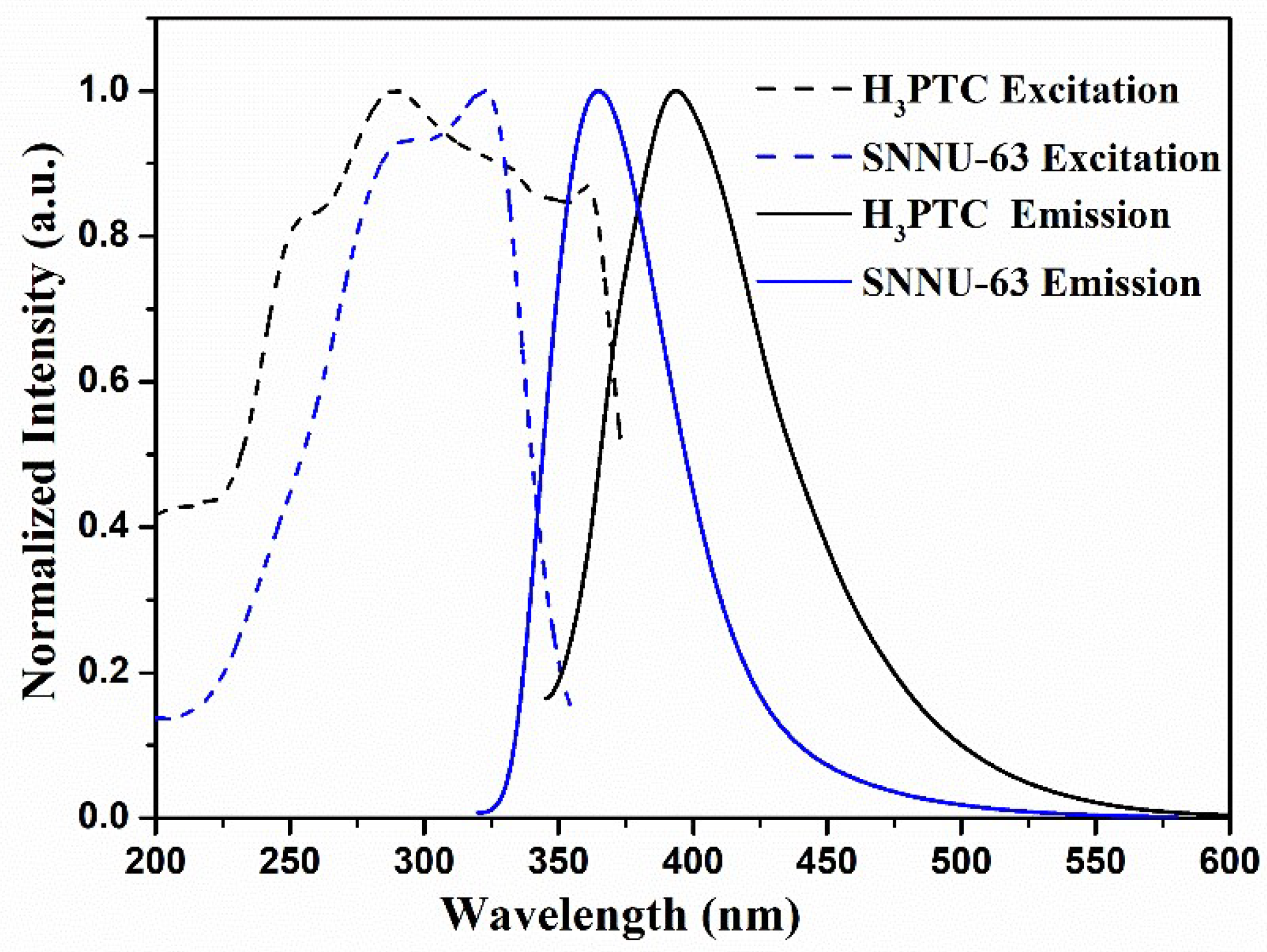
Considering that SNNU-63 has a unique structure, like the 3d10 configuration and a large ligand twist configuration, we measured the solid-state photoluminescence properties of the free ligand (H3PTC) and SNNU-63. Solid-state excitation and emission spectra at room temperature are shown in Figure 2. The free ligand shows the maximum emission peak at 393 nm under the 325 nm excitation, while SNNU-63 exhibits the maximum emission peak at 364 nm under the 300 nm excitation. Clearly, the maximum emission of SNNU-63 exhibits a blue-shift of 29 nm compared to that of the free ligand, which may arise from the different ligand conformations within SNNU-63 and H3PTC. To our knowledge, the ligand has a larger twist in the MOF skeleton compared to the free ligand, which can cause the MOF to have higher energy UV absorption than the free ligand. As predicted, the solid-state UV absorption maxima of the free ligand and SNNU-63 are at 325 and 300 nm, respectively (Figure S3). A blue shift (25 nm) of UV-vis absorption peak is consistent with the solid-state fluorescence’s result. Therefore, we propose that the blue-shift of the electronic absorption and emission spectra might be caused by different ligand twisted conformations within SNNU-63 and H3PTC [11,13]. Moreover, the CIE chromaticity coordinate of SNNU-63 is located in the blue region (x = 0.1584, y = 0.0566) (Figure S4), and the as-synthesized SNNU-63 exhibits the blue color under 254 nm UV light (Figure S5). Both the CIE coordinate and luminescent photo indicate SNNU-63 as a promising material for blue light-emitting diodes (LEDs).

3.2. Temperature-Dependent Emission Properties

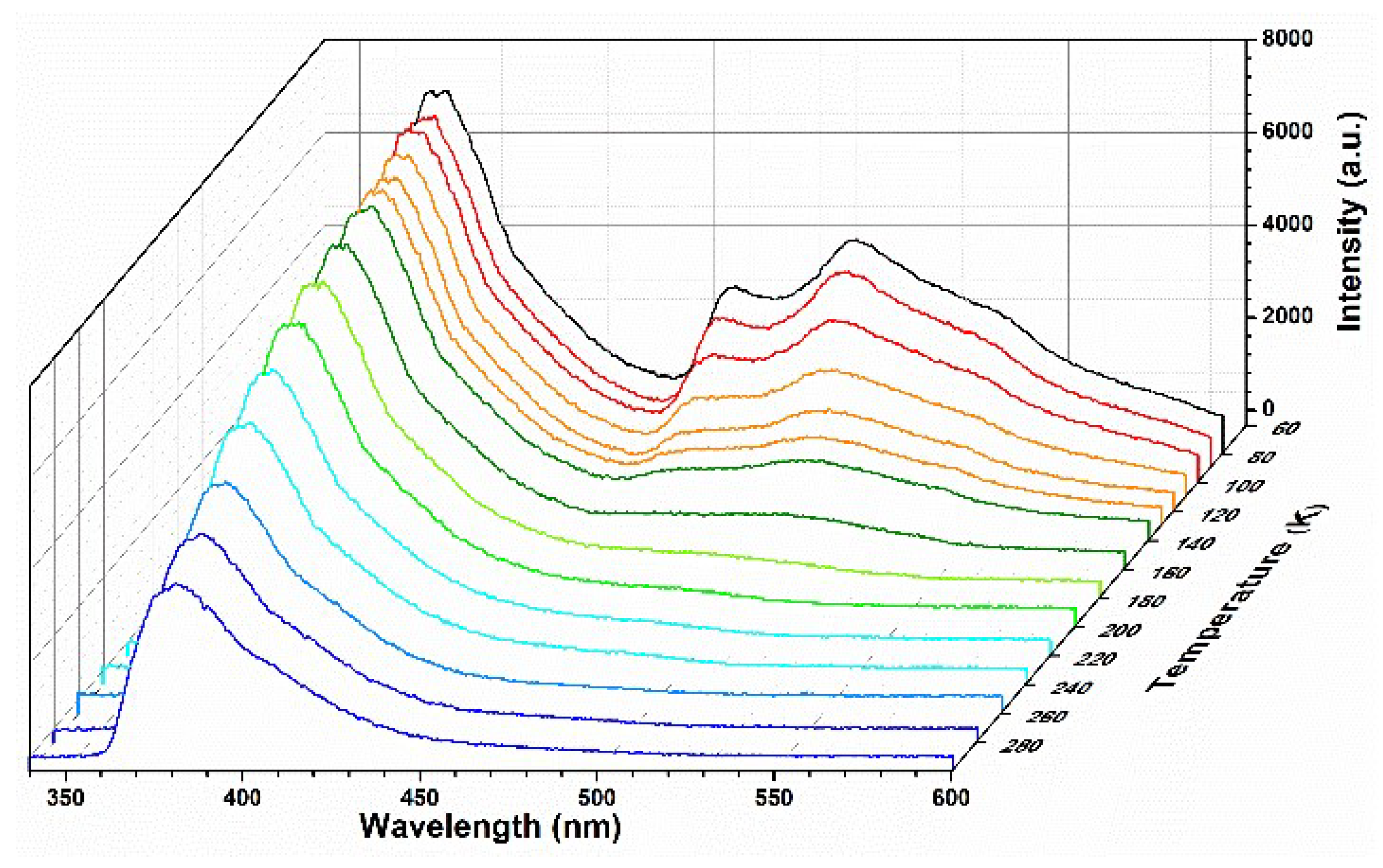
Considering SNNU-63 with unique the 3d10 configuration and the large ligand twisted conformation, we also investigated its temperature-dependent emission spectra in the range of 80–300 K (Figure 3). Obviously, when the temperature drops to 80 K, the MOF exhibits unique multi-emission centers, i.e., 381, 462, and 494 nm. This MOF at low temperature shows that the multi-emission centers are mainly derived from the limited ligand twisted conformations, and the reduced non-radiative attenuation for the benzene rings. When the temperature rises, the fluorescence intensities (I) of 462 and 494 nm gradually disappear, and only 381 nm is the main characteristic peak at 300 K.
The unique multi-emission centers of SNNU-63 at low temperatures have encouraged us to evaluate its application as a ratiometric thermometer in low-temperature detection. Here, we investigated the emission intensities at 381 and 494 nm to establish its potential as a ratiometric thermometer when excited 300 nm. The fluorescence intensity ratio I381/I494 as the self-calibrated thermometric parameter (Δ) with temperature was evaluated. As shown in Figure 4a, the temperature dependence of the fluorescence intensity ratio I381/I494 can be described by the Boltzmann equation (Δ = (A1 − A2)/(1 + exp((TT0)/dT)) + A2) (fitting parameters: A1 = 1.45, A2 = 17.62, T0 = 171.22, dT = 25.95), and a very good fit between Δ and temperature (80–240 K) (correlation coefficient R2 = 0.9969) can be obtained. These results demonstrate that SNNU-63 is a desirable candidate for highly sensitive temperature sensing.
To evaluate its practical application as a temperature sensor, we further investigated the relative sensitivity (Sr, Sr = (∂Δ/∂T)/Δ) in the range of 80–240 K. The calculated maximum relative sensitivity Sm = 2.60% K−1 is observed at 110 K (Figure 4b), which is comparable to that of the other reported MOFs with bimetallic Ln-MOF thermometers such as Tb0.80Eu0.20BPDA (1.19) [30], Nd0.577Yb0.423BDC-F4 (1.2) [31], and Eu0.37Tb0.63-BTC-a (0.68) [32]. Hence, this Ga-MOF is a good platform for temperature sensing in a wide temperature range.

3.3. Metal Ions Sensing Measurements

Owning to the excellent water stability and fluorescence properties of SNNU-63, we investigated the fluorescence response experiments of this MOF towards metal ions. The excitation and emission spectra of this MOF aqueous suspension are shown in Figure S6, i.e., the maximum emission intensity at 382 nm upon excitation at 288 nm. The metal ion sensing, based on SNNU-63, was performed by adding 2 mL aqueous solutions of metal salts (0.6 mM) (Mn+ = Sc3+, Al3+, Cd2+, K+, Ga3+, Zn2+, Ni2+, Na+, Co2+, Mg2+, Cu2+, Fe3+) with a 1 mL suspension of SNNU-63 (1 mg/mL). Clearly, the fluorescence emission intensity at 383 nm of SNNU-63 toward Fe3+ ion exhibits the highest quenching efficiency, while others metal ions show negligible effect on the fluorescence intensities (Figure 5a and Figure S7). It is noteworthy that the fluorescence quenching rate of this MOF towards Fe3+ ion is as high as 87% (Figure S8). The result demonstrates that SNNU-63 exhibits the high selectivity to Fe3+ ions in aqueous media via fluorescence quenching.
Considering that other metal ions coexist with Fe3+ ion in practical applications, the fluorescent emission intensities of SNNU-63 with different mixed ions before and after Fe3+ ion incorporation (0.3 mM) were recorded and compared (Figure 5b). Even in the presence of other metal ions, the fluorescence quenching efficiency of SNNU-63 towards Fe3+ ion was obvious. Hence, SNNU-63 exhibits a high selectivity for detecting Fe3+ ion in aqueous media.
To further investigate the detection limit and sensitivity for SNNU-63 toward Fe3+ ions, concentration dependence experiments were performed by adding different concentrations of Fe3+ ions, ranging from 0 to 0.667 mM (Figure 5c). It was very obvious that the luminescence intensity (I382nm) of SNNU-63 gradually decreased with the increase of the Fe3+ concentration. At a low concentration (0–0.04 mM) (Figure S9), the fluorescence quenching constant (Ksv) was used in the Stern–Volmer (SV) equation: (I0/I) = 1 + Ksv[M], where I0 and I are the luminescence intensity of SNNU-63 suspensions before and after adding different concentrations of Fe3+ ions, Ksv is the SV constant, and [M] is the molar concentration of Fe3+ ions. The Ksv value was evaluated to be 2.259 × 10⁴ M−1, which is comparable to that of the other stable MOFs toward Fe3+ sensing, such as BUT-15 (1.66 × 10⁴ M−1) [18] and Tb–HIAAC (2.5 × 10⁴ M−1) [33]. Based on the 3Sb/Ksv equation, the limit of detection (LOD) of SNNU-63 towards Fe3+ ion was evaluated to be 1.227 × 10⁻⁴ M. Overall, low LOD and high Ksv values enable SNNU-63 to detect Fe3+ with high sensitivity and accuracy. As a fluorescent probe, the reversible and recyclable experiments of SNNU-63 toward Fe3+ ion should be investigated. The result of four cycles demonstrates that SNNU-63 exhibits an excellent candidate for detecting Fe3+ with good recyclability and stability (Figure S10).
The rapid fluorescence quenching response and high selectivity of Fe3+ ion sensing for SNNU-63 prompted us to explore its fluorescence sensing mechanism. As shown in Figure 1, we determined that MOF materials can stably exist in the Fe3+ sensing system, which was proved by PXRD (Figure S11). Generally, the photoinduced electron transfer (PET) process can occur when the excited electrons of MOF are transferred to the LUMO of Fe3+ ion upon excitation [18]. On the other hand, a broad excitation of SNNU-63 showed the greatest degree of overlapping the absorption band of Fe3+ ions. However, no clear overlapping was observed between the excitation of SNNU-63 and the absorption spectra of other metal ions (Figure 5d). This result shows that the SNNU-63 towards Fe3+ sensing conforms to the fluorescence resonance energy transfer (FRET) process. Hence, the mechanism of stable SNNU-63 towards Fe3+ sensing is mainly attributed to both the PET and FRET processes.

4. Conclusions

In summary, a versatile luminescent Ga-MOF (SNNU-63) constructed with a 3d10 configuration and a large ligand twist configuration has been synthesized and studied. Due to the synergism between the metal center and ligand within the framework, this Ga-MOF should have excellent fluorescence performance and can be used for light emission and fluorescence sensing. The as-synthesized Ga-MOF exhibits a blue-shift of 29 nm compared to that of the free ligand. The CIE coordinate and luminescent photo demonstrate that this Ga-MOF is a promising material for blue light-emitting diodes. More importantly, this Ga-MOF exhibits multi-emission centers at 381, 462, and 494 nm at 80 K. The temperature dependence of fluorescence intensity ratio I381/I494 and the relative sensitivity analyses demonstrate that this Ga-MOF exhibits an excellent temperature sensing capability (80–240 K). In addition, the stable SNNU-63 exhibits an excellent selective detection of Fe3+ over other metal ions in aqueous medium. Overall, our work provides an effective method to develop and utilize luminescent Ga-MOFs materials.

Supplementary Materials

The following supporting information can be downloaded at: https://www.mdpi.com/article/10.3390/nano12224009/s1, Figure S1: Structure of the organic ligand (H3PTC). Figure S2: FT-IR spectra of the organic ligand and SNNU-63. Figure S3: The solid-state UV-Vis absorption spectra of SNNU-63 and the free H3PTC ligand at room temperature. Figure S4: The corresponding CIE 1931 chromaticity diagram of SNNU-63 at room temperature. Figure S5: Comparison of photo for SNNU-63 under the natural light (a) and UV 254 nm light (b). Figure S6: Excitation and emission spectra of SNNU-63 MOF aqueous suspension. Figure S7: Luminescence intensity (I382 nm) of SNNU-63 to different metal ions in aqueous (0.4 mM) under excitation of 288 nm. Figure S8: Fluorescence quenching of SNNU-63 dispersed in aqueous solutions of twelve different metal cations (0.4 mM). Figure S9: Linearity relationship of luminescent intensity of SNNU-63 and Fe3+ ion at low concentrations. Figure S10: The reversibility and recyclability of SNNU-63 based on the maximum emission intensity at 382 nm in water (1 mL 2 mg/mL) in the presence of 2 mL of a 0.6 mM aqueous solution of Fe3+ ions. Figure S11: Comparison of PXRD patterns of the as-synthesized MOF and the MOF after Fe3+ sensing.

Author Contributions

Data curation, W.S. and L.L.; methodology, J.Z. (Jinping Zhang) and H.Y., software, X.H.; writing—review and editing, J.Z. (Jianwei Zhang) and W.W. All authors have read and agreed to the published version of the manuscript.

Funding

The project was supported by the National Natural Science Foundation of China (21901229 and 22104079), the Natural Science Foundation of Henan Province (212300410218), the Science and Technology Fund of Henan Province (212102210616), the Program for Science & Technology Innovative Research Team (20IRTSTHN007), the Innovation Talents (22HASTIT028) and Key Scientific Research (22A150052) in the Universities of Henan Province.

Institutional Review Board Statement

Not applicable.

Informed Consent Statement

Informed consent was obtained from all subjects involved in the study.

Conflicts of Interest

The authors declare no conflict of interest.

References

  1. Zhao, D.; Timmons, D.J.; Yuan, D.; Zhou, H.-C. Tuning the topology and functionality of metal-organic frameworks by ligand design. Acc. Chem. Res. 2011, 44, 123–133. [Google Scholar] [CrossRef] [PubMed]
  2. Zhao, Y.; Zeng, H.; Zhu, X.-W.; Lu, W.; Li, D. Metal-organic frameworks as photoluminescent biosensing platforms: Mechanisms and applications. Chem. Soc. Rev. 2021, 50, 4484–4513. [Google Scholar] [CrossRef] [PubMed]
  3. Stavila, V.; Talin, A.A.; Allendorf, M.D. MOF-based electronic and opto-electronic devices. Chem. Soc. Rev. 2014, 43, 5994–6010. [Google Scholar] [CrossRef] [PubMed] [Green Version]
  4. Cui, Y.; Zhang, J.; He, H.; Qian, G. Photonic functional metal–organic frameworks. Chem. Soc. Rev. 2018, 47, 5740–5785. [Google Scholar] [CrossRef]
  5. Wu, S.; Min, H.; Shi, W.; Cheng, P. Multicenter metal-organic framework-based ratiometric fluorescent sensors. Adv. Mater. 2020, 32, 1805871. [Google Scholar] [CrossRef]
  6. Wu, Y.-P.; Tian, J.-W.; Liu, S.; Li, B.; Zhao, J.; Ma, L.-F.; Li, D.-S.; Lan, Y.-Q.; Bu, X. Bi-microporous metal-organic-frameworks with cubane [M4(OH)4] (M = Ni, Co) clusters and pore space partition for electrocatalytic methanol oxidation reaction. Angew. Chem. Int. Ed. 2019, 58, 12185–12189. [Google Scholar] [CrossRef]
  7. Zhao, Y.; Wang, Y.-J.; Wang, N.; Zheng, P.; Fu, H.-R.; Han, M.-L.; Ma, L.-F.; Wang, L.-Y. Tetraphenylethylene-decorated metal-organic frameworks as energy-transfer platform for the detection of nitro-antibiotics and white-light emission. Inorg. Chem. 2019, 58, 12700–12706. [Google Scholar] [CrossRef]
  8. Yang, X.-G.; Lu, X.M.; Zhai, Z.-M.; Zhao, Y.; Liu, X.-Y.; Ma, L.-F.; Zang, S.-Q. Facile synthesis of micro-scale MOF host-guest with long-last phosphorescence and enhanced optoelectronic performance. Chem. Commun. 2018, 54, 13563–13566. [Google Scholar]
  9. Sun, Z.; Khurshid, A.; Sohail, M.; Qiu, W.; Cao, D.; Su, S.-J. Encapsulation of dyes in luminescent metal-organic frameworks for white light emitting diodes. Nanomaterials 2021, 11, 2761. [Google Scholar] [CrossRef]
  10. Cai, K.; Zhao, N.; Zhang, N.; Sun, F.-X.; Zhao, Q.; Zhu, G.-S. A homochiral multifunctional metal-organic framework with rod-shaped secondary building units. Nanomaterials 2017, 7, 88. [Google Scholar] [CrossRef] [Green Version]
  11. Zhang, J.-W.; Li, H.; Li, J.-Q.; Chen, Y.; Qu, P.; Zhai, Q.-G. Enhancement of the fluorescence properties via introducing the tetraphenylethylene chromophores into a novel Mn-organic framework with a rare [Mn43-OH)2] cluster. Dalton Trans. 2021, 50, 17482–17486. [Google Scholar] [CrossRef] [PubMed]
  12. Dawood, S.; Dorris, A.; Davis, K.; Hammer, N.I.; Rathnayake, H. Synthesis, characterization, and photophysics of self-assembled Mn(II)-MOF with naphthalene chromophore. J. Phys. Chem. C 2021, 125, 792–802. [Google Scholar] [CrossRef]
  13. Wei, Z.; Gu, Z.-Y.; Arvapally, R.K.; Chen, Y.-P.; McDougald, R.N.; Ivy, J.F.; Yakovenko, A.A.; Feng, D.; Omary, M.A.; Zhou, H.-C. Rigidifying fluorescent linkers by metal-organic framework formation for fluorescence blue shift and quantum yield enhancement. J. Am. Chem. Soc. 2014, 136, 8269–8276. [Google Scholar] [CrossRef] [PubMed]
  14. Yu, L.; Wang, H.; Liu, W.; Teat, S.J.; Li, J. Blue-light-excitable, quantum yield enhanced, yellow-emitting, zirconium-based metal–organic framework phosphors formed by immobilizing organic chromophores. Cryst. Growth Des. 2019, 19, 6850–6854. [Google Scholar] [CrossRef]
  15. Yuan, S.; Qin, J.-S.; Lollar, C.T.; Zhou, H.-C. Stable metal-organic frameworks with group 4 metals: Current status and trends. ACS Cent. Sci. 2018, 4, 440–450. [Google Scholar] [CrossRef] [Green Version]
  16. Zhang, J.-W.; Ji, W.-J.; Hu, M.-C.; Li, S.-N.; Jiang, Y.-C.; Zhang, X.-M.; Qu, P.; Zhai, Q.-G. A superstable 3p-block metal-organic framework platform towards prominent CO2 and C1/C2-hydrocarbon uptake and separation performance and strong lewis acid catalysis for CO2 fixation. Inorg. Chem. Front. 2019, 6, 813–819. [Google Scholar] [CrossRef]
  17. Zhao, J.; Wang, Y.-N.; Dong, W.-W.; Wu, Y.-P.; Li, D.-S.; Zhang, Q.-C. A robust luminescent Tb(III)-MOF with lewis basic pyridyl sites for the highly sensitive detection of metal Ions and small molecules. Inorg. Chem. 2016, 55, 3265–3271. [Google Scholar] [CrossRef]
  18. Wang, B.; Yang, Q.; Guo, C.; Sun, Y.; Xie, L.-H.; Li, J.-R. Stable Zr(IV)-based metal-organic frameworks with predesigned functionalized ligands for highly selective detection of Fe(III) ions in water. ACS Appl. Mater. Interfaces 2017, 9, 10286–10295. [Google Scholar] [CrossRef]
  19. Xu, N.; Tan, Y.-X.; El-Sayed, E.-S.M.; Yuan, D. Two zirconium metal organic cages with S4 and D2d symmetry: Construction and detection of antibiotics. Cryst. Growth Des. 2022, 22, 2768–2773. [Google Scholar] [CrossRef]
  20. Cairns, A.J.; Eckert, J.; Wojtas, L.; Thommes, M.; Wallacher, D.; Georgiev, P.A.; Forster, P.M.; Belmabkhout, Y.; Ollivier, J.; Eddaoudi, M. Gaining insights on the H2-sorbent interactions: Robust soc-MOF platform as a case study. Chem. Mater. 2016, 28, 7353–7361. [Google Scholar] [CrossRef]
  21. Yan, B. Lanthanide-functionalized metal–organic framework hybrid systems to create multiple luminescent centers for chemical sensing. Acc. Chem. Res. 2017, 50, 2789–2798. [Google Scholar] [CrossRef] [PubMed]
  22. Cui, Y.; Song, R.; Yu, J.; Liu, M.; Wang, Z.; Wu, C.; Yang, Y.; Wang, Z.; Chen, B.; Qian, G. Dual-emitting MOF⊃dye composite for ratiometric temperature sensing. Adv. Mater. 2015, 27, 1420–1425. [Google Scholar] [CrossRef] [PubMed]
  23. Li, B.; Fan, H.-T.; Zang, S.-Q.; Li, H.-Y.; Wang, L.-Y. Metal–containing crystalline luminescent thermochromic materials. Coord. Chem. Rev. 2018, 377, 307–329. [Google Scholar]
  24. Liu, Y.; Lu, Y.-K.; Zhang, B.; Hou, L.; Wang, Y.-Y. Post-synthetic functionalization of Ni-MOF by Eu3+ ions: Luminescent probe for aspartic acid and magnetic property. Inorg. Chem. 2020, 59, 7531–7538. [Google Scholar] [CrossRef] [PubMed]
  25. Cheng, Y.; Gao, Y.; Lin, H.; Huang, F.; Wang, Y. Strategy design for ratiometric luminescence thermometry: Circumventing the limitation of thermally coupled levels. J. Mater. Chem. C 2018, 6, 7462–7478. [Google Scholar] [CrossRef]
  26. Wu, J.-X.; Yan, B. A dual-emission probe to detect moisture and water in organic solvents based on green-Tb3+ post-coordinated metal–organic frameworks with red carbon dots. Dalton Trans. 2017, 46, 7098–7105. [Google Scholar] [CrossRef] [PubMed]
  27. Li, Y.-T.; Zhang, J.-W.; Lv, H.-J.; Hu, M.-C.; Li, S.-N.; Jiang, Y.-C.; Zhai, Q.-G. Tailoring the pore environment of a robust Ga-MOF by deformed [Ga3O(COO)6] cluster for boosting C2H2 uptake and separation. Inorg. Chem. 2020, 59, 10368–10373. [Google Scholar] [CrossRef]
  28. Coelho, A. Topas-A: General Profile and Structure Analysis Software for Powder Diffraction Data; Bruker-AXS: Madison, WI, USA, 2005. [Google Scholar]
  29. Wu, Z.; Li, C.; Zhang, F.; Huang, S.; Wang, F.; Wang, X.; Jiao, H. High-performance ultra-narrow-band green-emitting phosphor LaMgAl11O19:Mn2+ for wide color-gamut WLED backlight displays. J. Mater. Chem. C 2022, 10, 7443–7448. [Google Scholar] [CrossRef]
  30. Zhao, D.; Rao, X.; Yu, J.; Cui, Y.; Yang, Y.; Qian, G. Design and synthesis of an MOF thermometer with high sensitivity in the physiological temperature range. Inorg. Chem. 2015, 54, 11193–11199. [Google Scholar] [CrossRef]
  31. Lian, X.; Zhao, D.; Cui, Y.; Yang, Y.; Qian, G. A near infrared luminescent metal-organic framework for temperature sensing in the physiological range. Chem. Commun. 2015, 51, 17676–17679. [Google Scholar] [CrossRef]
  32. Wang, H.; Zhao, D.; Cui, Y.; Yang, Y.; Qian, G. A Eu/Tb-mixed MOF for luminescent high-temperature sensing. J. Solid State Chem. 2017, 246, 341–345. [Google Scholar] [CrossRef]
  33. Huangfu, M.; Tian, X.; Zhao, S.; Wu, P.; Chu, H.; Zheng, X.; Tang, J.; Wang, J. Post-synthetic modification of a Tb-based metal-organic framework for highly selective and sensitive detection of metal ions in aqueous solution. New J. Chem. 2019, 43, 10232–10236. [Google Scholar] [CrossRef]
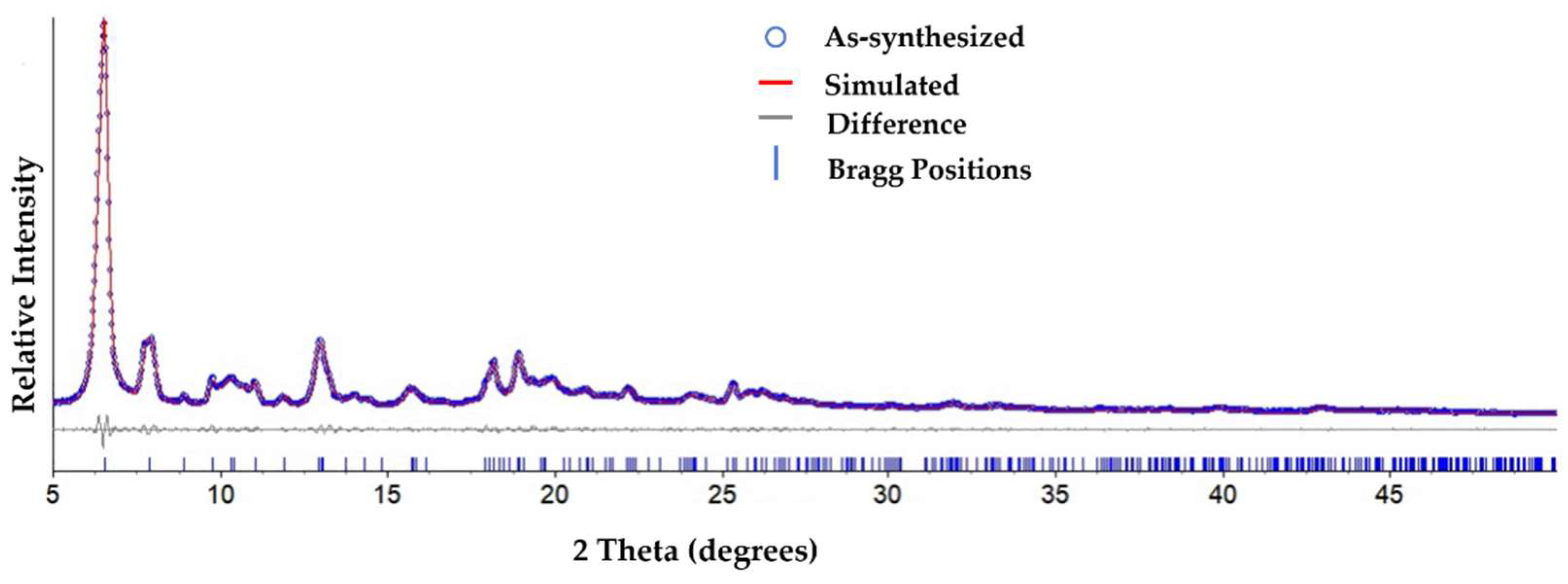
Figure 1. The Rietveld refinement XRD pattern of the as-synthesized SNNU-63.
Nanomaterials 12 04009 g001
Figure 2. Solid-state excitation and emission spectra of SNNU-63 and the free H3PTC ligand.
Nanomaterials 12 04009 g002
Figure 3. Temperature-dependent emission spectra of SNNU-63 in the range of 80–300 K.
Nanomaterials 12 04009 g003
Figure 4. (a) Temperature-dependent emission intensity ratio Δ(I493/I410) and the fitted curve of SNNU-63 in the range of 80–240 K; (b) temperature-dependent generalized relative sensitivity (Sr) of SNNU-63.
Nanomaterials 12 04009 g004
Figure 5. (a) Fluorescent spectra of SNNU-63 in the presence of 0.4 mM of different metal ions under an excitation of 288 nm. (b) The anti-interference performance of the coexistence of other metal ions and Fe3+ ions for SNNU-63. (c) Luminescence spectra of SNNU-63 toward Fe3+ ions with different concentrations of Fe3+ ranging from 0 to 0.667 mM. (d) Luminescence excitation spectrum of SNNU-63 suspensions and UV-vis absorption spectra of twelve different metal ion solutions.
Nanomaterials 12 04009 g005
Publisher’s Note: MDPI stays neutral with regard to jurisdictional claims in published maps and institutional affiliations.