Article

Biofilm Degradation by Seashell-Derived Calcium Hydroxide and Hydrogen Peroxide

Division of Biomedical Engineering, National Defense Medical College Research Institute, 3-2 Namiki, Tokorozawa-shi 359-8513, Japan
*
Authors to whom correspondence should be addressed.
Nanomaterials 2022, 12(20), 3681; https://doi.org/10.3390/nano12203681
Submission received: 16 September 2022 / Revised: 4 October 2022 / Accepted: 14 October 2022 / Published: 20 October 2022

Abstract

:
Microbial cells and self-produced extracellular polymeric substances assembled to form biofilms that are difficult to remove from surfaces, causing problems in various fields. Seashell-derived calcium hydroxide, a sustainable inorganic material, has shown high bactericidal activity even for biofilms due to its alkalinity. However, its biofilm removal efficacy is relatively low. Herein, we report a biofilm degradation strategy that includes two environmentally friendly reagents: seashell-derived calcium hydroxide and hydrogen peroxide. A biofilm model of Escherichia coli was prepared in vitro, treated with calcium hydroxide–hydrogen peroxide solutions, and semi-quantified by the crystal violet stain method. The treatment significantly improved biofilm removal efficacy compared with treatments by calcium hydroxide alone and hydrogen peroxide alone. The mechanism was elucidated from calcium hydroxide–hydrogen peroxide solutions, which suggested that perhydroxyl anion and hydroxyl radical generated from hydrogen peroxide, as well as the alkalinity of calcium hydroxide, enhanced biofilm degradation. This study showed that concurrent use of other reagents, such as hydrogen peroxide, is a promising strategy for improving the biofilm degradation activity of seashell-derived calcium hydroxide and will contribute to developing efficient biofilm removal methods.

1. Introduction

Biofilms are assemblies of microbial cells and self-produced extracellular polymeric substances, including polysaccharides and other biopolymers [1,2,3,4,5]. This lifestyle of bacteria is found on surfaces of living tissues, indwelling medical devices, industrial or potable water system piping, and natural aquatic systems. The biofilm state renders microbial cells highly resistant to harsh environmental factors, such as extreme temperature, extreme pH, high salinity, ultraviolet radiation, lack of nutrients, and antibiotics. Moreover, biofilms are sticky and viscoelastic due to the supramolecular structure of extracellular polymeric substances and therefore difficult to remove from surfaces [6]. Consequently, biofilms cause various problems in medical fields and food and other industries [7,8,9,10]. Harsh chemicals, such as chlorine, sodium hypochlorite, and other oxidizing agents, are typically required to kill and remove biofilms [9,11]. These reagents may damage tissues and materials, on which biofilms are attached, and produce harmful byproducts (e.g., trihalomethane) [12]. The development of effective and safe methods to eradicate biofilms remains challenging.
Recent studies have used calcium hydroxide (Ca(OH)2) and calcium oxide (CaO) prepared from seashells to develop effective and safe disinfectants [13,14]. Seashells are mainly composed of calcium carbonate and can be converted into CaO via heat treatments [13]. Subsequent hydration of CaO produces Ca(OH)2. These seashell-derived Ca(OH)2 and CaO have shown excellent microbicidal and virucidal activities due to their alkalinity [13,14]. Thus, their aqueous solutions, dispersions, and suspensions have been investigated as sustainable and halogen-free disinfectants [15,16,17,18,19,20,21]. Indeed, seashell-derived Ca(OH)2 and CaO can kill not only planktonic bacterial cells, but also biofilms [22,23,24], and thus they are promising for developing sustainable and halogen-free biofilm treatment methods. Nevertheless, the biofilm removal efficacy of seashell-derived Ca(OH)2 and CaO is relatively low despite their excellent bactericidal activities. Killed biofilms can still discharge endotoxins and facilitate the recolonization of new and viable biofilm [25,26,27,28]. Hence, methods to enhance the biofilm removal efficacy of seashell-derived Ca(OH)2 and CaO are highly sought.
Herein, we found that Ca(OH)2 and hydrogen peroxide (H2O2) synergize to effectively remove biofilm. H2O2 is an oxidizing agent that generates only water and oxygen as byproducts and is widely used for disinfection and bleaching [29,30,31]. Notably, alkaline conditions enhance the activity of H2O2: H2O2 is converted to perhydroxyl anion (HOO) and subsequently to hydroxyl radical (HO⋅) and other reactive species [29,30,32]. Thus, we hypothesized that the addition of H2O2 to Ca(OH)2 alkaline solutions would enhance biofilm removal efficacy. A model biofilm of Escherichia coli (E. coli) was prepared on polypropylene surfaces and treated with Ca(OH)2–H2O2 aqueous solutions. This treatment significantly decreased the amount of biofilms on the surface despite a slight decrease in the alkalinity of Ca(OH)2 by weakly acidic H2O2.

2. Materials and Methods

2.1. Materials

Ethanol, Ca(OH)2, and ~30 wt% H2O2 were purchased from FUJIFILM Wako Pure Chemical Corporation (Osaka, Japan). Brain heart infusion broth, Gram-Hucker’s stain solution I (20% ethanol, 2% crystal violet, and 0.8% ammonium oxalate), 5,5-dimethyl-1-pyrroline N-oxide, and saline were purchased from Nissui Pharmaceutical (Tokyo, Japan), Muto Pure Chemicals (Tokyo, Japan), Labotec (Tokyo, Japan), and Otsuka Pharmaceutical Factory (Tokushima, Japan), respectively. E. coli (ATCC 51813) was obtained from Microbiologics (St Cloud, MN, USA). Scallop shell-derived Ca(OH)2 was kindly donated by Hokkaido Lime (Hokkaido, Japan). Ultrapure water with a resistivity greater than 18.2 MΩ cm at 25 °C was supplied by an RFU464TA instrument (Advantec, Tokyo, Japan).

2.2. Preparation of Biofilms

Two hundred microliters of E. coli suspensions (ATCC 51813, ~1 × 10⁷ colony-forming units mL−1) in 1/5 brain heart infusion broth (7 g L−1) as the medium were added to each well of a 96-well polystyrene microplate (Thermo Fisher Scientific, Waltham, MA, USA). The microplates were then covered with 96-pin polypropylene lids (BMR-PP, BM Equipment, Tokyo, Japan) and incubated at 37 °C for 1 d to allow biofilms to form on the pins. The 96-pin lids containing biofilms were transferred to a 96-well plate containing fresh 1/5 brain heart infusion broth and incubated at 37 °C for 2 d to further grow biofilms. The medium was refreshed after 1 d.

2.3. Degradation and Semi-Quantification of Biofilms

After washing with saline, the 96-pin lids containing biofilms were transferred to 96-well microplates containing 250 µL of Ca(OH)2–H2O2 aqueous solutions in each well. The biofilms in Ca(OH)2–H2O2 solutions were incubated at room temperature for 10 min unless otherwise stated. After washing with saline, the 96-pin lids were transferred to 96-well microplates containing 250 µL of Gram-Hucker’s stain solution I in each well and incubated for 20 min. Stained biofilms were washed three times with saline and dried for more than 30 min. The dye crystal violet was extracted with ethanol (250 µL for each pin), followed by 10-fold dilution, with subsequent absorbance measurements performed at 595 nm using a Multiskan FC microplate reader (Thermo Fisher Scientific, Massachusetts, USA). The experiment was repeated three times independently. Statistical analysis was performed by parametric one-way analysis of variance (ANOVA) with Dunnett’s multiple comparison using GraphPad Prism9 software (GraphPad Software, San Diego, CA, USA).

2.4. Microscopic Observations of Biofilms

For scanning electron microscopy (SEM), the biofilms on pins were fixed with 0.1 M phosphate buffer solutions (pH 7.4) containing 2% paraformaldehyde and 2.5% glutaraldehyde, and subsequently with 0.1 M phosphate buffer solutions (pH 7.4) containing 1% osmium tetroxide. The samples were solvent-exchanged with 50, 70, and 95% ethanol, ethanol, ethanol dehydrated with a molecular sieve, and then isoamyl acetate. After critical point drying using an HCP-2 instrument (Hitachi, Tokyo, Japan), the samples were mounted on substrates using silver paste and coated with osmium. The surfaces were observed with a JSM-6340F instrument (JEOL, Tokyo, Japan) at an accelerating voltage of 5 kV.
For fluorescence microscopy, the biofilms on pins before and after the treatment with Ca(OH)2–H2O2 solutions were stained using LIVE/DEAD BacLight Bacterial Viability Kit (Thermo Fisher Scientific, Waltham, MA, USA). Specifically, the pins with biofilms were immersed in aqueous solutions of 4.8 µM SYTO 9 and 24 µM propidium iodide for 15 min in the dark. The samples were observed by a BZ-X800 instrument equipped with a Plan Apochromat 2x objective (Keyence, Osaka, Japan). Observations were conducted using an OP-87763 filter (excitation: 470 nm, absorption: 525 nm), an OP-87764 filter (excitation: 545 nm, absorption: 605 nm) for SYTO 9, and propidium iodide, respectively, at an exposure time of 1/6 s.

2.5. Characterization of Ca(OH)2–H2O2 Solutions

For pH measurements, 250 µL of Ca(OH)2–H2O2 aqueous solutions was prepared immediately before measurements. A pH meter F-74 equipped with a 9618S electrode (Horiba, Kyoto, Japan) was operated at room temperature.
For electron spin resonance (ESR) spectroscopy, Ca(OH)2 aqueous solutions and H2O2 aqueous solutions were mixed to prepare 0.1% Ca(OH)2–0.1% H2O2 solutions before incubation at room temperature for 1, 10, and 60 min. Fifty microliters of the mixtures were mixed with 10 µL of a radical trapping agent, 5,5-dimethyl-1-pyrroline N-oxide. An EMX-nano spectrometer (Bruker, Billerica, MA, USA) was operated at 9.65 GHz microwave frequency, 10 mV microwave power, and 5 scans.

3. Results and Discussion

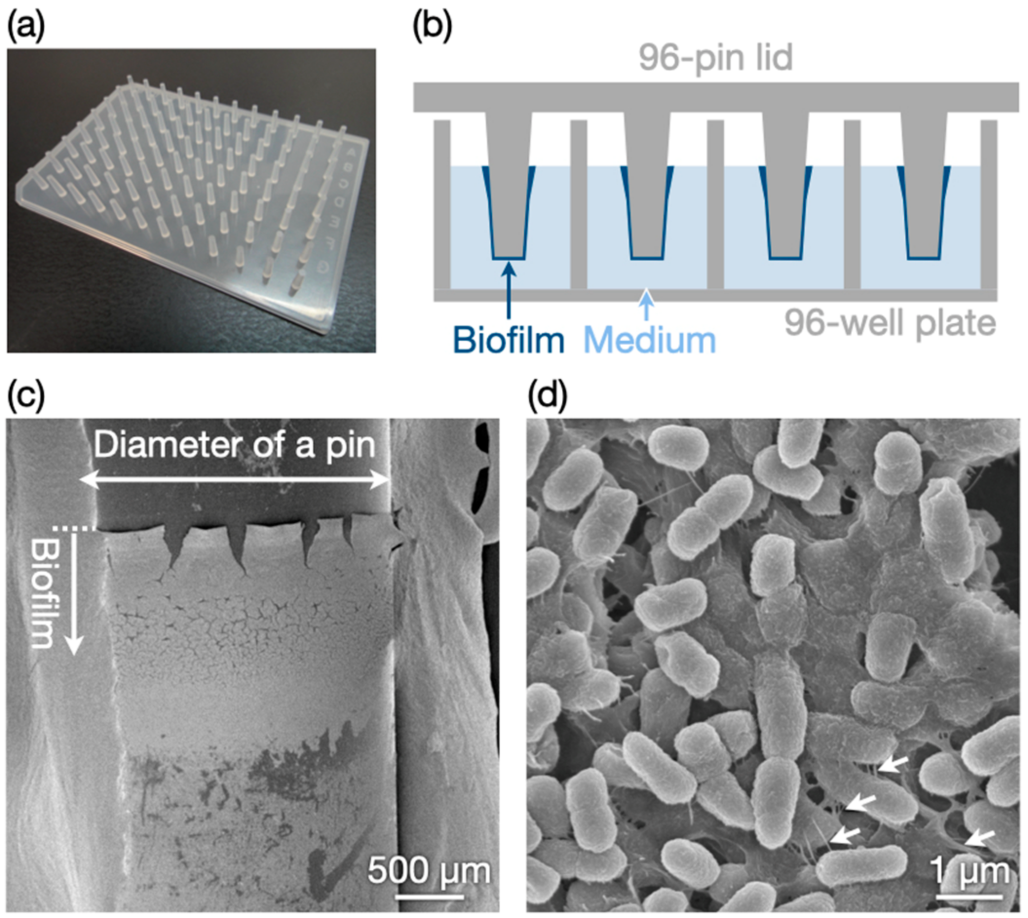
3.1. Biofilm Preparation

We adapted the in vitro biofilm preparation method using 96-pin plastic lids [33,34]. A 96-well microplate containing E. coli in a diluted liquid medium was covered with a 96-pin lid (Figure 1a) and incubated for 3 d. Biofilms formed on the pins in the liquid medium, as illustrated in Figure 1b. Notably, the biofilms were relatively thick at the upper edge near the air (Figure 1c). Figure 1d is an SEM image at a high magnification that revealed densely packed bacterial cells and nanofibrous materials around the cells. The nanofibrous materials appeared to be extracellular polymeric substances and/or flagella. The results confirmed the successful preparation of an in vitro biofilm model.

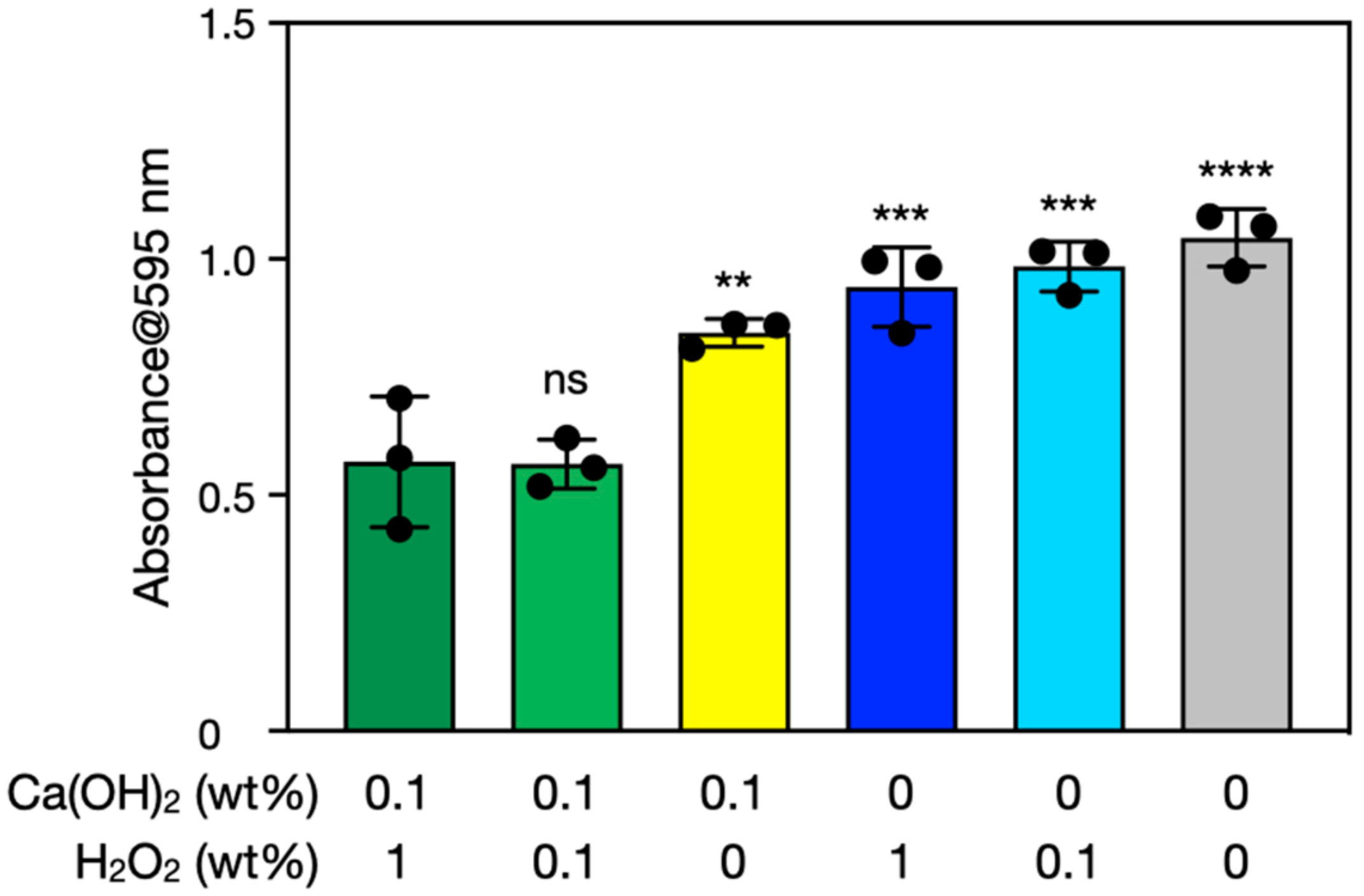
3.2. Biofilm Degradation by Ca(OH)2–H2O2

The prepared biofilms were immersed in Ca(OH)2–H2O2 solutions and incubated at room temperature for 10 min. Reagent-grade Ca(OH)2, rather than seashell-derived Ca(OH)2, was initially used for fundamental investigations before experiments using seashell-derived Ca(OH)2 (see Section 3.4). The amounts of biofilms were semi-quantified with the crystal violet stain method [35,36]. This method enabled us to semi-quantify biomolecules of bacterial cells and extracellular polymeric substances by measuring the absorbance of crystal violet extracted from the stained biofilms. Figure 2 shows results of biofilm semi-quantification before and after the treatments with Ca(OH)2 and/or H2O2 for 10 min. Solutions containing 0.1% Ca(OH)2 and 0.1% to 1% H2O2 significantly decreased the amount of biofilm on the surface. These decreases correspond to the removal of biofilms rather than bacterial death. Ca(OH)2–H2O2 removed significantly more biofilm than Ca(OH)2 alone or H2O2 alone. Thus, the concurrent use of H2O2 improves the biofilm removal efficacy of Ca(OH)2.
Ca(OH)2–H2O2 treatment time was varied from 10 min to 3 and 30 min (Figure 3). One-way ANOVA indicated nonsignificant differences between the groups (p > 0.05). The amount of biofilm hardly decreased after treatments for 3 and 30 min even with the use of both Ca(OH)2 and H2O2. The shorter treatment time proved insufficient to degrade biofilms. Meanwhile, the longer treatment time may have enabled readsorption of extracellular polymeric substances due to decreases in pH and reactive species concentrations (see Section 3.3). Thus, 10 min of treatment was considered optimal for biofilm degradation under the conditions investigated.
The biofilms treated with Ca(OH)2 and/or H2O2 were stained with SYTO 9 and propidium iodide for fluorescence microscopy. SYTO 9, a green fluorescence dye, permeates both intact and damaged membranes of the bacterial cells, and binds to nucleic acids. Meanwhile, propidium iodide, a red fluorescence dye, permeates only damaged membranes, and binds with a higher affinity to nucleic acids than SYTO 9. It is noted that extracellular polymeric substances should be non-specifically stained by these fluorescence dyes to a certain extent, causing background fluorescence. Fluorescence microscopy images of the treated biofilms are shown in Figure 4. The biofilms treated with Ca(OH)2–H2O2 exhibited less fluorescence of both SYTO 9 and propidium iodide than the other biofilm samples, indicating that the Ca(OH)2–H2O2 treatment killed bacteria and removed dead cells, which was also suggested by the crystal violet stain method.

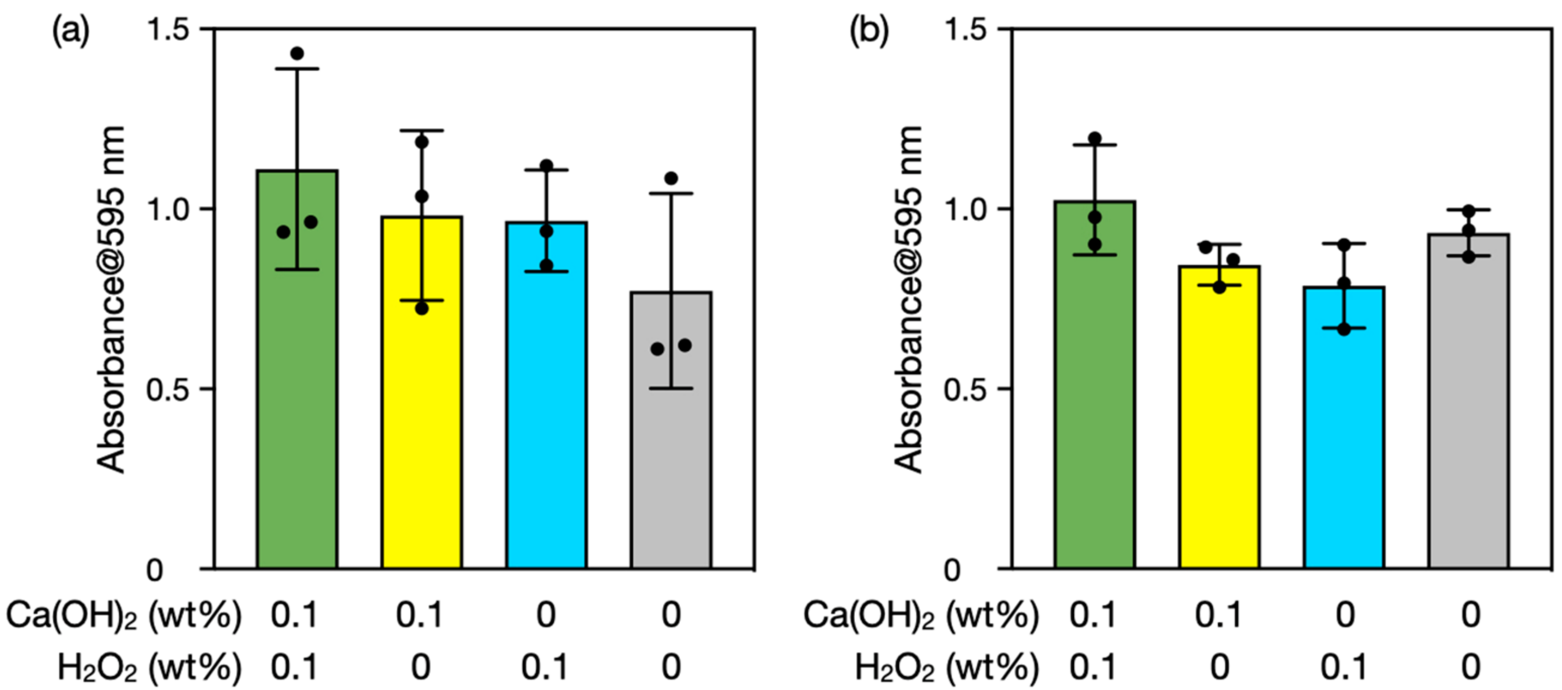
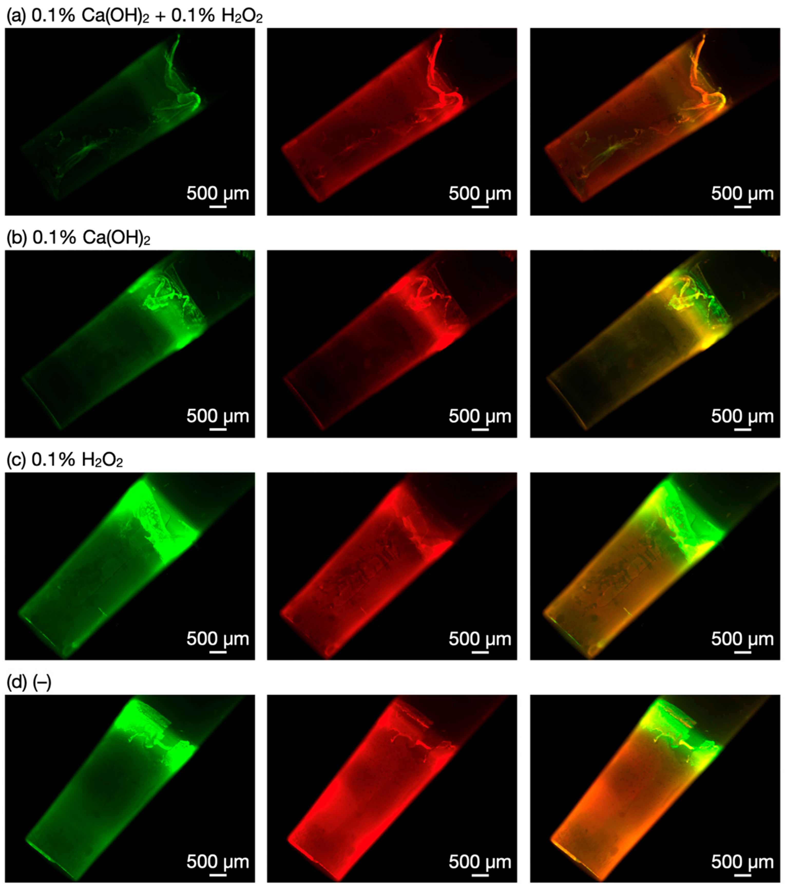
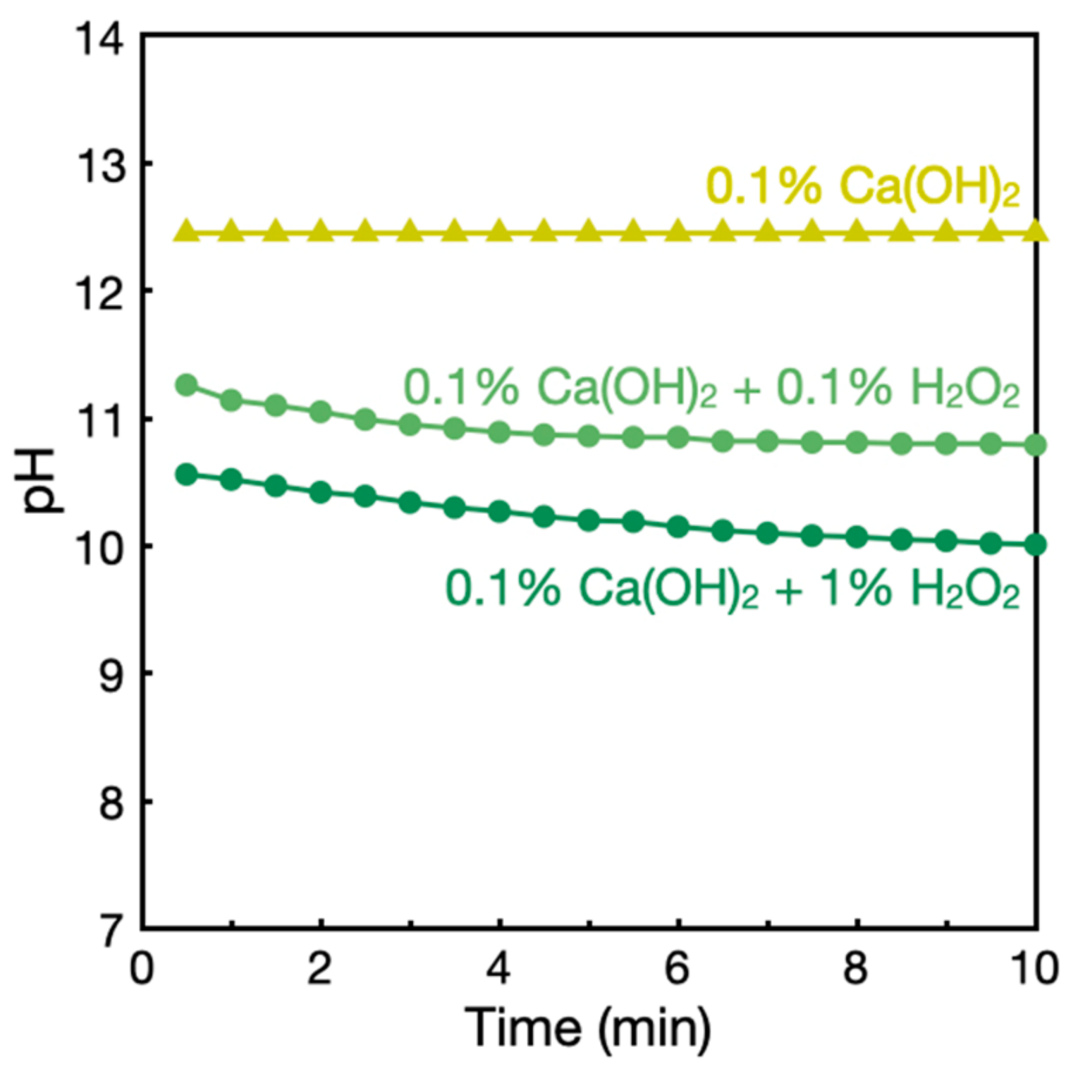
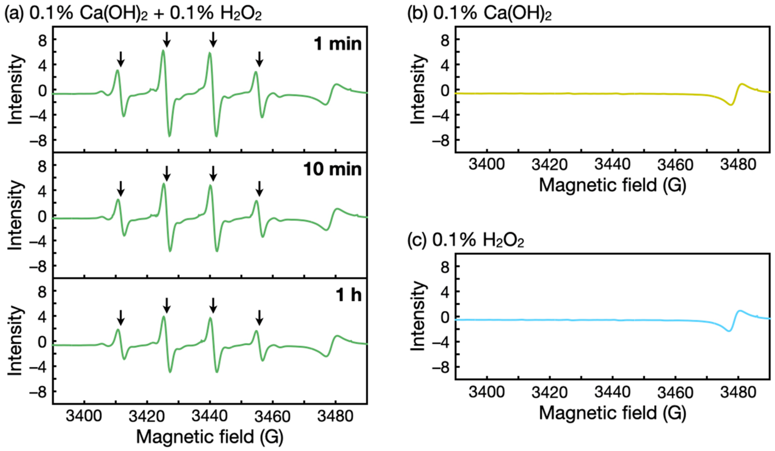
3.3. Investigations into Ca(OH)2–H2O2 Solutions

We characterized Ca(OH)2–H2O2 solutions to better understand biofilm degradation. Figure 5 presents the time courses of pH of the solutions. The addition of H2O2 decreased the alkalinity of Ca(OH)2 solutions probably due to the reaction of weakly acidic H2O2 (pKa = 11.6) with HO, producing HOO. HOO is a reactive species responsible for the bleaching action of H2O2 under alkaline conditions [29]. The pH further gradually decreased (Figure 5), which can be attributed to the decomposition of HOO and subsequent reproduction of HOO by consuming HO. Considering that a decrease in pH should reduce the bactericidal and biofilm removal activities of Ca(OH)2 derived mainly from its alkalinity, the improved biofilm removal activities of Ca(OH)2–H2O2 solutions seemed to originate from not only HO, but also reactive species generated from H2O2, such as HOO.
Other possible active species other than HOO include HO⋅, a highly reactive species that forms via a reaction between HOO and H2O2 [29,32]. Thus, we conducted ESR spectroscopy measurements using 5,5-dimethyl-1-pyrroline N-oxide as the radical trapping agent. ESR spectra of Ca(OH)2–H2O2 solutions showed four characteristic signals of HO⋅ (Figure 6a). These peaks were absent in spectra of Ca(OH)2 alone and H2O2 alone (Figure 6b,c). These results indicate that H2O2 with alkaline Ca(OH)2 generated highly reactive HO⋅, which contributed to the biofilm degradation. The HO⋅ signal intensity also gradually decreased (Figure 6a). This decrease in HO⋅ implies the decomposition of H2O2 over time and may explain the decreased biofilm removal efficacy at 30 min of the treatment (Figure 3b).
H2O2 decomposes to produce not only reactive species but also oxygen bubbles that may help remove biofilms via mechanical disruption [37]. Collectively, Ca(OH)2–H2O2 solutions, albeit weaker alkaline solutions than Ca(OH)2 solutions, better remove biofilm due to the synergy of HO, HOO, HO⋅, and possibly oxygen bubbles. Given the suggested mechanism based on chemically active species, the biofilm removal method using Ca(OH)2 and H2O2 will be effective to various bacteria strains and even species.

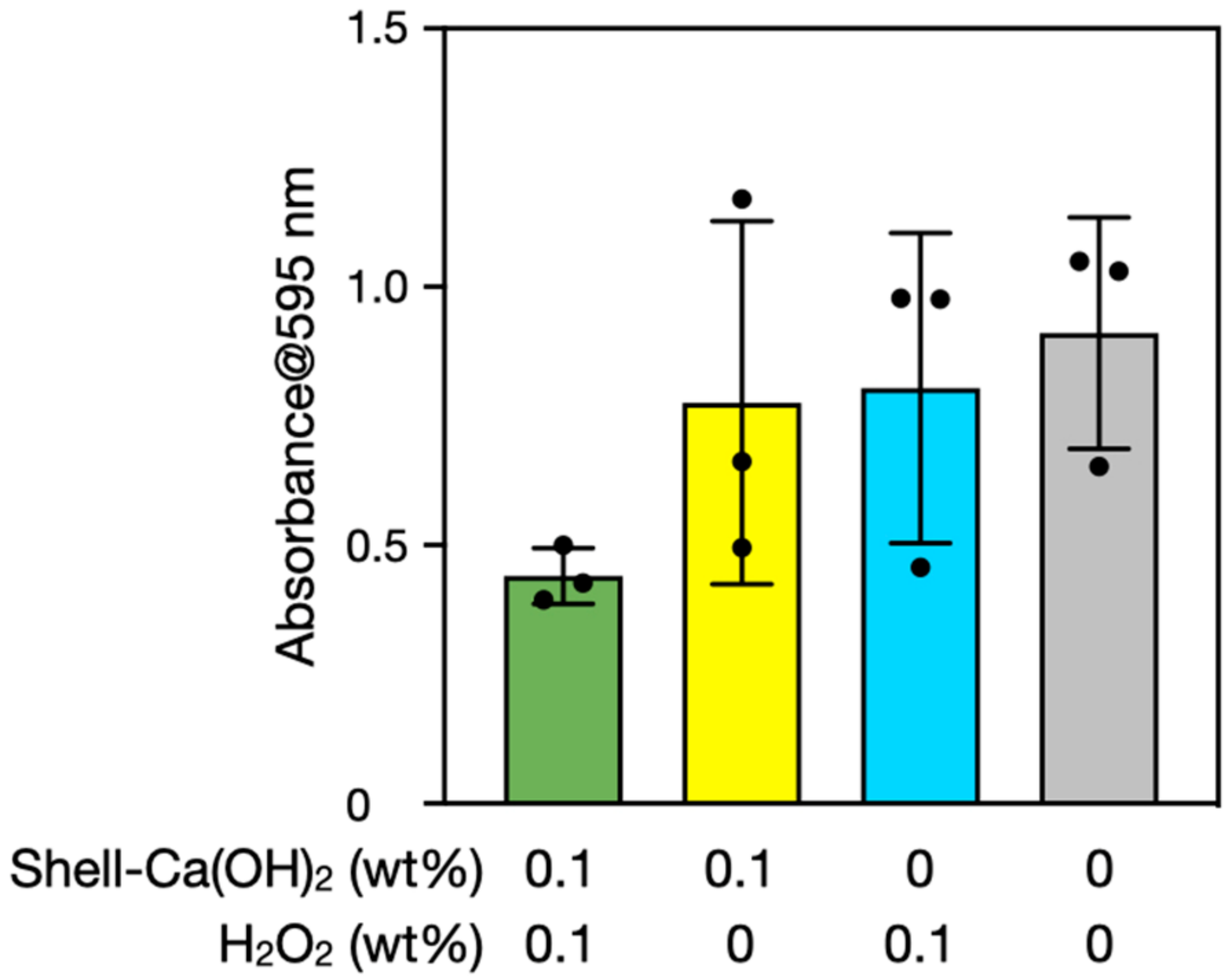
3.4. Biofilm Degradation Using Seashell-Derived Ca(OH)2

Finally, we performed experiments using scallop shell-derived Ca(OH)2 because naturally derived materials may contain impurities that influence H2O2 reactivities. Biofilms were therefore treated with scallop shell-derived Ca(OH)2 rather than reagent-grade Ca(OH)2. Figure 7 shows the results of biofilm semi-quantification by the crystal violet stain method before and after the treatments. One-way ANOVA indicated nonsignificant differences between the groups (p > 0.05). Nevertheless, average values showed a trend similar to those of the results using reagent-grade Ca(OH)2 (Figure 2), suggesting that scallop shell-derived Ca(OH)2 also has the potential to remove biofilms with the concurrent use of H2O2. Available natural sources of Ca(OH)2 include seashells, eggshells, and limestone.

4. Conclusions

We used Ca(OH)2 and H2O2 to degrade biofilms. This mixture removed a significantly greater amount of biofilm from the surface than Ca(OH)2 alone and H2O2 alone. Further, the 10 min treatment was considered optimal for biofilm degradation under the conditions investigated, as shorter and longer treatment time resulted in decreased efficacy. The higher biofilm degradation activity of Ca(OH)2–H2O2 solutions was attributed to the synergy of HO from Ca(OH)2 with reactive species from H2O2, such as HOO and HO⋅. The potential byproducts are environmentally friendly: CaCO3 and water from Ca(OH)2 and oxygen and water from H2O2. Our findings suggest that the concurrent use of other reagents, such as H2O2, is a promising strategy for improving the biofilm degradation activity of seashell-derived Ca(OH)2, and will inspire the development of more efficient biofilm removal methods.

Author Contributions

Conceptualization, Y.H., H.M., and S.N.; methodology, Y.H., Y.B., and S.H.; validation, H.M. and S.N.; investigation, Y.H., Y.B., and S.H.; writing—original draft preparation, Y.H.; writing—review and editing, H.M. and S.N.; supervision, S.N.; project administration, Y.H. All authors have read and agreed to the published version of the manuscript.

Funding

This work was partly supported by the Advanced Defense Medicine Research Program of the National Defense Medical College.

Institutional Review Board Statement

Not applicable.

Informed Consent Statement

Not applicable.

Data Availability Statement

Not applicable.

Acknowledgments

The authors wish to thank the National Defense Medical College Central Research Laboratory for SEM and fluorescence microscopy observations.

Conflicts of Interest

The authors declare no conflict of interest.

References

  1. Donlan, R.M. Biofilms: Microbial life on surfaces. Emerg. Infect. Dis. 2002, 8, 881–890. [Google Scholar] [CrossRef] [PubMed]
  2. Flemming, H.-C.; Wingender, J. The biofilm matrix. Nat. Rev. Microbiol. 2010, 8, 623–633. [Google Scholar] [CrossRef] [PubMed]
  3. Hobley, L.; Harkins, C.; MacPhee, C.E.; Stanley-Wall, N.R. Giving structure to the biofilm matrix: An overview of individual strategies and emerging common themes. FEMS Microbiol. Rev. 2015, 39, 649–669. [Google Scholar] [CrossRef] [PubMed] [Green Version]
  4. Yin, W.; Wang, Y.; Liu, L.; He, J. Biofilms: The microbial “protective clothing” in extreme environments. Int. J. Mol. Sci. 2019, 20, 3423. [Google Scholar] [CrossRef] [PubMed] [Green Version]
  5. Rumbaugh, K.P.; Sauer, K. Biofilm dispersion. Nat. Rev. Microbiol. 2020, 18, 571–586. [Google Scholar] [CrossRef] [PubMed]
  6. Hayta, E.N.; Ertelt, M.J.; Kretschmer, M.; Lieleg, O. Bacterial materials: Applications of natural and modified biofilms. Adv. Mater. Interfaces 2021, 8, 2101024. [Google Scholar] [CrossRef]
  7. Donlan, R.M.; Costerton, J.W. Biofilms: Survival mechanisms of clinically relevant microorganisms. Clin. Microbiol. Rev. 2002, 15, 167–193. [Google Scholar] [CrossRef] [Green Version]
  8. De Carvalho, C.C.C.R. Biofilms: Recent developments on an old battle. Recent Pat. Biotechnol. 2007, 1, 49–57. [Google Scholar] [CrossRef]
  9. Srey, S.; Jahid, I.K.; Ha, S.-D. Biofilm formation in food industries: A food safety concern. Food Control 2013, 31, 572–585. [Google Scholar] [CrossRef]
  10. Vestby, L.K.; Grønseth, T.; Simm, R.; Nesse, L.L. Bacterial biofilm and its role in the pathogenesis of disease. Antibiotics 2020, 9, 59. [Google Scholar] [CrossRef]
  11. Meyer, B. Approaches to prevention, removal and killing of biofilms. Int. Biodeterior. Biodegrad. 2003, 51, 249–253. [Google Scholar] [CrossRef]
  12. Gómez-López, V.M.; Marín, A.; Medina-Martínez, M.S.; Gil, M.I.; Allende, A. Generation of trihalomethanes with chlorine-based sanitizers and impact on microbial, nutritional and sensory quality of baby spinach. Postharvest Biol. Technol. 2013, 85, 210–217. [Google Scholar] [CrossRef]
  13. Sawai, J. Antimicrobial characteristics of heated scallop shell powder and its application. Biocontrol Sci. 2011, 16, 95–102. [Google Scholar] [CrossRef] [Green Version]
  14. Hata, Y.; Ishihara, M.; Hiruma, S.; Takayama, T.; Nakamura, S.; Ando, N. Recent progress in the development of disinfectants from scallop shell-derived calcium oxide for clinical and daily use. Biocontrol Sci. 2021, 26, 129–135. [Google Scholar] [CrossRef]
  15. Sawai, J.; Satoh, M.; Horikawa, M.; Shiga, H.; Kojima, H. Heated scallop-shell powder slurry treatment of shredded cabbage. J. Food Prot. 2001, 64, 1579–1583. [Google Scholar] [CrossRef]
  16. Bodur, T.; Yaldirak, G.; Kola, O.; Çaĝri-MehmetoĜlu, A. Inhibition of Listeria monocytogenes and Escherichia coli O157:H7 on frankfurters using scallop-shell powder. J. Food Saf. 2010, 30, 740–752. [Google Scholar] [CrossRef]
  17. Cagri-Mehmetoglu, A. Inhibition of Listeria monocytogenes and Salmonella enteritidis on chicken wings using scallop-shell powder. Poult. Sci. 2011, 90, 2600–2605. [Google Scholar] [CrossRef]
  18. Sato, Y.; Ohata, H.; Inoue, A.; Ishihara, M.; Nakamura, S.; Fukuda, K.; Takayama, T.; Murakami, K.; Hiruma, S.; Yokoe, H. Application of colloidal dispersions of bioshell calcium oxide (BiSCaO) for disinfection. Polymers 2019, 11, 1991. [Google Scholar] [CrossRef] [Green Version]
  19. Nakamura, S.; Ishihara, M.; Sato, Y.; Takayama, T.; Hiruma, S.; Ando, N.; Fukuda, K.; Murakami, K.; Yokoe, H. Concentrated bioshell calcium oxide (BiSCaO) water kills pathogenic microbes: Characterization and activity. Molecules 2020, 25, 3001. [Google Scholar] [CrossRef]
  20. Ishihara, M.; Hata, Y.; Hiruma, S.; Takayama, T.; Nakamura, S.; Sato, Y.; Ando, N.; Fukuda, K.; Murakami, K.; Yokoe, H. Safety of concentrated bioshell calcium oxide water application for surface and skin disinfections against pathogenic microbes. Molecules 2020, 25, 4502. [Google Scholar] [CrossRef]
  21. Hiruma, S.; Hata, Y.; Ishihara, M.; Takayama, T.; Nakamura, S.; Ando, N.; Fukuda, K.; Sato, Y.; Murakami, K.; Yokoe, H. Efficacy of bioshell calcium oxide water as disinfectants to enable face mask reuse. Biocontrol Sci. 2021, 26, 27–35. [Google Scholar] [CrossRef]
  22. Kubo, M.; Ohshima, Y.; Irie, F.; Kikuchi, M.; Sawai, J. Disinfection treatment of heated scallop-shell powder on biofilm of Escherichia coli ATCC 25922 surrogated for E. coli O157:H7. J. Biomater. Nanobiotechnol. 2013, 4, 10–19. [Google Scholar] [CrossRef] [Green Version]
  23. Sawai, J.; Nagasawa, K.; Kikuchi, M. Ability of heated scallop-shell powder to disinfect Staphylococcus aureus biofilm. Food Sci. Technol. Res. 2013, 19, 561–568. [Google Scholar] [CrossRef] [Green Version]
  24. Tsukuda, H.; Akimoto, T.; Fukikoshi, N.; Wada, R.; Sawai, J. Antibiofilm effects of heated scallop shell powder on Campylobacter jejuni biofilms. Membranes 2022, 12, 43. [Google Scholar] [CrossRef]
  25. Walker, J.T.; Bradshaw, D.J.; Fulford, M.R.; Marsh, P.D. Microbiological evaluation of a range of disinfectant products to control mixed-species biofilm contamination in a laboratory model of a dental unit water system. Appl. Environ. Microbiol. 2003, 69, 3327–3332. [Google Scholar] [CrossRef] [Green Version]
  26. Liaqat, I.; Sabri, A.N. Effect of biocides on biofilm bacteria from dental unit water lines. Curr. Microbiol. 2008, 56, 619–624. [Google Scholar] [CrossRef]
  27. Gawande, P.V.; LoVetri, K.; Yakandawala, N.; Romeo, T.; Zhanel, G.G.; Cvitkovitch, D.G.; Madhyastha, S. Antibiofilm activity of sodium bicarbonate, sodium metaperiodate and SDS combination against dental unit waterline-associated bacteria and yeast. J. Appl. Microbiol. 2008, 105, 986–992. [Google Scholar] [CrossRef]
  28. Akinbobola, A.B.; Amaeze, N.J.; Mackay, W.G.; Ramage, G.; Williams, C. ‘Secondary biofilms’ could cause failure of peracetic acid high-level disinfection of endoscopes. J. Hosp. Infect. 2021, 107, 67–75. [Google Scholar] [CrossRef]
  29. Fang, J.M.; Sun, R.C.; Salisbury, D.; Fowler, P.; Tomkinson, J. Comparative study of hemicelluloses from wheat straw by alkali and hydrogen peroxide extractions. Polym. Degrad. Stab. 1999, 66, 423–432. [Google Scholar] [CrossRef]
  30. Brooks, R.E.; Moore, S.B. Alkaline hydrogen peroxide bleaching of cellulose. Cellulose 2000, 7, 263–286. [Google Scholar] [CrossRef]
  31. Linley, E.; Denyer, S.P.; McDonnell, G.; Simons, C.; Maillard, J.-Y. Use of hydrogen peroxide as a biocide: New consideration of its mechanisms of biocidal action. J. Antimicrob. Chemother. 2012, 67, 1589–1596. [Google Scholar] [CrossRef] [PubMed] [Green Version]
  32. Qin, C.Q.; Du, Y.M.; Xiao, L. Effect of hydrogen peroxide treatment on the molecular weight and structure of chitosan. Polym. Degrad. Stab. 2002, 76, 211–218. [Google Scholar] [CrossRef]
  33. Harrison, J.J.; Turner, R.J.; Ceri, H. High-throughput metal susceptibility testing of microbial biofilms. BMC Microbiol. 2005, 5, 53. [Google Scholar] [CrossRef] [PubMed] [Green Version]
  34. Tsukatani, T.; Kawaguchi, T.; Suenaga, H.; Shiga, M.; Ikegami, T. Rapid and simple determination of minimum biofilm eradication concentration by a colorimetric microbial viability assay based on reduction of a water-soluble tetrazolium salt and combined effect of antibiotics against microbial biofilm. J. Microbiol. Biotechnol. Food Sci. 2016, 6, 677–680. [Google Scholar] [CrossRef] [Green Version]
  35. Merritt, J.H.; Kadouri, D.E.; O’Toole, G.A. Growing and analyzing static biofilms. Curr. Protoc. Microbiol. 2011, 22, 1B.1.1–1B.1.18. [Google Scholar] [CrossRef] [Green Version]
  36. Coffey, B.M.; Anderson, G.G. Biofilm formation in the 96-well microtiter plate. In Pseudomonas Methods and Protocols; Filloux, A., Ramos, J.-L., Eds.; Humana: New York, NY, USA, 2014; Volume 1149, pp. 631–641. [Google Scholar]
  37. Jang, H.; Rusconi, R.; Stocker, R. Biofilm disruption by an air bubble reveals heterogeneous age-dependent detachment patterns dictated by initial extracellular matrix distribution. npj Biofilms Microbiomes 2017, 3, 6. [Google Scholar] [CrossRef]
Figure 1. Preparation of an in vitro biofilm model. (a) Photograph of the 96-pin lid. (b) Schematic illustration of biofilm preparation. SEM images of prepared biofilms at (c) low and (d) high magnifications. The dotted line in (c) indicates the upper edge of the biofilms. The arrows in (d) indicate nanofibrous materials.
Nanomaterials 12 03681 g001
Figure 2. Semi-quantification of biofilms before and after the treatments with Ca(OH)2 and/or H2O2 for 10 min. The y-axis is the 595-nm absorbance of crystal violet extracted from the stained biofilms. The error bars represent the standard deviation of three individual trials. Statistical analysis was performed by parametric one-way ANOVA with Dunnett’s multiple comparison; ns, **, ***, and **** denote not significant, p < 0.01, p < 0.001, and p < 0.0001, respectively.
Nanomaterials 12 03681 g002
Figure 3. Semi-quantification of biofilms before and after the treatments with Ca(OH)2 and/or H2O2 for (a) 3 and (b) 30 min. The y-axis is the 595-nm absorbance of crystal violet extracted from the stained biofilms. The error bars represent the standard deviation of three individual trials.
Nanomaterials 12 03681 g003
Figure 4. Fluorescence microscopy images of biofilms after the treatments with (a) 0.1% Ca(OH)2–0.1% H2O2, (b) 0.1% Ca(OH)2, and (c) 0.1% H2O2 and (d) nontreated biofilms. Each image visualizes a polypropylene pin with biofilms. The green fluorescence (left) and the red fluorescence (middle) correspond to SYTO 9 and propidium iodide, respectively. Merged images of the two channels are shown in the right column.
Nanomaterials 12 03681 g004
Figure 5. pH time courses of the solutions of Ca(OH)2 and H2O2.
Nanomaterials 12 03681 g005
Figure 6. ESR spectra of (a) Ca(OH)2–H2O2 solutions, (b) Ca(OH)2 solutions, and (c) H2O2 solutions. The arrows in (a) indicate characteristic signals of HO⋅.
Nanomaterials 12 03681 g006
Figure 7. Semi-quantification of biofilms before and after the treatments with scallop shell-derived Ca(OH)2 and/or H2O2 for 10 min. The y-axis is the 595-nm absorbance of crystal violet extracted from the stained biofilms. The error bars represent the standard deviation of three individual trials.
Nanomaterials 12 03681 g007
Publisher’s Note: MDPI stays neutral with regard to jurisdictional claims in published maps and institutional affiliations.

