
Design of a Novel Nanosensors Based on Green Synthesized CoFe2O4/Ca-Alginate Nanocomposite-Coated QCM for Rapid Detection of Pb(II) Ions

 ,
,  and
and 
Abstract

1. Introduction
2. Materials and Methods
2.1. Materials
2.2. Preparation of the Clove Leaves Extract
2.3. Green Synthesis of CoFe2O4 Nanoparticles
2.4. Preparation of CoFe2O4/Ca-Alg Nanocomposite
2.5. Characterization
2.6. Quartz Crystal Microbalance (QCM)
3. Results and Discussion
3.1. Characterization of Green Synthesized CoFe2O4 Nanoparticles and CoFe2O4/Ca-Alg Nanocomposite
3.1.1. XRD
3.1.2. BET Surface Area and Porosity Properties
3.1.3. DLS and Zeta Potential
3.1.4. AFM
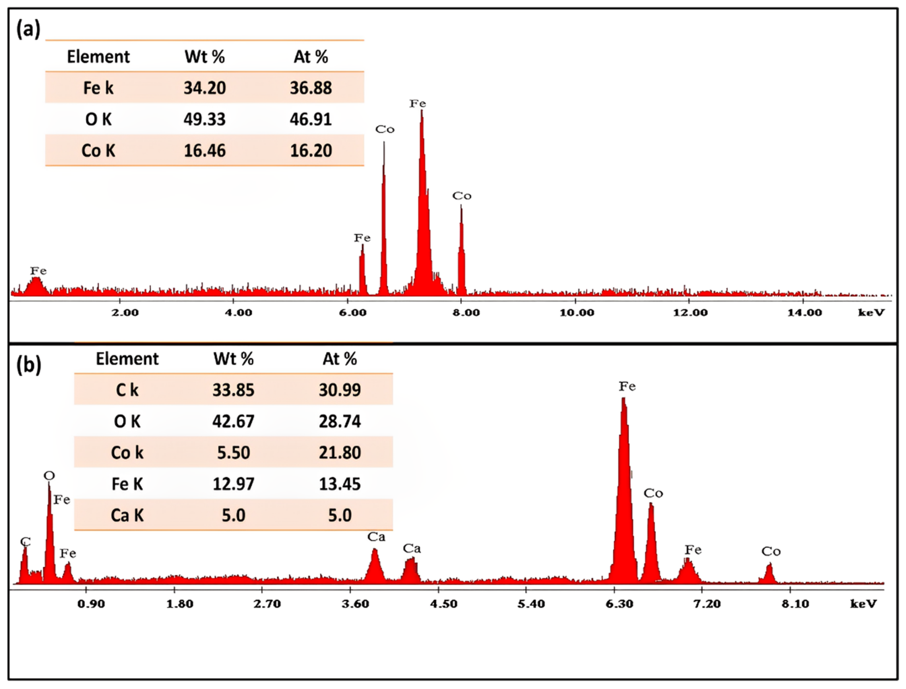
3.1.5. SEM and TEM
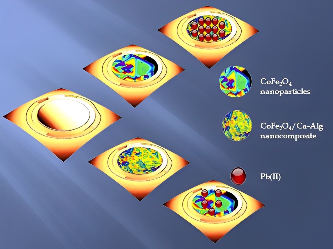
3.2. Green Synthesized CoFe2O4 Nanoparticles and CoFe2O4/Ca-Alg Nanocomposite-Coated QCM Nanosensors for Detecting Pb(II) Ions in the Aqueous Solutions
4. Conclusions
Supplementary Materials
Author Contributions
Funding
Data Availability Statement
Acknowledgments
Conflicts of Interest
References
- Yu, B.; Zhang, Y.; Shukla, A.; Shukla, S.S.; Dorris, K.L. The removal of heavy metals from aqueous solutions by sawdust adsorption—Removal of lead and comparison of its adsorption with copper. J. Hazard. Mater. 2001, 84, 83–94. [Google Scholar] [CrossRef]
- Tchounwou, P.B.; Yedjou, C.G.; Patlolla, A.K.; Sutton, D.J. Heavy metal toxicity and the environment. Mol. Clin. Environ. Toxicol. 2012, 101, 133–164. [Google Scholar] [CrossRef]
- Hamdy, A.; Mostafa, M.K.; Nasr, M. Techno-economic estimation of electroplating wastewater treatment using zero-valent iron nanoparticles: Batch optimization, continuous feed, and scaling up studies. Environ. Sci. Pollut. Res. 2019, 26, 25372–25385. [Google Scholar] [CrossRef] [PubMed]
- Hamdy, A. Experimental Study of the Relationship Between Dissolved Iron, Turbidity, and Removal of Cu(II) Ion From Aqueous Solutions Using Zero-Valent Iron Nanoparticles. Arab. J. Sci. Eng. 2020, 46, 5543–5565. [Google Scholar] [CrossRef]
- Hridya, T.; Varghese, E.; Harikumar, P. Removal of heavy metals from aqueous solution using porous (Styrene-divinylbenzene)/CuNi bimetallic nanocomposite microspheres. Environ. Nanotechnol. Monit. Manag. 2021, 16, 100606. [Google Scholar] [CrossRef]
- Jaishankar, M.; Tseten, T.; Anbalagan, N.; Mathew, B.B.; Beeregowda, K.N. Toxicity, mechanism and health effects of some heavy metals. Interdiscip. Toxicol. 2014, 7, 60–72. [Google Scholar] [CrossRef]
- Chowdhury, S.; Mazumder, M.A.J.; Al-Ahmed, A. Removal of lead ions (Pb2+) from water and wastewater: A review on the low-cost adsorbents. Appl. Water Sci. 2022, 12, 1–33. [Google Scholar] [CrossRef]
- Hamdy, A.; Ismail, S.H.; Ebnalwaled, A.A.; Mohamed, G.G. Characterization of Superparamagnetic/Monodisperse PEG-Coated Magnetite Nanoparticles Sonochemically Prepared from the Hematite Ore for Cd(II) Removal from Aqueous Solutions. J. Inorg. Organomet. Polym. Mater. 2020, 31, 397–414. [Google Scholar] [CrossRef]
- Gautam, R.K.; Sharma, S.K.; Mahiya, S.; Chattopadhyaya, M.C. CHAPTER 1. Contamination of Heavy Metals in Aquatic Media: Transport, Toxicity and Technologies for Remediation. In Heavy Metals in Water: Presence, Removal and Safety; RSC Publishing: Cambridge, UK, 2014; pp. 1–24. [Google Scholar] [CrossRef]
- Cherono, F.; Mburu, N.; Kakoi, B. Adsorption of lead, copper and zinc in a multi-metal aqueous solution by waste rubber tires for the design of single batch adsorber. Heliyon 2021, 7, e08254. [Google Scholar] [CrossRef]
- El-Wakeel, S.T.; Abdel-Karim, A.; Ismail, S.H.; Mohamed, G.G. Development of Ag-dendrites @Cu nanostructure for removal of selenium (IV) from aqueous solution. Water Environ. Res. 2022, 94, e10713. [Google Scholar] [CrossRef]
- Katowah, D.F.; Alsulami, Q.A.; Alam, M.M.; Ismail, S.H.; Asiri, A.M.; Mohamed, G.G.; Rahman, M.M.; Hussein, M.A. The Performance of Various SWCNT Loading into CuO–PMMA Nanocomposites Towards the Detection of Mn2+ Ions. J. Inorg. Organomet. Polym. Mater. 2020, 30, 5024–5041. [Google Scholar] [CrossRef]
- Cretescu, I.; Tutulea, M.D.; Sibiescu, D.; Stan, C. ELECTROCHEMICAL SENSORS FOR HEAVY METAL IONS DETECTION FROM AQUEOUS SOLUTIONS. Environ. Eng. Manag. J. 2012, 11, 463–470. [Google Scholar] [CrossRef]
- Razzak, S.A.; Faruque, M.O.; Alsheikh, Z.; Alsheikhmohamad, L.; Alkuroud, D.; Alfayez, A.; Hossain, S.M.Z.; Hossain, M.M. A comprehensive review on conventional and biological-driven heavy metals removal from industrial wastewater. Environ. Adv. 2022, 7, 100168. [Google Scholar] [CrossRef]
- Al-Qasmi, N.; Al-Gethami, W.; Alhashmialameer, D.; Ismail, S.H.; Sadek, A.H. Evaluation of Green-Synthesized Cuprospinel Nanoparticles as a Nanosensor for Detection of Low-Concentration Cd(II) Ion in the Aqueous Solutions by the Quartz Crystal Microbalance Method. Materials 2022, 15, 6240. [Google Scholar] [CrossRef] [PubMed]
- Sartore, L.; Barbaglio, M.; Borgese, L.; Bontempi, E. Polymer-grafted QCM chemical sensor and application to heavy metal ions real time detection. Sens. Actuators B Chem. 2011, 155, 538–544. [Google Scholar] [CrossRef]
- Yang, Z.-P.; Zhang, C.-J. Designing of MIP-based QCM sensor for the determination of Cu(II) ions in solution. Sens. Actuators B Chem. 2009, 142, 210–215. [Google Scholar] [CrossRef]
- Fernández, R.; Calero, M.; Jiménez, Y.; Arnau, A. A Real-Time Method for Improving Stability of Monolithic Quartz Crystal Microbalance Operating under Harsh Environmental Conditions. Sensors 2021, 21, 4166. [Google Scholar] [CrossRef]
- Cao, Z.; Guo, J.; Fan, X.; Xu, J.; Fan, Z.; Du, B. Detection of heavy metal ions in aqueous solution by P(MBTVBC-co-VIM)-coated QCM sensor. Sens. Actuators B Chem. 2011, 157, 34–41. [Google Scholar] [CrossRef]
- Thies, J.-W.; Kuhn, P.; Thürmann, B.; Dübel, S.; Dietzel, A. Microfluidic quartz-crystal-microbalance (QCM) sensors with specialized immunoassays for extended measurement range and improved reusability. Microelectron. Eng. 2017, 179, 25–30. [Google Scholar] [CrossRef]
- Atashbar, M.Z.; Bejcek, B.; Vijh, A.; Singamaneni, S. QCM biosensor with ultra thin polymer film. Sens. Actuators B Chem. 2005, 107, 945–951. [Google Scholar] [CrossRef]
- Diltemiz, S.E.; Keçili, R.; Ersöz, A.; Say, R. Molecular Imprinting Technology in Quartz Crystal Microbalance (QCM) Sensors. Sensors 2017, 17, 454. [Google Scholar] [CrossRef] [PubMed]
- Zhou, G. Real-Time, Selective Detection of Heavy Metal Ions in Water Using 2d Nanomaterials-based Field-effect Transistors. Master’s Thesis, The University of Wisconsin-Milwaukee, Milwaukee, WI, USA, 2017. [Google Scholar]
- Ebnalwaled, A.A.; Sadek, A.H.; Ismail, S.H.; Mohamed, G.G. Structural, optical, dielectric, and surface properties of polyimide hybrid nanocomposites films embedded mesoporous silica nanoparticles synthesized from rice husk ash for optoelectronic applications. Opt. Quantum Electron. 2022, 54, 1–31. [Google Scholar] [CrossRef]
- Maduraiveeran, G.; Jin, W. Nanomaterials based electrochemical sensor and biosensor platforms for environmental applications. Trends Environ. Anal. Chem. 2017, 13, 10–23. [Google Scholar] [CrossRef]
- Yalcin, B.; Ozcelik, S.; Icin, K.; Senturk, K.; Arda, L. Structural, optical, magnetic, photocatalytic activity and related biological effects of CoFe2O4 ferrite nanoparticles. J. Mater. Sci. Mater. Electron. 2021, 32, 13068–13080. [Google Scholar] [CrossRef]
- Kalam, A.; Al-Sehemi, A.G.; Assiri, M.; Du, G.; Ahmad, T.; Ahmad, I.; Pannipara, M. Modified solvothermal synthesis of cobalt ferrite (CoFe2O4) magnetic nanoparticles photocatalysts for degradation of methylene blue with H2O2/visible light. Results Phys. 2018, 8, 1046–1053. [Google Scholar] [CrossRef]
- Loan, N.T.T.; Lan, N.T.H.; Hang, N.T.T.; Hai, N.Q.; Anh, D.T.T.; Hau, V.T.; Van Tan, L.; Van Tran, T. CoFe2O4 Nanomaterials: Effect of Annealing Temperature on Characterization, Magnetic, Photocatalytic, and Photo-Fenton Properties. Processes 2019, 7, 885. [Google Scholar] [CrossRef]
- Gingasu, D.; Mindru, I.; Patron, L.; Calderon-Moreno, J.M.; Mocioiu, O.C.; Preda, S.; Stanica, N.; Nita, S.; Dobre, N.; Popa, M. Green synthesis methods of CoFe2O4 and Ag-CoFe2O4 nanoparticles using hibiscus extracts and their antimicrobial potential. J. Nanomater. 2016, 2016, 2106756. [Google Scholar] [CrossRef]
- Razavi, M.; Salahinejad, E.; Fahmy, M.; Yazdimamaghani, M.; Vashaee, D.; Tayebi, L. Green Chemical and Biological Synthesis of Nanoparticles and Their Biomedical Applications. In Green Process for Nanotechnology; Springer: Berlin/Heidelberg, Germany, 2015; pp. 207–235. [Google Scholar]
- Kombaiah, K.; Vijaya, J.J.; Kennedy, L.J.; Bououdina, M.; Ramalingam, R.J.; Al-Lohedan, H.A. Okra extract-assisted green synthesis of CoFe2O4 nanoparticles and their optical, magnetic, and antimicrobial properties. Mater. Chem. Phys. 2018, 204, 410–419. [Google Scholar] [CrossRef]
- Bao, Y.; He, J.; Song, K.; Guo, J.; Zhou, X.; Liu, S. Plant-Extract-Mediated Synthesis of Metal Nanoparticles. J. Chem. 2021, 2021, 1–14. [Google Scholar] [CrossRef]
- Sharma, D.; Kanchi, S.; Bisetty, K. Biogenic synthesis of nanoparticles: A review. Arab. J. Chem. 2019, 12, 3576–3600. [Google Scholar] [CrossRef]
- Lakhan, M.N.; Chen, R.; Shar, A.H.; Chand, K.; Shah, A.H.; Ahmed, M.; Ali, I.; Ahmed, R.; Liu, J.; Takahashi, K.; et al. Eco-friendly green synthesis of clove buds extract functionalized silver nanoparticles and evaluation of antibacterial and antidiatom activity. J. Microbiol. Methods 2020, 173, 105934. [Google Scholar] [CrossRef]
- Esmat, M.; Farghali, A.A.; Khedr, M.H.; El-Sherbiny, I.M. Alginate-based nanocomposites for efficient removal of heavy metal ions. Int. J. Biol. Macromol. 2017, 102, 272–283. [Google Scholar] [CrossRef] [PubMed]
- Tamhankar, P.M.; Kulkarni, A.M.; Watawe, S.C. Functionalization of Cobalt Ferrite Nanoparticles with Alginate Coating for Biocompatible Applications. Mater. Sci. Appl. 2011, 02, 1317–1321. [Google Scholar] [CrossRef]
- Rajaji, U.; Govindasamy, M.; Sha, R.; Alshgari, R.A.; Juang, R.-S.; Liu, T.-Y. Surface engineering of 3D spinel Zn3V2O8 wrapped on sulfur doped graphitic nitride composites: Investigation on the dual role of electrocatalyst for simultaneous detection of antibiotic drugs in biological fluids. Compos. Part B Eng. 2022, 242, 110017. [Google Scholar] [CrossRef]
- Vinoth, S.; Govindasamy, M.; Wang, S.-F. Solvothermal synthesis of silver tungstate integrated with carbon nitrides matrix composites for highly sensitive electrochemical nitrofuran derivative sensing in biological samples. Anal. Chim. Acta 2022, 1192, 339355. [Google Scholar] [CrossRef] [PubMed]
- Lu, Z.; Du, X.; Sun, M.; Zhang, Y.; Li, Y.; Wang, X.; Wang, Y.; Du, H.; Yin, H.; Rao, H. Novel dual-template molecular imprinted electrochemical sensor for simultaneous detection of CA and TPH based on peanut twin-like NiFe2O4/CoFe2O4/NCDs nanospheres: Fabrication, application and DFT theoretical study. Biosens. Bioelectron. 2021, 190, 113408. [Google Scholar] [CrossRef] [PubMed]
- Yildirim, A.; Dogac, Y.İ. An application of CoFe2O4/alginate magnetic beads: Drug delivery system of 5-fluorouracil. Int. J. Second. Metab. 2022, 9, 305–319. [Google Scholar] [CrossRef]
- Mostafazadeh, R.; Ghaffarinejad, A.; Tajabadi, F. A caffeic acid electrochemical sensor amplified with GNR/CoFe2O4@NiO and 1-Ethyl-3-methylimidazolium acetate; a new perspective for food analysis. Food Chem. Toxicol. 2022, 167, 113312. [Google Scholar] [CrossRef] [PubMed]
- Morais, P.V.; Orlandi, M.O.; Schöning, M.J.; Siqueira, J.R., Jr. Layer-by-Layer Films with CoFe2O4 Nanocrystals and Graphene Oxide as a Sensitive Interface in Capacitive Field-Effect Devices. ACS Appl. Nano Mater. 2022, 5, 5258–5267. [Google Scholar] [CrossRef]
- Al-Qasmi, N. Facial Eco-Friendly Synthesis of Copper Oxide Nanoparticles Using Chia Seeds Extract and Evaluation of Its Electrochemical Activity. Processes 2021, 9, 2027. [Google Scholar] [CrossRef]
- Albalah, M.A.; Alsabah, Y.A.; Mustafa, D.E. Characteristics of co-precipitation synthesized cobalt nanoferrites and their potential in industrial wastewater treatment. SN Appl. Sci. 2020, 2, 1–9. [Google Scholar] [CrossRef]
- Kuzmanović, M.; Božanić, D.K.; Milivojević, D.; Ćulafić, D.M.; Stanković, S.; Ballesteros, C.; Gonzalez-Benito, J. Sodium-alginate biopolymer as a template for the synthesis of nontoxic red emitting Mn2+-doped CdS nanoparticles. RSC Adv. 2017, 7, 53422–53432. [Google Scholar] [CrossRef]
- Kumar, K.V.; Gadipelli, S.; Wood, B.; Ramisetty, K.A.; Stewart, A.A.; Howard, C.A.; Brett, D.J.L.; Rodriguez-Reinoso, F. Characterization of the adsorption site energies and heterogeneous surfaces of porous materials. J. Mater. Chem. A 2019, 7, 10104–10137. [Google Scholar] [CrossRef]
- Xu, L.; Zhang, J.; Ding, J.; Liu, T.; Shi, G.; Li, X.; Dang, W.; Cheng, Y.; Guo, R. Pore Structure and Fractal Characteristics of Different Shale Lithofacies in the Dalong Formation in the Western Area of the Lower Yangtze Platform. Minerals 2020, 10, 72. [Google Scholar] [CrossRef]
- Fantauzzi, M.; Secci, F.; Angotzi, M.S.; Passiu, C.; Cannas, C.; Rossi, A. Nanostructured spinel cobalt ferrites: Fe and Co chemical state, cation distribution and size effects by X-ray photoelectron spectroscopy. RSC Adv. 2019, 9, 19171–19179. [Google Scholar] [CrossRef] [PubMed]
- Singh, M.N.; Sinha, A.K.; Ghosh, H. Determination of transition metal ion distribution in cubic spinel Co1.5Fe1.5O4using anomalous x-ray diffraction. AIP Adv. 2015, 5, 087115. [Google Scholar] [CrossRef]
- Jeevanantham, B.; Song, Y.; Choe, H.; Shobana, M. Structural and optical characteristics of cobalt ferrite nanoparticles. Mater. Lett. X 2021, 12, 100105. [Google Scholar] [CrossRef]
- Londoño-Calderón, C.L.; Londoño, O.M.; Muraca, D.; Arzuza, L.; Carvalho, P.; Pirota, K.R.; Knobel, M.; Pampillo, L.G.; Martínez-García, R. Synthesis and magnetic properties of cobalt-iron/cobalt-ferrite soft/hard magnetic core/shell nanowires. Nanotechnology 2017, 28, 245605. [Google Scholar] [CrossRef]
- Béjaoui, M.; Elmhamdi, A.; Pascual, L.; Pérez-Bailac, P.; Nahdi, K.; Martínez-Arias, A. Preferential Oxidation of CO over CoFe2O4 and M/CoFe2O4 (M= Ce, Co, Cu or Zr) Catalysts. Catalysts 2020, 11, 15. [Google Scholar] [CrossRef]


| Material | Surface Area (SBET), m2/g | Total Pore Volume, cc/g | Average Pore Size, nm | Micropore Volume, cc/g (DA Method) | Average Micropore Size, nm (DA Method) |
|---|---|---|---|---|---|
| CoFe2O4 nanoparticles | 63.21 | 0.068 | 2.17 | 0.066 | 1.55 |
| CoFe2O4/Ca-Alg nanocomposite | 34.05 | 0.083 | 4.86 | 0.047 | 1.17 |
Publisher’s Note: MDPI stays neutral with regard to jurisdictional claims in published maps and institutional affiliations. |
© 2022 by the authors. Licensee MDPI, Basel, Switzerland. This article is an open access article distributed under the terms and conditions of the Creative Commons Attribution (CC BY) license (https://creativecommons.org/licenses/by/4.0/).
Share and Cite
Al-Gethami, W.; Alhashmialameer, D.; Al-Qasmi, N.; Ismail, S.H.; Sadek, A.H. Design of a Novel Nanosensors Based on Green Synthesized CoFe2O4/Ca-Alginate Nanocomposite-Coated QCM for Rapid Detection of Pb(II) Ions. Nanomaterials 2022, 12, 3620. https://doi.org/10.3390/nano12203620
Al-Gethami W, Alhashmialameer D, Al-Qasmi N, Ismail SH, Sadek AH. Design of a Novel Nanosensors Based on Green Synthesized CoFe2O4/Ca-Alginate Nanocomposite-Coated QCM for Rapid Detection of Pb(II) Ions. Nanomaterials. 2022; 12(20):3620. https://doi.org/10.3390/nano12203620
Chicago/Turabian StyleAl-Gethami, Wafa, Dalal Alhashmialameer, Noha Al-Qasmi, Sameh H. Ismail, and Ahmed H. Sadek. 2022. "Design of a Novel Nanosensors Based on Green Synthesized CoFe2O4/Ca-Alginate Nanocomposite-Coated QCM for Rapid Detection of Pb(II) Ions" Nanomaterials 12, no. 20: 3620. https://doi.org/10.3390/nano12203620
APA StyleAl-Gethami, W., Alhashmialameer, D., Al-Qasmi, N., Ismail, S. H., & Sadek, A. H. (2022). Design of a Novel Nanosensors Based on Green Synthesized CoFe2O4/Ca-Alginate Nanocomposite-Coated QCM for Rapid Detection of Pb(II) Ions. Nanomaterials, 12(20), 3620. https://doi.org/10.3390/nano12203620