Article

Noble Metals Deposited LaMnO3 Nanocomposites for Photocatalytic H2 Production

by
Ahmed Hussain Jawhari
1,
Nazim Hasan
1,
Ibrahim Ali Radini
1,
Katabathini Narasimharao
2,* and
Maqsood Ahmad Malik
2,*
1
Department of Chemistry, Faculty of Science, Jazan University, Jazan 45142, Saudi Arabia
2
Chemistry Department, Faculty of Science, King Abdulaziz University, Jeddah 21589, Saudi Arabia
*
Authors to whom correspondence should be addressed.
Nanomaterials 2022, 12(17), 2985; https://doi.org/10.3390/nano12172985
Submission received: 20 July 2022 / Revised: 21 August 2022 / Accepted: 23 August 2022 / Published: 29 August 2022
(This article belongs to the Section Synthesis, Interfaces and Nanostructures)

Abstract

:
Due to the growing demand for hydrogen, the photocatalytic hydrogen production from alcohols present an intriguing prospect as a potential source of low-cost renewable energy. The noble metals (Ag, Au, Pd and Pt) deposited LaMnO3 nanocomposites were synthesized by a non-conventional green bio-reduction method using aqueous lemon peel extract, which acts as both reducing and capping agent. The successful deposition of the noble metals on the surface of LaMnO3 was verified by using powder XRD, FTIR, TEM, N2-physisorption, DR UV-vis spectroscopy, and XPS techniques. The photocatalytic activity of the synthesized nanocomposites was tested for photocatalytic H2 production under visible light irradiation. Different photocatalytic reaction parameters such as reaction time, pH, catalyst mass and reaction temperature were investigated to optimize the reaction conditions for synthesized nanocomposites. Among the synthesized noble metal deposited LaMnO3 nanocomposites, the Pt-LaMnO3 nanocomposite offered superior activity for H2 production. The enhanced photocatalytic activity of the Pt-LaMnO3 was found as a result from low bandgap energy, high photoelectrons generation and enhanced charge separation due to deposition of Pt nanoparticles. The effective noble metal deposition delivers a new route for the development of plasmonic noble metal-LaMnO3 nanocomposites for photocatalytic reforming of aqueous methanol to hydrogen.

1. Introduction

Hydrogen (H2) energy, as a clean, efficient, non-toxic, and renewable energy, has diverse applications, to promises towards the future energy structure [1,2]. Hydrogen energy can meet the increasing demand for sustainable clean energy. In addition, hydrogen can be used to generate electricity without harmful emissions in industrial applications. It is anticipated to be the cleanest energy source and an answer to the energy crisis and associated climate change issues [3]. H2 has received greater attention as it also has storage option; it allows energy to be carried and converted when required [4]. Currently, metal hydrides are utilized for conventional hydrogen storage, but their system’s storage capacity is inadequate. Fuel cell-based hydrogen is a better alternative [5,6]. Due to its low carbon emissions and high potential efficiency, the H2 fuel cell system offers an alternative to internal combustion engines for automobiles [7]. Because of the varied environmental advantages, higher efficiency and potential market, these systems have gained more attention [8].
Biomass is an important source of H2 [9] and it is mainly produced by biomass gasification, but this process is limited to specific applications [10]. Water electrolysis is a well proven technology, which is the future of H2 production, basically because it relies on an inexhaustible source, such as water. However, it uses a lot of energy, and this is a serious concern [11]. Photocatalytic water splitting [12] and photocatalytic reforming of bio-alcohols [13] are two promising methods for the sustainable production of H2, and the second process combines simultaneous H2 production [14]. H2 can be produced on a large scale through numerous technical processes. Among the thermal and nonthermal processes, the steam-reforming process exhibited four times higher H2 productivity compared to the photocatalytic non-thermal process [15]. Due to the expensive nature of H2 storage and transportation, the green methanol has become a safe and efficient alternative to promote and develop H2 energy [16]. The conventional thermal methanol-reforming reaction to produce H2 is an endothermic reaction; the energy required for the reaction is mainly provided by furnace heat, therefore, it cannot be termed as renewable energy [17].
Solar photocatalytic H2 production is ideally the best pathway to convert solar energy to chemical energy. Different semiconductors such as TiO2, ZnO, g-C3N4, Ag3PO4 and ZrO2 have been predominantly studied for photocatalytic H2 production from aqueous alcohols [18,19,20]. More recently, lanthanum-based perovskites have been proposed as photocatalytic material owing to its characteristic properties such as narrow bandgap (Eg = 1.86–2.36 eV), high stability as well as their environmental friendliness [21]. The photocatalytic properties of lanthanum (La) -based perovskites can further be enhanced via deposition of different metal nanoparticles. The presence of metal nanoparticles enhances charge transfer and light captivation through local surface plasmon resonance (LSPR), improving catalytic performance. Wang et.al. synthesized porous La-Ti based perovskite (La2Ti2O7) using CTAB as a template reagent for degradation of the dye azophloxine [22]. Moreover, the photocatalytic performance of La-based perovskites was enhanced using multiple complex or layered perovskite oxides containing Ln. Verduzco et al. synthesized a layered perovskite oxide (Sr2.7−xCaxLn0.3Fe2O7−δ) and demonstrated its use towards degradation of methylene blue (MB) dye under solar and UV irradiation [23]. Varying the stoichiometry or doping of perovskite with a cation (Ca-doping), of different valence states, can change the electronic structure, affecting the electrical and optical properties [24]. Yang et al. immobilized LaFeO3 and Au nanoparticles on the Cu2O surface as a ternary composite photocatalyst for rhodamine B degradation [25]. Numerous reports have shown that incorporation or decoration of Ln based perovskites with other metals or semiconductors results in pronounced enhancement in the photocatalysis [26,27,28,29]. The deposition of noble metal nanoparticles on the surface of polar semiconductor or insulator particles can enhance the photocatalytic activity by harvesting visible light due to the LSPR phenomenon, while the metal-semiconductor interface efficiently separates the photogenerated electrons and holes [30].
The present study focuses on the synthesis and characterization of noble metals deposited LaMnO3 nanocomposites for photocatalytic H2 production. A novel green procedure is adopted for the synthesis of bulk and noble metal deposited LaMnO3 nanocomposites. The H2 production efficacy of the indigenously synthesized, noble metal containing LaMnO3 nanocomposites was evaluated and discussed in this report for the first time.

2. Experimental

2.1. Materials

All chemicals, including Lanthanum(III) nitrate hexahydrate (La(NO3)3·6H2O), Manganese(II) nitrate hydrate (Mn(NO3)2·4H2O), Silver nitrate (AgNO3), Palladium(II) chloride (PdCl2), Platinum(II) chloride (PtCl2) and gold(III) chloride (AuCl3) are of analytical grade and purchased from Sigma-Aldrich, St. Louis, MO, USA.

2.2. Preparation of Nanomaterials

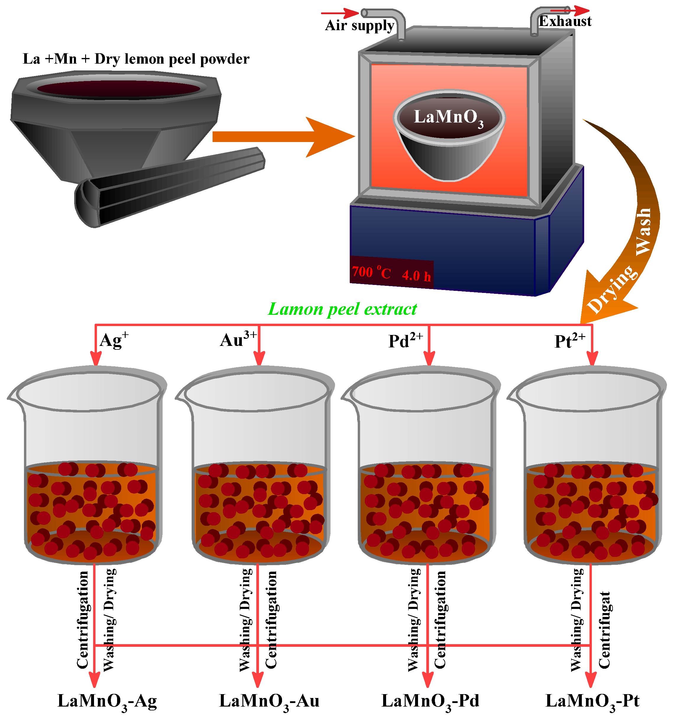
2.2.1. Synthesis of LaMnO3 by Green Solid-State Combustion Method

LaMnO3 nanocomposite was prepared by the green solid-state combustion method. First, a homogenous mixture of La(NO3)3 and Mn(NO3)2 was prepared by mixing 5.0 g of each metal precursor in an agate mortar and grounded using a pestle for 15 min at room temperature. To this homogenous precursor mixture, 20 g of fine, dried lemon peel powder was added and ground for another 30 min to acquire a perfect blend of the metal precursor and lemon peel powder. Then, the resultant powder was transferred into a ceramic crucible and calcined in a muffle furnace at 700 °C for 4 h. It was found that the mixture transformed into a fine, pitch-black powder suspended several times in double distilled water and absolute ethanol for washing; then, it was dried at 120 °C for 6 h in a vacuum oven.

2.2.2. Deposition of Noble Metals

Deposition with noble metals is an efficient strategy to extrapolate materials’ photocatalytic and optical performance. The deposition of LaMnO3 nanocomposites was carried out by the green bio-reduction method using aqueous lemon extract solution as a reducing and capping agent. The lemon peel extract has the major compound limonene, which is believed to act as a reducing agent. The fresh lemon peel extract was prepared by dispersing 10 g of fine dried lemon peel powder in 250 mL of distilled water and heating at 80 °C for 1 hr. The mixture was filtered using Whatman No. 1 filter paper by vacuum filtration. In four different beakers, 2 g of LaMnO3 nanocomposites was dispersed in 10 mL of double distilled water under constant stirring. To each beaker, 10 mL of 0.15 M aqueous solution of Ag/Pt/Au/Pd salt was added, under vigorous stirring, followed by the addition of lemon extract (20 mL). The initial reduction of noble metals was observed from the appearance of color change. The noble metal loaded LaMnO3 nanocomposites were collected by centrifugation, and the acquired material was washed several times with double distilled water followed by drying at 90 °C for 5 h. Figure 1 gives the schematic representation of the nanocomposite preparation procedure.

2.3. Characterization of Synthesized Nanomaterials

The PANalytical XpertPro diffractometer (PANalytical Inc., Westborough, MA, USA) was used to collect the XRD patterns of the powders. Applying the Debye–Scherer equation allowed for the determination of the crystallite size of the nanocomposites that were formed. Fourier transform infrared spectroscopy (FTIR) analysis of as prepared nanocomposites was performed on a Bruker ALPHA II FT-IR (Bruker Optics GmbH & Co. Rosenheim, Germany) spectrometer. For the transmission electron microscopy (TEM) investigation of the nanocomposites, a JEOL 2100HT microscope equipped with a 200 kV accelerating voltage was utilized, and images were captured using a Gatan digital camera. The X-ray photoelectron spectra of the nanocomposites were gathered with the use of an instrument called Thermo Scientific Escalab 250 Xi XPS. The Al K X-rays used in the experiment had a spot size of 650 mm. We were able to rectify the peak shift that occurred as a result of charge compensation by employing the binding energy of C1s peak. The information was gathered by performing 10–30 scans with a step size of 0.1 eV, a dwell length of 200 ms, and a pass energy of 100 eV.
Experiments with N2-physisorption employing the Quantachrome ASiQ adsorption device were used to collect information regarding the textural qualities of the materials. The reflectance spectra of the samples were taken with a Thermo-Scientific evolution UV-vis spectrophotometer that was equipped with an integrating sphere in the wavelength range of 200–800 nm. This was performed so that the optical properties of the samples could be determined. The Kubelka–Munk method was utilized to determine the bandgap energy values of each sample. The Kubelka–Munk factor, denoted by the letter K, was calculated with the help of the following equation: K = (1 − R)2/2R, where R refers to the percent reflectance. After the wavelengths (nm) were converted into energy (E), a curve was constructed by plotting (K*E)0.5 against E to determine the relationship between the two variables. The bandgap energy, denoted in eV, was found at the point on the curve where the two slopes intersected.

2.4. Photocatalytic Reforming of Methanol to Hydrogen

Under the influence of an argon environment, photocatalytic reactions were carried out in the liquid phase inside of a Pyrex flask. To equilibrate any adsorption process and achieve a homogeneous catalyst suspension, the catalyst, which weighed 150 mg, was disseminated by stirring at 500 rpm in 120 mL of a 20 vol% methanolic aqueous solution at 25 °C for thirty minutes while maintaining complete darkness. After that, the reactor was purged of its contents, and a Xe lamp with 300 W of power was used to irradiate the reaction zone with a flux of roughly 125 mW cm−2 for an hour. Using a closed gas circulation, evolved gases were brought into the sample loop of the gas chromatograph. To perform the H2 analysis, a Varian 3300 gas chromatograph was utilized. This instrument was equipped with a thermal conductivity detector and a 2 m MS 13X column. The samples were analyzed periodically.

3. Results and Discussion

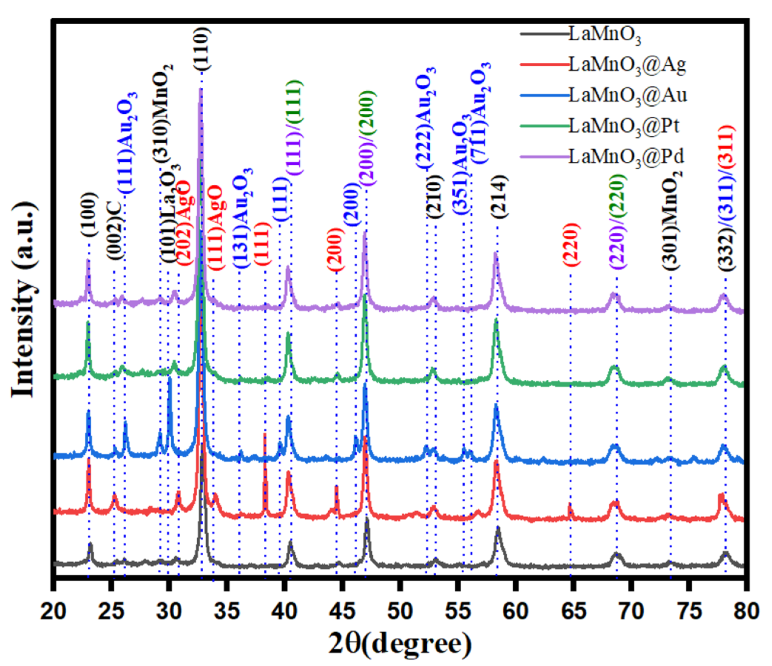
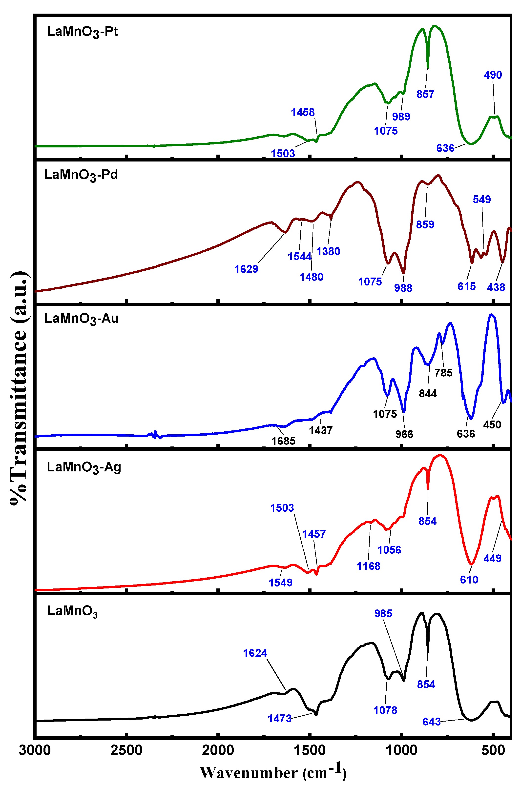
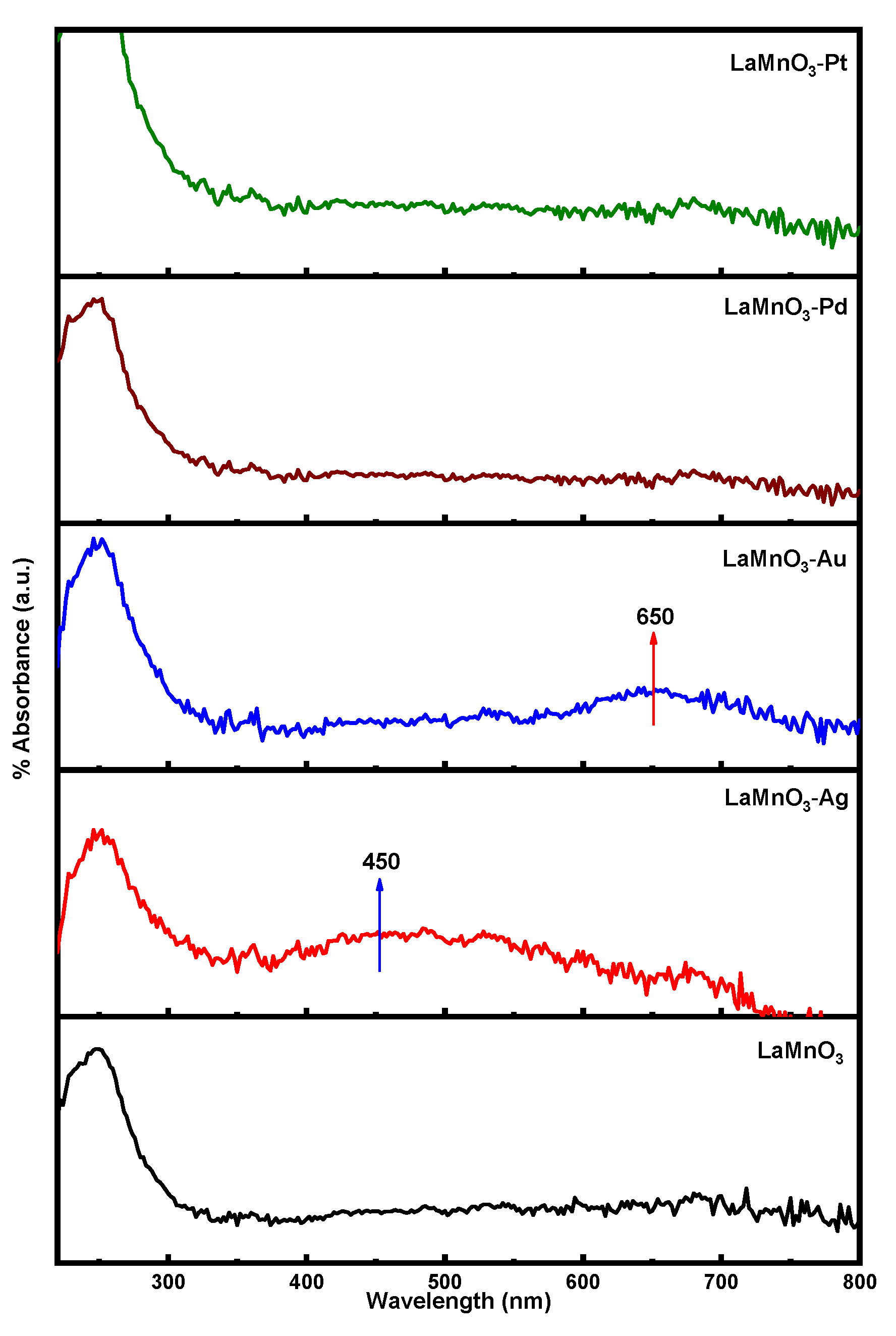
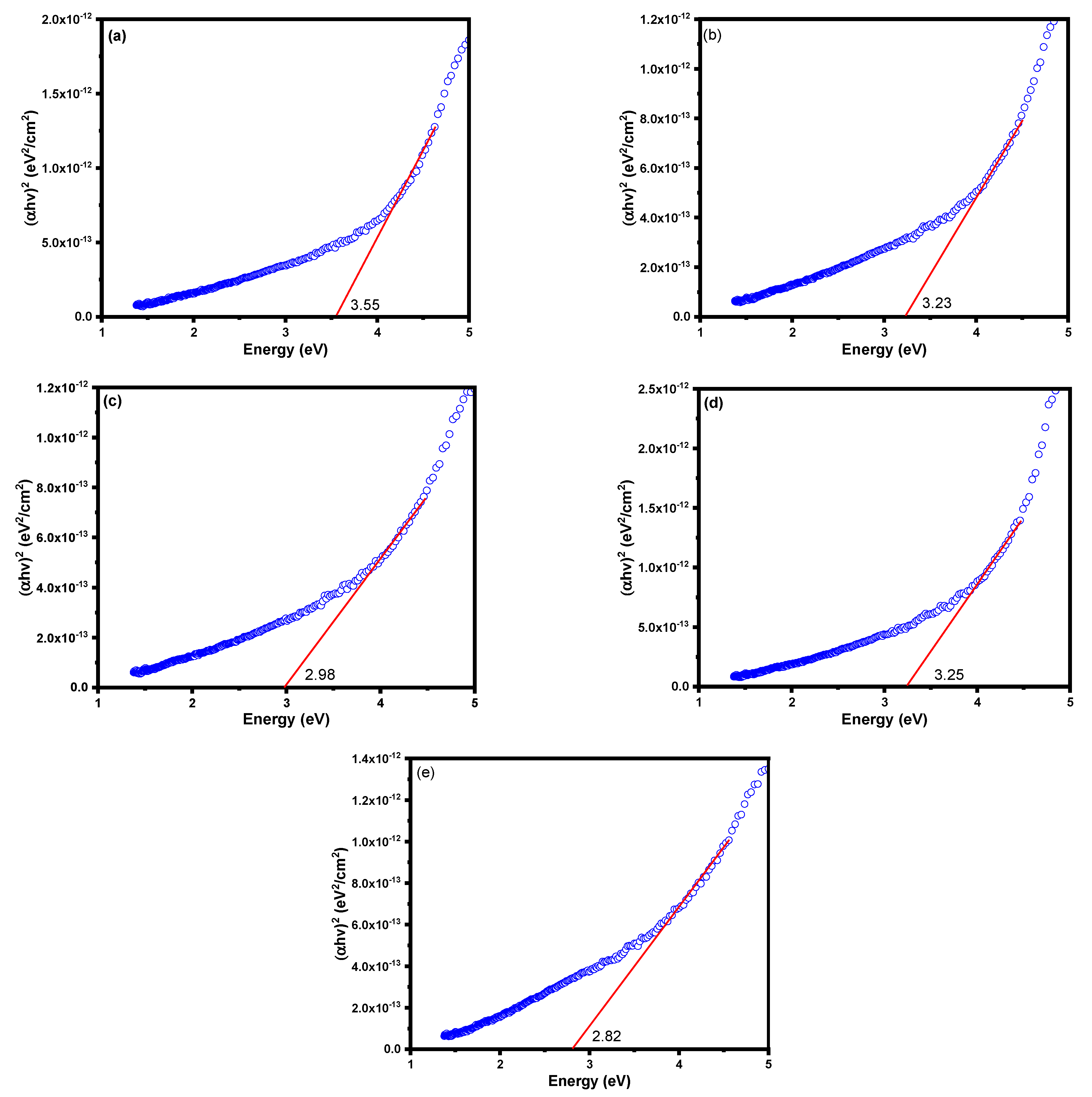
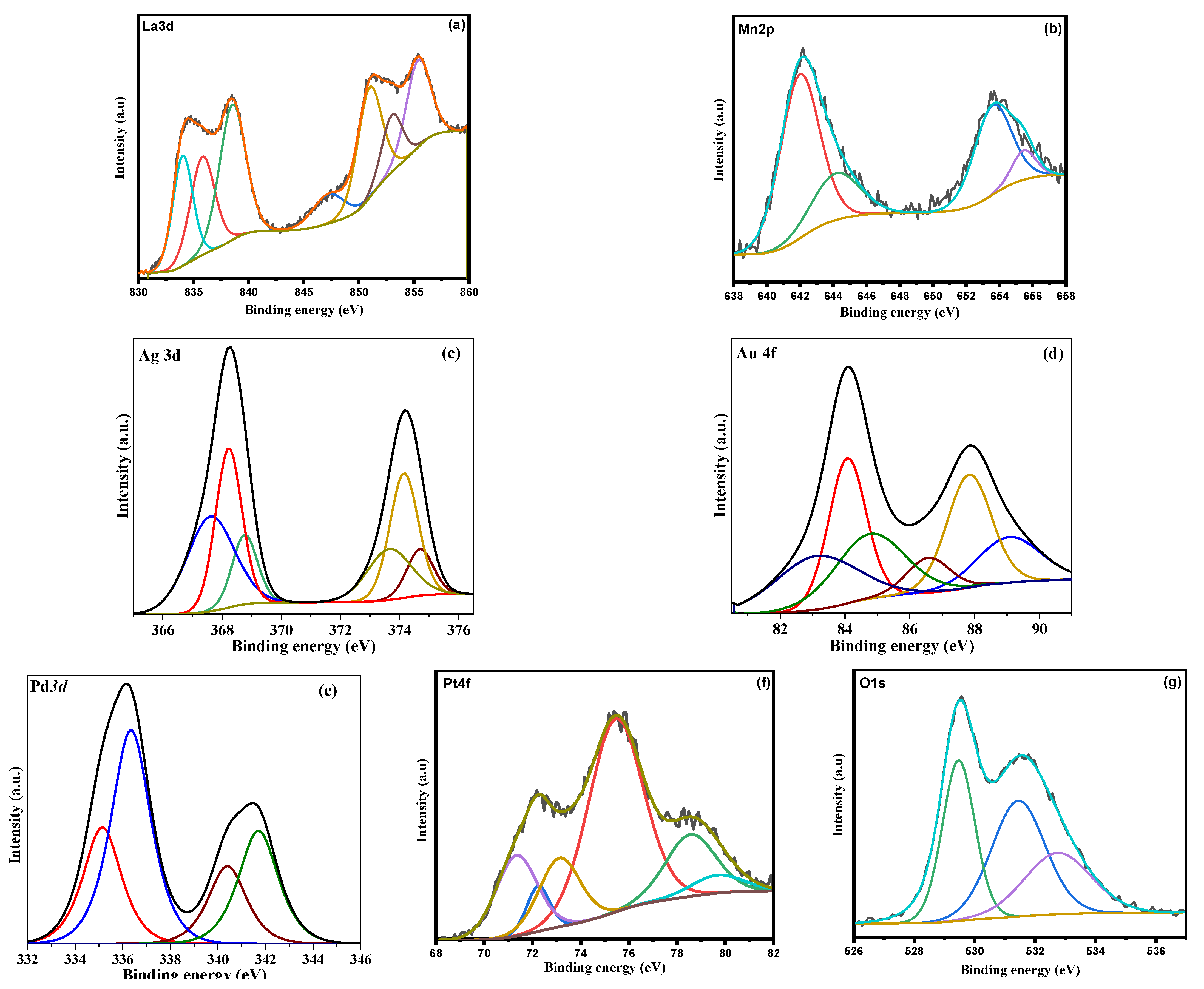
3.1. Characterization of LaMnO3 Nanocomposites

The X-ray diffraction (XRD) patterns for the synthesized samples were obtained to study their crystalline properties. The obtained patterns for bulk and the noble metal deposited LaMnO3 samples (Pt-LnMnO3, Pd-LnMnO3, Au-LnMnO3, and Ag-LnMnO3) are shown in Figure 2. The reflections at 2θ angles 33, 41, 58, 68 and 78° are indicative of the presence of crystalline LnMnO3 in all samples [31]. The characteristic reflections at 2θ angles of 38.12° (111), 44.27° (200), 64.42° (220), and 77.47° (311) for Ag (ICDD PDF file number 00-004-0783) [32], distinct peaks at 38.12, 44.27 and 64.42° were observed in case of Ag- LnMnO3 sample.
The XRD pattern of the Au-LnMnO3 sample exhibited additional reflections at 38.2° (111), 44.4° (200), 64.57° (220), and 77.54° (311) corresponding to the face-centered cubic structure of Au [ICDD PDF file number 00-004-0784]. The reflections at 39.6, 47.4 and 67.1° in case of Pt-LaMnO3 sample could be attributed to the reflections (111), (200), and (220) respectively, which are consistent with the face centered cubic (fcc) structure of Pt metal (JCPDS Card 04-0802]. The XRD pattern of Pd-LaMnO3 sample displays three peaks at 40.26, 45.78, and 68.67°, corresponding to (111), (200), and (220) planes, respectively. These can be indexed to the face-centered cubic (fcc) phase of Pd NPs [JCPDS ≠ 89-4897]. The observed XRD results clearly indicates the presence of noble metal crystallites in the synthesized samples.
The FT-IR spectra for bulk and noble metals deposited LaMnO3 samples are shown in Figure 3. The FT-IR bands were observed at 643, 854, 985, 1078, 1473, and 1624 cm−1 for all the samples. It was previously reported that significant absorption bands were around 600, 820, 1100, 1380, 1450, and 1650 cm−1 for LaMnO3 nanoparticles. The major IR absorption band in the range of 615-643 cm−1 is due to the stretching mode for Mn-O-Mn bonds associated with the octahedron MnO6. The presence of this vibrational band, which is a characteristic of the ABO3 type perovskite [33], indicates the successful synthesis of LaMnO3 structure. The bands around 1624 cm−1 are assumed to correspond to bending vibrations of the N-H bonds (secondary amines); the bands around 1380 and 1450 cm−1 are produced by bending vibrations in the bonds N-O (nitrates) [34]. As observed from the FT-IR spectrum in Figure 3, the deposition of noble metals resulted a small shift in the band position.
The morphology and particle sizes of different phases in the synthesized nanocomposites were studied using TEM analysis (Figure 4). As observed from the figure, the LaMnO3 nanoparticles with the sizes in the range of 10–25 nm was aggregated into clusters to form like nanorods with random distribution of particles. Apparently, the deposition of noble metals on the surface of LaMnO3 increased the agglomeration of particles probably due to usage of green bio-reduction using aqueous lemon extract. The TEM images of noble metal deposited samples clearly show dark nanoparticles of size in the range of 5–10 nm. The size variation in the noble metal nanoparticle size between the different noble metals was found to have no correlation with their atomic radius. It is probably because different metals possess different nucleation growth. The TEM images also show that all the samples exhibit meso to macro pore structure morphology as confirmed by the N2 adsorption isotherms of the samples (in later section). The meso and macro porosity is due to the random arrangement of the LaMnO3 particles. It was observed that Au, Ag and Pd containing samples possess large size pores, as shown by the TEM images. The surface nanoparticles and through-channels become obvious, which is due to the removal of different organic molecules (mainly citric acid) presented in the lemon extract during the thermal treatment. The HRTEM images clearly show LaMnO3 particles with a lattice-spacing distance of 0.28 nm all synthesized samples; this observation is in accordance with the lattice spacing of the (110) plane of LaMnO3 [35]. In addition, the presence of lattice fringes for metal and metal oxide nanoparticles (Ag, Au, Pd and Pt) appeared in the samples (Figure 4). These observations clearly corroborate the XRD analysis results.
The N2 adsorption-desorption isotherms and pore-size distribution patterns for synthesized samples are shown in Figure 5. As observed in the figure, all the samples are exhibiting the type IV isotherm with H3 type hysteresis loop, which is an indication that the samples possessed slit-shaped meso pores [36]. The shape of the hysteresis loop changed after the deposition of noble metals. The area of the hysteresis loop showed a distinct increase in the case of Pd, P, and Au deposited samples while it is decreased in the case of Ag deposited sample.
The textural properties of the samples, including the BET surface area, the average pore diameter and pore volume, are tabulated in Table 1. The BET surface area and pore volume values were highest for Pt-LaMnO3 sample. The least BET surface area was observed for the Ag-LaMnO3 sample, and the least pore size was that of LaMnO3 sample. The highest pore diameter was that of Pt-LaMnO3 and Au-LaMnO3 samples, closely followed by the Pd-LaMnO3 sample. The samples synthesized in this study exhibited BET surface area in the range of 12.2–20.1 m2/g, as shown in Table 1. These values are considerably high compared to those that have been attained by other methods of preparing perovskites (1–11 m2/g) [37]. The high surface area of the metal nanoparticles and through-channels of LaMnO3 become obvious, due to the removal of organic molecules during the thermal treatment. Additionally, there is an increase in the surface area from 14.6 to 20.1 m2/g when the Pt nanoparticles are deposited, this suggest that the dispersion of Pt on the LaMnO3 structure is high compared to Ag, Au and Pd metal particles, which has helped to enhance the surface area of the catalyst, hence improving the photocatalytic activity.
The DR UV-vis spectra of the bulk and noble metal deposited LnMnO3 samples are shown in Figure 6. The LnMnO3 sample exhibited absorption bands below 400 nm, which could be assigned to the charge transfer from valency band (VB) of O atoms to the conduction band (CB) of Ln atoms [38]. The DR UV-vis spectra of LnMnO3 deposited with noble metals exhibited an additional absorption band around 650 nm (visible region). The presence of Pt, Pd, Ag, and Au metal nanoparticles on the surface of LaMnO3 clearly resulted in appearance of absorption bands in the visible region. The excitation (charge transfer transition) of 4d electrons of Pt/Ag/Au/Pd metal nanoparticles into the CB is responsible for the presence of visible absorption bands [39]. The bandgap energy values were calculated from the Tauc plots, as shown in Figure 7. The bandgap values for all the samples were obtained by drawing a tangent to the slope. The data revealed that the bandgap energy for bulk LnMnO3 sample (3.55 eV) is higher than that of Ag-LnMnO3 (3.23 eV), Au-LnMnO3 (2.98 eV), Pd-LnMnO3 (3.25 eV), and Pt-LnMnO3 (2.82 eV). The deposition of noble metals is expected to increase the particle size; hence, a decrease in the bandgap energy value is expected. This decrease in bandgap energy could alter the photocatalytic activity under visible light radiation [40].
The XPS analysis for the synthesized samples was carried out to characterize the surface electronic properties of the samples. Figure 8 shows the representative deconvoluted XPS spectra of the samples. It is known that La 3d XPS peaks (Figure 8a) consist of La 3d3/2 and La 3d5/2 contributions and each spin orbit contribution have a doublet. The doublet appears due to the energy loss phenomenon induced by charge transfer from O 2p to La 4f [41]. The binding energies were appeared the range of 834–840 eV for La 3d3/2 contribution, which could be ascribed to La3+ species [42]. It is interesting to note that there is no considerable shift in the binding energies of La 3d peaks in the case of noble metal deposited LaMnO3 samples, indicating that noble metals were not incorporated into the LaMnO3 structure.
As shown in Figure 8b, the Mn 2p3/2 signal could be deconvoluted into two components with a binding energy of 642.2 and 644.2 eV, which could be assigned to Mn3+ ions and Mn4+ species, respectively, [43]. Similar spectra were observed in all the synthesized samples; therefore, the Mn could have existed in both Mn3+ and Mn4+ in noble metal deposited LaMnO3 samples. Previously, Chen et al. [44] reported that Mn3+ could be partially oxidized into Mn4+ on the catalyst surfaces, which could produce structural defects. It is possible that a similar phenomenon could have occurred in the synthesized LaMnO3 materials in this study. It is possible to distinguish the Mn state precisely by calculating the difference between Mn 3s peak and its satellite shake-up. The difference appeared to be around 5.3 eV, indicating that Mn is mainly presented in 3+ state in all the synthesized samples. It was previously reported that the Ag 3d5/2 peaks for surface bulk Ag0, Ag+, and Ag2+ species appear at 365.9, 366.4, and 367.1 eV, respectively [45]. However, in the present sample the Ag 3d5/2 peaks appeared at 367.6, 368.2 and 368.8 eV for metallic (Ag0) and oxidized (Ag+ and Ag2+) species, which are interacted with the LaMnO3 support. It is well known that a shift in binding energy occurs when surface species are interacted with the support. The Au 4f XPS spectrum of the Au-LaMnO3 sample shows two peaks related to the core-level Au 4f7/2 and Au 4f5/2 contributions, further deconvoluted each into three different peaks corresponding to three different Au species 83.2, 84.1 and 85.0 eV. It was previously reported that XPS peaks for metallic Au species appear in the range of 82.9–84.5 eV, while the peaks at 85.8 and 86.5 eV could be assigned to the oxidized Au species (Au+ & Au3+) [46]. Therefore, the two peaks at 83.2 and 84.1 eV that appeared in the sample could be assigned to metallic Au species interacted with LaMnO3 semiconductor, while the third peak at 85.0 eV could be attributed to the oxidized Au species. The deconvoluted Pd 3d XPS spectrum of the sample clearly shows the presence of four peaks at 336.4, 341.7, 335.1 and 340.4 eV, which could be assigned to Pd 3d5/2 and Pd 3d3/2 contributions of Pd2+ and Pd0 species, respectively, [47]. The percentage of peak areas of the Pd 3d states clearly indicates that the contribution for Pd2+ species is more compared to Pd0 species in the synthesized sample. Considering the general mechanism of deposition of recued noble metal species on a support, in which the noble metal precursor first gets hydrolyzed and subsequently reduced and deposited on the surface of the support, it is possible that part of the hydrolyzed noble metal species gets oxidized during the thermal treatment and remains as an oxidized form on the surface of the sample. The O 1s spectra of the samples could be deconvoluted into three different components with binding energy of 529.4, 531.4 and 533 eV, corresponding to lattice oxygen, adsorbed oxygen, and adsorbed H2O species, respectively [48].

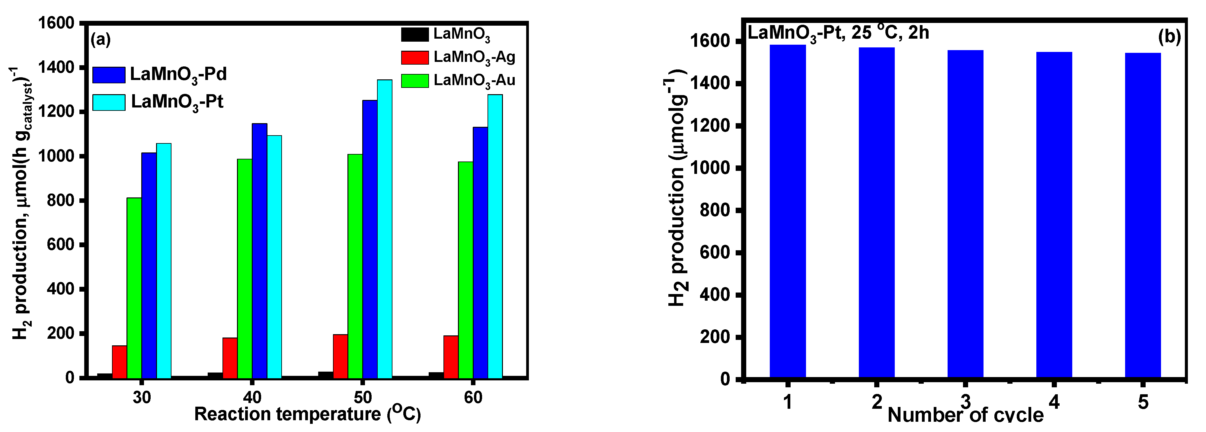
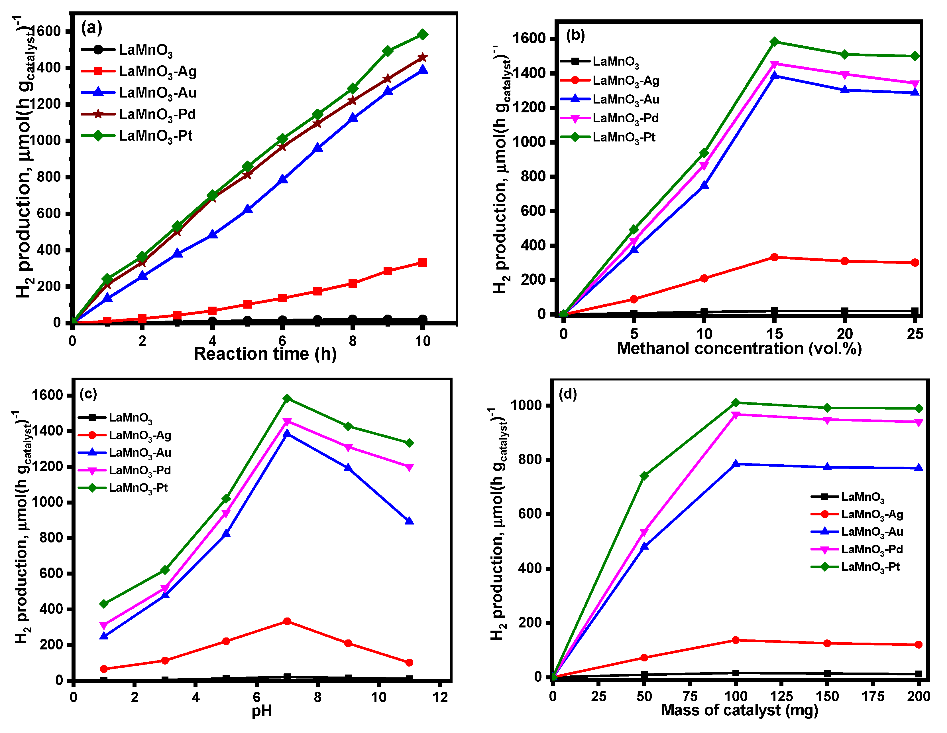
3.2. Photocatalytic Activity of Bulk and Noble Metal Deposited LaMnO3 Samples

Figure 9a shows the influence of reaction time on photocatalytic H2 production over synthesized LaMnO3 catalysts. The bulk LaMnO3 catalyst showed no photocatalytic H2 production under visible light irradiation. Of the noble metal deposited LaMnO3 catalysts, Ag-LaMnO3 showed the most negligible H2 production; however, the Au-LaMnO3, Pd-LaMnO3, and Pt-LaMnO3 samples exhibited significantly high photocatalytic H2 production as a function of reaction time. Figure 9b displays the effect of methanol concentration on the photocatalytic H2 production. The results showed a similar trend, as that observed in Figure 9a. A steady increase was observed up to 15 vol % of methanol, after which a slight decrease was observed in the case of all the noble metal deposited LaMnO3 catalysts.
Figure 9c provides the results related to the effect of pH on H2 production over the synthesized catalysts. As the pH of the methanol aqueous solution increases to 7, an exponential increase in H2 production activity was recorded; however, an increase of pH beyond 7 caused in decrease in the photocatalytic activity of all the noble metal LaMnO3 catalysts, confirming that the photo reforming activity of synthesized catalysts is influenced by the pH of the reaction mixture. Figure 9d provides the results from the study of the role of the mass of the catalyst on photocatalytic H2 production over all the synthesized LaMnO3 nanocomposite catalysts. As observed from the figure, when the catalyst mass is increased from 50 to 100 mg, a significant increase in photocatalytic methanol reformation activity was observed. However, with a further increase in catalyst mass, there is no significant change in the photocatalytic activity observed.
The influence of the reaction temperature on photocatalytic H2 production was studied and the obtained results are presented in Figure 10a. The results indicated that 50 °C is an ideal reaction temperature to obtain high H2 production. Up to 50 °C, a steady increase in H2 production was observed. With an increase in the reaction temperature (60 °C)., the reaction marginally slowed down. Korzhak et al. [49] studied photocatalytic H2 production over Cu-TiO2 nanocomposite catalysts at different reaction temperatures and observed that the reaction temperature has a considerable impact on the quantum yield. This is likely due to the thermal activation of product desorption. The observed decrease in H2 production beyond 50 °C may be caused by a decrease in the adsorption of methanol molecules on the catalyst surface. We have also reported a similar trend in the case of PtOx-TiO2 anatase nanomaterials [40]. Figure 10b provides the recyclability of the Pt-LaMnO3 catalyst. Specifically, the photocatalytic stability of the prototypical (highest active) Pt-LaMnO3 catalyst was revealed. Catalysts typically undergo photo-corrosion during the catalytic reaction; hence, it is widely known that investigating photocatalytic stability is important. As can be seen in Figure 10b, the Pt-LaMnO3 catalyst maintained its photocatalytic activity for H2 generation over an extended period. The structural stability and strong photo-corrosion resistance of the Pt system under the examined reaction conditions account for its recyclability [39].
With energy hv equal to or greater than the semiconductor bandgap, photon absorption starts photocatalytic reactions on semiconductors [50]. As a result, the photoinduced electrons move from the valence band (VB) to the conduction band (CB), resulting in the production of an electron-hole pair in the conduction band (CB). The resulting charge carriers can lead to the oxidation of electron donor species and the reduction of electron acceptor species, with the latter possessing a reduction potential that is higher in energy than the former. H+ ions serve as the electron acceptors in the photo-reforming reaction while the organic substrates, which are oxidized to CO2, serve as the electron donors. When organic molecules act as hole scavengers and undergo relatively quick and irreversible oxidation on the catalyst surface, the rate of photocatalytic H2 production is significantly accelerated [51]. Various nanocomposite materials, including ZnIn2S4-Au-TiO2 [52], Ag2O/Zn (O,S) Nanodiodes on Mesoporous SiO2 [53], ZnO/CdS hierarchical photocatalyst [54], Graphene oxide—Zn(O,S) photocatalyst [55] have been well established for their contribution to photocatalytic H2 evolution. The underlying mechanism in each case was somewhat the same; in the case of the noble metal deposited ZnO/CdS system, enhanced photocatalytic activity occurred because the heterostructure, not only facilitated an effective spatial separation of photo-induced electron-hole pairs, but also enhanced the redox ability of photocatalyst caused by an increase in redox potential. The work of the nanocomposites is to work towards effective spatial separation and enhanced redox ability.

4. Conclusions

In the present study, bulk LaMnO3 nanomaterial was synthesized by a combustion method using fine lemon powder as fuel. Novel green bio-reduction method was adopted for the deposition of various noble metals (Au, Ag, Pt, Pd) on the surface of LaMnO3. The characterization of the synthesized samples was accomplished by XRD, FT-IR, TEM, DR UV-vis, XPS, and N2 physisorption measurements. The existence of nanosized noble metal particles was observed from TEM image analysis. The deposition of noble metal led to improvement in visible-light absorption properties. The XPS results indicated the presence of both metallic and oxidized Ag, Au, Pd and Pt species on the surface of LaMnO3. The methanol reformation photocatalytic activity for bulk and noble metal deposited LaMnO3 samples was assessed. The results confirmed that the deposition of noble metals resulted substantial improvement in photocatalytic activity to H2 production. This enhancement is higher in case of Pt-LaMnO3 compared to other noble metal deposited samples. The increase in textural properties and the prevention of the recombination of photogenerated electrons and holes, thus, enhanced the photocatalytic activities of noble metal deposited LaMnO3 catalysts. The Pt deposited LaMnO3 catalyst has significant potential to produce H2 under visible light, which is both inexpensive and environmentally friendly, enabling the development of promises for H2 energy for the future.

Author Contributions

Conceptualization, M.A.M., A.H.J., N.H. and K.N.; Data curation, M.A.M., A.H.J., N.H. and K.N.; Formal analysis, I.A.R., M.A.M., A.H.J., N.H. and K.N.; Investigation, M.A.M., A.H.J., N.H. and K.N.; Methodology, M.A.M., and K.N.; Project administration, A.H.J.; Software, I.A.R., A.H.J. and N.H.; Supervision, A.H.J. and N.H.; Validation, M.A.M., A.H.J., N.H. and K.N.; Writing—original draft, M.A.M., A.H.J., N.H., K.N. and I.A.R.; Writing—review & editing, M.A.M., A.H.J., N.H., K.N. and I.A.R. All authors have read and agreed to the published version of the manuscript.

Funding

Deputyship for Research and Innovation, Ministry of Education in Saudi Arabia (project number ISP20-24).

Data Availability Statement

All data created is provided in this study.

Acknowledgments

The authors extend their appreciation to the Deputyship for Research and Innovation, Ministry of Education in Saudi Arabia, for funding this research work through project number ISP20-24.

Conflicts of Interest

The authors declare no conflict of interest.

References

  1. Liu, W.; Zuo, H.; Wang, J.; Xue, Q.; Ren, B.; Yang, F. The production and application of hydrogen in steel industry. Int. J. Hydrogen Energy 2021, 46, 10548–10569. [Google Scholar] [CrossRef]
  2. Okolie, J.A.; Patra, B.R.; Mukherjee, A.; Nanda, S.; Dalai, A.K.; Kozinski, J.A. Futuristic applications of hydrogen in energy, biorefining, aerospace, pharmaceuticals and metallurgy. Int. J. Hydrogen Energy 2021, 46, 8885–8905. [Google Scholar] [CrossRef]
  3. Ganguly, P.; Harb, M.; Cao, Z.; Cavallo, L.; Breen, A.; Dervin, S.; Dionysiou, D.D.; Pillai, S.C. 2D Nanomaterials for Photocatalytic Hydrogen Production. ACS Energy Lett. 2019, 4, 1687–1709. [Google Scholar] [CrossRef]
  4. Abdalla, A.M.; Hossain, S.; Nisfindy, O.B.; Azad, A.; Dawood, T.M.; Azad, A.K. Hydrogen production, storage, transportation and key challenges with applications: A review. Energy Convers. Manag. 2018, 165, 602–627. [Google Scholar] [CrossRef]
  5. Zamani, P.; Higgins, D.C.; Hassan, F.; Fu, X.; Choi, J.-Y.; Hoque, A.; Jiang, G.; Chen, Z. Highly active and porous graphene encapsulating carbon nanotubes as a non-precious oxygen reduction electrocatalyst for hydrogen-air fuel cells. Nano Energy 2016, 26, 267–275. [Google Scholar] [CrossRef]
  6. Acar, C.; Dincer, I. Comparative assessment of hydrogen production methods from renewable and non-renewable sources. Int. J. Hydrogen Energy 2014, 39, 1–12. [Google Scholar] [CrossRef]
  7. Manzardo, A.; Ren, J.; Mazzi, A.; Scipioni, A. A grey-based group decision-making methodology for the selection of hydrogen technologies in life cycle sustainability perspective. Int. J. Hydrogen Energy 2012, 37, 17663–17670. [Google Scholar] [CrossRef]
  8. Abbasi, M.; Farniaei, M.; Rahimpour, M.R.; Shariati, A. Enhancement of Hydrogen Production and Carbon Dioxide Capturing in a Novel Methane Steam Reformer Coupled with Chemical Looping Combustion and Assisted by Hydrogen Perm-Selective Membranes. Energy Fuels 2013, 27, 5359–5372. [Google Scholar] [CrossRef]
  9. Kondarides, D.I.; Daskalaki, V.M.; Patsoura, A.; Verykios, X.E. Hydrogen Production by Photo-Induced Reforming of Biomass Components and Derivatives at Ambient Conditions. Catal. Lett. 2008, 122, 26–32. [Google Scholar] [CrossRef]
  10. Silva, C.G.; Sampaio, M.J.; Marques, R.R.; Ferreira, L.A.; Tavares, P.B.; Silva, A.M.; Faria, J.L. Photocatalytic production of hydrogen from methanol and saccharides using carbon nanotube-TiO2 catalysts. Appl. Catal. B Environ. 2015, 178, 82–90. [Google Scholar] [CrossRef] [Green Version]
  11. Osterloh, F.E. Inorganic nanostructures for photoelectrochemical and photocatalytic water splitting. Chem. Soc. Rev. 2013, 42, 2294–2320. [Google Scholar] [CrossRef] [PubMed]
  12. Chen, S.; Takata, T.; Domen, K. Particulate photocatalysts for overall water splitting. Nat. Rev. Mater. 2017, 2, 17050. [Google Scholar] [CrossRef]
  13. Schneider, J.; Matsuoka, M.; Takeuchi, M.; Zhang, J.; Horiuchi, Y.; Anpo, M.; Bahnemann, D.W. Understanding TiO2 photocatalysis: Mechanisms and materials. Chem. Rev. 2014, 114, 9919. [Google Scholar] [CrossRef] [PubMed]
  14. Yu, C.-L.; Sakthinathan, S.; Chen, S.-Y.; Yu, B.-S.; Chiu, T.-W.; Dong, C. Hydrogen generation by methanol steam reforming process by delafossite-type CuYO2 nanopowder catalyst. Microporous Mesoporous Mater. 2021, 324, 111305. [Google Scholar] [CrossRef]
  15. Tountas, A.A.; Peng, X.; Tavasoli, A.V.; Duchesne, P.N.; Dingle, T.L.; Dong, Y.; Hurtado, L.; Mohan, A.; Sun, W.; Ulmer, U.; et al. Towards Solar Methanol: Past, Present, and Future. Adv. Sci. 2019, 6, 1801903. [Google Scholar] [CrossRef]
  16. Ren, S.J.; Shoemaker, W.R.; Wang, X.F.; Shang, Z.Y.; Klinghoffer, N.; Li, S.G.; Yu, M.; He, X.Q.; White, T.A.; Liang, X.H. Highly active and selective Cu-ZnO based catalyst for methanol and dimethyl ether synthesis via CO2 hydrogenation. Fuel 2019, 239, 1125–1133. [Google Scholar] [CrossRef]
  17. Perng, S.-W.; Wu, H.-W. Effect of depth and diameter of cylindrical cavity in a plate-type methanol steam reformer on estimated net power of PEMFC. Energy Convers. Manag. 2018, 177, 190–209. [Google Scholar] [CrossRef]
  18. Pastrana-Martínez, L.M.; Morales-Torres, S.; Papageorgiou, S.K.; Katsaros, F.K.; Romanos, G.E.; Figueiredo, J.L.; Faria, J.L.; Falaras, P.; Silva, A.M. Photocatalytic behaviour of nanocarbon–TiO2 composites and immobilization into hollow fibres. Appl. Catal. B Environ. 2013, 142–143, 101–111. [Google Scholar] [CrossRef]
  19. Woan, K.; Pyrgiotakis, G.; Sigmund, W. Photocatalytic carbon-nanotube–TiO2 composites. Adv. Mater. 2009, 21, 2233–2239. [Google Scholar] [CrossRef]
  20. Ahmmad, B.; Kusumoto, Y.; Somekawa, S.; Ikeda, M. Carbon nanotubes synergistically enhance photocatalytic activity of TiO2. Catal. Commun. 2008, 9, 1410–1413. [Google Scholar] [CrossRef]
  21. Serra, M.; Albero, J.; García, H. Photocatalytic activity of Au/TiO2 photocatalysts for H2 evolution: Role of the Au nanoparticles as a function of the irradiation wavelength. ChemPhysChem 2015, 16, 1842–1845. [Google Scholar] [CrossRef] [PubMed]
  22. Al-Azri, Z.H.N.; Al-Oufi, M.; Chan, A.; Waterhouse, G.I.; Idriss, H. Metal Particle Size Effects on the Photocatalytic Hydrogen Ion Reduction. ACS Catal. 2019, 9, 3946–3958. [Google Scholar] [CrossRef]
  23. Mei, B.; Han, K.; Mul, G. Driving Surface Redox Reactions in Heterogeneous Photocatalysis: The Active State of Illuminated Semiconductor-Supported Nanoparticles during Overall Water-Splitting. ACS Catal. 2018, 8, 9154–9164. [Google Scholar] [CrossRef]
  24. Nair, V.; Munoz-Batista, M.J.; Fernandez-García, M.; Luque, R.; Colmenares, C.J. Thermo-photocatalysis: Environmental and energy applications. ChemSusChem 2019, 12, 2098–2116. [Google Scholar] [CrossRef] [PubMed]
  25. Guan, S.; Li, R.; Sun, X.; Xian, T.; Yang, H. Construction of novel ternary Au/LaFeO3/Cu2O composite photocatalysts for RhB degradation via photo-Fenton catalysis. Mater. Technol. 2021, 36, 603–615. [Google Scholar] [CrossRef]
  26. Caudillo-Flores, U.; Agostini, G.; Marini, C.; Kubacka, A.; Fandez-García, M. Hydrogen thermo-photo production using Ru/TiO2: Heat and light synergistic effects. Appl. Catal. B Environ. 2019, 256, 117790. [Google Scholar] [CrossRef]
  27. Arabi, A.; Fazli, M.; Ehsani, M.H. Synthesis and characterization of calcium-doped lanthanum manganite nanowires as a photocatalyst for degradation of methylene blue solution under visible light irradiation. Bull. Mater. Sci. 2018, 41, 77. [Google Scholar] [CrossRef]
  28. Xu, J.; Sun, C.; Wang, Z.; Hou, Y.; Ding, Z.; Wang, S. Perovskite Oxide LaNiO3 Nanoparticles for Boosting H2 Evolution over Commercial CdS with Visible Light. Chem. Eur. J. 2018, 24, 18512. [Google Scholar] [CrossRef]
  29. Panahi, P.N.; Rasoulifard, M.H.; Babaei, S. Photocatalytic activity of cation (Mn) and anion (N) substitution in LaCoO3 nanoperovskite under visible light. Rare Met. 2020, 39, 139–146. [Google Scholar] [CrossRef]
  30. Bumajdad, A.; Madkour, M. Understanding the superior photocatalytic activity of noble metals modified titania under UV and visible light irradiation. Phys. Chem. Chem. Phys. 2014, 16, 7146–7158. [Google Scholar] [CrossRef]
  31. Susanti, Y.D.; Afifah, N.; Saleh, R. Ag modified LaMnO3 nanoparticles for methylene blue degradation via photosonocatalytic activities. IOP Conf. Ser. Mater. Sci. Eng. 2019, 496, 012037. [Google Scholar] [CrossRef]
  32. Vo, T.-T.; Dang, C.-H.; Doan, V.-D.; Dang, V.-S.; Nguyen, T.-D. Biogenic Synthesis of Silver and Gold Nanoparticles from Lactuca indica Leaf Extract and Their Application in Catalytic Degradation of Toxic Compounds. J. Inorg. Organomet. Polym. Mater. 2020, 30, 388–399. [Google Scholar] [CrossRef]
  33. Hernández, E.; Sagredo, V.; Delgado, G. Synthesis and magnetic characterization of LaMnO3 nanoparticles. Rev. Mex. Física 2015, 61, 166–169. [Google Scholar]
  34. LiK, K.; Li, X.; Zhu, K. Infrared Absorption spectra of Manganese Oxides. J. Appl. Phys 1997, 10, 6943. [Google Scholar]
  35. Han, X.; Wang, Y.; Hao, H.; Guo, R.; Hu, Y.; Jiang, W. Ce1–xLaxOy solid solution prepared from mixed rare earth chloride for soot oxidation. J. Rare Earths 2016, 34, 590–596. [Google Scholar] [CrossRef]
  36. Sing, K.S.W. Reporting physisorption data for gas/solid systems with special reference to the determination of surface area and porosity (recommendations 1984). Pure Appl. Chem. 1985, 57, 603–619. [Google Scholar] [CrossRef]
  37. Chen, C.; Zhou, J.; Geng, J.; Bao, R.; Wang, Z.; Xia, J.; Li, H. Perovskite LaNiO3/TiO2 step-scheme heterojunction with enhanced photocatalytic activity. Appl. Surf. Sci. 2020, 503, 144287. [Google Scholar] [CrossRef]
  38. Lee, B.-Y.; Park, S.-H.; Lee, S.-C.; Kang, M.; Park, C.-H.; Choung, S.-J. Optical properties of Pt-TiO2 catalyst and photocatalytic activities for benzene decomposition. Korean J. Chem. Eng. 2003, 20, 812–818. [Google Scholar] [CrossRef]
  39. Chen, H.-W.; Ku, Y.; Kuo, Y.-L. Effect of Pt/TiO2 characteristics on temporal behavior of o-cresol decomposition by visible light-induced photocatalysis. Water Res. 2007, 41, 2069–2078. [Google Scholar] [CrossRef]
  40. Alshehri, A.; Narasimharao, K. PtOx-TiO2 anatase nanomaterials for photocatalytic reformation of methanol to hydrogen: Effect of TiO2 morphology. J. Mater. Res. Technol. 2020, 9, 14907–14921. [Google Scholar] [CrossRef]
  41. Natile, M.M.; Ugel, E.; Maccato, C.; Glisenti, A. LaCoO3: Effect of synthesis conditions on properties and reactivity. Appl. Catal. B Environ. 2007, 72, 351–362. [Google Scholar] [CrossRef]
  42. Tzimpilis, E.; Moschoudis, N.; Stoukides, M.; Bekiaroglou, P. Preparation, active phase composition and Pd content of perovskite-type oxides. Appl. Catal. B Environ. 2008, 84, 607–615. [Google Scholar] [CrossRef]
  43. Popa, A.; Raita, O.; Stan, M.; Pana, O.; Borodi, G.; Giurgiu, L. Electron Paramagnetic Resonance of Mn-Doped Sn1−xMnxO2 Powders. Appl. Magn. Reson. 2012, 42, 453–462. [Google Scholar] [CrossRef]
  44. Chen, J.; Shen, M.; Wang, X.; Qi, G.; Wang, J.; Li, W. The influence of nonstoichiometry on LaMnO3 perovskite for catalytic NO oxidation. Appl. Catal. B Environ. 2013, 134–135, 251–257. [Google Scholar] [CrossRef]
  45. Weaver, J.F.; Hoflund, G.B. Surface Characterization Study of the Thermal Decomposition of Ag2O. Chem. Mater. 1994, 6, 1693–1699. [Google Scholar] [CrossRef]
  46. Villa, A.; Dimitratos, N.; Chan-Thaw, C.E.; Hammond, C.; Veith, G.M.; Wang, D.; Manzoli, M.; Prati, L.; Hutchings, G.J. Characterisation of gold catalysts. Chem. Soc. Rev. 2016, 45, 4953–4994. [Google Scholar] [CrossRef]
  47. Yao, W.; Wang, R.; Yang, X. LaCo1−xPdxO3 perovskite-type oxides: Synthesis, characterization and simultaneous removal of NOx and diesel soot. Catal. Lett. 2009, 130, 613–621. [Google Scholar] [CrossRef]
  48. Liu, Y.; Dai, H.; Deng, J.; Xie, S.; Yang, H.; Tan, W.; Han, W.; Jiang, Y.; Guo, G. Mesoporous Co3O4-supported gold nanocatalysts: Highly active for the oxidation of carbon monoxide, benzene, toluene, and o-xylene. J. Catal. 2014, 309, 408–418. [Google Scholar] [CrossRef]
  49. Korzhak, A.V.; Ermokhina, N.I.; Stroyuk, A.L.; Bukhtiyarov, V.K.; Raevskaya, A.E.; Litvin, V.I.; Kuchmiy, S.Y.; Ilyin, V.G.; Manorik, P.A. Photocatalytic hydrogen evolution over mesoporous TiO2/metal nanocomposites. J. Photochem. Photobiol. A Chem. 2008, 198, 126–134. [Google Scholar] [CrossRef]
  50. Fujishima, A.; Zhang, X.; Tryk, D.A. TiO2 photocatalysis and related surface phenomena. Surf. Sci. Rep. 2008, 63, 515–582. [Google Scholar] [CrossRef]
  51. Chiarello, G.L.; Aguirre, M.H.; Selli, E. Hydrogen production by photocatalytic steam reforming of methanol on noble metal-modified TiO2. J. Catal. 2010, 273, 182–190. [Google Scholar] [CrossRef]
  52. Yang, G.; Ding, H.; Chen, D.; Feng, J.; Hao, Q.; Zhu, Y. Construction of urchin-like ZnIn2S4-Au-TiO2 heterostructure with enhanced activity for photocatalytic hydrogen evolution. Appl. Catal. B Environ. 2018, 234, 260–267. [Google Scholar] [CrossRef]
  53. Gultom, N.S.; Abdullah, H.; Kuo, D.-H.; Ke, W.-C. Oriented p–n Heterojunction Ag2O/Zn(O,S) Nanodiodes on Mesoporous SiO2 for Photocatalytic Hydrogen Production. ACS Appl. Energy Mater. 2019, 2, 3228–3236. [Google Scholar] [CrossRef]
  54. Wang, S.; Zhu, B.; Liu, M.; Zhang, L.; Yu, J.; Zhou, M. Direct Z-scheme for enhanced photocatalytic H2-production activity. Appl. Catal. B Environ. 2019, 243, 19–26. [Google Scholar] [CrossRef]
  55. Gultom, N.S.; Abdullah, H.; Kuo, D.-H. Effects of graphene oxide and sacrificial reagent for highly efficient hydrogen production with the costless Zn(O,S) photocatalyst. Int. J. Hydrogen Energy 2019, 44, 29516–29528. [Google Scholar] [CrossRef]
Figure 1. Schematic representation of the preparation of bulk and noble metal deposited LaMnO3 catalysts.
Nanomaterials 12 02985 g001
Figure 2. XRD patterns for the bulk and noble metal deposited LaMnO3 nanocomposites.
Nanomaterials 12 02985 g002
Figure 3. FT-IR spectra for the bulk and noble metal deposited LaMnO3 nanocomposites.
Nanomaterials 12 02985 g003
Figure 4. TEM and HRTEM images of the synthesized nanocomposites.
Nanomaterials 12 02985 g004
Figure 5. (a) N2 adsorption-desorption isotherms and (b) pore size distribution patterns of the samples.
Nanomaterials 12 02985 g005
Figure 6. DR UV-vis spectra for the synthesized samples.
Nanomaterials 12 02985 g006
Figure 7. The Tauc plots for (a) LaMnO3, (b) LaMnO3-Ag, (c) LaMnO3-Au, (d) LaMnO3-Pd, and (e) LaMnO3-Pt samples.
Nanomaterials 12 02985 g007
Figure 8. XPS spectra of (a) La3d (b) Mn2p, (c) Ag3d (d) Au4f (e) Pd3d (d), (f) Pt4f and (g) O1s.
Nanomaterials 12 02985 g008
Figure 9. Photocatalytic H2 production, influence of (a) reaction time, (b) methanol concentration, (c) pH, and (d) mass of catalyst.
Nanomaterials 12 02985 g009
Figure 10. (a) Effect of reaction temperature on photocatalytic H2 production, (b) recyclability of the catalyst.
Nanomaterials 12 02985 g010
Table 1. Textural properties of the synthesized samples.
S. No.CatalystBET Surface Area (m2g−1)Pore Volume
(cm3g−1)
Pore Diameter
(Å)
1LaMnO314.60.01038
2Ag@ LaMnO312.20.01943
3Au@ LaMnO313.50.01564
4Pd@ LaMnO312.40.01861
5Pt@ LaMnO320.10.02364
Publisher’s Note: MDPI stays neutral with regard to jurisdictional claims in published maps and institutional affiliations.

