Article

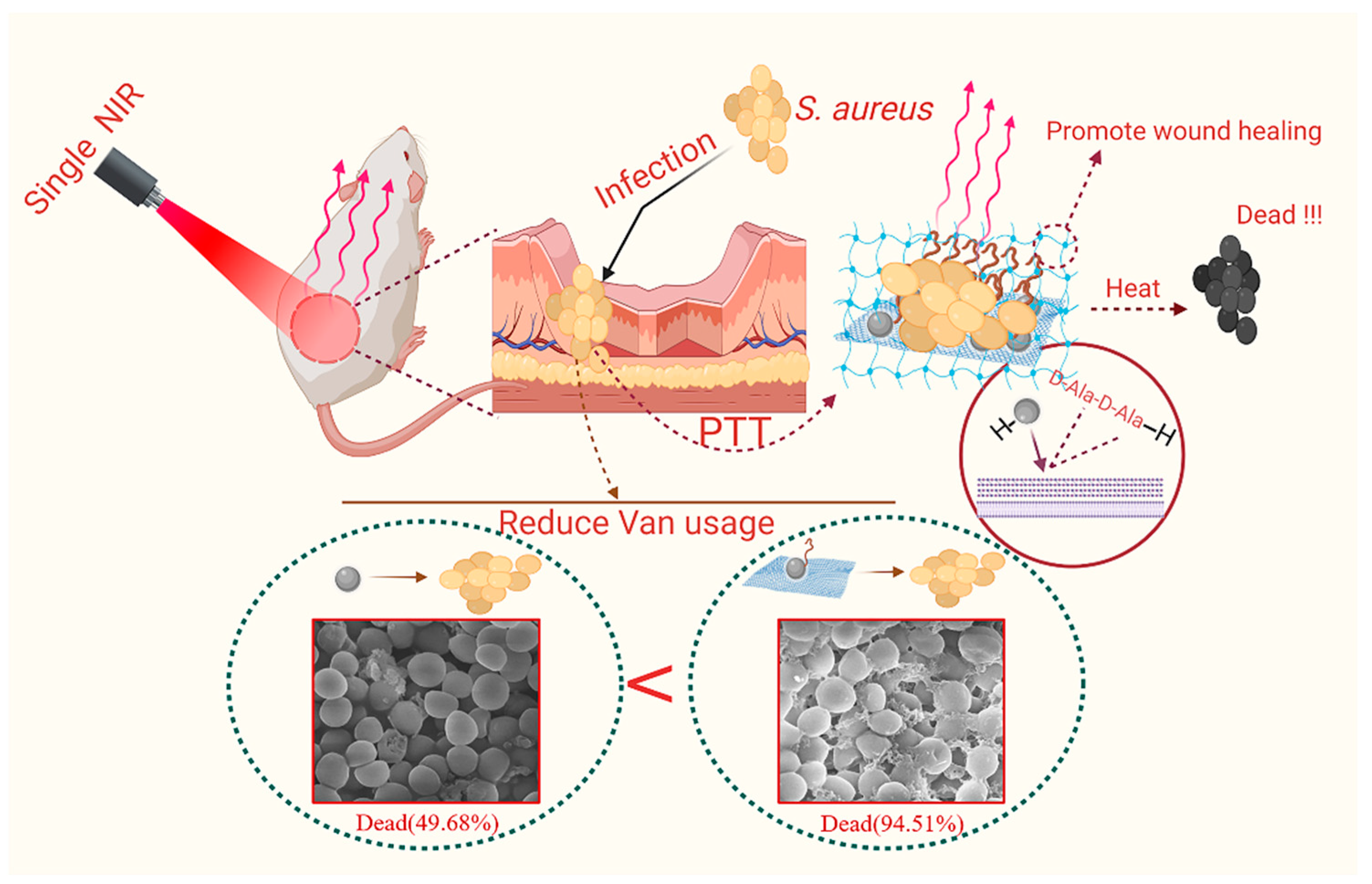
Synthesis of a Two-Dimensional Molybdenum Disulfide Nanosheet and Ultrasensitive Trapping of Staphylococcus Aureus for Enhanced Photothermal and Antibacterial Wound-Healing Therapy

1
School of Biological and Food Engineering, Anhui Polytechnic University, Wuhu 241000, China
2
Department of Microbiology and Immunology, Wannan Medical College, Wuhu 241002, China
*
Authors to whom correspondence should be addressed.
These authors contributed equally to this work.
Nanomaterials 2022, 12(11), 1865; https://doi.org/10.3390/nano12111865
Submission received: 22 April 2022 / Revised: 26 May 2022 / Accepted: 27 May 2022 / Published: 30 May 2022
(This article belongs to the Special Issue Luminescence Nanomaterials and Applications)

Abstract

:
Photothermal therapy has been widely used in the treatment of bacterial infections. However, the short photothermal effective radius of conventional nano-photothermal agents makes it difficult to achieve effective photothermal antibacterial activity. Therefore, improving composite targeting can significantly inhibit bacterial growth. We inhibited the growth of Staphylococcus aureus (S. aureus) by using an extremely low concentration of vancomycin (Van) and applied photothermal therapy with molybdenum disulfide (MoS2). This simple method used chitosan (CS) to synthesize fluorescein 5(6)-isothiocyanate (FITC)-labeled and Van-loaded MoS2-nanosheet hydrogels (MoS2-Van-FITC@CS). After modifying the surface, an extremely low concentration of Van could inhibit bacterial growth by trapping bacteria synergistically with the photothermal effects of MoS2, while FITC labeled bacteria and chitosan hydrogels promoted wound healing. The results showed that MoS2-Van-FITC@CS nanosheets had a thickness of approximately 30 nm, indicating the successful synthesis of the nanosheets. The vitro antibacterial results showed that MoS2-Van-FITC with near-infrared irradiation significantly inhibited S. aureus growth, reaching an inhibition rate of 94.5% at nanoparticle concentrations of up to 100 µg/mL. Furthermore, MoS2-Van-FITC@CS could exert a healing effect on wounds in mice. Our results demonstrate that MoS2-Van-FITC@CS is biocompatible and can be used as a wound-healing agent.

1. Introduction

Bacterial infections are consistently ranked as one of the leading causes of human mortality, with infection rates, mortality rates, and hospitalization costs increasing annually [1,2,3]. The high prevalence of bacterial infections has led to the misuse of antibiotics, resulting in the emergence of superbugs as bacteria become resistant to treatment. Unfortunately, superbugs are arising at a rate much faster than that of new antibiotic discovery, thereby leading to a growing threat of untreatable bacterial infections [4,5,6,7,8]. Therefore, new treatments are urgently needed.
With the rapid development of technologies in the fields of modern nanotechnology and biomedicine, numerous antibacterial inorganic nanoparticles (NPs), such as silver, gold, copper nanoparticles, alumina, zinc oxide, magnesium oxide, silica titanium dioxide, and graphene oxide NPs, as well as their composites, have been used in antibacterial therapy [3,9,10,11]. For example, two-dimensional (2D) graphene-based nanocomposites and analogues have been demonstrated to have wide application prospects because of their multi-functional antibacterial mechanisms, which include physical and chemical damage to bacterial cells [12,13]. Molybdenum disulfide (MoS2), a transition metal dichalcogenide, is similar to graphene. Currently, MoS2 has several potential applications in the field of biomedicine due to its unique electronic, optical, mechanical, and chemical properties, and MoS2 has been demonstrated to have unique optical properties in that it can convert itself into heat by absorbing light energy when irradiated with near-infrared (NIR) light. Through this property, MoS2 can kill bacteria [14,15,16].
Free MoS2 without irradiation does not have significant antibacterial effects. To improve the antibacterial ability of MoS2, we selected vancomycin (Van). Van is a peptide antibiotic that can kill bacteria, such as Staphylococcus aureus (S. aureus), residing on a wound’s surface [17,18]. Van has an attractive property in that it can target bacteria through hydrogen bonding to the terminal D-Ala-D-Ala sequence of the cytosolic peptide unit of Gram-positive bacteria. In other words, Van recognizes the D-Ala-D-Ala sequence on the cell surface of bacteria, increasing the half-life and effective working radius of the nano-antibacterial composites and enhancing the antibacterial effect [19,20,21,22]. Together, MoS2 and Van can target Gram-positive bacteria, which can further enhance the thermal response of MoS2 and promote bacterial growth inhibition while applying antibiotic therapy. Van has a primary amine group that binds covalently to fluorescein 5(6)-isothiocyanate (FITC), and we selected FITC-labeled Van because it can maintain both the fluorescent properties of FITC and the ability of Van to bind to the bacterial cell wall, thereby facilitating subsequent experimental validation.
To promote wound healing, it is important not only to clean the wound of germs but also to maintain an environment that is suitable for healing. A moist and clean environment accelerates the migration of epidermal cells, which facilitates skin cell granulation and division [23,24,25]. Polymer hydrogels are one of the most practiced soft-wet materials used for biomedical applications [26]. They can provide this type of environment for wounds, and thermosensitive hydrogels are easy to prepare. Chitosan (CS) is a biocompatible and weakly immunogenic material that can be degraded by enzymes in vivo, and the degradation products, namely oligosaccharides, are non-toxic. In addition, CS can enhance drug penetration by affecting the tightness between epithelial cells, which makes CS a valuable entity in the biomedical field [27]. As such, we prepared a temperature-sensitive hydrogel excipient by wrapping our synthesized MoS2-Van-FITC nanomaterials, which possess photothermic and chemotherapeutic properties, in a hydrogel. By physically mixing CS with a sodium β-glycerophosphate (β-GP) solution, a sol-gel phase transition was achieved at temperatures higher than 37 °C due to the enhanced hydrogen bonding, electrostatic attraction, and hydrophobic interactions between CS and β-GP [28].
In this study (Figure 1), Van not only efficiently inhibited the growth of bacteria on the wound surface but also enhanced the antibacterial effect by targeting bacteria through hydrogen bonding with the terminal D-Ala-D-Ala sequence of the cytosolic peptide unit of Gram-positive bacteria, increasing the half-life of the composite and the effective working radius of the photothermal treatment. Therefore, when MoS2 is combined with Van, the effective working radius of MoS2 PTT can be further increased to better inhibit bacterial growth while applying anti-infective therapy. The covalent binding of primary amine groups using FITC with MoS2-Van confers the nanocomposite to maintain the fluorescent properties of FITC, which can be observed for real-time detection. Additionally, photothermal treatment combined with chemotherapy cleaned the wound surface of pathogenic bacteria, thereby accelerating wound healing.

2. Materials and Methods

2.1. Materials

Raw MoS2 (99.5%), FITC (90%, mixture of 5- and 6-isomers), CS (≥99.8%), and Van HCL were purchased from Aladdin Chemical Reagent Co., Ltd. (Shanghai, China). The beef extract peptone agar medium, Staphylococcus aureus ATCC 25,923 cells (S. aureus, Gram-positive), and Escherichia coli ATCC 11,303 cells (E. coli, Gram-negative) were obtained from Anhui Polytechnic University.

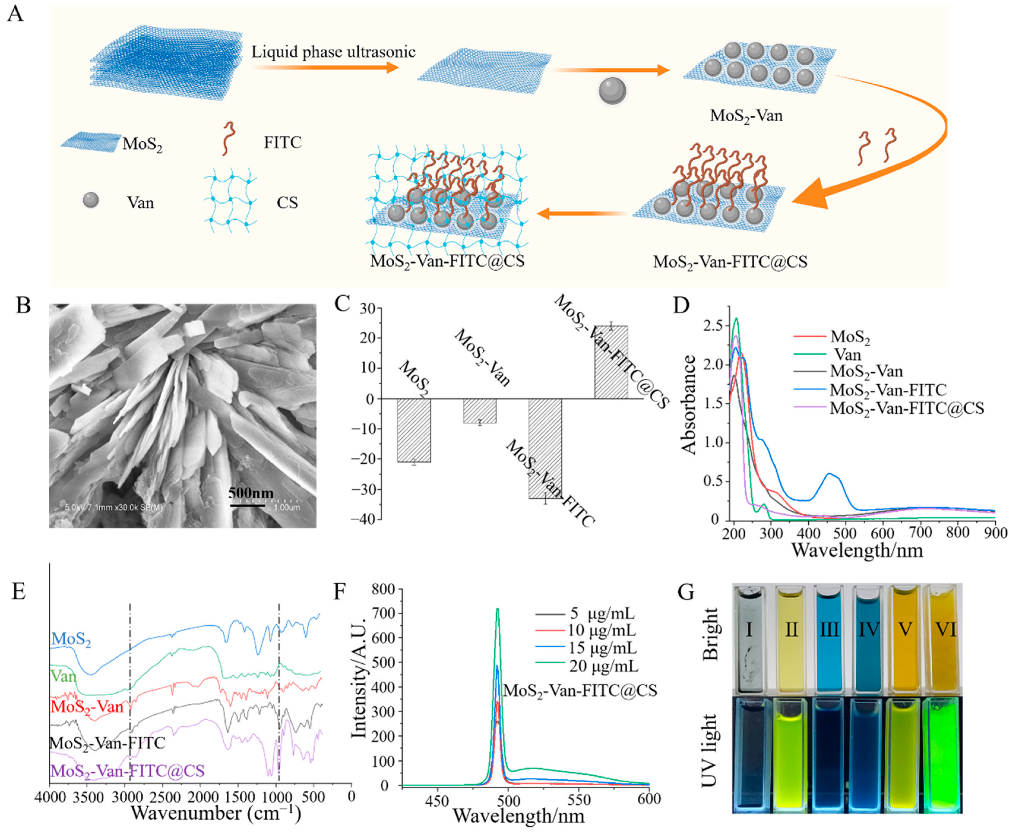
2.2. Synthesis of MoS2 Nanosheets

MoS2 nanosheets were synthesized by liquid ultrasonic stripping [29,30,31,32]. In brief, 0.5 g of MoS2 was combined with 50 mL of 45% ethanol to form the dispersion system of MoS2 (concentration, 10 μg/mL), which was sonicated in a water bath for 12 h. The solution was centrifuged at 12,000 rpm for 15 min, and the supernatant was collected and processed by a rotary evaporator to yield a thin film. The film was weighed, and MoS2 was resuspended in deionized water to form the MoS2 nanosheet aqueous solution.

2.3. Synthesis of MoS2-Van-FITC

Van is a heptapeptide-containing glycopeptide antibiotic with a primary amine moiety that binds covalently to FITC [33]. In brief, 4 mL of the MoS2 nanosheet aqueous solution (concentration, 0.002 g/mL) was aliquoted, 0.2 mg of Van was added to the solution, and the volume was adjusted to 20 mL. The solution was mixed on a magnetic stirrer at 250 rpm for 6 h and centrifuged at 12,000 rpm for 15 min. The precipitate was resuspended in 20 mL of pure water, and 1 mL of FITC was added. The solution was further mixed for 12 h. MoS2-Van-FITC was obtained by centrifugation.

2.4. Synthesis of the MoS2-Van-FITC@CS Hydrogel

To prepare the hydrogel, 300 mg of CS was added to 18 mL of 0.1 mol HCl solution and mixed on a magnetic stirrer until the CS solution was clarified. Thereafter, 2 mg of sodium β-glycerophosphate was added to 2 mL of 0.001 g/mL MoS2-Van-FITC solution, dissolved completely, and mixed for 4 h [27,34,35]. The MoS2-Van-FITC@CS hydrogel was obtained by heating in a water bath at 37 °C for 1 h.

2.5. Characterization

Ultraviolet-visible (UV–vis) absorption spectra were recorded by UV–vis spectroscopy (model S-3100, Scinco Co., Daejeon, Korea). Fluorescence spectra of MoS2-Van-FITC NPs were recorded by fluorescence spectrophotometry (model RF-5301PC, Shimadzu Corp., Kyoto, Japan). Fourier transform infrared (FT-IR) spectroscopy was performed with an FT-IR spectrometer (model Nicolette is50, Thermo Fisher Scientific, Waltham, MA, USA). The ultrastructural characteristics of synthesized MoS2 were observed via scanning electron microscopy (SEM; model S-4800, Hitachi, Tokyo, Japan). The Brookhaven Zeta Pals instrument was used to obtain zeta potential measurements and to characterize the optical properties of MoS2-Van-FITC (Brookhaven Instruments Corp., Holtsville, NY, USA). NPs of different concentrations were illuminated by using NIR irradiation at 808 nm at different power densities for 15 min, and the temperature was detected with an infrared camera with an accuracy of 0.1 °C [36,37].

2.6. In Vitro Antibacterial Assays

The minimum inhibitory concentration (MIC) of MoS2-Van-FITC NPs was determined using the 96-well microtitration plate dilution method. In brief, 100 μL of LB medium was added to each well of a sterile 96-well plate, and 100 μL of the drug solution was added to the first well, mixed, and diluted in multiples until the last well was mixed. Thereafter, 100 μL of the mixture was discarded, followed by the addition of 100 μL of the bacterial diluent to each well (final density of bacteria, 1 × 10⁶ CFU/mL). The positive controls were kanamycin and ampicillin. The cells of the NIR group were NIR irradiated (1.5 W/cm2) for 6 min and cultured at 37 °C for 12 h. The OD value was measured with a microplate reader [38]. The three independent measurements were averaged, and each treatment group had three wells.
Log-phase S. aureus cultures were inoculated (1:40) into medium containing MoS2-Van-FITC (100 μg/mL) or MoS2 (100 μg/mL) and incubated in a shaking incubator at 37 °C for 12 h. The irradiation power densities were 0.5 W/cm2, 1 W/cm2, and 1.5 W/cm2, and the irradiation times were 0 s, 150 s, and 300 s. Thereafter, the cells were coated on solid medium. The number of live bacteria was calculated using the CFU counting method.

2.7. Cellular Uptake Assays

To confirm the effect of Van in capturing S. aureus cells, we performed cellular uptake assays. Log-phase S. aureus cultures were incubated with different concentrations (15, 30, 45 µg/mL) of MoS2-Van-FITC NPs for 12 h. The cells treated with PBS served as the control. S. aureus cells were harvested and stained with 4ʹ-6-diamidino-2-phenylindole (DAPI; concentration, 5 µg/mL) for 15 min. Thereafter, the cells were washed twice with PBS, and the red and blue channels were examined under a fluorescence microscope.

2.8. LIVE–DEAD Assays

To further investigate the antibacterial ability of the drug, we conducted LIVE–DEAD assays. Log-phase S. aureus cultures were inoculated (1:40) in medium containing MoS2-Van-FITC (100 μg/mL), MoS2 (100 μg/mL), or Van (1 μg/mL) and incubated in a shaking incubator at 37°C for 12 h. The irradiation power density was 1.5 W/cm2, and the irradiation time was 300 s. Thereafter, the cells were stained with SYTO9 and propidium iodide for 30 min in the dark, washed twice with PBS, and red and green channels were examined under a fluorescence microscope. The number of non-viable bacterial cells was determined by the CytoFLEX system (Beckman Coulter, Brea, CA, USA) [2,39].

2.9. Cell Integrity Assays

Log-phase S. aureus cultures were treated with different concentrations (25, 50, 100 µg/mL) of MoS2-Van-FITC NPs. Van (1 µg/mL) and MoS2 (100 µg/mL) served as the controls. The PBS-treated group served as the blank. The irradiation power density was 1.5 W/cm2, and the irradiation time was 6 min. The cells were collected by centrifugation at 4000 rpm for 10 min, washed twice with PBS, and fixed with 2.5% glutaraldehyde at 4 °C for 12 h. The cells were subjected to gradient dehydration with different concentrations of ethanol (35%, 50%, 70%, 80%, 95%, 100%) for 20 min each time and then placed into acetone [40,41]. The specimens were observed under a scanning electron microscope.

2.10. Establishment of the Wound Mice Model

Kunming female mice (~25 g body weight; 4–5 weeks old) were obtained from the Model Animal Research Center of Nanjing University (Nanjing, China) and housed under standard environmental conditions (temperature, 22 ± 3 °C; humidity, 55 ± 5%; 12-h dark/12-h light cycles). All animals were housed according to the guidelines in the “Guide for the Care and Use of Laboratory Animals”. All animal studies were approved by Suzhou University (Suzhou, China) (approval number: 202010A415). After 7 days of acclimatization, an oval wound of approximately 1.5 cm in length was made by shaving the back of each mouse under anesthesia and adding 100 μL of activated S. aureus cells (1 × 10⁶ CFU/mL) dropwise to each wound for two consecutive days. The wound was treated after inflammation [14,42].

2.11. Wound Healing Assays

The mice whose wounds were infected with S. aureus were randomly divided into five groups (n = 5–7 mice) as follows: blank group, MoS2 group, MoS2-Van-FITC@CS group, NIR MoS2 group, and NIR MoS2-Van-FITC@CS group. The wound site of the blank group was treated with 100 μL of PBS, that of the MoS2 group was treated with 100 μL of MoS2 each day, that of the MoS2-Van-FITC@CS group was treated with 100 μL of MoS2-Van-FITC@CS hydrogel each day, that of the NIR MoS2 group was treated with MoS2, followed by irradiation at 1.5 W/cm2 for 5 min, and that of the NIR MoS2-Van-FITC@CS group was treated with MoS2-Van-FITC@CS, followed by irradiation at 1.5 W/cm2 for 5 min. The mice were photographed daily for eight consecutive days. The mice were euthanized, and their epidermises were harvested for hematoxylin–eosin staining [43].

2.12. Safety Evaluation of MoS2-Van-FITC@CS

To examine the toxic effects of NIR and MoS2-Van-FITC@CS on the heart, liver, spleen, lungs, and kidneys of mice, the wounded mice were divided into four groups as follows: control group, NIR irradiation group, MoS2-Van-FITC@CS group, and MoS2-Van-FITC@CS + NIR group. The irradiation power density was 1.5 W/cm2, the wavelength was 808 nm, and the irradiation time was 6 min. The mice were treated until their wounds healed, and they were euthanized 30 days after the end of treatment. Their organs were collected, and the cross-sections were stained with hematoxylin–eosin [44].

3. Results and Discussion

3.1. Characterization of MoS2-Van-FITC@CS

The nanocarriers were exfoliated by liquid phase ultrasound. The composite nanomaterials conjugated to FITC-labeled Van showed strong antibacterial effects. After irradiation, MoS2 converted light energy into heat energy, thereby killing the bacteria by actively trapping the cells through Van. MoS2-Van-FITC had good antibacterial and wound-promoting abilities through the temperature-sensitive hydrogel formed with chitosan (Figure 2A). To determine the synthesis of two-dimensional MoS2 nanosheets, the morphology and thickness of MoS2 NPs were observed via SEM (Figure 2B). The results of SEM showed that the flake NPs, which had a thickness of approximately 40 nm, were uniformly distributed. Zeta potentiometry can be used to determine the solid–liquid interfacial electrical properties of dispersed systems of particulate matter, so we can determine the successful synthesis of materials by using the potential changes of nanomaterials. The zeta potential of MoS2 was −21.4 ± 1.2 mV, whereas the loading of Van resulted in a potential of −8.3 ± 1.9 mV and a zeta potential of −33 ± 0.8 mV after labeling with FITC (Figure 2C). The synthesis of MoS2-Van-FITC@CS causes changes in the structure of a single component as a result of electron leaps between electronic energy levels in the valence and molecular orbitals, which can be observed by UV-vis spectrum (Figure 2D). In the UV-vis spectrum, MoS2 NPs were observed to have an absorption peak near 808 nm, showing a longitudinal surface plasmon resonance band, which indicated photothermal effects, whereas Van did not show an absorption peak near 808 nm. Van was adsorbed on MoS2, after which this characteristic peak significantly shifted. After FITC was decorated on the Van surface, the characteristic peak of MoS2-Van-FITC was significantly shifted, and a new characteristic peak was observed near 450 nm. MoS2-Van-FITC@CS also showed a shift in the characteristic peak compared to MoS2. FTIR spectroscopy allows the observation of the functional groups and chemical bonds contained in the material, in order to be able to determine the successful synthesis of MoS2-Van-FITC nanocomposites. FT-IR spectral analysis showed that the presence of Van resulted in an amino peak near 3000 cm−1, and a distinct peak at 1000 cm−1 in the fingerprint region after the loading of FITC (Figure 2E). A significant change was also found in the fingerprint region after the wrapping of chitosan. The fluorescence properties of MoS2-Van-FITC@CS are shown in Figure 2F,G. MoS2-Van-FITC@CS emitted fluorescence under UV light irradiation at 465 nm, and the fluorescence properties of the nanomaterials were further confirmed by the fluorescence spectra. Taken collectively, these findings indicate that MoS2, which has photothermal properties, was successfully synthesized, Van was successfully loaded, and the surface was modified by FITC. The morphological features of the MoS2-Van-FITC@CS hydrogel were indicative of its successful synthesis.

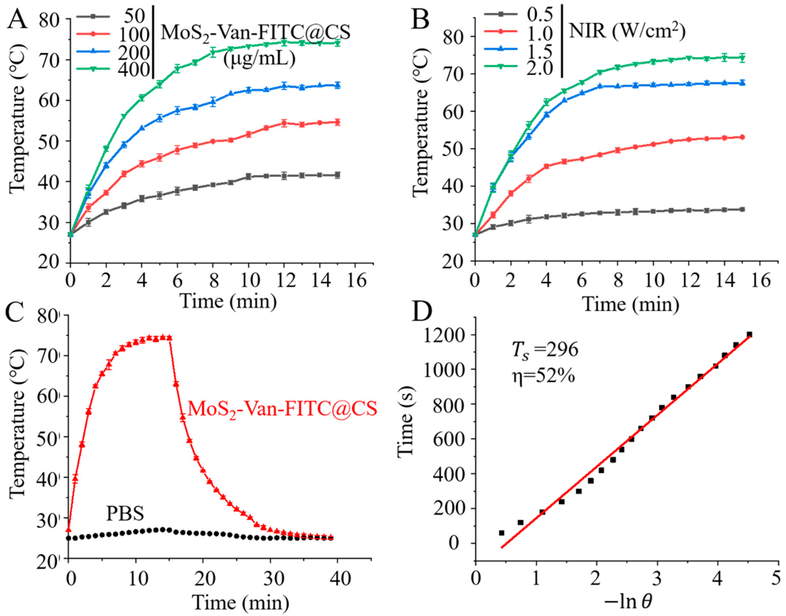
3.2. In Vitro Photothermal Efficiency

MoS2 is a photothermal agent that produces a large amount of heat to kill bacteria in the NIR region of 808 nm [45,46]. Therefore, we measured its photothermal conversion efficiency to understand the photothermal properties of MoS2. As expected, MoS2-Van-FITC acted as a good photothermal nanomaterial under NIR irradiation in that it converted light energy into heat energy, resulting in rapid warming (Figure 3A,B). Furthermore, the MoS2-Van-FITC concentration was 400 µg/mL, mPBS (mD) was 1.0 g, CH2O (CD) was 4.2 J/g/°C, ΔTmax was 47.3 °C (Figure 3C), I was 2 W, and τ s was 296 s (Figure 3D). Thus, the photothermal conversion efficiency (η) of MoS2-Van-FITC was 52%. Figure S1 shows the thermal images of MoS2-Van-FITC at different concentrations under an NIR irradiation at 808 nm. In summary, MoS2-Van-FITC has good photothermal conversion efficiency and can be used as a photothermal nanomaterial for killing bacteria.

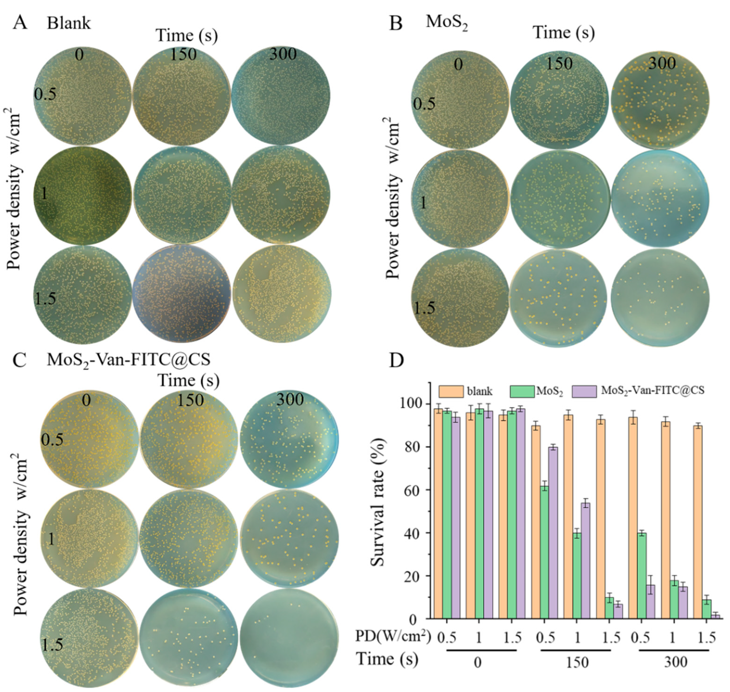
3.3. In Vitro Antibacterial Activity

We used S. aureus and E. coli as Gram-positive and Gram-negative bacteria, respectively, in subsequent experiments (Table 1). In the MIC test, we found that MoS2-Van-FITC + NIR had the highest killing effect against S. aureus, which may have been related to the fact that Van is a narrow-spectrum antibiotic that is only effective against Gram-positive bacteria. MoS2-Van-FITC + NIR showed the effects of common antibiotics at low doses, and the inhibition of growth made it difficult for bacteria to develop resistance (Table 1). Thermal images of the test in MIC were showed in Figure S2. Figure 4 shows the thermogram of the solution temperature increase after NIR irradiation. We found that our nanomaterials had a stronger growth inhibition ability against S. aureus. In the CFU test, we screened the power density and light time using power densities of 0.5, 1, and 1.5 W/cm2, and light times of 0, 150, and 300 s. We observed that a power density of 1.5 W/cm2 and a light time of 300 s inhibited bacterial growth. As shown in Figure 4, the inhibition rate of bacteria in the absence of MoS2 was low, and the survival rate of bacteria was 89%. However, the survival rate of bacteria gradually decreased after NIR irradiation, and the survival rate of bacteria was only 4.2% after treatment with MoS2 (100 μg/mL, 1.5 W/cm2, 300 s). After treatment with MoS2-Van-FITC + NIR (100 μg/mL), the survival rate of bacteria was 0.9% after an irradiation time of 300 s at a power density of 1.5 W/cm2. In the in vitro antibacterial test, the nanomaterials inhibited the growth of S. aureus cells, which played a role in the elimination of bacteria from the wound, thereby speeding up wound healing.

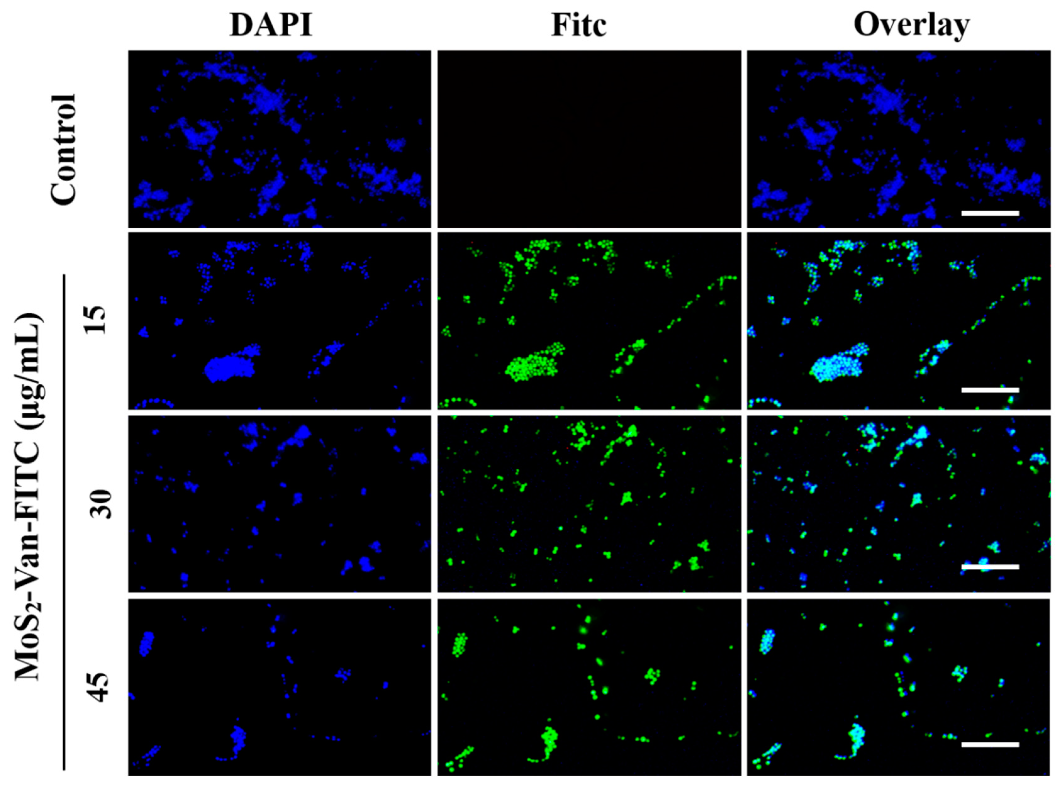
3.4. Cellular Uptake Assays

Van targets Gram-positive bacteria by binding to the hydrogen bond of the terminal D-Ala-D-Ala sequence of the cytosolic peptide of bacteria. It is also a heptapeptide-containing glycopeptide antibiotic with a primary amine moiety that binds covalently to FITC [46,47,48]. Van can label FITC on the bacterial surface; therefore, we verified the targeting of Van by DAPI staining. When MoS2-Van-FITC was used at a concentration of 15 μg/mL, most of the bacteria were labeled, similar to higher concentrations of 30 μg/mL and 45 μg/mL. However, as the concentration increased, the number of bacteria decreased, showing the excellent antibacterial ability of MoS2-Van-FITC (Figure 5). The results of cellular uptake assays indicated that MoS2-Van-FITC successfully targeted bacteria and showed excellent antibacterial ability, thereby achieving our goal of using combined chemotherapy and photothermal therapy to inhibit bacterial growth.

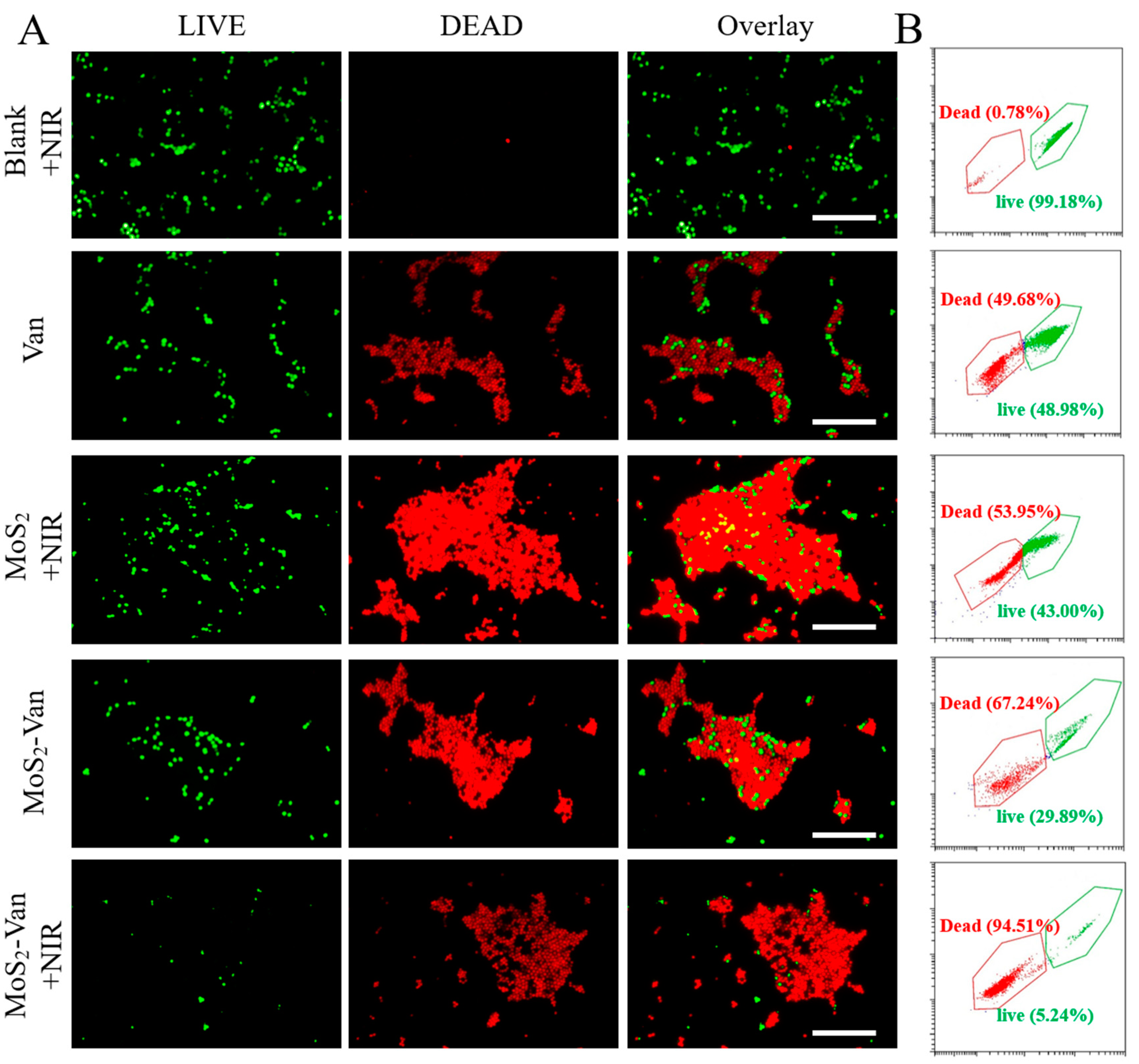
3.5. Fluorescent Staining Analysis of Antibacterial Activity

The CFU assay can detect only viable bacteria to determine the antibacterial activity of nanomaterials. To further examine the antibacterial activity of nanomaterials, we determined the number of viable and non-viable cells using the LIVE–DEAD assay to examine the antibacterial activity of MoS2-Van NPs. Viable bacterial cells were stained green, whereas non-viable bacterial cells were stained red (Figure 6A). No cell death was observed in the blank group. However, cell death was observed in the Van group, indicating that Van can inhibit bacterial growth. Similar results were obtained for the cells treated with MoS2 + NIR and MoS2-Van, with the MoS2-Van + NIR group exhibiting stronger inhibition of bacterial growth after NIR irradiation.
To further confirm that MoS2-Van+NIR reduced the survival of bacteria, the number of viable and non-viable bacterial cells was quantified via flow cytometry. As shown in Figure 6B, the apoptotic rate of the blank + NIR group was 0.78%. The apoptotic rate of MoS2 + NIR (100 µg/mL) after NIR irradiation was 53.95%. When the concentration of Van was 1 µg/mL, the apoptotic rate was 49.68%. The apoptotic rate of MoS2-Van (100 µg/mL) was 67.24% without irradiation, which was mainly due to the effects of Van, but MoS2 also played its own role after NIR irradiation, and the apoptotic rate was 94.51%. The increased antibacterial activity of MoS2-Van NPs was further confirmed by the quantitative analysis of viable and non-viable cells via flow cytometry.

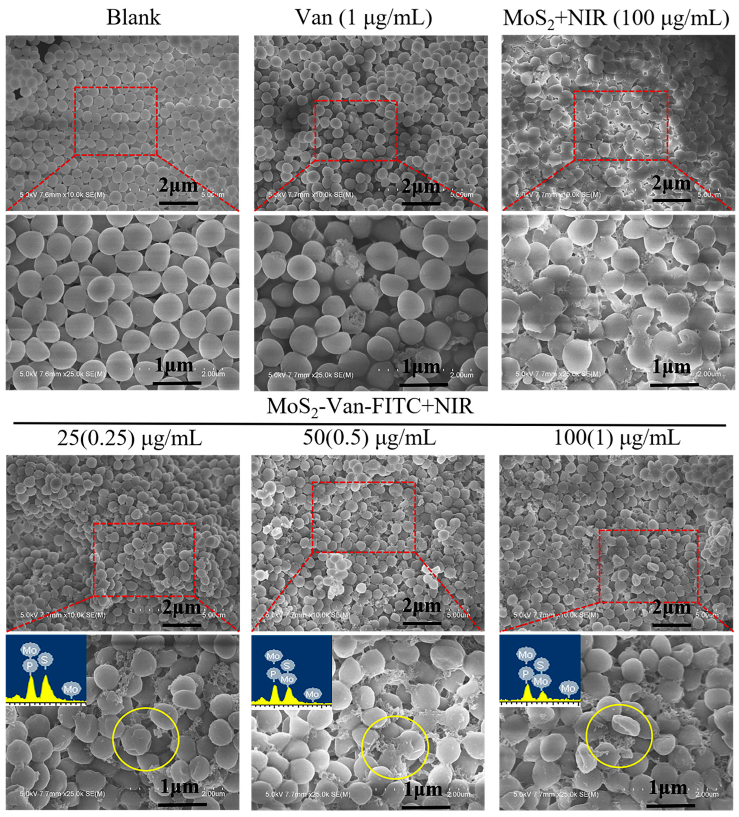
3.6. Cell Integrity Study

Based on our findings, MoS2-Van NPs + NIR showed efficient antibacterial activity against S. aureus. Because photothermal action mainly targets the bacterial cell surface, and the cell surface is also the site of action of Van, we speculate that changes in cell integrity may be the main mechanism behind the induction of apoptosis in bacteria. As such, we investigated the effects of MoS2-Van NPs + NIR on the cellular integrity of S. aureus by SEM.
The integrity of bacterial cells was examined via SEM, as shown in Figure 7. S. aureus cells in the blank group had normal cell morphology, including intact cell membranes. The results showed that NIR irradiation alone did not affect the structure of cells. The rupture and shrinkage of cells could be clearly seen after treatment with Van and MoS2 in the control group, and the enlarged area showed that the cells did not have intact cell membranes. In addition, there was cell leakage. In the MoS2-Van-FITC group, we observed more severe cell damage, even at a concentration of 25 µg/mL, compared to the blank group. As the concentration increased, the cell damage increased, indicating that MoS2-Van-FITC had a stronger antibacterial effect. The results from the elemental analysis chart showed that the bacterial surface did contain elemental sulfur and molybdenum, indicating that the bacterial surface contained MoS2. Taken collectively, these findings indicate that MoS2-Van-FITC NPs have very effective antibacterial activity compared to MoS2 NPs and Van alone.

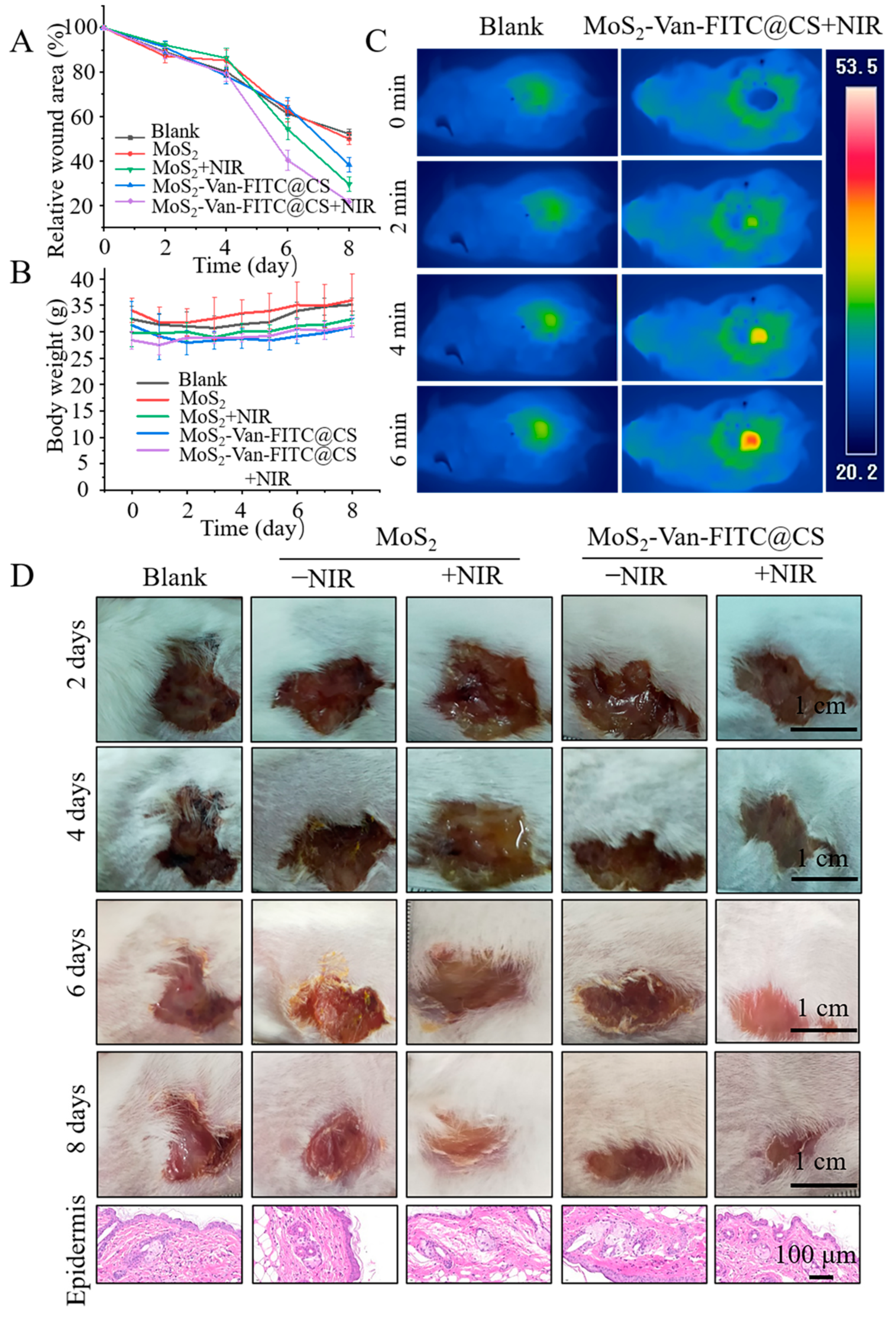
3.7. In Vivo Wound Healing Evaluation

We established a wound-healing mouse model and examined the pro-wound healing effects by directly applying NPs combined with irradiation. As shown in Figure 8, the MoS2-Van-FITC@CS hydrogel alone was slightly therapeutic, and the rate of wound healing increased with irradiation (Figure 8A). Heating, which increased the temperature of the nanomaterial to 50 °C by irradiation at 1.5 W/cm2 for 6 min, achieved a good therapeutic effect. The MoS2 group had the largest relative wound area, which did not heal. However, MoS2 decreased the relative wound area after NIR irradiation. MoS2-Van-FITC@CS hydrogel + NIR had the best therapeutic effect after NIR irradiation, where the relative wound area was reduced to 20.8%. The weight of mice in all groups decreased and then increased. The reason for the decrease in weight was likely due to the appearance of wounds, but as treatment progressed, the mice recovered and regained the weight. MoS2 was least effective, and the healing of mice treated with MoS2 was similar to that of the controls (Figure 8B). Immunohistochemical analysis was performed to examine the epidermis from the different groups of mice, and the abnormal histological features could be clearly seen in the stained sections (Figure 8D). No changes in morphology were observed in the epidermises of mice in the control group and the other four groups. The epidermises showed no significant damage compared to those of normal mice, indicating that the elevated temperature of the nanosheets during the photothermal treatment did not cause significant damage to the wounds. Therefore, the therapeutic effect of the MoS2-Van-FITC@CS hydrogel combined with NIR irradiation showed that there was no harm to the mice and their wound healing was accelerated. The preliminary photothermal imaging of mice also demonstrated the thermal response of NPs. In summary, the MoS2-Van-FITC@CS hydrogel combined with photothermolysis significantly promoted wound healing in mice.

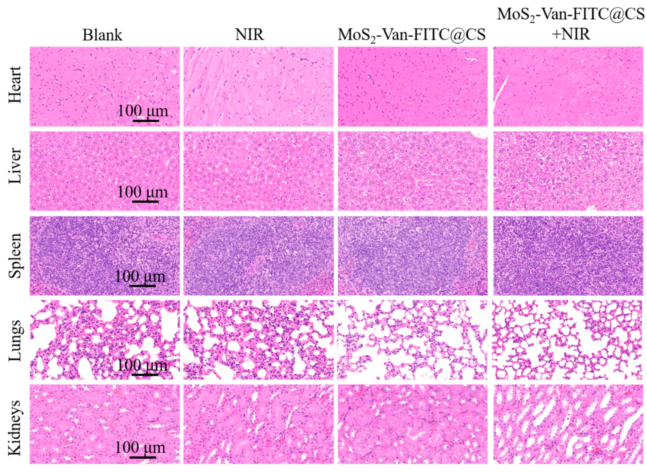
3.8. Biosafety Evaluation

The evaluation of toxicity is important for each new drug. As such, we evaluated the safety of our hydrogels in vivo. We applied the hydrogels to the wounds of healthy mice, followed by NIR irradiation, and performed a routine histological analysis of the major organs (Figure 9). Compared with the controls, no significant organ damage, tissue edema, cell death, or inflammatory cell infiltration was observed in the examined organs after treatment with NIR irradiation, MoS2-Van-FITC@CS, and MoS2-Van-FITC@CS + NIR, indicating that MoS2-Van-FITC@CS + NIR was non-toxic in mice.

4. Conclusions

In this study, the Van-modified MoS2-loaded nanosystem, which was encapsulated in a chitosan hydrogel, was established to examine its antibacterial activity and wound healing ability. The results showed that the thickness of MoS2 NPs was <100 nm, whereas other experiments revealed that the surface of MoS2 was successfully modified by Van. The antimicrobial activity was enhanced when Van was labeled. The photothermal characterization experiments confirmed that MoS2 had good photothermal conversion efficiency, and cellular uptake assays verified the active capture of S. aureus by Van, which significantly improved the photothermal inhibition of bacterial growth. The results of in vitro experiments indicated that NIR could significantly increase the antibacterial activity of MoS2 NPs, whereas those of flow cytometry showed that NPs could increase the apoptotic rate of bacterial cells. The morphological features of bacterial cells treated with MoS2-Van-FITC NPs and NIR irradiation were examined, and NPs were observed to disrupt the integrity of the bacterial cell wall. Furthermore, MoS2-Van-FITC combined with NIR irradiation could disrupt the cell morphology, induce apoptosis, and affect cell proliferation in vitro. In a wound healing assay, the MoS2-Van-FITC@CS hydrogel could accelerate wound healing. In summary, MoS2 in combination with NIR irradiation shows good applicability in the inhibition of bacterial growth, and the CS hydrogel in combination with a photothermal agent that actively traps S. aureus can disinfect the wound and maintain a moist environment to accelerate wound healing.

Supplementary Materials

The following supporting information can be downloaded at: https://www.mdpi.com/article/10.3390/nano12111865/s1, Figure S1: The thermal imaging images of MoS2-Van-FITC@CS at different concentrations (50, 100 and 200 µg/mL) and NIR irradiation (808 nm; 2 W/cm2); Figure S2: Thermal images of the test in MIC.

Author Contributions

Conceptualization, W.Z.; Data curation, W.Z., Z.K. and L.Z.; Formal analysis, W.Z. and Z.K.; Funding acquisition, P.S., W.L., L.G., Y.T. and F.G; Investigation, P.S., W.L. and C.T.; Methodology, P.S., W.L., C.T., F.G. and L.Z.; Project administration, L.G.; Resources, Y.T.; Writing—original draft, W.Z. and Z.K.; Writing—review & editing, Z.K. and L.Z. All authors have read and agreed to the published version of the manuscript.

Funding

The work was supported by the National natural science foundation of China (31671797), Anhui Nature Science Foundation (2008085QH397), Anhui Provincial Higher Education Institutes (KJ2020a0375 and KJ2021A0511), Anhui Polytechnic University (xjky2020064), Natural Science Foundation of Anhui University (KJ2020A118), Youth Key Talents Program of Wannan Medical College(wyqnyx202005).

Institutional Review Board Statement

The animal study protocol was approved by Suzhou University (Suzhou, China) (approval number: 202010A415).

Informed Consent Statement

Not applicable.

Data Availability Statement

Not applicable.

Conflicts of Interest

The authors have declared that no competing interests exist.

References

  1. Vlazaki, M.; Huber, J.; Restif, O. Integrating mathematical models with experimental data to investigate the within-host dynamics of bacterial infections. Pathog. Dis. 2019, 77, 14. [Google Scholar] [CrossRef] [PubMed]
  2. Li, W.; Song, P.; Xin, Y.; Kuang, Z.; Liu, Q.; Ge, F.; Zhu, L.; Zhang, X.; Tao, Y.; Zhang, W. The Effects of Luminescent CdSe Quantum Dot-Functionalized Antimicrobial Peptides Nanoparticles on Antibacterial Activity and Molecular Mechanism. Int. J. Nanomed. 2021, 16, 1849–1867. [Google Scholar] [CrossRef] [PubMed]
  3. Tang, M.; Zhang, J.; Yang, C.; Zheng, Y.; Jiang, H. Gold Nanoclusters for Bacterial Detection and Infection Therapy. Front. Chem. 2020, 8, 181. [Google Scholar] [CrossRef] [PubMed]
  4. Sun, D.; Zhang, W.; Mou, Z.; Chen, Y.; Guo, F.; Yang, E.; Wang, W. Transcriptome Analysis Reveals Silver Nanoparticle-Decorated Quercetin Antibacterial Molecular Mechanism. ACS Appl. Mater. Interfaces 2017, 9, 10047–10060. [Google Scholar] [CrossRef] [PubMed]
  5. Yang, X.; Zhang, W.; Zhao, Z.; Li, N.; Mou, Z.; Sun, D.; Cai, Y.; Wang, W.; Lin, Y. Quercetin loading CdSe/ZnS nanoparticles as efficient antibacterial and anticancer materials. J. Inorg. Biochem. 2017, 167, 36–48. [Google Scholar] [CrossRef] [PubMed]
  6. Carlie, S.; Boucher, C.E.; Bragg, R.R. Molecular basis of bacterial disinfectant resistance. Drug Resist. Updat. 2020, 48, 100672. [Google Scholar] [CrossRef]
  7. Fernández, J.; Bert, F.; Nicolas-Chanoine, M.-H. The challenges of multi-drug-resistance in hepatology. J. Hepatol. 2016, 65, 1043–1054. [Google Scholar] [CrossRef] [Green Version]
  8. Huang, S.; Liu, H.; Liao, K.; Hu, Q.; Guo, R.; Deng, K. Functionalized GO Nanovehicles with Nitric Oxide Release and Photothermal Activity-Based Hydrogels for Bacteria-Infected Wound Healing. ACS Appl. Mater. Interfaces 2020, 12, 28952–28964. [Google Scholar] [CrossRef]
  9. Wang, W.; Li, Y.; Wang, W.; Gao, B.; Wang, Z. Palygorskite/silver nanoparticles incorporated polyamide thin film nanocomposite membranes with enhanced water permeating, antifouling and antimicrobial performance. Chemosphere 2019, 236, 124396. [Google Scholar] [CrossRef]
  10. Sun, D.; Pang, X.; Cheng, Y.; Ming, J.; Xiang, S.; Zhang, C.; Lv, P.; Chu, C.; Chen, X.; Liu, G.; et al. Ultrasound-Switchable Nanozyme Augments Sonodynamic Therapy against Multidrug-Resistant Bacterial Infection. ACS Nano 2020, 14, 2063–2076. [Google Scholar] [CrossRef]
  11. Tao, B.; Lin, C.; Deng, Y.; Yuan, Z.; Shen, X.; Chen, M.; He, Y.; Peng, Z.; Hu, Y.; Cai, K. Copper-nanoparticle-embedded hydrogel for killing bacteria and promoting wound healing with photothermal therapy. J. Mater. Chem. B 2019, 7, 2534–2548. [Google Scholar] [CrossRef] [PubMed]
  12. Fan, X.; Yang, F.; Nie, C.; Yang, Y.; Ji, H.; He, C.; Cheng, C.; Zhao, C. Mussel-Inspired Synthesis of NIR-Responsive and Biocompatible Ag-Graphene 2D Nanoagents for Versatile Bacterial Disinfections. ACS Appl. Mater. Interfaces 2018, 10, 296–307. [Google Scholar] [CrossRef] [PubMed]
  13. Cao, W.; Yue, L.; Wang, Z. High antibacterial activity of chitosan—Molybdenum disulfide nanocomposite. Carbohydr. Polym. 2019, 215, 226–234. [Google Scholar] [CrossRef] [PubMed]
  14. Yin, W.; Yu, J.; Lv, F.; Yan, L.; Zheng, L.R.; Gu, Z.; Zhao, Y. Functionalized Nano-MoS2 with Peroxidase Catalytic and Near-Infrared Photothermal Activities for Safe and Synergetic Wound Antibacterial Applications. ACS Nano 2016, 10, 11000–11011. [Google Scholar] [CrossRef] [PubMed]
  15. Liu, Y.; Guo, Z.; Li, F.; Xiao, Y.; Zhang, Y.; Bu, T.; Jia, P.; Zhe, T.; Wang, L. Multifunctional Magnetic Copper Ferrite Nanoparticles as Fenton-like Reaction and Near-Infrared Photothermal Agents for Synergetic Antibacterial Therapy. ACS Appl. Mater. Interfaces 2019, 11, 31649–31660. [Google Scholar] [CrossRef]
  16. Zhao, X.; Chen, M.; Wang, H.; Xia, L.; Guo, M.; Jiang, S.; Wang, Q.; Li, X.; Yang, X. Synergistic antibacterial activity of streptomycin sulfate loaded PEG-MoS2/rGO nanoflakes assisted with near-infrared. Mater. Sci. Eng. C Mater. Biol. Appl. 2020, 116, 111221. [Google Scholar] [CrossRef]
  17. McMullen, A.R.; Lainhart, W.; Wallace, M.A.; Shupe, A.; Burnham, C.D. Evaluation of telavancin susceptibility in isolates of Staphylococcus aureus with reduced susceptibility to vancomycin. Eur. J. Clin. Microbiol. Infect. Dis. 2019, 38, 2323–2330. [Google Scholar] [CrossRef]
  18. Zhao, Z.; Yan, R.; Yi, X.; Li, J.; Rao, J.; Guo, Z.; Yang, Y.; Li, W.; Li, Y.Q.; Chen, C. Bacteria-Activated Theranostic Nanoprobes against Methicillin-Resistant Staphylococcus aureus Infection. ACS Nano 2017, 11, 4428–4438. [Google Scholar] [CrossRef]
  19. Han, D.; Yan, Y.; Wang, J.; Zhao, M.; Duan, X.; Kong, L.; Wu, H.; Cheng, W.; Min, X.; Ding, S. An enzyme-free electrochemiluminesce aptasensor for the rapid detection of Staphylococcus aureus by the quenching effect of MoS2-PtNPs-vancomycin to S2O82−/O2 system. Sens. Actuators B Chem. 2019, 288, 586–593. [Google Scholar] [CrossRef]
  20. Wu, Z.C.; Boger, D.L. Maxamycins: Durable Antibiotics Derived by Rational Redesign of Vancomycin. Acc. Chem. Res. 2020, 53, 2587–2599. [Google Scholar] [CrossRef]
  21. Blaskovich, M.A.T.; Hansford, K.A.; Gong, Y.; Butler, M.S.; Muldoon, C.; Huang, J.X.; Ramu, S.; Silva, A.B.; Cheng, M.; Kavanagh, A.M.; et al. Protein-inspired antibiotics active against vancomycin- and daptomycin-resistant bacteria. Nat. Commun. 2018, 9, 22. [Google Scholar] [CrossRef] [PubMed] [Green Version]
  22. Yang, C.; Ren, C.; Zhou, J.; Liu, J.; Zhang, Y.; Huang, F.; Ding, D.; Xu, B.; Liu, J. Dual Fluorescent- and Isotopic-Labelled Self-Assembling Vancomycin for in vivo Imaging of Bacterial Infections. Angew. Chem. Int. Ed. Engl. 2017, 56, 2356–2360. [Google Scholar] [CrossRef] [PubMed]
  23. Li, J.; Yu, F.; Chen, G.; Liu, J.; Li, X.L.; Cheng, B.; Mo, X.M.; Chen, C.; Pan, J.F. Moist-Retaining, Self-Recoverable, Bioadhesive, and Transparent in Situ Forming Hydrogels To Accelerate Wound Healing. ACS Appl. Mater. Interfaces 2020, 12, 2023–2038. [Google Scholar] [CrossRef] [PubMed]
  24. Nuutila, K.; Eriksson, E. Moist Wound Healing with Commonly Available Dressings. Adv. Wound Care 2021, 10, 685–698. [Google Scholar] [CrossRef] [PubMed]
  25. Basha, S.I.; Ghosh, S.; Vinothkumar, K.; Ramesh, B.; Kumari, P.H.P.; Mohan, K.V.M.; Sukumar, E. Fumaric acid incorporated Ag/agar-agar hybrid hydrogel: A multifunctional avenue to tackle wound healing. Mater. Sci. Eng. C Mater. Biol. Appl. 2020, 111, 110743. [Google Scholar] [CrossRef]
  26. Ganguly, S.; Das, P.; Itzhaki, E.; Hadad, E.; Gedanken, A.; Margel, S. Microwave-Synthesized Polysaccharide-Derived Carbon Dots as Therapeutic Cargoes and Toughening Agents for Elastomeric Gels. ACS Appl. Mater. Interfaces 2020, 12, 51940–51951. [Google Scholar] [CrossRef]
  27. Zheng, Y.; Wang, W.; Zhao, J.; Wu, C.; Ye, C.; Huang, M.; Wang, S. Preparation of injectable temperature-sensitive chitosan-based hydrogel for combined hyperthermia and chemotherapy of colon cancer. Carbohydr. Polym. 2019, 222, 115039. [Google Scholar] [CrossRef]
  28. Jiang, Y.; Meng, X.; Wu, Z.; Qi, X. Modified chitosan thermosensitive hydrogel enables sustained and efficient anti-tumor therapy via intratumoral injection. Carbohydr. Polym. 2016, 144, 245–253. [Google Scholar] [CrossRef]
  29. Zhao, H.; Wu, H.; Wu, J.; Li, J.; Wang, Y.; Zhang, Y.; Liu, H. Preparation of MoS2/WS2 nanosheets by liquid phase exfoliation with assistance of epigallocatechin gallate and study as an additive for high-performance lithium-sulfur batteries. J. Colloid. Interface Sci. 2019, 552, 554–562. [Google Scholar] [CrossRef]
  30. Ye, J.; Li, X.; Zhao, J.; Mei, X.; Li, Q. A Facile Way to Fabricate High-Performance Solution-Processed n-MoS2/p-MoS2 Bilayer Photodetectors. Nanoscale Res. Lett. 2015, 10, 454. [Google Scholar] [CrossRef] [Green Version]
  31. Yang, X.; Li, J.; Liang, T.; Ma, C.; Zhang, Y.; Chen, H.; Hanagata, N.; Su, H.; Xu, M. Antibacterial activity of two-dimensional MoS2 sheets. Nanoscale 2014, 6, 10126–10133. [Google Scholar] [CrossRef] [PubMed]
  32. Zhang, W.; Mou, Z.; Wang, Y.; Chen, Y.; Yang, E.; Guo, F.; Sun, D.; Wang, W. Molybdenum disulfide nanosheets loaded with chitosan and silver nanoparticles effective antifungal activities: In vitro and in vivo. Mater. Sci. Eng. C Mater. Biol. Appl. 2019, 97, 486–497. [Google Scholar] [CrossRef] [PubMed]
  33. Takai, H.; Kato, A.; Nakamura, T.; Tachibana, T.; Sakurai, T.; Nanami, M.; Suzuki, M. The importance of characterization of FITC-labeled antibodies used in tissue cross-reactivity studies. Acta Histochem. 2011, 113, 472–476. [Google Scholar] [CrossRef] [PubMed]
  34. Liu, T.; Li, J.; Shao, Z.; Ma, K.; Zhang, Z.; Wang, B.; Zhang, Y. Encapsulation of mesenchymal stem cells in chitosan/beta-glycerophosphate hydrogel for seeding on a novel calcium phosphate cement scaffold. Med. Eng. Phys. 2018, 56, 9–15. [Google Scholar] [CrossRef]
  35. Dang, Q.; Liu, K.; Zhang, Z.; Liu, C.; Liu, X.; Xin, Y.; Cheng, X.; Xu, T.; Cha, D.; Fan, B. Fabrication and evaluation of thermosensitive chitosan/collagen/alpha, beta-glycerophosphate hydrogels for tissue regeneration. Carbohydr. Polym. 2017, 167, 145–157. [Google Scholar] [CrossRef]
  36. Liu, H.; Zhu, X.; Guo, H.; Huang, H.; Huang, S.; Huang, S.; Xue, W.; Zhu, P.; Guo, R. Nitric oxide released injectable hydrogel combined with synergistic photothermal therapy for antibacterial and accelerated wound healing. Appl. Mater. Today 2020, 20, 100781. [Google Scholar] [CrossRef]
  37. Ganguly, S.; Das, P.; Das, T.K.; Ghosh, S.; Das, S.; Bose, M.; Mondal, M.; Das, A.K.; Das, N.C. Acoustic cavitation assisted destratified clay tactoid reinforced in situ elastomer-mimetic semi-IPN hydrogel for catalytic and bactericidal application. Ultrason. Sonochem. 2020, 60, 104797. [Google Scholar] [CrossRef]
  38. Sun, D.; Zhang, W.; Li, N.; Zhao, Z.; Mou, Z.; Yang, E.; Wang, W. Silver nanoparticles-quercetin conjugation to siRNA against drug-resistant Bacillus subtilis for effective gene silencing: In vitro and in vivo. Mater. Sci. Eng. C Mater. Biol. Appl. 2016, 63, 522–534. [Google Scholar] [CrossRef]
  39. Rekha, R.; Vaseeharan, B.; Vijayakumar, S.; Abinaya, M.; Govindarajan, M.; Alharbi, N.S.; Kadaikunnan, S.; Khaled, J.M.; Al-Anbr, M.N. Crustin-capped selenium nanowires against microbial pathogens and Japanese encephalitis mosquito vectors—Insights on their toxicity and internalization. J. Trace Elem. Med. Biol. 2019, 51, 191–203. [Google Scholar] [CrossRef]
  40. Lu, B.-Y.; Zhu, G.-Y.; Yu, C.-H.; Chen, G.-Y.; Zhang, C.-L.; Zeng, X.; Chen, Q.-M.; Peng, Q. Functionalized graphene oxide nanosheets with unique three-in-one properties for efficient and tunable antibacterial applications. Nano Res. 2020, 14, 185–190. [Google Scholar] [CrossRef]
  41. Sun, D.; Li, N.; Zhang, W.; Yang, E.; Mou, Z.; Zhao, Z.; Liu, H.; Wang, W. Quercetin-loaded PLGA nanoparticles: A highly effective antibacterial agent in vitro and anti-infection application in vivo. J. Nanoparticle Res. 2015, 18, 3. [Google Scholar] [CrossRef]
  42. Li, W.; Wang, Y.; Qi, Y.; Zhong, D.; Xie, T.; Yao, K.; Yang, S.; Zhou, M. Cupriferous Silver Peroxysulfite Superpyramids as a Universal and Long-Lasting Agent to Eradicate Multidrug-Resistant Bacteria and Promote Wound Healing. ACS Appl. Bio Mater. 2020, 4, 3729–3738. [Google Scholar] [CrossRef] [PubMed]
  43. Liang, Y.; Zhao, X.; Hu, T.; Chen, B.; Yin, Z.; Ma, P.X.; Guo, B. Adhesive Hemostatic Conducting Injectable Composite Hydrogels with Sustained Drug Release and Photothermal Antibacterial Activity to Promote Full-Thickness Skin Regeneration during Wound Healing. Small 2019, 15, e1900046. [Google Scholar] [CrossRef] [PubMed]
  44. Zhang, W.; Ding, X.; Cheng, H.; Yin, C.; Yan, J.; Mou, Z.; Wang, W.; Cui, D.; Fan, C.; Sun, D. Dual-Targeted Gold Nanoprism for Recognition of Early Apoptosis, Dual-Model Imaging and Precise Cancer Photothermal Therapy. Theranostics 2019, 9, 5610–5625. [Google Scholar] [CrossRef]
  45. Zhu, M.; Liu, X.; Tan, L.; Cui, Z.; Liang, Y.; Li, Z.; Yeung, K.W.K.; Wu, S. Photo-responsive chitosan/Ag/MoS2 for rapid bacteria-killing. J. Hazard. Mater. 2020, 383, 121122. [Google Scholar] [CrossRef]
  46. Xu, M.; Zhang, K.; Liu, Y.; Wang, J.; Wang, K.; Zhang, Y. Multifunctional MoS2 nanosheets with Au NPs grown in situ for synergistic chemo-photothermal therapy. Colloids Surf. B Biointerfaces 2019, 184, 110551. [Google Scholar] [CrossRef]
  47. Wang, C.; Gu, B.; Liu, Q.; Pang, Y.; Xiao, R.; Wang, S. Combined use of vancomycin-modified Ag-coated magnetic nanoparticles and secondary enhanced nanoparticles for rapid surface-enhanced Raman scattering detection of bacteria. Int. J. Nanomed. 2018, 13, 1159–1178. [Google Scholar] [CrossRef] [Green Version]
  48. Vimberg, V.; Gazak, R.; Szucs, Z.; Borbas, A.; Herczegh, P.; Cavanagh, J.P.; Zieglerova, L.; Zavora, J.; Adamkova, V.; Novotna, G.B. Fluorescence assay to predict activity of the glycopeptide antibiotics. J. Antibiot. 2019, 72, 114–117. [Google Scholar] [CrossRef]
Figure 1. Schematic illustration of the synergism between the two components of MoS2 and Van in MoS2-Van-FITC@CS, which was constructed and applied to wound healing in vivo with their highly efficiently antibacterial properties.
Nanomaterials 12 01865 g001
Figure 2. Synthesis and characterization of MoS2-Van-FITC@CS. (A) Synthesis illustration of MoS2-Van-FITC@CS. (B) SEM images of MoS2 NPs. (C) Zeta potential of MoS2, MoS2-Van, MoS2-Van-FITC, and MoS2-Van-FITC@CS. (D) UV-vis absorption spectra of MoS2, Van, MoS2-Van, MoS2-Van-FITC, and MoS2-Van-FITC@CS. (E) FT-IR spectrometry of MoS2, Van, MoS2-Van, MoS2-Van-FITC, and MoS2-Van-FITC@CS. (F) Digital images of I (Milli-water), II (FITC), III (MoS2), IV (MoS2-Van), V (MoS2-Van-FITC), and VI (MoS2-Van-FITC@CS) under bright and UV light. (G) Fluorescence emission spectra of MoS2-Van-FITC@CS.
Nanomaterials 12 01865 g002
Figure 3. In vitro photothermal efficiency. (A) The photothermal responses of MoS2-Van-FITC@CS. Different concentrations (50, 100, 200 and 400 µg/mL) were exposed to NIR irradiation (808 nm; 2 W/cm2). PBS served as a control. (B) The photothermal responses of MoS2-Van-FITC@CS (400 µg/mL), which was exposed to different power density of NIR irradiation (808 nm; 0.5, 1, 1.5 and 2 W/cm2). (C) The heating and cooling curves of MoS2-Van-FITC@CS (400 μg/mL) and PBS (laser irradiation at 808 nm). (D) The linear regression between the cooling period and −ln(θ) of the driving force temperature. Results shown are mean ± SD, n = 3.
Nanomaterials 12 01865 g003
Figure 4. In vitro antibacterial activity. (A) The results of the CFU assay for the blank group. PBS as a blank group. (B) The results of the CFU assay for MoS2 (100 μg/mL) for different times (0, 150 and 300 s) at different power densities (0.5, 1 and 1.5 W/cm2). (C) The results of the CFU assay for MoS2-Van-FITC (100 μg/mL) for different times (0, 150 and 300 s) at different power densities (0.5, 1 and 1.5 W/cm2). (D) Quantitative statistical results of (AC) (PD = Power Density). graphed using the Origin software. Results shown are mean ± SD, n = 3.
Nanomaterials 12 01865 g004
Figure 5. Fluorescence microscopy images of S. aureus cultures treated with MoS2-Van-FITC at various concentrations (15, 30, 45 µg/mL) for 12 h. PBS served as a control for the blank group. The cells stained with DAPI for 30 min were fluorescent blue, and those stained with MoS2-Van-FITC were fluorescent green. Scale bar = 15 μm. (DAPI, Ex = 358 nm and Em = 461 nm; FITC, Ex = 490 nm and Em = 525 nm).
Nanomaterials 12 01865 g005
Figure 6. Confocal fluorescence microscopy assay (A). S. aureus cultures after treatment with MoS2-Van NPs + NIR (100 µg/mL). Van solution (1 µg/mL), MoS2 NPs + NIR (100 µg/mL) and MoS2-Van (100 µg/mL) served as control groups. PBS served as a blank group. The cells were stained with SYTO 9 (green fluorescence) and PI (red fluorescence) for 30 min. The cells underwent NIR irradiation at 808 nm (1.5 W/cm2, 6 min). The results of the apoptotic assay by flow cytometry analysis were statistically analyzed by CytExpert software (version 2.4.0.28) (B). Scale bar = 15 μm. The data are expressed as mean ± SD (n = 3).
Nanomaterials 12 01865 g006
Figure 7. SEM images of S. aureus cells. The bacterial cultures were treated with MoS2-Van-FITC NPs at various concentrations (25, 50 and 100 µg/mL). The bacterial cultures treated with Van (1 µg/mL) and MoS2 NPs + NIR (100 µg/mL) served as control groups. PBS + NIR served as a blank group. The red squares indicate the enlarged regions. The blue area is the elemental analysis chart. The cells underwent NIR irradiation at 808 nm (1.5 W/cm2, 6 min).
Nanomaterials 12 01865 g007
Figure 8. In vivo wound healing analysis. (A) The relative wound area curve shows the MoS2-Van-FITC@CS hydrogel had a significant positive effect on wound healing. (B) The body weights of different groups after treatment. (C) The thermal imaging of mice after treatment. The cells underwent NIR irradiation at 808 nm (1.5 W/cm2, 6 min). (D) The photographs of wounds were taken every 2 days after treatment. MoS2-Van-FITC@CS hydrogel + NIR (100 µg/mL), MoS2 NPs (100 µg/mL), MoS2 NPs + NIR (100 µg/mL) and MoS2-Van-FITC@CS hydrogel served as control groups. PBS + NIR served as a blank group. The cells underwent NIR irradiation at 808 nm (1.5 W/cm2, 6 min). Scale bar = 1 cm. Results shown are mean ± SD, n = 5–7.
Nanomaterials 12 01865 g008
Figure 9. In vivo toxicity evaluation. The hematoxylin–eosin-stained images of major organs following different treatments of normal mice. Group 1 was the no treatment group, group 2 was the NIR irradiation (1.5 W/cm2, 6 min) group, group 3 was the MoS2-Van-FITC@CS group and group 4 was the MoS2-Van-FITC@CS + NIR (1.5 W/cm2, 6 min) group.
Nanomaterials 12 01865 g009
Table 1. Antibacterial activities with MICs values test (μg/mL).
MaterialsBacteria
S. aureusE. coli
MoS2>128>128
Van2>128
MoS2-Van-FITC6464
MoS2-Van-FITC@CS6464
MoS2 + NIR64128
MoS2-Van-FITC + NIR36128
MoS2-Van-FITC@CS + NIR36128
Kanamycin612
Ampicillin1824
Data are average values of at least three replicates.
Publisher’s Note: MDPI stays neutral with regard to jurisdictional claims in published maps and institutional affiliations.

