Article

Fe3O4-PEI Nanocomposites for Magnetic Harvesting of Chlorella vulgaris, Chlorella ellipsoidea, Microcystis aeruginosa, and Auxenochlorella protothecoides

1
Institute of Integrated Safety, Faculty of Materials Science and Technology, Slovak University of Technology, J. Bottu 25, 917 24 Trnava, Slovakia
2
Department of Nuclear Physics and Biophysics, Faculty of Mathematics, Physics and Informatics, Comenius University, Mlynská Dolina F1, 842 48 Bratislava, Slovakia
3
Institute of Electrical Engineering, Slovak Academy of Sciences, Dúbravská Cesta 9, 841 04 Bratislava, Slovakia
4
Centre for Nanodiagnostics of Materials, Faculty of Materials Science and Technology, Slovak University of Technology, Vazovova 5, 812 43 Bratislava, Slovakia
5
Institute of Materials Science, Faculty of Materials Science and Technology, Slovak University of Technology, J. Bottu 25, 917 24 Trnava, Slovakia
*
Author to whom correspondence should be addressed.
Nanomaterials 2022, 12(11), 1786; https://doi.org/10.3390/nano12111786
Submission received: 11 April 2022 / Revised: 10 May 2022 / Accepted: 19 May 2022 / Published: 24 May 2022
(This article belongs to the Special Issue Iron Oxide Nanomaterials)

Abstract

:
Magnetic separation of microalgae using magnetite is a promising harvesting method as it is fast, reliable, low cost, energy-efficient, and environmentally friendly. In the present work, magnetic harvesting of three green algae (Chlorella vulgaris, Chlorella ellipsoidea, and Auxenochlorella protothecoides) and one cyanobacteria (Microcystis aeruginosa) has been studied. The biomass was flushed with clean air using a 0.22 μm filter and fed CO2 for accelerated growth and faster reach of the exponential growth phase. The microalgae were harvested with magnetite nanoparticles. The nanoparticles were prepared by controlled co-precipitation of Fe2+ and Fe3+ cations in ammonia at room temperature. Subsequently, the prepared Fe3O4 nanoparticles were coated with polyethyleneimine (PEI). The prepared materials were characterized by high-resolution transmission electron microscopy, X-ray diffraction, magnetometry, and zeta potential measurements. The prepared nanomaterials were used for magnetic harvesting of microalgae. The highest harvesting efficiencies were found for PEI-coated Fe3O4. The efficiency was pH-dependent. Higher harvesting efficiencies, up to 99%, were obtained in acidic solutions. The results show that magnetic harvesting can be significantly enhanced by PEI coating, as it increases the positive electrical charge of the nanoparticles. Most importantly, the flocculants can be prepared at room temperature, thereby reducing the production costs.

1. Introduction

The consumption of conventional fossil fuels should be reduced as the reserves of raw materials are being depleted. High expectations are placed on the production of biofuels [1,2]. There has been a growing interest in microalgae exploitation over the past decade [2]. The microalgae are important single-cell photosynthetic microorganisms that are regarded as potential biomaterial sources for biofuel feedstock and nutrition [3,4]. The microalgal biofuel production represents a more environmentally friendly alternative to first-generation biofuels [5]. The microalgal production systems do not need fertile soils. They can be grown in marginal areas such as on non-arable lands or potentially in the ocean, thereby reducing competition for agricultural land and freshwater with food crops [6,7]. The microalgal production systems do not directly compete with the food chain. Furthermore, they can be used to convert CO2 to oxygen or for wastewater treatment [8,9,10,11].
Microalgae constitute a diverse group of single-cell photosynthetic organisms that include a wide range of eukaryotic algae and cyanobacteria [2]. Microalgae contain high-value nutrients [4]. Depending on species, the microalgae may produce several important lipids, and other oils [12,13,14]. Microalgae strains with high oil production capabilities are required for efficient biodiesel production [3,4]. Microalgae and cyanobacteria have a considerably higher oil production rate compared to conventional crops [15]. Chlorella species contain approximately 30% lipids (dry mass) [16,17]. Microcystis aeruginosa is a cyanobacteria (blue green algae) commonly observed in still waters (lakes and reservoirs), where it contributes to the development of eutrophication and bloom formation [18,19]. Although cyanobacteria are not eukaryotic phototrophs, as green algae are, they have high productivity and vast biomass [20,21]. Microcystis aeruginosa has a high lipid content [21]. As such, it is used for biofuel production. Furthermore, Microcystis aeruginosa can also be used in the sequestration of CO2 from the atmosphere [22,23].
Several sequential steps are involved in microalgal biodiesel production, including cell cultivation, harvesting, extraction of lipids, and fatty acid methyl ester generation. [24]. One of the problems that limits the use of microalgal biorefineries is the harvesting process [24,25]. It is more demanding compared to crop harvesting. The cost of the harvesting step can reach 20–30% of the total costs of algal-based biofuels’ production [26,27,28]. During microalgae harvesting, the water content is gradually removed from the microalgae culture medium through several subsequent techniques to concentrate biomass [29,30]. The choice of a suitable harvesting method is influenced by, for example, algae species (cell size, viability, and density, possible cell damage, strain properties, sedimentation rate, salt concentration) [29,31,32] and reuse of culture medium. It should be cheap and nontoxic when applied on a large scale. The suitability of the harvesting method depends on its energy demands, duration, financial needs, and finally, its environmental friendliness [33,34].
Several techniques have been developed for microalgae harvesting, including magnetic separation, centrifugation, flocculation, filtration, sedimentation, flotation, and electrophoresis [35]. Magnetic nanoparticles can be used to capture living algal cells rapidly and effectively, followed by low-energy magnetic isolation [36]. The magnetic separation using magnetic nanoparticles is regarded as one of the most promising methods. It is fast, energy efficient, low cost, environmentally friendly, scalable, and low contamination [1,37].
Magnetic nanoparticles are versatile materials with multiple applications [38,39]. They can be produced by several methods, including microemulsion [40,41], hydrothermal synthesis [42,43], thermal decomposition method [44,45], pyrolysis [46,47], sol-gel synthesis [48,49], and co-precipitation with bases [50,51]. Among these methods, the co-precipitation is the most used process [52]. It is easy to operate, and it can produce large volumes of nanoparticles. The co-precipitation is used to synthesize iron oxides and other ferrites [53,54]. A Fe3+/Fe2+ molar ratio of 2 is required for the synthesis of Fe3O4 nanoparticles. The formation of Fe3O4 by co-precipitation of Fe3+ and Fe2+ can be expressed by the following reaction [52,55]:
Fe2+ + 2Fe3+ + 8OH → Fe3O4 + 4H2O
In the Equation (1), a dropwise addition of ammonia is required to increase the pH of the solution. The co-precipitation is typically performed in an inert atmosphere (N2 or Ar) to avoid oxidation of Fe3O4 to Fe2O3. The reaction yield can be increased by vigorous stirring. The grain size and magnetic properties of the nanoparticles can be adjusted by controlling the reaction conditions [56,57]. Furthermore, the addition of different oxidizing and chelating agents, e.g., surfactants, saccharides, and polymers, is possible, which can influence the characteristic properties of the prepared nanoparticles [58].
A bare magnetite was used for the harvesting of Chlorella pyrenoidosa and C. minutissima [59], Nannochloropsis maritime [60], Scenedesmus obliquus [61], and Chlorella vulgaris [62,63,64]. The uncoated magnetic nanoparticles (NPs) were also studied in the harvesting of Microcystis aeruginosa [65]. Nevertheless, the electrostatic attraction between magnetite and algae species is not optimal. The functionalization of magnetite is usually necessary for higher harvesting efficiency or reactivation and for magnetite dosage reduction [66]. Polyethyleneimine (PEI) is a cationic polymer with repeating CH2CH2NH units. It can be easily protonated in acidic media. If adsorbed on the magnetite nanoparticles, it can functionalize them and modify their electrical charge [66]. PEI-coated nanoparticles were studied, for example, by Ge et al. for Scenedesmus dimorphus [67], Wang et al. [68] and Yang et al. [69] for Microcystis aeruginosa, and Hu et al. for Chlorella ellipsoidea [70].
The algal biomass increases with the culture time [71]. Hu et al. showed that the maximum harvesting efficiency could be obtained on days 14–18, i.e., when the biomass reaches the peak value [70,72]. As the algal biomass increases, the probability of interactions between the cells and nanoparticles also increases. The growth stage of algae has an influence on the lipid content and surface characteristics of the algal cells [33,73]. The number of functional groups on algal cells increases during the exponential growth phase, which enhances the adsorption capacity of the surface-functionalized magnetic particles [74]. The PEI-coated iron oxide nanoparticles can also be used to remove extracellular organic matter of the cells via charge neutralization. The harvesting and extracellular organic matter removal can be conducted simultaneously [68,75]. The nanoparticles can be removed by acid-base treatment and ultrasonication from the attached cells and re-used for further harvesting, which makes the process economical [76].
The above-mentioned studies of algae sorption to magnetite have been performed with magnetite nanoparticles synthetized at high temperatures (typically 80 °C). In the present study, we have prepared the iron oxide nanoparticles at room temperature and coated them with polyethyleneimine. The lower temperature was used to decrease the energy consumption of the process. The results show that the synthesis of PEI-coated Fe3O4 at 20 °C is feasible. The nanoparticles have comparable characteristics (particle size, microstructure, harvesting efficiency) to NPs prepared at 80 °C. The harvesting efficiencies can be significantly enhanced by PEI coating, as the polymer increases the positive electrical charge of the nanoparticles. In the present work, we study the magnetic harvesting of three green algae (Chlorella vulgaris, Chlorella ellipsoidea, and Auxenochlorella protothecoides) and one cyanobacteria (Microcystis aeruginosa) to explore the applicability of the process on several microorganisms. The results show that the magnetic harvesting with the nanoparticles synthesized at room temperature is applicable to both eukaryotic algae and cyanobacteria, making the process attractive for industrial use.

2. Materials and Methods

2.1. Magnetite Nanoparticles’ Synthesis

Magnetite nanoparticles (Fe3O4 NP) were synthesized by controlled co-precipitation of Fe2+ and Fe3+ chlorides in NH4OH. We used a previously reported method [70], however, the experiments were conducted at room temperature (20 °C) instead of 80 °C to decrease the production costs. The distilled water was de-oxygenated prior to the experiment by purging with flowing N2 for 30 min. For one dose of Fe3O4 NP, 1.98 g of FeCl2.4H2O and 5.4 g of FeCl3.6H2O were placed in a three-neck flask vessel and dissolved in 200 mL of deoxygenated distilled water. The resulting aqueous solution was vigorously stirred and constantly purged with flowing N2. After a dropwise addition of 20 mL of NH4OH (25 wt.%) and continuous stirring for 30 min, Fe3O4 NPs were precipitated. The concentration of the prepared Fe3O4 NP was 0.05 mol L−1. The nanoparticles were sedimented for 1 h. Subsequently, the precipitate was washed with distilled water. The decantation process was repeated three times. The sedimentation was aided by using a neodymium magnet.

2.2. PEI Coating Procedure

Decantated NPs from the co-precipitation were admixed with phosphate buffer (pH 7.3). Subsequently, polyethyleneimine (PEI) solution (1.2 kDa, 50% (w/v) in H2O, Sigma Aldrich, Bratislava, Slovakia) was added. The volume ratio of PEI:Fe3O4 was 9:1. The mixture was stirred at 150 rpm for 1 h, at laboratory temperature (20 °C). The prepared nanocomposites (NCs) were washed three times with distilled water, and stored in a sealed glass bottle for further use. All chemicals were analytical grade and were used without prior purification.

2.3. Transmission Electron Microscopy and X-ray Diffraction

The microstructure and particle size of Fe3O4 NPs and NCs were studied by a double-corrected, high-resolution scanning transmission electron microscope, JEOL JEM ARM 200cF (STEM resolution 0.78 Å, TEM resolution 1.1 Å, Tokyo, Japan). The samples for TEM observation were prepared by dropping the aqueous Fe3O4 solution onto a carbon layer-covered copper grid and air-dried. The particle size distribution was estimated from TEM images using ImageJ, a Java-based image processing program. An electron diffraction analysis was also employed to study the phase constitution of the prepared materials.
A PANalytical Empyrean X-ray diffractometer (XRD, Malvern Panalytical Ltd., Malvern, UK) was used to study the phase constitution of prepared nanoparticles and nanocomposites. The diffractometer was working with a CoKα1,2 radiation source and operating at 40 kV and 30 mA. The diffraction patterns were recorded at room temperature using Bragg–Brentano geometry. The measurements were carried out at 20° to 120° (2 Theta), with a step size of 0.02° and counting time of 98 s per step.

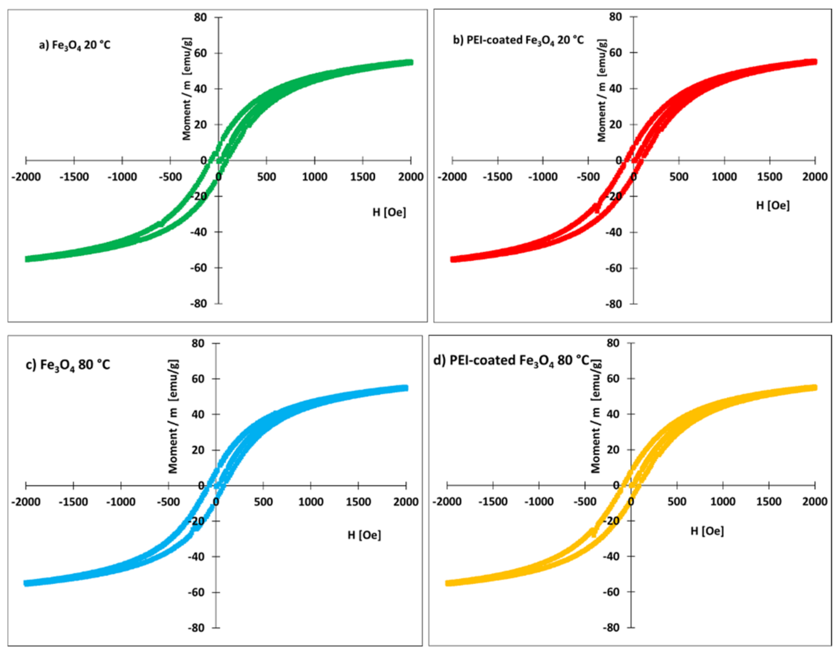
2.4. Magnetic Properties

The magnetic properties of the prepared and air-dried Fe3O4 NPs and NCs were studied by DC magnetometry. A plastic container with approximately 0.015 g of densely packed nanoparticle powder was placed in a vibrating sample magnetometer. The magnetic moment was measured at room temperature by generating an external homogeneous magnetic field with induction B, which was applied perpendicularly to the pre-dried sample. Magnetization loops were recorded between B = −2 T and B = +2 T (magnetic field strength between −20,000 and +20,000 Oe). A constant sweeping rate was used in all measurements.

2.5. Zeta Potential

Zeta potential measurements of the algae species, NPs, and NCs were performed using a Zetasizer Nano-ZS (Malvern, UK). The instrument uses a He-Ne laser with a wavelength of 633 nm and M3-PALS technology. The electrophoretic mobilities were converted into zeta potentials via the Henry equation in the Smoluchowski approximation [77]. The stock solution was 46 mg mL−1 for noncoated magnetite and 109 mg mL−1 for PEI-coated magnetite. The samples for electrokinetic potential measurements were prepared by micro-pipetting 2 μL of magnetic nanoparticles from the stock solution and mixing them with 998 μL of working buffer. In parallel, 100 μL of algae stock solution (concentration approximately 0.8 g L−1 DCW, in culture medium) was diluted in 900 μL of working buffer. Na-phosphate buffer (10 mmol L−1, NaH2PO4 and Na2HPO4) was used as a working buffer. The pH of 2.4–9.0 was adjusted by adding small aliquots of 1 mol L−1 of either HCl or NaOH. The tested solutions were measured in a disposable clear folded capillary zeta cell (DTS1070; Malvern, UK) at 25 °C. Values are reported as an average from 3 consecutive measurements, following an automatic measurement duration of 10–30 runs.

2.6. Microalgae Strains and Cultivation

Chlorella vulgaris (SAG 211-11b), Chlorella ellipsoidea (SAG 2111), Microcystis aeruginosa (SAG 46.80), and Auxenochlorella protothecoides (SAG 33.80) were obtained as sterile cultures from the algae collection of the University of Göttingen, Germany (SAG—Sammlung von Algenkulturen der Universität Göttingen). The biomass cultivation for the harvesting experiment was carried out in 1 L Erlenmeyer flasks using standard BG 11 cultivation medium. The biomass was illuminated at 2000 lx at 25 °C, with a light/dark cycle of 16/8 h. The biomass was flushed with clean air using a 0.22 μm filter. The algae were fed CO2 from the air to accelerate growth and accelerate reaching the exponential growth phase. The algae concentration (g L−1) was calculated using the calibration curve of the known optical density at 680 nm using a Genesys 8 spectrophotometer according to the dry cell weights determined gravimetrically after drying to constant weight at 110 °C.

2.7. Magnetite Harvesting Procedure

A known amount of either uncoated magnetite or PEI-coated magnetite was added to 50 mL of algae suspension. The flask was shaken manually for 90 s and subsequently placed on a permanent NdFeB block magnet (permanent magnetization 1.22–1.30 T, Magsy Ltd., Los Angeles, CA, USA) for 10 min. The optical densities of the remaining supernatant were measured at 680 nm using a Genesys 8 spectrophotometer. A harvesting efficiency, R (%), was calculated according to the following equation:
R = C 0 C e   C 0 × 100   % ,
In Equation (2), C0 is the initial concentration of the algae suspension (g L−1) and Ce is the concentration of algae in the supernatant after harvesting (g L−1).

2.8. Adsorption Experiments

Adsorption experiments were carried out with approximately 2-week-old microalgae. An optimal pH of 7.0 and dose of 5–30 mg of noncoated or PEI-coated magnetite were used at a constant temperature (25 °C), volume of algae 50 mL, reaction time 90 s, and stirring speed of approximately 120 r min−1. Two different isotherm models, Langmuir and Freundlich, have been tested (Table 1).
The algae cells with adsorbed Fe3O4-PEI NCs were transferred to a slide (base glass) and inspected with a light microscope, Carl Zeiss Jenavert.

2.9. Statistical Analysis

The algal experiments were performed at room temperature (20 ± 2 °C). We used triplicate sampling and testing. The results in this paper are presented as mean values calculated from three experiments. The standard deviation was also calculated from three independent measurements. The triplicated datasets of each experiment were analyzed statistically using one-way analysis of variance at a significance level of 0.05. The statistical analysis was integrated in the statistical software OriginPro 8.5.

3. Results

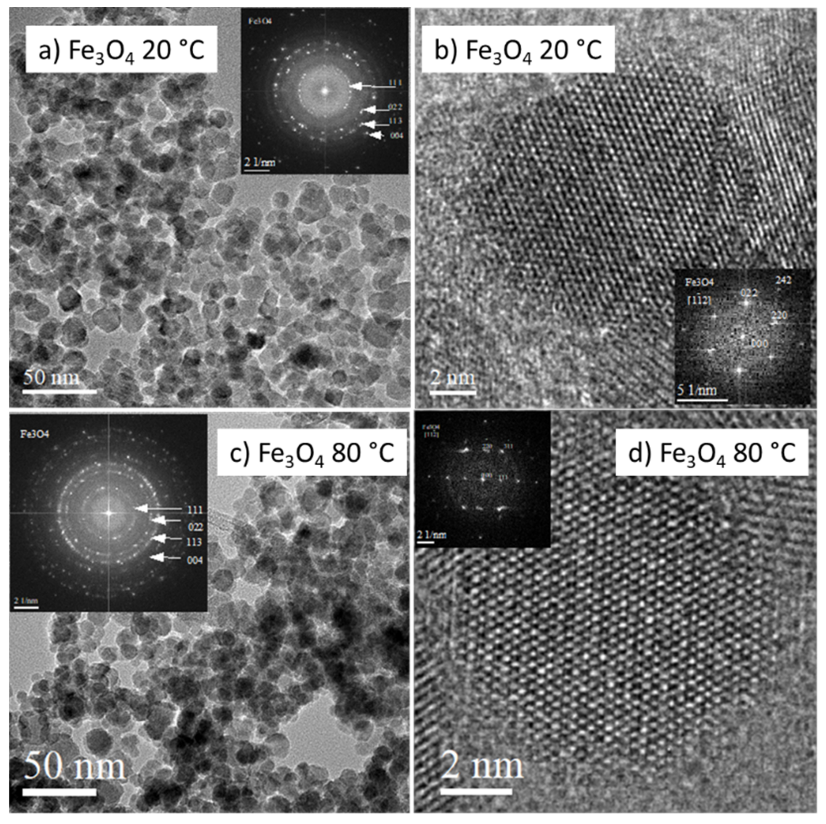
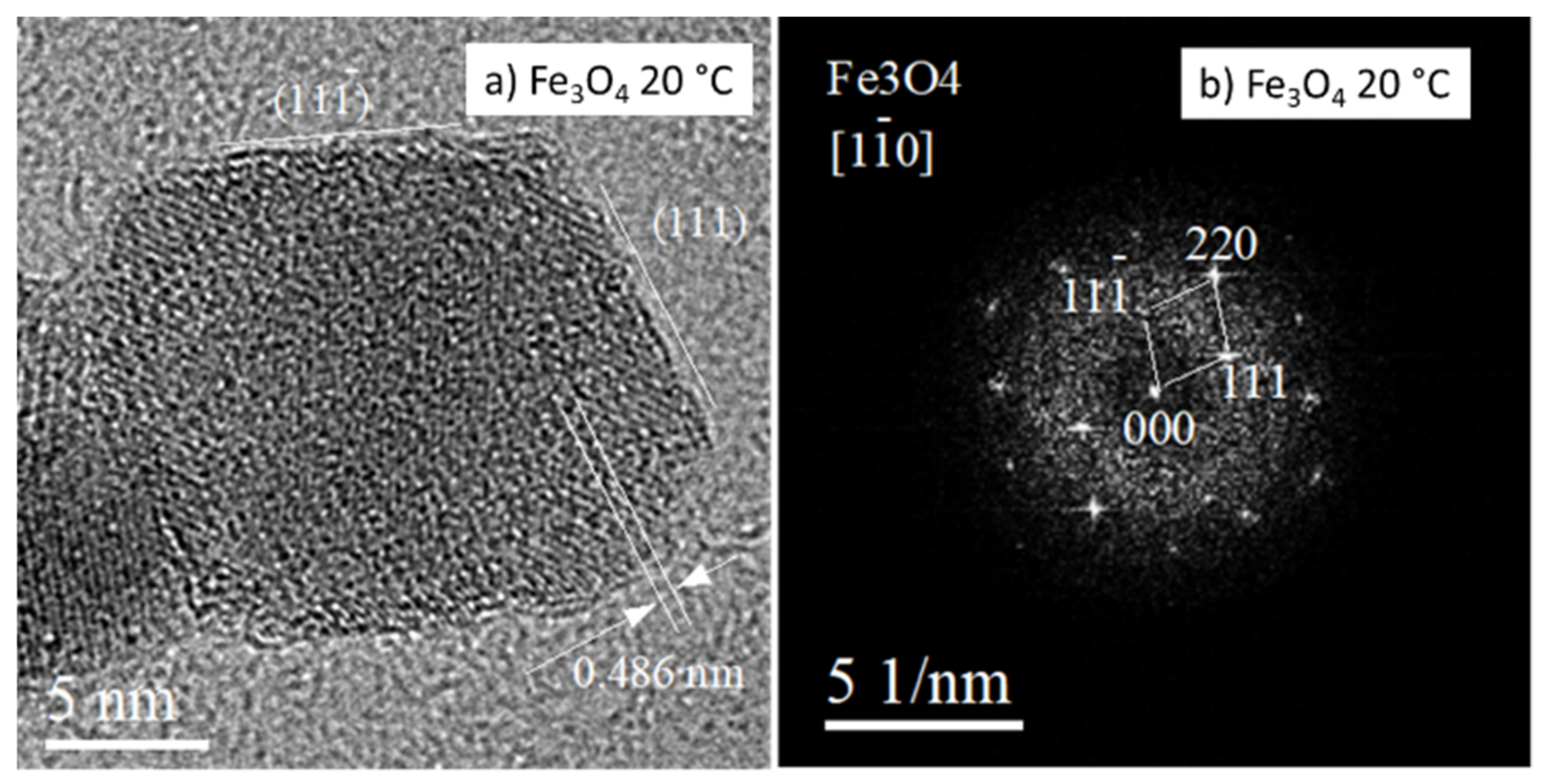
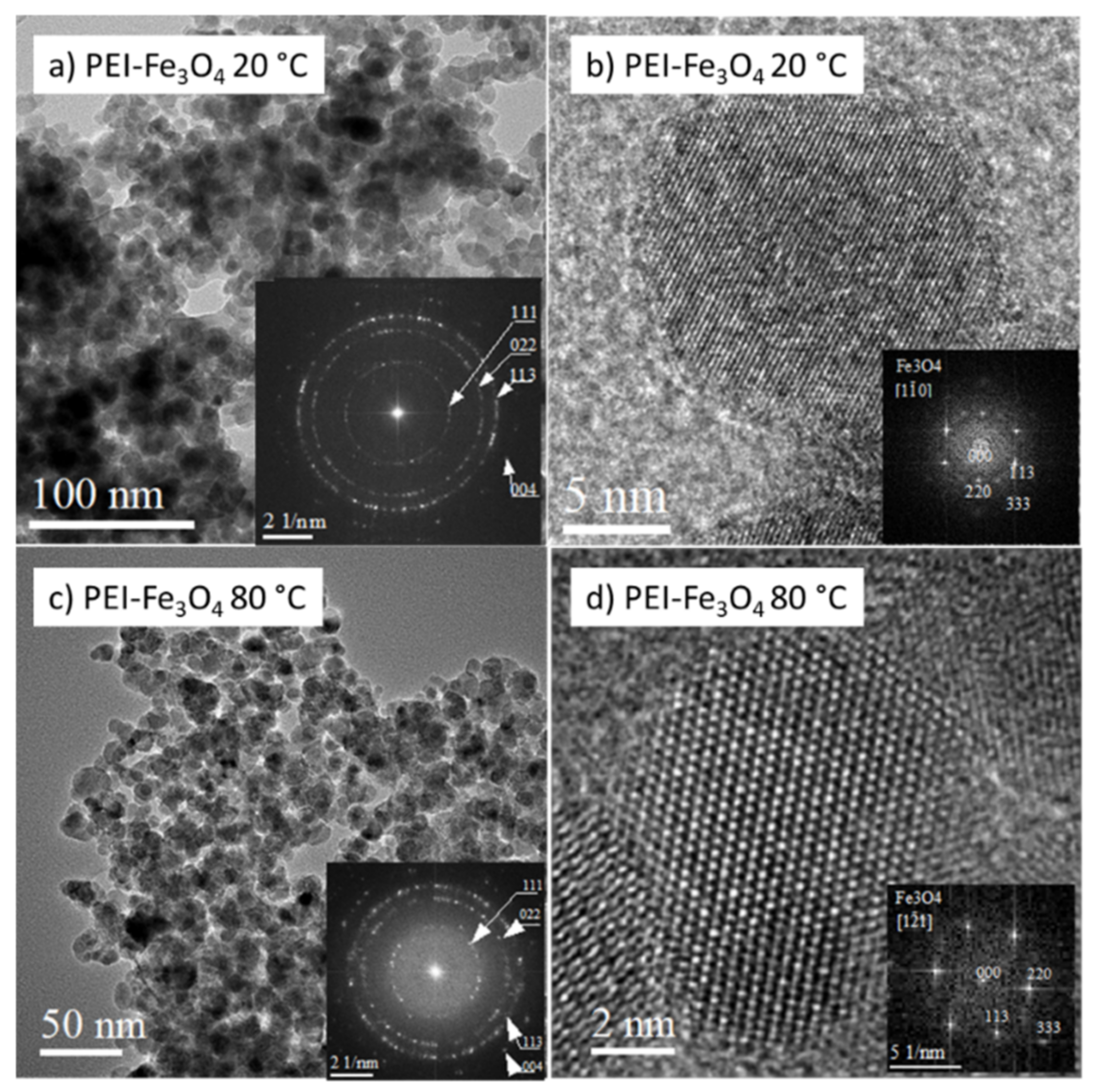
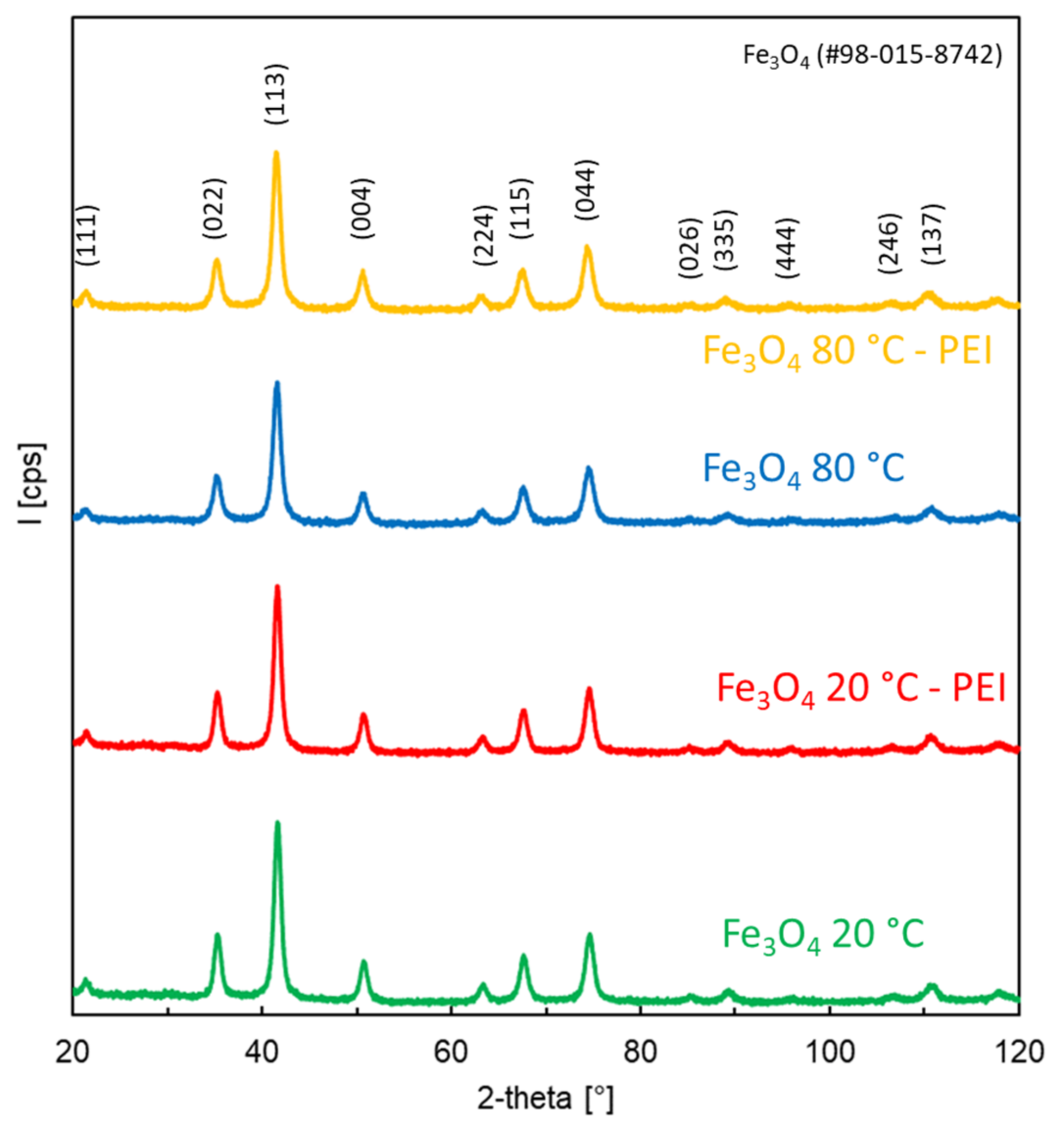
3.1. Characterization of Prepared Fe3O4 and Fe3O4-PEI NPs
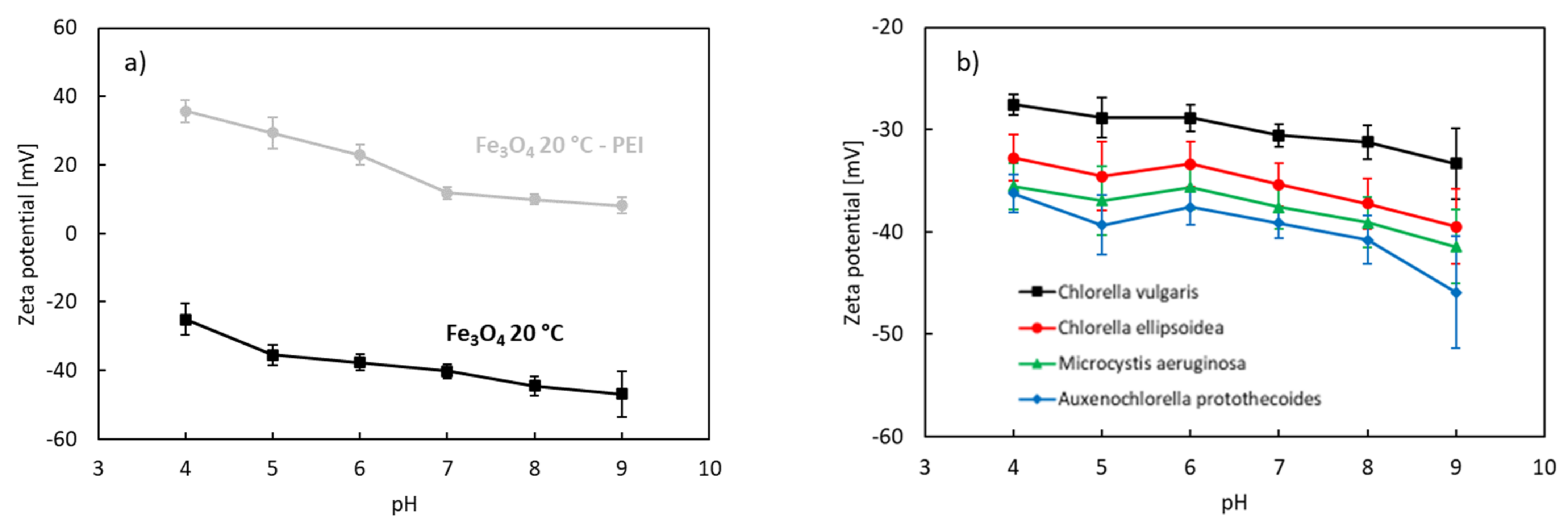
In this paper, we investigated the effect of the synthesis temperature on the microstructure and magnetic properties of PEI-coated Fe3O4 NPs. Originally, a relatively high temperature (80 °C) was applied for the co-precipitation of Fe3+ and Fe2+ in alkaline medium [70]. The high temperature was used to accelerate the chemical reaction. However, it was later reported that the synthesis temperature can be lowered and used to control the size of prepared Fe3O4 NPs [78,79].
The microstructure of magnetite Fe3O4 NPs prepared at 20 and 80 °C as recorded by TEM imaging is provided in Figure 1. The particle size distribution was relatively uniform, and an average particle diameter of ~10 nm was found. The prepared NPs exhibited mostly spherical morphology, however some of them were faceted. A more detailed image of a faceted particle produced at 20 °C is shown in Figure 2. As follows from the relevant fast Fourier transformation (FFT) pattern in Figure 2b, the particle exhibits octahedral morphology predominantly faceted by {111}-type planes. The aggregation of NPs is evident from Figure 1a,c.
The phase constitution of the produced NPs was studied using the selected area electron diffraction method (SAED) and by evaluation of the FFT patterns acquired from relevant HRTEM images (Figure 1b). Determined interplane distances of 0.485, 0.298, 0.255, and 0.212 nm correspond well with that reported for the 111, 022, 113, and 004 most-intense reflections of magnetite Fe3O4 phase (PDF No. 98-002-0596). Moreover, EELS spectroscopy was used to confirm the presence of magnetite in the samples. Quantitative EELS measurements showed that the amount of oxygen and iron in the NPs was 56.5 and 43.5 at%, respectively, which is close to chemical composition of magnetite Fe3O4.
The sharp-spotted rings in SAED/FFT patterns demonstrate a polycrystalline character of the samples and the good crystallinity of as-synthesized nanoparticles. This finding is in line with the more detailed HRTEM (high-resolution TEM) and ARTEM (atomic resolution TEM) images recorded from individual NPs prepared at 20 °C and also at 80 °C, in Figure 1 and Figure 2.
Several experimental approaches have been used to produce PEI-coated magnetic NPs, including hydrothermal, solvothermal, and co-precipitation methods [70,80,81]. However, the previously reported methods required either high temperatures, long reaction times, or several reaction steps. In our work, we have prepared PEI-coated Fe3O4 NPs at room temperature. The nanostructure of PEI-coated Fe3O4 NPs prepared at 20 °C is shown in Figure 3a. The image shows NPs of about 10 nm in size exhibiting mostly spherical morphology. However, faceted NPs, as shown in Figure 2b, are also seen in these images. The Fe3O4 NPs prepared at 80 °C and coated with PEI are shown in Figure 3c. The particle size is comparable to NPs prepared at 20 °C, confirming that the synthesis temperature can be reduced without affecting NPs’ size. Magnetite phase was confirmed by the estimation of relevant FFT patterns in both produced samples (Figure 3a,c). HRTEM and ARTEM images (Figure 3b,d) revealed that the atomic planes in NPs are well-ordered. Lattice defects, such as dislocations and stacking faults, were not detected in NPs.
The phase constitution of the prepared NPs and NCs was also studied by room-temperature X-ray diffraction. The results are presented in Figure 4. The prepared materials were crystalline. The XRD peaks can be assigned to Fe3O4 (PDF No. 98-015-8742). The cationic polymer did not affect the crystal structure of magnetite. Furthermore, there was practically no difference in the XRD patterns of NPs and NCs prepared at 20 and 80 °C, confirming that Fe3O4 can be prepared at room temperature.
The magnetization curves of the nanoparticles prepared at 20 and 80 °C are shown in Figure 5. The coated and uncoated nanoparticles had similar magnetization curves. The value of the remanent magnetization, Mr, was close to 7 emu/g. Remanent magnetization was nearly identical for both coated and uncoated nanoparticles. Thus, the organic coating does not adversely affect the value of remanent magnetization. For this reason, coated nanoparticles can be used for magnetic separation (collection) of algae from an aqueous medium.
The saturation of magnetization was close to 60 emu/g (Figure 5). This value is smaller than the previously reported 66.5 emu/g for PEI-coated Fe3O4 NPs prepared at 90 °C [82]. The smaller values are either related to the existence of nonmagnetic mass present in our samples or to nanoparticle interactions. A previous investigation [82] of the magnetic properties of PEI-coated Fe3O4 NPs suggested the existence of interacting particles, likely forming agglomerates, with a higher blocking temperature (>150 K), in which the surface spin disorder was weak and dominated by interparticle interactions. Nanoparticle agglomeration has also been observed in the present work (Figure 2a). The interparticle interactions could thus be responsible for the lower magnetic saturation.

3.2. Zeta Potential

Figure 6a shows the effect of pH on the electrokinetic zeta potential of uncoated and PEI-coated Fe3O4 nanoparticles synthesized at 20 °C. Figure 6b shows the zeta potential of the algae species tested.
The zeta potential of the uncoated nanoparticles was negative within the investigated pH range (4–9). The measured values correspond to the studies of Zhang [83], Plaza [84], Kim [85], and Savvidou [86] for magnetite nanoparticles produced by co-precipitation of iron sulfates or iron chlorides. The zeta potential increases with decreasing pH due to protonation. The isoelectric point is a point where the net electrical charge is 0. In our experiments, the isoelectric point of the uncoated magnetite nanoparticles produced by the co-precipitation method at 20 °C was estimated to be 2.0–3.0. These values are in line with the observations of Zhang et al. [83]. The zeta potential is affected not only by suspension conditions such as pH, temperature, ionic strength, and even the types of ions in the suspension, but also by particle properties such as size and concentration [87,88]. The decrease in the isoelectric point can indicate oxidation of magnetite to maghemite [89]. The final step of magnetite oxidation is maghemite [84]. Fe3O4 is not stable in the presence of oxygen, especially when stored in normal water conditions, and may undergo oxidation.
During co-precipitation of iron oxides in aqueous media (Equation (1)), surface hydroxyl groups are formed [90,91]. The hydroxyl groups are responsible for the amphoteric nature of iron oxides, leading to either positively or negatively charged surfaces depending on the pH of the solution and its ionic strength. The protonation strength values (pKa) of magnetite and its surface have been reported to be 4.4 (pKa1) and 9.0 (pKa2) [91]. The uncoated magnetic cores are prone to non-specific binding. Their stability in aqueous media is severely limited. Their colloidal stability is only achieved at extreme values and low ionic strengths. They do not have an adequate stability for most applications. To improve the stability, the bare magnetic materials are either encapsulated or coated with various chemical compounds, including surfactants and polymers [39,58,80]. The encapsulation/coating helps to stabilize the magnetic nanomaterials in aqueous solutions. The coating also helps to reduce oxidation and decrease the level of leaching of metal cations from the nanoparticle core. Furthermore, the coating process leads to an inherent inclusion of functional groups that allow further surface modification.
The surface functionalization of magnetite relies on chemical and physical forces [90]. The physical forces include electrostatic (Coulombic) interactions and van der Walls forces. Specific chemical interactions can be achieved by complexation with chelating agents. The zeta potentials of the PEI-coated nanoparticles were positive at pH 4.0–9.0 (Figure 6a). The presence of PEI thus brings a positive charge to magnetite. PEI is known for its high density of NH groups that can be easily protonated. The protonation is favored at low pH, as the concentration of H+ is high in acidic solutions, thereby making the surface of the nanocomposites more positively charged.
The membrane surfaces of microalgae cells are known to be terminated by functional groups –OH, –SH, and –COOH [25]. These groups can easily deprotonate. The algae species displayed a negative zeta potential within the investigated pH range (Figure 6b). Negative values decreased as pH increased due to deprotonation.

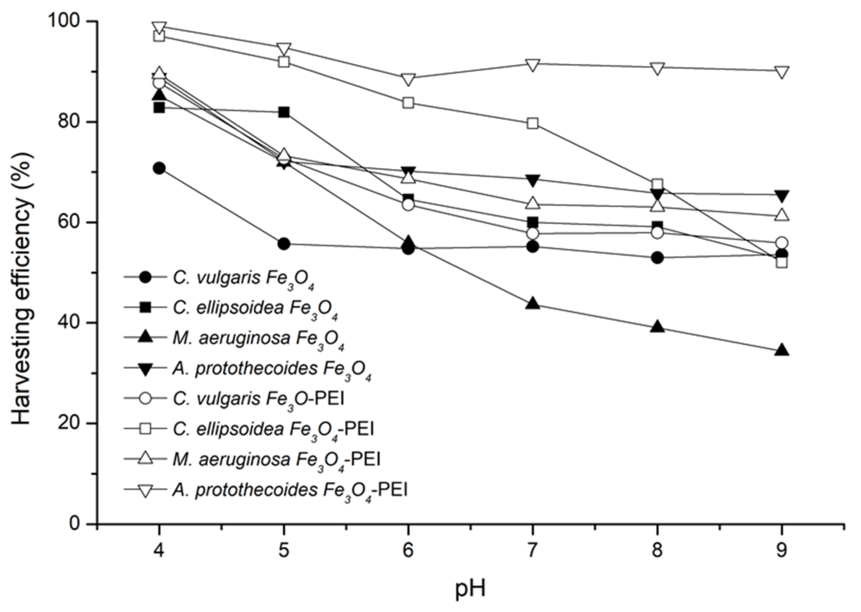
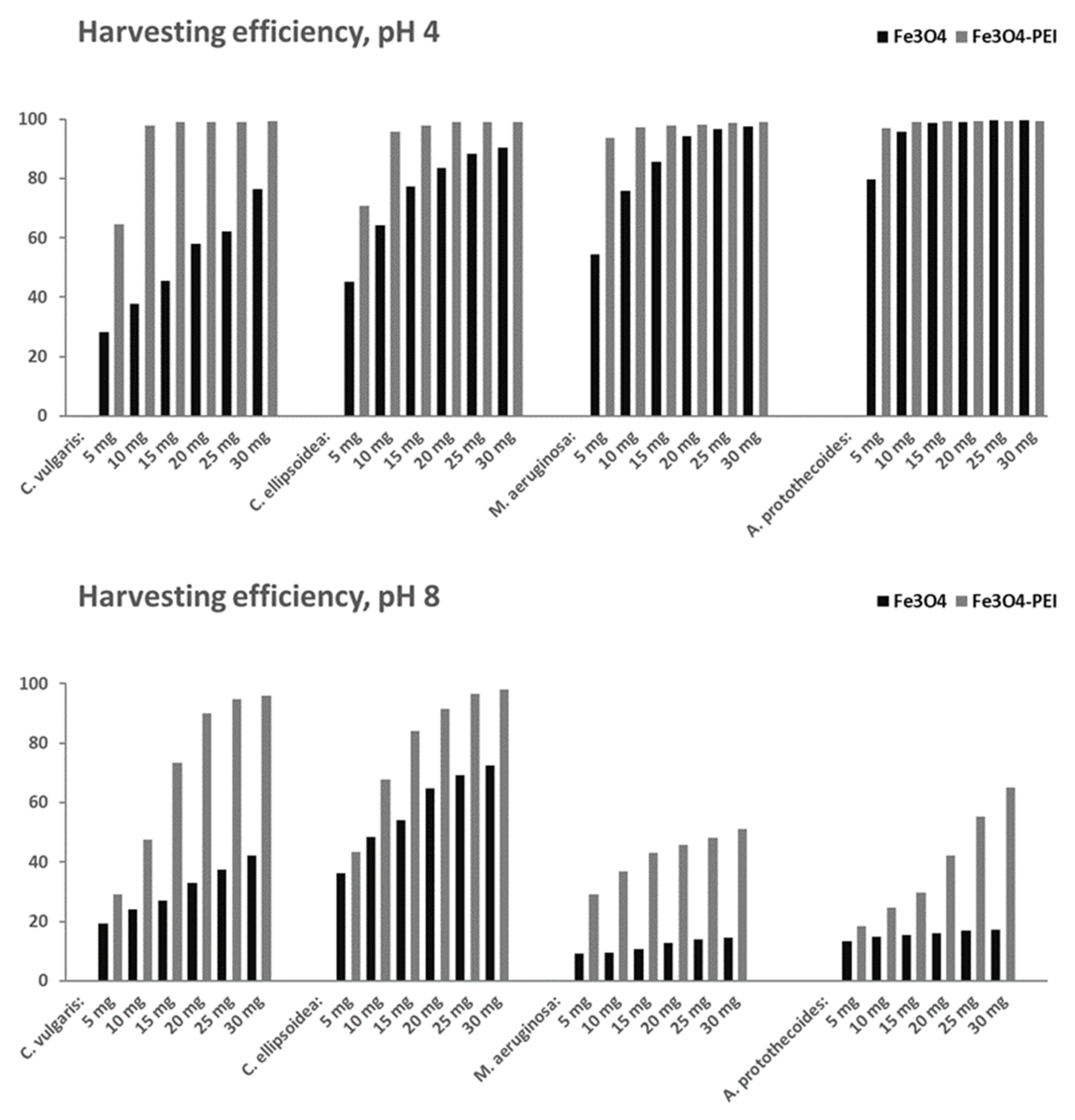
3.3. Magnetic Harvesting of Microalgae

Microalgae harvesting was studied at different pH levels (4–9) and different flocculant doses (5–30 mg). Harvesting efficiencies after 90 s of contact time are shown in Figure 7. Higher efficiencies, close to 100%, were obtained for PEI-coated Fe3O4 NPs. In the experiments, 10 mg of either NPs or NCs was used per testing bottle containing 50 mL of the algae suspension. The separation process was strongly pH-dependent. Harvesting efficiencies were found to decrease with increasing pH of the solution for uncoated and PEI-coated Fe3O4 NPs.
PEI-coated Fe3O4 NPs had higher harvesting efficiencies for all algae species tested within the investigated pH range. The harvesting efficiency of the uncoated magnetite at pH 8 was 39–53%, while for the PEI-coated magnetite it was 58–90%, respectively. Lowering the pH from 8.0 to 4.0 resulted in a significant increase in harvesting efficiencies. A. protothecoides and C. ellipsoidea reached harvesting efficiencies of 99% at pH 4 using PEI-coated Fe3O4 NPs as magnetic flocculants.
We also tested several different doses of uncoated magnetite and PEI-coated magnetite. In all cases, a higher dose resulted in higher efficiencies for both coated and uncoated magnetite. The results are presented in Figure 8. To reach a minimum efficiency of 90% at pH 4, 30 mg of Fe3O4 but less than 10 mg of Fe3O4-PEI were needed for C. ellipsoidea. At pH 8, 20 mg of Fe3O4-PEI caused a harvesting efficiency greater than 90%. The results again show that higher harvesting efficiencies were achieved in an acidic environment. To reach 98–99% harvesting efficiency, the optimal dosage at pH 4 is 10 mg of PEI-coated NP for C. vulgaris, C. ellipsoidea, M. aeruginosa, and A. protothecoides. For uncoated NPs, this level of harvesting efficiency was reached for only two algae species (C. ellipsoidea with the dose of 20 mg, and A. protothecoides with 15 mg). At pH 8, C. ellipsoidea reached 98% harvesting efficiency at the dose of PEI-coated NPs equal to 30 mg. This result shows that this algae species can be harvested in neutral conditions.
Harvesting efficiencies of the investigated green algae and cyanobacteria are compared in Table 2. Our results show comparable harvesting efficiencies to previously reported results [65,68,70,92,93]. The difference is minor and is attributable to either the utilization of non-specified Chlorella species, higher temperature during co-precipitation, higher pH of the solution (pH 7–8), or longer time used for the separation (up to 30 min). It can be observed that the decreased co-precipitation temperature causes a decrease in the harvesting efficiency. Hu et al. reached 97% of the harvest efficiency of C. vulgaris with PEI-coated NPs synthesized by the co-precipitation method at 80 °C with the dosage of 20 mg L−1 at pH 9 in 2 min [70]. In our study, 30 mg of PEI-coated NPs reached 96% of the harvesting efficiency at pH 8. On the other hand, only 80% of the harvesting efficiency with PEI-coated NPs was achieved in the study by Wang et al. [68] for Microcystis aeruginosa. In our study, we reached more than 93% of harvesting efficiencies with 5 mg of PEI-coated NPs. Therefore, it can be concluded that the harvesting efficiency for the PEI-coated magnetite NPs is comparable to previous studies.

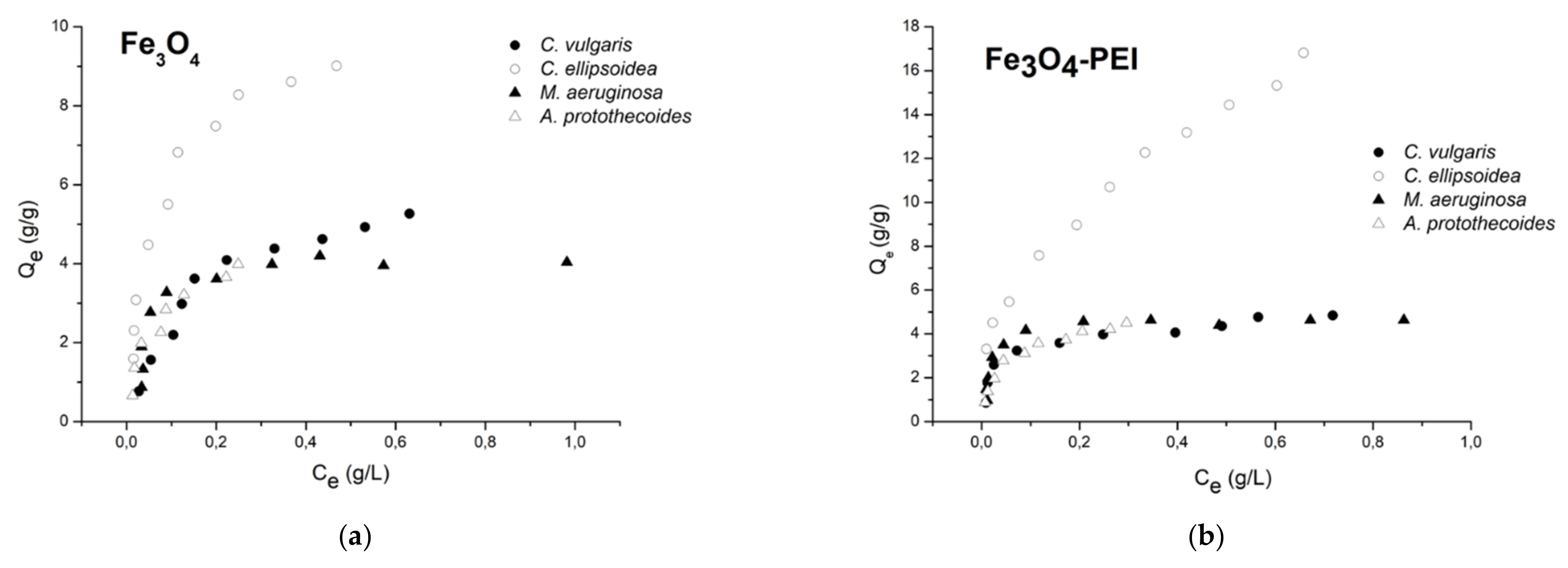
3.4. Adsorption Isotherms

A wide variety of adsorption isotherm models have been studied in the literature. The models can be classified as follows: (1) irreversible isotherms and one-parameter isotherms (e.g., Henry isotherm), (2) two-parameter isotherms (e.g., Langmuir, Freundlich, and Dubinin–Radushkevich, which are the most used), (3) three-parameter isotherms (e.g., Redlich–Peterson), and (4) more than three-parameter isotherms [94]. The adsorption isotherms illustrate the equilibrium relationship between the adsorption capacity (the equilibrium-adsorbed amounts) and the equilibrium concentration in the solution for a constant equilibrium pH and temperature of the solution [95]. In our work, the equilibrium between harvested microalgal cells and their concentration in the supernatant has been studied by the Langmuir and Freundlich isotherms. Experimental data for both uncoated and PEI-coated magnetite are displayed in Figure 9. Parameters estimated from linear and nonlinear Langmuir and Freundlich models are summarized in Table 3.
According to the data presented in Table 3, a better fit was found for the Langmuir than the Freundlich model, except for C. ellipsoidea and Fe3O4-PEI. This result agrees well with [35]. When comparing the utilization of linear and nonlinear models, we obtained better correlation in linear models for the Langmuir models than in the nonlinear models; however, when the Freundlich models were used, the nonlinear extrapolation was more accurate.
At pH 7, although the PEI coating brings an extra positive charge to the magnetite particles, the maximum adsorption capacity was lower. For example, for C. vulgaris, it was more than 26% lower (4.932 compared to 6.700 g g−1). The highest adsorption capacity was obtained in the case of coated magnetite and Chlorella ellipsoidea (18.612 g g−1), while the lowest adsorption capacity was obtained for noncoated magnetite and Microcystis aeruginosa (4.369 g g−1).
Studies of adsorption kinetics play an important role in identifying the required equilibration time, the optimal contact time, and the mechanism of the adsorption process [60]. The kinetic aspects of adsorption have not yet been studied in detail. However, it has been observed in our study that the sorption process of all algae species occurs rapidly, as the settling processes take only approximately 15–30 s.

3.5. Adsorption Mechanism

There exist four steps associated with materials’ transport during adsorption [94]. The first stage is solution phase transport, known as “bulk transport”. The bulk transport can occur instantaneously after the adsorbent is transported into the adsorbate solution. As such, its contribution to the overall rate of adsorption is negligible. The second stage is “film diffusion”. In the second stage, the adsorbate molecules are transferred from the bulk liquid phase to the adsorbent’s external surface through the hydrodynamic boundary layer or film. The third stage—interparticle diffusion—involves the diffusion of the molecules from the exterior into the pores of the adsorbent, along pore-wall surfaces, or both. The diffusion stage occurs slowly and may be rate-limiting. The last stage is an adsorptive attachment. It often occurs quickly, and therefore, it is not considered to be significant for the adsorption kinetics [94].
There also exist several possible mechanisms in algae harvesting with the uncoated and coated magnetite, including charge neutralization, patching, or adsorption bridging. In charge neutralization, the net charges of the microalgae particles are cancelled by adsorbing an equivalent number of opposite charges. An oppositely charged flocculant added into the culture medium increases the ionic strengths of the medium and the concentration of counter ions, but decreases the particle charges and the zeta potential. It allows the formation of van der Waals force of attraction to encourage initial aggregation. The electrostatic patch (patching) mechanism is the phenomenon in which a charged polymer binds to a particle with opposite charge. The polymer locally reverses the charge of the particle surface, resulting in patches of opposite charge on the particle surface. Patching occurs when unevenly distributed surface charges are incompletely neutralized. After that, particles may connect with each other through patches of opposite charge. In general, adsorption bridging occurs when long-chain polymers with high molecular weight and low charge density have been adsorbed on particles in such a way that long loops and tails extending or stretching into solution far beyond the electrical double-layer polymers or charged colloids simultaneously bind to the surface of two different particles to form a bridge between these particles [96,97,98,99,100,101,102].
The electrostatic interaction of algae and coated nanoparticles was important as the zeta potentials in the studied pH range were opposite. However, electrostatic forces may have not been sufficient when the non-coated magnetite was used, as the zeta potential of both species lay in the negative region (Figure 6). Savvidou et al. [86] suggested that Fe3O4 particles can be attached to microalgal cells by hydrogen bonding. Due to significant protonation of magnetite particles under acidic conditions, the chemical species of the hydrogen bond donor OH2+ can be formed in Fe3O4 and interact with the hydrogen bond acceptor groups present in C. vulgaris cells, such as amino or carboxy groups. The authors of [64] suggested that the principal mechanism of the algae harvesting process (C. vulgaris and tailor-made magnetic nanoparticles) was bridging. Furthermore, some authors [60] used an extended Derjaguin–Landau–Verwey–Overbeek (EDLVO) theory to demonstrate the contributions of the surface properties of membranes or flocculants to colloidal interactions. The EDLVO theory may be used to reveal the principles of interaction between the magnetic nanoparticles and algae cells. Although the electrostatic interaction is described by the net characteristic of all charged groups on the surface of microalgae cells and the magnetic surface of NP, it might appear in sophisticated bilayer microdomains [60]. This fact is confirmed by microscopy images of the system after adhesion, which show an agglomerated Fe3O4 structure that covers only a part of the microalgae cell wall and is heterogeneously distributed, leaving parts of the cell wall surface free (Figure 10). Furthermore, additional facts must be considered. The aggregation of superparamagnetic magnetite nanoparticles may also be affected by their magnetic properties, which are in competition with the repulsive forces of van der Waals and electrostatic interactions. The final agglomeration influences the mobility and reactivity of the nanoparticles and depends on several factors, that include the pH of the solution, the ionic strength, and the presence of organic matter. In biological experiments where nanoparticles are suspended in solution, the composition, density, viscosity, and physiochemical characteristics of the cell culture medium must also be considered, as they can interfere with the stability and aggregation of magnetic NPs [87,103].
The biochemical composition of the algae cell surface differs between species and is variable within a species, for example, exponential versus stationary phase cultures. Furthermore, microalgae often excrete significant amounts of organic matter, consisting of polysaccharides and proteins in growth medium, which can promote or inhibit floc formation [86]. Furthermore, chelating metal cations [104] can play an important role in the interaction with the flocculant as they are attached to the cell walls [97]. Another point is that algae are typically cultivated axenically only when storing the cultures, and afterward when the experiment is realized, cultivation is not sterile. The presence of bacteria that enter the system may also produce different kinds of extracellular polymeric substances, which can affect both the magnetic nanoparticles and the flocculation behavior of algae cells.

4. Conclusions

In the present work, magnetic harvesting of Chlorella vulgaris, Chlorella ellipsoidea, Microcystis aeruginosa, and Auxenochlorella protothecoides has been studied. Microalgae were obtained as sterile cultures from the algae collection of the University of Göttingen, Germany. The prepared microalgae were harvested with magnetite (Fe3O4) nanoparticles. The nanoparticles were prepared by controlled co-precipitation of Fe2+ and Fe3+ cations in ammonia at room temperature. Subsequently, the prepared Fe3O4 were coated with polyethyleneimine (PEI). The prepared materials were characterized by TEM, magnetometry, and zeta potential measurements. The following conclusions can be made:
  • The prepared NPs were spherical. The particle size distribution was relatively uniform and an average particle diameter of ~10 nm was found. The NPs prepared at 20 °C were smaller. However, the difference in nanoparticle diameter between materials prepared at 20 and 80 °C was not significant. The crystal structure of magnetite was confirmed by electron diffraction.
  • The zeta potential of the uncoated nanoparticles was negative within the investigated pH range (4–9). The zeta potentials of the PEI-coated nanoparticles were positive at pH 4–9. The presence of PEI thus brings a positive charge to magnetite.
  • The algae species displayed a negative zeta potential within the investigated pH range. Negative values decreased as pH increased due to deprotonation.
  • Microalgae harvesting was studied at different pH levels and different flocculant doses. Higher efficiencies, close to 100%, were obtained for PEI-coated Fe3O4 NPs.
  • The adsorption of magnetic flocculants on harvested microalgal cells has been studied by Langmuir and Freundlich isotherms. A better fit was found for the Langmuir isotherm, indicating a monolayer adsorption.
The results show that the synthesis of magnetic nanoparticles at 20 °C is feasible. The nanoparticles have comparable characteristics (particle size, microstructure, harvesting efficiency) to NPs prepared at 80 °C. The harvesting efficiencies can be significantly enhanced by PEI coating, as the polymer increases the positive electrical charge of the nanoparticles. High efficiencies, close to 100%, were obtained for PEI-coated Fe3O4 NPs at pH 4. Relatively high efficiencies can be obtained at pH 8, which makes the separation process feasible in neutral conditions. Furthermore, the magnetic harvesting with the nanoparticles synthesized at room temperature is applicable to both green algae and cyanobacteria, making the process attractive for industrial use.

Author Contributions

Conceptualization, K.G., A.K. and M.P.; methodology, K.G.; validation, A.K., K.G. and Z.S.; formal analysis, K.G. and A.K.; investigation, K.G., A.K., Z.S., E.S., Z.G. and M.Č.; resources, M.P. and Ľ.Č.; data curation, K.G., A.K., Z.S., Z.G., E.S., M.Č. and M.P.; writing—original draft preparation, K.G. and A.K.; writing—review and editing, M.P., Z.S., Z.G., E.S., M.Č. and Ľ.Č.; supervision, K.G., M.P. and Ľ.Č.; project administration, M.P. and Ľ.Č.; funding acquisition, Ľ.Č., M.Č., Z.G. and M.P. All authors have read and agreed to the published version of the manuscript.

Funding

This work was supported by the Slovak Research and Development Agency, project No. APVV-20-0124, Grant Agency VEGA of the Ministry of Education, Science, Research and Sport of the Slovak Republic, project No. VEGA 1/0756/20, and Grant Agency KEGA of the Ministry of Education, Science, Research and Sport of the Slovak Republic, project No. 020STU-4/2021. This publication was created on the basis of the major project “Advancing University Capacity and Competence in Research, Development and Innovation” (ITMS project code: 313021X329), supported by the Operational Programme Integrated Infrastructure and funded by the European Regional Development Fund. This publication was also supported by the Operational Programme Integrated Infrastructure for the project: “Scientific and Research Centre of Excellence SlovakION for Material and Interdisciplinary Research”, code of the project: ITMS2014+: 313011W085, co-financed by the European Regional Development Fund.

Data Availability Statement

Data are available from the corresponding author upon request.

Acknowledgments

Martin Kusý is acknowledged for his assistance with optical microscopy of algal cells.

Conflicts of Interest

The authors declare no conflict of interest.

References

  1. Seo, J.Y.; Kim, M.G.; Lee, K.; Lee, Y.C.; Na, J.G.; Jeon, S.G.; Park, S.B.; Oh, Y.K. Multifunctional Nanoparticle Applications to Microalgal Biorefinery. In Nanotechnology for Bioenergy and Biofuel Production, 2nd ed.; Rai, M., Silva, S.S.D., Eds.; Springer: Cham, Switzerland, 2017; pp. 59–87. [Google Scholar] [CrossRef]
  2. Gangl, D.; Zedler, J.A.Z.; Rajakumar, P.D.; Martinez, E.M.R.; Riseley, A.; Włodarczyk, A.; Purton, S.; Sakuragi, Y.; Howe, C.J.; Jensen, P.E.; et al. Biotechnological exploitation of microalgae. J. Exp. Bot. 2015, 66, 6975–6990. [Google Scholar] [CrossRef] [PubMed] [Green Version]
  3. Chhandama, M.V.L.; Satyan, K.B.; Changmai, B.; Vanlalveni, C.; Rokhum, S.L. Microalgae as a feedstock for the production of biodiesel: A review. Bioresour. Technol. Rep. 2021, 15, 100771. [Google Scholar] [CrossRef]
  4. Barkia, I.; Saari, N.; Manning, S.R. Microalgae for High-Value Products Towards Human Health and Nutrition. Mar. Drugs 2019, 17, 304. [Google Scholar] [CrossRef] [PubMed] [Green Version]
  5. Alalwan, H.A.; Alminshid, A.; Aljaafari, H.A. Promising evolution of biofuel generations. Subject review. Renew. Energy Focus 2019, 28, 127–139. [Google Scholar] [CrossRef]
  6. Winckelmann, D.; Bleeke, F.; Thomas, B.; Elle, C.; Klöck, G. Open pond cultures of indigenous algae grown on non-arable land in an arid desert using wastewater. Int. Aquat. Res. 2015, 7, 221–233. [Google Scholar] [CrossRef] [Green Version]
  7. Correa, D.F.; Beyer, H.L.; Possingham, H.P.; García-Ulloa, J.; Ghazoul, J.; Schenk, P.M. Freeing land from biofuel production through microalgal cultivation in the Neotropical region. Environ. Res. Lett. 2020, 15, 94094. [Google Scholar] [CrossRef]
  8. Onyeaka, H.; Miri, T.; Obileke, K.; Hart, A.; Anumudu, C.; Al-Sharify, Z.T. Minimizing carbon footprint via microalgae as a biological capture. Carbon Capture Sci. Technol. 2021, 1, 100007. [Google Scholar] [CrossRef]
  9. Prasad, R.; Gupta, S.K.; Shabnam, N.; Oliveira, C.Y.B.; Nema, A.K.; Ansari, F.A.; Bux, F. Role of Microalgae in Global CO2 Sequestration: Physiological Mechanism, Recent Development, Challenges, and Future Prospective. Sustainability 2021, 13, 13061. [Google Scholar] [CrossRef]
  10. Plöhn, M.; Spain, O.; Sirin, S.; Silva, M.; Escudero-Oñate, C.; Ferrando-Climent, L.; Allahverdiyeva, Y.; Funk, C. Wastewater treatment by microalgae. Physiol. Plant. 2021, 173, 568–578. [Google Scholar] [CrossRef]
  11. Wang, Y.; Ho, S.-H.; Cheng, C.-L.; Guo, W.-Q.; Nagarajan, D.; Ren, N.-Q.; Lee, D.-J.; Chang, J.-S. Perspectives on the feasibility of using microalgae for industrial wastewater treatment. Bioresour. Technol. 2016, 222, 485–497. [Google Scholar] [CrossRef]
  12. Udayan, A.; Pandey, A.K.; Sirohi, R.; Sreekumar, N.; Sang, B.-I.; Sim, S.J.; Kim, S.H.; Pandey, A. Production of microalgae with high lipid content and their potential as sources of nutraceuticals. Phytochem. Rev. 2022, 21, 1–28. [Google Scholar] [CrossRef]
  13. Sun, X.-M.; Ren, L.-J.; Zhao, Q.-Y.; Ji, X.-J.; Huang, H. Microalgae for the production of lipid and carotenoids: A review with focus on stress regulation and adaptation. Biotechnol. Biofuels 2018, 11, 272. [Google Scholar] [CrossRef] [Green Version]
  14. Tang, Y.; Rosenberg, J.N.; Bohutskyi, P.; Yu, G.; Betenbaugh, M.J.; Wang, F. Microalgae as a Feedstock for Biofuel Precursors and Value-Added Products: Green Fuels and Golden Opportunities. BioResources 2016, 11, 2850–2885. [Google Scholar] [CrossRef] [Green Version]
  15. Bošnjaković, M.; Sinaga, N. The Perspective of Large-Scale Production of Algae Biodiesel. Appl. Sci. 2020, 10, 8181. [Google Scholar] [CrossRef]
  16. Ferreira, G.F.; Pinto, L.F.R.; Filho, R.M.; Fregolente, L.V. A review on lipid production from microalgae: Association between cultivation using waste streams and fatty acid profiles. Renew. Sustain. Energy Rev. 2019, 109, 448–466. [Google Scholar] [CrossRef]
  17. Chisti, Y. Biodiesel from microalgae. Biotechnol. Adv. 2007, 25, 294–306. [Google Scholar] [CrossRef]
  18. Almanza, V.; Parra, O.; Bicudo, C.E.D.M.; Baeza, C.; Beltran, J.; Figueroa, R.; Urrutia, R. Occurrence of toxic blooms of Microcystis aeruginosa in a central Chilean (36° Lat. S) urban lake. Rev. Chil. Hist. Nat. 2016, 89, 1349. [Google Scholar] [CrossRef] [Green Version]
  19. Chen, M.; Tian, L.-L.; Ren, C.-Y.; Xu, C.-Y.; Wang, Y.-Y.; Li, L. Extracellular polysaccharide synthesis in a bloom-forming strain of Microcystis aeruginosa: Implications for colonization and buoyancy. Sci. Rep. 2019, 9, 1251. [Google Scholar] [CrossRef] [Green Version]
  20. Abed, R.; Dobretsov, S.; Sudesh, K. Applications of cyanobacteria in biotechnology. J. Appl. Microbiol. 2009, 106, 1–12. [Google Scholar] [CrossRef]
  21. Rós, P.D.; Silva, C.S.; Stenico, M.E.; Fiore, M.; Castro, H.F.D. Microcystis aeruginosa lipids as feedstock for biodiesel synthesis by enzymatic route. J. Mol. Catal. B Enzym. 2012, 84, 177–182. [Google Scholar] [CrossRef]
  22. Upendar, G.; Singh, S.; Chakrabarty, J.; Ghanta, K.C.; Dutta, S.; Dutta, A. Sequestration of carbon dioxide and production of biomolecules using cyanobacteria. J. Environ. Manag. 2018, 218, 234–244. [Google Scholar] [CrossRef] [PubMed]
  23. Ataeian, M.; Liu, Y.; Canon-Rubio, K.A.; Nightingale, M.; Strous, M.; Vadlamani, A. Direct capture and conversion of CO2 from air by growing a cyanobacterial consortium at pH up to 11.2. Biotechnol. Bioeng. 2019, 116, 1604–1611. [Google Scholar] [CrossRef] [PubMed] [Green Version]
  24. Vasistha, S.; Khanra, A.; Clifford, M.; Rai, M. Current advances in microalgae harvesting and lipid extraction processes for improved biodiesel production: A review. Renew. Sustain. Energy Rev. 2020, 137, 110498. [Google Scholar] [CrossRef]
  25. Branyikova, I.; Prochazkova, G.; Potocar, T.; Jezkova, Z.; Branyik, T. Harvesting of Microalgae by Flocculation. Fermentation 2018, 4, 93. [Google Scholar] [CrossRef] [Green Version]
  26. Bajpai, P. Harvesting and Drying of Algal Biomass. In Third Generation Biofuels; Springer: Singapore, 2019; pp. 29–36. [Google Scholar]
  27. Fasaei, F.; Bitter, J.; Slegers, P.; Boxtel, A.V. Techno-economic evaluation of microalgae harvesting and dewatering systems. Algal Res. 2018, 31, 347–362. [Google Scholar] [CrossRef]
  28. Suparmaniam, U.; Lam, M.K.; Uemura, Y.; Lim, J.W.; Lee, K.T.; Shuit, S.H. Insights into the microalgae cultivation technology and harvesting process for biofuel production: A review. Renew. Sustain. Energy Rev. 2019, 115, 109361. [Google Scholar] [CrossRef]
  29. Rawat, I.; Ranjith Kumar, R.; Mutanda, T.; Bux, F. Biodiesel from microalgae: A critical evaluation from laboratory to large scale production. Appl. Energy 2013, 103, 444–467. [Google Scholar] [CrossRef]
  30. Enamala, M.K.; Enamala, S.; Chavali, M.; Donepudi, J.; Yadavalli, R.; Kolapalli, B.; Aradhyula, T.V.; Velpuri, J.; Kuppam, C. Production of biofuels from microalgae—A review on cultivation, harvesting, lipid extraction, and numerous applications of microalgae. Renew. Sustain. Energy Rev. 2018, 94, 49–68. [Google Scholar] [CrossRef]
  31. Tran, D.-T.; Le, B.-H.; Lee, D.-J.; Chen, C.-L.; Wang, H.-Y.; Chang, J.-S. Microalgae harvesting and subsequent biodiesel conversion. Bioresour. Technol. 2013, 140, 179–186. [Google Scholar] [CrossRef]
  32. Brennan, L.; Owende, P. Biofuels from microalgae—A review of technologies for production, processing, and extractions of biofuels and co-products. Renew. Sustain. Energy Rev. 2010, 14, 557–577. [Google Scholar] [CrossRef]
  33. Wang, S.-K.; Stiles, A.R.; Guo, C.; Liu, C.-Z. Harvesting microalgae by magnetic separation: A review. Algal Res. 2015, 9, 178–185. [Google Scholar] [CrossRef]
  34. Japar, A.S.; Takriff, M.S.; Yasin, N.H.M. Harvesting microalgal biomass and lipid extraction for potential biofuel production: A review. J. Environ. Chem. Eng. 2017, 5, 555–563. [Google Scholar] [CrossRef]
  35. Singh, G.; Patidar, S.K. Microalgae harvesting techniques: A review. J. Environ. Manag. 2018, 217, 499–508. [Google Scholar] [CrossRef]
  36. Wang, F.; Guan, W.; Xu, L.; Ding, Z.; Ma, H.; Ma, A.; Terry, N. Effects of Nanoparticles on Algae: Adsorption, Distribution, Ecotoxicity and Fate. Appl. Sci. 2019, 9, 1534. [Google Scholar] [CrossRef] [Green Version]
  37. Nguyen, M.K.; Moon, J.Y.; Bui, V.K.H.; Oh, Y.K.; Lee, Y.C. Recent advanced applications of nanomaterials in microalgae biorefinery. Algal Res. 2019, 41, 101522. [Google Scholar] [CrossRef]
  38. Kudr, J.; Haddad, Y.; Richtera, L.; Heger, Z.; Cernak, M.; Adam, V.; Zitka, O. Magnetic Nanoparticles: From Design and Synthesis to Real World Applications. Nanomaterials 2017, 7, 243. [Google Scholar] [CrossRef]
  39. Nguyen, M.D.; Tran, H.-V.; Xu, S.; Lee, T.R. Fe3O4 Nanoparticles: Structures, Synthesis, Magnetic Properties, Surface Functionalization, and Emerging Applications. Appl. Sci. 2021, 11, 11301. [Google Scholar] [CrossRef]
  40. Hao, J.J.; Chen, H.L.; Ren, C.L.; Yan, N.; Geng, H.J.; Chen, X.G. Synthesis of superparamagnetic Fe3O4 nanocrystals in reverse microemulsion at room temperature. Mater. Res. Innov. 2010, 14, 324–326. [Google Scholar] [CrossRef]
  41. Liang, X.; Jia, X.; Cao, L.; Sun, J.; Yang, Y. Microemulsion Synthesis and Characterization of Nano-Fe3O4 Particles and Fe3O4 Nanocrystalline. J. Dispers. Sci. Technol. 2010, 31, 1043–1049. [Google Scholar] [CrossRef]
  42. Ahmadi, S.; Chia, C.-H.; Zakaria, S.; Saeedfar, K.; Asim, N. Synthesis of Fe3O4 nanocrystals using hydrothermal approach. J. Magn. Magn. Mater. 2012, 324, 4147–4150. [Google Scholar] [CrossRef]
  43. Wu, X.; Tang, J.; Zhang, Y.; Wang, H. Low temperature synthesis of Fe3O4 nanocrystals by hydrothermal decomposition of a metallorganic molecular precursor. Mater. Sci. Eng. B 2009, 157, 81–86. [Google Scholar] [CrossRef]
  44. Woo, S.; Kim, S.; Kim, H.; Cheon, Y.W.; Yoon, S.; Oh, J.-H.; Park, J. Charge-Modulated Synthesis of Highly Stable Iron Oxide Nanoparticles for In Vitro and In Vivo Toxicity Evaluation. Nanomaterials 2021, 11, 3068. [Google Scholar] [CrossRef] [PubMed]
  45. Park, J.; An, K.; Hwang, Y.; Park, J.-G.; Noh, H.-J.; Kim, J.-Y.; Park, J.-H.; Hwang, N.-M.; Hyeon, T. Ultra-large-scale syntheses of monodisperse nanocrystals. Nat. Mater. 2004, 3, 891–895. [Google Scholar] [CrossRef] [PubMed]
  46. Spivakov, A.; Lin, C.-R.; Chang, Y.-C.; Wang, C.-C.; Sarychev, D. Magnetic and Magneto-Optical Oroperties of Iron Oxides Nanoparticles Synthesized under Atmospheric Pressure. Nanomaterials 2020, 10, 1888. [Google Scholar] [CrossRef]
  47. Serga, V.; Burve, R.; Maiorov, M.; Krumina, A.; Skaudžius, R.; Zarkov, A.; Kareiva, A.; Popov, A.I. Impact of Gadolinium on the Structure and Magnetic Properties of Nanocrystalline Powders of Iron Oxides Produced by the Extraction-Pyrolytic Method. Materials 2020, 13, 4147. [Google Scholar] [CrossRef]
  48. Xu, J.; Yang, H.; Fu, W.; Du, K.; Sui, Y.; Chen, J.; Zeng, Y.; Li, M.; Zou, G. Preparation and magnetic properties of magnetite nanoparticles by sol–gel method. J. Magn. Magn. Mater. 2007, 309, 307–311. [Google Scholar] [CrossRef]
  49. Takai, Z.I.; Mustafa, M.K.; Asman, S.; Sekak, K.A. Preparation and characterization of magnetite (Fe3O4) nanoparticles by sol-gel method. Int. J. Nanoelectron. Mater. 2019, 12, 37–46. [Google Scholar]
  50. Gholizadeh, A. A comparative study of physical properties in Fe3O4 nanoparticles prepared by coprecipitation and citrate methods. J. Am. Ceram. Soc. 2017, 100, 3577–3588. [Google Scholar] [CrossRef]
  51. Arévalo, P.; Isasi, J.; Caballero, A.C.; Marco, J.; Hernandez, F.M. Magnetic and structural studies of Fe3O4 nanoparticles synthesized via coprecipitation and dispersed in different surfactants. Ceram. Int. 2017, 43, 10333–10340. [Google Scholar] [CrossRef]
  52. Majidi, S.; Sehrig, F.Z.; Farkhani, S.M.; Goloujeh, M.S.; Akbarzadeh, A. Current methods for synthesis of magnetic nanoparticles. Artif. Cells Nanomed. Biotechnol. 2016, 44, 722–734. [Google Scholar] [CrossRef]
  53. Houshiar, M.; Zebhi, F.; Razi, Z.J.; Alidoust, A.; Askari, Z. Synthesis of cobalt ferrite (CoFe2O4) nanoparticles using combustion, coprecipitation, and precipitation methods: A comparison study of size, structural, and magnetic properties. J. Magn. Magn. Mater. 2014, 371, 43–48. [Google Scholar] [CrossRef]
  54. Maaz, K.; Karim, S.; Mumtaz, A.; Hasanain, S.; Liu, J.; Duan, J. Synthesis and magnetic characterization of nickel ferrite nanoparticles prepared by co-precipitation route. J. Magn. Magn. Mater. 2008, 321, 1838–1842. [Google Scholar] [CrossRef]
  55. Borlido, L.; Azevedo, A.M.; Roque, A.; Aires-Barros, M. Magnetic separations in biotechnology. Biotechnol. Adv. 2013, 31, 1374–1385. [Google Scholar] [CrossRef]
  56. Eskandari, M.J.; Hasanzadeh, I. Size-controlled synthesis of Fe3O4 magnetic nanoparticles via an alternating magnetic field and ultrasonic-assisted chemical co-precipitation. Mater. Sci. Eng. B 2021, 266, 115050. [Google Scholar] [CrossRef]
  57. Shen, L.; Qiao, Y.; Guo, Y.; Meng, S.; Yang, G.; Wu, M.; Zhao, J. Facile co-precipitation synthesis of shape-controlled magnetite nanoparticles. Ceram. Int. 2014, 40, 1519–1524. [Google Scholar] [CrossRef]
  58. Ganapathe, L.S.; Mohamed, M.A.; Mohamad Yunus, R.; Berhanuddin, D.D. Magnetite (Fe3O4) Nanoparticles in Biomedical Application: From Synthesis to Surface Functionalisation. Magnetochemistry 2020, 6, 68. [Google Scholar] [CrossRef]
  59. Bharte, S.; Desai, K. Harvesting Chlorella species using magnetic iron oxide nanoparticles. Phycol. Res. 2018, 67, 128–133. [Google Scholar] [CrossRef]
  60. Fu, Y.; Hu, F.; Li, H.; Cui, L.; Qian, G.; Zhang, D.; Xu, Y. Application and mechanisms of microalgae harvesting by magnetic nanoparticles (MNPs). Sep. Purif. Technol. 2021, 265, 118519. [Google Scholar] [CrossRef]
  61. Egesa, D.; Chuck, C.J.; Plucinski, P. Multifunctional Role of Magnetic Nanoparticles in Efficient Microalgae Separation and Catalytic Hydrothermal Liquefaction. ACS Sustain. Chem. Eng. 2018, 6, 991–999. [Google Scholar] [CrossRef]
  62. Zhu, L.D.; Hiltunen, E.; Li, Z. Using magnetic materials to harvest microalgal biomass: Evaluation of harvesting and detachment efficiency. Environ. Technol. 2019, 40, 1006–1012. [Google Scholar] [CrossRef]
  63. Liu, P.; Wang, T.; Yang, Z.; Hong, Y.; Xie, X.; Hou, Y. Effects of Fe3O4 nanoparticle fabrication and surface modification on Chlorella sp. harvesting efficiency. Sci. Total Environ. 2020, 704, 135286. [Google Scholar] [CrossRef] [PubMed]
  64. Almomani, F. Algal cells harvesting using cost-effective magnetic nano-particles. Sci. Total Environ. 2020, 720, 137621. [Google Scholar] [CrossRef] [PubMed]
  65. Lin, Z.; Xu, Y.; Zhen, Z.; Fu, Y.; Liu, Y.; Li, W.; Luo, C.; Ding, A.; Zhang, D. Application, and reactivation of magnetic nanoparticles in Microcystis aeruginosa harvesting. Bioresour. Technol. 2015, 190, 82–88. [Google Scholar] [CrossRef] [PubMed]
  66. Barizão, A.C.D.L.; Oliveira, J.P.D.; Gonçalves, R.F.; Cassini, S.T. Nanomagnetic approach applied to microalgae biomass harvesting: Advances, gaps, and perspectives. Environ. Sci. Pollut. Res. 2021, 28, 44795–44811. [Google Scholar] [CrossRef]
  67. Ge, S.; Agbakpe, M.; Zhang, W.; Kuang, L. Heteroaggregation between PEI-coated magnetic nanoparticles and algae: Effect of particle size on algal harvesting efficiency. ACS Appl. Mater. Interfaces 2015, 7, 6102–6108. [Google Scholar] [CrossRef]
  68. Wang, C.; Yang, Y.; Hou, J.; Wang, P.; Miao, L.; Wang, X. Optimization of cyanobacterial harvesting and extracellular organic matter removal utilizing magnetic nanoparticles and response surface methodology: A comparative study. Algal Res. 2020, 45, 101756. [Google Scholar] [CrossRef]
  69. Yang, Y.; Hou, J.; Wang, P.; Wang, C.; Miao, L.; Ao, Y.; Xu, Y.; Wang, X.; Lv, B.; You, G.; et al. Interpretation of the disparity in harvesting efficiency of different types of Microcystis aeruginosa using polyethylenimine (PEI)-coated magnetic nanoparticles. Algal Res. 2018, 29, 257–265. [Google Scholar] [CrossRef]
  70. Hu, Y.R.; Guo, C.; Wang, F.; Wang, S.K.; Pan, F.; Liu, C.Z. Improvement of microalgae harvesting by magnetic nanocomposites coated with polyethylenimine. Chem. Eng. J. 2014, 242, 341–347. [Google Scholar] [CrossRef]
  71. Sánchez-Bayo, A.; Morales, V.; Rodríguez, R.; Vicente, G.; Bautista, L.F. Cultivation of Microalgae and Cyanobacteria: Effect of Operating Conditions on Growth and Biomass Composition. Molecules 2020, 25, 2834. [Google Scholar] [CrossRef]
  72. Hu, Y.-R.; Wang, F.; Wang, S.-K.; Liu, C.-Z.; Guo, C. Efficient harvesting of marine microalgae Nannochloropsis maritima using magnetic nanoparticles. Bioresour. Technol. 2013, 138, 387–390. [Google Scholar] [CrossRef]
  73. Aratboni, H.A.; Rafiei, N.; Garcia-Granados, R.; Alemzadeh, A.; Morones-Ramírez, J.R. Biomass and lipid induction strategies in microalgae for biofuel production and other applications. Microb. Cell Fact. 2019, 18, 178. [Google Scholar] [CrossRef] [Green Version]
  74. Shaoxian, S.; Huang, R.; Li, Y.; Song, S. The effect of growth phase on the surface properties of three oleaginous microalgae (Botryococcus sp. FACGB-762, Chlorella sp. XJ-445 and Desmodesmus bijugatus XJ-231). PLoS ONE 2017, 12, e0186434. [Google Scholar] [CrossRef]
  75. Yang, Y.; Fan, X.; Zhang, J.; Qiao, S.; Wang, X.; Zhang, X.; Miao, L.; Hou, J. A critical review on the interaction of iron-based nanoparticles with blue-green algae and their metabolites: From mechanisms to applications. Algal Res. 2022, 64, 102670. [Google Scholar] [CrossRef]
  76. Xu, Y.; Wang, X.; Fu, Y.; Hu, F.; Qian, G.; Liu, Q.; Sun, Y. Interaction energy and detachment of magnetic nanoparticles-algae. Environ. Technol. 2019, 41, 2618–2624. [Google Scholar] [CrossRef]
  77. Gerulová, K.; Bartošová, A.; Blinová, L.; Bártová, K.; Dománková, M.; Garaiová, Z.; Palcut, M. Magnetic Fe3O4-polyethyleneimine nanocomposites for efficient harvesting of Chlorella zofingiensis, Chlorella vulgaris, Chlorella sorokiniana, Chlorella ellipsoidea and Botryococcus braunii. Algal Res. 2018, 33, 165–172. [Google Scholar] [CrossRef]
  78. Martínez-Mera, I.; Espinosa-Pesqueira, M.E.; Pérez-Hernández, R.; Arenas-Alatorre, J. Synthesis of magnetite (Fe3O4) nanoparticles without surfactants at room temperature. Materials Letters 2007, 61, 4447–4451. [Google Scholar] [CrossRef]
  79. Saragi, T.; Depi, B.L.; Butarbutar, S.; Permana, B.; Risdiana. The impact of synthesis temperature on magnetite nanoparticles size synthesized by co-precipitation method. J. Phys. Conf. Ser. 2018, 1013, 12190. [Google Scholar] [CrossRef]
  80. Cai, H.; An, X.; Cui, J.; Li, J.; Wen, S.; Li, K.; Shen, M.; Zheng, L.; Zhang, G.; Shi, X. Facile Hydrothermal Synthesis and Surface Functionalization of Polyethyleneimine-Coated Iron Oxide Nanoparticles for Biomedical Applications. ACS Appl. Mater. Interfaces 2013, 5, 1722–1731. [Google Scholar] [CrossRef]
  81. Wang, Y.; Xu, F.; Zhang, L.; Wei, X. One-pot solvothermal synthesis of Fe3O4–PEI composite and its further modification with Au nanoparticles. J. Nanoparticle Res. 2012, 15, 1338. [Google Scholar] [CrossRef]
  82. Félix, L.; Martínez, M.A.R.; Salazar, D.G.P.; Coaquira, J.A.H. One-step synthesis of polyethyleneimine-coated magnetite nanoparticles and their structural, magnetic, and power absorption study. RSC Adv. 2020, 10, 41807–41815. [Google Scholar] [CrossRef]
  83. Zhang, J.; Lin, S.; Han, M.; Su, Q.; Xia, L.; Hui, Z. Adsorption Properties of Magnetic Magnetite Nanoparticle for Coexistent Cr(VI) and Cu(II) in Mixed Solution. Water 2020, 12, 446. [Google Scholar] [CrossRef] [Green Version]
  84. Plaza, R.C.; Arias, J.L.; Espín, M.; Jiménez, M.L.; Delgado, A.V. Aging Effects in the Electrokinetics of Colloidal Iron Oxides. J. Colloid Interface Sci. 2002, 245, 86–90. [Google Scholar] [CrossRef] [PubMed] [Green Version]
  85. Kim, M.; Jung, J.; Lee, J.; Na, K.; Park, S.; Hyun, J. Amphiphilic comb like polymers enhance the colloidal stability of Fe3O4 nanoparticles. Colloids Surf. B Biointerfaces 2010, 76, 236–240. [Google Scholar] [CrossRef] [PubMed]
  86. Savvidou, M.G.; Dardavila, M.M.; Georgiopoulou, I.; Louli, V.; Stamatis, H.; Kekos, D.; Voutsas, E. Optimization of Microalga Chlorella vulgaris Magnetic Harvesting. Nanomaterials 2021, 11, 1614. [Google Scholar] [CrossRef]
  87. Baldassarre, F.; Cacciola, M.; Ciccarella, G. A predictive model of iron oxide nanoparticles flocculation tuning Z-potential in aqueous environment for biological application. J. Nanoparticle Res. 2015, 17, 377. [Google Scholar] [CrossRef]
  88. Wang, N.; Hsu, C.; Zhu, L.; Tseng, S.; Hsu, J.P. Influence of metal oxide nanoparticles concentration on their zeta potential. J. Colloid Interface Sci. 2013, 407, 22–28. [Google Scholar] [CrossRef]
  89. Soares, S.F.; Fernandes, T.; Trindade, T.; Daniel-Da-Silva, A.L. Trimethyl Chitosan/Siloxane-Hybrid Coated Fe3O4 Nanoparticles for the Uptake of Sulfamethoxazole from Water. Molecules 2019, 24, 1958. [Google Scholar] [CrossRef] [Green Version]
  90. Petcharoen, K.; Sirivat, A. Synthesis and characterization of magnetite nanoparticles via the chemical co-precipitation method. Mater. Sci. Eng. B 2012, 117, 421–427. [Google Scholar] [CrossRef]
  91. Sun, Z.-X.; Su, F.-W.; Forsling, W.; Samskog, P.-O. Surface Characteristics of Magnetite in Aqueous Suspension. J. Colloid Interface Sci. 1998, 197, 151–159. [Google Scholar] [CrossRef]
  92. Xu, L.; Guo, C.; Wang, F.; Zheng, S.; Liu, C.Z. A simple and rapid harvesting method for microalgae by in situ magnetic separation. Bioresour. Technol. 2011, 102, 10047–10051. [Google Scholar] [CrossRef]
  93. Fraga-García, P.; Kubbutat, P.; Brammen, M.; Schwaminger, S.; Berensmeier, S. Bare Iron Oxide Nanoparticles for Magnetic Harvesting of Microalgae: From Interaction Behavior to Process Realization. Nanomaterials 2018, 8, 292. [Google Scholar] [CrossRef] [Green Version]
  94. Tran, H.N.; You, S.J.; Hosseini-Bandegharaei, A.; Chao, H.P. Mistakes, and inconsistencies regarding adsorption of contaminants from aqueous solutions: A critical review. Water Res. 2017, 120, 88–116. [Google Scholar] [CrossRef]
  95. Yin, Z.; Zhu, L.; Li, S.; Hu, T.; Chu, R.; Mo, F.; Hu, D.; Liu, C.; Li, B. A comprehensive review on cultivation and harvesting of microalgae for biodiesel production: Environmental pollution control and future directions. Bioresour. Technol. 2020, 301, 122804. [Google Scholar] [CrossRef]
  96. Yin, Z.; Zhang, L.; Hu, D.; Li, S.; Chu, R.; Liu, C.; Lv, Y.; Bao, J.; Xiang, M.; Zhu, L. Biocompatible magnetic flocculant for efficient harvesting of microalgal cells: Isotherms, mechanisms and water recycling. Sep. Purif. Technol. 2021, 279, 119679. [Google Scholar] [CrossRef]
  97. Vandamme, D.; Foubert, I.; Muylaert, K. Flocculation as a low-cost method for harvesting microalgae for bulk biomass production. Trends Biotechnol. 2013, 31, 233–239. [Google Scholar] [CrossRef] [Green Version]
  98. Chen, C.-Y.; Yeh, K.-L.; Aisyah, R.; Lee, D.-J.; Chang, J.-S. Cultivation, photobioreactor design and harvesting of microalgae for biodiesel production: A critical review. Bioresour. Technol. 2011, 102, 71–81. [Google Scholar] [CrossRef]
  99. Maćczak, P.; Kaczmarek, H.; Ziegler-Borowska, M. Recent Achievements in Polymer Bio-Based Flocculants for Water Treatment. Materials 2020, 13, 3951. [Google Scholar] [CrossRef]
  100. Lee, C.S.; Robinson, J.; Chong, M.F. A review on application of flocculants in wastewater treatment. Process Saf. Environ. Prot. 2014, 92, 489–508. [Google Scholar] [CrossRef]
  101. Toh, P.Y.; Ng, B.W.; Chong, C.H.; Ahmad, A.L.; Yang, J.W.; Chieh, D.C.J.; Lim, J.K. Magnetophoretic separation of microalgae: The role of nanoparticles and polymer binder in harvesting biofuel. RSC Adv. 2014, 4, 4114–4121. [Google Scholar] [CrossRef]
  102. Ge, S.; Agbakpe, M.; Wu, Z.; Kuang, L.; Zhang, W.; Wang, X. Influences of Surface Coating, UV Irradiation and Magnetic Field on the Algae Removal Using Magnetite Nanoparticles. Environ. Sci. Technol. 2015, 49, 1190–1196. [Google Scholar] [CrossRef]
  103. Niculescu, A.G.; Chircov, C.; Grumezescu, A.M. Magnetite nanoparticles: Synthesis methods—A comparative review. Methods 2022, 199, 16–27. [Google Scholar] [CrossRef] [PubMed]
  104. Roy, M.; Mohanty, K. A comprehensive review on microalgal harvesting strategies: Current status and future prospects. Algal Res. 2019, 44, 101683. [Google Scholar] [CrossRef]
Figure 1. Microstructure of naked Fe3O4 NPs prepared at 20 °C (a,b) and 80 °C (c,d), respectively. Low-magnification TEM images of NPs with relevant SAED pattern in insets (a,c). HRTEM/ARTEM images of individual NPs with relevant FFT patterns in insets (b,d).
Nanomaterials 12 01786 g001
Figure 2. HRTEM detail of naked NP produced at 20 °C (a). Relevant FFT pattern confirming octahedral morphology of NP faceted predominantly by {111}-type planes (b).
Nanomaterials 12 01786 g002
Figure 3. Microstructure of PEI-coated Fe3O4 NPs prepared at 20 °C (a,b) and 80 °C (c,d), respectively. Low-magnification TEM images of NPs with relevant FFT patterns in insets (a,c). HRTEM/ARTEM images of individual NPs with relevant FFT patterns in insets (b,d).
Nanomaterials 12 01786 g003
Figure 4. XRD patterns of Fe3O4 NPs prepared at 20 and 80 °C, and Fe3O4 NPs prepared at 20 and 80 °C and coated with PEI.
Nanomaterials 12 01786 g004
Figure 5. Magnetization curves of prepared nanomaterials.
Nanomaterials 12 01786 g005
Figure 6. Zeta potential of naked and PEI-coated Fe3O4 NPs prepared at 20 °C (a) and microalgae species (b). Standard deviations are included.
Nanomaterials 12 01786 g006
Figure 7. Harvesting efficiencies of uncoated and PEI-coated Fe3O4 NPs prepared at 20 °C (10 mg) at different pH levels.
Nanomaterials 12 01786 g007
Figure 8. Harvesting efficiencies (in %) of different doses of uncoated and PEI-coated Fe3O4 NPs prepared at 20 °C, at pH 4 (top) and 8 (bottom).
Nanomaterials 12 01786 g008
Figure 9. Adsorption isotherms for the sorption systems of tested algae and (a) non-coated magnetite and (b) PEI-coated magnetite prepared at 20 °C, pH 7.
Nanomaterials 12 01786 g009
Figure 10. Auxenochlorella protothecoides cells with adsorbed Fe3O4-PEI nanocomposites at different magnifications (a,b).
Nanomaterials 12 01786 g010
Table 1. Langmuir and Freundlich adsorption isotherms.
Nonlinear FormPlotLinear FormPlot
Langmuir Q e = Q m K L C e 1 + K L C e C e   v s .   Q e C e Q e = 1 Q m C e + 1 K L Q m C e   v s .   C e Q e
Freundlich Q e = K F C e 1 n F C e   v s .   Q e ln Q e = ln K F + 1 n F ln C e ln C e   v s .   ln Q e or
log C e   v s .   log Q e
In these equations: Qm is the maximum adsorption capacity (g g−1), KL is the Langmuir adsorption constant (L g−1), KF is the Freundlich constant related to the adsorption capacity (g g−1), and nF is the Freundlich heterogeneity factor of the adsorption sites (dimensionless).
Table 2. Harvesting efficiencies of green algae and cyanobacteria.
Microalgae/
Cyanobacteria
Algae DCW
(g L−1)
NPs TypeDosage
g Floculant/g of Dry Algae
pHContact Time
(s)
Harvesting Efficiency (%)Reference
M. aeruginosa1.788Fe3O40.11249085.2This study
M. aeruginosan.a.Fe3O40.58330099[65]
M. aeruginosa1.788Fe3O4-PEI0.11249089.4This study
M. aeruginosan.a.Fe3O4-PEI0.14–0.18370–9593-97[68]
C. ellipsoidea1.128Fe3O40.177 49082.9This study
C. ellipsoidean.a.Fe3O40.3 46090[92]
C. ellipsoidea1.128Fe3O4-PEI0.17749097.0This study
C. ellipsoidea0.75Fe3O4-PEI0.026412098[70]
C. vulgaris1.683Fe3O40.11949070.8This study
C. vulgarisn.a.Fe3O42.04120>70[93]
C. vulgaris1.683Fe3O4-PEI0.11949087.8This study
A. protothecoides0.746Fe3O40.26849088.8This study
A. protothecoides0.746Fe3O4-PEI0.26849099.0This study
Table 3. Adsorption isotherm parameters for the studied algae-NPs species at 25 °C. Microalgal growth stage: 14 days, pH 7. Explanation of abbreviations: Initial algae concentration (C0). Maximum adsorption capacity (Qm). Langmuir adsorption constant (KL). Freundlich adsorption constant (KF). Freundlich heterogeneity factor of adsorption sites (nF). Correlation coefficient (R). Constant separation factor (RL). Chi-squared test (χ2).
Microalgae SpeciesChlorella vulgarisChlorella ellipsoideaMicrocystis aeruginosaAuxenochlorella protothecoidesChlorella vulgarisChlorella ellipsoideaMicrocystis aeruginosaAuxenochlorella protothecoides
sorbentFe3O4Fe3O4Fe3O4Fe3O4Fe3O4-PEIFe3O4-PEIFe3O4-PEIFe3O4-PEI
dose (mg)10.05.010.05.010.02.510.05.0
modelLangmuir linearLangmuir linear
C0 (g L−1)1.68481.49791.78940.74631.68481.49791.78820.7463
Qm (g g−1)6.70010.2224.3694.8744.93218.6124.7354.871
KL (L g−1)5.66615.31017.95415.15323.3067.07051.25726.088
R20.9800.9940.9850.9620.9900.9470.9980.993
modelFreundlich linearFreundlich linear
C0 (g L−1)1.68481.49791.78940.74631.68481.49791.78820.7463
KF (g g−1)8.15415.0785.6988.7635.83018.2525.9267.956
1/nF0.5770.4540.3500.5110.3030.3820.2690.405
R20.9050.9090.6370.8620.8230.9860.7090.953
modelLangmuir nonlinearLangmuir nonlinear
C0 (g L−1)1.68481.49791.78940.74631.68481.49791.78820.7463
Qm (g g−1)6.51910.1054.5484.6834.60119.5234.8434.753
KL (L g−1)6.36416.03417.80017.25438.9645.60949.24528.084
R20.9720.9840.8860.9470.9380.9250.9450.980
χ20.06530.11780.20630.06920.10670.48990.10550.0309
RL0.08530.03990.03040.07200.01500.10630.01120.0455
modelFreundlich nonlinearFreundlich nonlinear
C0 (g L−1)1.68481.49791.78940.74631.68481.49791.78820.7463
KF (g g−1)6.75712.9084.72028.0845.36919.2685.3196.986
1/nF0.4340.3700.2550.4260.2450.4240.1990.345
R20.9120.9340.6990.9350.9140.9900.7550.964
χ20.20270.04910.44970.08460.14670.03350.47020.0573
Publisher’s Note: MDPI stays neutral with regard to jurisdictional claims in published maps and institutional affiliations.

