
Ultrasound-Assisted Synthesis of Luminescent Micro- and Nanocrystalline Eu-Based MOFs as Luminescent Probes for Heavy Metal Ions


 ,
,  ,
,  and
and
Abstract
:
1. Introduction
2. Materials and Methods
2.1. Reagents
2.2. Synthesis
2.3. Characterization
3. Results and Discussion
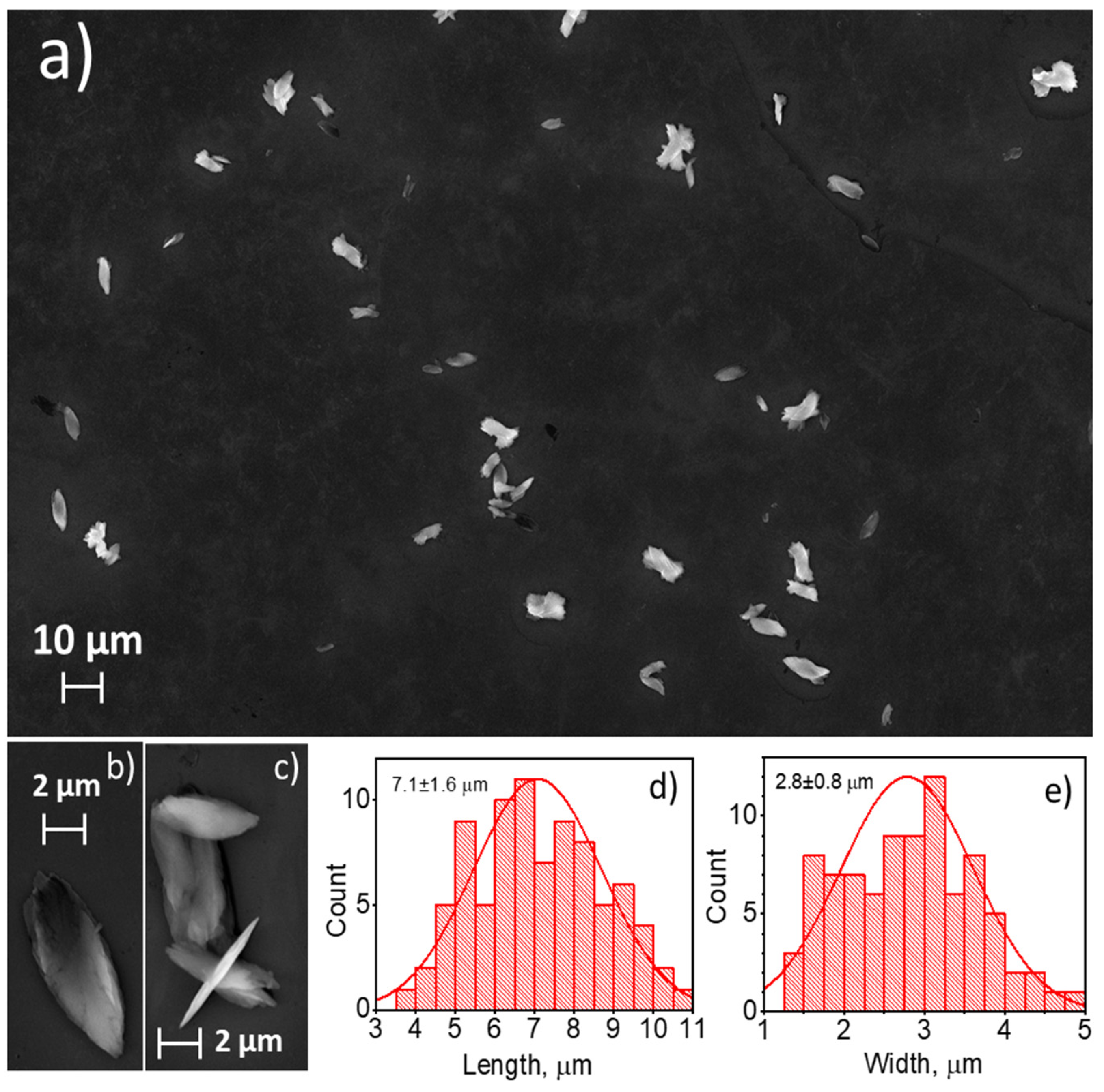
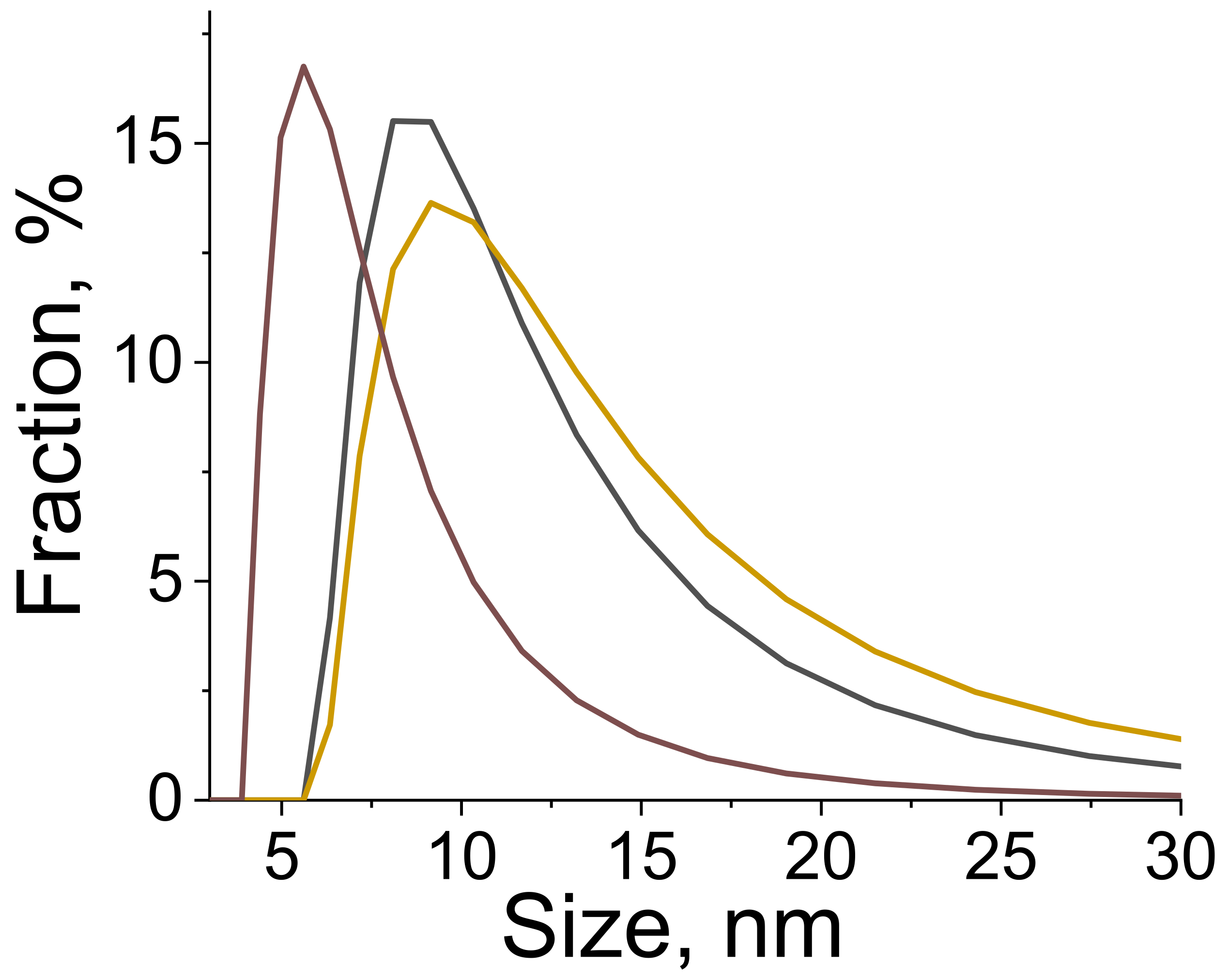
3.1. Morphology
3.2. Crystal Structure
3.3. Luminescent Properties
3.4. Sensing Transition Metal Cations
4. Conclusions
Supplementary Materials
Author Contributions
Funding
Data Availability Statement
Acknowledgments
Conflicts of Interest
References
- Zeng, X.; Zhang, Y.; Zhang, J.; Hu, H.; Wu, X.; Long, Z.; Hou, X. Facile colorimetric sensing of Pb2+ using bimetallic lanthanide metal-organic frameworks as luminescent probe for field screen analysis of lead-polluted environmental water. Microchem. J. 2017, 134, 140–145. [Google Scholar] [CrossRef]
- Cai, D.; Guo, H.; Wen, L.; Liu, C. Fabrication of hierarchical architectures of Tb-MOF by a “green coordination modulation method” for the sensing of heavy metal ions. CrystEngComm 2013, 15, 6702. [Google Scholar] [CrossRef]
- Fang, X.; Zong, B.; Mao, S. Metal-Organic Framework-Based Sensors for Environmental Contaminant Sensing. Nano-Micro Lett. 2018, 10, 64. [Google Scholar] [CrossRef] [PubMed] [Green Version]
- Feng, H.-J.; Xu, L.; Liu, B.; Jiao, H. Europium metal-organic frameworks as recyclable and selective turn-off fluorescent sensors for aniline detection. Dalton Trans. 2016, 45, 17392–17400. [Google Scholar] [CrossRef]
- Xu, H.; Liu, F.; Cui, Y.; Chen, B.; Qian, G. A luminescent nanoscale metal-organic framework for sensing of nitroaromatic explosives. Chem. Commun. 2011, 47, 3153. [Google Scholar] [CrossRef]
- Crawford, S.E.; Ohodnicki, P.R.; Baltrus, J.P. Materials for the photoluminescent sensing of rare earth elements: Challenges and opportunities. J. Mater. Chem. C 2020, 8, 7975–8006. [Google Scholar] [CrossRef]
- Mahata, P.; Mondal, S.K.; Singha, D.K.; Majee, P. Luminescent rare-earth-based MOFs as optical sensors. Dalton Trans. 2017, 46, 301–328. [Google Scholar] [CrossRef]
- Zhang, Y.; Yuan, S.; Day, G.; Wang, X.; Yang, X.; Zhou, H.-C. Luminescent sensors based on metal-organic frameworks. Coord. Chem. Rev. 2018, 354, 28–45. [Google Scholar] [CrossRef]
- Zhang, X.; Kang, X.; Cui, W.; Zhang, Q.; Zheng, Z.; Cui, X. Floral and lamellar europium(III)-based metal-organic frameworks as high sensitivity luminescence sensors for acetone. New J. Chem. 2019, 43, 8363–8369. [Google Scholar] [CrossRef]
- Lustig, W.P.; Mukherjee, S.; Rudd, N.D.; Desai, A.V.; Li, J.; Ghosh, S.K. Metal-organic frameworks: Functional luminescent and photonic materials for sensing applications. Chem. Soc. Rev. 2017, 46, 3242–3285. [Google Scholar] [CrossRef]
- Adusumalli, V.N.K.B.; Runowski, M.; Lis, S. 3,5-dihydroxy benzoic acid-capped caf2:Tb3+ nanocrystals as luminescent probes for the wo42- ion in aqueous solution. ACS Omega 2020, 5, 4568–4575. [Google Scholar] [CrossRef] [Green Version]
- Aslandukov, A.N.; Utochnikova, V.V.; Goriachiy, D.O.; Vashchenko, A.A.; Tsymbarenko, D.M.; Hoffmann, M.; Pietraszkiewicz, M.; Kuzmina, N.P. The development of a new approach toward lanthanide-based OLED fabrication: New host materials for Tb-based emitters. Dalton Trans. 2018, 47, 16350–16357. [Google Scholar] [CrossRef]
- Liu, D.; Lu, K.; Poon, C.; Lin, W. Metal-organic Frameworks as Sensory Materials and Imaging Agents. Inorg. Chem. 2014, 53, 1916–1924. [Google Scholar] [CrossRef]
- Younis, S.A.; Bhardwaj, N.; Bhardwaj, S.K.; Kim, K.-H.; Deep, A. Rare earth metal-organic frameworks (RE-MOFs): Synthesis, properties, and biomedical applications. Coord. Chem. Rev. 2021, 429, 213620. [Google Scholar] [CrossRef]
- Zhou, X.; Wang, H.; Jiang, S.; Xiang, G.; Tang, X.; Luo, X.; Li, L.; Zhou, X. Multifunctional Luminescent Material Eu(III) and Tb(III) Complexes with Pyridine-3,5-Dicarboxylic Acid Linker: Crystal Structures, Tunable Emission, Energy Transfer, and Temperature Sensing. Inorg. Chem. 2019, 58, 3780–3788. [Google Scholar] [CrossRef] [PubMed]
- Khudoleeva, V.; Tcelykh, L.; Kovalenko, A.; Kalyakina, A.; Goloveshkin, A.; Lepnev, L.; Utochnikova, V. Terbium-europium fluorides surface modified with benzoate and terephthalate anions for temperature sensing: Does sensitivity depend on the ligand? J. Lumin. 2018, 201, 500–508. [Google Scholar] [CrossRef]
- Dou, X.; Sun, K.; Chen, H.; Jiang, Y.; Wu, L.; Mei, J.; Ding, Z.; Xie, J. Nanoscale metal-organic frameworks as fluorescence sensors for food safety. Antibiotics 2021, 10, 358. [Google Scholar] [CrossRef] [PubMed]
- Puglisi, R.; Pellegrino, A.L.; Fiorenza, R.; Scirè, S.; Malandrino, G. A facile one-pot approach to the synthesis of gd-eu based metal-organic frameworks and applications to sensing of Fe3+ and Cr2O72− ions. Sensors 2021, 21, 1679. [Google Scholar] [CrossRef] [PubMed]
- Ding, S.B.; Wang, W.; Qiu, L.G.; Yuan, Y.P.; Peng, F.M.; Jiang, X.; Xie, A.J.; Shen, Y.H.; Zhu, J.F. Surfactant-assisted synthesis of lanthanide metal-organic framework nanorods and their fluorescence sensing of nitroaromatic explosives. Mater. Lett. 2011, 65, 1385–1387. [Google Scholar] [CrossRef]
- Li, Q.; Liu, H.; Alattar, M.; Jiang, S.; Han, J.; Ma, Y.; Jiang, C. The preferential accumulation of heavy metals in different tissues following frequent respiratory exposure to PM2.5 in rats. Sci. Rep. 2015, 5, 16936. [Google Scholar] [CrossRef] [Green Version]
- Jaishankar, M.; Tseten, T.; Anbalagan, N.; Mathew, B.B.; Beeregowda, K.N. Toxicity, mechanism and health effects of some heavy metals. Interdiscip. Toxicol. 2014, 7, 60–72. [Google Scholar] [CrossRef] [Green Version]
- Daiguebonne, C.; Kerbellec, N.; Guillou, O.; Bünzli, J.-C.; Gumy, F.; Catala, L.; Mallah, T.; Audebrand, N.; Gérault, Y.; Bernot, K.; et al. Structural and Luminescent Properties of Micro- and Nanosized Particles of Lanthanide Terephthalate Coordination Polymers. Inorg. Chem. 2008, 47, 3700–3708. [Google Scholar] [CrossRef]
- Zou, H.; Wang, L.; Zeng, C.; Gao, X.; Wang, Q.; Zhong, S. Rare-earth coordination polymer micro/nanomaterials: Preparation, properties and applications. Front. Mater. Sci. 2018, 12, 327–347. [Google Scholar] [CrossRef]
- Escudero, A.; Becerro, A.I.; Carrillo-carrión, C.; Núñez, N.O.; Zyuzin, M.V.; Laguna, M.; González-mancebo, D.; Ocaña, M.; Parak, W.J. Rare earth based nanostructured materials: Synthesis, functionalization, properties and bioimaging and biosensing applications. Nanophotonics 2017, 6, 881–921. [Google Scholar] [CrossRef]
- Wang, F.; Deng, K.; Wu, G.; Liao, H.; Liao, H.; Zhang, L.; Lan, S.; Zhang, J.; Song, X.; Wen, L. Facile and Large-Scale Syntheses of Nanocrystal Rare Earth Metal-organic Frameworks at Room Temperature and Their Photoluminescence Properties. J. Inorg. Organomet. Polym. Mater. 2012, 22, 680–685. [Google Scholar] [CrossRef]
- Yang, G.; Lin, W.; Lai, H.; Tong, J.; Lei, J.; Yuan, M.; Zhang, Y.; Cui, C. Ultrasonics Sonochemistry Understanding the relationship between particle size and ultrasonic treatment during the synthesis of metal nanoparticles. Ultrason. Sonochem. 2021, 73, 105497. [Google Scholar] [CrossRef] [PubMed]
- Liu, W.; Yin, R.; Xu, X.; Zhang, L.; Shi, W.; Cao, X. Structural Engineering of Low-Dimensional Metal-organic Frameworks: Synthesis, Properties, and Applications. Adv. Sci. 2019, 6, 1802373. [Google Scholar] [CrossRef] [PubMed]
- Taylor, K.M.L.; Jin, A.; Lin, W. Surfactant-Assisted Synthesis of Nanoscale Gadolinium Metal-Organic Frameworks for Potential Multimodal Imaging. Angew. Chem. Int. Ed. 2008, 47, 7722–7725. [Google Scholar] [CrossRef] [PubMed]
- Pellegrino, F.; Sordello, F.; Mino, L.; Prozzi, M.; Mansfeld, U.; Hodoroaba, V.-D.; Minero, C. Polyethylene Glycol as Shape and Size Controller for the Hydrothermal Synthesis of SrTiO3 Cubes and Polyhedra. Nanomaterials 2020, 10, 1892. [Google Scholar] [CrossRef] [PubMed]
- Yeung, H.H.M.; Sapnik, A.F.; Massingberd-Mundy, F.; Gaultois, M.W.; Wu, Y.; Fraser, D.A.X.; Henke, S.; Pallach, R.; Heidenreich, N.; Magdysyuk, O.V.; et al. Control of Metal-organic Framework Crystallization by Metastable Intermediate Pre-equilibrium Species. Angew. Chem. Int. Ed. 2019, 58, 566–571. [Google Scholar] [CrossRef] [PubMed]
- Dighe, A.V.; Nemade, R.Y.; Singh, M.R. Modeling and Simulation of Crystallization of Metal-organic Frameworks. Processes 2019, 7, 527. [Google Scholar] [CrossRef] [Green Version]
- Reineke, T.M.; Eddaoudi, M.; Fehr, M.; Kelley, D.; Yaghi, O.M. From Condensed Lanthanide Coordination Solids to Microporous Frameworks Having Accessible Metal Sites. J. Am. Chem. Soc. 1999, 121, 1651–1657. [Google Scholar] [CrossRef]
- Shannon, R.D. Revised Effective Ionic Radii and Systematic Studies of Interatomie Distances in in Halides and Chaleogenides. Acta Crystallogr. Sect. A 1976, A32, 751–767. [Google Scholar] [CrossRef]
- Haquin, V.; Etienne, M.; Daiguebonne, C.; Freslon, S.; Calvez, G.; Bernot, K.; Le Pollès, L.; Ashbrook, S.E.; Mitchell, M.R.; Bünzli, J.-C.; et al. Color and Brightness Tuning in Heteronuclear Lanthanide Terephthalate Coordination Polymers. Eur. J. Inorg. Chem. 2013, 2013, 3464–3476. [Google Scholar] [CrossRef] [Green Version]
- Utochnikova, V.V.; Grishko, A.Y.; Koshelev, D.S.; Averin, A.A.; Lepnev, L.S.; Kuzmina, N.P. Lanthanide heterometallic terephthalates: Concentration quenching and the principles of the “multiphotonic emission”. Opt. Mater. (Amst). 2017, 74, 201–208. [Google Scholar] [CrossRef]
- Binnemans, K. Interpretation of europium(III) spectra. Coord. Chem. Rev. 2015, 295, 1–45. [Google Scholar] [CrossRef] [Green Version]
- Carrera Jota, M.L.; García Murillo, A.; Carrillo Romo, F.; García Hernández, M.; Morales Ramírez, A.D.J.; Velumani, S.; de la Rosa Cruz, E.; Kassiba, A. Lu2O3:Eu3+ glass ceramic films: Synthesis, structural and spectroscopic studies. Mater. Res. Bull. 2014, 51, 418–425. [Google Scholar] [CrossRef]
- Liu, G.; Hong, G.; Sun, D. Synthesis and characterization of SiO2/Gd2O3:Eu core–shell luminescent materials. J. Colloid Interface Sci. 2004, 278, 133–138. [Google Scholar] [CrossRef]
- Kolesnikov, I.; Povolotskiy, A.; Mamonova, D.; Lahderanta, E.; Manshina, A.; Mikhailov, M. Photoluminescence Properties of Eu3+ Ions in Yttrium Oxide Nanoparticles: Defect vs Normal Sites. RSC Adv. 2016, 6, 76533–76541. [Google Scholar] [CrossRef]
- Golyeva, E.V.; Kolesnikov, I.E.; Lähderanta, E.; Kurochkin, A.V.; Mikhailov, M.D. Effect of synthesis conditions on structural, morphological and luminescence properties of MgAl2O4:Eu3+ nanopowders. J. Lumin. 2018, 194, 387–393. [Google Scholar] [CrossRef]
- Kolesnikov, I.E.; Kolokolov, D.S.; Kurochkin, M.A.; Voznesenskiy, M.A.; Osmolowsky, M.G.; Lähderanta, E.; Osmolovskaya, O.M. Morphology and doping concentration effect on the luminescence properties of SnO2:Eu3+ nanoparticles. J. Alloys Compd. 2020, 822, 153640. [Google Scholar] [CrossRef]
- Gorbunov, A.O.; Lindqvist-Reis, P.; Mereshchenko, A.S.; Skripkin, M.Y. Solvation and complexation of europium(III) ions in triflate and chloride aqueous-organic solutions by TRLF spectroscopy. J. Mol. Liq. 2017, 240, 25–34. [Google Scholar] [CrossRef]
- Hao, J.; Liu, F.; Liu, N.; Zeng, M.; Song, Y.; Wang, L. Ratiometric fluorescent detection of Cu2+ with carbon dots chelated Eu-based metal-organic frameworks. Sens. Actuators B Chem. 2017, 245, 641–647. [Google Scholar] [CrossRef]
- Chen, B.; Wang, L.; Xiao, Y.; Fronczek, F.R.; Xue, M.; Cui, Y.; Qian, G. A luminescent metal-organic framework with Lewis basic pyridyl sites for the sensing of metal ions. Angew. Chem. Int. Ed. 2009, 48, 500–503. [Google Scholar] [CrossRef] [PubMed]
- Xiao, Y.; Cui, Y.; Zheng, Q.; Xiang, S.; Qian, G.; Chen, B. A microporous luminescent metal-organic framework for highly selective and sensitive sensing of Cu2+ in aqueous solution. Chem. Commun. 2010, 46, 5503–5505. [Google Scholar] [CrossRef] [PubMed]
- Hao, Z.; Yang, G.; Song, X.; Zhu, M.; Meng, X.; Zhao, S.; Song, S.; Zhang, H. A europium(III) based metal-organic framework: Bifunctional properties related to sensing and electronic conductivity. J. Mater. Chem. A 2014, 2, 237–244. [Google Scholar] [CrossRef]
- Zhao, J.; Wang, Y.N.; Dong, W.W.; Wu, Y.P.; Li, D.S.; Zhang, Q.C. A Robust Luminescent Tb(III)-MOF with Lewis Basic Pyridyl Sites for the Highly Sensitive Detection of Metal Ions and Small Molecules. Inorg. Chem. 2016, 55, 3265–3271. [Google Scholar] [CrossRef] [PubMed]
- Wang, X.; Qin, T.; Bao, S.S.; Zhang, Y.C.; Shen, X.; Zheng, L.M.; Zhu, D. Facile synthesis of a water stable 3D Eu-MOF showing high proton conductivity and its application as a sensitive luminescent sensor for Cu2+ ions. J. Mater. Chem. A 2016, 4, 16484–16489. [Google Scholar] [CrossRef]
- Carboni, M.; Lin, Z.; Abney, C.W.; Zhang, T.; Lin, W. A metal-organic framework containing unusual eight-connected Zr-oxo secondary building units and orthogonal carboxylic acids for ultra-sensitive metal detection. Chem.-Eur. J. 2014, 20, 14965–14970. [Google Scholar] [CrossRef]
- Chen, Y.Z.; Jiang, H.L. Porphyrinic Metal-Organic Framework Catalyzed Heck-Reaction: Fluorescence “turn-On” Sensing of Cu(II) Ion. Chem. Mater. 2016, 28, 6698–6704. [Google Scholar] [CrossRef]
- Jin, J.-C.; Wu, J.; Yang, G.-P.; Wu, Y.-L.; Wang, Y.-Y. A microporous anionic metal-organic framework for a highly selective and sensitive electrochemical sensor of Cu2+ ions. Chem. Commun. 2016, 52, 8475–8478. [Google Scholar] [CrossRef]
- Luo, Y.H.; Xie, A.D.; Chen, W.C.; Shen, D.; Zhang, D.E.; Tong, Z.W.; Lee, C.S. Multifunctional anionic indium-organic frameworks for organic dye separation, white-light emission and dual-emitting Fe3+ sensing. J. Mater. Chem. C 2019, 7, 14897–14903. [Google Scholar] [CrossRef]
- Zhou, L.J.; Deng, W.H.; Wang, Y.L.; Xu, G.; Yin, S.G.; Liu, Q.Y. Lanthanide-Potassium Biphenyl-3,3′-disulfonyl-4,4′-dicarboxylate Frameworks: Gas Sorption, Proton Conductivity, and Luminescent Sensing of Metal Ions. Inorg. Chem. 2016, 55, 6271–6277. [Google Scholar] [CrossRef]
- Tang, Q.; Liu, S.; Liu, Y.; Miao, J.; Li, S.; Zhang, L.; Shi, Z.; Zheng, Z. Cation Sensing by a Luminescent Metal-organic Framework with Multiple Lewis Basic Sites. Inorg. Chem. 2013, 52, 2799–2801. [Google Scholar] [CrossRef] [PubMed]
- Liang, Y.-T.; Yang, G.-P.; Liu, B.; Yan, Y.-T.; Xi, Z.-P.; Wang, Y.-Y. Four super water-stable lanthanide–organic frameworks with active uncoordinated carboxylic and pyridyl groups for selective luminescence sensing of Fe3+. Dalton Trans. 2015, 44, 13325–13330. [Google Scholar] [CrossRef] [PubMed]
- Dang, S.; Ma, E.; Sun, Z.M.; Zhang, H. A layer-structured Eu-MOF as a highly selective fluorescent probe for Fe3+ detection through a cation-exchange approach. J. Mater. Chem. 2012, 22, 16920–16926. [Google Scholar] [CrossRef]
- Weng, H.; Yan, B. Lanthanide coordination polymers for multi-color luminescence and sensing of Fe3+. Inorg. Chem. Commun. 2016, 63, 11–15. [Google Scholar] [CrossRef]
- Zheng, M.; Tan, H.; Xie, Z.; Zhang, L.; Jing, X.; Sun, Z. Fast response and high sensitivity europium metal organic framework fluorescent probe with chelating terpyridine sites for Fe3+. ACS Appl. Mater. Interfaces 2013, 5, 1078–1083. [Google Scholar] [CrossRef]
- Li, Y.F.; Wang, D.; Liao, Z.; Kang, Y.; Ding, W.H.; Zheng, X.J.; Jin, L.P. Luminescence tuning of the Dy-Zn metal-organic framework and its application in the detection of Fe(III) ions. J. Mater. Chem. C 2016, 4, 4211–4217. [Google Scholar] [CrossRef]
- Xu, X.Y.; Yan, B. Eu(III)-functionalized MIL-124 as fluorescent probe for highly selectively sensing ions and organic small molecules especially for Fe(III) and Fe(II). ACS Appl. Mater. Interfaces 2015, 7, 721–729. [Google Scholar] [CrossRef]
- Dang, S.; Wang, T.; Yi, F.; Liu, Q.; Yang, W.; Sun, Z.M. A Nanoscale Multiresponsive Luminescent Sensor Based on a Terbium(III) Metal-Organic Framework. Chem.-Asian J. 2015, 10, 1703–1709. [Google Scholar] [CrossRef]
- Kang, Y.; Zheng, X.J.; Jin, L.P. A microscale multi-functional metal-organic framework as a fluorescence chemosensor for Fe(III), Al(III) and 2-hydroxy-1-naphthaldehyde. J. Colloid Interface Sci. 2016, 471, 1–6. [Google Scholar] [CrossRef]
- Zhao, X.-L.; Tian, D.; Gao, Q.; Sun, H.-W.; Xu, J.; Bu, X.-H. A chiral lanthanide metal-organic framework for selective sensing of Fe( iii ) ions. Dalton Trans. 2016, 45, 1040–1046. [Google Scholar] [CrossRef]
- Xu, H.; Hu, H.C.; Cao, C.S.; Zhao, B. Lanthanide Organic Framework as a Regenerable Luminescent Probe for Fe3+. Inorg. Chem. 2015, 54, 4585–4587. [Google Scholar] [CrossRef]
- Zhao, J.J.; Liu, P.Y.; Dong, Z.P.; Liu, Z.L.; Wang, Y.Q. Eu(III)-organic framework as a multi-responsive photoluminescence sensor for efficient detection of 1-naphthol, Fe3+ and MnO4− in water. Inorg. Chim. Acta 2020, 511, 119843. [Google Scholar] [CrossRef]
- Chen, S.; Shi, Z.; Qin, L.; Jia, H.; Zheng, H. Two new luminescent Cd(II)-metal−organic frameworks as bifunctional chemosensors for detection of cations Fe3+, anions CrO42−, and Cr2O72− in aqueous solution. Cryst. Growth Des. 2017, 17, 67–72. [Google Scholar] [CrossRef]
- Hou, B.L.; Tian, D.; Liu, J.; Dong, L.Z.; Li, S.L.; Li, D.S.; Lan, Y.Q. A Water-Stable Metal-Organic Framework for Highly Sensitive and Selective Sensing of Fe3+ Ion. Inorg. Chem. 2016, 55, 10580–10586. [Google Scholar] [CrossRef] [PubMed]
- Li, G.-P.; Liu, G.; Li, Y.-Z.; Hou, L.; Wang, Y.-Y.; Zhu, Z. Uncommon Pyrazoyl-Carboxyl Bifunctional Ligand-Based Microporous Lanthanide Systems: Sorption and Luminescent Sensing Properties. Inorg. Chem. 2016, 55, 3952–3959. [Google Scholar] [CrossRef]
- Wen, G.X.; Wu, Y.P.; Dong, W.W.; Zhao, J.; Li, D.S.; Zhang, J. An Ultrastable Europium(III)-Organic Framework with the Capacity of Discriminating Fe2+/Fe3+ Ions in Various Solutions. Inorg. Chem. 2016, 55, 10114–10117. [Google Scholar] [CrossRef] [PubMed]
- Cao, L.H.; Shi, F.; Zhang, W.M.; Zang, S.Q.; Mak, T.C.W. Selective Sensing of Fe3+ and Al3+ Ions and Detection of 2,4,6-Trinitrophenol by a Water-Stable Terbium-Based Metal-Organic Framework. Chem. Eur. J. 2015, 21, 15705–15712. [Google Scholar] [CrossRef] [PubMed]
- Qiu, L.; Zhu, C.; Chen, H.; Hu, M.; He, W.; Guo, Z. A turn-on fluorescent Fe3+ sensor derived from an anthracene-bearing bisdiene macrocycle and its intracellular imaging application. Chem. Commun. 2014, 50, 4631–4634. [Google Scholar] [CrossRef] [PubMed]
- Sarih, N.M.; Ciupa, A.; Moss, S.; Myers, P.; Slater, A.G.; Abdullah, Z.; Tajuddin, H.A.; Maher, S. Furo[3,2-c]coumarin-derived Fe3+ Selective Fluorescence Sensor: Synthesis, Fluorescence Study and Application to Water Analysis. Sci. Rep. 2020, 10, 7421. [Google Scholar] [CrossRef]
- Katowah, D.F.; Hussein, M.A.; Alam, M.M.; Ismail, S.H.; Osman, O.I.; Sobahi, T.R.; Asiri, A.M.; Ahmed, J.; Rahman, M.M. Designed network of ternary core-shell PPCOT/NiFe2O4/C-SWCNTs nanocomposites. A Selective Fe3+ ionic sensor. J. Alloys Compd. 2020, 834, 155020. [Google Scholar] [CrossRef]
- Wang, H.H.; Zhou, L.J.; Wang, Y.L.; Liu, Q.Y. Terbium-biphenyl-3,3′-disulfonyl-4,4′-dicarboxylate framework with sulfonate sites for luminescent sensing of Cr3+ ion. Inorg. Chem. Commun. 2016, 73, 94–97. [Google Scholar] [CrossRef]
- Zhang, P.-P.; Song, B.; Li, Z.; Zhang, J.-J.; Ni, A.-Y.; Chen, J.; Ni, J.; Liu, S.; Duan, C. A “turn-on” Cr3+ ion probe based on non-luminescent metal-organic framework-new strategy to prepare a recovery probe. J. Mater. Chem. A 2021, 9, 13552–13561. [Google Scholar] [CrossRef]
- El-Shishtawy, R.M.; Rahman, M.M.; Sheikh, T.A.; Nadeem Arshad, M.; Al-Zahrani, F.A.M.; Asiri, A.M. A New Cr3+ Electrochemical Sensor Based on ATNA/Nafion/Glassy Carbon Electrode. Materials 2020, 13, 2695. [Google Scholar] [CrossRef]


| Sample | C(EuCl3) | C(Na2bdc) | PEG-6000 | Ultrasonication | Stirring |
|---|---|---|---|---|---|
| 1 | 1 mM | 2 mM | - | - | + |
| 2 | 1 mM | 2 mM | 20% | + | + |
| 3 | 0.5 mM | 1 mM | - | + | + |
| Sample | a, Å | b, Å | c, Å | α, deg. | β, deg. | γ, deg. | V, Å3 |
|---|---|---|---|---|---|---|---|
| Tb2bdc3·4H2O | 6.14 | 10.07 | 10.10 | 102.25 | 91.12 | 101.52 | 596.63 |
| Eu2bdc3·4H2O (1) | 6.20 | 9.85 | 10.29 | 102.15 | 89.75 | 105.10 | 592.91 |
| Eu2bdc3·4H2O (2) | 6.16 | 9.8 | 10.22 | 101.84 | 90.27 | 104.86 | 582.19 |
| Sample | Ar (s−1) | Anr (s−1) | Atotal (s−1) | η (%) | Φ (%) |
|---|---|---|---|---|---|
| 1 | 371 | 2193 | 2564 | 14.5 | 10 ± 1 |
| 2 | 290 | 2405 | 2695 | 10.8 | 5 ± 1 |
| 3 | 150 | 8545 | 8695 | 1.7 | 1.5 ± 0.5 |
| Sensing Material | Method | Target Contaminant | LOD | Ref. |
|---|---|---|---|---|
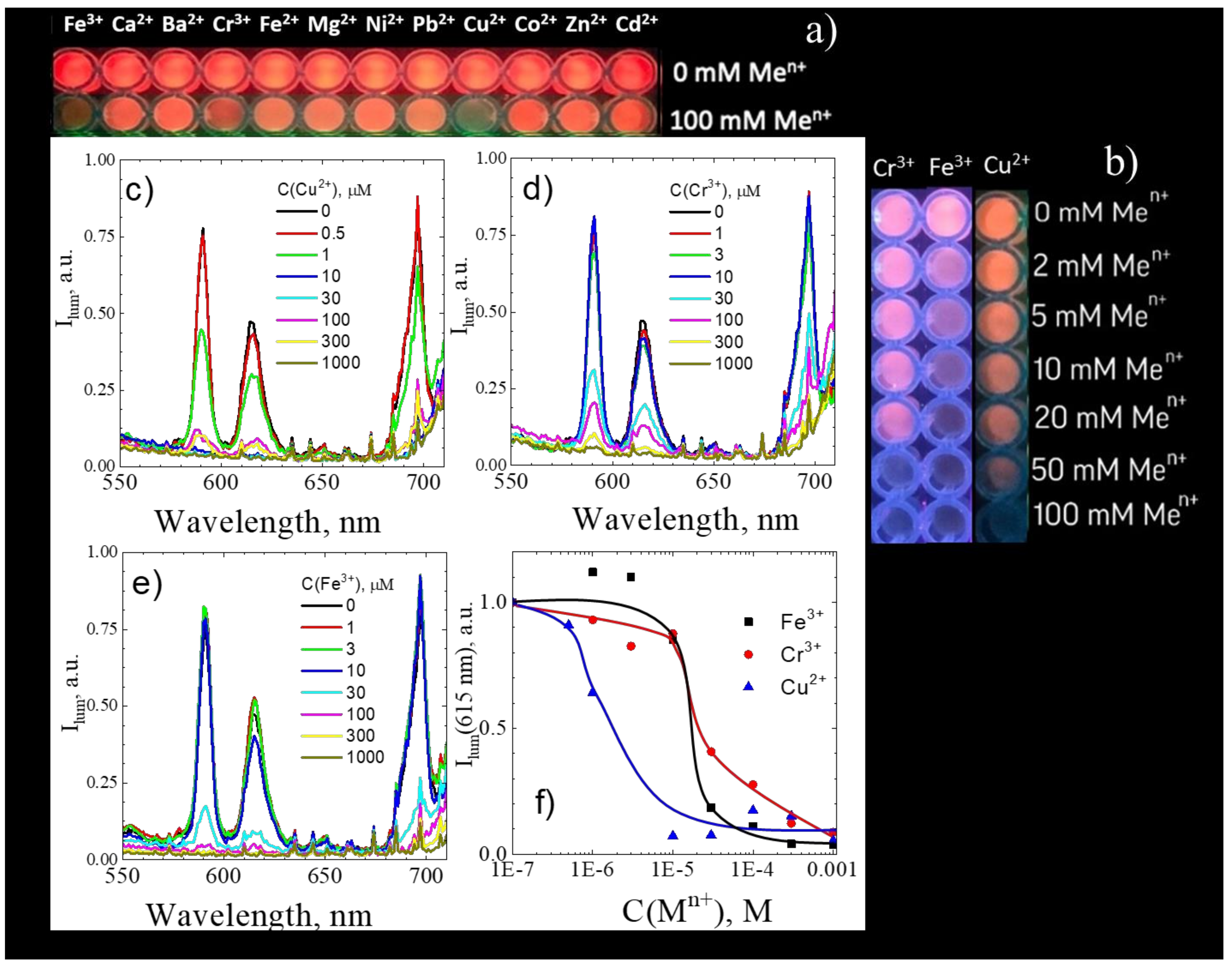
| Eu2(bdc)3·4H2O | luminescent | Cu2+ | 1 μM | Current work |
| Tb(BTC)(H2O) | luminescent | Cu2+ | 10 μM | [2] |
| CDs@Eu-DPA MOFs | luminescent | Cu2+ | 26.3 nM | [3,43] |
| [Eu(PDC)1.5(DMF)]·(DMF)0.5(H2O)0.5 | luminescent | Cu2+ | 10 mM | [7,44] |
| Eu2(FMA)2(OX)(H2O)4·4H2O | luminescent | Cu2+ | 100 μM | [7,45] |
| [Eu4(BPT)4(DMF)2(H2O)8] | luminescent | Cu2+ | 10 μM | [7,46] |
| [Tb3(L)2(HCOO)(H2O)5]·DMF·4H2O | luminescent | Cu2+ | 100 μM | [7,47] |
| [Eu(ox)2(H2O)](Me2NH2)(H2O)3 | luminescent | Cu2+ | 10 μM | [10,48] |
| Zr6(O)8(OH2)8(tpdc)4 | luminescent | Cu2+ | 1 μM | [10,49] |
| PCN-222-Pd(II) | luminescent | Cu2+ | 50 nM | [10,50] |
| Me2NH2@MOF-1 | electrochemical | Cu2+ | 10 pM | [3,51] |
| Eu2(bdc)3 nanoparticles | luminescent | Fe3+ | 30 μM | Current work |
| [Me2NH2][In(abtc)]·solvents | luminescent | Fe3+ | 34.5 μM | [52] |
| [LnK(BPDSDC)(DMF)(H2O)]·x(solvent) | luminescent | Fe3+ | 10 μM | [10,53] |
| [Eu(BTPCA)(H2O)]·2DMF·3H2O | luminescent | Fe3+ | 10 μM | [7,54] |
| [Eu(HL)(H2O)2]n·2H2O | luminescent | Fe3+ | 1 μM | [7,55] |
| EuL | luminescent | Fe3+ | 100 μM | [7,56] |
| [H2NMe2]3[Tb(DPA)3] | luminescent | Fe3+ | 10 μM | [7,57] |
| Eu (4′-(4-carboxyphenyl)-2,2′: 6′,2″-terpyridine)3 | luminescent | Fe3+ | 100 μM | [7,58] |
| [H(H2O)8][DyZn4(imdc)4(im)4] | luminescent | Fe3+ | 1 mM | [7,59] |
| Eu3+@Ga2(OH)4(C9O6H4) | luminescent | Fe3+ | 0.28 μM | [7,60] |
| nTbL | luminescent | Fe3+ | 10 μM | [7,61] |
| [Eu(atpt)1.5(phen)(H2O)]n | luminescent | Fe3+ | 500 μM | [7,62] |
| [(CH3)2NH2] ·[Tb(bptc)]·xS | luminescent | Fe3+ | 10 μM | [7,63] |
| Tb-BTB | luminescent | Fe3+ | 10 μM | [7,64] |
| [Eu3(BDC)4.5(H2O)(DMF)2] | luminescent | Fe3+ | 1 μM | [7,65] |
| [Cd(L)(BPDC)]·2H2O | luminescent | Fe3+ | 2 μM | [8,66] |
| [Cd(L)(SDBA)(H2O)]∙0.5H2O | luminescent | Fe3+ | 2 μM | [8,66] |
| [Zn5(hfipbb)4(trz)2(H2O)2] | luminescent | Fe3+ | 10 μM | [8,67] |
| [Eu(Hpzbc)2(NO3)]·H2O | luminescent | Fe3+ | 10 μM | [8,68] |
| [Eu(L)(H2O)2]·NMP·H2O | luminescent | Fe3+ | 100 nM | [8,69] |
| [Tb(L1)1.5(H2O)]·3H2O | luminescent | Fe3+ | 10 μM | [10,70] |
| Bisdiene macrocycle | luminescent | Fe3+ | 0.58 μM | [71] |
| 2-(cyclohexylamino)-3-phenyl-4Hfuro [3,2-c]chromen-4-one | luminescent | Fe3+ | 1.73 μM | [72] |
| [Me2NH2][In(abtc)]·solvents | luminescent | Fe3+ | 34.5 μM | [53] |
| PPCOT/NiFe2O4/C-SWCNT | electrochemical | Fe3+ | 100 pM | [73] |
| Eu2(bdc)3 nanoparticles | luminescent | Cr3+ | 30 μM | Current work |
| Tb(BTC)(H2O) | luminescent | Cr3+ | 10 μM | [2] |
| [TbK(BPDSDC)(DMF)(H2O)2] | 10 μM | [8,74] | ||
| [Eu2L3(DMF)3]·2DMF·5H2O | luminescent | Cr3+ | 75.2 nM | [75] |
| ATNA deriviative | electrochemical | Cr3+ | 130 pM | [76] |
Publisher’s Note: MDPI stays neutral with regard to jurisdictional claims in published maps and institutional affiliations. |
© 2020 by the authors. Licensee MDPI, Basel, Switzerland. This article is an open access article distributed under the terms and conditions of the Creative Commons Attribution (CC BY) license (https://creativecommons.org/licenses/by/4.0/).
Share and Cite
Kolesnik, S.S.; Nosov, V.G.; Kolesnikov, I.E.; Khairullina, E.M.; Tumkin, I.I.; Vidyakina, A.A.; Sysoeva, A.A.; Ryazantsev, M.N.; Panov, M.S.; Khripun, V.D.; et al. Ultrasound-Assisted Synthesis of Luminescent Micro- and Nanocrystalline Eu-Based MOFs as Luminescent Probes for Heavy Metal Ions. Nanomaterials 2021, 11, 2448. https://doi.org/10.3390/nano11092448
Kolesnik SS, Nosov VG, Kolesnikov IE, Khairullina EM, Tumkin II, Vidyakina AA, Sysoeva AA, Ryazantsev MN, Panov MS, Khripun VD, et al. Ultrasound-Assisted Synthesis of Luminescent Micro- and Nanocrystalline Eu-Based MOFs as Luminescent Probes for Heavy Metal Ions. Nanomaterials. 2021; 11(9):2448. https://doi.org/10.3390/nano11092448
Chicago/Turabian StyleKolesnik, Stefaniia S., Viktor G. Nosov, Ilya E. Kolesnikov, Evgenia M. Khairullina, Ilya I. Tumkin, Aleksandra A. Vidyakina, Alevtina A. Sysoeva, Mikhail N. Ryazantsev, Maxim S. Panov, Vasiliy D. Khripun, and et al. 2021. "Ultrasound-Assisted Synthesis of Luminescent Micro- and Nanocrystalline Eu-Based MOFs as Luminescent Probes for Heavy Metal Ions" Nanomaterials 11, no. 9: 2448. https://doi.org/10.3390/nano11092448
APA StyleKolesnik, S. S., Nosov, V. G., Kolesnikov, I. E., Khairullina, E. M., Tumkin, I. I., Vidyakina, A. A., Sysoeva, A. A., Ryazantsev, M. N., Panov, M. S., Khripun, V. D., Bogachev, N. A., Skripkin, M. Y., & Mereshchenko, A. S. (2021). Ultrasound-Assisted Synthesis of Luminescent Micro- and Nanocrystalline Eu-Based MOFs as Luminescent Probes for Heavy Metal Ions. Nanomaterials, 11(9), 2448. https://doi.org/10.3390/nano11092448