
Local Structure and Redox Properties of Amorphous CeO2-TiO2 Prepared Using the H2O2-Modified Sol-Gel Method

Abstract
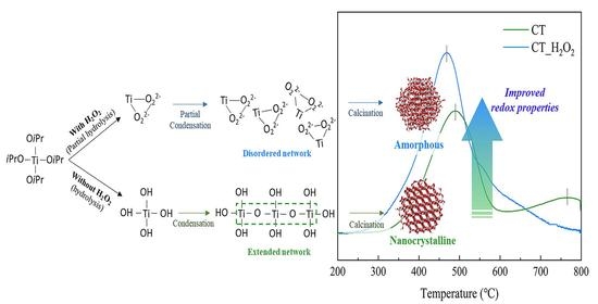
:
1. Introduction
2. Experimental Procedure
2.1. Sample Preparation
2.2. Characterization
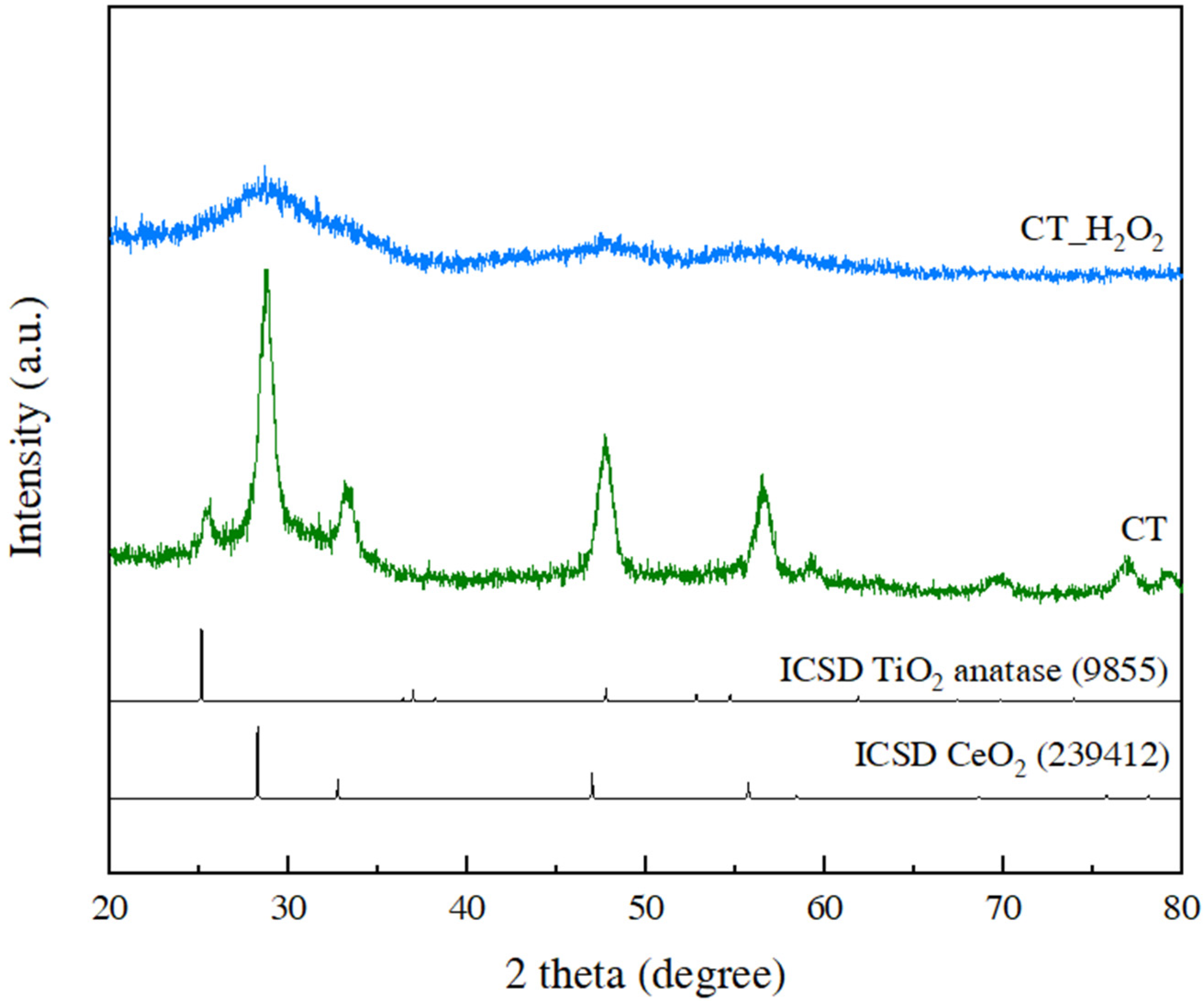
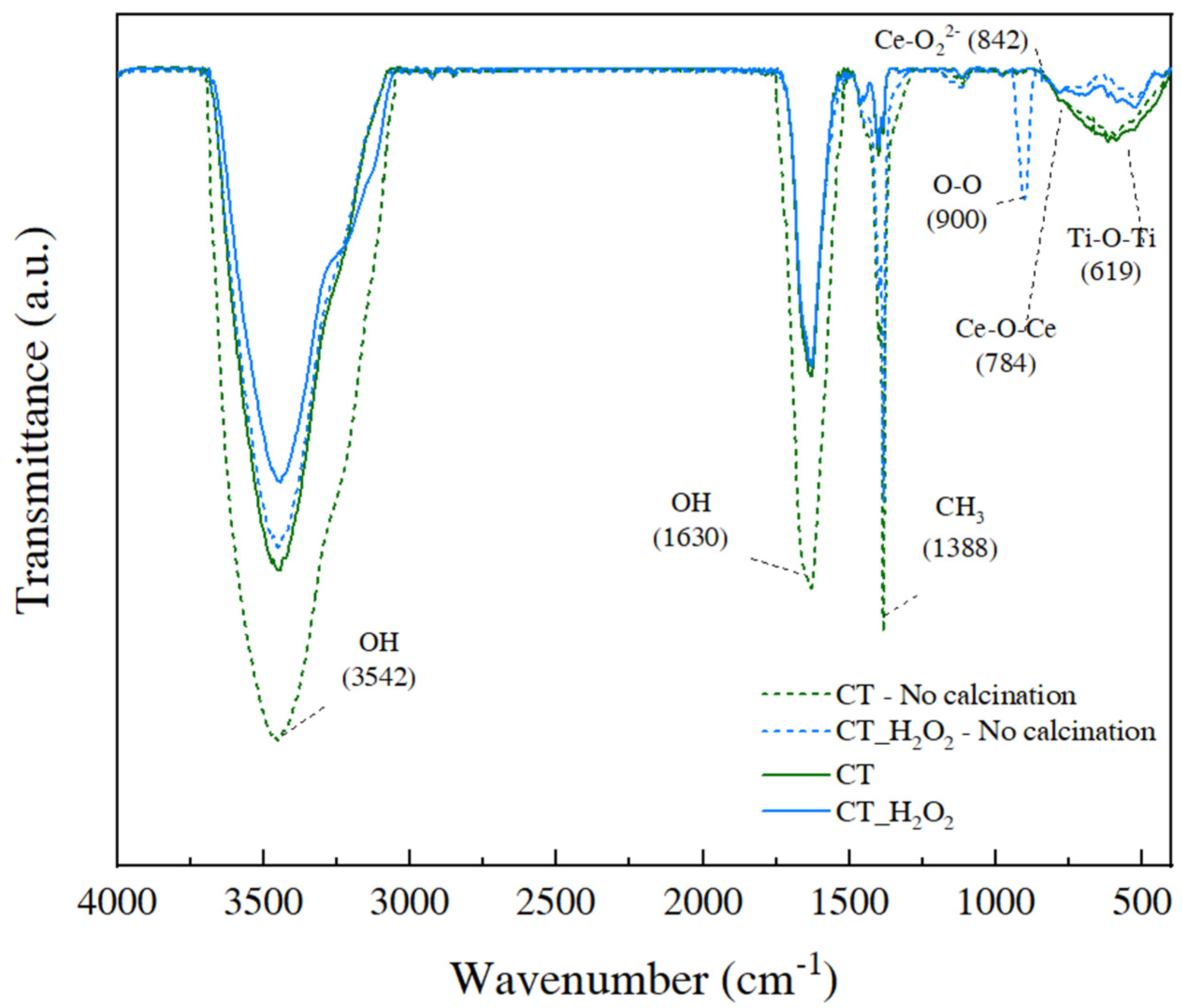
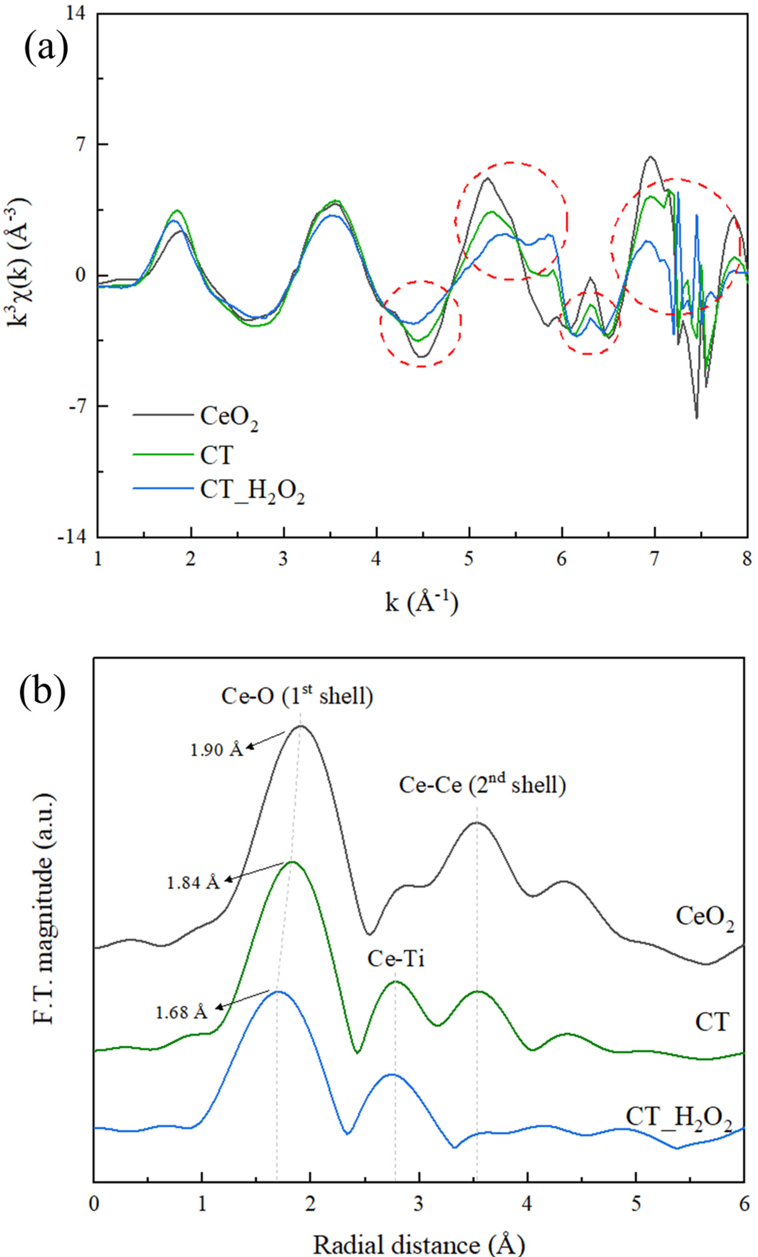
3. Results and Discussion
4. Conclusions
Author Contributions
Funding
Data Availability Statement
Conflicts of Interest
References
- Oskam, G. Metal oxide nanoparticles: Synthesis, characterization and application. J. Sol-Gel Sci. Technol. 2006, 37, 161–164. [Google Scholar] [CrossRef]
- Trovarelli, A.; Boaro, M.; Rocchini, E.; de Leitenburg, C.; Dolcetti, G. Some recent developments in the characterization of ceria-based catalysts. J. Alloys Compd. 2001, 323, 584–591. [Google Scholar] [CrossRef]
- Li, P.; Xin, Y.; Li, Q.; Wang, Z.; Zhang, Z.; Zheng, L. Ce–Ti amorphous oxides for selective catalytic reduction of NO with NH3: Confirmation of Ce–O–Ti active sites. Environ. Sci. Technol. 2012, 46, 9600–9605. [Google Scholar] [CrossRef]
- Fang, N.; Ding, Y.; Liu, C.; Chen, Z. Effect of crystalline/amorphous structure on light absorption and carrier separationof CeO2-TiO2 heterojunctions. Appl. Surf. Sci. 2018, 452, 49–57. [Google Scholar] [CrossRef]
- Zhu, H.; Qin, Z.; Shan, W.; Shen, W.; Wang, J. Pd/CeO2–TiO2 catalyst for CO oxidation at low temperature: A TPR study with H2 and CO as reducing agents. J. Catal. 2004, 225, 267–277. [Google Scholar] [CrossRef]
- Curry-Hyde, H.E.; Musch, H.; Baiker, A. Selective catalytic reduction of nitric oxide over amorphous and crystalline chromia: I. Comparative study of activities. Appl. Catal. 1990, 65, 211–223. [Google Scholar] [CrossRef]
- Tang, X.; Hao, J.; Xu, W.; Li, J. Low temperature selective catalytic reduction of NOx with NH3 over amorphous MnOx catalysts prepared by three methods. Catal. Commun. 2007, 8, 329–334. [Google Scholar] [CrossRef]
- Li, L.; Shao, Q.; Huang, X. Amorphous oxide nanostructures for advanced electrocatalysis. Chem. Eur. J. 2020, 26, 3943–3960. [Google Scholar] [CrossRef]
- Gao, X.; Jiang, Y.; Zhong, Y.; Luo, Z.; Cen, K. The activity and characterization of CeO2-TiO2 catalysts prepared by the sol–gel method for selective catalytic reduction of NO with NH3. J. Hazard. Mater. 2010, 174, 734–739. [Google Scholar] [CrossRef]
- Tian, J.; Sang, Y.; Zhao, Z.; Zhou, W.; Wang, D.; Kang, X.; Liu, H.; Wang, J.; Chen, S.; Cai, H.; et al. Enhanced photocatalytic performances of CeO2/TiO2 nanobelt heterostructures. Small 2013, 9, 3864–3872. [Google Scholar] [CrossRef]
- Gao, X.; Jiang, Y.; Fu, Y.; Zhong, Y.; Luo, Z.; Cen, K. Preparation and characterization of CeO2/TiO2 catalysts for selective catalytic reduction of NO with NH3. Catal. Commun. 2010, 11, 465–469. [Google Scholar] [CrossRef]
- Frenzer, G.; Maier, W.F. Amorphous porous mixed oxides: Sol-gel ways to a highly versatile class of materials and catalysts. Annu. Rev. Mater. Res. 2006, 36, 281–331. [Google Scholar] [CrossRef]
- Simonsen, M.E.; Søgaard, E.G. Sol–gel reactions of titanium alkoxides and water: Influence of pH and alkoxy group on cluster formation and properties of the resulting products. J. Sol-Gel Sci. Technol. 2010, 53, 485–497. [Google Scholar] [CrossRef] [Green Version]
- Debecker, D.P.; Mutin, P.H. Non-hydrolytic sol–gel routes to heterogeneous catalysts. Chem. Soc. Rev. 2012, 41, 3624–3650. [Google Scholar] [CrossRef] [PubMed]
- Seok, S.I.; Ahn, B.Y.; Pramanik, N.C.; Kim, H.; Hong, S.I. Preparation of nanosized rutile TiO2 from an aqueous peroxotitanate solution. J. Am. Ceram. Soc. 2006, 89, 1147–1149. [Google Scholar] [CrossRef]
- Shen, C.; Shaw, L.L. FTIR analysis of the hydrolysis rate in the sol–gel formation of gadolinia-doped ceria with acetylacetonate precursors. J. Sol-Gel Sci. Technol. 2010, 53, 571–577. [Google Scholar] [CrossRef]
- Livage, J.; Sanchez, C. Sol-gel chemistry. J. Non-Cryst. Solids 1992, 145, 11–19. [Google Scholar] [CrossRef]
- Li, S.; Xu, Q.; Uchaker, E.; Cao, X.; Cao, G. Comparison of amorphous, pseudohexagonal and orthorhombic Nb2O5 for high-rate lithium ion insertion. CrystEngComm 2016, 18, 2532–2540. [Google Scholar] [CrossRef] [Green Version]
- Zou, J.; Gao, J.; Xie, F. An amorphous TiO2 sol sensitized with H2O2 with the enhancement of photocatalytic activity. J. Alloys Compd. 2010, 497, 420–427. [Google Scholar] [CrossRef]
- Gao, Y.; Masuda, Y.; Seo, W.S.; Ohta, H.; Koumoto, K. TiO2 nanoparticles prepared using an aqueous peroxotitanate solution. Ceram. Int. 2004, 30, 1365–1368. [Google Scholar] [CrossRef]
- Mohammadi, M.R.; Fray, D.J. Nanostructured TiO2–CeO2 mixed oxides by an aqueous sol–gel process: Effect of Ce: Ti molar ratio on physical and sensing properties. Sens. Actuators B Chem. 2010, 150, 631–640. [Google Scholar] [CrossRef]
- Sharma, N.; Deepa, M.; Varshney, P.; Agnihotry, S.A. FTIR and absorption edge studies on tungsten oxide based precursor materials synthesized by sol–gel technique. J. Non-Cryst. Solids 2002, 306, 129–137. [Google Scholar] [CrossRef]
- Damatov, D.; Mayer, J.M. (Hydro)peroxide ligands on colloidal cerium oxide nanoparticles. Chem. Commun. 2016, 52, 10281–10284. [Google Scholar] [CrossRef]
- Alsharaeh, E.H.; Bora, T.; Soliman, A.; Ahmed, F.; Bharath, G.; Ghoniem, M.G.; Abu-Salah, K.M.; Dutta, J. Sol-gel-assisted microwave-derived synthesis of anatase Ag/TiO2/GO nanohybrids toward efficient visible light phenol degradation. Catalysts 2017, 7, 133. [Google Scholar] [CrossRef]
- Wandre, T.M.; Gaikwad, P.N.; Tapase, A.S.; Garadkar, K.; Vanalakar, S.A.; Lokhande, P.D.; Sasikala, R.; Hankare, P.P. Sol–gel synthesized TiO2–CeO2 nanocomposite: An efficient photocatalyst for degradation of methyl orange under sunlight. J. Mater. Sci. Mater. Electron. 2016, 27, 825–833. [Google Scholar] [CrossRef]
- Sanchez, C.; Livage, J.; Henry, M.; Babonneau, F. Chemical modification of alkoxide precursors. J. Non-Cryst. Solids 1988, 100, 65–76. [Google Scholar] [CrossRef]
- Purcar, V.; Rădiţoiu, V.; Nicolae, C.A.; Frone, A.N.; RĂDIŢOIU, A.; Raduly, F.M.; ŞOMOGHI, R.; Gabor, R.A.; Căprărescu, S. Synthesis and morpho-structural properties of TiO2-based materials. J. Optoelectron. Adv. Mater. 2019, 21, 281–286. [Google Scholar]
- Ksapabutr, B.; Gulari, E.; Wongkasemjit, S. Sol–gel derived porous ceria powders using cerium glycolate complex as precursor. Mater. Chem. Phys. 2006, 99, 318–324. [Google Scholar] [CrossRef]
- Teo, B.K. EXAFS: Basic Principles and Data Analysis; Springer Science & Business Media: Berlin/Heidelberg, Germany, 2012; Volume 9. [Google Scholar]
- Jo, S.H.; Lee, H. Local structure and short-range ordering of MnO2–Ce(1−x) ZrxO2/TiO2. Mater. Charact. 2015, 110, 102–108. [Google Scholar] [CrossRef]
- Jo, S.H.; Lee, I.; Park, H.; Lee, H. Experimental evidence and mechanism of the oxygen storage capacity in MnO2-Ce(1−x)ZrxO2/TiO2 catalyst for low-temperature SCR. Ceram. Int. 2017, 43, 5182–5188. [Google Scholar] [CrossRef]
- Vayssilov, G.N.; Lykhach, Y.; Migani, A.; Staudt, T.; Petrova, G.P.; Tsud, N.; Skála, T.; Bruix, A.; Illas, F.; Prince, K.C.; et al. Support nanostructure boosts oxygen transfer to catalytically active platinum nanoparticles. Nat. Mater. 2011, 10, 310–315. [Google Scholar] [CrossRef] [PubMed]
- Park, G.; Kim, M.; Cha, J.; Kim, H.; Lee, H. Local atomic structure and enhanced catalytic activity of W doped CeWTiOx catalysts. J. Solid State Chem. 2020, 292, 121689. [Google Scholar] [CrossRef]
- Neri, G.; Pistone, A.; Milone, C.; Galvagno, S. Wet air oxidation of p-coumaric acid over promoted ceria catalysts. Appl. Catal. B Environ. 2002, 38, 321–329. [Google Scholar] [CrossRef]
- Liu, J.; Wang, L.L.; Fei, Z.Y.; Chen, X.; Tang, J.H.; Cui, M.F.; Qiao, X. Structure and properties of amorphous CeO2@TiO2 catalyst and its performance in the selective catalytic reduction of NO with NH3. J. Fuel Chem. Technol. 2016, 44, 954–960. [Google Scholar] [CrossRef]
- Fei, Z.; Yang, Y.; Wang, M.; Tao, Z.; Liu, Q.; Chen, X.; Cui, M.; Zhang, Z.; Tang, J.; Qiao, X. Precisely fabricating Ce-O-Ti structure to enhance performance of Ce-Ti based catalysts for selective catalytic reduction of NO with NH3. Chem. Eng. J. 2018, 353, 930–939. [Google Scholar] [CrossRef]


| Sample | Surface Oxygen Reduction Temp. (°C) | Bulk Oxygen Reduction Temp. (°C) | H2 Consumption (mmol/g) | BET Surface Area (m2/g) |
|---|---|---|---|---|
| CT | 490 | 797 | 1110 | 103.0 |
| CT_H2O2 | 468 | Not observed | 1354 | 113.1 |
Publisher’s Note: MDPI stays neutral with regard to jurisdictional claims in published maps and institutional affiliations. |
© 2021 by the authors. Licensee MDPI, Basel, Switzerland. This article is an open access article distributed under the terms and conditions of the Creative Commons Attribution (CC BY) license (https://creativecommons.org/licenses/by/4.0/).
Share and Cite
Kim, M.; Park, G.; Lee, H. Local Structure and Redox Properties of Amorphous CeO2-TiO2 Prepared Using the H2O2-Modified Sol-Gel Method. Nanomaterials 2021, 11, 2148. https://doi.org/10.3390/nano11082148
Kim M, Park G, Lee H. Local Structure and Redox Properties of Amorphous CeO2-TiO2 Prepared Using the H2O2-Modified Sol-Gel Method. Nanomaterials. 2021; 11(8):2148. https://doi.org/10.3390/nano11082148
Chicago/Turabian StyleKim, Myungju, Gwanhee Park, and Heesoo Lee. 2021. "Local Structure and Redox Properties of Amorphous CeO2-TiO2 Prepared Using the H2O2-Modified Sol-Gel Method" Nanomaterials 11, no. 8: 2148. https://doi.org/10.3390/nano11082148
APA StyleKim, M., Park, G., & Lee, H. (2021). Local Structure and Redox Properties of Amorphous CeO2-TiO2 Prepared Using the H2O2-Modified Sol-Gel Method. Nanomaterials, 11(8), 2148. https://doi.org/10.3390/nano11082148