
Nano-Biomaterials for Retinal Regeneration


Abstract
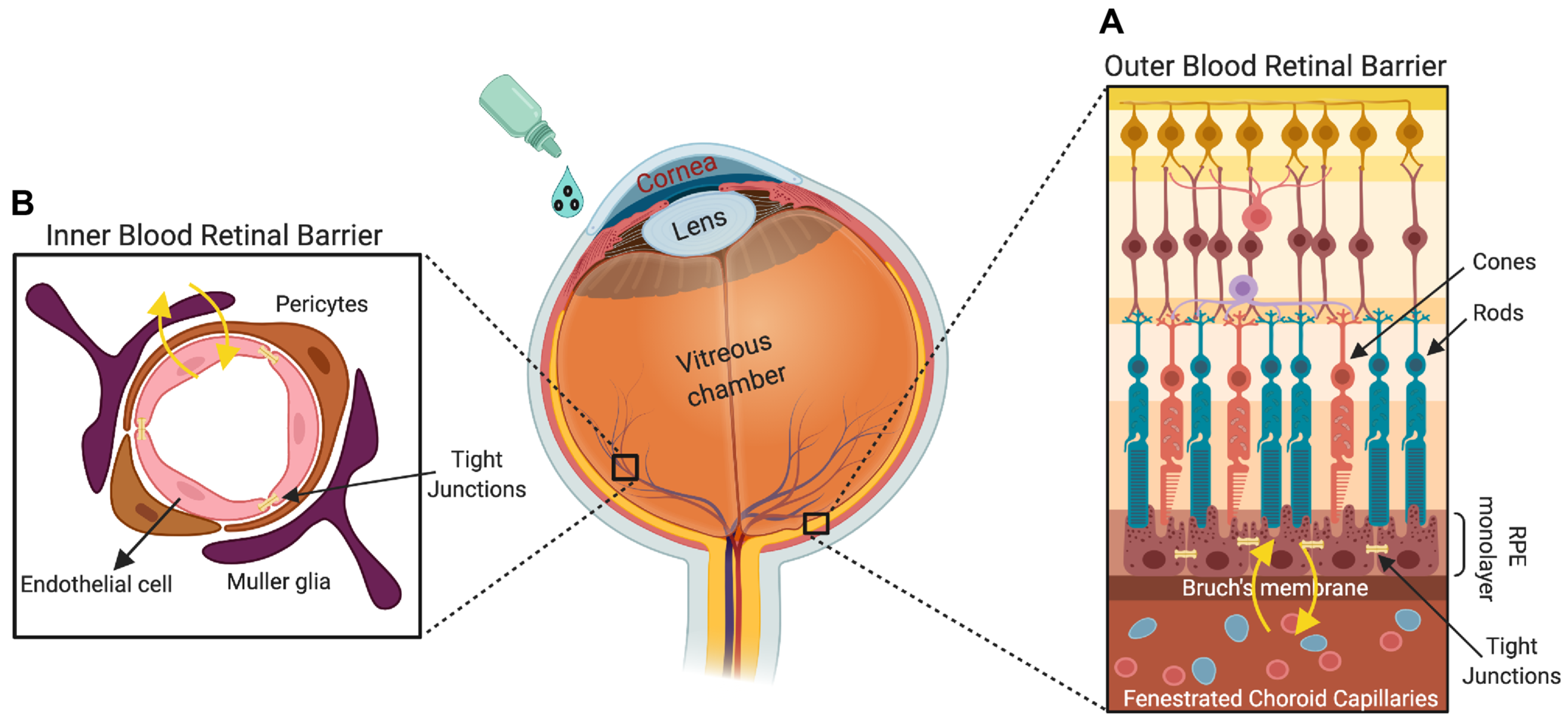
1. Introduction
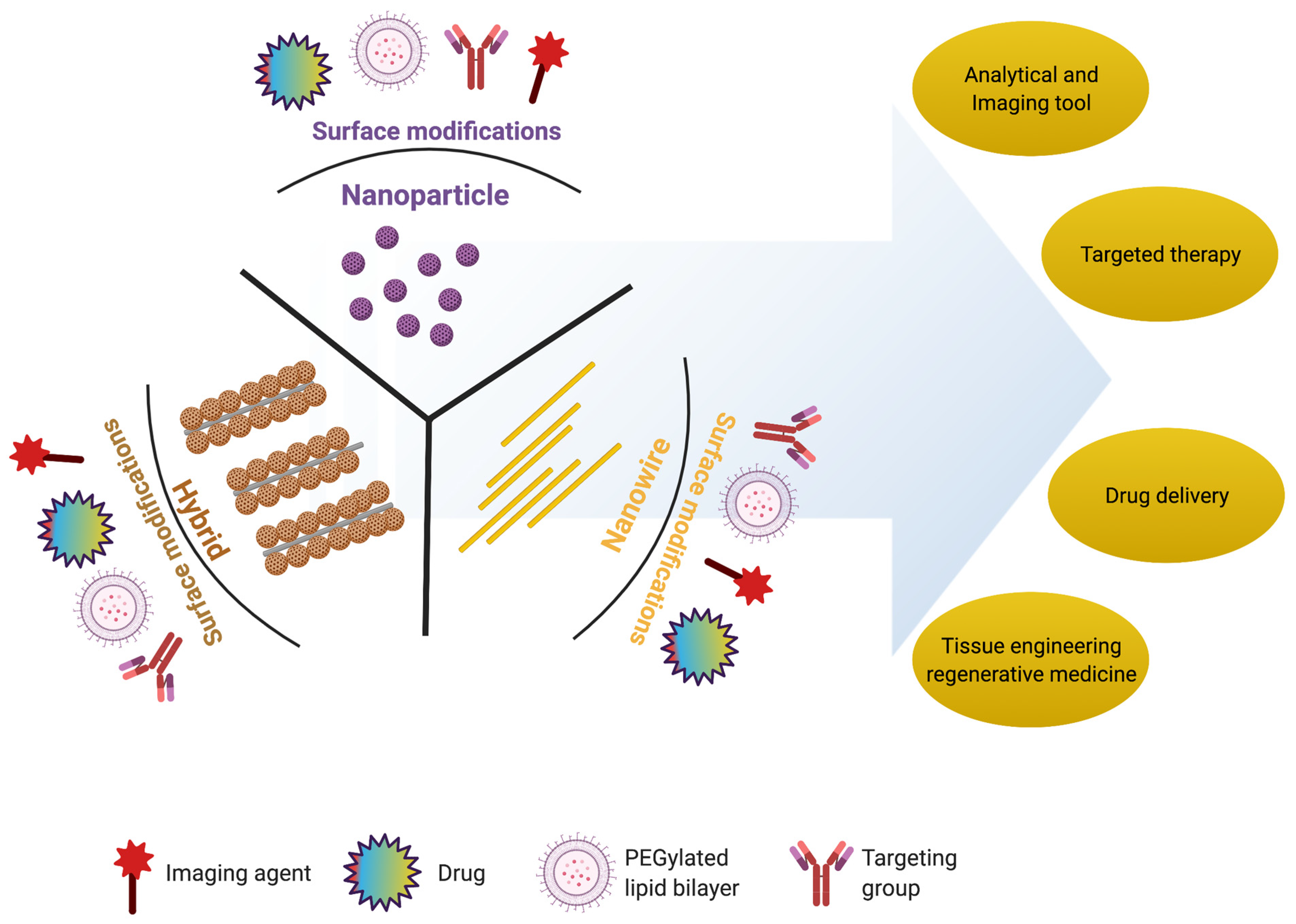
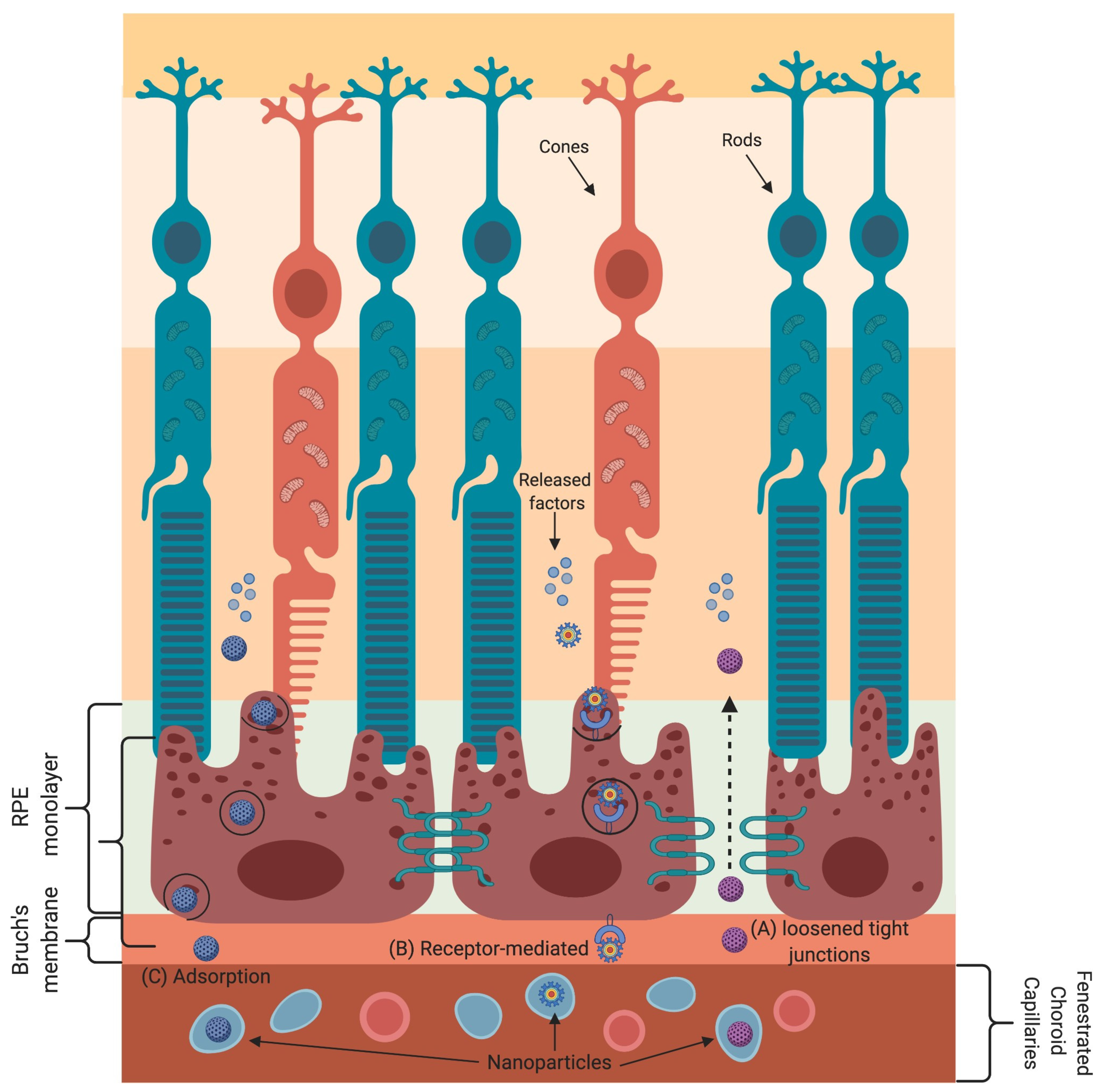
2. Nanomaterials for Retinal Regeneration

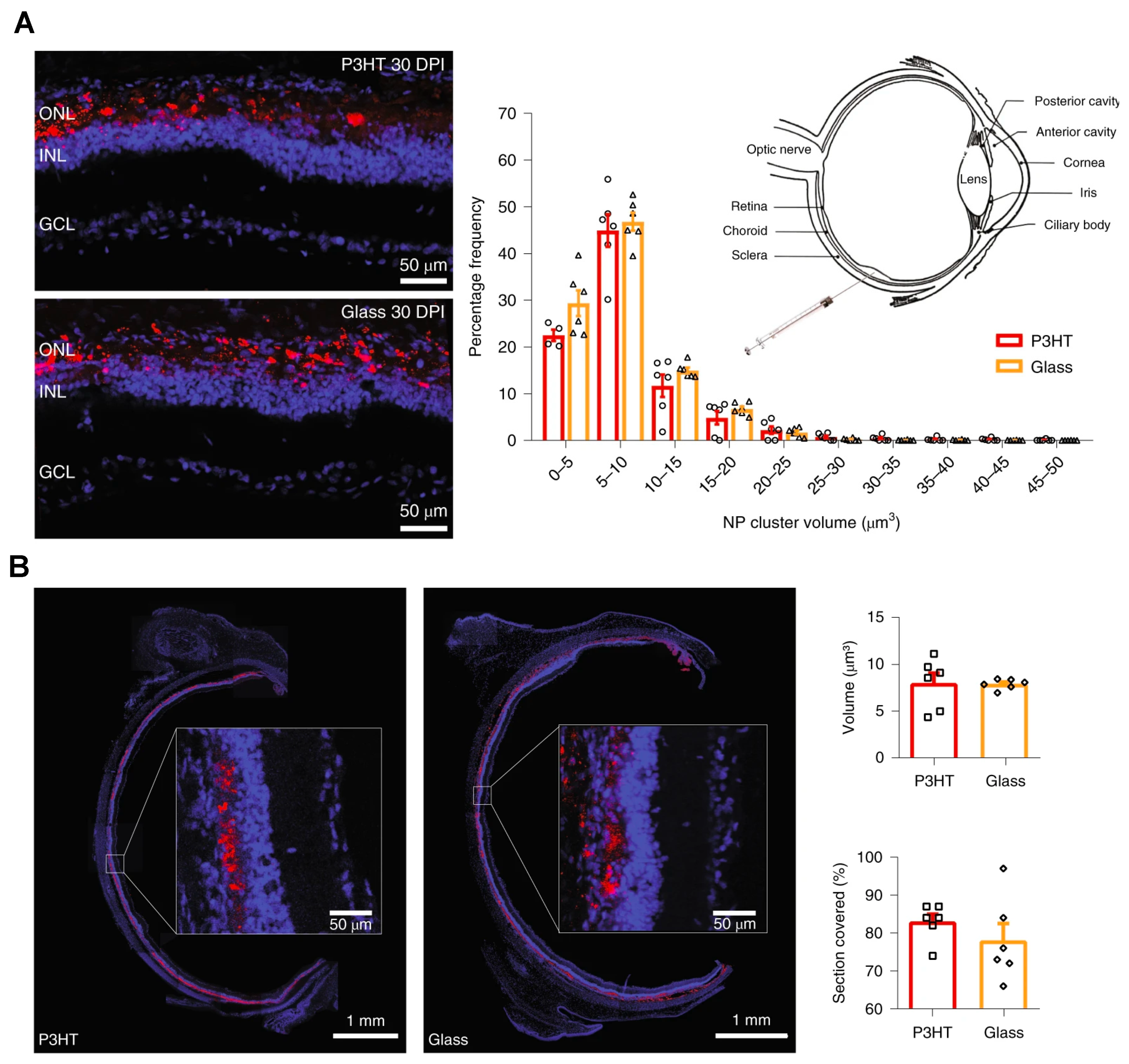
| Nanostructure | Nanomaterial | Size Range (nm) | Target Tissue/Cells | Ref. |
|---|---|---|---|---|
| Nanoparticles | Gold (Au) (diameter) | 3–5 | Choroidal and retinal endothelial cells | [42] |
| 10–12 | Retina of rabbit | [43] | ||
| 10–20 | Photoreceptor precursor transplantation | [44] | ||
| 80 | Retinal cells | [45] | ||
| 20–80 | Nucleus and mitochondria of retinal cells | [46] | ||
| 5–20 | Blood–retinal barrier | [44,47,48] | ||
| Gold (Au) nanodisk | Thickness: 20 Diameter: 160 | Retina | [47] | |
| Silver (Ag) (diameter) | 20–80 | Bovine retinal endothelial cells | [49] | |
| 40–50 | Porcine retinal endothelial cells | [50] | ||
| Superparamagnetic iron oxide nanoparticles | Diameter: 5–20 | Retina | [51] | |
| Magnetite | 10 | Retina and cells | [52,53] | |
| NWs | Poly (ε-caprolactone) (PCL) membranes | Length: 2500 | Implantation into subretinal space | [54] |
| Gallium phosphide (GaP) | Length: 500–4000 | Retinal cells | [55] | |
| n-type silicon | Length: 4400 | Retinal cells | [56] | |
| Gold (Au) nanorods | Thickness: 10–35 | Retinal cells and photoreceptors | [57] | |
| Hybrid nanostructure | Gold NPs coated over titania (TiO2) NWs | Au NP diameter: 5–15 TiO2 NW length: 2000 | Artificial photoreceptors | [54,58,59,60,61] |
| Gallium phosphide (GaP) rod and cone | Length: 20–2500 | Ganglion cells, and bipolar cells | [55] | |
| Gold NPs coated over silicon NWs | Au NP diameter: 5–10 NW length: 500–2500 | Artificial photoreceptors | [62,63] | |
| Thin film functionalized with the NPs | Diameter: 5–50 | Photoreceptors | [64,65] | |
| p–n junction silicon NWs | NW length: 10–120 | Membranes of live bipolar cells | [66] | |
| Au-coated carbon nanotube (Au-CNT) | Au NP diameter: 5–20 CNT length: 500–2500 | Subretinal space of mice | [67] | |
| Iridium oxide (IrOx) combined with reduced graphene oxide | IrOx diameter: 2–25 CNT length: 2–2500 | Subretinal implant into live mice | [68] | |
| Iridium oxide (IrOx) coated with CNT | IrOx diameter: 5–25 CNT length: 500–2500 | Retinal cells/tissues | [52,69,70,71,72] | |
| Core–shell-structured β-NaYF4:20%Yb, 2%Er@β-NaYF4 nanoparticles | Diameter: 30–40 | Subretinal space of mice | [15] | |
| Nanoscaffolds | Natural polymer: gelatin, fibrin, chitosan, laminin, and hyaluronic acid | Diameter/porosity: 100–200 | Extracellular matrix and cell attachment | [73,74,75,76,77,78] |
| Synthetic polymer: poly (lactic-co-glycolic acid) (PLGA), poly (ε-caprolactone) (PCL), poly (L-lactic acid) (PLA), polyimide, and poly (l-lactide-co-ε-caprolactone) | Diameter/porosity: 50–500 | RPE, biological activity, extracellular matrix, and cell attachment | [79,80,81,82] | |
| Biohybrid: nanofibers of Bruch’s membrane | Diameter/porosity: 100–200 | RPE and biological activity | [83] |
2.1. Nanoparticles
2.2. Nanowires
2.3. Hybrid Nanostructures
2.4. Nanoscaffolds
3. Studies on the Application of Nano-Biomaterials for Retinal Regeneration
3.1. In Vitro Studies on Nano-Biomaterial Implantation and Imaging
3.2. In Vivo Studies on Nano-Biomaterial Implantation and Imaging
3.3. Therapeutic Studies on Nano-Biomaterial Implantation and Imaging
4. Challenges and Future Perspectives
5. Method of Literature Search
Author Contributions
Funding
Institutional Review Board Statement
Informed Consent Statement
Data Availability Statement
Acknowledgments
Conflicts of Interest
Abbreviations
| ARPE-19 | Adult retinal pigment epithelial |
| Au-CNT | Gold coating on carbon nanotube |
| AuNPs | Gold NPs |
| Au-TiO2 | Gold-nanoparticle-decorated titania |
| CNT | Carbon nanotube |
| CPCB-RPE1 | California Project to Cure Blindness-Retinal Pigment Epithelium 1 |
| CT | Computed tomography |
| FDA | Food and Drug Administration |
| iPSC | Induced pluripotent stem cell |
| IR | Infrared |
| iRPE | Induced retinal pigment epithelial |
| mfERG | Multifocal electroretinography |
| MRI | Magnetic resonance imaging |
| MIONPs | Magnetic iron oxide nanoparticles |
| NIR | Near-infrared |
| NPs | Nanoparticles |
| NWs | Nanowires |
| OCT | Optical coherence tomography |
| pbUNCPs | Photoreceptor-binding upconversion nanoparticles |
| PCL | Poly (ε-caprolactone) |
| PDA | Polydopamine |
| P3HT | Poly (3-hexylthiophene) |
| POSS | Polyhedral oligomeric silsesquioxane |
| PLA | Poly (L-lactic acid) |
| PLGA | Poly (lactic-co-glycolic acid) |
| PMMA | Poly (methyl methacrylate) |
| PTT | Photothermic therapy |
| ROS | Reactive oxygen species |
| RPCs | Retinal progenitor cells |
| RPE | Retinal pigment epithelium |
| SEM | Scanning electron microscopy |
| Si NWs | Silicon nanowires |
| TEM | Transmission electron microscope |
| TUNEL | Deoxynucleotidyl transferase dUTP nick end labeling |
References
- Alzaraa, A.; Gravante, G.; Chung, W.Y.; Al-Leswas, D.; Bruno, M.; Dennison, A.R.; Lloyd, D.M. Targeted microbubbles in the experimental and clinical setting. Am. J. Surg. 2012, 204, 355–366. [Google Scholar] [CrossRef]
- Engel, E.; Michiardi, A.; Navarro, M.; Lacroix, D.; Planell, J.A. Nanotechnology in regenerative medicine: The materials side. Trends Biotechnol. 2008, 26, 39–47. [Google Scholar] [CrossRef]
- Willyard, C. Timeline: Regrowing the Body. Nature 2016, 540, S50–S51. [Google Scholar] [CrossRef]
- Yang, Y.; Leong, K.W. Nanoscale surfacing for regenerative medicine. Wiley Interdiscip. Rev. Nanomed. Nanobiotechnol. 2010, 2, 478–495. [Google Scholar] [CrossRef]
- Doshi, N.; Mitragotri, S. Designer biomaterials for nanomedicine. Adv. Funct. Mater. 2009, 19, 3843–3854. [Google Scholar] [CrossRef]
- Mitragotri., S.; Lahann, J. Physical approaches to biomaterial design. Nat. Mater. 2009, 8, 15–23. [Google Scholar] [CrossRef]
- Dobrovolskaia, M.A.; McNeil, S.E. Immunological properties of engineered nanomaterials. Nat. Nanotechnol. 2007, 2, 469–478. [Google Scholar] [CrossRef] [PubMed]
- Howes, P.D.; Chandrawati, R.; Stevens, M.M. Colloidal nanoparticles as advanced biological sensors. Science 2014, 346, 1247390. [Google Scholar] [CrossRef] [PubMed]
- Etheridge, M.L.; Campbell, S.A.; Erdman, A.G.; Haynes, C.L.; Wolf, S.M.; McCullough, J. The big picture on nanomedicine: The state of investigational and approved nanomedicine products. Nanomedicine 2013, 9, 1–14. [Google Scholar] [CrossRef] [PubMed]
- Karl, M.O.; Reh, T.A. Regenerative medicine for retinal diseases: Activating endogenous repair mechanisms. Trends Mol. Med. 2010, 16, 193–202. [Google Scholar] [CrossRef] [PubMed]
- Zarbin, M.A.; Montemagno, C.; Leary, J.F.; Ritch, R. Regenerative nanomedicine and the treatment of degenerative retinal diseases. Wiley Interdiscip. Rev. Nanomed. Nanobiotechnol. 2012, 4, 113–137. [Google Scholar] [CrossRef]
- Mains, J.; Wilson, C.G. The vitreous humor as a barrier to nanoparticle distribution. J. Ocul. Pharmacol. Ther. 2013, 29, 143–150. [Google Scholar] [CrossRef]
- Mitragotri, S.; Anderson, D.G.; Chen, X.; Chow, E.K.; Ho, D.; Kabanov, A.V.; Karp, J.M.; Kataoka, K.; Mirkin, C.A.; Petrosk, S.H.; et al. Accelerating the translation of nanomaterials in biomedicine. ACS Nano 2015, 9, 6644–6654. [Google Scholar] [CrossRef] [PubMed]
- Tang, J.; Qin, N.; Chong, Y.; Diao, Y.; Yiliguma; Wang, Z.; Xue, T.; Jiang, M.; Zhang, J.; Zheng, G. Nanowire arrays restore vision in blind mice. Nat. Commun. 2018, 9, 786. [Google Scholar] [CrossRef] [PubMed]
- Ma, Y.; Bao, J.; Zhang, Y.; Li, Z.; Zhou, X.; Wan, C.; Huang, L.; Zhao, Y.; Han, G.; Xue, T. Mammalian near-infrared image vision through injectable and self-powered retinal nanoantennae. Cell 2019, 177, 243–255. [Google Scholar] [CrossRef]
- Chen, J.; Patil, S.; Seal, S.; Mcginnis, J.F. Rare earth nanoparticles prevent retinal degeneration induced by intracellular peroxides. Nat. Nanotechnol. 2006, 1, 142–150. [Google Scholar] [CrossRef] [PubMed]
- Liu, X.L.; Chen, S.; Zhang, H.; Zhou, J.; Fan, H.M.; Liang, X.J. Magnetic nanomaterials for advanced regenerative medicine: The promise and challenges. Adv. Mater. 2019, 31, e1804922. [Google Scholar] [CrossRef]
- Desai, N. Challenges in development of nanoparticle-based therapeutics. AAPS J. 2012, 14, 282–295. [Google Scholar] [CrossRef]
- Hao, R.; Xing, R.; Xu, Z.; Hou, Y.; Gao, S.; Sun, S. Synthesis, functionalization, and biomedical applications of multifunctional magnetic nanoparticles. Adv. Mater. 2010, 22, 2729–2742. [Google Scholar] [CrossRef]
- Gao, Y.; Lim, J.; Teoh, S.H.; Xu, C. Emerging translational research on magnetic nanoparticles for regenerative medicine. Chem. Soc. Rev. 2015, 44, 6306–6329. [Google Scholar] [CrossRef]
- Xu, C.; Sun, S. New forms of superparamagnetic nanoparticles for biomedical applications. Adv. Drug Deliv. Rev. 2013, 65, 732–743. [Google Scholar] [CrossRef] [PubMed]
- Liu, X.L.; Yang, Y.; Ng, C.T.; Zhao, L.Y.; Zhang, Y.; Bay, B.H.; Fan, H.M.; Ding, J. Magnetic Vortex Nanorings: A new class of hyperthermia agent for highly efficient in vivo regression of tumors. Adv. Mater. 2015, 27, 1939–1944. [Google Scholar] [CrossRef] [PubMed]
- Onoshima, D.; Yukawa, H.; Baba, Y. Multifunctional quantum dots-based cancer diagnostics and stem cell therapeutics for regenerative medicine. Adv. Drug Deliv. Rev. 2015, 95, 2–14. [Google Scholar] [CrossRef]
- de Mel, A.; Oh, J.T.; Ramesh, B.; Seifalian, A.M. Biofunctionalized quantum dots for live monitoring of stem cells: Applications in regenerative medicine. Regen Med. 2012, 7, 335–347. [Google Scholar] [CrossRef] [PubMed]
- Zhu, L.; Chang, D.W.; Dai, L.; Hong, Y. DNA damage induced by multiwalled carbon nanotubes in mouse embryonic stem cells. Nano Lett. 2007, 7, 3592–3597. [Google Scholar] [CrossRef]
- Tran, P.A.; Zhang, L.T.; Webster, A.J. Carbon nanofibers and carbon nanotubes in regenerative medicine. Adv. Drug Deliv. Rev. 2009, 61, 1097–1114. [Google Scholar] [CrossRef]
- Golovin, Y.I.; Gribanovsky, S.L.; Golovin, D.Y.; Zhigachev, A.O.; Klyachko, N.L.; Majouga, A.G.; Sokolsky, M.; Kabanov, A.V. The dynamics of magnetic nanoparticles exposed to non-heating alternating magnetic field in biochemical applications: Theoretical study. J. Nanopart. Res. 2017, 19, 59. [Google Scholar] [CrossRef]
- Muller, D.J.; Helenius, J.; Alsteens, D.; Dufrene, Y.F. Force probing surfaces of living cells to molecular resolution. Nat. Chem. Biol. 2009, 5, 383–390. [Google Scholar] [CrossRef] [PubMed]
- Wu, C.; Shen, Y.; Chen, M.; Wang, K.; Li, Y.; Cheng, Y. Recent advances in magnetic-nanomaterial-based mechanotransduction for cell fate regulation. Adv. Mater. 2018, 30, e1705673. [Google Scholar] [CrossRef] [PubMed]
- Monzel, C.; Vicario, C.; Piehler, J.; Coppey, M.; Dahan, M. Magnetic control of cellular processes using biofunctional nanoparticles. Chem. Sci. 2017, 8, 7330–7338. [Google Scholar] [CrossRef]
- Lee, N.; Yoo, D.; Ling, D.; Cho, M.H.; Hyeon, T.; Cheon, J. Iron oxide based nanoparticles for multimodal imaging and magnetoresponsive therapy. Chem. Rev. 2015, 115, 10637–10689. [Google Scholar] [CrossRef]
- Zhao, Q.; Yan, Z.; Chen, C.; Chen, J. Spinels: Controlled preparation, oxygen reduction/evolution reaction application, and beyond. Chem. Rev. 2017, 117, 10121–10211. [Google Scholar] [CrossRef]
- Wu, L.; Garcia, A.M.; Li, Q.; Sun, S. Organic phase syntheses of magnetic nanoparticles and their applications. Chem. Rev. 2016, 116, 10473–10512. [Google Scholar] [CrossRef] [PubMed]
- Noh, S.H.; Na, W.; Jang, J.T.; Lee, J.H.; Lee, E.J.; Moon, S.H.; Lim, Y.; Shin, J.S.; Cheon, J. Nanoscale magnetism control via surface and exchange anisotropy for optimized ferrimagnetic hysteresis. Nano Lett. 2012, 12, 3716–3721. [Google Scholar] [CrossRef] [PubMed]
- Reimer, P.; Balzer, T. Ferucarbotran (Resovist): A new clinically approved res-specific contrast agent for contrast-enhanced MRI of the liver: Properties, clinical development, and applications. Eur. Radiol. 2003, 13, 1266–1276. [Google Scholar] [CrossRef] [PubMed]
- Shu, W.; Wang, Y.; Liu, C.; Li, R.; Pei, C.; Lou, W.; Lin, S.; Di, W.; Wan, J. construction of a plasmonic chip for metabolic analysis in cervical cancer screening and evaluation. Small Methods 2020, 4, 1900469. [Google Scholar] [CrossRef]
- Liu, J.; Cai, C.; Wang, Y.; Liu, Y.; Huang, L.; Tian, T.; Yao, Y.; Wei, J.; Chen, R.; Zhang, K.; et al. A biomimetic plasmonic nanoreactor for reliable metabolite detection. Adv. Sci. 2020, 7, 1903730. [Google Scholar] [CrossRef]
- Hu, D.; Zou, L.; Gao, Y.; Jin, Q.; Ji, J. Emerging nanobiomaterials against bacterial infections in postantibiotic era. View 2020, 1, 20200014. [Google Scholar] [CrossRef]
- Huang, L.; Gurav, D.D.; Wu, S.; Xu, W.; Vedarethinam, V.; Yang, J.; Su, H.; Wan, X.; Fang, Y.; Shen, B.; et al. A multifunctional platinum nanoreactor for point-of-care metabolic analysis. Matter 2019, 1, 1669–1680. [Google Scholar] [CrossRef]
- Yang, J.; Wang, R.; Huang, L.; Zhang, M.; Niu, J.; Bao, C.; Shen, N.; Dai, M.; Guo, Q.; Wang, Q.; et al. Urine metabolic fingerprints encode subtypes of kidney diseases. Angew. Chem. Int. Ed. Engl. 2020, 59, 1703–1710. [Google Scholar] [CrossRef]
- Cao, J.; Shi, X.; Gurav, D.D.; Huang, L.; Su, H.; Li, K.; Niu, J.; Zhang, M.; Wang, Q.; Jiang, M.; et al. Metabolic fingerprinting on synthetic alloys for medulloblastoma diagnosis and radiotherapy evaluation. Adv. Mater. 2020, 32, 2000906. [Google Scholar] [CrossRef]
- Chan, C.M.; Hsiao, C.Y.; Li, H.J.; Fang, J.Y.; Chang, D.C.; Hung, C.F. The inhibitory effects of gold nanoparticles on vegf-a-induced cell migration in choroid-retina endothelial cells. Int. J. Mol. Sci. 2020, 21, 109. [Google Scholar] [CrossRef] [PubMed]
- Bakri, S.J.; Pulido, J.S.; Mukherjee, P.; Marler, R.J.; Mukhopadhyay, D. Absence of histologic retinal of intravitreal nanogold in a rabbit model. Retina 2008, 28, 147–149. [Google Scholar] [CrossRef] [PubMed]
- Kim, J.H.; Kim, J.H.; Kim, K.W.; Kim, M.H.; Yu, Y.S. Intravenously administered gold nanoparticles pass through the blood-retinal barrier depending on the particle size and induce no retinal toxicity. Nanotechnology 2009, 20, 505101. [Google Scholar] [CrossRef] [PubMed]
- Karakocak, B.B.; Raliya, R.; Davis, J.T.; Chavalmane, S.; Wang, W.N.; Ravi, N.; Biswas, P. Biocompatibility of gold nanoparticles in retinal pigment epitheial cell line. Toxicol. Vitro 2016, 37, 61–69. [Google Scholar] [CrossRef] [PubMed]
- Soderstjerna, E.; Bauer, P.; Cedervall, T.; Abdshill, H.; Johansson, F.; Johansson, U.E. Silver and gold nanoparticles exposure to in vitro cultured retina-studies on nanoparticle internalization, apoptosis, oxidative stress, glial- and microglial activity. PLoS ONE 2014, 9, e105359. [Google Scholar] [CrossRef] [PubMed]
- Song, H.B.; Wi, J.S.; Jo, D.H.; Kim, J.H.; Lee, S.W. Intraocular application of gold nanodisks optically tuned for optical coherence tomography: Inhibitory effect on retinal neovascularization without unbearable toxicity. Nanomedicine 2017, 13, 1901–1911. [Google Scholar] [CrossRef]
- Kim, S.J. Novel approaches for retinal drug and gene delivery. Transl. Vis. Sci. Technol. 2014, 3, 7. [Google Scholar] [CrossRef][Green Version]
- Sheikpranbabu, S.; Kalishwaralal, K.; Venkataraman, D.; Eom, S.H.; Park, J.; Gurunathan, S. Silver nanoparticles inhibit vegf-and il-1β-induced vascular permeability via src dependent pathway in porcine retinal endothelial cells. J. Nanobiotechnol. 2009, 7, 8. [Google Scholar] [CrossRef]
- Maya-Vetencourt., J.F.; Manfredi, G.; Mete, M.; Colombo, E.; Bramini, M.; Di Marco, S.; Shmal, D.; Mantero, G.; Dipalo, M.; Rocchi, A.; et al. Subretinally injected semiconducting polymer nanoparticles rescue vision in a rat model of retinal dystrophy. Nat. Nanotechnol. 2020, 15, 698–708. [Google Scholar] [CrossRef]
- Sharma, R.; Khristov, V.; Rising, A.; Jha, B.S.; Dejene, R.; Hotaling, N.; Li, Y.; Stoddard, J.; Stankewicz, C.; Wan, Q.; et al. Clinical-grade stem cell-derived retinal pigment epithelium patch rescues retinal degeneration in rodents and pigs. Sci. Transl. Med. 2019, 11, eaat5580. [Google Scholar] [CrossRef]
- Wang, K.; Tang, R.Y.; Zhao, X.B.; Li, J.J.; Lang, Y.R.; Jiang, X.X.; Sun, H.J.; Lin, Q.X.; Wang, C.Y. Covalent bonding of yigsr and rgd to pedot/pss/mwcnt-cooh composite material to improve the neural interface. Nanoscale 2015, 7, 18677–18685. [Google Scholar] [CrossRef]
- Ito, A.; Takizawa, Y.; Honda, H.; Hata, K.I.; Kagami, H.; Ueda, M.; Kobayashi, T. Tissue engineering using magnetite nanoparticles and magnetic force: Heterotypic layers of cocultured hepatocytes and endothelial cells. Tissue Eng. 2004, 10, 833–840. [Google Scholar] [CrossRef] [PubMed]
- Redenti, S.; Tao, S.L.; Yang, J.; Gu, P.; Klassen, H.; Saigal, S.; Desai, T.; Young, M.J. Retinal tissue engineering using mouse retinal progenitor cells and a novel biodegradable, thin-film poly (e-caprolactone) nanowire scaffold. J. Ocul. Biol. Dis. Inform. 2008, 1, 19–29. [Google Scholar] [CrossRef]
- Piret, G.; Perez, M.T.; Prinz, C.N. Neurite outgrowth and synaptophysin expression of postnatal cns neurons on gap nanowire arrays in long-term retinal cell culture. Biomaterials 2013, 34, 875–887. [Google Scholar] [CrossRef] [PubMed]
- Piret, G.; Perez, M.T.; Prinz, C.N. Substrate porosity induces phenotypic alterations in retinal cells cultured on silicon nanowires. RSC Adv. 2014, 4, 27888–27897. [Google Scholar] [CrossRef]
- Gabriele Sandrian, M.; Wollstein, G.; Schuman, J.S.; Bilonick, R.A.; Ling, Y.; Ishikawa, H.; Kagemann, L.; McKenna, K.C. Inflammatory Response to intravitreal injection of gold nanorods. Br. J. Ophthalmol. 2012, 96, 1522–1529. [Google Scholar] [CrossRef]
- Gurunathan, S.; Lee, K.; Kalishwaralal, K.; Sheikpranbabu, S.; Vaidyanathan, R.; Eom, S.H. Antiangiogenic properties of silver nanoparticles. Biomaterials 2009, 30, 6341–6350. [Google Scholar] [CrossRef] [PubMed]
- Dvir, T.; Timko, B.P.; Kohane, D.S.; Langer, R. Nanotechnological strategies for engineering complex tissues. Nat. Nanotechnol. 2011, 6, 13–22. [Google Scholar] [CrossRef] [PubMed]
- Tang, J.; Huo, Z.; Brittman, S.; Gao, H.; Yang, P. Solution-processed core-shell nanowires for efficient photovoltaic cells. Nat. Nanotechnol. 2011, 6, 568–572. [Google Scholar] [CrossRef] [PubMed]
- Christiansen, A.T.; Tao, S.L.; Smith, M.; Wnek, G.E.; Prause, J.U.; Young, M.J.; Klassen, H.; Kaplan, H.J.; la Cour, M.; Kiilgaard, J.F. Subretinal implantation of electrospun, short nanowire, and smooth poly (ε-caprolactone) scaffolds to the subretinal space of porcine eyes. Stem Cells Int. 2012, 2012, 454295. [Google Scholar] [CrossRef]
- Kang, M.; Lee, H.; Kang, T.; Kim, B. Synthesis, properties, and biological application of perfect crystal gold nanowires: A review. J. Mater. Sci. Technol. 2015, 31, 573–580. [Google Scholar] [CrossRef]
- Lee, S.; Jung, S.W.; Ahn, J.; Yoo, H.J.; Oh, S.J.; Cho, D.D. Microelectrode array with integrated nanowire fet switches for high-resolution retinal prosthetic systems. J. Micromech. Microeng. 2014, 24, 075018. [Google Scholar] [CrossRef]
- SanMartin, A.; Johansson, F.; Samuelson, L.; Prinz, C.N. Microarray analysis reveals moderate gene expression changes in cortical neural stem cells cultured on nanowire arrays. J. Nanosci. Nanotechnol. 2014, 14, 4880–4885. [Google Scholar] [CrossRef]
- Gautam, V.; Rand, D.; Hanein, Y.; Narayan, K. A polymer optoelectronic interface provides visual cues to a blind retina. Adv. Mater. 2014, 26, 1751–1756. [Google Scholar] [CrossRef] [PubMed]
- Parameswaran, R.; Carvalho-de- Souza, J.L.; Jiang, Y.; Burke, M.J.; Zimmerman, J.F.; Koehler, K.; Phillips, A.W.; Yi, J.; Adams, E.J.; Bezanilla, F.; et al. Photoelectrochemical modulation of neuronal activity with free-standing coaxial silicon nanowires. Nat. Nanotechnol. 2018, 13, 260–266. [Google Scholar] [CrossRef] [PubMed]
- Vafaiee, M.; Mohammadpour, R.; Vossoughi, M.; Elham Asadian, E.; Janahmadi, M.; Sasanpour, P. Carbon nanotube modified microelectrode array for neural interface. Front. Bioeng. Biotechnol. 2021, 8, 582713. [Google Scholar] [CrossRef]
- Carretero, N.M.; Lichtenstein, M.P.; Pérez, E.; Cabana, L.; Suñol, C.; Casañ-Pastor, N. IrOx–carbon nanotube hybrids: A nanostructured material for electrodes with increased charge capacity in neural systems. Acta Biomater. 2014, 10, 4548–4558. [Google Scholar] [CrossRef]
- Pérez, E.; Lichtenstein, M.; Suñol, C.; Casañ-Pastor, N. Coatings of nanostructured pristine graphene-irox hybrids for neural electrodes: Layered stacking and the role of non-oxygenated graphene. Mater. Sci. Eng. C Mater. Biol. Appl. 2015, 55, 218–226. [Google Scholar] [CrossRef]
- Carretero, N.M.; Lichtenstein, M.; Pérez, E.; Sandoval, S.; Tobias, G.; Suñol, C.; Casan-Pastor, N. Enhanced charge capacity in iridium oxide-graphene oxide hybrids. Electrochim. Acta 2015, 157, 369–377. [Google Scholar] [CrossRef]
- Xiao, H.; Zhang, M.; Xiao, Y.; Che, J. A feasible way for the fabrication of single walled carbon nanotube/polypyrrole composite film with controlled pore size for neural interface. Colloids Surf. B Biointerfaces 2015, 126, 138–145. [Google Scholar] [CrossRef] [PubMed]
- Zhou, H.; Cheng, X.; Rao, L.; Li, T.; Duan, Y.Y. Poly (3, 4-ethylenedioxythiophene)/multiwall carbon nanotube composite coatings for improving the stability of microelectrodes in neural prostheses applications. Acta Biomater. 2013, 9, 6439–6449. [Google Scholar] [CrossRef]
- Rose, J.B.; Pacelli, S.; Haj, A.J.E.; Dua, H.S.; Hopkinson, A.; White, L.J.; Rose, F.R.A.J. Gelatin-based materials in ocular tissue engineering. Materials 2014, 7, 3106–3135. [Google Scholar] [CrossRef] [PubMed]
- White, C.E.; Olabisi, R.M. Scaffolds for retinal pigment epithelial cell transplantation in age-related macular degeneration. J. Tissue Eng. 2017, 8. [Google Scholar] [CrossRef]
- Noorani, B.; Tabandeh, F.; Yazdian, F.; Soheili, Z.-S.; Shakibaie, M.; Rahmani, S. Thin natural gelatin/chitosan nanofibrous scaffolds for retinal pigment epithelium cells. Int. J. Polym. Mater. Polym. Biomater. 2018, 67, 754–763. [Google Scholar] [CrossRef]
- Karamichos, D. Ocular Tissue Engineering: Current and Future Directions. J. Funct. Biomater. 2015, 6, 77–80. [Google Scholar] [CrossRef]
- Hynes, S.R.; Lavik, E.B. A tissue-engineered approach towards retinal repair: Scaffolds for cell transplantation to the subretinal space. Graefes Arch. Clin. Exp. Ophthalmol. 2010, 248, 763–778. [Google Scholar] [CrossRef]
- Pritchard, C.D.; Arnér, K.M.; Neal, R.A.; Neeley, W.L.; Bojo, P.; Bachelder, E.; Holz, J.; Watson, N.; Botchwey, E.A.; Langer, R.S.; et al. The use of surface modified poly (glycerol-co-sebacic acid) in retinal transplantation. Biomaterials 2010, 31, 2153–2162. [Google Scholar] [CrossRef]
- Warnke, P.H.; Alamein, M.; Skabo, S.; Stephens, S.; Bourke, R.; Heiner, P.; Liu, Q. Primordium of an artificial Bruch’s membrane made of nanofibers for engineering of retinal pigment epithelium cell monolayers. Acta Biomater. 2013, 9, 9414–9422. [Google Scholar] [CrossRef] [PubMed]
- McHugh, K.J.; Tao, S.L.; Saint-Geniez, M. Porous poly(ε-caprolactone) scaffolds for retinal pigment epithelium transplantation. Investig. Ophthalmol. Vis. Sci. 2014, 55, 1754–1762. [Google Scholar] [CrossRef] [PubMed]
- Giordano, G.G.; Thomson, R.C.; Ishaug, S.L.; Mikos, A.G.; Cumber, S.; Garcia, C.A.; Lahiri-Munir, D. Retinal pigment epithelium cells cultured on synthetic biodegradable polymers. J. Biomed. Mater. Res. 1997, 34, 87–93. [Google Scholar] [CrossRef]
- Ilmarinen, T.; Hiidenmaa, H.; Kööbi, P.; Nymark, S.; Sorkio, A.; Wang, J.H.; Stanzel, B.V.; Thieltges, F.; Alajuuma, P.; Oksala, O.; et al. Ultrathin Polyimide Membrane as Cell Carrier for Subretinal Transplantation of Human Embryonic Stem Cell Derived Retinal Pigment Epithelium. PLoS ONE 2015, 10, e0143669. [Google Scholar] [CrossRef] [PubMed]
- Xiang, P.; Wu, K.C.; Zhu, Y.; Xiang, L.; Li, C.; Chen, D.L.; Chen, F.; Xu, G.; Wang, A.; Li, M.; et al. A novel Bruch’s membrane-mimetic electrospun substrate scaffold for human retinal pigment epithelium cells. Biomaterials 2014, 35, 9777–9788. [Google Scholar] [CrossRef] [PubMed]
- Kim, J.H.; Kim, M.H.; Jo, D.H.; Yu, Y.S.; Lee, T.J.; Kim, J.H. The inhibition of retinal neovascularization by gold nanoparticles via suppression of vegfr-2 activation. Biomaterials 2011, 32, 1865–1871. [Google Scholar] [CrossRef] [PubMed]
- Joris, F.; Manshian, B.B.; Peynshaert, K.; De Smedt, S.C.; Braeckmans, K.; Soenen, S.J. Assessing nanoparticle toxicity in cell-based assays: Influence of cell culture parameters and optimized models for bridging the in vitro-in vivo gap. Chem. Soc. Rev. 2013, 42, 8339–8359. [Google Scholar] [CrossRef]
- Li, Y.; Yue, T.T.; Yang, K.; Zhang, X.R. Molecular modeling of the relationship between nanoparticle shape anisotropy and endocytosis kinetics. Biomaterials 2012, 33, 4965–4973. [Google Scholar] [CrossRef]
- Ngwa, W.; Makrigiorgos, G.M.; Berbeco, R. Gold nanoparticle enhancement of stereotactic radiosurgery for neovascular age-related macular degeneration. Phys. Med. Biol. 2012, 57, 6371–6380. [Google Scholar] [CrossRef]
- Ngwa, W.; Makrigiorgos, G.M.; Berbeco, R. SU-E-T-408: Enhancing stereotactic radiosurgery for neovascular age-related macular degeneration, using gold nanoparticles. Med. Phys. 2012, 39, 3798. [Google Scholar] [CrossRef]
- Farjo, K.M.; Ma, J.X. The potential of nanomedicine therapies to treat neovascular disease in the retina. J. Angiogenes Res. 2010, 2, 21. [Google Scholar] [CrossRef]
- Diebold, Y.; Calonge, M. Applications of nanoparticles in ophthalmology. Prog. Retin. Eye Res. 2010, 29, 596–609. [Google Scholar] [CrossRef]
- Hayashi, A.; Naseri, A.; Pennesi, M.E.; de Juan, E., Jr. Subretinal delivery of immunoglobulin g with gold nanoparticles in the rabbit eye. Jpn. J. Ophthalmol. 2009, 53, 249–256. [Google Scholar] [CrossRef]
- De Matteis, V.; Cascione, M.; Cristina, C.; Rinaldi, R. Engineered gold nanoshells killing tumor cells: New perspectives. Curr. Pharm. Des. 2019, 25, 1477–1489. [Google Scholar] [CrossRef]
- Weissleder, R. A clearer vision for in vivo imaging. Nat. Biotechnol. 2001, 19, 316–317. [Google Scholar] [CrossRef] [PubMed]
- Hammarin, G.; Persson, H.; Dabkowska, A.P.; Prinz, C.N. Enhanced laminin adsorption on nanowires compared to flat surfaces. Colloids Surf. B. Biointerfaces 2014, 122, 85–89. [Google Scholar] [CrossRef]
- Wang, R.; Huang, X.; Liu, G.; Wang, W.; Dong, F.; Li, Z. Fabrication and characterization of a parylene-based three-dimensional microelectrode array for use in retinal prosthesis. J. Microelectromech. Syst. 2010, 19, 367–374. [Google Scholar] [CrossRef]
- Pan, Y.L.; Noda, T.; Sasagawa, K.; Tokuda, T.; Ohta, J. Sputtering condition optimization of sputtered irox and tin stimulus electrodes for retinal prosthesis. IEEJ Trans. 2013, 8, 310–312. [Google Scholar] [CrossRef]
- Stett, A.; Barth, W.; Weiss, S.; Haemmerle, H.; Zrenner, E. Electrical multisite stimulation of the isolated chicken retina. Vis. Res. 2000, 40, 1785–1795. [Google Scholar] [CrossRef]
- Loudin, J.; Simanovskii, D.; Vijayraghavan, K.; Sramek, C.; Butterwick, A.; Huie, P.; Mclean, G.Y.; Palanker, D.V. Optoelectronic retinal prosthesis: System design and performance. J. Neural Eng. 2007, 4, S72–S84. [Google Scholar] [CrossRef]
- Sealy, C. Nanowires promise new ways to restore vision and movement. Nano Today 2018, 20, 1–2. [Google Scholar]
- Chinh, V.D.; Speranza, G.; Migliaresi, C.; Van Chuc, N.; Tan, V.M.; Phuong, N.T. Synthesis of gold nanoparticles decorated with multiwalled carbon nanotubes (Au-MWCNTs) via cysteaminium chloride functionalization. Sci. Rep. 2019, 9, 5667. [Google Scholar] [CrossRef] [PubMed]
- Shi, X.; Xiao, Y.; Xiao, H.; Harris, G.; Wang, T.; Che, J. Topographic guidance based on microgrooved electroactive composite films for neural interface. Colloids Surf. B Biointerfaces 2016, 145, 768–776. [Google Scholar] [CrossRef]
- Kolarcik, C.L.; Catt, K.; Rost, E.; Albrecht, I.N.; Bourbeau, D.; Du, Z.; Kozai, T.D.; Luo, X.; Weber, D.J.; Cui, X.T. Evaluation of poly (3,4-ethylenedioxythiophene)/carbon nanotube neural electrode coatings for stimulation in the dorsal root ganglion. J. Neural Eng. 2014, 12, 016008. [Google Scholar] [CrossRef]
- Yanai, A.; Häfeli, U.O.; Metcalfe, A.L.; Soema, P.; Addo, L.; Gregory-Evans, C.Y.; Po, K.; Shan, X.; Moritz, O.L.; Gregory-Evans, K. Focused magnetic stem cell targeting to the retina using superparamagnetic iron oxide nanoparticles. Cell Transplant. 2012, 21, 1137–1148. [Google Scholar] [CrossRef] [PubMed]
- Ito, A.; Hibino, E.; Kobayashi, C.; Terasaki, H.; Kagami, H.; Ueda, M.; Kobayashi, T.; Honda, H. Construction and delivery of tissue-engineered human retinal pigment epithelial cell sheets, using magnetite nanoparticles and magnetic force. Tissue Eng. 2005, 11, 489–496. [Google Scholar] [CrossRef] [PubMed]
- Yang, P.; Zhang, S.; Chen, X.; Liu, X.; Wang, Z.; Li, Y. Recent developments in polydopamine fluorescent nanomaterials. Mater. Horiz. 2020, 7, 746. [Google Scholar] [CrossRef]
- Li, Z.; Li, H.; Zhang, J.; Liu, X.; Gu, Z.; Li, Y. Ultrasmall nanoparticle ROS scavengers based on polyhedral oligomeric silsesquioxanes. Chin. J. Polym. Sci. 2020, 38, 1149–1156. [Google Scholar] [CrossRef]
- Li, Z.; Hu, J.; Yang, L.; Zhang, X.; Liu, X.; Wang, Z.; Li, Y. Integrated POSS-dendrimer nanohybrid materials: Current status and future perspective. Nanoscale 2020, 12, 11395–11415. [Google Scholar] [CrossRef]
- Hotaling, N.A.; Khristov, V.; Wan, Q.; Sharma, R.; Jha, B.S.; Lotfi, M.; Maminishkis, A.; Simon, C.G., Jr.; Bharti, K. Nanofiber scaffold-based tissue-engineered retinal pigment epithelium to treat degenerative eye diseases. J. Ocul. Pharmacol. Ther. 2016, 32, 272–285. [Google Scholar] [CrossRef] [PubMed]
- Santos-Ferreira, T.; Llonch, S.; Borsch, O.; Postel, K.; Haas, J.; Ader, M. Retinal transplantation of photoreceptors results in donor–host cytoplasmic exchange. Nat. Commun. 2016, 7, 13028. [Google Scholar] [CrossRef] [PubMed]
- Biazar, E.; Baradaran-Rafii, A.; Heidari-keshel, S.; Tavakolifard, S. Oriented nanofibrous silk as a natural scaffold for ocular epithelial regeneration. J. Biomater. Sci. Polym. Ed. 2015, 26, 1139–1151. [Google Scholar] [CrossRef]
- Komez, A.; Baran, E.T.; Erdem, U.; Hasirci, N.; Hasirci, V. Construction of a patterned hydrogel-fibrous mat bilayer structure to mimic choroid and bruch’s membrane layers of retina. J. Biomater. Res. A 2016, 104, 2166–2177. [Google Scholar] [CrossRef] [PubMed]
- Shrestha, B.K.; Shrestha, S.; Baral, E.R.; Lee, J.Y.; Kim, B.S.; Park, C.H.; Kim, C.S. π-Conjugated polyaniline-assisted flexible titania nanotubes with controlled surface morphology as regenerative medicine in nerve cell growth. Chem. Eng. J. 2019, 360, 701–713. [Google Scholar] [CrossRef]
- Jaggessar, A.; Mathew, A.; Wang, H.; Tesfamichael, T.; Yan, C.; Yarlagadda, P.K. Mechanical, bactericidal and osteogenic behaviours of hydrothermally synthesised tio2 nanowire arrays. J. Mech. Behav. Biomed. Mater. 2018, 80, 311–319. [Google Scholar] [CrossRef] [PubMed]
- Lin, H.I.; Kuo, S.W.; Yen, T.J.; Lee, O.K. Si NWs biophysically regulate the fates of human mesenchymal stem cells. Sci. Rep. 2018, 8, 12913. [Google Scholar] [CrossRef]
- Li, Z.; Persson, H.; Adolfsson, K.; Oredsson, S.; Prinz, C.N. Morphology of living cells cultured on nanowire arrays with varying nanowire densities and diameters. Sci. China Life Sci. 2018, 61, 427–435. [Google Scholar] [CrossRef]
- Masse, F.; Ouellette, M.; Lamoureux, G.; Boisselier, E. Gold nanoparticles in ophthalmology. Med. Res. Rev. 2019, 39, 302–327. [Google Scholar] [CrossRef]
- Chemla, Y.; Betzer, O.; Markus, A.; Farah, N.; Motiei, M. Gold nanoparticles for multimodal high-resolution imaging of transplanted cells for retinal replacement therapy. Nanomedicine 2019, 14, 1857–1871. [Google Scholar] [CrossRef]
- Leow, S.N.; Luu, C.D.; Hairul Nizam, M.H.; Mok, P.L.; Ruhaslizan, R.; Wong, H.S.; Halim, W.H.W.A.; Ng, M.H.; Ruszymah, B.H.I.; Chowdhury, S.R.; et al. Safety and efficacy of human wharton’s jelly-derived mesenchymal stem cells therapy for retinal degeneration. PLoS ONE 2015, 10, e0128973. [Google Scholar] [CrossRef]
- Thumann, G.; Viethen, A.; Gaebler, A.; Walter, P.; Kaempf, S.; Johnen, S.; Salz, A.K. The in vitro and in vivo behaviour of retinal pigment epithelial cells cultured on ultrathin collagen membranes. Biomaterials 2009, 30, 287–294. [Google Scholar] [CrossRef]
- Heidari, R.; Soheili, Z.S.; Samiei, S.; Ahmadieh, H.; Davari, M.; Nazemroaya, F.; Bagheri, A.; Deezagi, A. Alginate as a cell culture substrate for growth and differentiation of human retinal pigment epithelial cells. Appl. Biochem. Biotechnol. 2015, 175, 2399–2412. [Google Scholar] [CrossRef]
- Lai, J.-Y.; Li, Y.-T. Evaluation of cross-linked gelatin membranes as delivery carriers for retinal sheets. Mater. Sci. Eng. C 2010, 30, 677–685. [Google Scholar] [CrossRef]
- Kundu, J.; Michaelson, A.; Talbot, K.; Baranov, P.; Young, M.; Carrier, R. Decellularized retinal matrix: Natural platforms for human retinal progenitor cell culture. Acta Biomater. 2016, 31, 61–70. [Google Scholar] [CrossRef] [PubMed]
- Sabir, M.I.; Xu, X.; Li, L. A review on biodegradable polymeric materials for bone tissue engineering applications. J. Mater. Sci. 2009, 44, 5713–5724. [Google Scholar] [CrossRef]
- Shahmoradi, S.; Yazdian, F.; Tabandeh, F.; Soheili, Z.S.; Hatamian Zarami, A.S.; Navaei-Nigjeh, M. Controlled surface morphology and hydrophilicity of polycaprolactone toward human retinal pigment epithelium cells. Mater. Sci. Eng. C Mater. Biol. Appl. 2017, 73, 300–309. [Google Scholar] [CrossRef] [PubMed]
- Jin, S.; Yao, H.; Weber, J.L.; Melkoumian, Z.K.; Ye, K. A synthetic, xeno-free peptide surface for expansion and directed differentiation of human induced pluripotent stem cells. PLoS ONE 2012, 7, e50880. [Google Scholar]
- Calejo, M.T.; Ilmarinen, T.; Jongprasitkul, H.; Skottman, H.; Kellomäki, M. Honeycomb porous films as permeable scaffold materials for human embryonic stem cell-derived retinal pigment epithelium. J. Biomed. Mater. Res. A 2016, 104, 1646–1656. [Google Scholar] [CrossRef]
- Peng, C.H.; Chuang, J.H.; Wang, M.L.; Jhan, Y.Y.; Chien, K.H.; Chung, Y.C.; Hung, K.H.; Chang, C.C.; Lee, C.K.; Tseng, W.L.; et al. Laminin modification subretinal bio-scaffold remodels retinal pigment epithelium-driven microenvironment in vitro and in vivo. Oncotarget 2016, 7, 64631–64648. [Google Scholar] [CrossRef]
- Rahmani, S.; Tabandeh, F.; Faghihi, S.; Amoabediny, G.; Shakibaie, M.; Noorani, B.; Yazdian, F. Fabrication and characterization of poly(ε-caprolactone)/gelatin nanofibrous scaffolds for retinal tissue engineering. Int. J. Polym. Mater. Polym. Biomater. 2018, 67, 27–35. [Google Scholar] [CrossRef]
- White, C.; DiStefano, T.; Olabisi, R. The influence of substrate modulus on retinal pigment epithelial cells. J. Biomed. Mater. Res. A. 2017, 105, 1260–1266. [Google Scholar] [CrossRef]
- Thomson, H.A.; Treharne, A.J.; Walker, P.; Grossel, M.C.; Lotery, A.J. Optimisation of polymer scaffolds for retinal pigment epithelium (RPE) cell transplantation. Br. J. Ophthalmol. 2011, 95, 563–568. [Google Scholar] [CrossRef]
- Zhang, D.; Ni, N.; Chen, J.; Yao, Q.; Shen, B.; Zhang, Y.; Zhu, M.; Wang, Z.; Ruan, J.; Wang, J.; et al. Electrospun SF/PLCL nanofibrous membrane: A potential scaffold for retinal progenitor cell proliferation and differentiation. Sci. Rep. 2015, 5, 14326. [Google Scholar] [CrossRef] [PubMed]
- Sepahvandi, A.; Eskandari, M.; Moztarzadeh, F. Fabrication and characterization of SrAl2O4: Eu(2+)Dy(3+)/CS-PCL electrospun nanocomposite scaffold for retinal tissue regeneration. Mater. Sci. Eng. C Mater. Biol. Appl. 2016, 66, 306–314. [Google Scholar] [CrossRef] [PubMed]
- Nasehi, F.; Karshenas, M.; Nadri, S.; Barati, G.; Salim, A. Core-shell fibrous scaffold as a vehicle for sustained release of retinal pigmented epithelium-derived factor (PEDF) for photoreceptor differentiation of conjunctiva mesenchymal stem cells. J. Biomed. Mater. Res. A 2017, 105, 3514–3519. [Google Scholar] [CrossRef]
- Wu, W.; Yan, L.; Chen, S.; Li, Q.; Gu, Z.; Xu, H.; Yin, Z.Q. Investigating oxidation state-induced toxicity of PEGylated graphene oxide in ocular tissue using gene expression profiles. Nanotoxicology 2018, 12, 819–835. [Google Scholar] [CrossRef]
- Osakada, F.; Ikeda, H.; Mandai, M.; Wataya, T.; Watanabe, K.; Yoshimura, N.; Akaike, A.; Sasai, Y.; Takahashi, M. Toward the generation of rod and cone photoreceptors from mouse, monkey and human embryonic stem cells. Nat. Biotechnol. 2008, 26, 215–224. [Google Scholar] [CrossRef] [PubMed]
- Bernards, D.A.; Bhisitkul, R.B.; Wynn, P.; Steedman, M.R.; Lee, O.T.; Wong, F.; Thoongsuwan, S.; Desai, T.A. Ocular biocompatibility and structural integrity of micro- and nanostructured poly(caprolactone) films. J. Ocul. Pharmacol. Ther. 2013, 29, 249–257. [Google Scholar] [CrossRef]
- Tao, S.; Young, C.; Redenti, S.; Zhang, Y.; Klassen, H.; Desai, T.; Young, M.J. Survival, migration and differentiation of retinal progenitor cells transplanted on micro-machined poly (methyl methacrylate) scaffolds to the subretinal space. Lab Chip 2007, 7, 695–701. [Google Scholar] [CrossRef]
- Thomas, B.B.; Zhu, D.; Zhang, L.; Thomas, P.B.; Hu, Y.; Nazari, H.; Stefanini, F.; Falabella, P.; Clegg, D.O.; Hinton, D.R.; et al. Survival and functionality of hesc-derived retinal pigment epithelium cells cultured as a monolayer on polymer substrates transplanted in rcs rats. Investig. Ophthalmol. Vis. Sci. 2016, 57, 2877–2887. [Google Scholar] [CrossRef]
- Koss, M.J.; Falabella, P.; Stefanini, F.R.; Pfister, M.; Thomas, B.B.; Kashani, A.H.; Brant, R.; Zhu, D.; Clegg, D.O.; Hinton, D.R.; et al. Subretinal implantation of a monolayer of human embryonic stem cell-derived retinal pigment epithelium: A feasibility and safety study in Yucatán minipigs. Graefes Arch. Clin. Exp. Ophthalmol. 2016, 254, 1553–1565. [Google Scholar] [CrossRef]
- Wang, Y.; Liu, W.; Yuan, B.; Yin, X.; Li, Y.; Li, Z.; Cui, J.; Yuan, X.; Li, Y. The application of Methylprednisolone Nanoscale Zirconium-Porphyrin Metal-Organic Framework (MPS-NPMOF) in the treatment of photoreceptor degeneration. Int. J. Nanomed. 2019, 14, 9763–9776. [Google Scholar] [CrossRef]
- Yang, P.; Dong, Y.; Huang, D.; Zhu, C.; Liu, H.; Pan, X.; Wu, C. Silk fibroin nanoparticles for enhanced bio-macromolecule delivery to the retina. Pharm. Dev. Technol. 2019, 24, 575–583. [Google Scholar] [CrossRef]
- Marcus, M.; Smith, A.; Maswadeh, A.; Shemesh, Z.; Zak, I.; Motiei, M.; Schori, H.; Margel, S.; Sharoni, A.; Shefi, O. Magnetic Targeting of Growth Factors Using Iron Oxide Nanoparticles. Nanomaterials 2018, 8, 707. [Google Scholar] [CrossRef] [PubMed]
- Bennett, J.; Chung, D.C.; Maguire, A. Gene delivery to the retina: From mouse to man. Methods Enzym. 2012, 507, 255–257. [Google Scholar]
- Lassota, N.; Kiilgaard, J.F.; Prause, J.U.; la Cour, M. Correlation between clinical and histological features in a pig model of choroidal neovascularization. Graefes Arch. Clin. Exp. Ophthalmol. 2006, 244, 394–398. [Google Scholar] [CrossRef] [PubMed]
- Kashani, A.H.; Uang, J.; Mert, M.; Rahhal, F.; Chan, C.; Avery, R.L.; Dugel, P.; Chen, S.; Lebkowski, J.; Clegg, D.O.; et al. Surgical method for implantation of a biosynthetic retinal pigment epithelium monolayer for geographic atrophy: Experience from a phase 1/2a study. Ophthalmol. Retina 2020, 4, 264–273. [Google Scholar] [CrossRef] [PubMed]
- Kashani, A.H.; Lebkowski, J.S.; Rahhal, F.M.; Avery, R.L.; Salehi-Had, H.; Dang, W.; Lin, C.M.; Mitra, D.; Zhu, D.; Thomas, B.B.; et al. A bioengineered retinal pigment epithelial monolayer for advanced, dry age-related macular degeneration. Sci. Transl. Med. 2018, 10, eaao4097. [Google Scholar] [CrossRef]
- Yang, J.W.; Yu, Z.Y.; Cheng, S.J.; Chung, J.H.Y.; Liu, X.; Wu, C.Y.; Lin, S.F.; Chen, G.Y. Graphene oxide-based nanomaterials: An insight into retinal prosthesis. Int. J. Mol. Sci. 2020, 21, 2957. [Google Scholar] [CrossRef]
- Yan, J.; Peng, X.; Cai, Y.; Cong, W. Development of facile drug delivery platform of ranibizumab fabricated PLGA-PEGylated magnetic nanoparticles for age-related macular degeneration therapy. J. Photochem. Photobiol. B 2018, 183, 133–136. [Google Scholar] [CrossRef] [PubMed]
- Chittasupho, C.; Kengtrong, K.; Chalermnithiwong, S.; Sarisuta, N. Anti-angiogenesis by dual action of R5K peptide conjugated itraconazole nanoparticles. AAPS Pharm. Sci. Tech. 2020, 21, 74. [Google Scholar] [CrossRef]
- Mahlumba, P.; Kumar, P.; du Toit, L.C.; Poka, M.S.; Ubanako, P.; Choonara, Y.E. Fabrication and characterisation of a photo-responsive, injectable nanosystem for sustained delivery of macromolecules. Int. J. Mol. Sci. 2021, 22, 3359. [Google Scholar] [CrossRef]
- Jung, S.W.; Shin, J.Y.; Pi, K.; Goo, Y.S.; Cho, D.D. Neuron stimulation device integrated with silicon nanowire-based photodetection circuit on a flexible substrate. Sensors 2016, 16, 2035. [Google Scholar] [CrossRef] [PubMed]
- Mahaling, B.; Srinivasarao, D.A.; Raghu, G.; Kasam, R.K.; Bhanuprakash Reddy, G.; Katti, D.S. A non-invasive nanoparticle mediated delivery of triamcinolone acetonide ameliorates diabetic retinopathy in rats. Nanoscale 2018, 10, 16485–16498. [Google Scholar] [CrossRef] [PubMed]
- Radwan, S.E.; El-Kamel, A.; Zaki, E.I.; Burgalassi, S.; Zucchetti, E.; El-Moslemany, R.M. Hyaluronic-coated albumin nanoparticles for the non-invasive delivery of apatinib in diabetic retinopathy. Int. J. Nanomed. 2021, 16, 4481–4494. [Google Scholar] [CrossRef]


| Analysis | Nanomaterial | Form | Size (nm) | Cell Response | Ref. |
|---|---|---|---|---|---|
| In vitro | Poly (ε-caprolactone) (PCL) | NWs | Length: 2500 | ↑ expression of PKC and recoverin in RPCs; cells undergo differentiation | [54] |
| Gallium phosphide (GaP) | NWs | Length: 500–4000 | Extended growth of retinal cells | [61] | |
| n-type silicon | NWs | Length: 440 | Long-term and dense growth of mouse retinal cells | [95] | |
| Gold (Au) | Nanoparticle | Diameter: 5–100 | ARPE-19 cells undergo apoptosis upon AuNP internalization | [77] | |
| Diameter: 10–12 | Gold nanoparticles inhibit proliferation of ARPE-19 cells; no cytotoxicity | [16] | |||
| Diameter: 80 | Highly viable mesenchymal stem cells undergo differentiation and secrete various trophic factors | [15] | |||
| Gold (Au), silver (Ag) | Nanoparticle | Diameter: 20–80 | Increase uptake into retinal cells; ↑ apoptosis, oxidative stress, and microglia activation | [58] | |
| Gold (Au) | Nanodisk | Diameter: 160 | Inhibition of in vitro angiogenesis without cellular toxicity of HRMECs | [56] | |
| Hybrid nanoscaffolds | Combination of Antheraea pernyi silk fibroin (RWSF), PCL, and gelatin | Diameter/porosity: 90–210 | Increased expression of RPE marker genes (CRALBP, PEDF, VEGF, MITF, and PMEL 17 among others) | [83] | |
| In vivo | Poly (ε-caprolactone) (PCL) membranes | NWs | Length: 2500 | Successful implantation into subretinal space with limited tissue disruption and no inflammation | [54] |
| Gold (Au), titania (TiO2) | Au nanoparticle coated TiO2 NWs | AuNPs diameter: 5–15, TiO2 NW length: 2000 | AuNP-decorated TiO2 NW arrays restore light-sensitive visual responses in degenerated photoreceptors | [14] | |
| Gold (Au) | Nanodisk | Diameter: 160 | Intravitreal injection attenuates neovascularization in mouse model of oxygen-induced retinopathy | [56] | |
| Gold (Au) | Nanoparticle | Diameter: 20–100 | Intravitreal injection of gold nanoparticles passed through the blood–retinal barrier with no structural abnormality or cell death | [91] | |
| Gold (Au) | Nano-gold | Not reported | No retinal or optic nerve toxicity by intravitreal injection of nano-gold | [43,91] | |
| Gold (Au), poly (strenesulfate) | Poly (strenesulfate) or anti-CD90.2 antibody-coated Au nanorods (PSS-AuNRs) | Not reported | Intravitreal injection obscured the retinal signal and induced ocular inflammation | [57] | |
| Nanoscaffolds | Nanofibrous porous membrane | Diameter/porosity: 680 | Bruch’s membrane thickness changes with aging, and it correlates with RPE function | [83] | |
| Therapeutic | Gold (Au) | Nanoparticles | Diameter: 20 | AuNP-labeled photoreceptor precursor transplantation provides high-resolution long-term tracking and cell survival with no toxic effects on retina or cells | [91,117] |
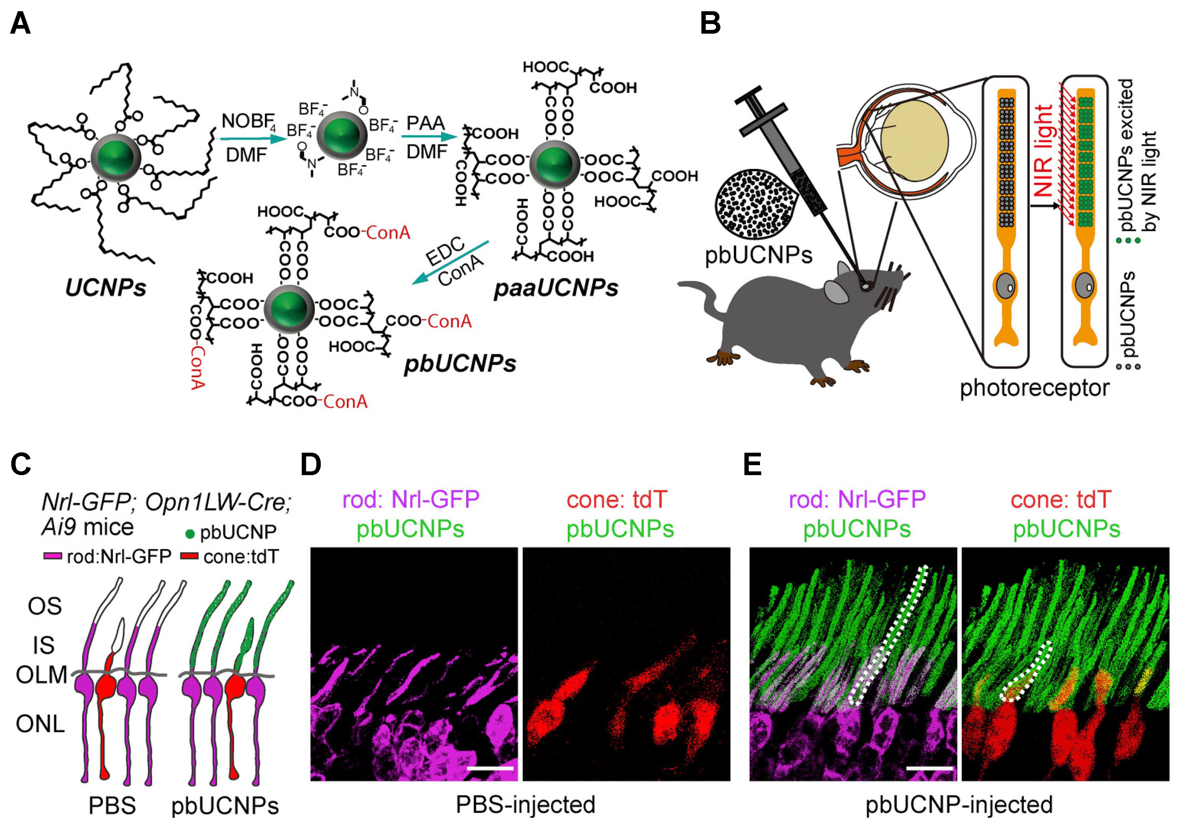
| Core–shell-structured β-NaYF4:20%Yb, 2%Er@β-NaYF4 | Nanoparticle (core–shell-structured upconversion nanoparticles (UCNPs)) | Diameter: 35–40 | Retinal pbUCNP injection extends the visual spectrum to the near infra-red range in mice | [15] | |
| Synthetic nanoscaffolds | Nanofibrous scaffolds | Diameter/porosity: 100–200 | Used as a cell replacement therapy | [108] |
Publisher’s Note: MDPI stays neutral with regard to jurisdictional claims in published maps and institutional affiliations. |
© 2021 by the authors. Licensee MDPI, Basel, Switzerland. This article is an open access article distributed under the terms and conditions of the Creative Commons Attribution (CC BY) license (https://creativecommons.org/licenses/by/4.0/).
Share and Cite
Sharma, R.; Sharma, D.; Hazlett, L.D.; Singh, N.K. Nano-Biomaterials for Retinal Regeneration. Nanomaterials 2021, 11, 1880. https://doi.org/10.3390/nano11081880
Sharma R, Sharma D, Hazlett LD, Singh NK. Nano-Biomaterials for Retinal Regeneration. Nanomaterials. 2021; 11(8):1880. https://doi.org/10.3390/nano11081880
Chicago/Turabian StyleSharma, Rahul, Deepti Sharma, Linda D. Hazlett, and Nikhlesh K. Singh. 2021. "Nano-Biomaterials for Retinal Regeneration" Nanomaterials 11, no. 8: 1880. https://doi.org/10.3390/nano11081880
APA StyleSharma, R., Sharma, D., Hazlett, L. D., & Singh, N. K. (2021). Nano-Biomaterials for Retinal Regeneration. Nanomaterials, 11(8), 1880. https://doi.org/10.3390/nano11081880