Review

Cross-Species Comparisons of Nanoparticle Interactions with Innate Immune Systems: A Methodological Review

by
Benjamin J. Swartzwelter
1,
Craig Mayall
2,
Andi Alijagic
3,
Francesco Barbero
4,
Eleonora Ferrari
5,
Szabolcs Hernadi
6,
Sara Michelini
7,
Natividad Isabel Navarro Pacheco
8,
Alessandra Prinelli
9,
Elmer Swart
10 and
Manon Auguste
11,*
1
Institute of Biochemistry and Cell Biology, National Research Council, 80131 Napoli, Italy
2
Department of Biology, Biotechnical Faculty, University of Liubljana, 1000 Ljubljana, Slovenia
3
Institute for Biomedical Research and Innovation, National Research Council, 90146 Palermo, Italy
4
Institut Català de Nanosciència i Nanotecnologia (ICN2), Bellaterra, 08193 Barcelona, Spain
5
Center for Plant Molecular Biology–ZMBP Eberhard-Karls University Tübingen, 72076 Tübingen, Germany
6
School of Biosciences, Cardiff University, Cardiff CF10 3AX, UK
7
Department of Biosciences, Paris-Lodron University Salzburg, 5020 Salzburg, Austria
8
Institute of Microbiology of the Czech Academy of Sciences, 142 20 Prague, Czech Republic
9
AvantiCell Science, Ltd., Ayr KA6 5HW, UK
10
UK Centre for Ecology and Hydrology, Wallingford OX10 8BB, UK
11
Department of Earth Environment and Life Sciences, University of Genova, 16126 Genova, Italy
*
Author to whom correspondence should be addressed.
Nanomaterials 2021, 11(6), 1528; https://doi.org/10.3390/nano11061528
Submission received: 18 February 2021 / Revised: 4 June 2021 / Accepted: 7 June 2021 / Published: 9 June 2021

Abstract

:
Many components of the innate immune system are evolutionarily conserved and shared across many living organisms, from plants and invertebrates to humans. Therefore, these shared features can allow the comparative study of potentially dangerous substances, such as engineered nanoparticles (NPs). However, differences of methodology and procedure between diverse species and models make comparison of innate immune responses to NPs between organisms difficult in many cases. To this aim, this review provides an overview of suitable methods and assays that can be used to measure NP immune interactions across species in a multidisciplinary approach. The first part of this review describes the main innate immune defense characteristics of the selected models that can be associated to NPs exposure. In the second part, the different modes of exposure to NPs across models (considering isolated cells or whole organisms) and the main endpoints measured are discussed. In this synergistic perspective, we provide an overview of the current state of important cross-disciplinary immunological models to study NP-immune interactions and identify future research needs. As such, this paper could be used as a methodological reference point for future nano-immunosafety studies.

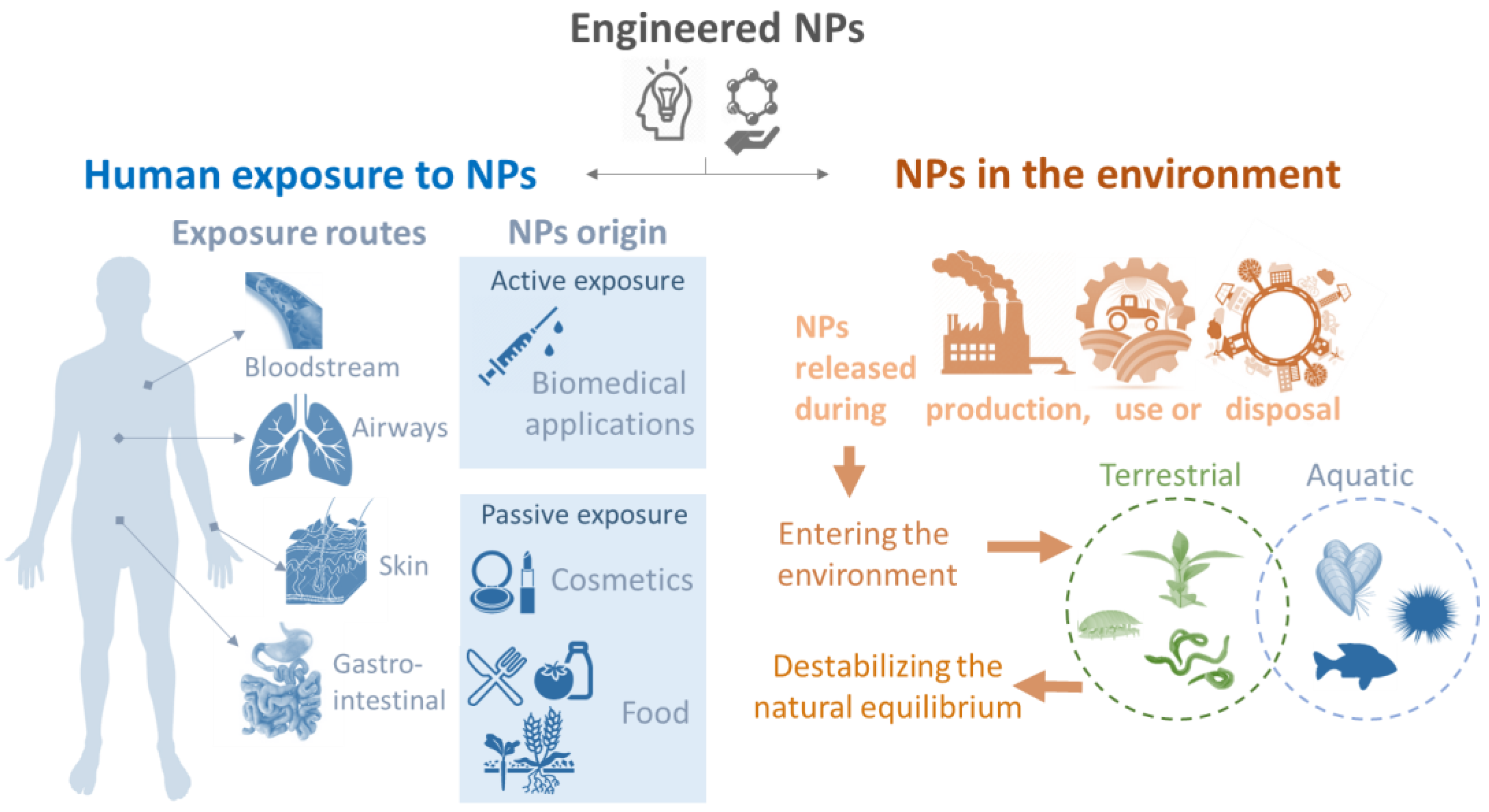
1. General Introduction: The Need for Studying Nanoparticle–Immune System Interactions

Over the last twenty years, there has been a significant growth in the research, development, and production of engineered NPs [1]. When materials are downsized to the nanoscale, novel physical and chemical properties emerge, conferring them with new and unique behaviors. Depending on their nature (e.g., composition, size, shape, surface state), these materials have remarkable optical, magnetic, electrical, catalytic, structural, and chemical properties, which can be exploited in many different sectors such as automotive, agricultural, pharmaceutical, and biomedical fields [2,3,4,5]. It is estimated that the global nanomaterial production in 2014 was between 0.3 and 1.6 million tons, with SiO2, TiO2 and ZnO nanomaterials being the most abundantly produced [6].
The wide utilization and increasing production of NPs has inevitably lead to an increase in humans and environmental exposure to these materials although exposure routes are not necessarily identical for different organisms. The expected increased exposure in human and environmental organisms has given rise to concerns regarding potential safety risks. The main exposure routes to NPs in both humans and environmental species are highlighted and summarized in Figure 1.
In humans, the first main exposure pathway is via intentional introduction of NPs, for instance during medical administration. The ability of some NPs to interact with molecular and cellular processes and to be target specific makes their use in drug delivery an attractive application. They have long been known to play an effective role in vaccination, acting not only as antigen carriers, but also as adjuvants that activate innate immunity and thereby increase the efficacy of antigen presentation [7]. They can also be valuable tools in medical imaging and diagnosis, and innovative new therapies [8]. Alongside the potential benefits of nanoparticle-based therapies, there is also a risk associated with parenteral introduction of novel substances, and thus there is a need to ensure that NPs will not negatively impact the normal functioning of the immune system [9,10,11,12,13]. Other interactions can arise from passive exposure such as through cosmetic products or food. Although NPs will likely first interact with epithelial and mucosal barriers, in some cases they are able to cross these barriers or potentially cause adverse effects, for example by interacting with the natural gut microbiome [14].
Although most NPs are not directly applied in the environment, many NPs used in consumer products or industry are expected to be released into the environment during production, use or during the disposal of products containing NPs [15]. Over the past decade, an increasing number of products containing NPs have been introduced into agricultural practices with the aim of increasing crop yield and reducing production costs [16]. In addition, the use of wastewater treatment plant biosolids as crop fertilizers can facilitate release of NPs into the terrestrial environment leading to exposure in soil organisms [17]. NPs can also reach aquatic environments, including seashores, through landfill leachates, or direct disposal of wastes (e.g., consumer products containing plastics) [18]. Once in the water, NPs can remain in suspension in the water column, interacting with planktonic organisms, or due to interactions with organic matter and/or their higher density, NPs can aggregate and deposit on the seafloor. This has been reflected by several models predicting NP concentrations within different regions which showed higher concentrations of NPs in sediments than surface water [19]. Therefore, benthic and sediment dwelling organisms are expected to be exposed to NPs, due to their feeding habit (e.g., filter, deposit feeders) [20]. In addition, some marine invertebrates possess an open (or semiopen) circulatory system, which is in direct contact with the external environment, eventually contributing to increased exposure.
Considering the many possible exposure and entry routes of NPs, defining common parameters for assessing organism-NP interactions is fundamental for allowing comparisons at different taxonomic levels. Innate immunity is a shared feature for every multicellular organism and the effector mechanisms of the innate immune system are the first line of defense that detect and protect the body from nonself objects such as NPs [21,22,23]. As is the case for natural pathogens, NPs have the potential to induce an immune response. In cases where NPs can elicit an immune response, there is a need to study the type and degree of this response, and the NP-immune interaction mechanisms rather than remaining limited to only measurements of acute toxicity. Comparative immunology, by its multidisciplinary approach may unravel fundamental mechanisms activated by NPs and help further global understanding regarding the effects of NPs.
Experiments in a laboratory are first necessary to allow the understanding of basic mechanisms under controlled conditions. However, carefully chosen models and assessment parameters are important with regard to future translocation to more realistic environmental exposure. To this end, models within this review have been selected which can be good indicators and representatives of their regional and global distribution and which are easy to maintain under laboratory conditions. Environmental models can be therefore compared across taxa and even to human cells, through both in vitro and/or in vivo approaches according to the model possibilities (Figure 2). Plant models are a compelling place to begin for assessment of NP-immune interactions. In particular Arabidopsis thaliana, a small flowering plant belonging to the Brassicaceae family, which is widely used in crop science studies and was also the first plant genome to be fully sequenced [24,25,26]. Among terrestrial invertebrates, earthworms belonging to the family Lumbricidae (Eisenia fetida) are abundant in the soil and play an essential role in soil formation, by facilitating nutrient cycling, fragmenting biomass and aeration of soil through bioturbation [27,28]. Similarly, terrestrial isopods, such as Porcelio scaber, are crustaceans which evolved to live on land, inhabiting the top-soil level. They are decomposers and play an important role in returning nutrients to the soil [29,30,31]. Their feeding habits makes it likely they will come into contact with environmental pollutants, including NPs, and thus represent interesting model species to study these interactions. The Mediterranean mussel Mytilus galloprovincialis and the sea urchin Paracentrotus lividus are both sessile marine invertebrates. Mussels are able to filter large quantities of water which they use for breathing and feeding, while sea urchins graze on the seafloor layer. These qualities, as well as the ease with which they can be harvested along seashores, make these good models in which to study invertebrate interactions with NPs [20,32,33,34].
This work is supported by the EU PANDORA project [35], which devoted effort to study the effects and mechanisms of action of NPs on the innate immunity of different models from across the tree of life. The general outcomes of the project were previously reported, summarizing the main findings but also to set future perspective and research direction in this field [21,36]. The remainder of this review will focus on the translatable aspects of experimental methodology, parameters and endpoints used, the suitably of the selected models when considering investigating NP effects, and the possibilities regarding research at the whole organism level (in vivo) or with isolated cells (in vitro).
Here we aim to: (i) give a short overview of the characteristics of various relevant innate immune models from across tree of life; and (ii) provide a comparative analysis of the methods used to study the interaction of NPs with these innate immune models.

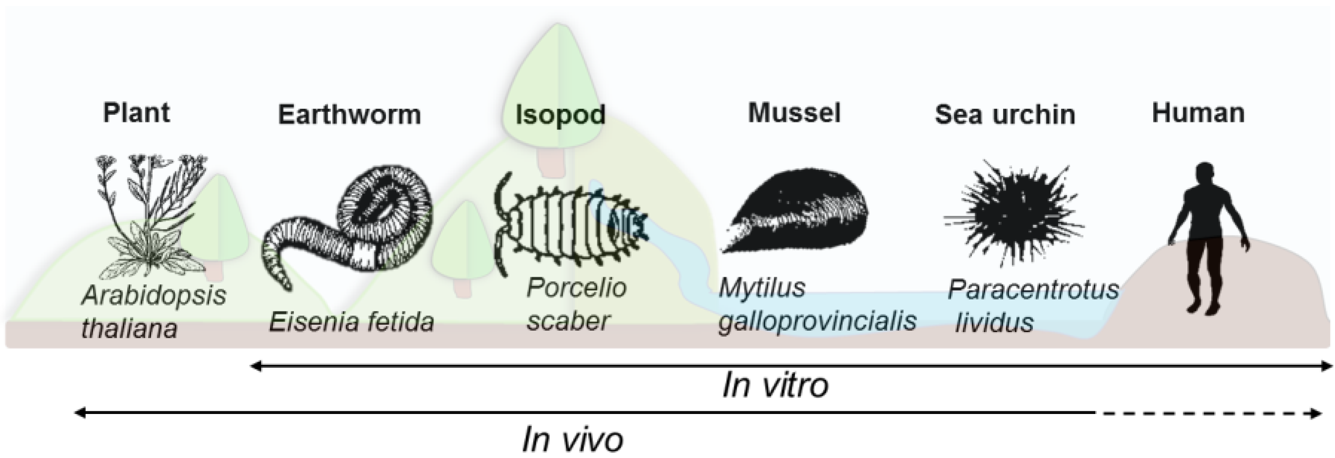
2. Short Description of the Innate Immune System for the Models of Interest

2.1. Generalities and Conserved Innate Immune Traits across the Selected Models

The ability to mount an immune response against external threats is a characteristic of every living organism. While increasing levels of immune complexity are found in higher organisms, at every stage of evolution there is present a basic initial host defense that has been characterized as innate immunity. Innate immunity is a fast, standardized, nonspecific response which includes multiple levels of defense mechanisms, beginning with physicochemical barriers (e.g., shell, mucosal or epithelial barrier) [37,38]. Further mechanisms of defense rely on dual components of the immune system, the immune cells (e.g., monocytes, macrophages for vertebrates, or hemocytes, coelomocytes for invertebrates) and the production of humoral factors. Innate immune cells found in the circulating fluids of invertebrates can have different names and the cellular portion of their innate immunity relies on these unique cells. These cells can be subdivided into different cell populations, such as granular or hyaline cells, and they have distinct roles and can trigger a specific response upon encountering threatening nonself material. Only plants lack these specialized immune cells, but in plants all the cells are believed to be able to mount a defensive response to foreign attack [39]. Complex machinery, including cells and humoral factors is involved in recognition of nonself material, and especially in detecting domains called pathogen/microbe associated molecular patterns (PAMPs/MAMPs) that are typically displayed on the surface of bacterial, fungal, and parasitic organisms and virus-infected cells. Host recognition of nonself will involve a large range of cell membrane bound and scattered pattern recognition receptors (PRRs). Although the distinctive PRRs can vary between models, the main concept is consistent, and different PRRs share a similar role upon recognition of nonself particles. PRRs in humans, much like their invertebrate homologues, are responsible for initiating innate immune cell responses including the initiation of phagocytosis or endocytosis, cellular motility, and beginning the processes leading to inflammatory reactions [40]. Upon successful recognition, the pathogen detecting cell will initiate a process of destruction or sequestration to eliminate eventual danger, and later repair the stress or damage caused by this unexpected material. Most invertebrate immune cells, similarly to human macrophages or monocytes, are involved in phagocytosis, which remains one of the most efficient mechanisms to clear nonself material. The induction of some global defense mechanisms can be easily observed across different models, such as the production of reactive oxygen species (ROS) and nitrogen radicals (RNS), synthesis and secretion of antibacterial and antifungal proteins, cytokine-like proteins, and hydrolytic enzymes. Antimicrobial peptides (AMPs—small cationic, amphipathic molecules) are very well studied and highly involved in invertebrate immunity. They can tag objects or induce direct destruction by destabilizing biological membranes, which make them effective against large range of unicellular organisms like bacteria, yeast, fungi, and also some protozoans and enveloped viruses [41]. Circulating fluid also contains a large panel of enzymes (released by immune cells) with hemolytic, proteolytic and cytotoxic roles (one of the most common being lysozyme).
The encapsulation of foreign objects and activation of enzymatic cascades that regulate melanization and coagulation of hemolymph are also common defense mechanisms encountered in invertebrates. Indeed, phenoloxidase is considered among the most important components of the invertebrate immune system, especially in insects and crustaceans. The phenoloxidase cascade produces the antimicrobial molecule melanin, as well as inducing multiple potent bioactive agents such as peroxinectin and ROS, that aid in phagocytosis and cell adhesion. Melanization is essential in wound healing, encapsulation, and nodulation. Proper modulation of this enzyme is crucial to ensure survival of the organism. The majority of species activate the phenoloxidase cascade using the proPO enzyme [42,43,44].
Although general immune features are conserved across the previously described models and organisms, adaptations exist in each case that address the organism particular vulnerabilities and environmental stresses. These adaptations occur according to the organism’s lifestyle, habits and need, which might cause certain parameters to be more important than others in some species to deal with threats some models are more likely to encounter. In line with this, the next section aims to report the main mechanisms and characteristics of innate immune responses for the selected models, and in particular those known to be activated upon exposure to NPs. A summary is presented in Table 1.

2.2. Model Specific Immune System Characteristics

2.2.1. Plants

Plants lack specialized mobile immune cells, and every cell is believed to be capable of initiating an immune defense against pathogens and invaders. Two layers of innate immune responses, i.e., pattern triggered immunity (PTI) and effector-triggered immunity (ETI), provide an efficient defense that keeps most pathogens and external attacks under control [39,45]. The activation of PTI relies on PRRs perceptions of MAMPs/PAMPs to trigger complex immune responses. PRRs are solely on the cell surface of plant cells and among them the most studied is the plant flagellin receptor-FLS2 [46,47,48,49,50,51]. The second line of defense, ETI, is mediated by nucleotide-binding domain leucine-rich repeat (NB-LRR) disease resistance proteins (NLR), which induce defense responses leading to a hypersensitive programmed cell death [52]. NLRs can detect effectors directly or by indirect surveillance of the effector action on other host target proteins [39]. Both lines of defense share parts of their defense signaling pathways [53,54]. After pathogen detection, multiple morphological and physiological responses are induced, such as ion fluxes over the plasma membrane, including Ca2+- and H+-influx; production of ROS and antimicrobial compounds (phytoalexins); activation of mitogen-activated protein kinases (MAPKs) and calcium-dependent protein kinases (CDPKs). In consequence, the transcriptome will be reprogramed by activation of a subset of transcription factors; callose deposition; stomatal closure; restriction of nutrient transfer from the cytosol to the apoplast and programmed cell death [55,56]. Phytohormones such as salicylic acid, abscisic acid, jasmonic acid, and ethylene have a critical role in the plant’s responses to specific pathogens [57]. Defense hormones can be transported within and between plants to alert distant tissues and confer systemic immunity [55,58].

2.2.2. Earthworms

Earthworms are protostome animals that have large coelomic cavities throughout the length of the animal. The coelomic cavity is typically nonsterile, open to the outer environment through dorsal pores, allowing the entrance of fungi, bacteria, and protozoans. Coelomocytes can be classified into two major cell types: amoebocytes and eleocytes. Amoebocytes (hyaline and granular) are involved in various immune responses including phagocytosis, encapsulation, and the production of antimicrobial molecules [59,60]. Eleocytes display more nutritive and accessory functions [59,61]. Three types of PRRs have so far been identified: coelomic cytolytic factor (CCF) [62], toll-like receptors (TLR) [63], and lipopolysaccharide-binding protein/bacterial permeability-increasing protein (LBP/BPI) [64]. CCF has two recognition domains that can interact with bacterial or fungal MAMPs, which in turn triggers the proPO cascade [42,65]. A range of antimicrobial molecules including lysozyme and the hemolytic proteins fetidins and lysenins are involved in the elimination of the microorganisms [66,67,68,69].

2.2.3. Isopods

The hemocytes of Porcellio scaber originate in the hematopoietic glands located along the animals dorsal vessel, and can be split into granular and hyaline hemocytes [70,71]. Hyaline (absence of granules) hemocytes are mainly responsible for phagocytosis [71]. Semigranular cells also show some phagocytic ability but seem more involved in encapsulation and nodulation. Granular cells are predominantly involved with the phenoloxidase system [72], and along with semigranular cells are thought to produce AMPs and be involved in antioxidant defense [73,74]. In P. scaber, the PO cascade is initiated by hemocyanin [44]. In addition, for defense they are able to produce, RNS, ROS, and AMPs like other invertebrates [75,76]. Genomic mining of the terrestrial isopod, Armadillidium vulgare, revealed genes for specific AMPs including anti-lipopolysaccharide factor (ALF) 1 and 2, crustin 1, 2, and 3, and I type lysozyme, and pathogen recognition genes C-type lectins 1, 2, and 3 and peroxinectin-like A and B [77].

2.2.4. Mussels

The innate effector cells of Mytilus, hemocytes, are composed of granulocytes and hyalinocytes. Mature granulocytes are among the first lines of cell defense for the elimination of invaders via phagocytic processes [78]. In Mytilus, a large range of PRRs, anchored on the cell outer membrane and secreted are encountered, with lectins the most dominant group. Other classes of soluble PRRs are found, like C-terminal fibrinogen related domain-FReD-containing proteins, which have been shown to improve the rate of phagocytosis. TLRs and peptidoglycan recognition proteins-PGRPs, and others have been recently discovered but further study remains to be done to properly appraise their mechanisms of action (see [79] for more details). Moreover, Mytilus possesses the complement system pathway and relies on the involvement of C1qDC (C1q domain-containing) proteins [80]. Several signaling transduction pathways have been reported to be present in bivalves such as the mitogen-activated protein kinase (MAPK), nuclear factor-κB (NF-κB), the complement component, the toll pathways, and the JAK-STAT pathway (reviewed in [79]). Additionally, as with other species, hemocytes can trigger the production and release of several factors such as ROS, nitric oxide-NO, hydrolytic enzymes (e.g., lysozyme), and AMPs. Several AMPs have been identified in Mytilus such as mytilin, myticin, mytimicin (with an antifungal and/or antibacterial role) and defensins [81]. In extreme cases and for larger objects, hemocytes can encapsulate foreign matter via the coordination of several hemocytes and the release of cytotoxic products (enzymes or ROS) to degrade the material, followed by cellular reabsorption of the debris [82]. Finally, the proPO cascade, while present, remains relatively unstudied in bivalves [83].

2.2.5. Sea Urchins

Sea urchins contain circulating immune cells called coelomocytes which can be subdivided into four classes and are able to infiltrate into different tissues [84,85]. The macrophage-like phagocytes can encapsulate and internalize nonself particles, red amoebocytes release the bactericidal pigment echinochrome A, and white/colorless amoebocytes operate the cytotoxic/cytolytic response, while vibratile cells most probably degranulate and trigger immune cell aggregation [85]. Recently, their genome sequences have revealed the presence of a vast array of immune-related genes, including those coding for PRRs such as TLRs, NLRs or SRCR domain-containing proteins, and complement proteins (Complement C3 homologue) [86,87]. Moreover, lectins are also important in sea urchins and in addition to their role in opsonization, they show lytic functions, and are involved in wound repair [88]. Sea urchins contain several humoral factors including hemolysin and agglutinin which can be induced upon cell activation. ROS are also produced during immune responses. Echinoderms possess many different AMPs with various modes of action depending on the species, among them paracentrin 1 in P. lividus, showing an antimicrobial role [89]. Interestingly, the phagocytes can contain AMPs but they are not released into the extracellular medium, instead they play a role within the phagolysosome [88]. Finally, sea urchins belong to deuterostome lineage which makes them phylogenetically near to chordates, sharing several common traits with mammalians, especially with regard to cytokine production [90,91].

2.2.6. Human Cells

Human and other mammalian immune responses are organized within two branches: the innate immune response which is characteristic of all eukaryotes, and additionally a highly specific adaptive immune response, individualized for distinct pathogens. As NPs do not display highly specified and unique surface patterns, it is the innate branch of human immunity that is tasked with responses to NP exposure. Nonself particles and pathogenic threats that enter human circulation may activate humoral components such as AMPs and complement alongside innate immune cells. Cells participating in the human innate immune response include granulocytes, such as neutrophils (which primarily function to overwhelm pathogenic invaders through large numbers and phagocytic mechanisms), and myeloid-derived cells, including monocytes, macrophages, and dendritic cells (DCs). Monocytes represent about 2–8% of the leukocytes in circulation at any given time [92] and generally patrol the circulatory system for signs of foreign particles or internal damage. They can be recruited in tissue via resident cells releasing chemokines such as CCL2 [93]. As monocytes attempt to engulf foreign particles by phagocytosis, simultaneous chemokines and cytokines are secreted that signal for a broader inflammatory and immune response. They can be further involved in the resolution of an inflammatory reaction, assisting in tissue repair [94]. Macrophages, are tissue resident and represent up to 15% of the cells in a given tissue [95]. Functionally two broad classes of macrophage exist, M1 which display a more inflammatory phenotype involved in early immune response (killing and defending); and later M2, which display more phagocytic and tissue repair oriented traits [96,97,98]. Finally, DCs are known as antigen-presenting cells, acting as the bridge between human innate and adaptive immunity. They play a role in the generation of pathogen-specific T-cells and B-cell antibodies. Of the PRRs in humans, toll-like receptors (TLRs) play the most prominent role in the detection of extracellular pathogens [99], where they recognize substances such as bacterially associated carbohydrate patterns or RNA sequences associated with viruses [100]. Other PRRs that can be found on the membrane of human innate immune cells include scavenger receptors, which detect various polymers and lipoproteins [101], and C-type lectin receptors including dectin-1, which recognizes B-glucan components of various fungi [102]. In addition, they are some intracellular PRRs found in cytosol, such as NOD-like receptors (NLRs) and rig-I-like receptors (RLRs), which recognize a large range of PAMPs [103,104,105]. The most notable difference with invertebrates, is the diversity and number of types of cells involved in the immune response.

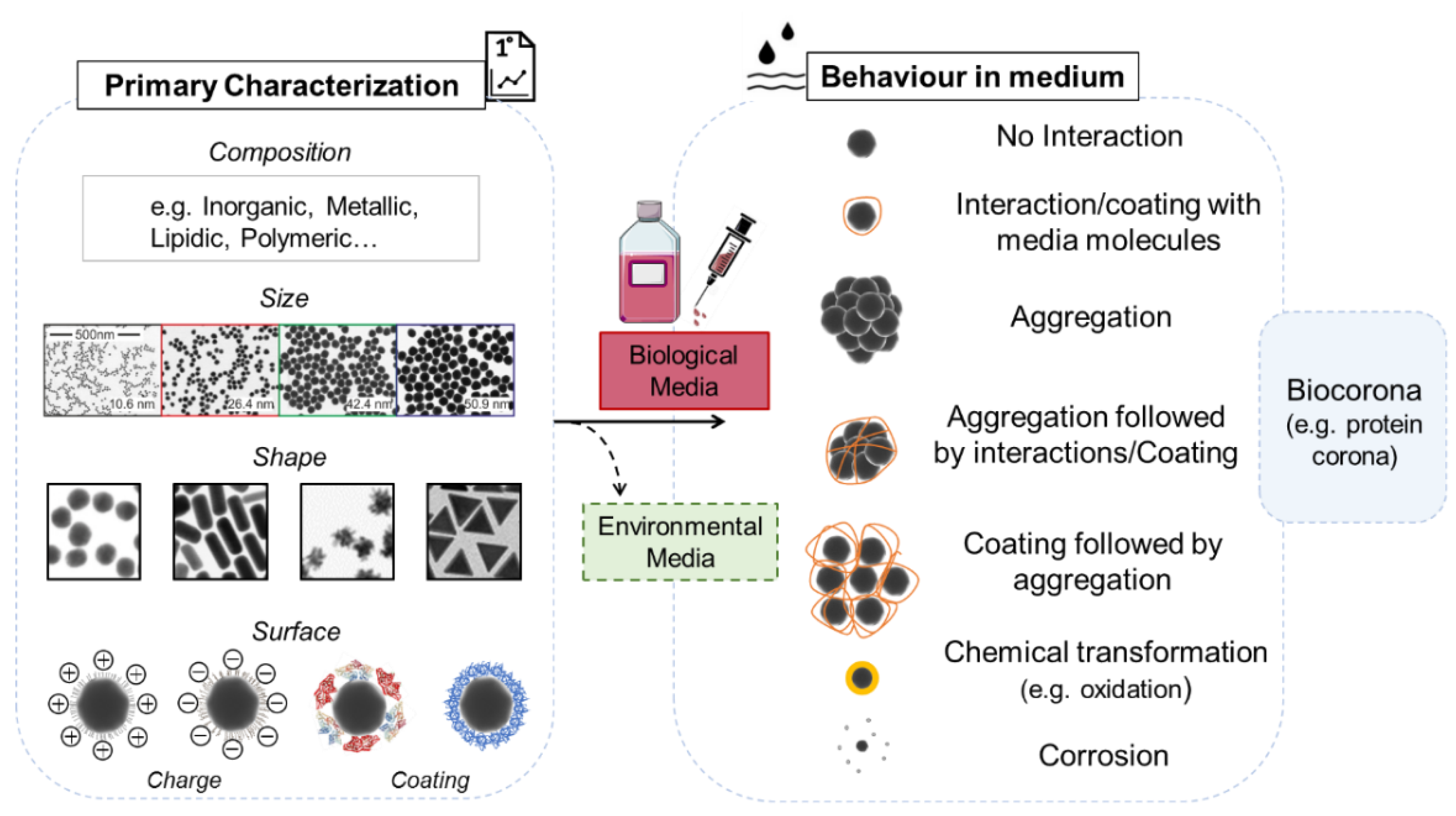
3. Parameters Assessed: From NPs to Innate Immune Responses

3.1. NPs: What to Consider When You Use a Biological System?

The physico-chemical characteristics of a NP and its behavior in different exposure media are fundamental considerations when attempting to understand the interactions of NPs within a biological model. It is important to take into account that the relatively large surface area to volume, the low coordination of atoms at the surface, and their colloidal nature cause NPs to display physical and chemical characteristics that differ from their bulk counterparts. It is also fundamental to understand the characteristics of the final object that living organisms will encounter and to correlate the pristine and final NP features with the potential effects on living organisms. The main NP characteristics to be considered are reported in Figure 3.

3.1.1. Primary Characterization

The first determination of primary characteristics includes the description of the material composition, the nominal size, shape, and surface charge (zeta potential). Moreover, characterization of NP coating and other surface modifications are crucial to consider (Figure 3 left panel).

3.1.2. Behavior in Medium

In addition to the known properties of a chosen NP following synthesis, once exposed to biological conditions (e.g., medium or circulating fluid), NPs can display unpredicted new characteristics. NPs can have a propensity to move towards a more stable thermodynamic state via different means: aggregation (which can mean escaping from the nanoscale), formation of a coating composed of various molecules, chemical transformations, particle corrosion, and dissolution [1]. All these transformations can change the identity of the NP or produce new chemical entities (e.g., reactive metal ions), modifying their behavior and their potential associated risk and interactions (Figure 3 right panel). Therefore, the determination of NP characteristics in exposure medium needs to be assessed. This generally includes the aggregation state (Z-average), the change in surface charge (zeta potential), and the dispersion index (PdI). Moreover, the evolution over time of these parameters can also be of value for a full appraisal of the NPs dynamic in the exposure medium. All these analyses are usually performed using DLS (dynamic light scattering) analysis, or electron microscopy (TEM and SEM) depending on the material being investigated. Additionally, careful controls have to be performed in order to avoid artifacts due to the presence of chemicals, often used to stabilize the particles [106,107], or contaminants, such as bacterial lipopolysaccharide (LPS), that can cause false positives in an immune assay [108].
Another routinely measured parameter is the presence and composition of the molecular biocorona, where components of biological fluids can be adsorbed by the NP, forming a corona on its surface. Usually, they are believed to be mostly constituted by proteins (protein corona -PC) but other macromolecules including lipids present in the medium can also contribute to its formation. The presence of this supplementary layer on top of the NPs can in turn affect the NPs behavior and interactions with the surrounding media. However, this corona depends on both the biological fluid (plasma, or otherwise) composition and the properties of the NPs, including size, curvature, surface functionalization, and charge. The composition of the corona is theoretically divided into the soft corona (weakly bound) and the hard corona (tightly bound), but it is dynamic and the ligand on the top can be exchanged and replaced over time, according to the affinity of the macromolecule for the NPs [109,110]. The PC is the biological identity of the NP and represents what cells “see” and with which they will interact [111,112,113,114,115,116,117,118]. Consequently, recognition by immune cells can be different and specific from one type of NP to another, which means that they will interact with the protein on the surface rather than the NP itself. This results in the triggering of defense mechanisms different from those observed in medium free of proteins. This does not apply only to mammalian plasma, but it has been demonstrated in the biological fluids of different terrestrial and marine invertebrates, including earthworms, bivalve, and sea urchins, in which the composition and effects on immune parameters appeared different for each NP type [119,120,121,122]. For these reasons, the PC is an important parameter to consider under laboratory conditions and needs to be characterized with precision during the exposure event.
All the previously cited characteristics can also be applied to environmental media [123,124,125,126]. The NPs will be subjected to other factors like abiotic physico-chemical parameters (such as pH, ionic strength, temperature) which can influence their dispersion, aggregation, agglomeration [127,128], interaction with molecules present in their environment, and adsorption to macro-organic matter (e.g., eco-corona) [117]. Scientific literature is being produced on the physico-chemical transformation of NPs due to their exposure to aquatic and terrestrial scenarios, correlating the environments and particle properties with the observed changes. Consideration of this should be taken into account in future studies working with environmental scenario experiments [129,130,131,132].

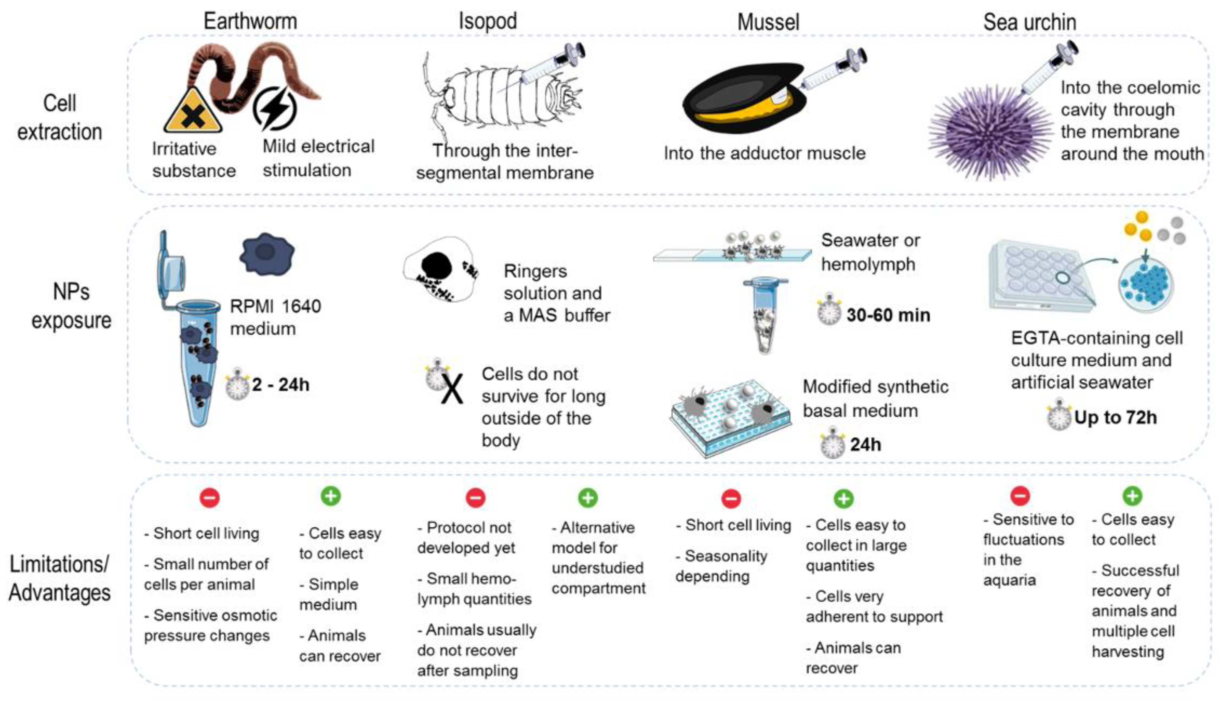
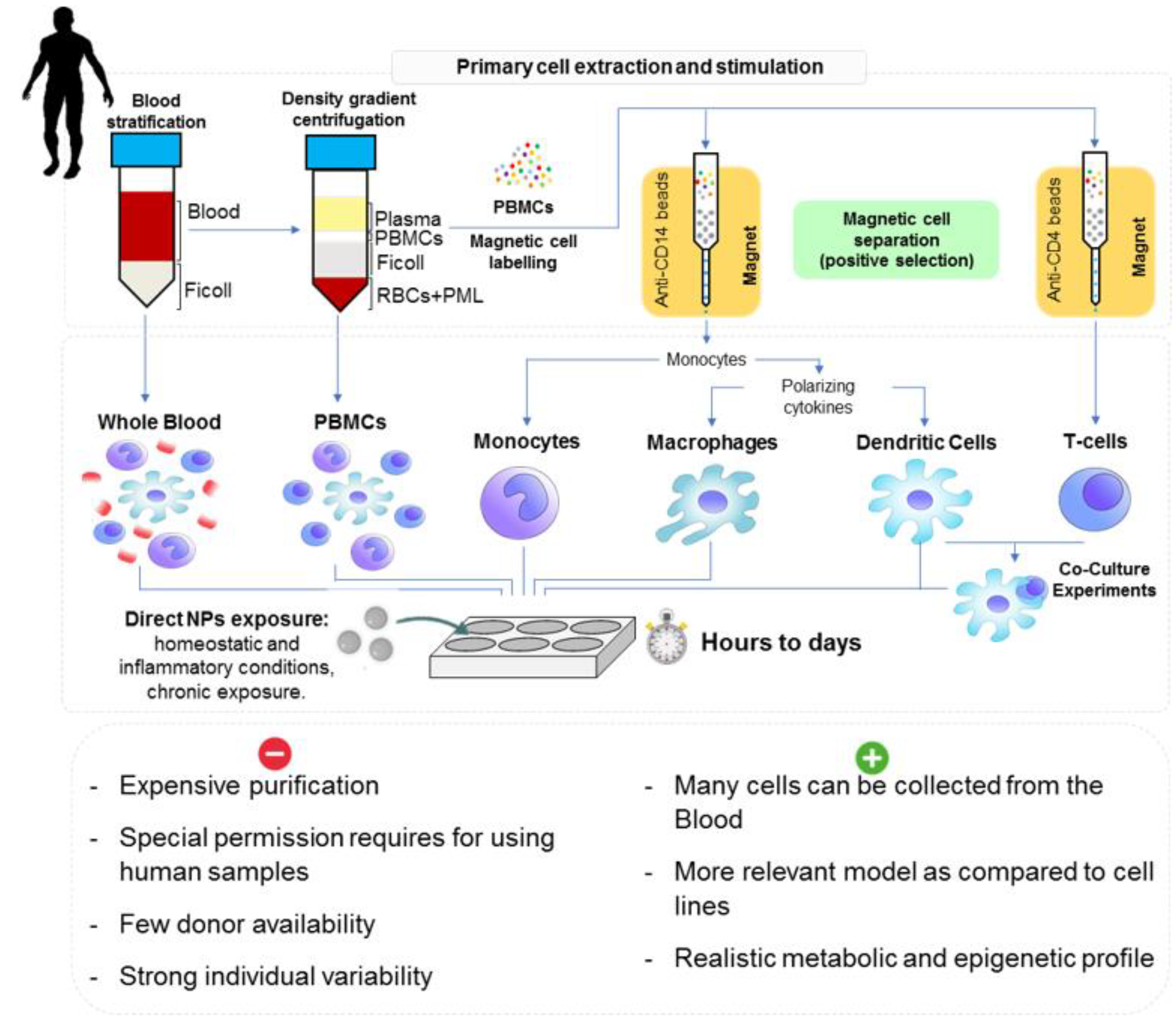
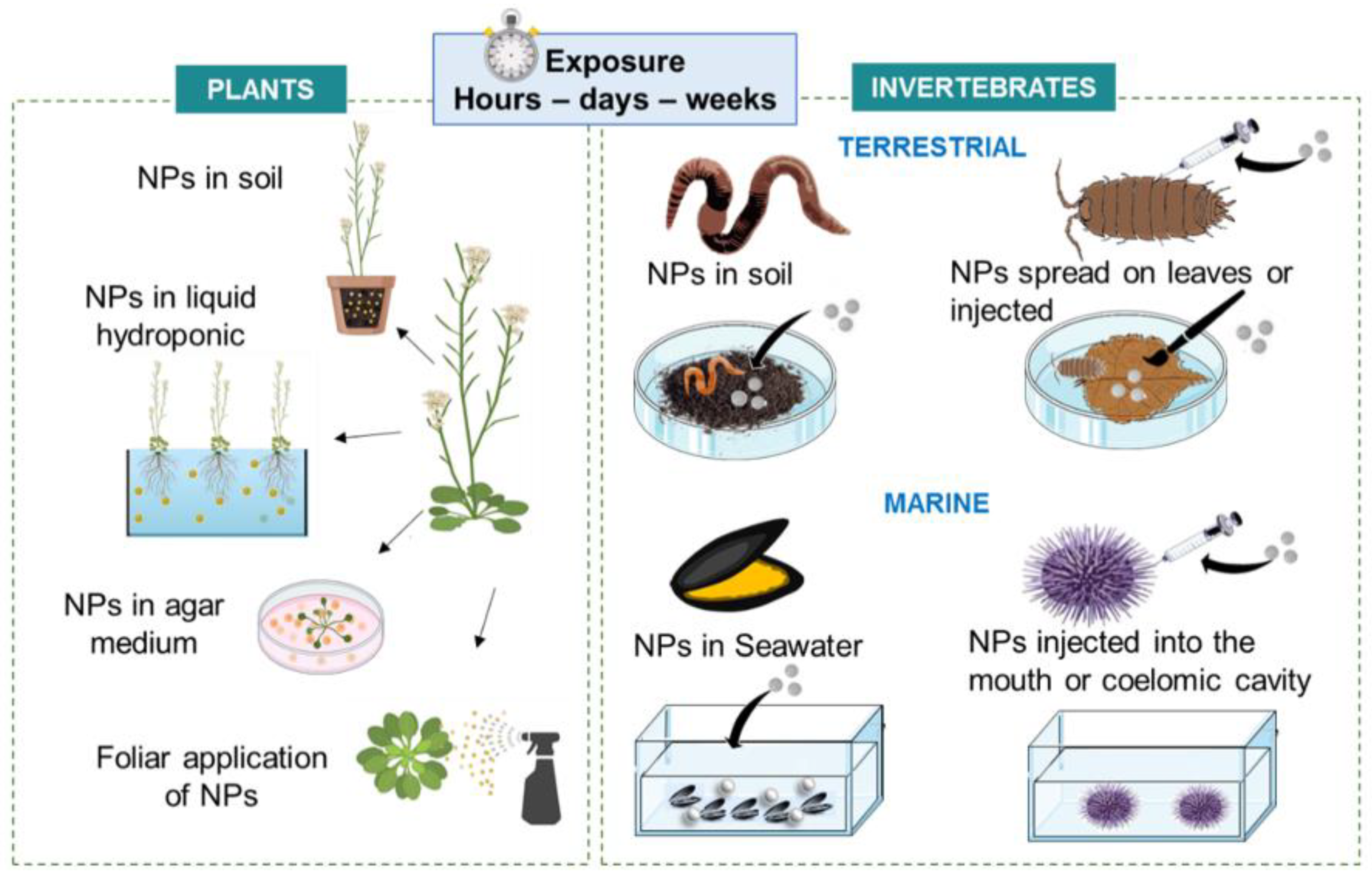
3.2. Models, Cell Culture and Mode of Exposure

In experimental science, the use of in vitro assays is being promoted as sustainable alternative for a large range of product testing, including NPs, following the 3R principle (replacement, reduction, and refinement). The extraction of immune cells, separation, culture and feasibility to maintain such isolated primary cells varies across models. To illustrate, a summary of the different methods of cell harvesting and exposure for both invertebrates and human primary cells are reported in Figure 4 and Figure 5.

3.2.1. Nonmammalian In Vitro Assays

In many invertebrates, coelomocytes/hemocytes act as the first line of defense against nonself objects. The induction of functional responses with these cells is often rapidly observed, helping to counteract the limitation of the relative short-term lifespan of cells in cultures (ranging from a few hours to a few days depending on the model). As a natural defense mechanism, earthworms can extrude their coelomic fluid, through dorsal pores. Therefore, coelomocytes can be extracted by mild electrical stimulation or by exposing the animals to an irritative substance [133,134]. Immediately upon collection, the coelomocytes need to be stabilized in a culture medium in order preserve cell viability. Recent studies show that RPMI 1640 medium is the optimal medium for earthworm coelomocytes culturing, as well as the assessment of NP toxicity towards coelomocytes [135,136]. Critical for the successful culturing of coelomocytes is the adjustment of osmolality of the medium so that it reflects that of the coelomic fluid [137,138,139]. Exposure time for in vitro assays depends on cell viability and may range from between 2 to 72 h, with 24 h being the optimum time for cell cultures in RPMI 1640, according to some investigations [136,137,140]. For terrestrial isopods, the culturing of hemocytes did not show hopeful results yet. Hemolymph can be collected by puncturing through the intersegmental membrane on the dorsal side of the isopod with a sterile needle and collecting the hemolymph with a micropipette. With the use of ringers solution and a MAS (mitochondrial assay solution) buffer, cells appeared to hardly survive for even a few hours outside the body. The selection of a suitable medium is still needed to be identified and adapted for keeping hemocytes alive without showing excessive levels of stress [141].
In marine invertebrates, in vitro experiments are much more abundant, in particular, experiments using hemocytes of the marine mussel M. galloprovincialis extracted via a non-invasive method. This method can provide a first line of investigation for testing several types of substances, including NPs [142,143,144,145]. Mytilus hemolymph is easy to collect via the adductor muscle and fluid quantities are sufficient (depending on the season, volume can be as high as several ml per animal) to perform various experiments [146]. Short-term exposures (≤1 h) have shown rapid activation of hemocyte functional parameters but longer exposure times (up to 24 h) have shown the induction of further immune or stress parameters. For short experiments, hemocytes can be maintained in a natural hemolymph or seawater suspension, in tubes or as monolayers on glass slides. Longer culture times were more successful when modified synthetic basal medium (Basal Medium Eagle) was used in microwell plates. These in vitro experiments are possible due to the cells ability to quickly adhere to supports (<20 min) [147,148]. Ex-vivo tissue explant has also been used (e.g., gills) to study the first interactions and potential uptake of NPs [149].
The coelomocytes from sea urchin once extracted are placed in cell culture plates and kept in EGTA-containing cell culture medium and artificial seawater [84,85,150]. The coelomocytes can be kept for a long period of time (over two weeks), with regular medium replenishment and without the addition of the special growth factors or nutrients [90,151].
Finally, as plants do not possess specialized mobile immune cells, in vitro research is not typically suitable/realizable, and the main experiments in laboratories are made on the full plant or tissues excisions (see next section). Each piece of tissue should respond upon exposure as each single cell is able to launch an effective immune response [152].

3.2.2. Human Cell Models

The study of human cells offers a wide range of possibilities not currently developed for invertebrate models. Multiple cell types, coculture conditions, and cell maturation or differentiation programs exist to define more precisely the interactions with NPs. Several important models used in mammalian systems to test NP–immune system interactions are listed below.
Figure 5. The different in vitro NP exposure possibilities available for human primary cells and the procedure of cell extraction and preparation (Reprinted with permission from Michelini et al. (2021) [153]. Copyright 2021, Copyright Royal Society of Chemistry).
Nanomaterials 11 01528 g005
In vitro modelling of human innate immunity is usually conducted using monocytes, macrophages, or dendritic cells, as these cells are responsible for directing the innate immune response from pathogen recognition to phagocytosis to inflammation, and even to eventual antigen presentation and induction of adaptive immunity. Cell lines for each of these cell types exist and are frequently used due to their easy experimental repeatability and scalability, with THP-1 (monocytes) and U937 (macrophages) being the most frequently reported [154]. However, cell lines are truly limited to the representative phenotype observed at the time of culture, and even this is susceptible to mutations that do not represent the true reactivity of healthy human cells. More robust models of innate immune responsiveness utilize primary cells, which are collected directly from donors and may be isolated using techniques that select for the desired cell type. Primary cells are representative of an individual’s current in vivo condition, and lack the altered metabolic and epigenetic profile inherent to cell lines. Furthermore, as monocytes are found abundantly in circulation, and since they can be precursors for both macrophages and DCs, the differentiation of monocytes in culture into primary differentiated macrophages or DCs is an effective tool to create models of innate immune responses in vitro. However, models utilizing primary cells must contend with individual variability as the immune experience and capacity is different between donors [155].
The whole blood assay is one of the most simple and rapid tests for assessing the immune activating capacity of novel substances within a human system. Typically, blood is drawn from healthy donors and immediately exposed to the substance under investigation, with 250 µL of blood typically diluted in 750 µL of RPMI plus the tested material and incubated for 24–48 h [156]. Peripheral blood mononuclear cell (PBMCs) can also be isolated from the whole blood using Ficoll–Paque density gradient centrifugation [157]. Magnetic cell separation using some CD-4 beads can be used to isolate monocytes, and later growth factors can be added to differentiate macrophages such as macrophage colony stimulating factor (M-CSF) or DC GM-CSF and IL-4 [94,158].
The monocyte activation test (MAT) models (or using macrophage and DCs) can assess the exposure and response of monocytes to NPs, and many parameters may be assessed following activation [159,160,161]. Usually cells (in the range of ~500,000 cells/mL) in plate culture can be directly exposed to NPs added to the wells, and tests on monocyte/macrophage/DC activation are typically completed within 24 h. Oftentimes, PBMC culture is conducted in round bottom wells, which simulate a lymph node in which communication between myeloid cells and lymphocytes occurs.
Finally, NPs can also interact with other cells present in blood, such as DCs, which link with adaptive immunity. Similar principles of the MAT test can also be applied for testing NPs, but also cocultures with T-cells of self or foreign origin [153]. These types of test can mimic autoimmunity or the mixed lymphocytes reaction and are of interest for the use of NPs in vaccines and immunotherapy [162].
Experiments considering primary isolated cells offer other advantages by representing a simplified model, limiting interfering factors, which could help to spotlight NPs mechanisms of action before performing further experiments; e.g., coculture, tissue models, or even using whole-organism in vivo experiments. Moreover, these in vitro experimentations allow easier comparison between models, particularly, with human cells. In addition to the commonly known pros for in vitro assessment such as cheap cost, fewer animals used and relatively fast results, a list of the more important pros and cons for each model, with special input for in vitro assays, is presented in Figure 4 and Figure 5, lower panel. Although they provide a simplified set up and can be used to try and understand some of the basic mechanisms, they do not represent the true exposure pathway. Additionally, large variations in the exposure time and the culture methods between different models persist. In invertebrates, the immune cells are usually easy to collect, except for isopods, and in large quantity. There are some species-specific difficulties in experimentation, such as molt cycles in isopods or seasonality with reproductive period in mussels that can impact immune measurements. Moreover, some cells are more sensitive and fragile to handle compared to others. Usually, the immune cells from invertebrates are viable for shorter times in culture, as basal parameters are quickly impacted. For humans, in addition to regulatory hurdles, donor availability is restricted for the obtaining of primary immune cells. Each donor is usually considered independently, which can reflect stronger variabilities in responses. However, from one whole blood sample many cells can be collected and offer a large range of possible assays after purification.

3.3. Whole Model Exposure Experiments

In vivo experiments allow for evaluation of the effects and mechanisms of action of NPs in organisms at different levels of biological organization (molecular, cellular, tissue level). They provide a realistic scenario of the exposure pathways as encountered under natural conditions. For controlled laboratory experiments, the mode of exposure to NPs needs to be adapted for each model; a summary is presented in Figure 6.
These tests are usually conducted in environmentally relevant mediums (soil or water), through feeding experiments or through breathing and filtering experiments for aquatic species. As the selected models are usually easy to maintain in laboratory conditions for long periods of time, requiring little space and maintenance (e.g., feeding), the exposure time (acute, semichronic and chronic exposure) can range from hours, to days and weeks.
Plants can germinate and grow directly in the presence of the NP in the growth medium, i.e., soil, hydroponic nutrient solutions or agar-solidified agents, or they can be exposed at subsequent development stages. Despite the presence of cell walls, that can represent a barrier preventing NPs entering into the plant cell and cytoplasm, NPs might be absorbed through root or leaf and be potentially transported to the shoot or to other points through the phloem (vascular system) [163,164,165,166]. NPs can be also dispensed onto the plants surface by foliar spray application [167]. After entrance into the leaf tissue, NPs can diffuse into the intercellular space, the apoplast, or membranes and cause secondary effects. Moreover, temporality is important, and it is necessary to understand the course of plant growth and development, from seed germination to root elongation and shoot emergence, in relation to NP exposure [168]. The following investigations can assess the NP uptake by cells and further nanophytotoxicity, focusing on the toxicity symptoms of plants.
In vivo earthworm exposures are typically conducted in soils following well-described and standardized procedures (e.g., [27]) that can also be applied for NPs [140,169]. However, care must be taken when it comes to the mixing of NPs with soils, adjusting the parameters depending on the form (i.e., as solution dispersion or as powder) in which the NPs are supplied [170]. Furthermore, coexposures with infectious microbes are also important in order to establish whether an exposure to NPs has an effect on the ability of a host to maintain immunity [171]. A methodological approach to investigate the impact NPs have on the earthworm’s ability to maintain immunity when coexposed with infectious bacteria has been recently established [140].
A major benefit of working with the terrestrial isopod, P. scaber is that they are able to be exposed to the NPs in a manner similar to how they would be exposed in nature. NP suspensions can be spread on leaves that P. scaber eat, and both the leaf and animal are then placed in a petri dish. During the experiment, feeding rate, defecation rate, and mortality can be monitored. This also allows for modelling of real-world impacts of NPs on the organism from behavioral changes, like feeding avoidance and mortality, to cellular immune responses. However, the gut of P. scaber is covered in a thick cuticle which is believed to stop the translocation of NPs from the gut into the hemocoel where the hemocytes are, so the immunological effects of the NPs might not be seen when ingested [172]. There is the possibility for an alternative exposure scenario, with injection experiments delivering substances directly into the animals hemocoel, allowing for the study of the direct interaction of a known concentration of NPs with the hemocytes. This ensures NP and immune cell interaction [141,173].
Mussels are suspension feeders and are able to filter large amounts of water (up to 3 L per hour) implying that, in a short period of time, they can easily uptake the NPs present in the seawater of experiment tanks. For this reason, the NPs can be directly added to the seawater and the ventilation system allows for constant movement of water within the tank. To study the first immune defense response, short term experiments (24 h to 96 h), have been shown to be sufficient to induce the activation of the immune system [142]. Moreover, the use of artificial seawater (ASW) implies the absence of organic matter or other substances that could interact with the NP suspension; together with a constant salt content and as such are reproducible for all periods of the year. The experiments are mainly conducted in the spring and summer periods where mussels are at their healthiest. During experiments, mussels are not fed and can readily survive several days without feeding [146]. This is necessary for NP experiments, as the presence of microalgae could interact with the NPs. In this context, the study relies only on the uptake of the NPs in seawater. To mimic a more realistic exposure, other studies have performed longer-period experiments to consider the interactions between food intake and the NPs, but this generally focused more on physiology and tissue changes and not strictly the immune response [148,174]. Biological uptake routes are dependent on NP properties and may occur as direct uptake in gill tissues and/or through transference from the cilia to the digestive system. Moreover, the agglomeration of particles in seawater has been shown to facilitate NP ingestion by suspension feeding bivalves, and their potential translocation from the gut to the circulatory system [175,176]. However, this internalization pathway seems to vary according to the NPs and some can be captured and excreted in pseudofaeces (mixture of mucus and undigested particles) before arriving to the stomach, resulting in lower tissue accumulation and higher depuration [177].
The existing in vivo studies utilizing sea urchins mainly focus on the immune status of the animal after exposure to NPs via the injection of the NP suspension into the mouth or directly into the coelomic cavity (through the soft peristomal membrane surrounding the mouth). Consequently, NPs injected orally partially cross the intestinal epithelium, invade the coelomic fluid and are then engulfed by phagocytes, while the remaining particles pass the digestive system and can be excreted [178]. On the other hand, NPs injected into the coelomic cavity can directly interact and be recognized by phagocytes [85,179].
In general, the in vivo passive exposure experiments consider more realistic exposure pathways (feeding, breathing) of NPs. However, for more simplistic set up and to be sure that NPs encounter immune cells, NP suspensions can be also injected into the animal. Results obtained from in vivo tests can provide a good proxy of interactions of immune cells in situ and thereby in vivo tests are crucial to resolve the issue of whether NPs pose an immune threat to living organisms. These models have been shown to be easy to maintain in the laboratory, and exposure experiments allow for the effects of NPs to be studied at different levels of the organism. For future experiments, mesocosms will help to mimic environmental scenarios before further studies in the environment.

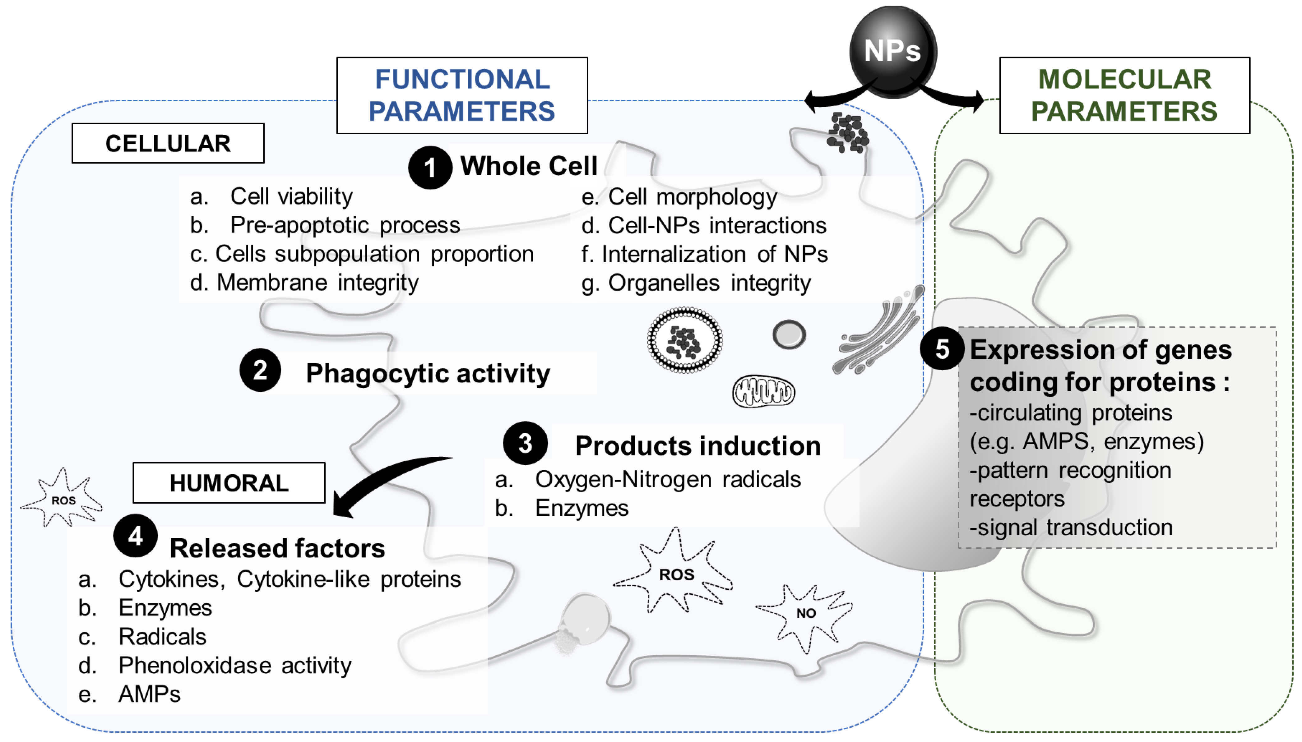
3.4. Innate Immune Parameters of Interest

As highlighted in Figure 7, a variety of endpoints can be used to compare immune system–nanoparticle interactions between different models. This includes functional responses, which comprise the biochemical assessment of cellular and humoral responses, and molecular responses that aim to evaluate changes in the expression of immune-related genes. Because there are many methods available to quantify functional responses, here we provide a comparative overview of these methods to show which are most appropriate for the purpose of NP testing and cross-species comparisons (see Table 2).

3.4.1. Whole Cell Response

Parameters looking at the whole immune cell concerns the immune cells viability, the membrane integrity, all the different types of interactions they can have with NPs, and their potential changes in morphology (Figure 7, first point). The first important cellular responses that can be used to investigate nanoparticle—immune system interactions is (immune) cell viability. Although cell death and apoptosis are part of a normal immune response, studies have shown that NPs are able to cause excessive mortality in immune cells with possible adverse effects on immunocompetence. There are several methods available that can measure cell viability (Table 2). A common method used in human cell lines is the measurement of the release of LDH or ATP through biochemical assays [153,182]. An alternative is the staining of living or dead cells using fluorescent probes (e.g., fluorescein diacetate–FDA or propidium iodide–PI) for observation using flow cytometry fluorescent microscopy. In some models, cell viability may also be studied by measuring metabolic activity through cell-permeable fluorescent reduction such as CTB or the colorimetric MTT. In some models the use of counter stain dye such as trypan blue or nigrosine [141,192] or the use of DNA-binding florescent dyes are alternative methods for assessing cell viability [150]. There are several methods available to measure the number of cells that are in the process of dying (apoptosis). Apoptosis and preapoptosis evaluation methods, which are available for several models, can be used as early markers for cell viability through the use of specific fluorescent dyes (e.g., annexin V binding, apostain, tetramethylrhodamine, ethylester perchlorate-TMRE or DAPI labelling) [139,197]. Cell viability is probably the best described immune parameter in most species (Table 2); therefore, this parameter is one of most relevant to assess in cross-species comparison. In addition to measuring the overall immune cell viability, quantifying changes in the ratios of different subpopulations of immune cells (e.g., total hemocyte counts-THC) can often give a more detailed view of the impact of NPs on immune cell viability [141,179,199,200]. In human cells, fluorescence-activated cell sorting (FACS) is a common method in which fluorescent antibody-tags can be used to determine a large range of parameters but also to discriminate sub-cell populations [153].
The subcellular effects of NP exposure can be identified via assessment of the integrity of organelles, membranes, and other cellular compartments. Lysosomal functional integrity is an evolutionarily conserved marker of stress (including NPs) and of an individuals’ health status, and is commonly evaluated by measuring neural red retention or uptake [185,189,248]. Other approaches that can be used to assess the effects of NPs on organelles include methods measuring trans-golgi apparatus integrity and internal membrane polarization [178,179,200].
Another crucial step in the characterization of NP–immune system interactions is assessing whether immune cells are able to internalize NPs. The internalization of NPs has been observed for different types of NPs across the selected models and was recently reviewed in [36]. There are several techniques available to detect the internalization of NPs. These include transmission electron microscopy (TEM) which can image internalized particles and scanning electron microscopy (SEM) which helps to visualize membrane-bound particles and can give a direct image of the particles and cells following contact. They can also provide details on how the interaction occurs as well as the state of the NPs (e.g., agglomeration, aggregation, precipitation). These techniques when coupled to an EDX system (energy dispersive X-ray) can be used to perform chemical characterization of the NPs’ surfaces. TEM and SEM are descriptive techniques that can provide valuable information but makes quantification difficult between models. Some research has reported the use of fluorescently labelled particles to help to measure particle uptake, although the use of such labelled particles requires additional controls to rule out any effects linked to the leakage of fluorescent dyes [249]. In general, NP internalization in human cells has been well reported but for invertebrate, similar methods often require adjustments to be made (as for example, the salt or osmotic concentrations during fixation) [90,141,153,205,206,209]. For plants, TEM can be used to verify the entry of NPs into the cells [203]. In addition, these kinds of techniques can reveal the change in cell morphologies and subcellular structures (e.g., vacuoles, phagosomes, endosomes) upon NP exposure and give hints regarding the general activation or damages that the cell has undergone [203,207].

3.4.2. Phagocytic Activity

While an immune response towards NPs could be part of normal immune functioning, overstimulation of the immune system resulting in damage or suppression leading to a compromised immune functioning may pose a threat to the organism. Such suppression of immune functioning caused by NPs could be studied via the assessment of the immune cells capacity to phagocytose and the consequent changes on index and rates (Figure 7, second point, Table 2). In earthworms, mussels, and sea urchins, phagocytosis can be evaluated by using fluorescence beads or yeast (using neutral red stained zymosan) [139,145,150,181,216,250].

3.4.3. Cytotoxic Factors

Upon contact with NPs, cells can be activated and produce cytotoxic factors inside the cells in order to help to remove internalized foreign particles (Figure 7, third point). Among them, the oxidative burst, which involves the production of several radicals from oxygen (ROS) and nitrogen (RNS) derivatives. To quantify ROS, several methods, including the use of fluorescent probes (e.g., DCF or calcein), UV-vis spectroscopy (e.g., cytochrome C reduction) or histochemical staining, can be used and many of which have been adjusted for use across the model organisms (Table 2). Moreover, lipid peroxidation can be measured as a proxy for the damages caused by oxidative stress to the membranes, even if it is more frequently analyzed in tissues than in individual cells [138,139]. As for the quantification of RNS and more commonly nitric oxide (NO), in isopods, mussels, and earthworms, NO levels in the hemolymph can be measured spectrophotometrically from hemolymph samples using Griess reagent [141,234,250].
Lysozyme is an evolutionary conserved enzyme that catalyzes the hydrolysis of peptidoglycan and plays a role in the innate immunity of many organisms including earthworms, mussels, sea urchins, and plants [67,234,251,252]. The quantification of the release of lysozyme into the extracellular medium is based on the lysis activity of Micrococcus lysodeikticus which can be determined spectrophotometrically. Fluorescent probes can be also used to monitor the evolution of the lysosomal compartment and acidification in the cell upon exposure to NPs [200].

3.4.4. Humoral Factors

Humoral immune responses play a crucial role in immunity by facilitating communication between immune cells and directing the extracellular destruction of foreign objects. Upon activation of the immune cells, some factors can be released into the extracellular medium (Figure 7, fourth point, Table 2). In mammals, cytokine production is a key driver of cellular immune responses [238]. Analyzing the extra- and intracellular concentration of cytokines secreted by (human) immune cells is a well-established method to test the effects and safety of NPs [253]. Many techniques have been developed to detect single or multiple cytokines and factors secreted by cells. These include classic methods such as western blot, ELISA, and bio-chemiluminescence assays, and many commercial possibilities for multiplex assays including legendplex or Ella multiplex technology [153,209,254]. Interestingly, in sea urchins, cytokine IL-6 can be detected in immune cells and secretome using western blot analysis after exposure to NPs [151].
Lysozyme and radicals can also be released by immune cells in the extracellular medium, to directly destruct foreign particles in close proximity to the cell. In addition, there are species specific released factors such as the hemolytic protein lysesin found in the coelomic fluid of earthworms.
Lastly, an important immunological parameter often analyzed in invertebrate models is the measurement of phenoloxidase (PO) activity. This enzyme, produced via the pro-PO cascade, is involved in the production of melanin [43,141]. Upon detecting a melanized pathogen or object, immune cells quickly encapsulate the material resulting in the elimination of the threat. PO activity can be assessed by monitoring the formation of a reddish-brown pigments in the hemolymph from an individual organism using spectrophotometry [44,173,240]. In bivalves, the presence of PO has been reported but its basal levels, the variation across species and especially its response to NP exposure remain poorly understood [83,255]. In the sea urchin Strongylocentrotus nudus coelomocytes, three proteins with PO-like activities have been identified using electrophoretic methods [241].

3.4.5. Molecular Response

Increasingly, humoral responses can be measured through genetic or omics approaches (e.g., quantitative PCR or full-transcriptome sequencing) (Table 2). A main advantage of using these approaches over biochemical ones is their high-throughput potential and increased specificity. Furthermore, genetic or omics approaches allow for the assessment of entire immunological pathways instead of focusing on specific biochemical endpoints. However, major limitations of these methods are that they require species specific primers and the availability of transcriptomes, which are currently lacking for many invertebrate species. Moreover, gene expression is highly regulated and time-dependent, so careful consideration must be given to the experimental model in terms of stimulation/exposure time, and cell collection technique.
Genes involved in different immune-related functions, such as oxidative stress response, humoral factors (e.g., AMPs), and receptor proteins, are available for some organisms and the main immune related genes known to be activated upon NP exposure are reported in Table 2.
A whole genome transcript is under development and will be available for P. scaber. Using this and genes previously annotated in other more commonly used crustacean species, primers specific for P. scaber immune-related genes can be designed (Hernadi, Mayall personal communication). Moreover, gene expression offers alternative possibilities to study several proteins involved in the immune response that biochemical tests that evaluate their activity or functionality are not feasible, such as the effects of NPs on AMP modulation. In plants, microarray-based studies are good tools to monitor the expression of candidate genes involved in the plant defense responses after interaction with NPs [204]. Additionally, an important future issue for environmental molecular biology is to establish whether an up- or downregulation of a certain gene correlates to a modification in the levels of related proteins [256]. The study of transcriptomic changes in cells, tissues, or full invertebrate organisms after exposure to NPs is now emerging but is still in its early phase. Transcriptome analysis can highlight pathways being activated but it should also be accompanied by the study of functional parameters for a fuller, deeper understanding [228,257,258]. In addition, changes in protein repertoires (proteomics approach) have shown interesting outcomes; however, studying of the combined immune response to NP exposure remains in its infancy [259,260,261].

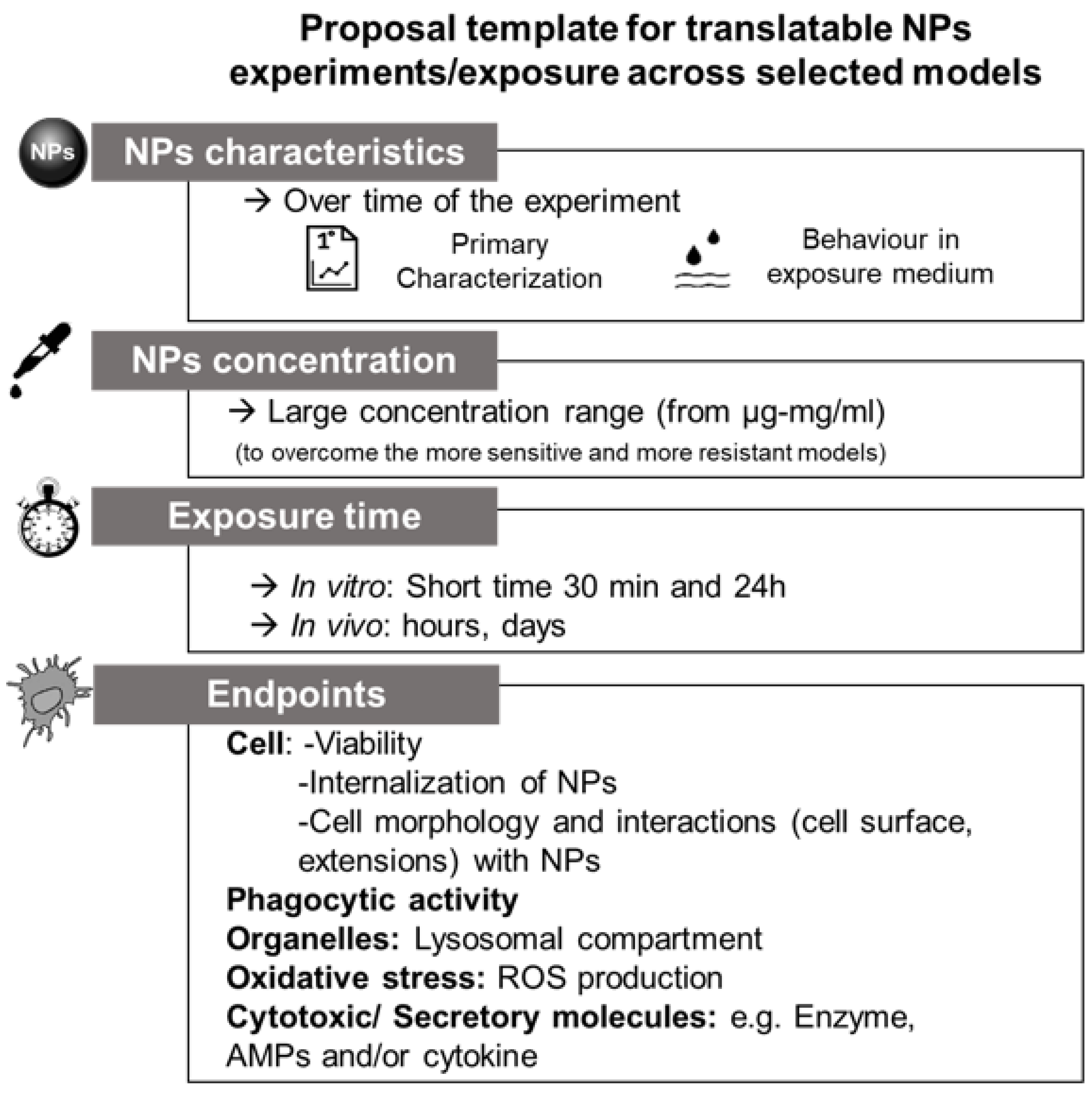
4. Proposal for Future Cross-Species Evaluations and Conclusions

During the PANDORA project [35], several studies were conducted on the innate immune response of different models exposed to a large range of different NPs. Based on the outcomes of these studies, several conclusions can be made which may help to guide future (comparative) studies on nanoparticle–immune system interactions (Figure 8).
Because chemical conditions of mediums strongly affect the form and state of NPs and thereby the behavior of NPs, it is crucial to characterize the physico-chemical properties of NPs in the exposure medium as well as in their pristine form (e.g., after production). As the behavior and the interaction of NPs with immune systems are also time-dependent, experimental design will need to critically consider exposure duration as well. In vitro models can be considered as the prime focus for studying nanoparticle–immune system interactions. However, in vitro models are not available for all immune model species (e.g., isopods, plants), limiting comparative studies based on in vitro testing. In vivo experiments are crucial to study nanoparticle–immune system interactions under more realistic conditions. Studies using invertebrates, which are well-established and can be conducted on a routine basis, may serve as good alternatives to in vivo mammalian testing models such as mice or rats.
Here, exposure route and exposure concentration will need to be critically considered as these factors are likely to significantly affect immune cells and their interaction with the NPs.
In this review, we provided an overview of the methods used to characterize nanoparticle immune responses in various organisms across the tree of life (Table 2). Among cellular parameters, it appears that methods to assess cell viability (including assessments of subpopulations) and NPs internalization by immune cells are well described in most organisms. Phagocytic activity is a crucial parameter to be evaluated for immune cells, however, some models lack the methods to study this parameter (e.g., isopods) or do not rely on this type of response (e.g., plants). Moreover, as methods for microscopy are universally available, measurements of the change in morphology and external interactions of NPs can be studied in most immune models.
Due to a lack of general knowledge on the composition and functioning of humoral immunity in most organisms other than mammal/human models, it remains difficult to identify the most relevant humoral parameter for cross-species evaluations. The exception being oxidative (and nitrosative) stress, for which methods are well described in most species.
Further work is needed to identify the interorganism comparability of otherwise species-specific markers, especially in invertebrates. In order to fully characterize NP-immune responses across species from the tree of life, there is a need for the identification of markers indicative for both pro- and anti-inflammatory responses, as are currently already available for human models (e.g., [253]). The development of such markers will require fundamental research on the innate immune systems of organisms other than human models. Thorough investigations in species from across the tree of life will help to understand how NPs interact with the innate immune system under different conditions and environments which may guide the future development of NPs that are immunologically safer-by-design.

Author Contributions

Conceptualization, B.J.S., C.M. and M.A.; writing—original draft preparation, C.M., B.J.S., A.A., F.B., E.F., S.H., S.M., N.I.N.P., A.P., E.S., M.A.; writing—review and editing, B.J.S., C.M., E.S., M.A.; supervision, M.A. All authors have read and agreed to the published version of the manuscript.

Funding

All authors were supported by the EU H2020 project PANDORA, grant number 671881.

Institutional Review Board Statement

Not applicable.

Informed Consent Statement

Not applicable.

Acknowledgments

The authors warmly acknowledge the project coordinator Diana Boraschi for her strong support during the whole project period, and all the PIs (L. Canesi, D. Drobne, A. Duschl, M.A. Ewart, J. Horejs-Hoeck, P. Italiani, B. Kemmerling, P. Kille, A. Pinsino, P. Procházková, V.F. Puntes, D.J. Spurgeon, C. Svendsen, C.J. Wilde) for the offered opportunity. The authors wish also to acknowledge Paola Cesaroni for her immense organizational help. Special thanks to our Project Officer Giuliana Donini for making PANDORA a successful story.

Conflicts of Interest

The authors declare no conflict of interest.

References

  1. Casals, E.; Gonzalez, E.; Puntes, V.F. Reactivity of Inorganic Nanoparticles in Biological Environments: Insights into Nanotoxicity Mechanisms. J. Phys. D Appl. Phys. 2012, 45, 443001. [Google Scholar] [CrossRef] [Green Version]
  2. Aitken, R.J.; Chaudhry, M.Q.; Boxall, A.B.A.; Hull, M. Manufacture and Use of Nanomaterials: Current Status in the UK and Global Trends. Occup. Med. 2006, 56, 300–306. [Google Scholar] [CrossRef] [PubMed] [Green Version]
  3. Maynard, A.D.; Aitken, R.J.; Butz, T.; Colvin, V.; Donaldson, K.; Oberdorster, G.; Philbert, M.A.; Ryan, J.; Seaton, A.; Stone, V. Safe Handling of Nanotechnology. Nature 2006, 444, 267–269. [Google Scholar] [CrossRef] [PubMed]
  4. Schmid, K.; Riediker, M. Use of Nanoparticles in Swiss Industry: A Targeted Survey. Environ. Sci. Technol. 2008, 42, 2253–2260. [Google Scholar] [CrossRef] [Green Version]
  5. Bogart, L.K.; Pourroy, G.; Murphy, C.J.; Puntes, V.; Pellegrino, T.; Rosenblum, D.; Peer, D. Nanoparticles for Imaging, Sensing, and Therapeutic Intervention. ACS Nano 2014, 8, 16. [Google Scholar] [CrossRef] [PubMed]
  6. Pulit-Prociak, J.; Banach, M. Silver Nanoparticles—A Material of the Future…? Open Chem. 2016, 14. [Google Scholar] [CrossRef]
  7. Boraschi, D. From Antigen Delivery System to Adjuvanticy: The Board Application of Nanoparticles in Vaccinology. Vaccines 2015, 10, 930–939. [Google Scholar] [CrossRef] [Green Version]
  8. Lehner, R. Intelligent Nanomaterials for Medicine: Carrier Platforms and Targeting Strategies in the Context of Clinical Application. Nanomedicine 2013, 16, 742–757. [Google Scholar] [CrossRef]
  9. Boraschi, D. Interaction of Engineered Nanomaterials with the Immune System: Health-Related Safety and Possible Benefits. Curr. Opin. Toxicol. 2018, 10, 74–83. [Google Scholar] [CrossRef]
  10. Moghimi, S.M.; Hunter, A.C.; Murray, J.C. Nanomedicine: Current Status and Future Prospects. FASEB J. 2005, 19, 311–330. [Google Scholar] [CrossRef] [Green Version]
  11. Nel, A.; Xia, T. Toxic Potential of Materials at the Nanolevel. Science 2006, 311, 622–627. [Google Scholar] [CrossRef] [Green Version]
  12. Dobrovolskaia, M.; McNeil, S. Immunological Properties of Engineered Nanomaterials. Nat. Nanotechnol. 2007, 2, 10. [Google Scholar] [CrossRef]
  13. Fadeel, B.; Garcia-Bennett, A.E. Better Safe than Sorry: Understanding the Toxicological Properties of Inorganic Nanoparticles Manufactured for Biomedical Applications. Adv. Drug Deliv. Rev. 2010, 13, 362–374. [Google Scholar] [CrossRef]
  14. Jiang, Z.; Jacob, J.A.; Li, J.; Wu, X.; Wei, G.; Vimalanathan, A.; Mani, R.; Nainangu, P.; Rajadurai, U.M.; Chen, B. Influence of Diet and Dietary Nanoparticles on Gut Dysbiosis. Microb. Pathog. 2018, 118, 61–65. [Google Scholar] [CrossRef]
  15. Spurgeon, D.J.; Lahive, E.; Schultz, C.L. Nanomaterial Transformations in the Environment: Effects of Changing Exposure Forms on Bioaccumulation and Toxicity. Small 2020, 16. [Google Scholar] [CrossRef]
  16. Mittal, D.; Kaur, G.; Singh, P.; Yadav, K.; Ali, S.A. Nanoparticle-Based Sustainable Agriculture and Food Science: Recent Advances and Future Outlook. Front. Nanotechnol. 2020, 2, 579954. [Google Scholar] [CrossRef]
  17. Bundschuh, M. Nanoparticles in the Environment: Where Do We Come from, Where Do We Go To? Environ. Sci. Eur. 2018, 30, 6. [Google Scholar] [CrossRef] [Green Version]
  18. Graca, B.; Zgrundo, A.; Zakrzewska, D.; Rzodkiewicz, M.; Karczewski, J. Origin and Fate of Nanoparticles in Marine Water-Preliminary Results. Chemosphere 2018, 206, 359–368. [Google Scholar] [CrossRef]
  19. Gottschalk, F.; Lassen, C.; Kjoelholt, J.; Christensen, F.; Nowack, B. Modeling Flows and Concentrations of Nine Engineered Nanomaterials in the Danish Environment. Int. J. Environ. Res. Public Health 2015, 12, 5581–5602. [Google Scholar] [CrossRef] [Green Version]
  20. Rocha, T.L.; Gomes, T.; Sousa, V.S.; Mestre, N.C.; Bebianno, M.J. Ecotoxicological Impact of Engineered Nanomaterials in Bivalve Molluscs: An Overview. Mar. Environ. Res. 2015, 111, 74–88. [Google Scholar] [CrossRef]
  21. Boraschi, D.; Alijagic, A.; Auguste, M.; Barbero, F.; Ferrari, E.; Hernadi, S.; Mayall, C.; Michelini, S.; Pacheco, N.I.N.; Prinelli, A.; et al. Addressing Nanomaterial Immunosafety by Evaluating Innate Immunity across Living Species. Small 2020, 16. [Google Scholar] [CrossRef] [PubMed]
  22. Swartzwelter, B.J.; Fux, A.C.; Johnson, L.; Swart, E.; Hofer, S.; Hofstätter, N.; Geppert, M.; Italiani, P.; Boraschi, D.; Duschl, A.; et al. The Impact of Nanoparticles on Innate Immune Activation by Live Bacteria. Int. J. Mol. Sci. 2020, 23, 9695. [Google Scholar] [CrossRef] [PubMed]
  23. Boraschi, D.; Duschl, A. Nanoparticles and the Immune System: Safety and Effects; Elsevier/AP: Amsterdam, The Netherlands, 2014; ISBN 978-0-12-408085-0. [Google Scholar]
  24. Mitchell-Olds, T. Arabidopsis Thaliana and Its Wild Relatives: A Model System for Ecology and Evolution. Trends Ecol. Evol. 2001, 16. [Google Scholar] [CrossRef]
  25. Bevan, M.; Walsh, S. The Arabidopsis Genome: A Foundation for Plant Research. Genome Res. 2005, 15, 1632–1642. [Google Scholar] [CrossRef] [PubMed] [Green Version]
  26. The Arabidopsis Genome Iniative. Analysis of the Genome Sequence of the Flowering Plant Arabidopsis Thaliana. Nature 2000, 408, 796–815. [Google Scholar] [CrossRef] [Green Version]
  27. OECD. Test No 222. Earthworm Reproduction Test (Eisenia Fetida/ Eisenia Andrei); OECD: Paris, France, 2016. [Google Scholar]
  28. Edwards, C.A.; Bohlen, P.J. Biology and Ecology of Earthworms; Chapman&Hall: London, UK, 1996. [Google Scholar]
  29. Van Gestel, C.A.M.; Loureiro, S.; Zidar, P. Terrestrial Isopods as Model Organisms in Soil Ecotoxicology: A Review. Zookeys 2018, 801, 127–162. [Google Scholar] [CrossRef]
  30. Malev, O. Effects of CeO2 Nanoparticles on Terrestrial Isopod Porcellio Scaber: Comparison of CeO2 Biological Potential with Other Nanoparticles. Arch. Environ. Contam. Toxicol. 2017, 72, 303–311. [Google Scholar] [CrossRef]
  31. Novak, S.; Romih, T.; Dra, B.; Ferraris, P.; Sorieul, S.; Zieba, M.; Sebastian, V.; Arruebo, M.; Ho, S.B. The in Vivo Effects of Silver Nanoparticles on Terrestrial Isopods, Porcellio Scaber, Depend on a Dynamic Interplay between Shape, Size and Nanoparticle Dissolution Properties. Analyst 2019, 2. [Google Scholar] [CrossRef]
  32. Beyer, J.; Green, N.W.; Brooks, S.; Allan, I.J.; Ruus, A.; Gomes, T.; Bråte, I.L.N.; Schøyen, M. Blue Mussels (Mytilus Edulis spp.) as Sentinel Organisms in Coastal Pollution Monitoring: A Review. Mar. Environ. Res. 2017, 130, 338–365. [Google Scholar] [CrossRef]
  33. Fernández Robledo, J.A.; Yadavalli, R.; Allam, B.; Pales Espinosa, E.; Gerdol, M.; Greco, S.; Stevick, R.J.; Gómez-Chiarri, M.; Zhang, Y.; Heil, C.A.; et al. From the Raw Bar to the Bench: Bivalves as Models for Human Health. Dev. Comp. Immunol. 2019, 92, 260–282. [Google Scholar] [CrossRef]
  34. Chou, H.-Y.; Lun, C.M.; Smith, L.C. SpTransformer Proteins from the Purple Sea Urchin Opsonize Bacteria, Augment Phagocytosis, and Retard Bacterial Growth. PLoS ONE 2018, 13, e0196890. [Google Scholar] [CrossRef]
  35. Pandora-H2020. Available online: https://www.Pandora-H2020.Eu/ (accessed on 16 February 2021).
  36. Pinsino, A.; Bastús, N.G.; Busquets-Fité, M.; Canesi, L.; Cesaroni, P.; Drobne, D.; Duschl, A.; Ewart, M.-A.; Gispert, I.; Horejs-Hoeck, J.; et al. Probing the Immune Responses to Nanoparticles across Environmental Species. A Perspective of the EU Horizon 2020 Project PANDORA. Environ. Sci. Nano 2020, 7, 3216–3232. [Google Scholar] [CrossRef]
  37. Soderhall, K. Invertebrate Immunity. Advances in Experimental Medicine and Biology; Landes Bioscience and Springer Science+Business Media, LLC: New York, NY, USA, 2010; ISBN 978-1-4419-8058-8. [Google Scholar]
  38. Canesi, L.; Procházková, P. The Invertebrate Immune System as a Model for Investigating the Environmental Impact of Nanoparticles. In Nanoparticles and the Immune System Safety and Effects; Boraschi, D., Duschl, A., Eds.; Academic Press: Cambridge, MA, USA, 2014; pp. 91–112. [Google Scholar]
  39. Jones, J.D.G.; Dangl, J.L. The Plant Immune System. Nature 2006, 444, 323–329. [Google Scholar] [CrossRef] [Green Version]
  40. Takeuchi, O.; Akira, S. Pattern Recognition Receptors and Inflammation. Cell 2010, 140, 805–820. [Google Scholar] [CrossRef] [Green Version]
  41. Sarma, J.V.; Ward, P.E. The Complement System. Cell Tissue Res. 2011, 343, 227–235. [Google Scholar] [CrossRef]
  42. Bilej, M.; Baetselier, P.D.; Dijck, E.V.; Stijlemans, B.; Colige, A.; Beschin, A. Distinct Carbohydrate Recognition Domains of an Invertebrate Defense Molecule Recognize Gram-Negative and Gram-Positive Bacteria. J. Biol. Chem. 2001, 276, 45840–45847. [Google Scholar] [CrossRef] [Green Version]
  43. Soderhall, K.; Cerenius, L. Role of the Prophenoloxidase-Activating System in Invertebrate Immunity. Curr. Opin. Immunol. 1998, 10, 23–28. [Google Scholar] [CrossRef]
  44. Jaenicke, E.; Fraune, S.; May, S.; Irmak, P.; Augustin, R.; Meesters, C.; Decker, H.; Zimmer, M. Is Activated Hemocyanin Instead of Phenoloxidase Involved in Immune Response in Woodlice? Dev. Comp. Immunol. 2009, 33, 1055–1063. [Google Scholar] [CrossRef]
  45. Motion, G.B.; Huitema, E. Nuclear Processes Associated with Plant Immunity and Pathogen Susceptibility. Brief Funct. Genom. 2015, 14, 243–252. [Google Scholar] [CrossRef] [Green Version]
  46. Coll, N.; Epple, P.; Dangl, J. Programmed Cell Death in the Plant Immune System. Cell Death Differ. 2011, 18, 1247–1256. [Google Scholar] [CrossRef] [Green Version]
  47. Newman, M.-A. MAMP (Microbe-Associated Molecular Pattern) Triggered Immunity in Plants. Front. Plant Sci. 2013, 4, 139. [Google Scholar] [CrossRef] [PubMed] [Green Version]
  48. Miescher-Institut, F.; Box, P.O. FLS2: An LRR Receptor–like Kinase Involved in the Perception of the Bacterial Elicitor Flagellin in Arabidopsis. Mol. Cell 2000, 5, 1003–1011. [Google Scholar]
  49. Boutrot, F.; Zipfel, C. Function, Discovery, and Exploitation of Plant Pattern Recognition Receptors for Broad-Spectrum Disease Resistance. Annu. Rev. Phytopathol. 2017, 55, 257–286. [Google Scholar] [CrossRef] [PubMed]
  50. Dangl, J.L.; Horvath, D.M.; Staskawicz, B.J. Pivoting the Plant Immune System from Dissection to Deployment. Science 2013, 14. [Google Scholar] [CrossRef] [PubMed] [Green Version]
  51. Gust, A.A.; Pruitt, R.; Nürnberger, T. Sensing Danger: Key to Activating Plant Immunity. Trends Plant Sci. 2017, 22, 779–791. [Google Scholar] [CrossRef] [PubMed]
  52. Sukarta, O.C.A. Structure-Informed Insights for NLR Functioning in Plant Immunity. Semin. Cell Dev. Biol. 2016, 56, 134–149. [Google Scholar] [CrossRef]
  53. Thomma, B.P.H.J.; Nürnberger, T.; Joosten, M.H.A.J. Of PAMPs and Effectors: The Blurred PTI-ETI Dichotomy. Plant. Cell 2011, 23, 4–15. [Google Scholar] [CrossRef] [Green Version]
  54. Tao, Y.; Xie, Z.; Chen, W.; Glazebrook, J.; Chang, H.-S.; Han, B.; Zhu, T.; Zou, G.; Katagiri, F. Quantitative Nature of Arabidopsis Responses during Compatible and Incompatible Interactions with the Bacterial Pathogen Pseudomonas Syringae. Plant Cell 2003, 15, 317–330. [Google Scholar] [CrossRef] [Green Version]
  55. Bigeard, J. Signaling Mechanisms in Pattern-Triggered Immunity (PTI). Mol. Plant 2015, 8, 521–539. [Google Scholar] [CrossRef] [Green Version]
  56. Chagas, F.O.; Pessotti, R.D.C.; Caraballo-Rodrıguez, A.M.; Pupo, M.T. Chemical Signaling Involved in Plant–Microbe Interactions. Chem. Soc. Rev. 2018, 47, 1652–1704. [Google Scholar] [CrossRef]
  57. Halim, V.A.; Vess, A.; Scheel, D.; Rosahl, S. The Role of Salicylic Acid and Jasmonic Acid in Pathogen Defence. Plant Biol. 2006, 8, 307–313. [Google Scholar] [CrossRef] [Green Version]
  58. Kachroo, P.; Kachroo, A. The Roles of Salicylic Acid and Jasmonic Acid in Plant Immunity. In Molecular Plant Immunity; Sessa, G., Ed.; Wiley-Blackwell: Oxford, UK, 2012; pp. 55–79. ISBN 978-1-118-48143-1. [Google Scholar]
  59. Bilej, M.; Procházková, P.; Silerova, M.; Joskova, R. Earthworm immunity. In Invertebrate Immunity; Landes Bioscience and Springer Science: New York, NY, USA, 2010; pp. 66–79. [Google Scholar]
  60. Cooper, E.L.; Kauschke, E.; Cossarizza, A. Digging for Innate Immunity since Darwin and Metchnikoff. BioEssays 2002, 24, 319–333. [Google Scholar] [CrossRef]
  61. Molnár, L. Cold-Stress Induced Formation of Calcium and Phosphorous Rich Chloragocyte Granules (Chloragosomes) in the Earthworm Eisenia Fetida. Comp. Biochem. Physiol. 2012, 11, 199–209. [Google Scholar] [CrossRef]
  62. Beschin, A.; Bilej, M.; Hanssens, F.; Raymakers, J.; Dyck, E.V.; Revets, H.; Brys, L.; Gomez, J.; Baetselier, P.D.; Timmermans, M. Identification and Cloning of a Glucan- and Lipopolysaccharide- Binding Protein from Eisenia Foetida Earthworm Involved in the Activation of Prophenoloxidase Cascade. J. Biol. Chem. 1998, 273, 24948–24954. [Google Scholar] [CrossRef] [Green Version]
  63. Škanta, F.; Roubalová, R.; Dvořák, J.; Procházková, P.; Bilej, M. Molecular Cloning and Expression of TLR in the Eisenia Andrei Earthworm. Dev. Comp. Immunol. 2013, 41, 694–702. [Google Scholar] [CrossRef]
  64. Škanta, F. LBP/BPI Homologue in Eisenia Andrei Earthworms. Dev. Comp. Immunol. 2016, 54, 1–6. [Google Scholar] [CrossRef]
  65. Silerova, M.; Prochazkova, P.; Joskova, R.; Josens, G.; Beschin, A.; De Baetselier, P.; Bilej, M. Comparative Study of the CCF-like Pattern Recognition Protein in Different Lumbricid Species. Dev. Comp. Immunol. 2006, 30, 765–771. [Google Scholar] [CrossRef]
  66. Cotuk, A.; Dales, R.P. Lyzomyme Activity in the Coelomic Fluid and Coelomocytes of the Earthworm Eisenia Foetida Sav. in Relation to Bacterial Infection. Comp. Biochem. Physiol. 1984, 78A, 469–474. [Google Scholar] [CrossRef]
  67. Joskova, R.; Šilerová, M. Identification and Cloning of an Invertebrate-Type Lysozyme from Eisenia Andrei. Dev. Comp. Immunol. 2009, 33, 932–938. [Google Scholar] [CrossRef]
  68. Lassegues, M.; Milochau’, A.; Doignon, F.; Pasquier, L.D.; Valembois, P. Sequence and Expression of an Eisenia-Fetida-Derived CDNA Clone That Encodes the 40-KDa Fetidin Antibacterial Protein. JBIC J. Biol. Inorg. Chem. 1997, 246, 756–762. [Google Scholar]
  69. Sekizawa, Y.; Hagiwara, K.; Nakajima, T.; Kobayashi, H. A Novel Protein, Lysenin, That Causes Contraction of the Isolated Rat Aorta: Its Purification from the Coelomic Fluid of the Earthworm Eisenia Foetida. Biomed. Res. 1996, 17, 197–203. [Google Scholar] [CrossRef] [Green Version]
  70. Chevalier, F.; Herbinière-Gaboreau, J.; Bertaux, J.; Raimond, M.; Morel, F.; Bouchon, D.; Grève, P.; Braquart-Varnier, C. The Immune Cellular Effectors of Terrestrial Isopod Armadillidium Vulgare: Meeting with Their Invaders, Wolbachia. PLoS ONE 2011, 6, e18531. [Google Scholar] [CrossRef] [PubMed]
  71. Kostanjšek, R. Pathogenesis, Tissue Distribution and Host Response to Rhabdochlamydia Porcellionis Infection in Rough Woodlouse Porcellio Scaber. J. Invertebr. Pathol. 2015, 125, 56–67. [Google Scholar] [CrossRef]
  72. Liu, H. Phenoloxidase Is an Important Component of the Defense against Aeromonas Hydrophila Infection in a Crustacean, Pacifastacus Leniusculus. J. Biol. Chem. 2007, 282, 33593–33598. [Google Scholar] [CrossRef] [PubMed] [Green Version]
  73. Zhou, Y. Ginger Extract Extends the Lifespan of Drosophila Melanogaster through Antioxidation and Ameliorating Metabolic Dysfunction. J. Funct. Foods 2018, 49, 295–305. [Google Scholar] [CrossRef]
  74. Jiravanichpaisal, P.; Lee, B.L.; Söderhäll, K. Cell-Mediated Immunity in Arthropods: Hematopoiesis, Coagulation, Melanization and Opsonization. Immunobiology 2006, 211, 213–236. [Google Scholar] [CrossRef] [PubMed]
  75. Yeh, F.-C.; Wu, S.-H.; Lai, C.-Y.; Lee, C.-Y. Demonstration of Nitric Oxide Synthase Activity in Crustacean Hemocytes and Anti-Microbial Activity of Hemocyte-Derived Nitric Oxide. Comp. Biochem. Physiol. Part B Biochem. Mol. Biol. 2006, 144, 11–17. [Google Scholar] [CrossRef]
  76. Rosa, R.; Barracco, M. Antimicrobial Peptides in Crustaceans. Invert. Surviv. J. 2010, 7, 262–284. [Google Scholar]
  77. Chevalier, F.; Herbinière-Gaboreau, J.; Charif, D.; Mitta, G.; Gavory, F.; Wincker, P.; Grève, P.; Braquart-Varnier, C.; Bouchon, D. Feminizing Wolbachia: A Transcriptomics Approach with Insights on the Immune Response Genes in Armadillidium Vulgare. BMC Microbiol. 2012, 12, S1. [Google Scholar] [CrossRef] [Green Version]
  78. Canesi, L.; Pruzzo, C. Specificity of Innate Immunity in bivalves: A Lesson From Bacteria. In Lessons in Immunity: From Single-Cell Organisms to Mammals; Ballarin, L., Cammarata, M., Eds.; Academic Press: Cambridge, MA, USA, 2016; pp. 79–92. [Google Scholar]
  79. Gerdol., M.; Gomez-Chiari, M.; Castillo, M.G.; Figueras, A.; Fiorito, G.; Moreira, R.; Novoa, B.; Pallavicini, A.; Ponte, G.; Roumbedakis, K.; et al. Immunity in Molluscs: Recognition and Effector Mechanisms, with a Focus on Bivalvia. In Advances in Comparative Immunology; Cooper, E., Ed.; Springer: Berlin/Heidelberg, Germany, 2018; pp. 225–342. [Google Scholar]
  80. Pezzati, E.; Canesi, L.; Damonte, G.; Salis, A.; Marsano, F.; Grande, C.; Vezzulli, L.; Pruzzo, C. Susceptibility of V Ibrio Aestuarianu s 01/032 to the Antibacterial Activity of M Ytilus Haemolymph: Identification of a Serum Opsonin Involved in Mannose-Sensitive Interactions: Vibrio Aestuarianus and Bivalve Haemocytes. Environ. Microbiol. 2015, 17, 4271–4279. [Google Scholar] [CrossRef]
  81. Song, L.; Wang, L.; Qiu, L.; Zhang, H. Bivalve immunity. In Invertebrate Immunity; Landes Bioscience and Springer Science: New York, NY, USA, 2010. [Google Scholar]
  82. Allam, B.; Raftos, D. Immune Responses to Infectious Diseases in Bivalves. J. Invertebr. Pathol. 2015, 131, 121–136. [Google Scholar] [CrossRef] [Green Version]
  83. Luna-Acosta, A.; Breitwieser, M.; Renault, T.; Thomas-Guyon, H. Recent Findings on Phenoloxidases in Bivalves. Mar. Pollut. Bull. 2017, 122, 5–16. [Google Scholar] [CrossRef]
  84. Pinsino, A. Sea Urchin Immune Cells as Sentinels of Environmental Stress. Dev. Comp. Immunol. 2015, 49, 198–205. [Google Scholar] [CrossRef]
  85. Smith, L.C.; Arizza, V.; Hudgell, M.A.B.; Barone, G.; Bodnar, A.G.; Buckley, K.M.; Cunsolo, V.; Dheilly, N.M.; Franchi, N.; Fugmann, S.D.; et al. Echinodermata: The complex immune system in echinoderms. In Advances in Comparative Immunology; Cooper, E.L., Ed.; Springer International Publishing AG: New York, NY, USA, 2018; pp. 409–501. [Google Scholar]
  86. Sea Urchin Genome Sequencing Consortium. The Genome of the Sea Urchin Strongylocentrotus Purpuratus. Science 2006, 10, 941–952. [Google Scholar] [CrossRef] [Green Version]
  87. Rast, J.P.; Smith, L.C.; Loza-Coll, M.; Hibino, T.; Litman, G.W. Genomic Insights into the Immune System of the Sea Urchin. Science 2006, 314, 952–956. [Google Scholar] [CrossRef] [Green Version]
  88. Echinoderm Antimicrobial peptides: The ancient arms of the Deuterostome inna immune system. In Lessons in Immunity: From Single Cell Organisms to Mammals; Academic Press: Cambridge, MA, USA; Elsevier Inc.: Amsterdam, The Netherlands, 2016; pp. 145–153.
  89. Schillaci, D.; Arizza, V.; Parrinello, N.; Di Stefano, V.; Fanara, S.; Muccilli, V.; Cunsolo, V.; Haagensen, J.J.A.; Molin, S. Antimicrobial and Antistaphylococcal Biofilm Activity from the Sea Urchin Paracentrotus Lividus: Antimicrobial and Antistaphylococcal Biofilm Activity. J. Appl. Microbiol. 2010, 108, 17–24. [Google Scholar] [CrossRef]
  90. Alijagic, A. Gold Nanoparticles Coated with Polyvinylpyrrolidone and Sea Urchin Extracellular Molecules Induce Transient Immune Activation. J. Hazard. Mater. 2021, 402, 123793. [Google Scholar] [CrossRef]
  91. Liu, M.-C.; Liao, W.-Y.; Buckley, K.M.; Yang, S.Y.; Rast, J.P.; Fugmann, S.D. AID/APOBEC-like Cytidine Deaminases Are Ancient Innate Immune Mediators in Invertebrates. Nat. Commun. 2018, 9, 1948. [Google Scholar] [CrossRef]
  92. Chernecky, C.C.; Berger, B.J. Laboratory Tests and Diagnostic Procedures-E-Book; Elsevier Health Science: St. Louis, MO, USA, 2012. [Google Scholar]
  93. Tsou, C.-L.; Peters, W.; Si, Y.; Slaymaker, S.; Aslanian, A.M.; Weisberg, S.P.; Mack, M.; Charo, I.F. Critical Roles for CCR2 and MCP-3 in Monocyte Mobilization from Bone Marrow and Recruitment to Inflammatory Sites. J. Clin. Investig. 2007, 117, 902–909. [Google Scholar] [CrossRef] [Green Version]
  94. Italiani, P. From Monocytes to M1/M2 Macrophages: Phenotypical vs. Functional Differentiation. Front. Immunol. 2014, 5, 514. [Google Scholar] [CrossRef] [Green Version]
  95. Bain, C.C.; Bravo-Blas, A.; Scott, C.L.; Perdiguero, E.G.; Geissmann, F.; Henri, S.; Malissen, B.; Osborne, L.C.; Mowat, A.M. Constant Replenishment from Circulating Monocytes Maintains the Macrophage Pool in Adult Intestine. Nat. Immunol. 2014, 15, 929–937. [Google Scholar] [CrossRef] [PubMed] [Green Version]
  96. Mills, C.D.; Kincaid, K.; Alt, J.M.; Heilman, M.J.; Hill, A.M. M-1/M-2 Macrophages and the Th1/Th2 Paradigm. J. Immunol. 2000, 164, 6166–6173. [Google Scholar] [CrossRef] [PubMed] [Green Version]
  97. Mosser, D.M.; Edwards, J.P. Exploring the Full Spectrum of Macrophage Activation. Nat. Rev. Immunol. 2008, 8, 958–969. [Google Scholar] [CrossRef] [PubMed]
  98. Hoppstädter, J.; Seif, M.; Dembek, A.; Cavelius, C.; Huwer, H.; Kraegeloh, A.; Kiemer, A.K. M2 Polarization Enhances Silica Nanoparticle Uptake by Macrophages. Front. Pharmacol. 2015, 6. [Google Scholar] [CrossRef] [Green Version]
  99. Medzhitov, R.; Preston-Hurlburt, P.; Janewayr, C.A. A Human Homologue of the Drosophila Toll Protein Signals Activation of Adaptive Immunity. Nat. Cell Biol. 1997, 388, 4. [Google Scholar] [CrossRef]
  100. Diebold, S.S.; Kaisho, T.; Hemmi, H.; Akira, S. Innate Antiviral Responses by Means of TLR7-Mediated Recognition of Single-Stranded RNA. Science 2004, 303, 4. [Google Scholar] [CrossRef]
  101. Peiser, L.; Mukhopadhyay, S.; Gordon, S. Scavenger Receptors in Innate Immunity. Curr. Opin. Immunol. 2002, 14, 123–128. [Google Scholar] [CrossRef]
  102. Brown, G.D.; Taylor, P.R.; Reid, D.M.; Willment, J.A.; Williams, D.L.; Martinez-Pomares, L.; Wong, S.Y.C.; Gordon, S. Dectin-1 Is A Major β-Glucan Receptor On Macrophages. J. Exp. Med. 2002, 196, 407–412. [Google Scholar] [CrossRef] [Green Version]
  103. Davis, B.K.; Wen, H.; Ting, J.P.-Y. The Inflammasome NLRs in Immunity, Inflammation, and Associated Diseases. Annu. Rev. Immunol. 2011, 29, 707–735. [Google Scholar] [CrossRef] [Green Version]
  104. Kato, H.; Takeuchi, O.; Sato, S.; Yoneyama, M.; Yamamoto, M.; Matsui, K.; Uematsu, S.; Jung, A.; Kawai, T.; Ishii, K.J.; et al. Differential Roles of MDA5 and RIG-I Helicases in the Recognition of RNA Viruses. Nat. Cell Biol. 2006, 441, 101–105. [Google Scholar] [CrossRef]
  105. Martinon, F. NLRs Join TLRs as Innate Sensors of Pathogens. Trends Immunol. 2005, 26, 447–454. [Google Scholar] [CrossRef]
  106. Connor, E.E.; Mwamuka, J.; Gole, A.; Murphy, C.J.; Wyatt, M.D. Gold Nanoparticles Are Taken Up by Human Cells but Do Not Cause Acute Cytotoxicity. Small 2005, 1, 325–327. [Google Scholar] [CrossRef]
  107. Barbero, F.; Moriones, O.H.; Bastús, N.G.; Puntes, V. Dynamic Equilibrium in the Cetyltrimethylammonium Bromide–Au Nanoparticle Bilayer, and the Consequent Impact on the Formation of the Nanoparticle Protein Corona. Bioconjugate Chem. 2019, 30, 2917–2930. [Google Scholar] [CrossRef]
  108. Li, Y.; Boraschi, D. Endotoxin Contamination: A Key Element in the Interpretation of Nanosafety Studies. Nanomedicine 2016, 11, 269–287. [Google Scholar] [CrossRef] [Green Version]
  109. Treuel, L.; Docter, D.; Maskos, M.; Stauber, R.H. Protein Corona-from Molecular Adsorption to Physiological Complexity. Beilstein J. Nanotechnol. 2015, 6, 857–873. [Google Scholar] [CrossRef] [Green Version]
  110. Fleischer, C.C.; Payne, C.K. Nanoparticle–Cell Interactions: Molecular Structure of the Protein Corona and Cellular Outcomes. Acc. Chem. Res. 2014, 47, 2651–2659. [Google Scholar] [CrossRef]
  111. Lundqvist, M.; Stigler, J.; Elia, G.; Lynch, I.; Cedervall, T.; Dawson, K.A. Nanoparticle Size and Surface Properties Determine the Protein Corona with Possible Implications for Biological Impacts. Proc. Natl. Acad. Sci. USA 2008, 105, 14265–14270. [Google Scholar] [CrossRef] [Green Version]
  112. Casals, E.; Pfaller, T.; Duschl, A.; Oostingh, G.J.; Puntes, V. Time Evolution of the Nanoparticle Protein Corona. ACS Nano 2010, 4, 3623–3632. [Google Scholar] [CrossRef]
  113. Monopoli, M.P.; Walczyk, D.; Campbell, A.; Elia, G.; Lynch, I.; Baldelli Bombelli, F.; Dawson, K.A. Physical−Chemical Aspects of Protein Corona: Relevance to in Vitro and in Vivo Biological Impacts of Nanoparticles. J. Am. Chem. Soc. 2011, 133, 2525–2534. [Google Scholar] [CrossRef]
  114. Tenzer, S.; Docter, D.; Kuharev, J.; Musyanovych, A.; Fetz, V.; Hecht, R.; Schlenk, F.; Fischer, D.; Kiouptsi, K.; Reinhardt, C.; et al. Rapid Formation of Plasma Protein Corona Critically Affects Nanoparticle Pathophysiology. Nat. Nanotech. 2013, 8, 772–781. [Google Scholar] [CrossRef]
  115. Silvio, D.D. Effect of Protein Corona Magnetite Nanoparticles Derived from Bread in Vitro Digestion on Caco-2 Cells Morphology and Uptake. Int. J. Biochem. 2016, 75, 212–222. [Google Scholar] [CrossRef] [PubMed]
  116. Piella, J.; Bastús, N.G.; Puntes, V. Size-Dependent Protein–Nanoparticle Interactions in Citrate-Stabilized Gold Nanoparticles: The Emergence of the Protein Corona. Bioconjugate Chem. 2017, 28, 88–97. [Google Scholar] [CrossRef] [PubMed]
  117. Barbero, F.; Russo, L.; Vitali, M.; Piella, J.; Salvo, I.; Borrajo, M.L.; Busquets-Fité, M.; Grandori, R.; Bastús, N.G.; Casals, E.; et al. Formation of the Protein Corona: The Interface between Nanoparticles and the Immune System. Semin. Immunol. 2017, 34, 52–60. [Google Scholar] [CrossRef] [PubMed]
  118. Saha, K.; Rahimi, M.; Yazdani, M.; Kim, S.T.; Moyano, D.F.; Hou, S.; Das, R.; Mout, R.; Rezaee, F.; Mahmoudi, M.; et al. Regulation of Macrophage Recognition through the Interplay of Nanoparticle Surface Functionality and Protein Corona. ACS Nano 2016, 10, 4421–4430. [Google Scholar] [CrossRef] [PubMed] [Green Version]
  119. Hayashi, Y.; Miclaus, T.; Scavenius, C.; Kwiatkowska, K.; Sobota, A. Species Differences Take Shape at Nanoparticles: Protein Corona Made of the Native Repertoire Assists Cellular Interaction. Environ. Sci. Technol. 2013, 47, 14367–14375. [Google Scholar] [CrossRef]
  120. Canesi, L.; Balbi, T.; Fabbri, R.; Salis, A.; Damonte, G.; Volland, M.; Blasco, J. Biomolecular Coronas in Invertebrate Species: Implications in the Environmental Impact of Nanoparticles. NanoImpact 2017, 8, 89–98. [Google Scholar] [CrossRef]
  121. Marques-Santos, L.F.; Grassi, G.; Bergami, E.; Faleri, C.; Balbi, T.; Salis, A.; Damonte, G.; Canesi, L.; Corsi, I. Cationic Polystyrene Nanoparticle and the Sea Urchin Immune System: Biocorona Formation, Cell Toxicity, and Multixenobiotic Resistance Phenotype. Nanotoxicology 2018, 12, 847–867. [Google Scholar] [CrossRef]
  122. Grassi, G.; Landi, C.; Della Torre, C.; Bergami, E.; Bini, L.; Corsi, I. Proteomic Profile of the Hard Corona of Charged Polystyrene Nanoparticles Exposed to Sea Urchin Paracentrotus Lividus Coelomic Fluid Highlights Potential Drivers of Toxicity. Environ. Sci. Nano 2019, 6, 2937–2947. [Google Scholar] [CrossRef]
  123. Mueller, N.C.; Nowack, B. Exposure Modeling of Engineered Nanoparticles in the Environment. Environ. Sci. Technol. 2008, 42, 4447–4453. [Google Scholar] [CrossRef]
  124. Nowack, B. Nanosilver Revisited Downstream. Science 2010, 330, 1054–1055. [Google Scholar] [CrossRef]
  125. Nowack, B.; Krug, H.F.; Height, M. 120 Years of Nanosilver History: Implications for Policy Makers. Policy Anal. 2011, 45, 1177–1183. [Google Scholar]
  126. Dale, A.L.; Casman, E.A.; Lowry, G.V.; Lead, J.R.; Viparelli, E.; Baalousha, M. Modeling Nanomaterial Environmental Fate in Aquatic Systems. Environ. Sci. Technol. 2015, 49, 2587–2593. [Google Scholar] [CrossRef]
  127. Nasser, F.; Constantinou, J.; Lynch, I. Nanomaterials in the Environment Acquire an “Eco-Corona” Impacting Their Toxicity to Daphnia Magna—A Call for Updating Toxicity Testing Policies. Proteomics 2020, 20, 1800412. [Google Scholar] [CrossRef] [Green Version]
  128. Saavedra, J.; Stoll, S.; Slaveykova, V.I. Influence of Nanoplastic Surface Charge on Eco-Corona Formation, Aggregation and Toxicity to Freshwater Zooplankton. Environ. Pollut. 2019, 252, 715–722. [Google Scholar] [CrossRef] [Green Version]
  129. Barbero, F.; Mayall, C.; Drobne, D.; Saiz-Poseu, J.; Bastús, N.G.; Puntes, V. Formation and Evolution of the Nanoparticle Environmental Corona: The Case of Au and Humic Acid. Sci. Total Environ. 2021, 768, 144792. [Google Scholar] [CrossRef]
  130. Batley, G.E.; Kirby, J.K.; Mclaughlin, M.J. Fate and Risks of Nanomaterials in Aquatic and Terrestrial Environments. Accounts Chem. Res. 2013, 46, 854–862. [Google Scholar] [CrossRef]
  131. Nowack, B.; Rose, J. Potential Scenarios for Nanomaterial Release and Subsequent Alteration in the Environment. Environ. Toxicol. Chem. 2011, 31, 50–59. [Google Scholar] [CrossRef]
  132. Peijnenburg, W.J.G.M.; Baalousha, M.; Chen, J.; Chaudry, Q.; Von der kammer, F.; Kuhlbusch, T.A.J.; Lead, J.; Nickel, C.; Quik, J.T.K.; Renker, M.; et al. A Review of the Properties and Processes Determining the Fate of Engineered Nanomaterials in the Aquatic Environment. Crit. Rev. Environ. Sci. Technol. 2015, 45, 2084–2134. [Google Scholar] [CrossRef] [Green Version]
  133. Svendsen, C.; Spurgeon, D.J.; Hankard, P.K.; Weeks, J.M. A Review of Lysosomal Membrane Stability Measured by Neutral Red Retention: Is It a Workable Earthworm Biomarker? Ecotoxicol. Environ. Saf. 2004, 57, 20–29. [Google Scholar] [CrossRef]
  134. Eyambe, G.S.; Goven, A.J.; Fitzpatrick, L.C.; Venables, B.J.; Cooper, E.L. A Non-Invasive Technique for Sequential Collection of Earthworm (Lumbricus Terrestris) Leukocytes during Subchronic Immunotoxicity Studies. Lab. Anim. 1991, 25, 61–67. [Google Scholar] [CrossRef] [Green Version]
  135. Garcia-Velasco, N. Selection of an Optimal Culture Medium and the Most Responsive Viability Assay to Assess AgNPs Toxicity with Primary Cultures of Eisenia Fetida Coelomocytes. Ecotoxicol. Environ. Saf. 2019, 183, 109545. [Google Scholar] [CrossRef] [PubMed]
  136. Yang, Y.; Xiao, Y.; Li, M.; Ji, F.; Hu, C.; Cui, Y. Evaluation of Complex Toxicity of Canbon Nanotubes and Sodium Pentachlorophenol Based on Earthworm Coelomocytes Test. PLoS ONE 2017, 12, e0170092. [Google Scholar] [CrossRef] [PubMed]
  137. Hayashi, Y. Time-Course Profiling of Molecular Stress Responses to Silver Nanoparticles in the Earthworm Eisenia Fetida. Ecotoxicol. Environ. Saf. 2013, 98, 219–226. [Google Scholar] [CrossRef] [PubMed]
  138. Semerad, J.; Pacheco, N.I.N.; Grasserova, A.; Prochazkova, P.; Pivokonsky, M.; Pivokonska, L.; Cajthaml, T. In Vitro Study of the Toxicity Mechanisms of Nanoscale Zero-Valent Iron (NZVI) and Released Iron Ions Using Earthworm Cells. Nanomaterials 2020, 10, 2189. [Google Scholar] [CrossRef]
  139. Pacheco, N.I.N.; Roubalova, R.; Semerad, J.; Grasserova, A.; Benada, O.; Kofronova, O.; Cajthaml, T.; Dvorak, J.; Bilej, M.; Prochazkova, P. In Vitro Interactions of TiO2 Nanoparticles with Earthworm Coelomocytes: Immunotoxicity Assessment. Nanomaterials 2021, 11, 250. [Google Scholar] [CrossRef]
  140. Swart, E.; Dvorak, J.; Hernádi, S.; Goodall, T.; Kille, P.; Spurgeon, D.; Svendsen, C.; Prochazkova, P. The Effects of In Vivo Exposure to Copper Oxide Nanoparticles on the Gut Microbiome, Host Immunity, and Susceptibility to a Bacterial Infection in Earthworms. Nanomaterials 2020, 10, 1337. [Google Scholar] [CrossRef]
  141. Dolar, A. Modulations of Immune Parameters Caused by Bacterial and Viral Infections in the Terrestrial Crustacean Porcellio Scaber: Implications for Potential Markers in Environmental Research. Dev. Comp. Immunol. 2020, 113, 103789. [Google Scholar] [CrossRef]
  142. Canesi, L.; Ciacci, C.; Balbi, T. Invertebrate Models for Investigating the Impact of Nanomaterials on Innate Immunity: The Example of the Marine Mussel Mytilus spp. CBNT 2017, 2, 77–83. [Google Scholar] [CrossRef]
  143. Barrick, A.; Guillet, C.; Mouneyrac, C.; Châtel, A. Investigating the Establishment of Primary Cultures of Hemocytes from Mytilus Edulis. Cytotechnology 2018, 70, 1205–1220. [Google Scholar] [CrossRef]
  144. Katsumiti, A.; Tomovska, R.; Cajaraville, M.P. Intracellular Localization and Toxicity of Graphene Oxide and Reduced Graphene Oxide Nanoplatelets to Mussel Hemocytes in Vitro. Aquat. Toxicol. 2017, 188, 138–147. [Google Scholar] [CrossRef]
  145. Sendra, M.; Volland, M.; Balbi, T.; Fabbri, R.; Yeste, M.P.; Gatica, J.M.; Canesi, L.; Blasco, J. Cytotoxicity of CeO2 Nanoparticles Using in Vitro Assay with Mytilus Galloprovincialis Hemocytes: Relevance of Zeta Potential, Shape and Biocorona Formation. Aquat. Toxicol. 2018, 200, 13–20. [Google Scholar] [CrossRef]
  146. Balbi, T.; Fabbri, R.; Montagna, M.; Camisassi, G.; Canesi, L. Seasonal Variability of Different Biomarkers in Mussels (Mytilus Galloprovincialis) Farmed at Different Sites of the Gulf of La Spezia, Ligurian Sea, Italy. Mar. Pollut. Bull. 2017, 116, 348–356. [Google Scholar] [CrossRef]
  147. Katsumiti, A.; Gilliland, D.; Arostegui, I.; Cajaraville, M.P. Cytotoxicity and Cellular Mechanisms Involved in the Toxicity of CdS Quantum Dots in Hemocytes and Gill Cells of the Mussel Mytilus Galloprovincialis. Aquat. Toxicol. 2014, 153, 39–52. [Google Scholar] [CrossRef]
  148. Canesi, L.; Auguste, M.; Bebianno, M.J. Sublethal Effects of Nanoparticles on Aquatic Invertebrates, from Molecular to Organism Level. In Ecotoxicology of Nanoparticles in Aquatic Systems; Blasco, J., Corsi, I., Eds.; CRC Press: Boca Raton, FL, USA, 2019; pp. 38–61. [Google Scholar]
  149. Kádár, E.; Lowe, D.M.; Solé, M.; Fisher, A.S.; Jha, A.N.; Readman, J.W.; Hutchinson, T.H. Uptake and Biological Responses to Nano-Fe versus Soluble FeCl3 in Excised Mussel Gills. Anal. Bioanal. Chem. 2010, 396, 657–666. [Google Scholar] [CrossRef]
  150. Pinsino, A.; Alijagic, A. Sea Urchin Paracentrotus Lividus Immune Cells in Culture: Formulation of the Appropriate Harvesting and Culture Media and Maintenance Conditions. Biol. Open 2019, 8, 7. [Google Scholar] [CrossRef] [Green Version]
  151. Alijagic, A.; Gaglio, D.; Napodano, E.; Russo, R.; Costa, C.; Benada, O.; Kofronova, O.; Pinsino, A. Titanium Dioxide Nanoparticles Temporarily Influence the Sea Urchin Immunological State Suppressing Inflammatory-Relate Gene Transcription and Boosting Antioxidant Metabolic Activity. J. Hazard. Mater. 2020, 11. [Google Scholar] [CrossRef]
  152. Nurnberger, T.; Brunner, F.; Kemmerling, B.; Piater, L. Innate Immunity in Plants and Animals: Striking Similarities and Obvious Differences. Immunol. Rev. 2004, 198, 249–266. [Google Scholar] [CrossRef]
  153. Michelini, S.; Barbero, F.; Prinelli, A.; Steiner, P.; Weiss, R.; Verwanger, T.; Andosch, A.; Lütz-Meindl, U.; Puntes, V.F.; Drobne, D.; et al. Gold Nanoparticles (AuNPs) Impair LPS-Driven Immune Responses by Promoting a Tolerogenic-like Dendritic Cell Phenotype with Altered Endosomal Structures. Nanoscale 2021. [Google Scholar] [CrossRef]
  154. Koeffler, H.P. Human Myeloid Leukemia Cell Lines: A Review. Blood 1980, 56, 344–350. [Google Scholar] [CrossRef] [Green Version]
  155. Kroll, A.; Pillukat, M.H.; Hahn, D.; Schnekenburger, J. Current in Vitro Methods in Nanoparticle Risk Assessment: Limitations and Challenges. Eur. J. Pharm. Biopharm. 2009, 72, 370–377. [Google Scholar] [CrossRef]
  156. Pott, G.; Chan, E.; Dinarello, C.A.; Shapiro, L. A-1-Antitrypsin Is an Endogenous Inhibitor of Proinflammatory Cytokine Production in Whole Blood. J. Leukoc. Biol. 2009, 85, 11. [Google Scholar] [CrossRef] [PubMed] [Green Version]
  157. Beguin, Y.; Noizat-Pirenne, F.; Pirenne, J.; Gathy, R.; Dehart, I.; Igot, D.; Baudrihaye, M.; Delacroix, D.; Franchimontl, P. Direct stimulation of cytokines (il-lp, tnf-a, il-6, il-2, ifn-y and gm-csf) in whole blood. I. Comparison with isolated pbmc stimulation. Cytokine 1992, 4, 239–248. [Google Scholar]
  158. Kiertscher, S.M.; Roth, M.D. Human CD14 + Leukocytes Acquire the Phenotype and Function of Antigen-Presenting Dendritic Cells When Cultured in GM-CSF and IL-4. J. Leukoc. Biol. 1996, 59, 208–218. [Google Scholar] [CrossRef] [PubMed]
  159. Pfeiffer, I.A. Leukoreduction System Chambers Are an Efficient, Valid, and Economic Source of Functional Monocyte-Derived Dendritic Cells and Lymphocytes. Immunobiology 2013, 218, 1392–1401. [Google Scholar] [CrossRef]
  160. Arts, R.J.W. BCG Vaccination Protects against Experimental Viral Infection in Humans through the Induction of Cytokines Associated with Trained Immunity. Cell Host Microbe 2018, 23, 89–100.e5. [Google Scholar] [CrossRef] [Green Version]
  161. Pfaller, T.; Colognato, R.; Nelissen, I.; Favilli, F.; Casals, E.; Ooms, D.; Leppens, H.; Ponti, J.; Stritzinger, R.; Puntes, V.; et al. The Suitability of Different Cellular in Vitro Immunotoxicity and Genotoxicity Methods for the Analy. Nanotoxicology 2010, 4, 52–72. [Google Scholar] [CrossRef]
  162. Irvine, D.J.; Hanson, M.C.; Rakhra, K.; Tokatlian, T. Synthetic Nanoparticles for Vaccines and Immunotherapy. Chem. Rev. 2015, 115, 11109–11146. [Google Scholar] [CrossRef] [Green Version]
  163. Siddiqui, D.M.H.; Al-Whaibi, M.H.; Mohammad, F. Nanotechnology and Plant. Sciences: Nanoparticles and Their Impact on Plants; Springer: New York, NY, USA; Berlin/Heidelberg, Germany, 2015; ISBN 978-3-319-14501-3. [Google Scholar]
  164. Ma, X.; Geiser-Lee, J.; Deng, Y.; Kolmakov, A. Interactions between Engineered Nanoparticles (ENPs) and Plants: Phytotoxicity, Uptake and Accumulation. Sci. Total Environ. 2010, 408, 3053–3061. [Google Scholar] [CrossRef]
  165. Koelmel, J.; Leland, T.; Wang, H.; Amarasiriwardena, D.; Xing, B. Investigation of Gold Nanoparticles Uptake and Their Tissue Level Distribution in Rice Plants by Laser Ablation-Inductively Coupled-Mass Spectrometry. Environ. Pollut. 2013, 174, 222–228. [Google Scholar] [CrossRef]
  166. Avellan, A.; Schwab, F.; Masion, A.; Chaurand, P.; Borschneck, D.; Vidal, V.; Rose, J.; Santaella, C.; Levard, C. Nanoparticle Uptake in Plants: Gold Nanomaterial Localized in Roots of Arabidopsis Thaliana by X-Ray Computed Nanotomography and Hyperspectral Imaging. Environ. Sci. Technol. 2017, 51, 8682–8691. [Google Scholar] [CrossRef] [Green Version]
  167. Kolenčík, M.; Ernst, D.; Komár, M.; Urík, M.; Šebesta, M.; Dobročka, E.; Černý, I.; Illa, R.; Kanike, R.; Qian, Y.; et al. Effect of Foliar Spray Application of Zinc Oxide Nanoparticles on Quantitative, Nutritional, and Physiological Parameters of Foxtail Millet (Setaria Italica L.) under Field Conditions. Nanomaterials 2019, 9, 1559. [Google Scholar] [CrossRef] [Green Version]
  168. Al-Khaishany, M.Y. Role of Nanoparticles in Plants. In Nanotechnology and Plant Sciences; Siddiqui, M.H., Al-Whaibi, M.H., Mohammad, F., Eds.; Springer International Publishing: Cham, Swizterland, 2015; pp. 19–35. ISBN 978-3-319-14501-3. [Google Scholar]
  169. Hayashi, Y.; Miclaus, T.; Engelmann, P.; Autrup, H.; Sutherland, D.S.; Scott-Fordsmand, J.J. Nanosilver Pathophysiology in Earthworms: Transcriptional Profiling of Secretory Proteins and the Implication for the Protein Corona. Nanotoxicology 2016, 10, 303–331. [Google Scholar] [CrossRef]
  170. Waalewijn-Kool, P.L.; Ortiz, M.D. Effect of Different Spiking Procedures on the Distribution and Toxicity of ZnO Nanoparticles in Soil. Ecotoxicol. 2012, 21, 1797–1804. [Google Scholar] [CrossRef] [Green Version]
  171. Boraschi, D.; Oostingh, G.J.; Casals, E.; Italiani, P.; Nelissen, I.; Puntes, V.F.; Duschl, A. Nano-Immunosafety: Issues in Assay Validation. J. Phys. Conf. Ser. 2011, 304, 9. [Google Scholar] [CrossRef]
  172. Moret, Y.; Moreau, J. The Immune Role of the Arthropod Exoskeleton. Invert. Surviv. J. 2012, 9, 200–206. [Google Scholar]
  173. Mayall, C.; Dolar, A.; Jemec Kokalj, A.; Novak, S.; Razinger, J.; Barbero, F.; Puntes, V.; Drobne, D. Stressor-Dependant Changes in Immune Parameters in the Terrestrial Isopod Crustacean, Porcellio Scaber: A Focus on Nanomaterials. Nanomaterials 2021, 11, 934. [Google Scholar] [CrossRef]
  174. Duroudier, N.; Katsumiti, A.; Mikolaczyk, M.; Schäfer, J.; Bilbao, E.; Cajaraville, M.P. Dietary Exposure of Mussels to PVP/PEI Coated Ag Nanoparticles Causes Ag Accumulation in Adults and Abnormal Embryo Development in Their Offspring. Sci. Total Environ. 2019, 655, 48–60. [Google Scholar] [CrossRef]
  175. Ward, J.E.; Kach, D.J. Marine Aggregates Facilitate Ingestion of Nanoparticles by Suspension-Feeding Bivalves. Mar. Environ. Res. 2009, 68, 137–142. [Google Scholar] [CrossRef] [Green Version]
  176. Barmo, C.; Ciacci, C.; Canonico, B.; Fabbri, R.; Cortese, K.; Balbi, T.; Marcomini, A.; Pojana, G.; Gallo, G.; Canesi, L. In Vivo Effects of N-TiO2 on Digestive Gland and Immune Function of the Marine Bivalve Mytilus Galloprovincialis. Aquat. Toxicol. 2013, 132–133, 9–18. [Google Scholar] [CrossRef]
  177. Auguste, M. In Vivo Immunomodulatory and Antioxidant Properties of Nanoceria (NCeO2) in the Marine Mussel Mytilus Galloprovincialis. Comp. Biochem. Physiol. Part C Toxicol. Pharmacol. 2019, 219, 95–102. [Google Scholar] [CrossRef]
  178. Falugi, C. Toxicity of Metal Oxide Nanoparticles in Immune Cells of the Sea Urchin. Mar. Environ. Res. 2012, 76, 114–121. [Google Scholar] [CrossRef] [PubMed]
  179. Pinsino, A.; Russo, R.; Bonaventura, R.; Brunelli, A.; Marcomini, A.; Matranga, V. Titanium Dioxide Nanoparticles Stimulate Sea Urchin Immune Cell Phagocytic Activity Involving TLR/P38 MAPK-Mediated Signalling Pathway. Sci. Rep. 2015, 5, 14492. [Google Scholar] [CrossRef] [PubMed] [Green Version]
  180. Chivasa, S.; Ndimba, B.K.; Simon, W.J.; Lindsey, K.; Slabas, A.R. Extracellular ATP Functions as an Endogenous External Metabolite Regulating Plant Cell Viability. Plant Cell 2005, 17, 3019–3034. [Google Scholar] [CrossRef] [PubMed] [Green Version]
  181. Bigorgne, E.; Foucaud, L.; Caillet, C.; Giamberini, L.; Nahmani, J.; Thomas, F.; Rodius, F. Cellular and Molecular Responses of E. Fetida Cœlomocytes Exposed to TiO2 Nanoparticles. J. Nanopart. Res. 2012, 14, 1–17. [Google Scholar] [CrossRef]
  182. Oostingh, G.J.; Casals, E.; Italiani, P.; Colognato, R.; Stritzinger, R.; Ponti, J.; Pfaller, T.; Kohl, Y.; Ooms, D.; Favilli, F.; et al. Problems and Challenges in the Development and Validation of Human Cell-Based Assays to Determine Nanoparticle-Induced Immunomodulatory Effects. Part. Fibre Toxicol. 2011, 8, 8. [Google Scholar] [CrossRef] [Green Version]
  183. Jones, K.; Kim, D.W.; Park, J.S.; Khang, C.H. Live-Cell Fluorescence Imaging to Investigate the Dynamics of Plant Cell Death during Infection by the Rice Blast Fungus Magnaporthe Oryzae. BMC Plant Biol. 2016, 16, 69. [Google Scholar] [CrossRef] [Green Version]
  184. Huang, C.-N.; Cornejo, M.J.; Bush, D.S.; Jones, R.L. Estimating Viability of Plant Protoplasts Using Double and Single Staining. Protoplasma 1986, 135, 80–87. [Google Scholar] [CrossRef]
  185. Ciacci, C.; Canonico, B.; Bilaniĉovă, D.; Fabbri, R.; Cortese, K.; Gallo, G.; Marcomini, A.; Pojana, G.; Canesi, L. Immunomodulation by Different Types of N-Oxides in the Hemocytes of the Marine Bivalve Mytilus Galloprovincialis. PLoS ONE 2012, 7, e36937. [Google Scholar] [CrossRef]
  186. Moyen, N.E.; Bump, P.A.; Somero, G.N.; Denny, M.W. Establishing Typical Values for Hemocyte Mortality in Individual California Mussels, Mytilus Californianus. Fish Shellfish Immunol. 2020, 100, 70–79. [Google Scholar] [CrossRef]
  187. de Araújo, R.F., Jr.; de Araújo, A.A.; Pessoa, J.B.; Freire Neto, F.P.; da Silva, G.R.; Leitão Oliveira, A.L.; de Carvalho, T.G.; Silva, H.F.; Eugênio, M.; Sant’Anna, C.; et al. Anti-Inflammatory, Analgesic and Anti-Tumor Properties of Gold Nanoparticles. Pharmacol. Rep. 2017, 69, 12. [Google Scholar] [CrossRef]
  188. Ikegawa, H.; Yamamoto, Y.; Matsumoto, H. Cell Death Caused by a Combination of Aluminum and Iron in Cultured Tobacco Cells. Physiol. Plant. 1998, 104, 474–478. [Google Scholar] [CrossRef]
  189. Katsumiti, A.; Gilliland, D.; Arostegui, I.; Cajaraville, M.P. Mechanisms of Toxicity of Ag Nanoparticles in Comparison to Bulk and Ionic Ag on Mussel Hemocytes and Gill Cells. PLoS ONE 2015, 10, e0129039. [Google Scholar] [CrossRef]
  190. Fernández-Bautista, N.; Domínguez-Núñez, J.; Moreno, M.M.; Berrocal-Lobo, M. Plant Tissue Trypan Blue Staining During Phytopathogen Infection. Bio-Protocol 2016, 6. [Google Scholar] [CrossRef] [Green Version]
  191. Gupta, S.; Kushwah, T.; Yadav, S. Earthworm Coelomocytes as Nanoscavenger of ZnO NPs. Nanoscale Res. Lett 2014, 9, 259. [Google Scholar] [CrossRef] [Green Version]
  192. Parisi, M.G. Effects of Organic Mercury on Mytilus Galloprovincialis Hemocyte Function and Morphology. J. Comp. Physiol. B 2021, 191, 143–158. [Google Scholar] [CrossRef]
  193. Murano, C.; Bergami, E.; Liberatori, G.; Palumbo, A.; Corsi, I. Interplay Between Nanoplastics and the Immune System of the Mediterranean Sea Urchin Paracentrotus Lividus. Front. Mar. Sci. 2021, 8, 647394. [Google Scholar] [CrossRef]
  194. Karlsson, H.L.; Cronholm, P.; Gustafsson, J.; Moller, L. Copper Oxide Nanoparticles Are Highly Toxic: A Comparison between Metal Oxide Nanoparticles and Carbon Nanotubes. Chem. Res. Toxicol. 2008, 21, 1726–1732. [Google Scholar] [CrossRef]
  195. Watanabe, M.; Setoguchi, D.; Uehara, K.; Ohtsuka, W.; Watanabe, Y. Apoptosis-like Cell Death of Brassica Napus Leaf Protoplasts. New Phytol. 2002, 156, 417–426. [Google Scholar] [CrossRef]
  196. Wang, H.; Zhu, X.; Li, H.; Cui, J.; Liu, C.; Chen, X.; Zhang, W. Induction of Caspase-3-like Activity in Rice Following Release of Cytochrome-f from the Chloroplast and Subsequent Interaction with the Ubiquitin-Proteasome System. Sci. Rep. 2015, 4, 5989. [Google Scholar] [CrossRef] [Green Version]
  197. Canesi, L.; Ciacci, C.; Bergami, E.; Monopoli, M.P.; Dawson, K.A.; Papa, S.; Canonico, B.; Corsi, I. Evidence for Immunomodulation and Apoptotic Processes Induced by Cationic Polystyrene Nanoparticles in the Hemocytes of the Marine Bivalve Mytilus. Mar. Environ. Res. 2015, 111, 34–40. [Google Scholar] [CrossRef]
  198. Kumar, G.; Degheidy, H.; Casey, B.J.; Goering, P.L. Flow Cytometry Evaluation of in Vitro Cellular Necrosis and Apoptosis Induced by Silver Nanoparticles. Food Chem. Toxicol. 2015, 85, 45–51. [Google Scholar] [CrossRef]
  199. Irizar, A. Establishment of Toxicity Thresholds in Subpopulations of Coelomocytes (Amoebocytes vs. Eleocytes) of Eisenia Fetida Exposed in Vitro to a Variety of Metals: Implications for Biomarker Measurements. Ecotoxicology 2015, 24, 1004–1013. [Google Scholar] [CrossRef] [PubMed]
  200. Auguste, M.; Canesi, L. Shift in Immune Parameters After Repeated Exposure to Nanoplastics in the Marine Bivalve Mytilus. Front. Immunol. 2020, 11, 11. [Google Scholar] [CrossRef] [PubMed]
  201. Rocha, T.L.; Gomes, T.; Cardoso, C.; Letendre, J.; Pinheiro, J.P.; Sousa, V.S.; Teixeira, M.R.; Bebianno, M.J. Immunocytotoxicity, Cytogenotoxicity and Genotoxicity of Cadmium-Based Quantum Dots in the Marine Mussel Mytilus Galloprovincialis. Mar. Environ. Res. 2014, 101, 29–37. [Google Scholar] [CrossRef] [PubMed]
  202. Tomic, S.; Ðokic, J.; Vasilijic, S.; Ogrinc, N.; Rudolf, R.; Pelicon, P.; Vučević, D.; Milosavljevic, P.; Rupnik, M.S.; Friedrich, B. Size-Dependent Effects of Gold Nanoparticles Uptake on Maturation and Antitumor Functions of Human Dendritic Cells In Vitro. PLoS ONE 2014, 9, e96584. [Google Scholar] [CrossRef] [Green Version]
  203. Yan, A.; Chen, Z. Impacts of Silver Nanoparticles on Plants: A Focus on the Phytotoxicity and Underlying Mechanism. IJMS 2019, 20, 1003. [Google Scholar] [CrossRef]
  204. Taylor, A.F.; Rylott, E.L.; Anderson, C.W.N.; Bruce, N.C. Investigating the Toxicity, Uptake, Nanoparticle Formation and Genetic Response of Plants to Gold. PLoS ONE 2014, 9, e93793. [Google Scholar] [CrossRef] [Green Version]
  205. Hayashi, Y. Earthworms and Humans in Vitro: Characterizing Evolutionarily Conserved Stress and Immune Responses to Silver Nanoparticles. Environ. Sci. Technol. 2012, 46, 4166–4173. [Google Scholar] [CrossRef]
  206. Auguste, M.; Mayall, C.; Barbero, F.; Hočevar, M.; Alberti, S.; Grassi, G.; Puntes, V.F.; Drobne, D.; Canesi, L. Functional and Morphological Changes Induced in Mytilus Hemocytes by Selected Nanoparticles. Nanomaterials 2021, 11, 470. [Google Scholar] [CrossRef]
  207. Canesi, L.; Ciacci, C.; Fabbri, R.; Balbi, T.; Salis, A.; Damonte, G.; Cortese, K.; Caratto, V.; Monopoli, M.P.; Dawson, K.; et al. Interactions of Cationic Polystyrene Nanoparticles with Marine Bivalve Hemocytes in a Physiological Environment: Role of Soluble Hemolymph Proteins. Environ. Res. 2016, 150, 73–81. [Google Scholar] [CrossRef]
  208. Katsumiti, A.; Arostegui, I.; Oron, M.; Gilliland, D.; Valsami-Jones, E.; Cajaraville, M.P. Cytotoxicity of Au, ZnO and SiO2 NPs Using in Vitro Assays with Mussel Hemocytes and Gill Cells: Relevance of Size, Shape and Additives. Nanotoxicology 2015, 10, 185–193. [Google Scholar] [CrossRef]
  209. Swartzwelter, B.J.; Barbero, F.; Verde, A.; Mangini, M.; Pirozzi, M.; Luca, A.C.D.; Puntes, V.F.; Leite, L.C.C.; Italiani, P.; Boraschi, D. Gold Nanoparticles Modulate BCG-Induced Innate Immune Memory in Human Monocytes by Shifting the Memory Response towards Tolerance. Nanomaterials 2019, 9, 1354. [Google Scholar] [CrossRef] [Green Version]
  210. Kurepa, J.; Paunesku, T.; Vogt, S.; Arora, H.; Rabatic, B.M.; Lu, J.; Wanzer, M.B.; Woloschak, G.E.; Smalle, J.A. Uptake and Distribution of Ultrasmall Anatase TiO2 Alizarin Red S Nanoconjugates in Arabidopsis thaliana. Nano Lett. 2010, 10, 2296–2302. [Google Scholar] [CrossRef] [Green Version]
  211. Timmers, A.C.J.; Tirlapur, U.K.; Schel, J.H.N. Vacuolar Accumulation of Acridine Orange and Neutral Red in Zygotic and Somatic Embryos of Carrot (Daucus Carota L.). Protoplasma 1995, 188, 236–244. [Google Scholar] [CrossRef]
  212. Weeks, J.M.; Svendsen, C. Neutral Red Retention by Lysosomes from Earthworm (Lumbricus rubellus) Coelomocytes: A Simple Biomarker of Exposure to Soil Copper. Environ. Toxicol. Chem. 1996, 15, 1801–1805. [Google Scholar] [CrossRef]
  213. Long, J. Internalization, Cytotoxicity, Oxidative Stress and Inflammation of Multi-Walled Carbon Nanotubes in Human Endothelial Cells: Influence of Pre-Incubation with Bovine Serum Albumin. RSC Adv. 2018, 8, 9253–9260. [Google Scholar] [CrossRef] [Green Version]
  214. Liu, Y.; Schiff, M.; Czymmek, K.; Tallóczy, Z.; Levine, B.; Dinesh-Kumar, S.P. Autophagy Regulates Programmed Cell Death during the Plant Innate Immune Response. Cell 2005, 121, 567–577. [Google Scholar] [CrossRef] [Green Version]
  215. Patel, S.; Dinesh-Kumar, S.P. Arabidopsis ATG6 Is Required to Limit the Pathogen-Associated Cell Death Response. Autophagy 2008, 4, 20–27. [Google Scholar] [CrossRef] [Green Version]
  216. Auguste, M. Effects of Nanosilver on Mytilus Galloprovincialis Hemocytes and Early Embryo Development. Aquat. Toxicol. 2018, 203, 107–116. [Google Scholar] [CrossRef] [Green Version]
  217. Borges, J.; Porto-Neto, L.; Mangiaterra, M.; Jensch-Junior, B.; da Silva, J. Phagocytosis in Vitro and in Vivo in the Antarctic Sea Urchin Sterechinus Neumayeri at 0 °C. Polar Biol. 2002, 25, 891–897. [Google Scholar] [CrossRef]
  218. Gustafson, H.H.; Holt-Casper, D.; Grainger, D.W.; Ghandehari, H. Nanoparticle Uptake: The Phagocyte Problem. Nano Today 2015, 10, 487–510. [Google Scholar] [CrossRef] [PubMed] [Green Version]
  219. Thwala, M.; Musee, N.; Sikhwivhilu, L.; Wepener, V. The Oxidative Toxicity of Ag and ZnO Nanoparticles towards the Aquatic Plant Spirodela Punctuta and the Role of Testing Media Parameters. Environ. Sci. Processes Impacts 2013, 15, 1830. [Google Scholar] [CrossRef] [PubMed]
  220. Sharma, P.; Bhatt, D.; Zaidi, M.G.H.; Saradhi, P.P.; Khanna, P.K.; Arora, S. Silver Nanoparticle-Mediated Enhancement in Growth and Antioxidant Status of Brassica Juncea. Appl. Biochem. Biotechnol. 2012, 167, 2225–2233. [Google Scholar] [CrossRef] [PubMed]
  221. Moreira, R.; Romero, A.; Rey-Campos, M.; Pereiro, P.; Rosani, U.; Novoa, B.; Figueras, A. Stimulation of Mytilus Galloprovincialis Hemocytes With Different Immune Challenges Induces Differential Transcriptomic, MiRNomic, and Functional Responses. Front. Immunol. 2020, 11, 606102. [Google Scholar] [CrossRef]
  222. Magesky, A.; de Oliveira Ribeiro, C.A.; Beaulieu, L.; Pelletier, É. Silver Nanoparticles and Dissolved Silver Activate Contrasting Immune Responses and Stress-Induced Heat Shock Protein Expression in Sea Urchin: Nanosilver and Dissolved Ag Effects in Sea Urchins. Environ. Toxicol. Chem. 2017, 36, 1872–1886. [Google Scholar] [CrossRef]
  223. Minai, L.; Yeheskely-Hayon, D.; Yelin, D. High Levels of Reactive Oxygen Species in Gold Nanoparticle-Targeted Cancer Cells Following Femtosecond Pulse Irradiation. Sci. Rep. 2013, 3, srep02146. [Google Scholar] [CrossRef] [Green Version]
  224. Shi, J. Inflammatory Caspases Are Innate Immune Receptors for Intracellular LPS. Nat. Cell Biol. 2014, 514, 187–192. [Google Scholar] [CrossRef]
  225. Reddy Pullagurala, V.L.; Adisa, I.O.; Rawat, S.; Kalagara, S.; Hernandez-Viezcas, J.A.; Peralta-Videa, J.R.; Gardea-Torresdey, J.L. ZnO Nanoparticles Increase Photosynthetic Pigments and Decrease Lipid Peroxidation in Soil Grown Cilantro (Coriandrum Sativum). Plant Physiol. Biochem. 2018, 132, 120–127. [Google Scholar] [CrossRef]
  226. Capolupo, M.; Valbonesi, P.; Fabbri, E. A Comparative Assessment of the Chronic Effects of Micro- and Nano-Plastics on the Physiology of the Mediterranean Mussel Mytilus Galloprovincialis. Nanomaterials 2021, 11, 649. [Google Scholar] [CrossRef]
  227. Paciorek, P. Products of Lipid Peroxidation as a Factor in the Toxic Effect of Silver Nanoparticles. Materials 2020, 13, 2460. [Google Scholar] [CrossRef]
  228. Chen, W.; Provart, N.J.; Glazebrook, J.; Katagiri, F.; Chang, H.-S.; Eulgem, T.; Mauch, F.; Luan, S.; Zou, G.; Whitham, S.A.; et al. Expression Profile Matrix of Arabidopsis Transcription Factor Genes Suggests Their Putative Functions in Response to Environmental Stresses. Plant Cell 2002, 14, 559–574. [Google Scholar] [CrossRef]
  229. Tripathi, D.K.; Singh, S.; Singh, S.; Srivastava, P.K.; Singh, V.P.; Singh, S.; Prasad, S.M.; Singh, P.K.; Dubey, N.K.; Pandey, A.C.; et al. Nitric oxide alleviates silver nanoparticles (AgNps)-induced phytotoxicity in Pisum sativum seedlings. Plant Physiol. Biochem. 2017, 110, 167–177. [Google Scholar] [CrossRef]
  230. Homa, J.; Zorska, A.; Wesolowski, D.; Chadzinska, M. Dermal Exposure to Immunostimulants Induces Changes in Activity and Proliferation of Coelomocytes of Eisenia Andrei. J. Comp. Physiol. B 2013, 183, 313–322. [Google Scholar] [CrossRef] [Green Version]
  231. Ma, J.S.; Kim, W.J.; Kim, J.J.; Kim, T.J.; Ye, S.K.; Song, M.D.; Kang, H.; Kim, D.W.; Moon, W.K.; Lee, K.H. Gold Nanoparticles Attenuate LPS-Induced NO Production through the Inhibition of NF-ΚB and IFN-β/STAT1 Pathways in RAW264.7 Cells. Nitric Oxide 2010, 23, 214–219. [Google Scholar] [CrossRef]
  232. Sakthivel, M.; Karthikeyan, N.; Palani, P. Detection and analysis of lysozyme activity in some tuberous plants and calotropis procera’s latex. J. Phytol. 2010, 2, 65–72. [Google Scholar]
  233. Fiołka, M.J.; Zagaja, M.P.; Hułas-Stasiak, M.; Wielbo, J. Activity and Immunodetection of Lysozyme in Earthworm Dendrobaena Veneta (Annelida). J. Invertebr. Pathol. 2012, 109, 83–90. [Google Scholar] [CrossRef]
  234. Auguste, M.; Lasa, A.; Balbi, T.; Pallavicini, A.; Vezzulli, L.; Canesi, L. Impact of Nanoplastics on Hemolymph Immune Parameters and Microbiota Composition in Mytilus Galloprovincialis. Mar. Environ. Res. 2020, 159, 105017. [Google Scholar] [CrossRef]
  235. Shimizu, M.; Kohno, S.; Kagawa, H.; Ichise, N. Lytic Activity and Biochemical Properties of Lysozyme in the Coelomic Fluid of the Sea UrchinStrongylocentrotus Intermedius. J. Invertebr. Pathol. 1999, 73, 214–222. [Google Scholar] [CrossRef]
  236. Pagliara, P.; Stabili, L. Zinc Effect on the Sea Urchin Paracentrotus Lividus Immunological Competence. Chemosphere 2012, 89, 563–568. [Google Scholar] [CrossRef]
  237. Ragland, S.A.; Criss, A.K. From Bacterial Killing to Immune Modulation: Recent Insights into the Functions of Lysozyme. PLoS Pathog. 2017, 13, e1006512. [Google Scholar] [CrossRef]
  238. Dinarello, C.A. Historical Insights into Cytokines. Eur. J. Immunol. 2007, 37, S34–S45. [Google Scholar] [CrossRef] [PubMed] [Green Version]
  239. Smith-Garvin, J.E.; Koretzky, G.A.; Jordan, M.S. T Cell Activation. Annu. Rev. Immunol. 2009, 27, 591–619. [Google Scholar] [CrossRef] [PubMed]
  240. Procházková, P.; Silerova, M.; Stijlemans, B.; Dieu, M.; Halada, P.; Joskova, R.; Beschin, A.; De Baetselier, P.; Bilej, M. Evidence for Proteins Involved in Prophenoloxidase Cascade Eisenia Fetida Earthworms. J. Comp. Physiol. B 2006, 176, 581–587. [Google Scholar] [CrossRef] [PubMed]
  241. Cheng, Y. Identification and Characterization of Proteins with Phenoloxidase-like Activities in the Sea Urchin Strongylocentrotus Nudus. Fish Shellfish. Immunol. 2015, 47, 117–121. [Google Scholar] [CrossRef] [PubMed]
  242. Kumar, V.; Guleria, P.; Kumar, V.; Yadav, S.K. Gold Nanoparticle Exposure Induces Growth and Yield Enhancement in Arabidopsis Thaliana. Sci. Total Environ. 2013, 462–468. [Google Scholar] [CrossRef]
  243. Bergami, E.; Krupinski Emerenciano, A.; González-Aravena, M.; Cárdenas, C.A.; Hernández, P.; Silva, J.R.M.C.; Corsi, I. Polystyrene Nanoparticles Affect the Innate Immune System of the Antarctic Sea Urchin Sterechinus Neumayeri. Polar Biol. 2019, 42, 743–757. [Google Scholar] [CrossRef]
  244. Mincarelli, L. Evaluation of Gene Expression of Different Molecular Biomarkers of Stress Response as an Effect of Copper Exposure on the Earthworm EIsenia Andrei. Ecotoxicology 2019, 28, 938–948. [Google Scholar] [CrossRef]
  245. Chan, S.L.; Mukasa, T.; Santelli, E.; Low, L.Y.; Pascual, J. The Crystal Structure of a TIR Domain from Arabidopsis Thaliana Reveals a Conserved Helical Region Unique to Plants. Protein Sci. 2009. [Google Scholar] [CrossRef] [Green Version]
  246. Vasilichin, V.A.; Tsymbal, S.A.; Fakhardo, A.F.; Anastasova, E.I.; Marchenko, A.S.; Shtil, A.A.; Vinogradov, V.V.; Koshel, E.I. Effects of Metal Oxide Nanoparticles on Toll-Like Receptor MRNAs in Human Monocytes. Nanomaterials 2020, 10, 127. [Google Scholar] [CrossRef] [Green Version]
  247. Iizasa, S.; Iizasa, E.; Matsuzaki, S.; Tanaka, H.; Kodama, Y.; Watanabe, K.; Nagano, Y. Arabidopsis LBP/BPI Related-1 and -2 Bind to LPS Directly and Regulate PR1 Expression. Sci. Rep. 2016, 6, 27527. [Google Scholar] [CrossRef]
  248. OSPAR. Background Document and Technical Annexes for Biological Effects Monitoring. 2013. Available online: https://mcc.jrc.ec.europa.eu/documents/OSPAR/OSPAR_CoordinatedEnvironmentalMonitoringProgramme_CEMP.pdf (accessed on 8 June 2021).
  249. Conte, C.; Dal Poggetto, G.; Swartzwelter, B.; Esposito, D.; Ungaro, F.; Laurienzo, P.; Boraschi, D.; Quaglia, F. Surface Exposure of PEG and Amines on Biodegradable Nanoparticles as a Strategy to Tune Their Interaction with Protein-Rich Biological Media. Nanomaterials 2019, 9, 1354. [Google Scholar] [CrossRef] [Green Version]
  250. Gautam, A. Immunotoxicity of Copper Nanoparticle and Copper Sulfate in a Common Indian Earthworm. Ecotoxicol. Environ. Saf. 2018, 148, 620–631. [Google Scholar] [CrossRef]
  251. Dvořák, J.; Mančíková, V.; Pižl, V.; Elhottová, D.; Šilerová, M.; Roubalová, R.; Škanta, F.; Procházková, P.; Bilej, M. Microbial Environment Affects Innate Immunity in Two Closely Related Earthworm Species Eisenia Andrei and Eisenia Fetida. PLoS ONE 2013, 8, e79257. [Google Scholar]
  252. Dvořák, J. Sensing Microorganisms in the Gut Triggers the Immune Response in Eisenia Andrei Earthworms. Dev. Comp. Immunol. 2016, 57, 67–74. [Google Scholar] [CrossRef]
  253. Bhattacharya, K. Fundamentals of Qualitative Research A Practical Guide; Routledge: London, UK, 2017. [Google Scholar]
  254. Banchereau, J.; Steinman, R.M. Dendritic Cells and the Control of Immunity. Nature 1998, 392, 245–252. [Google Scholar] [CrossRef]
  255. Buffet, P.-E.; Richard, M.; Caupos, F.; Vergnoux, A.; Perrein-Ettajani, H.; Luna-Acosta, A.; Akcha, F.; Amiard, J.-C.; Amiard-Triquet, C.; Guibbolini, M.; et al. A Mesocosm Study of Fate and Effects of CuO Nanoparticles on Endobenthic Species (Scrobicularia Plana, Hediste Diversicolor). Environ. Sci. Technol. 2013, 130110104824003. [Google Scholar] [CrossRef]
  256. van Straalen, N.M.; Feder, M.E. Ecological and Evolutionary Functional Genomics—How Can It Contribute to the Risk Assessment of Chemicals? Environ. Sci. Technol. 2012, 46, 3–9. [Google Scholar] [CrossRef]
  257. Maleck, K.; Levine, A.; Eulgem, T.; Morgan, A.; Schmid, J.; Lawton, K.A.; Dietrich, R.A. The Transcriptome of Arabidopsis Thaliana during Systemic Acquired Resistance. Nat. Genet. 2000, 26, 8. [Google Scholar] [CrossRef]
  258. Détrée, C.; Gallardo-Escárate, C. Single and Repetitive Microplastics Exposures Induce Immune System Modulation and Homeostasis Alteration in the Edible Mussel Mytilus Galloprovincialis. Fish Shellfish Immunol. 2018, 83, 52–60. [Google Scholar] [CrossRef]
  259. Felice, B.D.; Parolini, M. Can Proteomics Be Considered as a Valuable Tool to Assess the Toxicity of Nanoparticles in Marine Bivalves? J. Mar. Sci. Eng. 2020, 8, 1033. [Google Scholar] [CrossRef]
  260. Duroudier, N. Changes in Protein Expression in Mussels Mytilus Galloprovincialis Dietarily Exposed to PVP/PEI Coated Silver Nanoparticles at Different Seasons. Aquat. Toxicol. 2019, 210, 56–68. [Google Scholar] [CrossRef] [PubMed]
  261. Syu, Y. Impacts of Size and Shape of Silver Nanoparticles on Arabidopsis Plant Growth and Gene Expression. Plant Physiol. Biochem. 2014, 83, 57–64. [Google Scholar] [CrossRef] [PubMed]
Figure 1. The different exposure pathways of engineered NPs that can interact with human or environmental species.
Nanomaterials 11 01528 g001
Figure 2. The different models discussed in the current review, and their main experimental usage in the laboratory: in vivo (whole organism experiments) and in vitro (isolated cells or cell lines).
Nanomaterials 11 01528 g002
Figure 3. The different characteristics of NPs and parameters to investigate when they are in suspension media for laboratory experiments.
Nanomaterials 11 01528 g003
Figure 4. The different in vitro approaches and NPs exposure parameters encountered across the selected models.
Nanomaterials 11 01528 g004
Figure 6. NP exposure approaches using the whole organism with the different exposure pathways across the selected models.
Nanomaterials 11 01528 g006
Figure 7. Summary of the different endpoints measured in immune cells after exposure to NPs.
Nanomaterials 11 01528 g007
Figure 8. Proposal template for translatable NP experiments across the models of interest (plants, terrestrial and marine invertebrates, and human cells).
Nanomaterials 11 01528 g008
Table 1. Summary of the main defense mechanisms involved in innate immunity at different levels of the models discussed.
NameInnate Immune Cell TypesWhole Organism Level DefenseCellular ResponseHumoral/Extracellular FactorsRecognition & Activation
Plant
Arabidopsis thaliana
All cells Cell wall
Waxy epidermal cuticle
MAMP-triggered immunity
Effector triggered
Immunity
Hypersensitive response
ROS production
Hormones (ethylene, JA, SA)
Antimicrobial secreted peptides
PRRs:
RLKs
RLPs
NLRs
Earthworm
Eisenia fetida
Amoebocytes (granular and hyaline) EleocytesSkin
Mucus
Expulsion by dorsal pore
Autotomy
Phagocytosis
Agglutination-encapsulation
ProPO cascade → melanization
AMPs (lumbricin I)
Bacteriolytic enzyme (lysozyme)
Hemolytic, proteolytic and cytotoxic proteins (fetidin and lysenins)
ROS production
PRRs: CCF (lectinlike domain)
TLR
LBP/BPI
Terrestrial isopod
Porcelio scaber
Hemocyte
Granular and hyaline
CuticlePhagocytosis
Encapsulation
ProPO cascade → melanization
AMPs
ROS/NO production
PRRs:
TLR
Marine mussel
Mytilus galloprovincialis
Hemocyte
Granular and hyaline
Shell barrier
Mucus layer
Pseudo-
feces
Phagocytosis
Encapsulation
ProPO
AMPs (mytilin, myticin, mytimicin), Defensins
Complement system (C1qDC)
Bacteriolytic enzyme-Lysozyme
ROS /NO production
PRRs: lectins
PGRPS
TLR
C1qDC
FRED
Sea urchin
Paracentrotus lividus
Macrophage-like phagocytes, amoebocytes (colorless, red); vibratile cellsTest
Gut barrier
Faeces
Phagocytosis
Encapsulation
ROS production, AMPs (strongylocins, centrocins, paracentrin 1), lysozymePRRs:
TLRs
NLRs
SRCR domain-containing proteins
HumanMonocytes
Macrophages
DCs
Granulocytes 1
Epithelial and mucosal tissuePhagocytosis
Inflammation
Granulocyte recruitment
Antigen presentation
Complement
antibodies, AMPs
NETs, ROS/NO
PRRs:TLRs, NLRs, Scavenger Receptors, RLRs, CLRs,
1 Other innate cell types exist that are not discussed, including natural killer cells and innate lymphoid cells. Refer to the main text for the meaning of the abbreviations.
Table 2. Overview of studies demonstrating the use of cellular and humoral parameters to characterize the immune responses of organisms.
PlantsEarthwormsIsopodsMusselsSea
Urchins
Human
1. Whole cell
Cell viability
LDH or ATP release[180][181] [153,182]
Fluorescent probes (FDA or PI)[183,184][138,139] [185,186][178][187]
Metabolic activity (MTT or CTB)[188][181] [189] [153]
Blue tryptan[190][191][173][192][193][194]
(Pre)-apoptosis (Annexin-V, DAPI, PI)[195,196][138,139][141][197][121][198]
Cell subpopulation or polarization [139,199][141,173][200,201][179][153,202]
NP internalization
TEM/SEM[203,204][181,205][141][189,206,207,208][90,151,178][153,209]
Organelles
Neural red uptake/ release[210,211][212] [185,189][150,179][213]
Lysosome acidification[214,215] [200][179]
Other organelles integrity (Trans-Golgi apparatus, Mitochondria) [216][178,179]
2. Phagocytic activity
Phagocytosis
Phagocytic activity (index, rate) [139,181] [174,216][150,217][218]
3. Cytotoxic factors
Oxygen and nitrogen radicals
ROS production[219,220][139,205] [146,208,221][151,222][223,224]
Lipid peroxidase activity[220,225][138,139] [226] [227]
RNS (including NO) production[228,229][230][173][185] [231]
Hydrolytic enzymes
Lysozyme[232][233] [234][235,236][237]
Other species specific enzymes lysenin [119]
4. Humoral factors
Cytokines
IL, TNF, IF secretion [151][94,238,239]
Melanization
Phenoloxidase activation [230,240][44][83][241]
5. Gene expression
Oxidative stress genes
Antioxidant defense and detoxification genes (e.g., CAT, SOD)[242][140,195] [176,200][243]
Circulating protein genes
Signal transduction protein, enzymes, AMPs (general and species-specific) Lysenin/Fetidin [141,170,192]
CCF
[181,244]
mytilin, myticin, EPp
[176,200]
[231]
Receptor protein genes
TLR[245][244] [177][151,179][246]
LBP/BPI (LPS-binding protein/bacterial permeability-increasing protein)[247][64] [243]
Publisher’s Note: MDPI stays neutral with regard to jurisdictional claims in published maps and institutional affiliations.

