
Highly Sensitive Fluorescent Detection of Acetylcholine Based on the Enhanced Peroxidase-Like Activity of Histidine Coated Magnetic Nanoparticles


Abstract
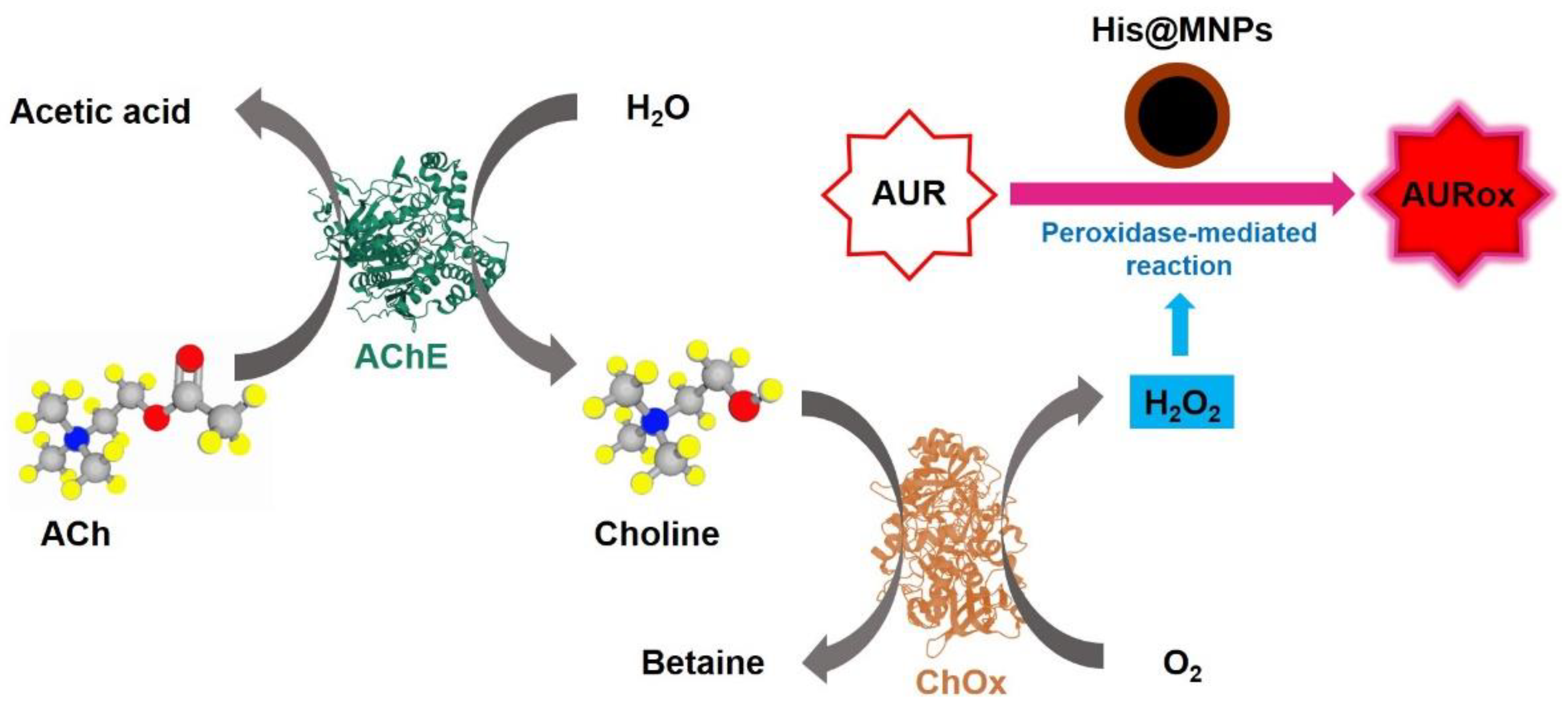
:1. Introduction
2. Materials and Methods
2.1. Materials
2.2. One-Pot Synthesis of Histidine and other Amino Acids Coated Magnetic Nanoparticles
2.3. Characterization of His@MNPs
2.4. Investigation for the Peroxidase-Like Activity of His@MNPs and Controls
2.5. Detection of H2O2, Choline, and ACh Using His@MNPs
3. Results and Discussion
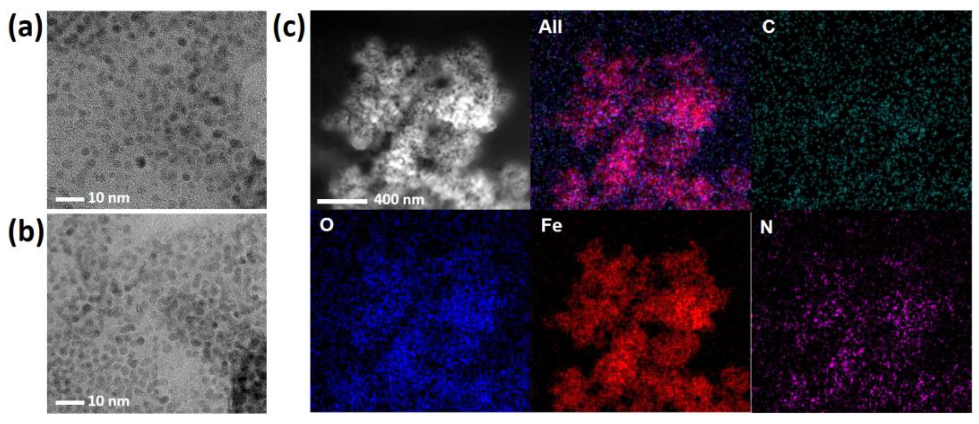
3.1. Synthesis and Characterization of His@MNPs
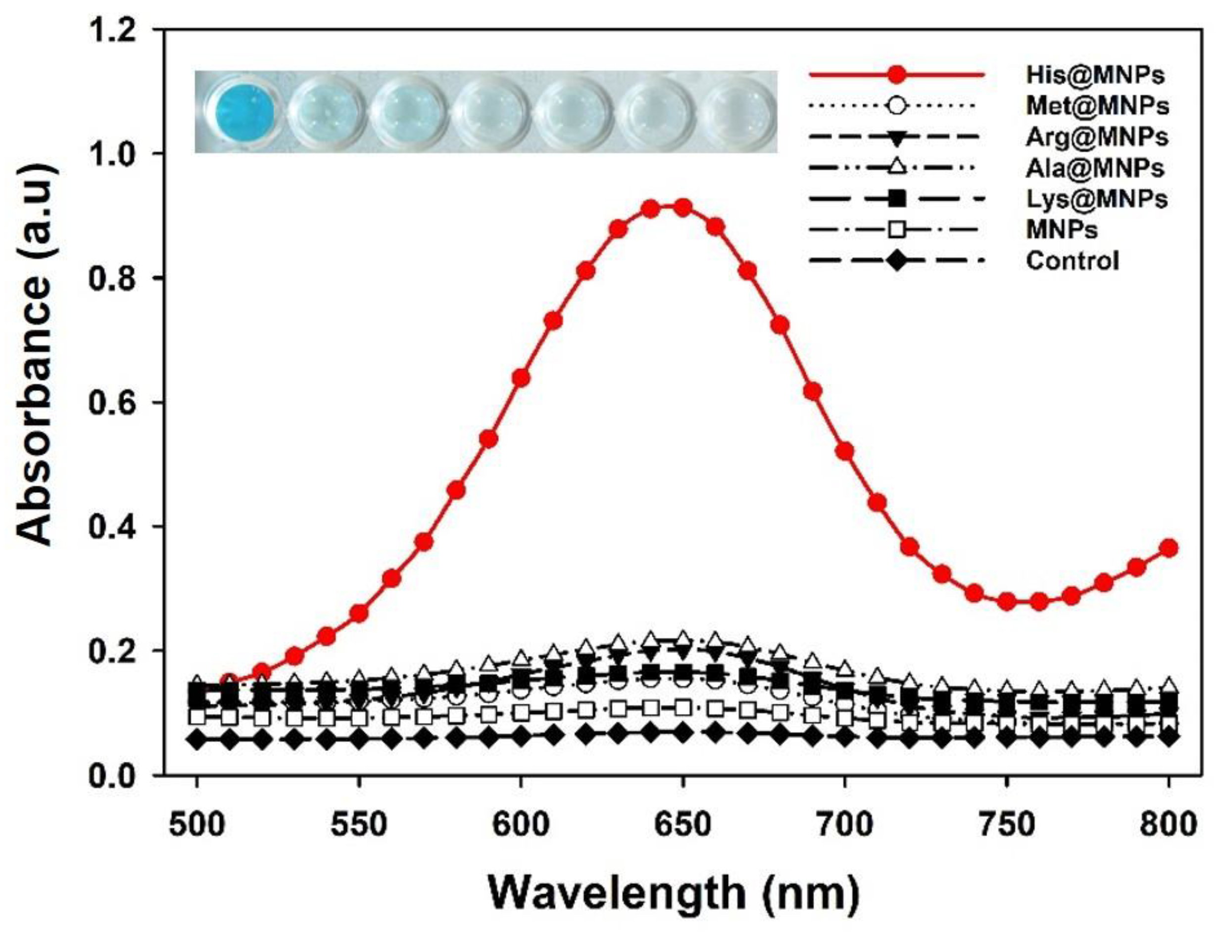
3.2. Enhanced Peroxidase-Like Activity of His@MNPs
3.3. Highly Sensitive Fluorescent Detection of Choline and ACh
4. Conclusions
Supplementary Materials
Author Contributions
Funding
Data Availability Statement
Conflicts of Interest
References
- Bolat, E.Ö.; Tiğ, G.A.; Pekyardımcı, Ş. Fabrication of an amperometric acetylcholine esterase-choline oxidase biosensor based on MWCNTs-Fe3O4 NPs-CS nanocomposite for the determination of aetylcholine. J. Electroanal. Chem. 2017, 758, 241–248. [Google Scholar] [CrossRef]
- Gou, J.; Wu, S.; Wang, Y.; Zhao, M.A. A label-free fluorescence biosensor based on a bifunctional MIL-101 (Fe) nanozyme for sensitive detection of choline and acetylcholine at nanomolar level. Sens. Actuator B Chem. 2020, 312, 128021. [Google Scholar]
- Kong, D.; Jin, R.; Zhao, X.; Li, H.; Yan, X.; Liu, F.; Sun, P.; Gao, Y.; Liang, X.; Lin, Y.J.; et al. Protein–inorganic hybrid nanoflower-rooted agarose hydrogel platform for point-of-care detection of acetylcholine. ACS Appl. Mater. Interfaces 2019, 11, 11857–11864. [Google Scholar] [CrossRef] [PubMed]
- Hampel, H.; Mesulam, M.; Cuello, A.C.; Farlow, M.R.; Giacobini, E.; Grossberg, G.T.; Khachaturian, A.S.; Vergallo, A.; Cavedo, E.; Snyder, P.J.; et al. The cholinergic system in the pathophysiology and treatment of Alzheimer’s disease. Brain 2018, 141, 1917–1933. [Google Scholar] [CrossRef] [PubMed]
- Castro, D.M.; Dillon, C.; Machnicki, G.; Allegri, R.F. The economic cost of Alzheimer’s disease: Family or public-health burden? Dement. Neuropsychol. 2010, 4, 262–267. [Google Scholar] [CrossRef] [PubMed]
- Zhao, L.H. Alzheimer’s disease facts and figures. Alzheimers Dement. 2020, 16, 391–460. [Google Scholar]
- Dunphy, R.; Burrinsky, D.J. Detection of choline and acetylcholine in a pharmaceutical preparation using high performance liquid chromatography/electrospray ionization mass spectrometry. Analysis 2003, 31, 905–915. [Google Scholar] [CrossRef]
- He, S.-B.; Wu, G.-W.; Deng, H.-H.; Liu, A.-L.; Lin, X.-H.; Xia, X.-H.; Chen, W. Choline and acetylcholine detection based on peroxidase-like activity and protein antifouling property of platinum nanoparticles in bovine serum albumin scaffold. Biosens. Bioelectron. 2014, 62, 331–336. [Google Scholar] [CrossRef]
- Craig, L.A.; Hong, N.S.; McDonald, R.J. Revisiting the cholinergic hypothesis in the development of Alzheimer’s disease. Neurosci. Biobehav. Rev. 2011, 35, 1397–1409. [Google Scholar] [CrossRef] [Green Version]
- Huang, Y.; Ren, J.; Qu, X. Nanozymes: Classification, catalytic mechanisms, activity regulation, and applications. Chem. Rev. 2019, 119, 4357–4412. [Google Scholar] [CrossRef]
- Gao, L.; Zhuang, J.; Nie, L.; Zhang, J.; Zhang, Y.; Gu, N.; Wang, T.; Feng, J.; Yang, D.; Perrett, S.J. Intrinsic peroxidase-like activity of ferromagnetic nanoparticles. Nat. Nanotechnol. 2007, 2, 577–583. [Google Scholar] [CrossRef]
- Liu, L.; Zhu, S.; Wei, Y.; Liu, X.; Jiao, S.; Yang, J. Ultrasensitive detection of miRNA-155 based on controlled fabrication of AuNPs@MoS2 nanostructures by atomic layer deposition. Biosens. Bioelectron. 2019, 144, 111660. [Google Scholar] [CrossRef]
- Wang, L.; Hu, Z.; Wu, S.; Pan, J.; Xu, X.; Niu, X. A peroxidase-mimicking Zr-based MOF colorimetric sensing array to quantify and discriminate phosphorylated proteins. Anal. Chim. Acta 2020, 1121, 26–34. [Google Scholar] [CrossRef] [PubMed]
- Loynachan, C.N.; Thomas, M.R.; Gray, E.R.; Richards, D.A.; Kim, J.; Miller, B.S.; Brookes, J.C.; Agarwal, S.; Chudasama, V.; McKendry, R.A. Platinum nanocatalyst amplification: Redefining the gold standard for lateral flow immunoassays with ultrabroad dynamic range. ACS Nano 2018, 12, 279–288. [Google Scholar] [CrossRef] [PubMed]
- Nguyen, P.T.; Ahn, H.T.; Kim, M.I. Reagent-free colorimetric assay for galactose using agarose gel entrapping nanoceria and galactose oxidase. Nanomaterials 2020, 10, 895. [Google Scholar] [CrossRef]
- Tripathi, K.M.; Ahn, H.T.; Chung, M.; Le, X.A.; Saini, D.; Bhati, A.; Sonkar, S.K.; Kim, M.I.; Kim, T. N, S, and P-Co-doped carbon quantum dots: Intrinsic peroxidase activity in a wide pH range and its antibacterial applications. ACS Biomater. Sci. Eng. 2020, 6, 5527–5537. [Google Scholar] [CrossRef] [PubMed]
- Duan, D.; Fan, K.; Zhang, D.; Tan, S.; Liang, M.; Liu, Y.; Zhang, J.; Zhang, P.; Liu, W.; Qiu, X.; et al. Nanozyme-strip for rapid local diagnosis of Ebola. Biosens. Bioelectron. 2015, 74, 134–141. [Google Scholar] [CrossRef] [PubMed]
- Shin, H.Y.; Kim, B.-G.; Cho, S.; Lee, J.; Na, H.B.; Kim, M.I. Visual determination of hydrogen peroxide and glucose by exploiting the peroxidase-like activity of magnetic nanoparticles functionalized with a poly (ethylene glycol) derivative. Microchim. Acta 2017, 184, 2115–2122. [Google Scholar] [CrossRef]
- Srinivasan, B.; Li, Y.; Jing, Y.; Xu, Y.; Yao, X.; Xing, C.; Wang, J.-P. A detection system based on giant magnetoresistive sensors and high--moment magnetic nanoparticles demonstrates zeptomole sensitivity: Potential for personalized medicine. Angew. Chem. Int. Ed. 2009, 121, 2802–2805. [Google Scholar] [CrossRef]
- Wang, Y.; Dostalek, J.; Knoll, W. Magnetic nanoparticle-enhanced biosensor based on grating-coupled surface plasmon resonance. Anal. Chem. 2011, 83, 6202–6207. [Google Scholar] [CrossRef]
- Yang, L.; Ren, X.; Tang, F.; Zhang, L. A practical glucose biosensor based on Fe3O4 nanoparticles and chitosan/nafion composite film. Biosens. Bioelectron. 2009, 25, 889–895. [Google Scholar] [CrossRef] [PubMed]
- Cardoso, V.F.; Francesko, A.; Ribeiro, C.; Bañobre-López, M.; Martins, P.; Lanceros-Mendez, S. Advances in magnetic nanoparticles for biomedical applications. Adv. Healthc. Mater. 2018, 7, 1700845. [Google Scholar] [CrossRef] [PubMed]
- Slimani, Y.; Hannachi, E. Magnetic nanosensors and their potential applications. In Nanosensors for Smart Cities; Han, B., Nguyen, T.A., Singh, P.K., Eds.; Elsevier: London, UK, 2020; Chapter 9; pp. 143–155. [Google Scholar]
- Dhavale, R.P.; Dhavale, R.; Sahoo, S.C.; Kollu, P.; Jadhav, S.U.; Patil, P.S.; Dongale, T.D.; Chougale, A.D.; Patil, P.B. Chitosan coated magnetic nanoparticles as carriers of anticancer drug Telmisartan: pH-responsive controlled drug release and cytotoxicity studies. J. Phys. Chem. Solids 2021, 148, 109749. [Google Scholar] [CrossRef]
- Sahin, S.; Ozmen, I. Determination of optimum conditions for glucose-6-phosphate dehydrogenase immobilization on chitosan-coated magnetic nanoparticles and its characterization. J. Mol. Catal. B Enzym. 2016, 133, S25–S33. [Google Scholar] [CrossRef]
- Komersová, A.; Kovářová, M.; Komers, K.; Lochař, V.; Čegan, A. Why is the hydrolytic activity of acetylcholinesterase pH dependent? Kinetic study of acetylcholine and acetylthiocholine hydrolysis catalyzed by acetylcholinesterase from electric eel. Z. Naturforsch. C J. Biosci. 2018, 73, 345–351. [Google Scholar] [CrossRef] [PubMed]
- Silman, H.; Karline, A. Effect of local pH changes caused by substrate hydrolysis on the activity of membrane-bound acetylcholinesterase. Proc. Natl. Acad. Sci. USA 1967, 58, 1664–1668. [Google Scholar] [CrossRef] [PubMed] [Green Version]
- Silva, V.A.; Andrade, P.L.; Silva, M.P.C.; Bustamante, A.; Valladares, L.D.L.S.; Aguiar, J.A. Synthesis and characterization of Fe3O4 nanoparticles coated with fucan polysaccharides. J. Magn. Magn. Mater. 2013, 343, 138–143. [Google Scholar] [CrossRef] [Green Version]
- Zhang, W.; Niu, X.; Meng, S.; Li, X.; He, Y.; Pan, J.; Qiu, F.; Zhao, H.; Lan, M. Histidine-mediated tunable peroxidase-like activity of nanosized Pd for photometric sensing of Ag+. Sens. Actuator B Chem. 2018, 273, 400–407. [Google Scholar] [CrossRef]
- Fan, K.; Wang, H.; Xi, J.; Liu, Q.; Meng, X.; Duan, D.; Gao, L.; Yan, X. Optimization of Fe3O4 nanozyme activity via single amino acid modification mimicking an enzyme active site. Chem. Commun. 2017, 53, 424–427. [Google Scholar] [CrossRef] [Green Version]
- Huang, X.; Xia, F.; Nan, Z. Fabrication of FeS2/SiO2 double mesoporous hollow spheres as an artificial peroxidase and rapid determination of H2O2 and glutathione. ACS Appl. Mater. Interfaces 2020, 12, 46539–46548. [Google Scholar] [CrossRef]
- Huang, Z.; He, W.; Shen, H.; Han, G.; Wang, H.; Su, P.; Song, J.; Yang, Y. NiCo2S4 microflowers as peroxidase mimic: A multi-functional platform for colorimetric detection of glucose and evaluation of antioxidant behavior. Talanta 2021, 230, 122337. [Google Scholar] [CrossRef]
- Karim, M.D.; Singh, M.; Weerathunge, P.; Bian, P.; Zheng, R.; Dekiwadia, C.; Ahmed, T.; Walia, S.; Gaspera, E.D.; Singh, S.; et al. Visible-light-triggered reactive oxygen species mediated antibacterial activity of peroxidase mimic CuO nanorods. ACS Appl. Nano Mater. 2018, 1, 1694–1704. [Google Scholar] [CrossRef]
- Kim, M.S.; Cho, S.; Joo, S.H.; Lee, J.; Kwak, S.K.; Kim, M.I.; Lee, J. N-and B-codoped graphene: A strong candidate to replace natural peroxidase in sensitive and selective bioassays. ACS Nano 2019, 13, 4312–4321. [Google Scholar] [CrossRef]
- Valekar, A.H.; Batule, B.S.; Kim, M.I.; Cho, K.-H.; Hong, D.-Y.; Lee, U.-H.; Chang, J.-S.; Park, H.G.; Hwang, Y.K. Novel amine-functionalized iron trimesates with enhanced peroxidase-like activity and their applications for the fluorescent assay of choline and acetylcholine. Biosens. Bioelectron. 2018, 100, 161–168. [Google Scholar] [CrossRef] [PubMed]
- Su, L.; Yu, X.; Qin, W.; Dong, W.; Wu, C.; Zhang, Y.; Mao, G.; Feng, S. One-step analysis of glucose and acetylcholine in water based on the intrinsic peroxidase-like activity of Ni/Co LDHs microspheres. J. Mater. Chem. B 2017, 5, 116–122. [Google Scholar] [CrossRef] [PubMed]


| Substrate | [E] (mM) | Vmax (µM s−1) | Km (mM) | kcat (s−1) | Ref. | |
|---|---|---|---|---|---|---|
| MIL-101(Fe) | TMB | - | 1.0083 | 0.585 | - | [2] |
| H2O2 | - | 0.5138 | 1.89 | - | ||
| BSA-Cu3(PO4)2 nanoflower | TMB | - | 0.099 | 0.1709 | - | [3] |
| H2O2 | - | 0.063 | 1.00 | - | ||
| HRP | TMB | 2.5 × 10⁻¹¹ | 0.1 | 0.434 | 4 × 10³ | [11] |
| H2O2 | 2.5 × 10⁻¹¹ | 0.087 | 3.70 | 3.48 × 10³ | ||
| NSP-carbon quantum dot | TMB | - | 0.188 | 0.47 | - | [16] |
| H2O2 | - | 69.50 | 32.61 | - | ||
| FeS2/SiO2 | TMB | - | 0.31 | 0.948 | 2.22 | [31] |
| H2O2 | - | 1.181 | 0.0126 | 1.29 | ||
| MNP | TMB | 9.7 × 10⁻⁶ | 0.18 | 0.142 | 1.88 | This work |
| H2O2 | 9.7 × 10⁻⁶ | 567.83 | 689.38 | 5.9 × 10⁴ | ||
| His@MNP | TMB | 9.7 × 10⁻⁶ | 0.51 | 0.149 | 5.29 | This work |
| H2O2 | 9.7 × 10⁻⁶ | 676.45 | 381.62 | 3.9 × 10⁴ |
| Sample | Added Value (μM) | Measured Value a (μM) | SD b | CV c (%) | Recovery d (%) |
|---|---|---|---|---|---|
| 1 | 3 | 3.01 | 0.23 | 7.54 | 100.20 |
| 2 | 5 | 5.07 | 0.06 | 1.16 | 101.47 |
| 3 | 8 | 8.15 | 0.31 | 3.85 | 101.92 |
Publisher’s Note: MDPI stays neutral with regard to jurisdictional claims in published maps and institutional affiliations. |
© 2021 by the authors. Licensee MDPI, Basel, Switzerland. This article is an open access article distributed under the terms and conditions of the Creative Commons Attribution (CC BY) license (https://creativecommons.org/licenses/by/4.0/).
Share and Cite
Cheon, H.J.; Nguyen, Q.H.; Kim, M.I. Highly Sensitive Fluorescent Detection of Acetylcholine Based on the Enhanced Peroxidase-Like Activity of Histidine Coated Magnetic Nanoparticles. Nanomaterials 2021, 11, 1207. https://doi.org/10.3390/nano11051207
Cheon HJ, Nguyen QH, Kim MI. Highly Sensitive Fluorescent Detection of Acetylcholine Based on the Enhanced Peroxidase-Like Activity of Histidine Coated Magnetic Nanoparticles. Nanomaterials. 2021; 11(5):1207. https://doi.org/10.3390/nano11051207
Chicago/Turabian StyleCheon, Hong Jae, Quynh Huong Nguyen, and Moon Il Kim. 2021. "Highly Sensitive Fluorescent Detection of Acetylcholine Based on the Enhanced Peroxidase-Like Activity of Histidine Coated Magnetic Nanoparticles" Nanomaterials 11, no. 5: 1207. https://doi.org/10.3390/nano11051207
APA StyleCheon, H. J., Nguyen, Q. H., & Kim, M. I. (2021). Highly Sensitive Fluorescent Detection of Acetylcholine Based on the Enhanced Peroxidase-Like Activity of Histidine Coated Magnetic Nanoparticles. Nanomaterials, 11(5), 1207. https://doi.org/10.3390/nano11051207