
Modelling of Dynamic Behaviour in Magnetic Nanoparticles

 , , , , and
Abstract
1. Introduction
- To model the behaviour of particles: allow for the optimisation of particles for a given application without the need for extensive empirical testing;
- To predict a particle’s properties, magnetic field properties and environmental parameters, such as viscosity, based on the behaviour of the MNPs.
2. Theory
3. Methods
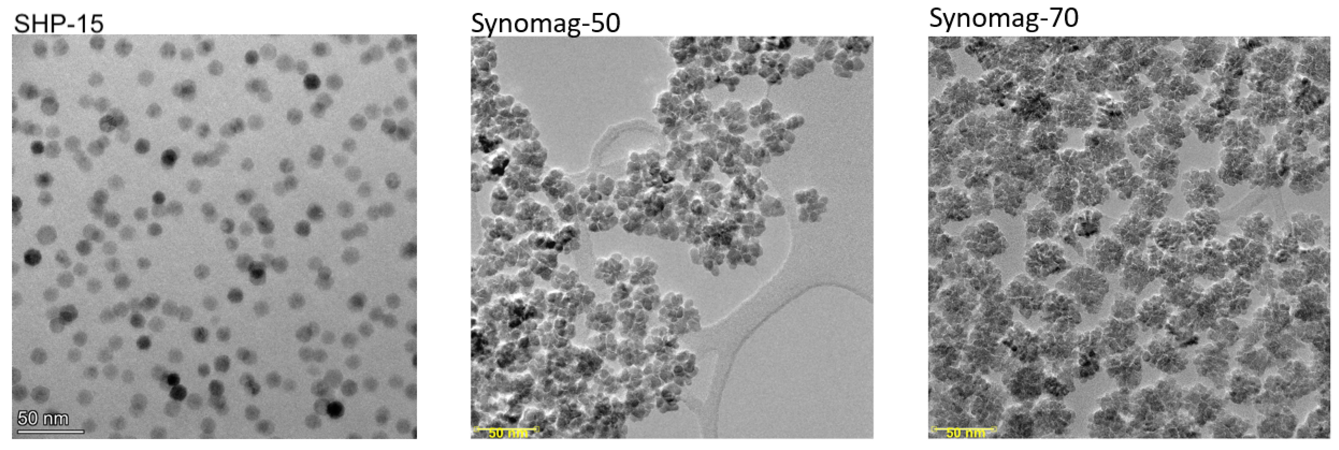
3.1. MNP Samples
3.2. Data Acquisition for Experimental Observations
3.3. Model
3.4. Model Validation
4. Results
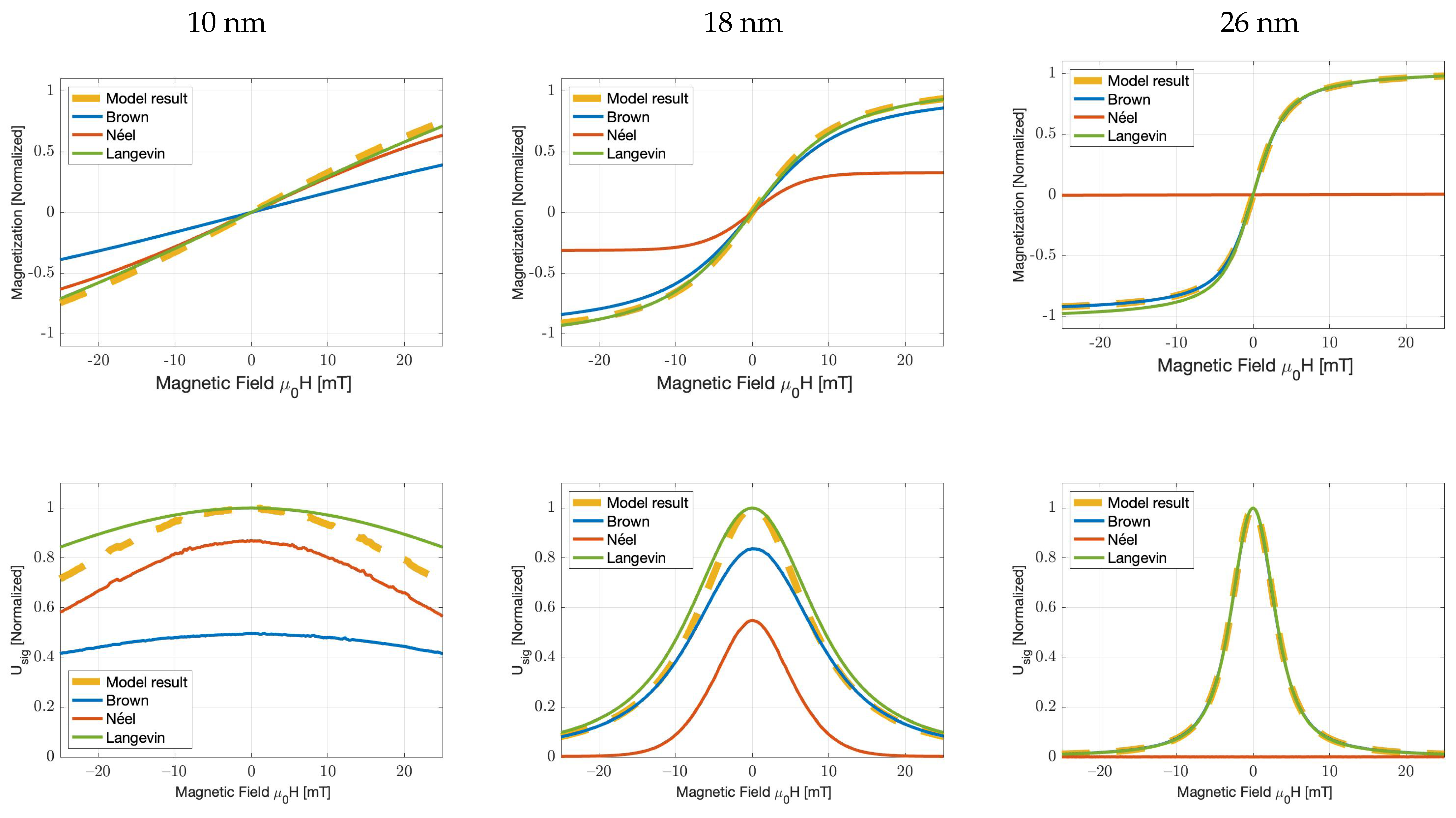
4.1. Numerical Modelling of Brownian and Néel Dominated M–H Curves
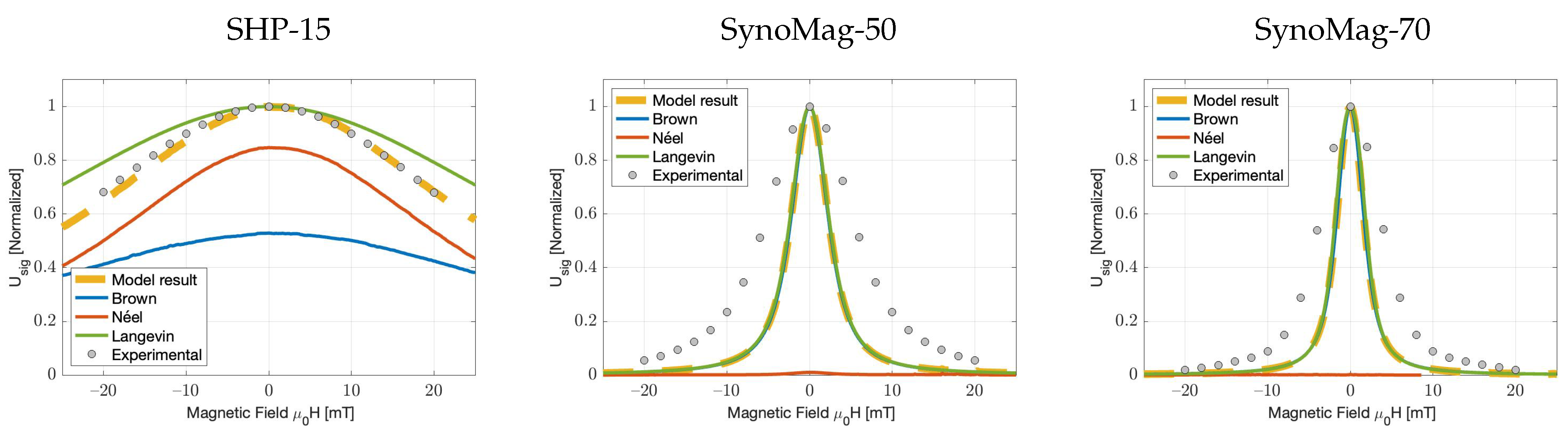
4.2. Experimental Verification of Particle Response Functions
5. Conclusions and Discussion
Author Contributions
Funding
Institutional Review Board Statement
Informed Consent Statement
Data Availability Statement
Acknowledgments
Conflicts of Interest
References
- Pankhurst, Q.A.; Connolly, J.; Jones, S.K.; Dobson, J. Applications of magnetic nanoparticles in biomedicine. J. Phys. D Appl. Phys. 2003, 36, R167–R181. [Google Scholar] [CrossRef]
- Ganguly, S.; Margel, S. Review: Remotely controlled magneto-regulation of therapeutics from magnetoelastic gel matrices. Biotechnol. Adv. 2020, 44, 107611. [Google Scholar] [CrossRef] [PubMed]
- Krishnan, K.M. Biomedical nanomagnetics: A spin through possibilities in imaging, diagnostics, and therapy. IEEE Trans. Magn. 2010, 46, 2523–2558. [Google Scholar] [CrossRef]
- Ganguly, S.; Neelam; Grinberg, I.; Margel, S. Layer by layer controlled synthesis at room temperature of tri-modal (MRI, fluorescence and CT) core/shell superparamagnetic IO/human serum albumin nanoparticles for diagnostic applications. Polym. Adv. Technol. 2021, 32, 3909–3921. [Google Scholar] [CrossRef]
- Yoo, D.; Lee, J.H.; Shin, T.H.; Cheon, J. Theranostic Magnetic Nanoparticles. Accounts Chem. Res. 2011, 44, 863–874. [Google Scholar] [CrossRef] [PubMed]
- Douek, M.; Klaase, J.; Monypenny, I.; Kothari, A.; Zechmeister, K.; Brown, D.; Wyld, L.; Drew, P.; Garmo, H.; Agbaje, O.; et al. Sentinel Node Biopsy Using a Magnetic Tracer Versus Standard Technique: The SentiMAG Multicentre Trial. Ann. Surg. Oncol. 2013, 21, 1237–1245. [Google Scholar] [CrossRef]
- Visscher, M.; Waanders, S.; Krooshoop, H.; ten Haken, B. Selective detection of magnetic nanoparticles in biomedical applications using differential magnetometry. J. Magn. Magn. Mater. 2014, 365, 31–39. [Google Scholar] [CrossRef]
- Waanders, S.; Visscher, M.; Wildeboer, R.; Odekerk, T.; Krooshoop, H.; ten Haken, B. A handheld SPIO-based sentinel lymph node mapping device using differential magnetometry. Phys. Med. Biol. 2016, 61, 8120–8134. [Google Scholar] [CrossRef]
- Wu, K.; Su, D.; Saha, R.; Liu, J.; Chugh, V.K.; Wang, J.P. Magnetic Particle Spectroscopy: A Short Review of Applications Using Magnetic Nanoparticles. ACS Appl. Nano Mater. 2020, 3, 4972–4989. [Google Scholar] [CrossRef]
- Gleich, B.; Weizenecker, J. Tomographic imaging using the nonlinear response of magnetic particles. Nature 2005, 435, 1214–1217. [Google Scholar] [CrossRef]
- Rosensweig, R.E. Heating magnetic fluid with alternating magnetic field. J. Magn. Magn. Mater. 2002, 252, 370–374. [Google Scholar] [CrossRef]
- Felderhof, B.U.; Jones, R.B. Mean field theory of the nonlinear response of an interacting dipolar system with rotational diffusion to an oscillating field. J. Phys. Condens. Matter 2003, 15, 4011–4024. [Google Scholar] [CrossRef]
- Felderhof, B.U.; Jones, R.B. Nonlinear response of a dipolar system with rotational diffusion to an oscillating field. J. Phys. Condens. Matter 2003, 15, S1363–S1378. [Google Scholar] [CrossRef]
- Rauwerdink, A.M.; Weaver, J.B. Viscous effects on nanoparticle magnetization harmonics. J. Magn. Magn. Mater. 2010, 322, 609–613. [Google Scholar] [CrossRef]
- Rauwerdink, A.M.; Weaver, J.B. Harmonic phase angle as a concentration-independent measure of nanoparticle dynamics. Med. Phys. 2010, 37, 2587–2592. [Google Scholar] [CrossRef]
- Yoshida, T.; Bai, S.; Hirokawa, A.; Tanabe, K.; Enpuku, K. Effect of viscosity on harmonic signals from magnetic fluid. J. Magn. Magn. Mater. 2015, 380, 105–110. [Google Scholar] [CrossRef]
- Kahmann, T.; Rösch, E.L.; Enpuku, K.; Yoshida, T.; Ludwig, F. Determination of the effective anisotropy constant of magnetic nanoparticles—Comparison between two approaches. J. Magn. Magn. Mater. 2021, 519, 167402. [Google Scholar] [CrossRef]
- Du, Z.; Sun, Y.; Higashi, O.; Noguchi, Y.; Enpuku, K.; Draack, S.; Janssen, K.J.; Kahmann, T.; Zhong, J.; Viereck, T.; et al. Effect of core size distribution on magnetic nanoparticle harmonics for thermometry. Jpn. J. Appl. Phys. 2019, 59, 010904. [Google Scholar] [CrossRef]
- Anand, M. Thermal and dipolar interaction effect on the relaxation in a linear chain of magnetic nanoparticles. J. Magn. Magn. Mater. 2021, 522, 167538. [Google Scholar] [CrossRef]
- Gilbert, T. Classics in Magnetics A Phenomenological Theory of Damping in Ferromagnetic Materials. IEEE Trans. Magn. 2004, 40, 3443–3449. [Google Scholar] [CrossRef]
- Brown, W.F. Thermal fluctuations of a single-domain particle. Phys. Rev. 1963, 130, 1677–1686. [Google Scholar] [CrossRef]
- Deissler, R.J.; Wu, Y.; Martens, M.A. Dependence of Brownian and Néel relaxation times on magnetic field strength. Med. Phys. 2014, 41, 012301. [Google Scholar] [CrossRef] [PubMed]
- Ferguson, R.M.; Khandhar, A.P.; Krishnan, K.M. Tracer design for magnetic particle imaging (invited). J. Appl. Phys. 2012, 111, 07B318. [Google Scholar] [CrossRef]
- Weizenecker, J.; Borgert, J.; Gleich, B. A simulation study on the resolution and sensitivity of magnetic particle imaging. Phys. Med. Biol. 2007, 52, 6363–6374. [Google Scholar] [CrossRef] [PubMed]
- Croft, L.R.; Goodwill, P.W.; Conolly, S.M. Relaxation in X-Space Magnetic Particle Imaging. IEEE Trans. Med Imaging 2012, 31, 2335–2342. [Google Scholar] [CrossRef]
- Reeves, D.B.; Weaver, J.B. Approaches for Modeling Magnetic Nanoparticle Dynamics. Crit. Rev. Trade Biomed. Eng. 2014, 42, 85–93. [Google Scholar] [CrossRef]
- Chung, S.H.; Hoffmann, A.; Bader, S.D.; Liu, C.; Kay, B.; Makowski, L.; Chen, L. Biological sensors based on Brownian relaxation of magnetic nanoparticles. Appl. Phys. Lett. 2004, 85, 2971–2973. [Google Scholar] [CrossRef]
- Fannin, P.C.; Charles, S.W. The study of a ferrofluid exhibiting both Brownian and Neel relaxation. J. Phys. D Appl. Phys. 2000, 22, 187–191. [Google Scholar] [CrossRef]
- Kötitz, R.; Weitschies, W.; Trahms, L.; Semmler, W. Investigation of Brownian and Neel relaxation in magnetic fluids—Condens. Matter. J. Magn. Magn. Mater. 1999, 201, 102–104. [Google Scholar] [CrossRef]
- Draack, S.; Viereck, T.; Nording, F.; Janssen, K.J.; Schilling, M.; Ludwig, F. Determination of dominating relaxation mechanisms from temperature-dependent Magnetic Particle Spectroscopy measurements. J. Magn. Magn. Mater. 2019, 474, 570–573. [Google Scholar] [CrossRef]
- Kötitz, R.; Weitschies, W.; Trahms, L.; Brewer, W.; Semmler, W. Determination of the binding reaction between avidin and biotin by relaxation measurements of magnetic nanoparticles. J. Magn. Magn. Mater. 1999, 194, 62–68. [Google Scholar] [CrossRef]
- Ludwig, F.; Remmer, H.; Kuhlmann, C.; Wawrzik, T.; Arami, H.; Ferguson, R.M.; Krishnan, K.M. Self-consistent magnetic properties of magnetite tracers optimized for magnetic particle imaging measured by ac susceptometry, magnetorelaxometry and magnetic particle spectroscopy. J. Magn. Magn. Mater. 2014, 360, 169–173. [Google Scholar] [CrossRef] [PubMed]
- Ferguson, R.M.; Khandhar, A.P.; Arami, H.; Hua, L.; Hovorka, O.; Krishnan, K.M. Tailoring the magnetic and pharmacokinetic properties of iron oxide magnetic particle imaging tracers. Biomed. Tech./Biomed. Eng. 2013, 58, 493–507. [Google Scholar] [CrossRef] [PubMed]
- Sun, Y.; Ye, N.; Wang, D.; Du, Z.; Bai, S.; Yoshida, T. An Improved Method for Estimating Core Size Distributions of Magnetic Nanoparticles via Magnetization Harmonics. Nanomaterials 2020, 10, 1623. [Google Scholar] [CrossRef]
- Van De Loosdrecht, M.M.; Draack, S.; Waanders, S.; Schlief, J.G.; Krooshoop, H.J.; Viereck, T.; Ludwig, F.; Ten Haken, B. A novel characterization technique for superparamagnetic iron oxide nanoparticles: The superparamagnetic quantifier, compared with magnetic particle spectroscopy. Rev. Sci. Instrum. 2019, 90, 024101. [Google Scholar] [CrossRef] [PubMed]
- Martens, M.; Deissler, R.; Wu, Y.; Lisa, B.; Yao, Z.; Robert, B.; Mark, G. Modeling the Brownian relaxation of nanoparticle ferrofluids: Comparison with experiment. In Proceedings of the 2013 International Workshop on Magnetic Particle Imaging, IWMPI 2013, Berkeley, CA, USA, 23–24 March 2013; p. 022303. [Google Scholar] [CrossRef]
- Weizenecker, J.; Gleich, B.; Rahmer, J.; Borgert, J. Micro-magnetic simulation study on the magnetic particle imaging performance of anisotropic mono-domain particles. Phys. Med. Biol. 2012, 57, 7317–7327. [Google Scholar] [CrossRef] [PubMed]
- Shliomis, M.I.; Stepanov, V.I. Theory of the dymamic susceptibility of magnetic fluids. Adv. Chem. Phys. Relax. Phenom. Condens. Matter 1994, 87, 1–30. [Google Scholar]
- Lehlooh, A.F.; Mahmood, S.H.; Williams, J.M. On the particle size dependence of the magnetic anisotropy energy constant. Phys. B Condens. Matter 2002, 321, 159–162. [Google Scholar] [CrossRef]
- Arami, H.; Ferguson, R.M.; Khandhar, A.P.; Krishnan, K.M. Size-dependent ferrohydrodynamic relaxometry of magnetic particle imaging tracers in different environments. Med. Phys. 2013, 40, 071904. [Google Scholar] [CrossRef]
- Weizenecker, J. The Fokker–Planck equation for coupled Brown-Néel-rotation. Phys. Med. Biol. 2018, 63, 035004. [Google Scholar] [CrossRef]
- Eberbeck, D.; Wiekhorst, F.; Wagner, S.; Trahms, L. How the size distribution of magnetic nanoparticles determines their magnetic particle imaging performance. Appl. Phys. Lett. 2011, 98, 182502. [Google Scholar] [CrossRef]
- Fannin, P. Magnetic Spectroscopy as an Aide in Understanding Magnetic Fluids. Ferrofluids 2002, 19–32. [Google Scholar] [CrossRef]
- Ludwig, F.; Kuhlmann, C.; Wawrzik, T.; Dieckhoff, J.; Lak, A.; Kandhar, A.P.; Ferguson, R.M.; Kemp, S.J.; Krishnan, K.M. Dynamic Magnetic Properties of Optimized Magnetic Nanoparticles for Magnetic Particle Imaging. IEEE Trans. Magn. 2014, 50, 1–4. [Google Scholar] [CrossRef]


| Shell | ||||||
|---|---|---|---|---|---|---|
| (nm) | (nm) | (kJ m nm) | (kJ m) | (kA m) | ||
| SHP-15 | 0.150 | 5.0 | 205 | |||
| Synomag-D50 | 0.150 | 9.5 | 420 | |||
| Synomag-D70 | 0.150 | 9.5 | 420 |
| SHP-15 (Extrapolated) | Synomag®-D50 | Synomag®-D70 | ||||
|---|---|---|---|---|---|---|
| FWHM (% diff) | MoR | FWHM (% diff) | MoR | FWHM (% diff) | MoR | |
| Model | 11.3 | 0.02 | −56.7 | 0.30 | −55.1 | 0.30 |
| Langevin | 49.5 | 0.07 | −55.2 | 0.29 | −51.2 | 0.27 |
Publisher’s Note: MDPI stays neutral with regard to jurisdictional claims in published maps and institutional affiliations. |
© 2021 by the authors. Licensee MDPI, Basel, Switzerland. This article is an open access article distributed under the terms and conditions of the Creative Commons Attribution (CC BY) license (https://creativecommons.org/licenses/by/4.0/).
Share and Cite
Rietberg, M.T.; Waanders, S.; Horstman-van de Loosdrecht, M.M.; Wildeboer, R.R.; ten Haken, B.; Alic, L. Modelling of Dynamic Behaviour in Magnetic Nanoparticles. Nanomaterials 2021, 11, 3396. https://doi.org/10.3390/nano11123396
Rietberg MT, Waanders S, Horstman-van de Loosdrecht MM, Wildeboer RR, ten Haken B, Alic L. Modelling of Dynamic Behaviour in Magnetic Nanoparticles. Nanomaterials. 2021; 11(12):3396. https://doi.org/10.3390/nano11123396
Chicago/Turabian StyleRietberg, Max Tigo, Sebastiaan Waanders, Melissa Mathilde Horstman-van de Loosdrecht, Rogier R. Wildeboer, Bennie ten Haken, and Lejla Alic. 2021. "Modelling of Dynamic Behaviour in Magnetic Nanoparticles" Nanomaterials 11, no. 12: 3396. https://doi.org/10.3390/nano11123396
APA StyleRietberg, M. T., Waanders, S., Horstman-van de Loosdrecht, M. M., Wildeboer, R. R., ten Haken, B., & Alic, L. (2021). Modelling of Dynamic Behaviour in Magnetic Nanoparticles. Nanomaterials, 11(12), 3396. https://doi.org/10.3390/nano11123396