Article

Dye Sensitization for Ultraviolet Upconversion Enhancement

1
Institute of Nanochemistry and Nanobiology, Shanghai University, Shanghai 200444, China
2
Department of Stomatology, Tongji Hospital, Tongji Medical College, Huazhong University of Science and Technology, Wuhan 430030, China
3
School of Stomatology, Tongji Medical College, Huazhong University of Science and Technology, Wuhan 430030, China
4
Hubei Province Key Laboratory of Oral and Maxillofacial Development and Regeneration, Wuhan 430022, China
*
Authors to whom correspondence should be addressed.
Nanomaterials 2021, 11(11), 3114; https://doi.org/10.3390/nano11113114
Submission received: 20 October 2021 / Revised: 8 November 2021 / Accepted: 11 November 2021 / Published: 18 November 2021
(This article belongs to the Special Issue Luminescence Nanomaterials and Applications)

Abstract

:
Upconversion nanocrystals that converted near-infrared radiation into emission in the ultraviolet spectral region offer many exciting opportunities for drug release, photocatalysis, photodynamic therapy, and solid-state lasing. However, a key challenge is the development of lanthanide-doped nanocrystals with efficient ultraviolet emission, due to low conversion efficiency. Here, we develop a dye-sensitized, heterogeneous core–multishelled lanthanide nanoparticle for ultraviolet upconversion enhancement. We systematically study the main influencing factors on ultraviolet upconversion emission, including dye concentration, excitation wavelength, and dye-sensitizer distance. Interestingly, our experimental results demonstrate a largely promoted multiphoton upconversion. The underlying mechanism and detailed energy transfer pathway are illustrated. These findings offer insights into future developments of highly ultraviolet-emissive nanohybrids and provide more opportunities for applications in photo-catalysis, biomedicine, and environmental science.

1. Introduction

Lanthanide-doped upconversion nanoparticles can absorb near-infrared (NIR) laser light and emit visible and ultraviolet light, with potential applications in bioimaging [1,2,3,4,5], biotherapy [6,7,8,9,10,11,12], and so on. In particular, the applications of these nanoparticles in optogenetic [13,14], photothermal [15,16], and photodynamic [17,18,19] therapy could be achieved via ultraviolet (UV) light emission under NIR excitation. Although UV light can be obtained by Nd3+- and Yb3+-sensitized upconversion [17,18,20,21], it is challenging to realize the high luminescence intensity needed to satisfy the minimum requirement of biological applications. This obstacle can be addressed in several ways: by controlling dopant composition [22], nanoparticle phase and size [23], excitation beam pulse width [24], and nanoparticle core–shell design [21,25,26,27,28,29]. Very recently, our group has made significant progress in overcoming the difficulty using an upconverted excitation lock-in (UCEL) strategy [30].
Hybrid systems are composed of inorganic nanoparticles and an organic dye, which can significantly strengthen the absorbance and expand the absorbance spectra of inorganic nanoparticles [31], leading to enhancement of their emission intensities. It has been demonstrated that NIR dye can effectively enhance the upconversion emission of lanthanide-doped nanoparticles [14,32,33,34]. However, previous studies have mainly focused on the analysis of visible upconversion emission. Little effort has been made to develop a hybrid nanoparticle with enhanced UV luminescence.
In this study, we developed IR-806-loaded upconversion nanoparticles (Gd-CSYS2S3@IR-806) with enhanced upconversion emission in the UV spectral region. The key factors that influence upconversion emission were studied, including dye concentration, excitation wavelength, and distance between the dye and the sensitizer Nd3+ (Scheme 1). We also demonstrated the dominant effect of ligand loading on multiphoton upconversion. In addition, the upconversion mechanism and the energy transfer pathway in Gd-CSYS2S3@IR-806 hybrid nanoparticles were carefully studied. This study provides new insights into the mechanistic understanding of UV upconversion luminescence in hybrid nanoparticles and enables new opportunities for these nanomaterials in a broad range of applications.

2. Materials and Methods

2.1. Materials

Gd(CH3CO2)3·xH2O (99.9%), Nd(CH3CO2)3·xH2O (99.9%), Y(CH3CO2)3·xH2O (99.9%), Yb(CH3CO2)3·xH2O (99.9%), Tm(CH3CO2)3·xH2O (99.9%), NaOH (>98%), NH4F (>98%), chloroform(99.9%), oleic acid (OA, 90%), and 1-octadecene (ODE, 90%) were all purchased from Sigma-Aldrich (Shanghai, China). IR-806 is supported by Dr. Sanyang Han from the University of Cambridge. IR-780 iodide, 4-mercaptobenzoic acid, and N,N-dimethylformamide (DMF, anhydrous, 99.8%), as raw materials of IR-806, were obtained from Sigma-Aldrich (London, UK). Dichloromethane (DCM, AR) and diethyl ether (AR), as solvents for the synthesis of IR-806, were obtained from Lab-Scan (London, United Kingdom). Chloroform (AR), cyclohexane (AR), and ethanol (AR) were purchased from Sinopharm Chemical Reagent Co., Ltd. (Shanghai, China). All chemicals and reagents were used as received without further purification unless otherwise noted.

2.2. Characterization

Luminescence emission measurements were obtained with an FS5 (Edinburgh, UK) conjugated with 808 nm (CNI, MDL-III-808-2.5W, China), 793 nm (CNI, MDL-III-793-2.0W, China), and 980 nm (CNI, MDLIII-980-2.0W, China) diode lasers at room temperature. The decay curves were recorded by a lifetime spectrometer (FS5, Edinburgh, UK), in conjunction with pulsed 808 nm, 793 nm, and 980 nm diode lasers and a picosecond pulsed light emitting diode (EPLED-270). Low-resolution transmission electron microscopy (TEM) measurements were carried out on an HT7700 field emission transmission electron microscope operated at an acceleration voltage of 120 kV. The energy-dispersive X-ray (EDX) spectrum was obtained with an HT7700 field emission transmission electron microscope equipped with an Oxford Instruments system. High-resolution TEM images were obtained using an FEI Talos F200S transmission electron microscope operated at an acceleration voltage of 200 kV. HAADF-STEM and elemental mapping images were obtained using an FEI Talos F200X transmission electron microscope. Powder X-ray diffraction (XRD) analysis was performed on a Rigaku D/Max-2200 system equipped with a rotating anode and a Cu Kα radiation source (λ = 0.15418 nm). The excitation power density was measured using a TS5 laser power densitometer (Changchun New Industries Optoelectronics Technology, China). UV–vis absorption spectra were obtained using a PerkinElmer LAMBDA 750 ultraviolet–visible–near-infrared spectrometer and a Hitachi U-3010 spectrophotometer. All spectra were recorded under identical experimental conditions unless otherwise noted. Key experiments were repeated three times, and all other experiments were repeated twice.

2.3. Method

2.3.1. Synthesis of NaGdF4:49%Yb,1%Tm Core Nanocrystals

NaGdF4 doped with 49 mol % of Yb and 1 mol % of Tm (NaGdF4:49%Yb,1%Tm) was synthesized via a modified literature procedure [28,35,36]. A water solution of Gd(CH3CO2)3 (0.067 g; 0.2 mmol), Yb(CH3CO2)3 (0.069 g; 0.196 mmol), and Tm(CH3CO2)3 (0.001 g; 0.004 mmol) was combined with OA (5 mL) and ODE (5 mL) in a 50 mL two-neck round-bottom flask. The mixture was heated to 150 °C and maintained at this temperature for 1.5 h to form the lanthanide–oleate precursor. After cooling to 50 °C, a methanol solution consisting of NH4F (0.05 g; 1.36 mmol) and NaOH (0.04 g; 1 mmol) was added to the mixture and stirred for 30 min. The solution was heated to 100 °C for 20 min in vacuo to remove methanol. The resulting solution was quickly heated to 300 °C and maintained at this temperature for 1.5 h with nitrogen before cooling to room temperature. The obtained nanocrystals were precipitated by centrifugation at 8000 rpm for 5 min and then washed with cyclohexane and ethanol three times. The core nanoparticles were dispersed in cyclohexane (4 mL) for further shell coating.

2.3.2. Synthesis of NaGdF4:49%Yb,1%Tm@NaYF4:20%Yb Core–Shell Nanocrystals

The synthesis procedure for core–shell nanoparticles was similar to that in our previous paper [36]. We use the obtained NaGdF4:49%Yb,1%Tm nanocrystals as seeds for subsequent shell coating. NaYF4 with 20 mol % of Yb (NaYF4:20%Yb) precursor was prepared via the same procedure as mentioned above, except that different amounts of OA (3 mL) and ODE (7 mL) were used. After cooling to 80 °C, the cyclohexane solution of NaGdF4:Yb/Tm nanoparticles (4 mL) was added and kept at 80 °C for 30 min to remove cyclohexane. Then, a methanol solution of NH4F (0.05 g; 1.36 mmol) and NaOH (0.04 g; 1 mmol) was added to the mixture and stirred at 50 °C for 30 min. Subsequently, the mixture was heated to 100 °C for 20 min in vacuo to remove methanol. The solution was then heated to 300 °C for 1.5 h under a nitrogen atmosphere. After cooling to room temperature, the core–shell nanoparticles were collected and washed using the same post-treatment approach as for core nanocrystals. NaGdF4@NaGdF4:49%Yb,1%Tm and NaYF4@NaGdF4:49%Yb,1%Tm were synthesized using a similar method to core–shell nanocrystals except for the use of NaGdF4 and NaYF4 as seeds.

2.3.3. Synthesis of NaGdF4:49%Yb,1%Tm@NaYF4:20%Yb@NaGdF4:50%Nd,10%Yb and NaGdF4:49%Yb,1%Tm@NaYF4:20%Yb@NaGdF4:50%Nd,10%Yb@NaGdF4 Core–Multishell Nanocrystals

The following multishelled core–shell nanoparticles were prepared using a procedure similar to the NaGdF4:49%Yb,1%Tm@NaYF4:20%Yb core–shell nanoparticles: NaGdF4@NaGdF4:49%Yb,1%Tm@ NaYF4:20%Yb; NaYF4@NaGdF4:49%Yb,1%Tm@NaYF4: 20%Yb; NaGdF4@NaGdF4:49%Yb, 1%Tm@NaYF4:20%Yb@NaGdF4:50%Nd,10%Yb; NaYF4 @NaGdF4:49%Yb,1%Tm@NaYF4:20%Yb@NaGdF4:50%Nd,10%Yb;NaGdF4@NaGdF4:49%Yb,1%Tm@NaYF4:20%Yb@NaGdF4:50%Nd,10%Yb@NaGdF4; NaYF4@NaGdF4:49%Yb,1% Tm@NaYF4:20%Yb@ NaGdF4:50%Nd,10%Yb@NaGdF4.

2.3.4. Preparation of Dye-Sensitized Upconversion Nanoparticles

The synthesis of IR-806 followed a well-established method [32]. Then, the IR-806 was dissolved in CHCl3 (0.01 mg/mL). The as-prepared core–multishell nanocrystals were centrifuged and dissolved in CHCl3 to a final concentration of 0.375 mg/mL. The samples were prepared by adding different amounts of IR-806 to Gd-CSYS2S3 CHCl3 solution (4 mL) and stirring for 2 h at a speed of 700 rpm at room temperature before UV–vis–NIR absorption and standard fluorescence measurements. All samples were prepared and measured in a dark environment.

3. Results

3.1. Synthesis of Core–Multishell Upconversion Nanoparticles

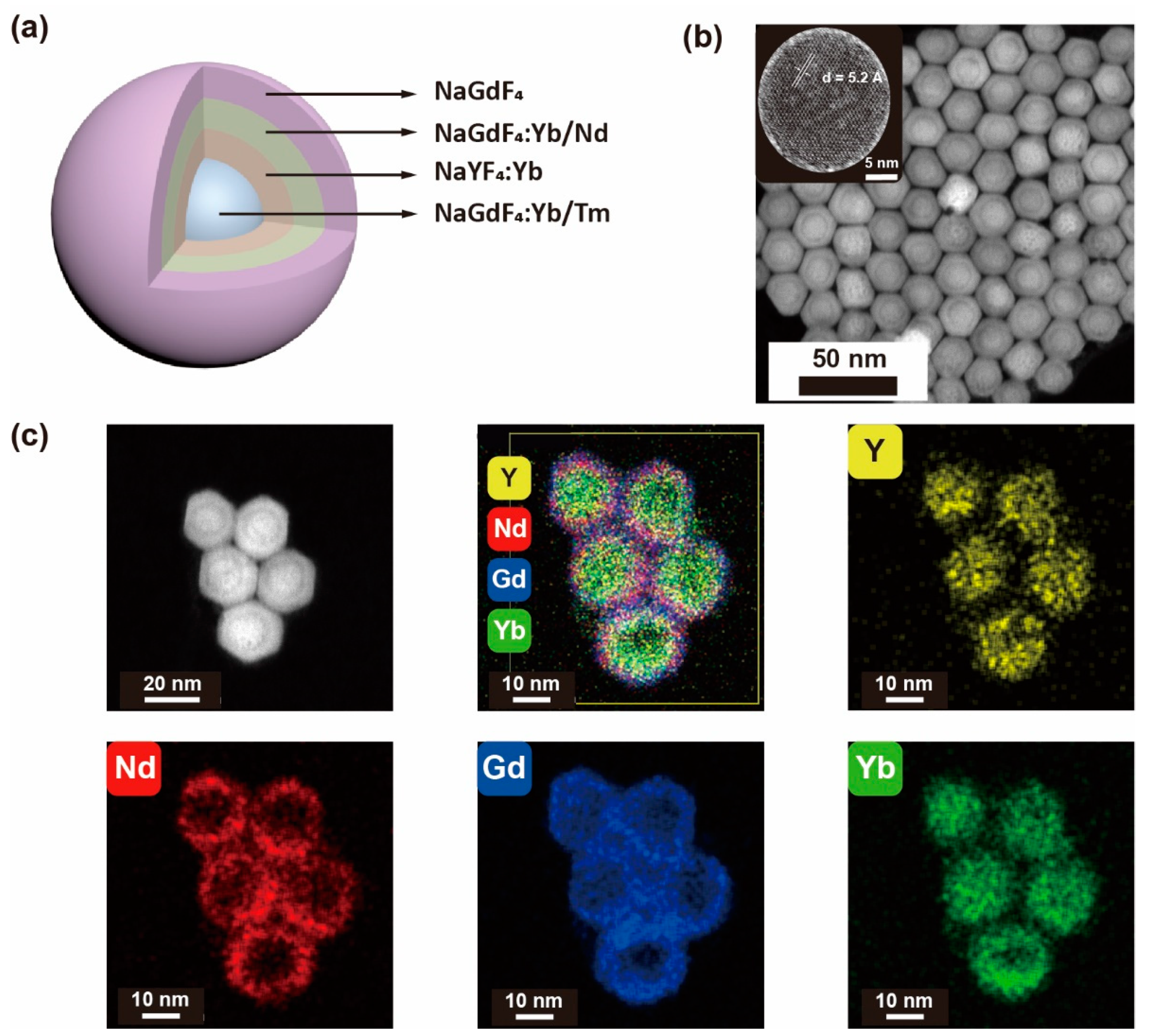
We previously designed a heterogeneous core–multishell nanoparticle with enhanced UV upconversion emission, involving six- and five-photon upconversion processes [30]. The optimum doping concentration and nanoparticle design were determined according to our previous reports [36]. From our previous photoluminescence results, the optimized nanostructure was determined to be NaGdF4:49%Yb/1%Tm@NaGdF4:20%Yb@ NaGdF4:10%Yb/50%Nd@NaGdF4. Recently, we found that when the NaGdF4:20%Yb was replaced with NaYF4:20%Yb, UV emission was significantly enhanced due to the effective suppression of energy consumption induced by interior energy traps. Herein, we chose this heterogeneous nanostructure as an experimental model to further enhance upconversion emission in the UV range. We first synthesized the core–multishell NaGdF4:49%Yb,1%Tm@NaYF4:20%Yb@NaGdF4:10%Yb,50%Nd@NaGdF4 (Gd-CSYS2S3) nanoparticles using a layer epitaxial growth method. Transmission electron microscopy (TEM) images showed that the nanoparticles had a uniform size of about 28 nm and the thickness of each layer was ~2 nm (Figure S1). The as-prepared nanoparticles were identified as the hexagonal phase by X-ray powder diffraction (XRD, JCPDS file number 27-0699, Figure S2). In addition, the constitution of the heterogeneous core–multishell nanostructures was confirmed by high-angle annular dark-field scanning transmission electron microscopy (HAADF-STEM), elemental mapping images and energy dispersive X-ray (EDX) spectra (Figure 1b,c, Figures S3 and S4), where brighter regions correspond to heavier elements (Gd, Yb, and Nd) and lighter regions correspond to lighter elements (Y).

3.2. Remarkable UV Enhancement

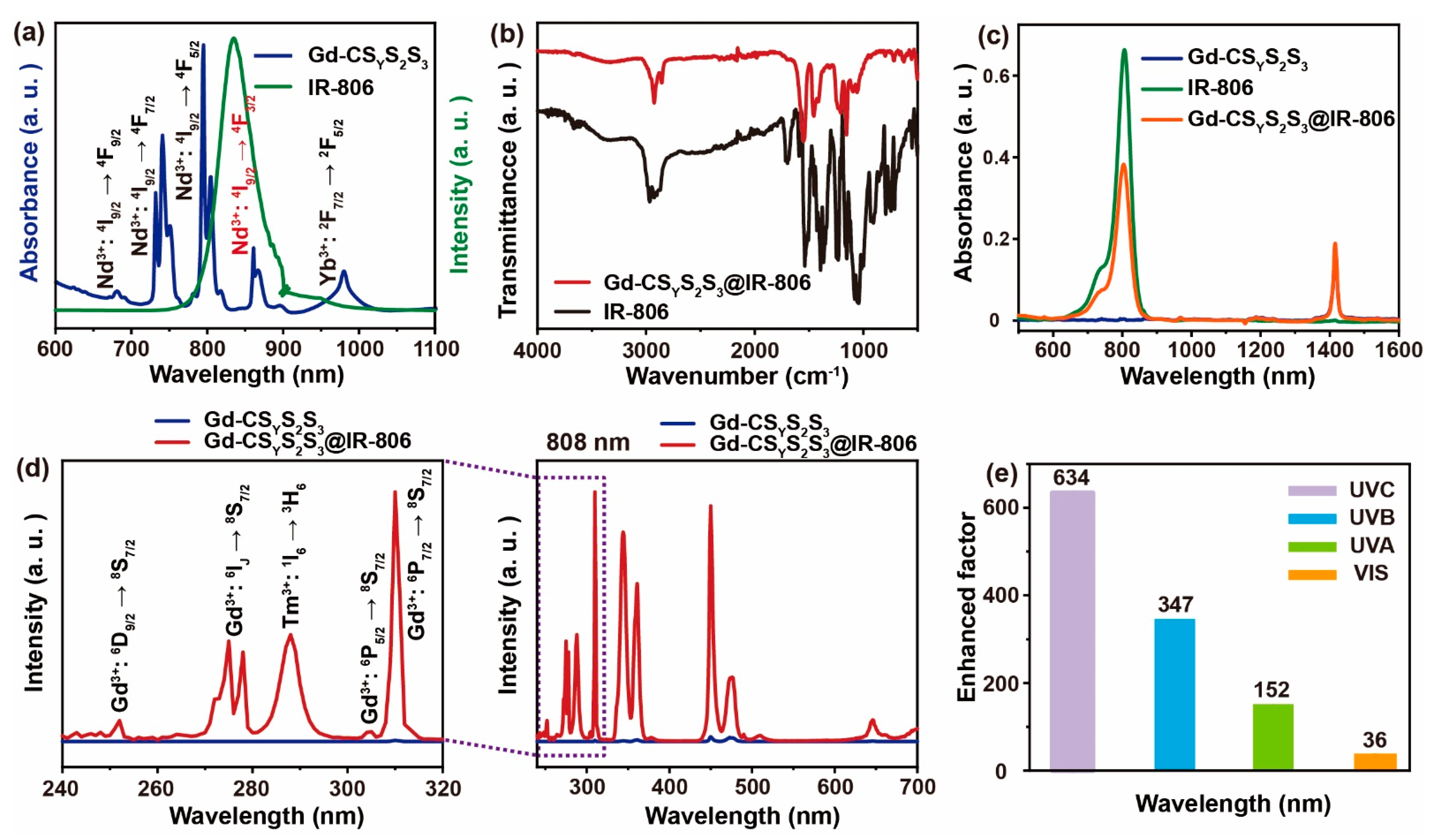
To enhance upconversion emission in the UV range, we chose a near-infrared (NIR) fluorescent dye (IR-806) to sensitize upconversion nanoparticles, due to its intense absorption in the NIR range [33]. As shown in Figure 2a, the fluorescence spectrum of IR-806 has considerable overlap with the absorbance of Gd-CSYS2S3 nanoparticles with Nd3+ (4F3/24I9/2) sensitizer, ensuring an effective energy transfer from IR-806 to the nanoparticles. We then utilized a modified Hummelen’s method to load the IR-806 onto the surface of Gd-CSYS2S3 nanoparticles [32]. In addition, free IR-806 has an absorption band at 1708 cm−1 in the FTIR spectrum, corresponding to the stretching mode of –COOH. The absorption band at 1708 cm−1 disappeared when IR-806 was bound on to the surface of the nanoparticles. Nevertheless, absorption bands at 1560 and 1450 cm−1 were observed on Gd-CSYS2S3@IR-806, corresponding to the antisymmetric and symmetric vibration modes of the –COO group. This indicates that the IR-806 carboxylic acid group was bound onto the surface of Gd-CSYS2S3, since the carboxylic region changed [33]. The successful preparation of IR-806-loaded Gd-CSYS2S3 nanoparticles was demonstrated by Fourier-transform infrared spectroscopy (FTIR) analysis (Figure 2b), which is consistent with the previous report [32]. In addition, we also compared the absorption spectra of Gd-CSYS2S3, IR-806, and Gd-CSYS2S3@IR-806. As shown in Figure 2c, Gd-CSYS2S3@IR-806 nanoparticles showed an intense absorbance band peaking at ~800 nm, further proving the successful loading of IR-806. Consequently, we observed more than 70-fold enhancements in Tm3+ emission over the whole wavelength range from 240–700 nm by Gd-CSYS2S3@IR-806 compared with Gd-CSYS2S3 nanoparticles, owing to the fact that the absorption cross section of Gd-CSYS2S3 was significantly enhanced after IR-806 loading. Furthermore, we also observed more than 600-fold, 300-fold, 150-fold, and 30-fold enhancements in UVC (240–280 nm), UVB (280–320 nm), UVA (320–400 nm), and visible (400–700 nm) regions, respectively (Figure 2e). Similarly, we synthesized the NaGdF4:18%Yb,2%Er@NaYF4:20%Yb@NaGdF4:10%Yb, 50%Nd@NaGdF4 nanoparticles with IR-806 loading. The emission intensity in the UV spectral region increased by more than 60 times, while the intensity in the visible region increased by only 30 times (Figure S5). Taken together, these results demonstrated that the overall enhancements were dominated by increased emission in the UV spectral regions, which is consistent with the dominant effect of ligand coordination on multiphoton upconversion [37]. Notably, the enhancement factors in the UV spectral region are remarkably larger than those in the visible region, offering enticing prospects for NIR light-mediated UV upconversion nanoparticles.

3.3. Optimum Weight Ratio between IR-806 and Nanoparticles

We determined the optimum weight ratio of Gd-CSYS2S3:IR-806 by setting a series of weight gradients from 120:1 to 180:1 (mNPs:mIR-806). As shown in Figure 3a, the optimum weight ratio was determined to be 160:1. The optimized number of dye molecules on the surface of Gd-CSYS2S3 nanoparticles was calculated to be 395 [32]. Note that the absorbance of Gd-CSYS2S3@IR-806 increased as IR-806 increased. However, when the weight ratio of Gd-CSYS2S3: IR-806 was smaller than 160:1, the emission intensity decreased due to fluorescence quenching caused by dye self-quenching. Due to the critical role of the Nd3+ sensitizers in mediating energy transfer from the dye to the upconversion nanoparticles, we verified that the optimum doping concentration of Nd3+ was 50 mol% (Figure S6). We then quantified the energy transfer efficiency of IR-806 to Gd-CSYS2S3 by measuring the lifetime of the IR-806 in a pair of Gd-CSYS2S3 samples with and without Nd3+ nanoparticles. Due to energy trapping by Nd3+, the lifetime is shortened from 1.20 ns to 1.13 ns for Gd-CSYS2S3@IR-806. However, the lifetime of IR-806 was essentially unchanged after loading on Gd-CSYS90%Y,10%YbS3@IR-806, due to the absence of Nd3+ dopants. The energy transfer efficiency was calculated to be 5.8% according to the following equation [38]:
E = 1   τ D A τ D

3.4. The Effect of Excitation Wavelength on UV Upconversion Emission

To investigate the enhancement effect on upconversion emission under 793, 808, and 980 nm excitation, we measured two series of Gd-CSYS2S3 nanoparticles with different amounts of IR-806 loading. As shown in Figure 3d, the emission intensities of Gd-CSYS2S3 were slightly improved after IR-806 loading under 793 nm excitation. In contrast, their emission intensities decreased under 980 nm excitation (Figure 3e). These results can be ascribed to poor matching between the excitation wavelengths (793 nm and 980 nm) and the absorption of IR-806. We then normalized the luminescence spectra of Gd-CSYS2S3 nanoparticles under three different excitation wavelengths. We found that the ratio was unchanged for UVC, UVB, UVA, and visible spectral regions under 793 nm and 980 nm excitation. In contrast, the normalized intensity of the UVC spectral region clearly increased (Figure S7), indicating effective energy transfer from IR-806 to the nanoparticles under 808 nm excitation.

3.5. The Effect of IR-806 Sensitizer Distance on UV Upconversion

To study the effect of the distance between Nd3+ and IR-806 on UV upconversion emission, we synthesized a pair of nanoparticles: Gd-CSYS2S3 and Gd-CSYS2 (without the third shell protection) shown in Figure 4a. Comparing the emission intensities of Gd-CSYS2S3 and Gd-CSYS2 with and without IR-806, the emission intensities of the Gd-CSYS2 nanoparticles without shell protection increased by more than 230 times overall, while only 70-fold enhancement was observed in Gd-CSYS2S3, which has 2 nm thickness shell protection. Furthermore, UV and visible emission intensities increased more than 500-fold and 130-fold, respectively, for the nanoparticles without shell protection (Figure 4b). Notably, the transfer efficiency decreased as 1/R6 [39]. Therefore, the enhancement factor decreased as the distance between the dye and the sensitizer increased.
Similarly, we synthesized two pairs of nanoparticles: NaGdF4@ NaGdF4:49%Yb,1%Tm@NaYF4:20%Yb@NaGdF4:10%Yb,50%Nd@NaGdF4 (Gd-CS1SYS3S4) vs. NaGdF4@NaGdF4:49%Yb,1%Tm@NaYF4:20%Yb@NaGdF4:10%Yb,50%Nd (Gd-CS1SYS3) and NaYF4@NaGdF4:49%Yb,1%Tm@NaYF4:20%Yb@NaGdF4:10%Yb,50%Nd@ NaGdF4 (Y-CS1SYS3S4) vs. NaYF4@NaGdF4:49%Yb,1%Tm@NaYF4:20%Yb@NaGdF4:10%Yb, 50%Nd (Y-CS1SYS3) (Figure S8). The core–multishell structures are illustrated in Figure S9. To study the effect of different structures on emission enhancement, NaGdF4 and NaYF4 without any dopants were used as a core to shorten the distance between the NaGdF4:49%Yb,1%Tm emissive layer and IR-806. The emission intensities of IR-806 grafted on Gd-CS1SYS3 and Gd-CS1SYS3S4 increased 99 and 20 times, respectively, while the luminescence intensity in the UV region increased by 118 and 25 times and that in the visible region increased by 82 and 16 times, respectively. Moreover, the emission intensities of Y-CS1SYS3 and Y-CS1SYS3S4 improved by 72 and 18 times after IR-806 loading. We also observed 81-fold and 22-fold enhancements in the UV spectral region and 63-fold and 14-fold enhancements in the visible region (Figure S10). These results are also consistent with our luminescence analysis, in that a significant enhancement in the UV luminescence of Gd-CSYS2S3 nanoparticles was observed compared to the visible range (Figure S11).

3.6. Energy Transfer Mechanism

As shown in Scheme 2, IR-806 effectively absorbs the laser energy due to the absorption cross section under 808 nm excitation. To generate an efficient dye sensitization process, Nd3+ plays a critical role in bridging the energy transfer from the dye to the upconversion nanoparticles. Nd3+ ions trap the energy from the 808 nm laser and IR-806 mainly via the fluorescence–resonance energy transfer process and then gather photons at the 4F5/2 energy state. Subsequently, relaxing to the 4F3/2 energy state, Nd3+ transfers the energy to Yb3+ by an efficient energy transfer process. As an energy migrator, the excited Yb3+ populates the energy states of Tm3+ and gives rise to emission at 475 nm (1G43H6), 450 nm (1D23F4), 360 nm(1D23H6), 345 nm(1I63H5), and 290 nm(1I63H6). Apart from emitting, Tm3+ serves as an energy donor donating energy to the Gd3+ ions via a five-photon process. Meanwhile, the six-photon upconversion process of 253 nm (6D9/28S7/2) and the five-photon upconversion processes of 273 nm (6IJ8S7/2), 276 nm (6IJ8S7/2), 279 nm (6IJ8S7/2), 306 nm (6P5/28S7/2), and 310 nm (6P7/28S7/2) are observed with the assistance of the appropriate energy matching of the following transition of 2F5/22F7/2 (9750 cm−1, Yb3+): 6PJ6DJ (∼8750 cm−1, Gd3+). Notably, the utilization of an optically inert NaYF4 host lattice with Yb3+ dopants as the interlayer plays a decisive role in protecting the energy by cooperative dye and Nd3+ sensitization from interior lattice defects, making it possible to effectively further increase UV via dye sensitizing.

3.7. Back Energy Transfer from Nanoparticles to IR-806

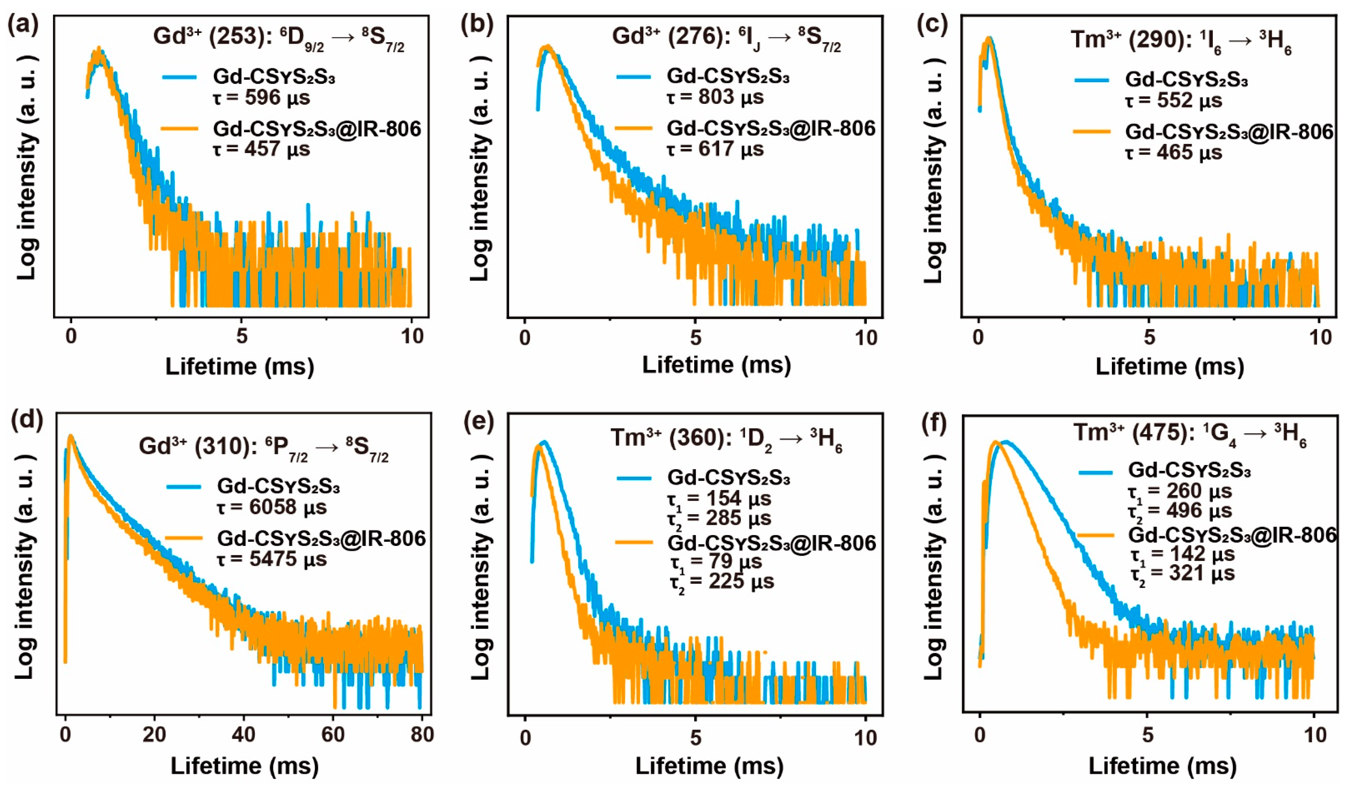
As well as increasing the luminescence intensity, a back energy transfer process from IR-806 to Gd-CSYS2S3 occurred. As depicted in Figure 5 and Figure S12, the lifetime of Gd3+ at 253, 276, and 310 nm, and Tm3+ at 290, 345, 475, and 650 nm slightly decreased after IR-806 loading, which can be ascribed to the nonradiative energy transfer from Gd3+ and Tm3+ to IR-806 [40,41,42].

4. Discussion

In this study, we developed a dye-sensitized heterogeneous lanthanide nanoparticle to regulate the energy transfer pathway for UV enhancement by 808 nm excitation. We systematically studied the influence of dye concentration, excitation wavelength, and distance between the dye and the sensitizer Nd3+ on upconversion emission, especially in the UV spectral region. Dye loading can improve the absorption of excitation light and thus improve the efficiency of energy-transfer-mediated upconversion. Moreover, our experimental results demonstrated a strengthened multiphoton upconversion process, which can be ascribed to the dominant effect of ligand loading on upconversion emission from high-lying energy states. The fundamentals gained from our investigations may provide insights into promoting the multiphoton upconversion process and the future design of organic–inorganic hybrid luminescent nanoparticles for applications in photocatalysis, biomedicine, environmental science, and more.

Supplementary Materials

The following are available online at https://www.mdpi.com/article/10.3390/nano11113114/s1. Figure S1: TEM and size distribution of Gd-CSYS2S3 nanoparticles; Figure S2: XRD of Gd-CSYS2S3 nanoparticles; Figure S3: EDX of Gd-CSYS2S3 nanoparticles; Figure S4: EDX lining analysis of Gd-CSYS2S3 nanoparticles; Figure S5: luminescence emission of NaGdF4:18%Yb,2%Er@NaYF4:20%Yb@NaGdF4:10%Yb, 50%Nd@NaGdF4 with and without IR-806 loading; Figure S6: luminescence emission of Gd-CSYS2S3 nanoparticles with different Nd3+ doping before and after IR-806 loading; Figure S7: normalized intensity of luminescence spectra of Gd-CSYS2S3 with various contents of IR-806; Figure S8: TEM images of as-synthesized nanoparticles with different structures for distance effect studies; Figure S9: schematic illustration of five types of core–multishell structures including Gd-CSYS2S3, Y-CS1SYS3, Y-CS1SYS3S4, Gd-CS1SYS3, and Gd-CS1SYS3S4; Figure S10: luminescence spectra of as-synthesized nanoparticles with different structures for distance effect studies; Figure S11: normalized intensities of luminescence spectra of corresponding nanoparticles for distance effect studies; Figure S12: the lifetime decay of Tm3+ at 650 nm in Gd-CSYS2S3 and Gd-CSYS2S3@IR-806 nanoparticles under 808 nm excitation.

Author Contributions

Conceptualization, Q.S. and C.H; methodology, Q.S. and M.W.; validation, Q.S., C.H. and M.W.; investigation, M.W., Q.S., H.W. and S.W.; resources, Q.S.; data curation, M.W.; writing—original draft preparation, M.W.; writing—review and editing, Q.S. and C.H; visualization, M.W.; supervision, Q.S. and C.H.; project administration, Q.S.; funding acquisition, C.H. and Q.S. All authors have read and agreed to the published version of the manuscript.

Funding

This research was funded by the National Natural Science Foundation of China (Nos. 82002893 and 21701109).

Data Availability Statement

All of the relevant data are available from the correspondence authors upon reasonable request. Source data are provided with this paper.

Acknowledgments

The authors acknowledge Han for helpful discussions. The authors thank Jin and Guan for their help with the HAADF-STEM measurements.

Conflicts of Interest

The authors declare no conflict of interest.

References

  1. Gu, Y.; Guo, Z.; Yuan, W.; Kong, M.; Liu, Y.; Liu, Y.; Gao, Y.; Feng, W.; Wang, F.; Zhou, J.; et al. High-sensitivity imaging of time-domain near-infrared light transducer. Nat. Photon. 2019, 13, 525–531. [Google Scholar] [CrossRef]
  2. Zhu, X.; Su, Q.; Feng, W.; Li, F. Anti-stokes shift luminescent materials for bio-applications. Chem. Soc. Rev. 2017, 46, 1025–1039. [Google Scholar] [CrossRef]
  3. Zhao, J.; Chu, H.; Zhao, Y.; Lu, Y.; Li, L. A NIR light gated DNA nanodevice for spatiotemporally controlled imaging of MicroRNA in cells and animals. J. Am. Chem. Soc. 2019, 141, 7056–7062. [Google Scholar] [CrossRef]
  4. Wang, Y.-F.; Liu, G.-Y.; Sun, L.-D.; Xiao, J.-W.; Zhou, J.-C.; Yan, C.-H. Nd3+-sensitized upconversion nanophosphors: Efficient In Vivo bioimaging probes with minimized heating effect. ACS Nano 2013, 7, 7200–7206. [Google Scholar] [CrossRef]
  5. Xu, J.; Yang, P.; Sun, M.; Bi, H.; Liu, B.; Yang, D.; Gai, S.; He, F.; Lin, J. Highly emissive dye-sensitized upconversion nanostructure for dual-photosensitizer photodynamic therapy and bioimaging. ACS Nano 2017, 11, 4133–4144. [Google Scholar] [CrossRef]
  6. Yang, D.; Ma, P.A.; Hou, Z.; Cheng, Z.; Li, C.; Lin, J. Current advances in lanthanide ion (Ln3+)-based upconversion nanomaterials for drug delivery. Chem. Soc. Rev. 2015, 44, 1416–1448. [Google Scholar] [CrossRef] [PubMed] [Green Version]
  7. Idris, N.M.; Gnanasammandhan, M.K.; Zhang, J.; Ho, P.C.; Mahendran, R.; Zhang, Y. In Vivo photodynamic therapy using upconversion nanoparticles as remote-controlled nanotransducers. Nat. Med. 2012, 18, 1580–1585. [Google Scholar] [CrossRef] [PubMed]
  8. Chen, G.; Qiu, H.; Prasad, P.N.; Chen, X. Upconversion nanoparticles: Design, nanochemistry, and applications in theranostics. Chem. Rev. 2014, 114, 5161–5214. [Google Scholar] [CrossRef] [PubMed]
  9. Fan, W.; Bu, W.; Shi, J. On the latest three-stage development of nanomedicines based on upconversion nanoparticles. Adv. Mater. 2016, 28, 3987–4011. [Google Scholar] [CrossRef]
  10. Dai, Y.; Xiao, H.; Liu, J.; Yuan, Q.; Ma, P.A.; Yang, D.; Li, C.; Cheng, Z.; Hou, Z.; Yang, P.; et al. In Vivo multimodality imaging and cancer therapy by near-infrared light-triggered trans-platinum pro-drug-conjugated upconverison nanoparticles. J. Am. Chem. Soc. 2013, 135, 18920–18929. [Google Scholar] [CrossRef]
  11. Bansal, A.; Zhang, Y. Photocontrolled nanoparticle delivery systems for biomedical applications. Acc. Chem. Res. 2014, 47, 3052–3060. [Google Scholar] [CrossRef]
  12. Zheng, W.; Zhou, S.; Xu, J.; Liu, Y.; Huang, P.; Liu, Y.; Chen, X. Ultrasensitive luminescent in vitro detection for tumor markers based on inorganic lanthanide nano-bioprobes. Adv. Sci. 2016, 3, 1600197. [Google Scholar] [CrossRef] [PubMed]
  13. Chen, S.; Weitemier Adam, Z.; Zeng, X.; He, L.; Wang, X.; Tao, Y.; Huang Arthur, J.Y.; Hashimotodani, Y.; Kano, M.; Iwasaki, H.; et al. Near-infrared deep brain stimulation via upconversion nanoparticle–mediated optogenetics. Science 2018, 359, 679–684. [Google Scholar] [CrossRef] [Green Version]
  14. Wu, X.; Zhang, Y.; Takle, K.; Bilsel, O.; Li, Z.; Lee, H.; Zhang, Z.; Li, D.; Fan, W.; Duan, C.; et al. Dye-sensitized core/active shell upconversion nanoparticles for optogenetics and bioimaging applications. ACS Nano 2016, 10, 1060–1066. [Google Scholar] [CrossRef] [PubMed] [Green Version]
  15. Liu, J.; Zheng, X.; Yan, L.; Zhou, L.; Tian, G.; Yin, W.; Wang, L.; Liu, Y.; Hu, Z.; Gu, Z.; et al. Bismuth sulfide nanorods as a precision nanomedicine for in vivo multimodal imaging-guided photothermal therapy of tumor. ACS Nano 2015, 9, 696–707. [Google Scholar] [CrossRef] [PubMed]
  16. Zhu, X.; Feng, W.; Chang, J.; Tan, Y.-W.; Li, J.; Chen, M.; Sun, Y.; Li, F. Temperature-feedback upconversion nanocomposite for accurate photothermal therapy at facile temperature. Nat. Commun. 2016, 7, 10437. [Google Scholar] [CrossRef]
  17. Zuo, J.; Tu, L.; Li, Q.; Feng, Y.; Que, I.; Zhang, Y.; Liu, X.; Xue, B.; Cruz, L.J.; Chang, Y.; et al. Near infrared light sensitive ultraviolet–blue nanophotoswitch for imaging-guided “Off–On” therapy. ACS Nano 2018, 12, 3217–3225. [Google Scholar] [CrossRef]
  18. Chan, M.-H.; Pan, Y.-T.; Chan, Y.-C.; Hsiao, M.; Chen, C.-H.; Sun, L.; Liu, R.-S. Nanobubble-embedded Inorganic 808 nm excited upconversion nanocomposites for tumor multiple imaging and treatment. Chem. Sci. 2018, 9, 3141–3151. [Google Scholar] [CrossRef] [Green Version]
  19. Liu, C.; Liu, B.; Zhao, J.; Di, Z.; Chen, D.; Gu, Z.; Li, L.; Zhao, Y. Nd3+-sensitized upconversion Metal–Organic frameworks for mitochondria-targeted amplified photodynamic therapy. Angew. Chem. Int. Ed. 2020, 59, 2634–2638. [Google Scholar] [CrossRef]
  20. Zheng, K.; Qin, W.; Cao, C.; Zhao, D.; Wang, L. NIR to VUV: Seven-photon upconversion emissions from Gd3+ ions in fluoride nanocrystals. J. Phys. Chem. Lett. 2015, 6, 556–560. [Google Scholar] [CrossRef]
  21. Chen, X.; Jin, L.; Kong, W.; Sun, T.; Zhang, W.; Liu, X.; Fan, J.; Yu, S.F.; Wang, F. Confining energy migration in upconversion nanoparticles towards deep ultraviolet lasing. Nat. Commun. 2016, 7, 10304. [Google Scholar] [CrossRef]
  22. Zhao, C.; Kong, X.; Liu, X.; Tu, L.; Wu, F.; Zhang, Y.; Liu, K.; Zeng, Q.; Zhang, H. Li+ Ion doping: An approach for improving the crystallinity and upconversion emissions of NaYF4:Yb3+, Tm3+ Nanoparticles. Nanoscale 2013, 5, 8084–8089. [Google Scholar] [CrossRef] [PubMed] [Green Version]
  23. Shi, F.; Wang, J.; Zhang, D.; Qin, G.; Qin, W. Greatly enhanced size-tunable ultraviolet upconversion luminescence of monodisperse β-NaYF4:Yb,Tm nanocrystals. J. Mater. Chem. 2011, 21, 13413–13421. [Google Scholar] [CrossRef]
  24. Dawson, P.; Romanowski, M. Excitation modulation of upconversion nanoparticles for switch-like control of ultraviolet luminescence. J. Am. Chem. Soc. 2018, 140, 5714–5718. [Google Scholar] [CrossRef] [PubMed]
  25. Wang, F.; Wang, J.; Liu, X. Direct evidence of a surface quenching effect on size-dependent luminescence of upconversion nanoparticles. Angew. Chem. Int. Ed. 2010, 49, 7456–7460. [Google Scholar] [CrossRef] [PubMed]
  26. Wang, F.; Deng, R.; Wang, J.; Wang, Q.; Han, Y.; Zhu, H.; Chen, X.; Liu, X. Tuning upconversion through energy migration in core-shell nanoparticles. Nat. Mater. 2011, 10, 968–973. [Google Scholar] [CrossRef] [PubMed]
  27. Sun, T.; Li, Y.; Ho, W.L.; Zhu, Q.; Chen, X.; Jin, L.; Zhu, H.; Huang, B.; Lin, J.; Little, B.E.; et al. Integrating temporal and spatial control of electronic transitions for bright multiphoton upconversion. Nat. Commun. 2019, 10, 1811. [Google Scholar] [CrossRef]
  28. Su, Q.; Han, S.; Xie, X.; Zhu, H.; Chen, H.; Chen, C.-K.; Liu, R.-S.; Chen, X.; Wang, F.; Liu, X. The effect of surface coating on energy migration-mediated upconversion. J. Am. Chem. Soc. 2012, 134, 20849–20857. [Google Scholar] [CrossRef]
  29. Xie, X.; Gao, N.; Deng, R.; Sun, Q.; Xu, Q.-H.; Liu, X. Mechanistic investigation of photon upconversion in Nd3+-sensitized core–shell nanoparticles. J. Am. Chem. Soc. 2013, 135, 12608–12611. [Google Scholar] [CrossRef]
  30. Su, Q.; Wei, H.-L.; Liu, Y.; Chen, C.; Guan, M.; Wang, S.; Su, Y.; Wang, H.; Chen, Z.; Jin, D. Six-photon Upconverted Excitation Energy Lock-in for Ultraviolet-C Enhancement. Nat. Commun. 2021, 12, 4367. [Google Scholar] [CrossRef]
  31. Wen, S.; Zhou, J.; Schuck, P.J.; Suh, Y.D.; Schmidt, T.W.; Jin, D. Future and challenges for hybrid upconversion nanosystems. Nat. Photon. 2019, 13, 828–838. [Google Scholar] [CrossRef]
  32. Zou, W.; Visser, C.; Maduro, J.A.; Pshenichnikov, M.S.; Hummelen, J.C. Broadband dye-sensitized upconversion of near-infrared light. Nat. Photon. 2012, 6, 560–564. [Google Scholar] [CrossRef] [Green Version]
  33. Shao, Q.; Li, X.; Hua, P.; Zhang, G.; Dong, Y.; Jiang, J. Enhancing the upconversion luminescence and photothermal conversion properties of ~800 nm excitable core/shell nanoparticles by dye molecule sensitization. J. Colloid Interface Sci. 2017, 486, 121–127. [Google Scholar] [CrossRef] [PubMed]
  34. Xue, B.; Wang, D.; Zhang, Y.; Zuo, J.; Chang, Y.; Tu, L.; Liu, X.; Yuan, Z.; Zhao, H.; Song, J.; et al. Regulating the color output and simultaneously enhancing the intensity of upconversion nanoparticles via a dye sensitization strategy. J. Mater. Chem. C. 2019, 7, 8607–8615. [Google Scholar] [CrossRef]
  35. Wang, F.; Deng, R.; Liu, X. Preparation of core-shell NaGdF4 nanoparticles doped with luminescent lanthanide ions to be used as upconversion-based probes. Nat. Protoc. 2014, 9, 1634–1644. [Google Scholar] [CrossRef] [PubMed]
  36. Wang, S.; Shen, B.; Wei, H.-L.; Liu, Z.; Chen, Z.; Zhang, Y.; Su, Y.; Zhang, J.-Z.; Wang, H.; Su, Q. Comparative investigation of the optical spectroscopic and thermal effect in Nd3+-doped nanoparticles. Nanoscale 2019, 11, 10220–10228. [Google Scholar] [CrossRef]
  37. Xu, H.; Han, S.; Deng, R.; Su, Q.; Wei, Y.; Tang, Y.; Qin, X.; Liu, X. Anomalous upconversion amplification induced by surface reconstruction in lanthanide sublattices. Nat. Photon. 2021, 15, 732–737. [Google Scholar] [CrossRef]
  38. Muhr, V.; Würth, C.; Kraft, M.; Buchner, M.; Baeumner, A.J.; Resch-Genger, U.; Hirsch, T. Particle-size-dependent förster resonance energy transfer from upconversion nanoparticles to organic dyes. Anal. Chem. 2017, 89, 4868–4874. [Google Scholar] [CrossRef]
  39. Ray, P.C.; Fan, Z.; Crouch, R.A.; Sinha, S.S.; Pramanik, A. Nanoscopic optical rulers beyond the fret distance limit: Fundamentals and applications. Chem. Soc. Rev. 2014, 43, 6370–6404. [Google Scholar] [CrossRef]
  40. Deng, R.; Wang, J.; Chen, R.; Huang, W.; Liu, X. Enabling förster resonance energy transfer from large nanocrystals through energy migration. J. Am. Chem. Soc. 2016, 138, 15972–15979. [Google Scholar] [CrossRef]
  41. Tu, D.; Liu, L.; Ju, Q.; Liu, Y.; Zhu, H.; Li, R.; Chen, X. Time-Resolved FRET biosensor based on amine-functionalized lanthanide-doped NaYF4 nanocrystals. Angew. Chem. Int. Ed. 2011, 50, 6306–6310. [Google Scholar] [CrossRef] [PubMed]
  42. Kong, M.; Gu, Y.; Liu, Y.; Shi, Y.; Wu, N.; Feng, W.; Li, F. Luminescence lifetime–Based In Vivo detection with responsive rare earth–dye nanocomposite. Small 2019, 15, 1904487. [Google Scholar] [CrossRef] [PubMed]
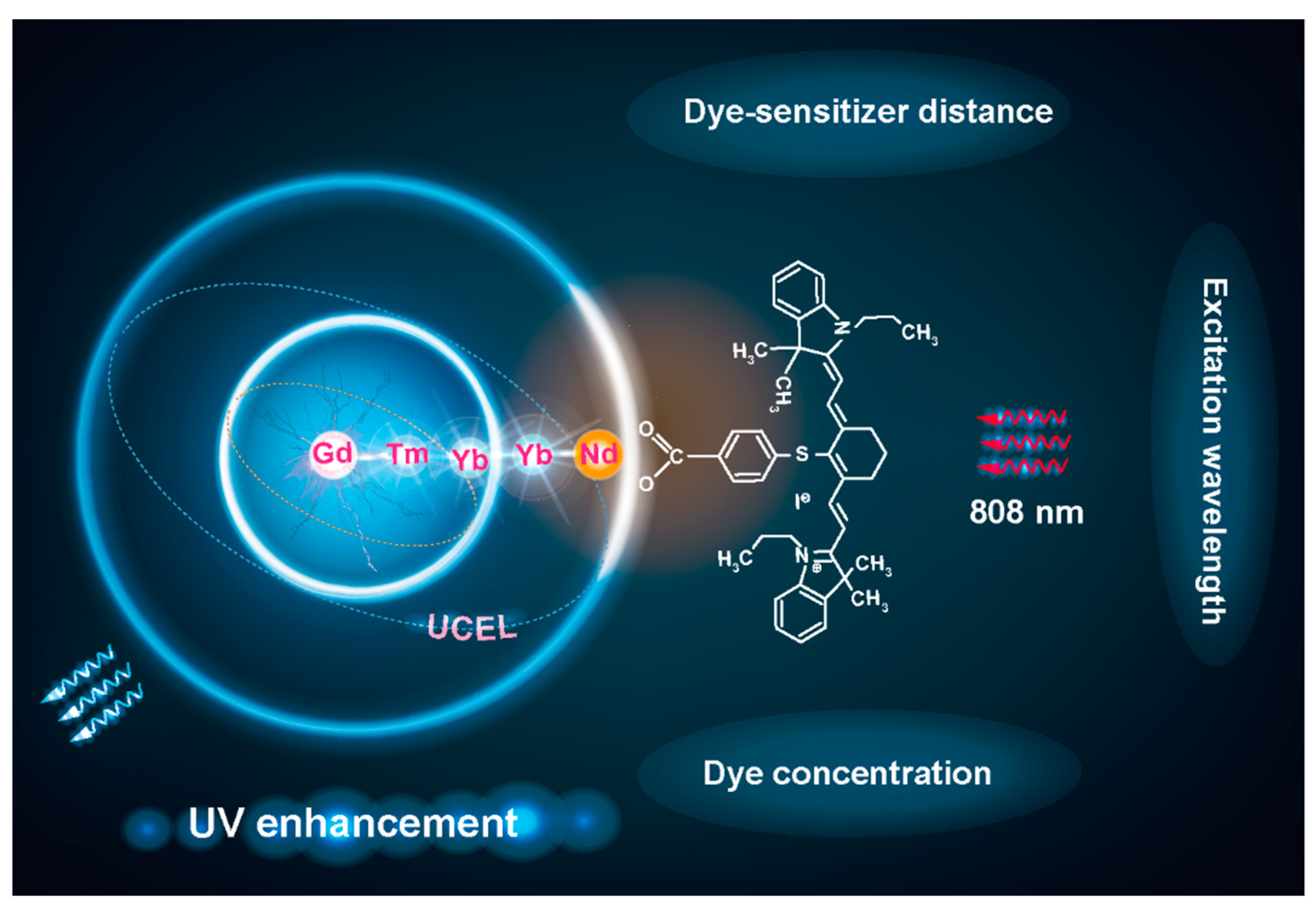
Scheme 1. Schematic illustration of the key factors that influence UV enhancement in IR-806-loaded upconversion nanoparticles, including dye concentration, excitation wavelength, and dye–sensitizer distance.
Nanomaterials 11 03114 sch001
Figure 1. Schematic illustration and characterization of Gd-CSYS2S3 heterogeneous nanoparticles. (a) Diagrammatic representation of Gd-CSYS2S3 nanostructure. (b) High-angle annular dark-field scanning transmission electron microscopy (HAADF-STEM) image of Gd-CSYS2S3 nanoparticles. Inset: high-resolution TEM of as-prepared Gd-CSYS2S3 nanoparticle. (c) HAADF-STEM image and elemental mapping image of Gd-CSYS2S3 nanoparticles, revealing the spatial distribution of the Y, Nd, Gd, and Yb elements in the heterogeneous nanoparticles.
Nanomaterials 11 03114 g001
Figure 2. Preparation and characterization of Gd-CSYS2S3@IR-806. (a) IR-806 emission spectrum and Gd-CSYS2S3 nanoparticles absorption spectrum. (b) FTIR of Gd-CSYS2S3@IR-806 and IR-806. (c) The absorption spectra of Gd-CSYS2S3, IR-806, and Gd-CSYS2S3@IR-806. (d) Emission spectra of Gd-CSYS2S3 with and without IR-806 loading under 808 nm CW diode laser at a power density of 10 W/cm2. (e) The enhancement factors of upconversion emission were obtained by comparing the results for samples with and without IR-806 loading. The emission intensities were calculated by integrating the spectral intensities in the UVC (240–280 nm), UVB (280–320 nm), UVA (320–400 nm), and visible (400–650 nm) ranges.
Nanomaterials 11 03114 g002
Figure 3. Optimizing the weight ratio of Gd-CSYS2S3 to IR-806 and calculating the energy transfer efficiency. (a) The emission spectrum of Gd-CSYS2S3 (4 mL in CHCl3, 0.375 mg/mL) after adding various masses of IR-806 dye under 808 nm excitation. (b) The absorption spectrum of Gd-CSYS2S3 (4 mL in CHCl3, 0.375 mg/mL) with various masses IR-806 dye. (c) The decay curves of Gd-CSYS2S3, Gd-CSYS2(90%, 10%Yb)S3@IR-806, and Gd-CSYS2S3@IR-806. (d,e) The emission spectra of Gd-CSYS2S3 (4 mL in CHCl3, 0.375 mg/mL) after adding various masses of IR-806 dye under 793 nm and 980 nm excitation, respectively.
Nanomaterials 11 03114 g003
Figure 4. The effect of the distance between IR-806 and sensitizer Nd3+ on upconversion emission. (a) Schematic illustration of the nanostructural design to study the distance effect on upconversion emission. (b) The emission spectra of Gd-CSYS2S3, Gd-CSYS2S3@IR-806, Gd-CSYS2, Gd-CSYS2@IR-806 under 808 nm excitation.
Nanomaterials 11 03114 g004
Scheme 2. Schematic illustration of the mechanism for cascade energy transfer in Gd-CSYS2S3@IR-806. Upon 808 nm laser excitation, IR-806 first absorbs excitation energy and transfers it to Nd3+. Next, Yb3+ accepts the energy from Nd3+, contributing to populating photons in the 3P2 state of Tm3+ through a continuous five-photon energy transfer process and then relaxing to the 1I6 state of Tm3+. Trapping the energy from both five-photon upconversion from Tm3+ and one-photon upconversion from Yb3+, six-photon and five-photon upconversion luminescence from 6DJ, 6IJ, and 6PJ state of Gd3+ is observed.
Nanomaterials 11 03114 sch002
Figure 5. The decreased lifetime of Tm3+ and Gd3+ for Gd-CSYS2S3@IR-806. (af) The Tm3+ and Gd3+ lifetime decay curves of Gd-CSYS2S3 and Gd-CSYS2S3@IR-806 at 253, 276, 290, 310, 360, and 475 nm under 808 nm excitation, respectively.
Nanomaterials 11 03114 g005
Publisher’s Note: MDPI stays neutral with regard to jurisdictional claims in published maps and institutional affiliations.