Review

Advances in Nanomaterials-Based Electrochemical Biosensors for Foodborne Pathogen Detection

1
BioSense Institute, University of Novi Sad, 21102 Novi Sad, Serbia
2
Micalis Institute, INRAE, AgroParisTech, Université Paris-Saclay, 78350 Jouy-en-Josas, France
3
Department of Agriculture Food, Environmental and Animal Sciences, University of Udine, 33100 Udine, Italy
4
Institute for Multidisciplinary Research, University of Belgrade, 11030 Belgrade, Serbia
*
Authors to whom correspondence should be addressed.
Nanomaterials 2021, 11(10), 2700; https://doi.org/10.3390/nano11102700
Submission received: 29 September 2021 / Revised: 7 October 2021 / Accepted: 9 October 2021 / Published: 13 October 2021
(This article belongs to the Special Issue Functional Nanomaterials for Biosensing Applications)

Abstract

:
Electrochemical biosensors utilizing nanomaterials have received widespread attention in pathogen detection and monitoring. Here, the potential of different nanomaterials and electrochemical technologies is reviewed for the development of novel diagnostic devices for the detection of foodborne pathogens and their biomarkers. The overview covers basic electrochemical methods and means for electrode functionalization, utilization of nanomaterials that include quantum dots, gold, silver and magnetic nanoparticles, carbon nanomaterials (carbon and graphene quantum dots, carbon nanotubes, graphene and reduced graphene oxide, graphene nanoplatelets, laser-induced graphene), metal oxides (nanoparticles, 2D and 3D nanostructures) and other 2D nanomaterials. Moreover, the current and future landscape of synergic effects of nanocomposites combining different nanomaterials is provided to illustrate how the limitations of traditional technologies can be overcome to design rapid, ultrasensitive, specific and affordable biosensors.

1. Introduction

Pathogen diagnostics are currently critical for applications in healthcare, food safety analysis and environmental monitoring. Foodborne and waterborne pathogens (i.e., bacteria, fungi, viruses and some parasites) cause infections in humans via contaminated food or water. The high incidence of infection caused by foodborne pathogens indicates that the prevention, surveillance and management of foodborne diseases need to be strengthened [1,2,3].
The traditional technologies to detect pathogens in food and water are constrained by delayed analysis times, expensive and laborious sample preparation steps and the need for highly trained personnel. The major conventional detection methods can be classified as counting methods, immune-assays and polymerase chain reaction (PCR)-based methods. Counting bacterial colonies on microbiological culture plates is inherently a complex, time-consuming and error-prone method. The detection time takes 3 to 9 days while up to 2 weeks are needed for confirmation of positive results. The confirmation includes observation of the bacterial colony color and morphology together with biochemical tests in a specific medium that is performed after pathogen isolation. Alternatively, immunoassays, such as the enzyme-linked immunosorbent assay (ELISA), lateral flow and dot blot immunoassay, enable detection of pathogen antigens [4,5,6,7,8]. They can use monoclonal or polyclonal antibodies that specifically bind to the targeted pathogen and can be applied for testing large-scale samples and for the on-site detection of pathogens. However, they usually show low sensitivity and thus have to be confirmed by an additional test. In contrast, PCR-based methods allow rapid and highly specific pathogen diagnosis. In spite of these advantages, PCR-based methods have some limitations. For instance, they can produce false negative results due to a DNA polymerase inhibition by food matrix molecules and ions, which may completely block amplification of target DNA, or false positive results due to the cross-amplification of PCR-generated fragments of non-target DNA.
Biosensors provide a promising tool for such applications due to their portability and simplicity of utilization. The most used types of sensors are by construction optical (plasmonic, UV-Vis/Infrared spectroscopy, Raman, attenuated total reflection), electrochemical, electromagnetic, mechanical, airflow and acoustic. The principal issue in all these technologies is to enable sensitive and selective detection of pathogens in complex food samples that contain low analyte concentrations. Nonspecific adsorption of biomolecules presented in the sample (originated from either the matrix or microorganisms that constitute normal sample microflora) at the biosensor surface can drastically obstruct detection performance, diminish the signal intensity and specificity of the biosensor and increase background “noise”.
In the last years, innovative and portable biosensors have emerged as they overcome limitations of traditional and molecular detection technologies and even other biosensors concerning the quantitative detection and screening of pathogens in clinical, environmental and food analysis [9,10]. Among different biosensors, electrochemical platforms are the most popular because they are highly specific towards the analyte and can be adapted for multiplex analysis providing high analytical accuracy even in complex food matrices of various composition, densities and pH. Electrochemical detection of a pathogen exploits a working electrode modified with specific recognition elements (such as antibody, aptamer, DNA probe) ensuring the selectivity, sensitivity and specificity of the measurements. Various strategies and concepts have been developed to prevent nonspecific binding to the electrode surface in biosensors. The concept of such strategies primarily relies on the fabrication method, sample composition, electrochemical technique and performance of each detection principle. Recent literature highlights that different nanomaterials are incorporated into electrochemical biosensors as enhancers, labeling factors or immobilizer supports to enable the overall feasibility of the platform for diagnostic/detection applications.
In this review, we present some basic principles of the electrochemical methods used in biosensors and the state-of-the-art nanomaterial-based electrochemical biosensors for foodborne pathogen (virus, bacteria and bacterial toxin) detection.

2. Electrochemical Methods and Electrode Functionalization

Electrochemical biosensors transduce biochemical events into electrical signals (current, potential, impedance or resistance). They can be divided into biocatalytic using enzymes as recognition elements and affinity (biocomplexing) biosensors using selective and strong binding biomolecules. Affinity biosensors can be further divided into immunosensors based on antibodies or nanobodies, aptasensors based on DNA or RNA aptamers and genosensors based on single strand DNA (ssDNA). Besides, some electrochemical biosensors for pathogen detection use peptides, phages, microRNA, antibiotics or molecularly imprinted polymers (MIPs) as recognition elements [10]. Various electrochemical techniques with different signal mechanisms exist, as illustrated in Figure 1. Their applicability and efficiency depend on the target properties and design of the sensor platform. Voltammetry is performed under controlled potentials when the measured current reflects electron transfer between the sample and the electrode surface. It is possible to measure current values during the potential sweeping towards and backwards while cycling (cyclic voltammetry). By holding the potential of the electrode constant (amperometry), or holding the current constant (potentiometry), the obtained information in the timescale gives the change in current and potential, respectively. Capacitance, as one of the electric properties of (bio)molecular and biological layers at the surface of the electrochemical electrode, represents important information of the layer charging effect while sensing certain molecules/pathogens. Electrochemical impedance spectroscopy (impedance/resistance of the system) is usually employed for measuring the impedance of the catalytic layer that changes upon target binding to the immobilized recognition element. Electrochemical impedance spectroscopy is frequently used in detecting pathogen microorganisms due to its high sensitivity. Increase in the diameter of electrochemical impedance plotted in a Nyquist diagram shows the increase in system impedance. It is directly proportional to the electron transfer resistance of the system and enables quantitative detection. Potentiostatic systems usually work in a three-electrode format (working, auxiliary and reference electrode) while conductometry and electrochemical impedance spectroscopy are mainly performed in a two-electrode format (working and auxiliary).
An ideal electrochemical sensor should achieve high sensitivity and specificity, a wide dynamic range of detection, measurement reproducibility, rapid response with real-time analysis and multiple uses. It should also be portable, user-friendly and cost-effective with self-calibration and self-cleaning. To enable simultaneous detection of several targets in the same sample (multiplexing), electrochemical biosensors can be combined with microfluidic systems and integrated with microelectronics. The development of screen-printed electrodes is important in making sensors economical and widely commercially available. Furthermore, data post-processing plays a very important role in obtaining credible and accurate detection results. Many of these properties are an issue in detecting analytes in complex matrices. Affinity biosensors, especially, may have difficulty operating in samples such as food matrices due to nonspecific adsorption on the electrode surfaces that compromises the performance of the device.
The electrode material, its design and fabrication may significantly increase the sensor specificity and selectivity. Surface chemistry is used to immobilize recognition elements onto the working electrode and to prevent a background signal [11]. To eliminate the matrix effect, common strategies involve electrode functionalization using specific surface chemistry and additional electrode covering with poly(ethylene glycol) (PEG) or oligo(ethylene glycol) (OEG) layers that effectively passivate the electrode [12]. Usually, immobilization of biomolecules is performed via amine-, carboxyl-, aldehyde- and thiol- conjugation, depending on the chemical reactivity of the electrode material and its modifications. Finally, to enable sensitive electrochemical detection, a redox indicator is added to the sample. Ferrocene is the most commonly used redox indicator [10], but others such as protamine [13], tripropylamine [14] or methylene blue [15] also enable estimation of the target concentration by measuring changes in peak intensity. For instance, target binding on the electrode surface may decrease peak intensities of the redox indicator due to the higher electron transfer resistance of the electrode system while increasing the concentration of the captured target. To simplify biosensor utilization and to increase signal intensities, the redox marker can be immobilized onto the electrode surface as shown for influenza A virus detection using conducting copolypyrrole integrating ferrocenyl group electrodes [16,17]. Finally, in cases when the analyte can undergo oxido-reduction on the working electrode itself, no additional redox marker is needed [18].

3. Nanomaterial-Based Electrochemical Biosensors

Many types of sensing electrochemical devices come up, and some of them represent a scaled-down lab to a single chip (lab on a chip). However, despite the intense development of electrochemical biosensors, their high sensitivity and reproducibility remain challenging [19]. Employing various nanomaterials may improve analytical performances of electrochemical sensors by signal enhancement [9,20]. Association of nanomaterials with the electrode increases surface area which can boost loading capacities and mass transport of reactants, resulting in signal amplification. Moreover, nanomaterials can be carriers of redox probes to provide sensitive detection or can improve dynamics of redox exchanges, which significantly amplifies the read-out [21].
Nanomaterials are generally classified as 0D—quantum dots, carbon dots, nanoparticles, 1D—nanotubes, nanowires, nanorods, 2D—nanoplates, nanosheets, nanodisks and 3D—nanoflowers, nanocones, nanoballs [22] (Figure 2). In all 0–3D forms, nanomaterials have been extensively incorporated into electrode construction in electrochemical biosensors applied in the detection of foodborne pathogens [23]. Generally, 0D nanomaterials comprise nanoparticles, usually metal or metal oxide nanoparticles [24,25,26], carbon and quantum dots [27,28] with nanoscale dimensions. Metal nanoparticles, most commonly gold nanoparticles, are often selected for application in electrochemical biosensors for detection of foodborne pathogens due to their high conductivity and biocompatibility and retention of biomolecule activity over time [29,30]. Quantum dots (QDs) have great potential for application in small size electrochemical biosensing devices due to their small compact size and good and stable performance [31]. In terms of material type, quantum dots can be classified as metal QDs, carbon dots (CDs) and graphene quantum dots (GQDs). One-dimensional (1D) nanomaterials applied in electrochemical biosensing generally include carbon nanotubes [32] and metal oxides synthesized in the form of nanowires, nanotubes or nanorods [33]. Two-dimensional (2D) nanomaterials have come into the limelight starting with the discovery of graphene in 2004 [34]. Besides atomic layer thickness, tunable electronic properties, good mechanical strength and chemical activity, they feature a high surface-to-volume ratio making them good candidates for electrochemical biosensing, gas sensing, energy conversion, storage devices and many other biomedical applications [35,36,37,38]. Besides graphene and its derivatives, research has focused on the development of other emerging 2D nanomaterials including boron nitride, graphite carbon nitride, transition metal dichalcogenides, MXenes, black phosphorous, transition metal oxides and also, more recently, heterostructures incorporating at least one 2D nanomaterial [39,40,41]. Metal oxides, when exfoliated into monolayers, can form a 2D oxide nanostructure [40]. Most common 3D nanomaterial structures applied in electrochemical biosensing of foodborne pathogens are generally various metal oxides, often grown in the form of nanoflowers [42] or other 3D structures, and, more recently, carbon allotropes such as laser-induced graphene [43,44]. Especially, nanomaterials with intrinsic conductivity, such as metal oxide, carbon nanomaterials and metal nanoparticles, significantly improve the sensing devices that relied on electrical signal. Moreover, synergic effects can be achieved by combining two or more 0–3D nanomaterials, forming a nanocomposite heterostructure on the same electrode [45,46,47,48].
Different surface modifications involving thiols, amines and silanes are performed to functionalize electrodes carrying nanomaterials in order to attach biorecognition elements to the electrode in a stable way. Proper functionalization is one of the key elements in biosensor development since maintaining the full biological activity upon immobilization allows optimal analytical properties of the biosensor [49]. For instance, DNA probes thiolated at 5′ or 3′, or peptides modified with a cysteine residue at one end, covalently bind to gold film or gold nanoparticles [14], while an antibody can be immobilized covalently via amino links on a gold electrode surface with a previously attached self-assembled thiol layer that was activated with a mixture of 1-ethyl-3-(3-dimethylaminopropyl)carbodiimide (EDC) and N-hyrdoxysuccinimide (NHS) [50]. In addition, non-covalent binding of recognition elements to the electrode via streptavidin–biotin interaction allows for a highly stable biosensor surface [51,52].

3.1. Metal Oxide Nanomaterials

Metal oxides are semiconductor materials due to their crystalline ordering, electronic band structure, specific surface and quantum related properties. According to the semi-classical theory [53], the conductivity of a semiconductor can be easily modified/enhanced by changing the concentration or the mobility of free charge carriers. Such features represent an ideal starting point for the design of electrochemical biosensors for pathogen detection. Previous studies have shown that the concentration of free charge carriers in metal oxide materials can be modified by oxygen vacancy concentration, doping, particle size, temperature, humidity, electromagnetic radiation and surface adsorbed species [26,33]. Metal oxide nanomaterials can be synthesized in various morphologies ranging from 0 to 3D, providing an interesting playground for the design of electrochemical biosensors [25]. They are low cost, highly biocompatible, show an antimicrobial effect and have a large catalytic area and electrocatalytic activity [45]. The synthesis procedure has a significant influence on metal oxide nanoparticle morphology and resulting properties [26,33]. Some examples of metal oxide nanostructures include nanoparticles [54,55], nanowires [56], nanocubes [57], nanosheets [58], flower-like structures [59], etc.
Recent research has focused on nanocomposite heterostuctures, where different characteristics of each component lead to improved performance and characteristics of electrochemical biosensors for food pathogen detection [46]. Metal oxide 0–3D nanomaterials have great potential to improve the biorecognition performance, where focus is on engineering the microstructure, as shown by Zhai et al., where a 3D networked carbon nanowall/diamond supporting CuO architecture was developed combining microwave plasma chemical vapor deposition of the hybrid carbon nanowall/diamond film on fluorine tin oxide (FTO) glass substrate. It was then used as a template for deposition of Cu nanoparticles by magnetron sputtering followed by growth of CuO nanoparticles by an electrochemical method [45]. Fatema et al. performed a comparative study of two mesoporous nanocomposites, ZrO2-Ag-G-SiO2 and In2O3-G-SiO2 (Figure 3), for rapid and highly efficient detection of Escherichia coli using cyclic voltammetry, achieving detection in the range from 101 CFU/mL to 1010 CFU/mL [46].
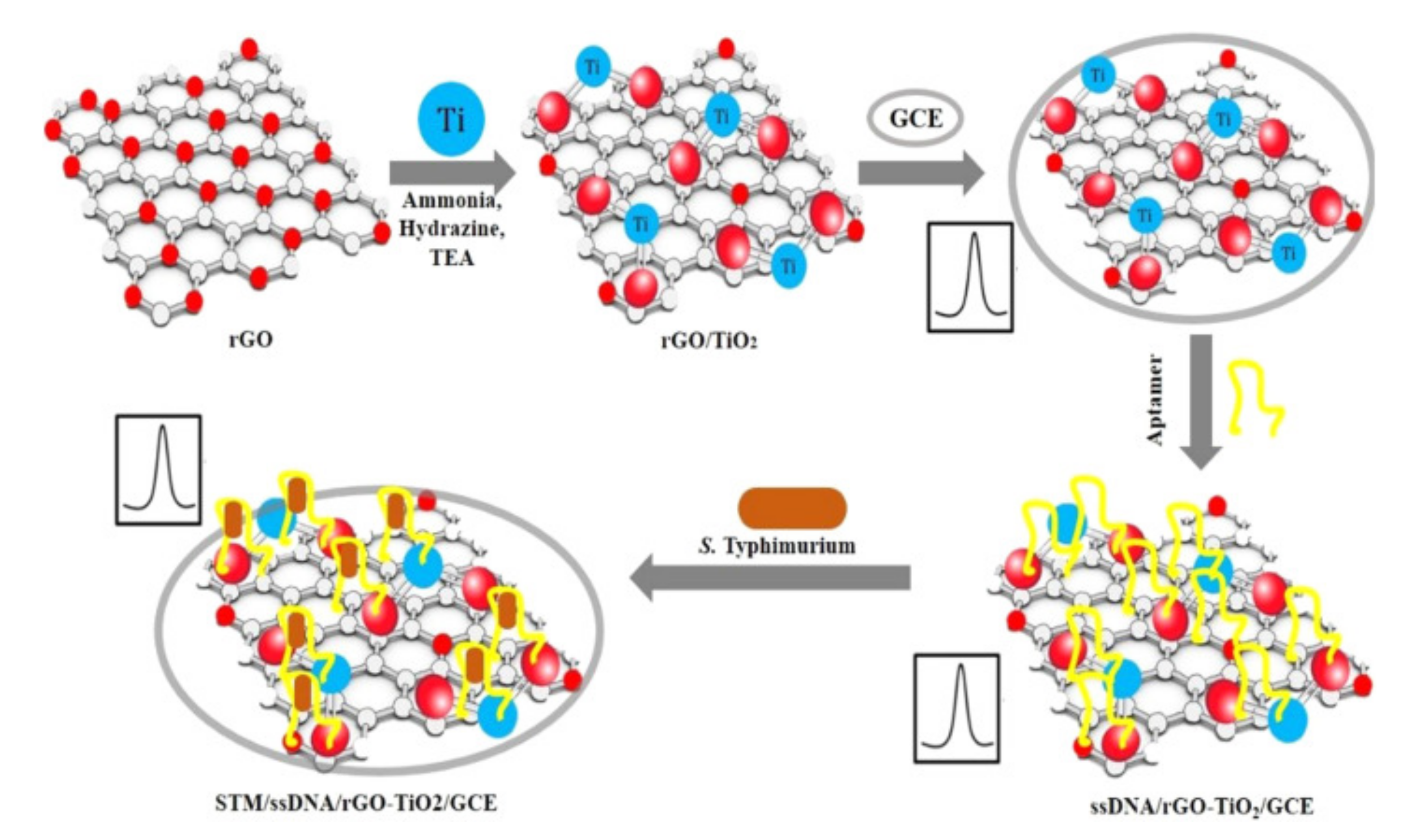
Several reports have indicated significant sensing improvements obtained by using metal oxide nanoparticles in electrochemical biosensors. Muniandy et al. [60], developed a reduced graphene oxide–nano TiO2 composite for an aptasensor used in the detection of Salmonella enterica (Figure 4). The bacterial cells captured by the aptamers incorporated on the electrode surface were a physical obstacle for electron transfer, which decreased the voltammetric signal proportionally to the bacterial concentration. Performance of the sensor was evaluated using cyclic voltammetry and electronic impedance spectroscopy. The developed aptasensor exhibited high sensitivity with a wide detection range (1–108 CFU/mL), low detection limit of 1 CFU/mL, good selectivity for Salmonella strains and acceptable long-term stability. Nadzirah et al. [61] used pure TiO2 nanoparticles (NPs) and fabricated interdigitated electrodes for E. coli detection. A specific ssDNA probe was immobilized on the electrode surface upon its chemical functionalization with (3-aminopropyl) triethoxysilane (APTES) to provide contact between the organic and inorganic surfaces of a ssDNA probe and TiO2 NPs. The obtained genosensor showed high sensitivity since it was able to detect as low as 1.0 × 10⁻¹³ M of E. coli O157:H7 DNA in bacterial lystes, with a high specificity and reproducibility.
Teng et al. [62] showed that ZnO nanorods in an electrochemical immunoassay for detection of E. coli led to signal enhancement. They immobilized both the detection antibody (anti-E. coli polyclonal antibody) and the redox probe (ferrocene) onto the surface of ZnO nanorods which surfaces were coated with a layer of silica. When coated with silica, the nanorods form core–shell nanorods that can be easily modified with various functional groups. The obtained complex antibody-ZnO-ferrocene was incubated with an E. coli contaminated sample and then washed and deposited on to a gold electrode carrying a capturing anti-E. coli antibody. The designed immunoassay showed a detection limit of 50 cfu/mL. In another study, Purwidyantri et al. [63] used ZnO nanograss decorated with Au nanospeckles to develop a sensing platform for Staphylococcus epidermidis, based on DNA hybridization. Applying the thermal evaporation, Au nanoparticles were deposited on the hydrothermally synthesized ZnO nanograss. The deposition increased by approximately two-fold the effective surface area and diffusion coefficient compared to the non-speckled ZnO nanograss. The fabricated genosensor carrying a DNA probe complementary to the 16S region in the genome of S. epidermidis attained a limit of detection (LoD) of 0.506 pM.
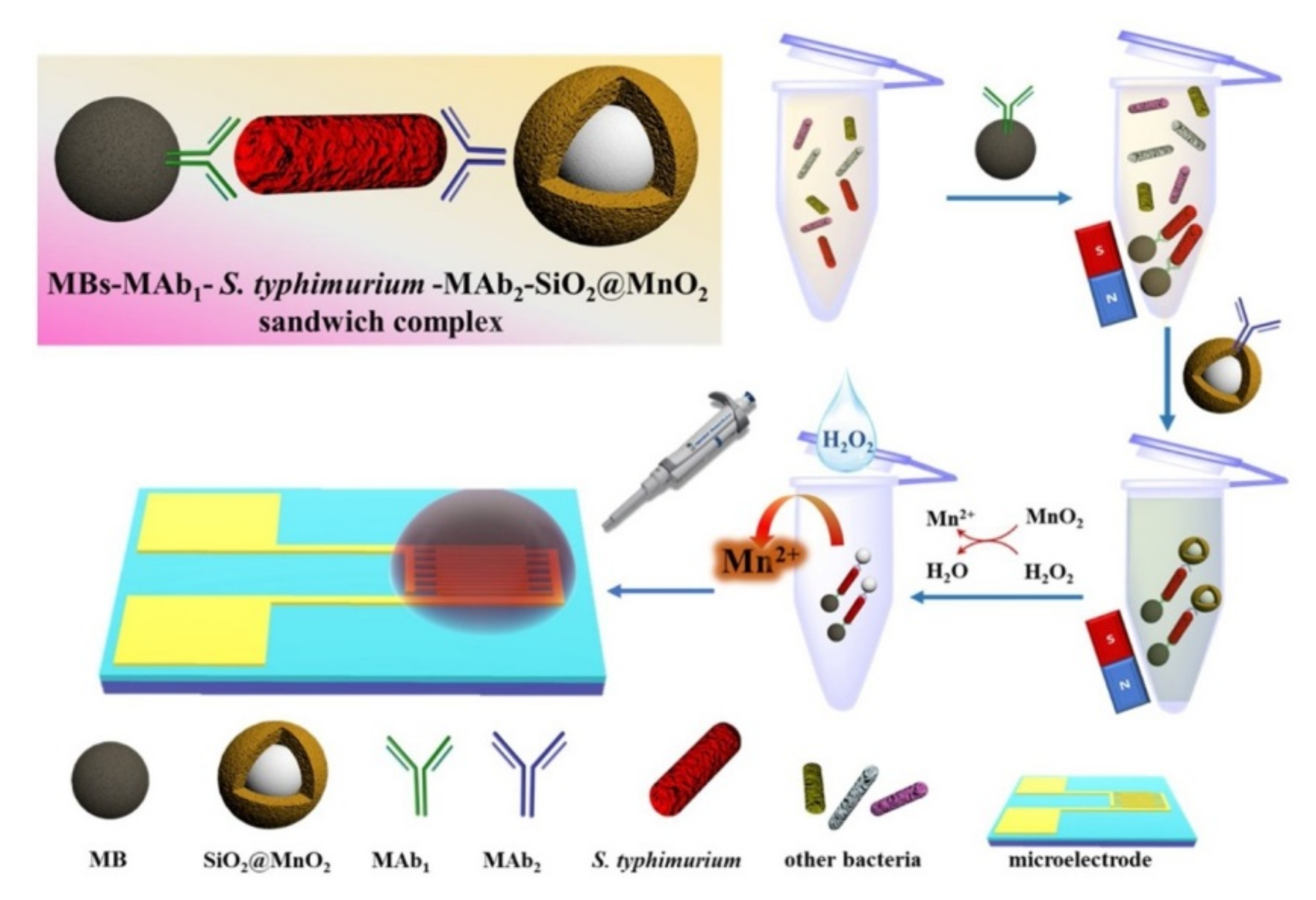
Earth abundant transition metal oxides are showing great potential for electrochemical applications, including electrochemical biosensors for foodborne pathogens [64]. Thus, rapid detection of Salmonella typhimurium was achieved using an SiO2@MnO2 nanocomposite impedance biosensor developed on interdigitated array microelectrodes combined with immunomagnetic separation [65]. Magnetic beads were used to capture monoclonal antibodies and separate S. typhimirium cells rapidly from samples, while detection was achieved by release of Mn2+ by H2O2 monitored as a change in impedance, as shown in Figure 5.
An electrochemical genosensor based on SnO2 nanocrystalline quantum dots was developed by Patel et al. [66] for detection of Vibrio cholerae using the DNA hybridization principle. The electrode was obtained by electrophoretic deposition of SnO2-QDs onto indium-tin oxide coated glass substrate. Subsequently, a DNA probe was attached to SnO2 NPs via its phosphate groups. The study showed that SnO2 NPs provided not only an effective surface for DNA probe immobilization, but also enhanced electron transport and improved signal read-out. The fabricated sensor detected V. choleare complementary DNA sequences with the limit of detection of 31.5 ng/µL and showed high long-term stability.
Bacteria remain the most commonly detected pathogen with metal oxide based electrochemical biosensors. Moreover, current trends are focused on the use of metal oxide photocatalytic properties in inactivation and elimination of bacteria [67,68,69,70]. For instance, a multifunctional electrochemical platform was obtained by combining ZnO, colloidal Ag and vancomycin [71]. AgNPs/3D-ZnO nanorod arrays, functionalized with vancomycin, were immobilized onto the working electrode. The platform detected S. aureus with a detection limit of 330 CFU/mL and eliminated bacterial cells with 50% efficiency at low bacterial concentrations of about 103 CFU/mL.

3.2. Carbon Nanomaterials

Carbon materials have long been a main companion in electrochemical sensor electrodes, in the form of either glassy carbon or activated carbon due to high chemical inertness and a high specific surface area needed for catalytic material impregnation. The discovery of new carbon allotropes, such as fullerene, carbon nanotubes (CNTs) [72,73] and graphene [74], has triggered active investigation of their application in different types of biosensors. Graphene has a unique two-dimensional honeycomb lattice structure, while the structure of CNTs consists of cylindrical graphene rolled up into a seamless cylinder with a diameter of the order of a nanometer. They provide unprecedentedly high specific surface areas up to 2630 m2/g [75] and 1315 m2/g [76], respectively, combined with a high electrical conductivity and charge carrier mobility. A number of reviews have discussed the perspectives of graphene and carbon nanomaterial application as materials for electrodes to improve electrochemical sensors [77,78,79], including foodborne pathogen detection [80].
Application of carbon nanomaterials, including graphene-related materials, is fostered by a deeper understanding of their physical and chemical properties as well as scalable production, processing and functionalization methods [81]. Examples of electrochemical biosensors utilizing different carbon nanomaterials are summarized in Table 1. The development of stable solutions of graphene and carbon nanotubes makes them prospective for electrodes integrated with conventional technologies for low-cost disposable electrochemical sensors in point-of-need devices. The graphene and carbon nanotube technology combines well with screen printed technologies for portable electrochemical sensors development [82], as well as paper based devices [83]. In foodborne pathogen detection, graphene-based electrochemical sensors also bring advances as they can operate directly in biological and food matrices. Graphene-based composites with functional nanomaterials and bioreceptors (antibodies, aptamers, DNA probes, etc.) provide low LoD down to pico/femto molar concentrations, and reduction of the time of analysis [80].
Electrochemical sensors based on graphene screen-printed disposable electrodes were found to be useful for analysis of meat adulteration [84]. For instance, graphene-based electrochemical biosensors combined with a loop-mediated isothermal amplification (LAMP) assay were used for V. parahaemolyticus toxR gene detection in seafood products [85]. The nonspecific interaction of the DNA backbone by π-π stacking on graphene-modified screen-printed carbon electrode was used for analysis of amplicons on the picogram level.

3.2.1. Graphene Nanoplatelets (GNPs)

In spite of the superior properties of graphene, its bare form of a monolayer atomic sheet is rarely used for electrochemical sensing applications. The technology of scalable monolayer graphene production based on chemical vapor deposition (CVD) is still in development to become low cost. Moreover, the defects and active sites in graphene are highly desirable for the binding of molecules and increasing sensitivity and selectivity [86]. Thus, bare graphene does not meet these demands because of its ideal crystalline structure, which would require additional treatment and, subsequently, increase the complexity of electrochemical electrode preparation.
In contrast, graphene nanoplatelets (GNPs) are a robust graphene-derived material with a 3D structure formed by multiple graphene layers, with properties similar to single-layer graphene. They provide a stable solution in organic solvents without the need for special chemical pre-treatment. GNPs and monolayer graphene were compared in a capacitive sensing platform for foodborne pathogenic E. coli O157:H7 detection [87]. The CVD-grown graphene was deposited on a silicon substrate with electrical contacts. Antibodies specific to E. coli were immobilized on graphene surface for the selective response during impedance measurements (Figure 6). Higher sensitivity was demonstrated for the monolayer graphene-based sensor, compared to the GNPs, with sensitivities of 10 cells/mL and 100 cells/mL, respectively. Nevertheless, the technology of preparation of less-defective graphene sensing monolayers was more complicated.
GNPs have been applied in the first electrochemical paper-based biosensor. Paper was coated by GNPs and Poly(N-isopropyl acrylamide) (PNIPAm) followed by Au deposition [88]. This biosensor can be applied directly with liquid samples without the use of a bioreceptor. Detection of bacterial cells, Gram negative E. coli and positive S. mutans and B. subtilis, was performed by monitoring the electrical resistance. The achieved detection LoD was only 5 cells/mL.

3.2.2. Graphene Oxide

Graphene derivatives, such as graphene oxide (GO) and reduced graphene oxide (rGO), are preferable materials for electrochemical electrode modification due to a low-cost scalable technology of production and processing in integrated devices [86,89]. The difference between GO and rGO is the number of oxygen molecules present, hence the conductivity. GO shows insulating or semi-conducting behavior, while rGO is electrically conductive but its conductivity also depends on the degree of reduction. Full reduction of GO is still difficult to achieve, while partial reduction of GO is rather easy.
Table 1. Detection of pathogens in food with carbon-based integrated electrochemical sensors.
NanomaterialTarget PathogenWorking Electrode/Nanomaterial Recognition ComplexEC
Technique
Linear
Range
LODFood MatrixReferences
Graphite feltE. coli
O157:H7
GF-GCEOSWV-400 cells/mLBeef[90]
SalmonellaGF-GCEOSWV-103 cells/mL-[91]
GrapheneE. coli
O157:H7
Cx-Gnfs/ITOEIS10−6 M–10−17 M1 × 10−17 M-[92]
S. aureusssDNA/GNDs-Zeo/PADCV/DPV 0.1 nMFruit juice[93]
Vibrio parahaemolyticusSPGEsCV8 × 10 to 8 × 10⁶ CFU/mL2 CFU/25 gSeafood[85]
E. coli
S. mutans
B. subtilis
PNIPAm-GR/Au platformEIS101–105 cells/mL5 cells/mLWater
Milk
[88]
E. coli O157:H7SiO2-MG
SiO2-GNPs
EIS10–107 cells/mL10–100 cells/mL-[87]
GOE. coli O157:H7ssDNA/GO/CSGCEEIS1 × 10−14 to 1 × 10⁻⁸ M3.584 × 10−15 M-[94]
SalmonellaGCE/GO/AuNPsEIS2.4–2.4 × 103 CFU/mL3 CFU/mL-[95]
S. TyphimuriumSPCE/rG-GOEIS-10 CFU/mLOrange juice
Water
[96]
rGOE. coli O157:H7SPCE/PANI-AuNPs-Ab1; rGO-NR-Au@Pt-Ab2
(measurement of H2O2 reduction)
CV8.9 × 103–8.9 × 10⁹ CFU/mL2840 CFU/mLMilk
Pork
[97]
E. colirGO/Al2O3FET1–100 CFU/µL104 CFU/mLRiver water[98]
E. colirGO–CysCuEIS10–108 CFU/mL3.8 CFU/mLWater
Fruit
Juice
Milk
[99]
E. coli O157:H7rGO–NR–Au@PtCV4.0 × 103–4.0 × 10⁸ CFU/mL4.0 × 102 CFU/mLPork
Milk
[100]
L. monocytogenesp-rGO/AuNPs/CILEDPV1.0 × 10− 13–1.0 × 10− 6 M3.17 × 10− 14 M-[101]
SalmonellaPPy-rGO/GCE/AuNPsDPV1.0 × 10−16–1.0 × 10⁻¹⁰ M
9.6–9.6 × 10⁴ CFU/mL
4.7 × 10⁻¹⁷ M DNA
8.07 CFU/mL
-[102]
SalmonellarGO-MWCNTEIS75 to 7.5 × 10⁵ CFU/mL25 CFU/mLChicken meat[103]
SalmonellarGO–CHIDPV10–106 CFU/mL10 CFU/mLChicken meat[104]
S. entericarGO-TiO2CV & EIS101–108 CFU/mL10 CFU/mLChicken meat[59]
LIGS. entericaLIGEIS25 to 105 CFU/mL13 ± 7 CFU/mLChicken broth[44]
E. coli O157:H7AuNPs-LIG EIS102−108 CFU/mL102 CFU/mL-[105]
SWCNTS. aureusSWCNTEIS-104 CFU/mL-[106]
S. aureusSWCNTDPV10–106 CFU/mL13 CFU/mLMilk[107]
MWCNTKlebsiella pneumoniae Enterococcus faecalis
E. coli
ClavA-CNTs-CysEIS102–106 CFU/mL102 CFU/mL-[108]
E. coliPPy/AuNP/MWCNT/CHIamperometry30–306 CFU/mL30 CFU/mL-[109]
E. coli
O157:H7
ITO/MWCNT/PEIEIS1–104 CFU/mL1 CFU/mL [110]
S. entericac-MWCNT/AuNPCV0.0976–100 ng/µL0.5 pg/mLMilk[111]
S. enteritidisMWCNT/ITOCV10−1–10−8 CFU/mL5.5 × 101 CFU/mL 6.7 × 10 CFU/mL-[112]
S. TyphimuriumSPCE/MWCNTDPV10–106 CFU mL−17.9 CFU/mLMilk[113]
S. aureusc-MWCNTs-PEIDPV-5 CFU mL−1Milk[114]
Listeria monocytogenesMWCNT/fiber electrodeDPV102–105 CFU/mL1.07 × 102 CFU/mLMilk[115]
Ab, antibody; c-MWCNT, carboxylated multi-walled carbon nanotube; CILE, carbon ionic liquid electrode; CSGCE, chitosan (CS) hybrid nanocomposites modified glassy carbon electrode (GCE); CHI, chitosan; ClavA, antimicrobial peptide clavanin A; CNTs, carbon nanotubes; CV, cyclic voltammetry Cx-Gnfs, carboxylated graphene nanoflakes; DPV, differential pulse voltammetry; EIS, electrochemical impedance spectroscopy; FET, Field-Effect Transistor; GCE, glassy carbon electrode; GF, graphite felt; GND, graphene nano dots; GNP, graphene nanoplatelets; GO, graphene oxide; GR, graphene nanoplatelet; ITO, indium tin oxide; LIG, laser induced graphene; MG, monolayered graphene; MWCNT, multi-walled carbon nanotubes; NR, neutral red; OSWV, Osteryoung square wave voltammetry; p-rGO, partially reduced graphene; PAD, paper analytical device; PANI, regenerative leucoemeraldine base polyaniline; PEI, polyethyleneimine; PNIPAm, poly(N-isopropylacrylamide) polymer; PPy, polyrrole; rG-GO, reduced graphene-graphene oxide; rGO, reduced graphene oxide; rGO-CHI, electrochemically-reduced graphene oxide-chitosan; rGO-CysCu, graphene wrapped copper (II) assisted cysteine hierarchical structure; rGO-TiO2, reduced graphene oxide-titanium dioxide; SPCE, screen-printed carbon electrode; SPGE, screen-printed graphene electrodes; SWCNT, single-walled carbon nanotube; Zeo, zeolite.
GO is soluble in aqueous solutions without the need for surfactant addition that is typically the case for graphene and carbon nanotubes. Moreover, the naturally high concentration of defects in GO [116] allow easy functionalization with specific receptor molecules. GO combined with chitosan has been demonstrated to be an excellent means for electron transfer for the detection of short DNA sequences achieving the detection limit of 3.584 × 10⁻¹⁵ M [94]. Paper-based sensors with screen-printed electrodes modified by a Nafion/PPy/GO composite were proposed for the detection of lipopolysaccharides (LPSs), which are a marker for Gram-negative bacteria [117]. Raw264.7 macrophage cells were used as a recognition element. The cells were grown in a 3D structure in a Nafion/PPy/GO composite scaffold, serving as a NO gas release to be electrochemically oxidized and detected as a differential pulse voltammetry signal change. GO was shown to be both a good electrical conductor and biocompatible material for cell growth. A sensitivity of 3 pg/mL of LPSs was demonstrated in peach and orange juice.
A rGO-based FET sensor passivated with a layer of Al2O3 was functionalized with specific antibody immobilized on gold nanoparticles [98]. This sensor was developed to detect E. coli in water. Detection was performed by monitoring the change in electrical conductivity of the rGO channel. The LoD was 103 cells/mL. The sensitivity can be improved using rGO modified with cysteine (Cys) in the presence of Cu2+ -ions. Such electrochemical immunosensor achieved a LoD of 3.8 CFU/mL of E. coli O157:H7 through maintaining the antibody bioactivity [99]. In addition, the biosensor was able to distinguish pathogenic E. coli O157:H7 from nonpathogenic E. coli strains.
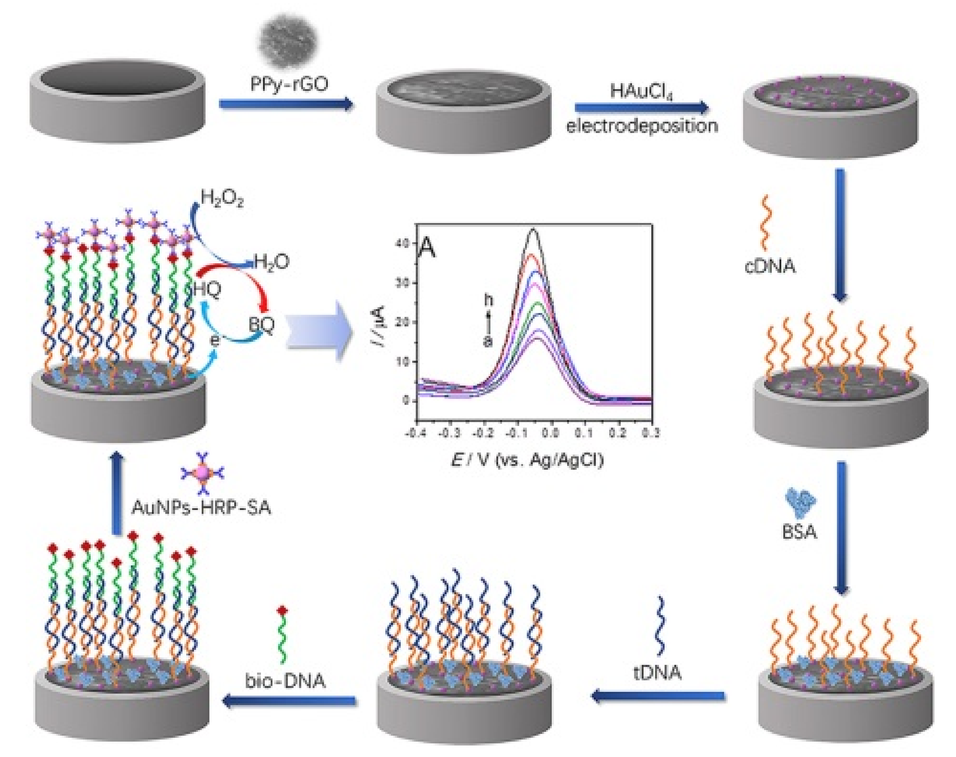
A rapid and sensitive electrochemical invA gene biosensor for the detection of Salmonella was designed by applying a polypyrrole-rGO nanocomposite on a glassy carbon electrode [102], as shown in Figure 7. Signal amplification was achieved using horseradish peroxidase streptavidin biofunctionalized AuNPs. The LoD was 8.07 CFU/mL with a detection range 9.6–9.6 × 10⁴ CFU/mL.

3.2.3. Laser-Induced Graphene

Recently, novel methods of direct graphene-based electrode writing were applied for portable sensor development [118]. Laser-induced graphene (LIG) is a very simple and scalable technology of porous graphene material production by a local thermal treatment of polymers like polyimide [119]. The obtained material combines the advantages of graphene like a high surface area, electrical conductivity with numerous active centers for surface modifications with different receptors [120].
A one step method was proposed to create an electrochemical substrate composed of 3D porous graphene and gold nanoparticles [105]. The aim was to improve the detection performance with a more stable sensor due to the synergic effect of the two nanocomponents. The antibodies were immobilized on the NPs-LIG substrate for the detection of the E. coli O157:H7. Despite the use of NPs, the limit of detection achieved was 102 CFU/mL.
In another study, LIG electrodes modified with polyclonal antibodies were used for the highly selective detection of Salmonella enterica serovar Typhimurium [44]. The developed immunoassay demonstrated the linear range of 25 × 105 CFU/mL with a low detection limit of 13 CFU/mL in spiked chicken broth samples and a response time of 22 min. Notably, no special preparation of samples was needed to perform measurements.

3.2.4. Carbon Nanotubes (CNTs)

Carbon nanotubes were used as electrode materials long before the graphene. CNTs are divided into single-walled carbon nanotubes (SWCNTs) and multi-walled carbon nanotubes (MWCNTs) based on the number of graphene sheets [121]. SWCNTs have a diameter range of 0.5 nm to 12 nm but the smallest diameter of SWCNTs is 0.4 nm with different tube lengths starting from several micrometers depending on manufacturing and treatment techniques. MWCNTs consist of multi-rolled layers of graphene inserted one into the other and the number of graphene walls may reach more than 25 walls with a spacing of 0.34 nm. The outside diameter of MWCNTs ranges from 1 nm to 50 nm while the inside diameter is several nanometers. Nevertheless, the problem of good aqueous suspension of carbon nanotubes still prevents their wide usage in integrated electrodes. In addition, proper functionalization of nanotubes is needed which reduces the electrical properties of these nanomaterials.
SWCNT composites were used for highly sensitive detection of bacterial and virus model species E. coli O157:H7 and the bacteriophage T7, respectively [122]. The carbon nanotube was used as a transduction element aligned in parallel to bridge two gold electrodes. To provide recognition, SWNTs were functionalized with specific antibodies. The sensor exhibited excellent selectivity, sensitivity and a fast response time of about 5 min in the case of T7 detection, while the response time for the detection of E. coli was 60 min.
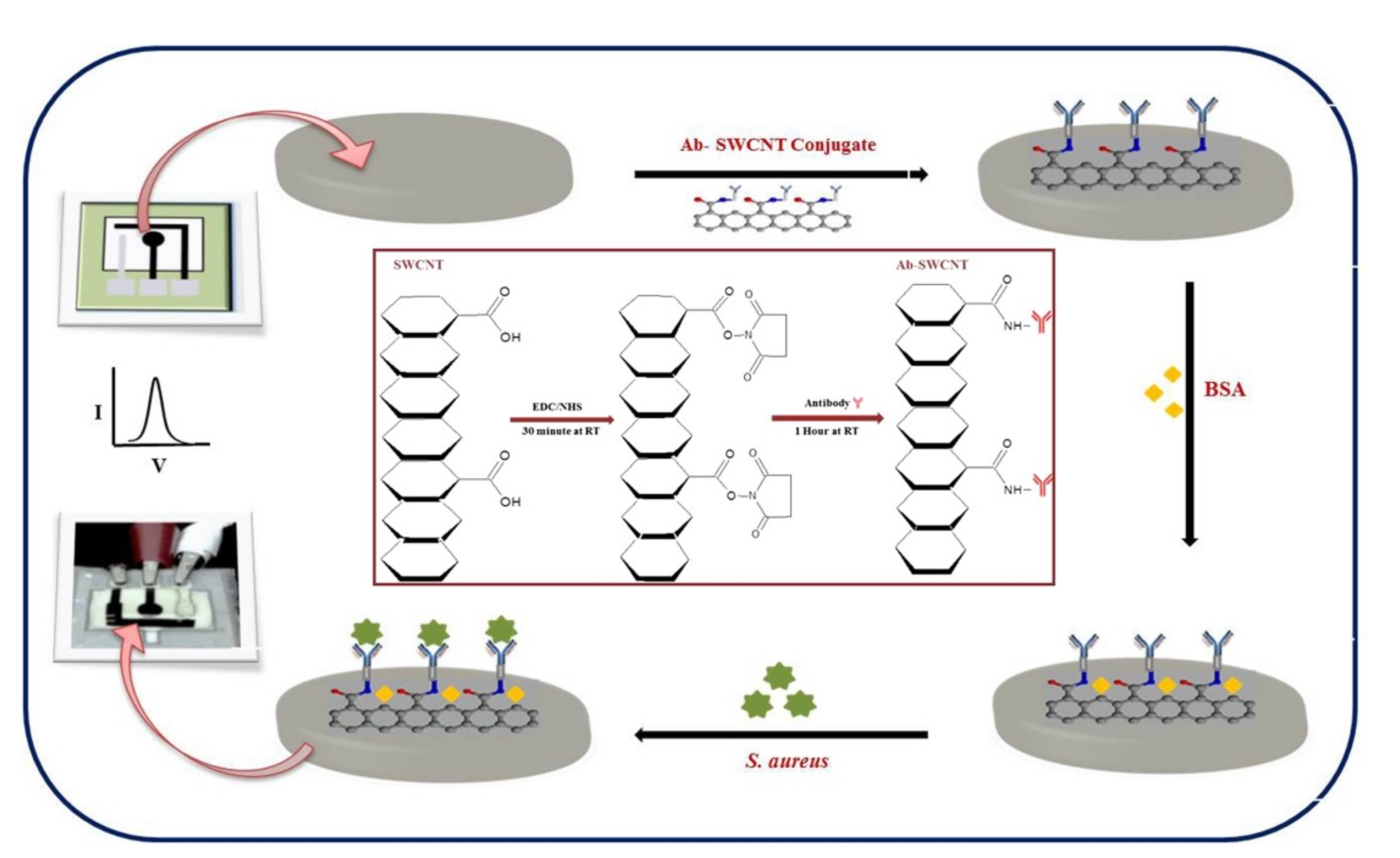
SWCNTs with immobilized antibodies were integrated into a disposable bio-nano combinatorial junction sensor for detection of E. coli K-12 [123]. Measurements were performed on gold tungsten wires coated with polyethyleneimine with aligned functionalized SWCNTs to form a crossbar junction. Changes in electrical current observed after the SWCNT surface interaction with bacterial cells were monitored to evaluate the sensor’s performance. The biosensor had a LoD of 102 CFU/mL with a detection time of less than 5 min. A low-cost paper-based electrochemical immunosensor was developed utilizing an antibody-SWCNT bioconjugate for rapid detection of S. aureus using differential pulse voltammetry (Figure 8), achieving a detection time of 30 min with a detection limit of 13 CFU/mL in spiked milk samples [107].
As a material modification, MWCNTs is better than SWCNTs as it is stiffer, easier and cheaper to produce on a large scale, and several studies have demonstrated to have better sensitivity. Indeed, MWCNTs deposited on an Indium tin oxide (ITO) electrode and modified with aptamers to detect S. enteritidis and S. thyphimuri achieved a detection limit of 5.5 × 101 CFU/mL and 6.7 × 101 CFU/mL, respectively [112]. Measurements were performed in food samples using cyclic voltammetry and electrochemical impedance spectroscopy techniques to characterize the electrochemical properties and conductivity of the aptasensor. The impedance measured at the aptamer/MWCNT/ITO electrode surface increased after exposure to target Salmonella cells, due to the capturing of Salmonella by the immobilized aptamers. A promising electrode substrate was developed with c-MWCNTs to confer an electrical conductivity at bacterial cellulose fibre (BCF) [114]. The BCF was modified with poly- ethyleneimine cation (PEI) to allow the immobilization of phages used as a bioreceptor for S. aureus. The LoD of 5 CFU/mL and 2 CFU/mL was found in milk and phosphate buffer saline, respectively, with effective discrimination between dead and live cells and within only 30 min. Moreover, the produced electrodes were maintained stable for over 6 weeks.
Carbon nanomaterials are often used as one of the components in nanocomposite electrochemical biosensors for foodborne pathogen detection. For example, grapheme oxide as part of mesoporous nanocomposite for detection of E.coli [46]. A rapid and sensitive detection in the dynamic range from 101 CFU/mL to 108 CFU/mL with a detection limit of 101 CFU/mL of S. enterica was achieved with a nanocomposite of rGO and CNT modified with an amino-modified DNA aptamer [124].
The low-cost carbon materials, including graphene and carbon nanofibers, provide a large specific surface area, high electron transfer rate and good catalytic properties, which is of high importance for development of sensing platforms that can be miniaturized for point-of-need testing.

3.3. Gold Nanoparticles

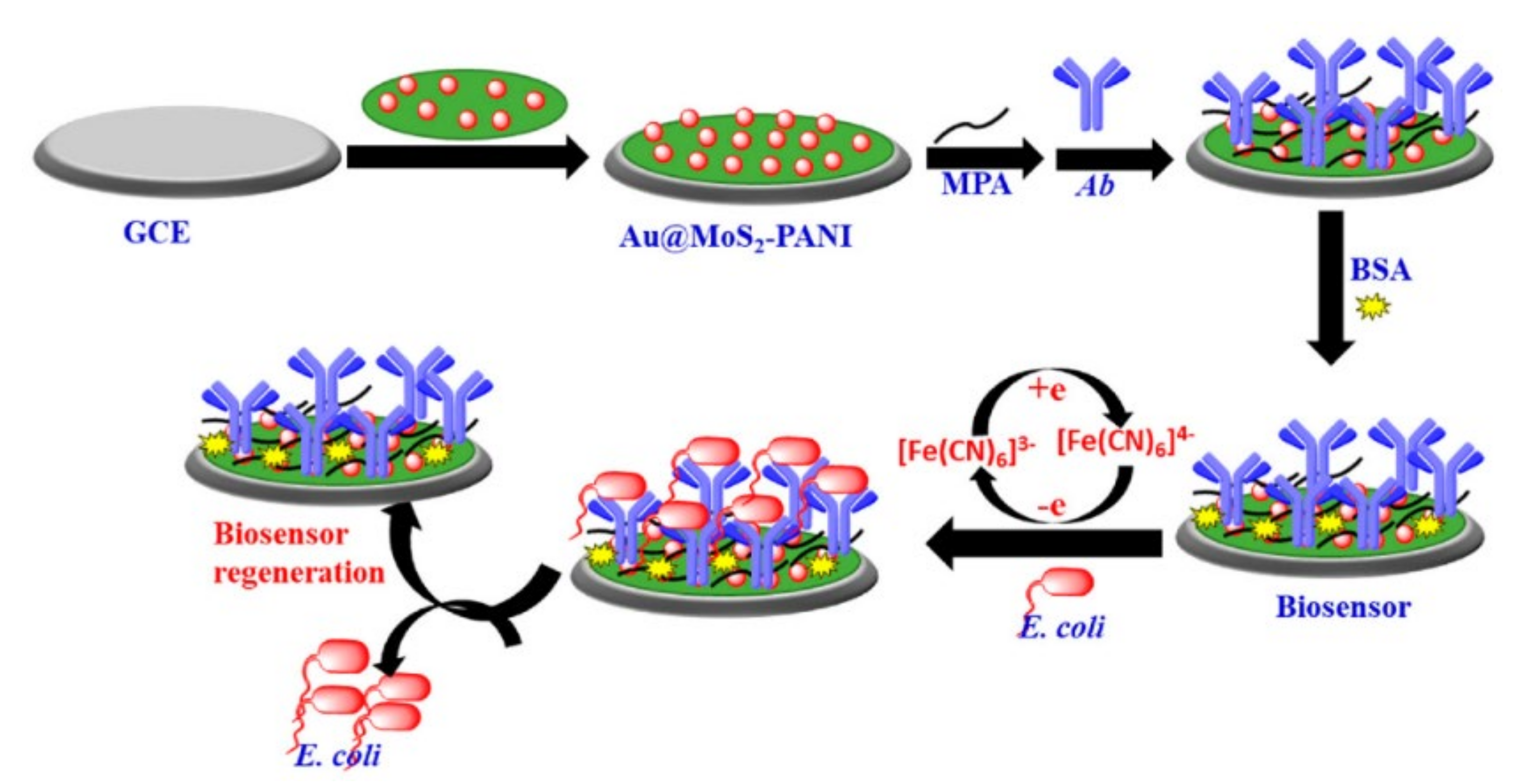
Gold nanoparticles (AuNPs) have been increasingly used in the design of electrochemical biosensors for their biocompatibility, conductivity, catalytic activity, stability and high surface-to-volume ratio [125]. Deposition of AuNPs onto gold electrodes enables a significant increase in the electrode surface area for target recognition and, consequently, its analytical performance [126,127]. When AuNPs are immobilized on the surface of electrodes made of other materials (such as carbon, graphene, paper, etc.), they increase the surface biocompatibility, promote electron transfer between electrode and immobilized molecules and enable easy bio-conjugation of recognition elements besides increasing the electrode surface area. Raj et al. [128] developed a label-free electrochemical biosensor for the detection of E. coli based on a glassy carbon electrode with immobilized a complex of polyaniline nanocomposites (PANI), gold nanoparticles and MoS2 (Au@MoS2–PANI), in order to increase conductivity, stability and electro-activity of the electrode. The surface of AuNPs were treated with mercaptopropionic acid to covalently immobilize antibodies against E. coli and to minimize the nonspecific adsorption on the electrode surface. The biosensor was simple, rapid and specific, with a LoD of 10 CFU/mL and a linear detection range of 10–107 CFU/mL. A schematic representation of this electrode construction is shown in Figure 9. In another study, AuNPs were immobilized on a carbon screen-printed electrode to increase the stability and efficacy of the electrochemical biosensor for the label-free detection of E. coli [29]. The modified electrode was treated with N-(γ-Maleimidobutyryloxy) succinimide (GMBS) to create -NHS groups for cross-linking of E. coli O157-specific polyclonal antibodies. The analysis showed rapid and efficient pathogen detection with a dynamic range of 10–106 CFU/mL and a LoD of 15 CFU/mL.
The electrocatalytic properties of AuNPs towards hydrogen evolution reaction was employed for rapid and highly sensitive immunodetection of E. coli O157:H7 in minced beef and water [129]. The test was performed in a sandwich format where superparamagnetic microbeads modified with the first antibody were used to perform pre-concentration/purification of the bacterial cells from the sample and AuNPs modified with the second antibody provided the catalytic reaction. The method showed a LoD of 457 CFU/mL in minced beef and 309 CFU/mL in water. When compared with a commercial lateral flow kit in terms of LoD, specificity, reproducibility and detection range, the electrochemical method showed clear advantages. Similarly, the magneto-immunoassay and AuNPs as label for electrochemical detection was developed for the detection of Salmonella enterica subsp. enterica serovar Typhimurium LT2 (S) in skimmed milk by Alfonso et al. [130]. A magnet is incorporated under the screen-printed carbon electrode to attach magnetic beads carrying Salmonella specific antibodies. Beads were added to milk samples to pre-concentrate bacterial cells and then deposited onto the electrode. A sandwich was created using AuNPs modified with antibodies to provide a redox signal. Applying differential pulse voltammetry, a linear range from 103 cells/mL to 106 cells/mL and a LoD of 143 cells/mL was found for skimmed milk samples contaminated with Salmonella. AuNP modified screen-printed carbon electrodes were combined with magnetic nanoparticles coupled to specific peptides via a streptavidin interaction to achieve multiplexed electrochemical detection of Listeria monocytogenes and Staphylococcus aureus with a low detection limit of 9 CFU/mL and 3 CFU/mL, respectively [131].
Magnetic and gold nanoparticles have also been combined in impedance biosensors. For instance, Wang et al. [132] used urease-modified AuNPs to amplify the signal of impedance biosensors implemented with magnetic nanoparticles for the detection of Listeria monocytogenes. Bacterial cells captured between magnetic nanoparticles decorated with a monoclonal antibody and AuNPs–urease complex carrying the polyclonal antibody were resuspended in urea to catalyze its hydrolysis into ammonium and carbonate ions. Generated ions were detected by a screen-printed interdigitated electrode. The technique, characterized by low cost and high specificity, gave a linear range from 1.9 × 10³ CFU/mL to 1.9 × 10⁶ CFU/mL, and a LoD of 1.6 × 10³ CFU/mL, in spiked lettuce samples.
Expensive mono- and poly-clonal antibodies can be replaced with lectins that recognize LPS on the bacterial surface. Oliveira et al. [133] immobilized Cramol L lectin on AuNPs functionalized with l-cysteine. Cramol L is a Cratylia mollis lectin used as the recognition interface by making hydrogen bonds with methyl-α-d-mannoside in LPS. To build the biosensor the gold electrode surface was covered by a poly (vinyl chloride-vinyl acetate maleic acid) layer to attach Au-cysteine-Cramol L nanoparticles through the electrostatic interactions. Bovine serum albumin was used to block the remaining non-functionalized electrode surface. The sensor, tested on E. coli, Serratia marcescens, Salmonella enterica and Klebsiella pneumoniae, was able to selectively discriminate bacterial species due to their different LPS composition with a high sensitivity.
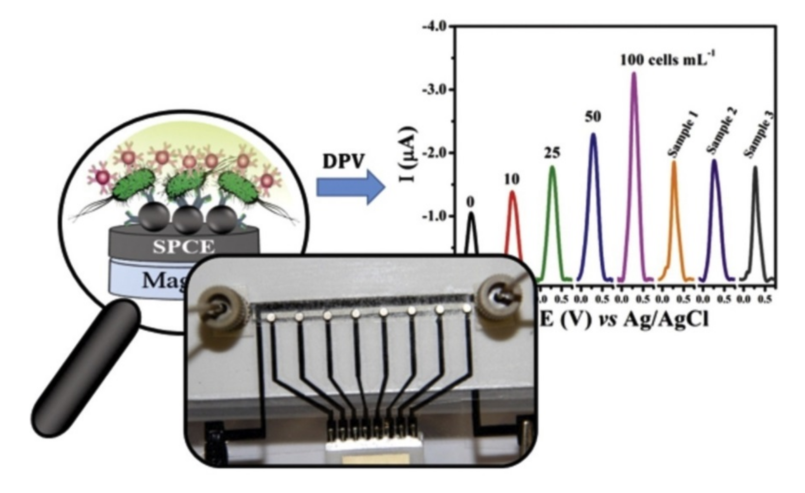
Although AuNPs based electrochemical biosensors have been extensively employed, their complexity is still an issue limiting the general application, especially in complex food matrices. Usually a multistep procedure, it involves user manual interventions during the test, such as for repetitive washing, loading of samples and reagents. These steps increase the time of analysis and cause imprecise result. Attempts have been made to automatize manual interventions by coupling microfluidic with electrochemical cell. Microfluidic can also enable multiplex detection of different pathogens in the same sample. A disposable microfluidic device for Salmonella typhimurium detection in milk was proposed by de Oliveira et al. [134]. The microfluidic device allowed the simultaneous measurement of eight samples by a magneto-immunoassay, as illustrated in Figure 10. The bacteria were captured from the sample by magnetic beads modified with a monoclonal antibody. A sandwich was then completed with AuNPs labeled with a polyclonal antibody. The complex was injected into the device and magnetically placed on the electrode surface. This approach allowed to obtain an easy to use and rapid detection, with a LOD of 7.7 cells/mL.
A sandwich-type electrochemical immunosensor for the detection of L. monocytogenes proposed to use 3,4,9,10-perylene tetracarboxylic acid/graphene ribbon nanohybrids as a sensing platform and ferrocene/AuNPs as a signal amplifier [135]. A low detection limit of 6 CFU/mL and linear range of 10–2 × 10⁴ CFU/mL was achieved, showing that incorporation of nanomaterials, such as graphene and AuNPs, enables improved sensing properties.
Electrochemical biosensors based on AuNPs have been successfully applied for virus detection [136]. For instance, the Middle East respiratory syndrome coronavirus (MERS-CoV), which is one of the highly pathogenic viruses, was found to contaminate dairy products [137]. It was detected by the competitive assay carried out on an array of carbon electrodes modified with gold nanoparticles [136]. The electrode array enabled multiplexed detection of different strains of CoVs through the indirect competition between free virus in the sample and immobilized MERS-CoV protein S1 or a fixed concentration of antibody added to the sample. By using ferrocyanide/ferricyanide as a redox probe, voltammetric measurements performed within 20 min showed low detection limit of 1.0 pg/mL for MERS-CoV and high selectivity.

3.4. Other 0–3D Nanomaterials

Besides AuNPs, silver nanoparticles (AgNPs) have also been applied for electrochemical detection. For instance, chitosan stabilized AgNPs were applied for electrochemical detection of negatively charged LPS, enabling detection of E. coli in the range 10–107 CFU/mL [138].
The high benefits of graphene as a transducer layer for working electrodes in electrochemical biosensors have resulted in an increased interest in the 2D nanomaterial family for application in electrochemical sensing [33]. Semiconductor 2D materials, such as transition metals dichalcogenides (TMDC) and transition metal carbides and carbonitrides (MXenes), have a high surface area and conductivity and possess an intrinsic energy band providing sensitivity to the weak changes in the charge state on electrodes. They can thus greatly improve the performance of electrochemical sensors [139]. A technological process of defect-free 2D materials production is still based on the CVD method, which is an expensive technological process demanding high-purity precursors. In contrast, the richness of defects and boundary grains in MXene production is very simple and inexpensive and better adapted for applications in portable electrochemical devices [140]. Two-dimensional transition metal materials provide a robust sensing surface due to their structural stability and excellent electrochemical properties such as conductivity, catalytic performance and abundant active sites. Previous reviews have discussed the perspective of the electrochemical sensor development based on molybdenum disulfide (MoS2) [141] and MXene nanomaterials [142].
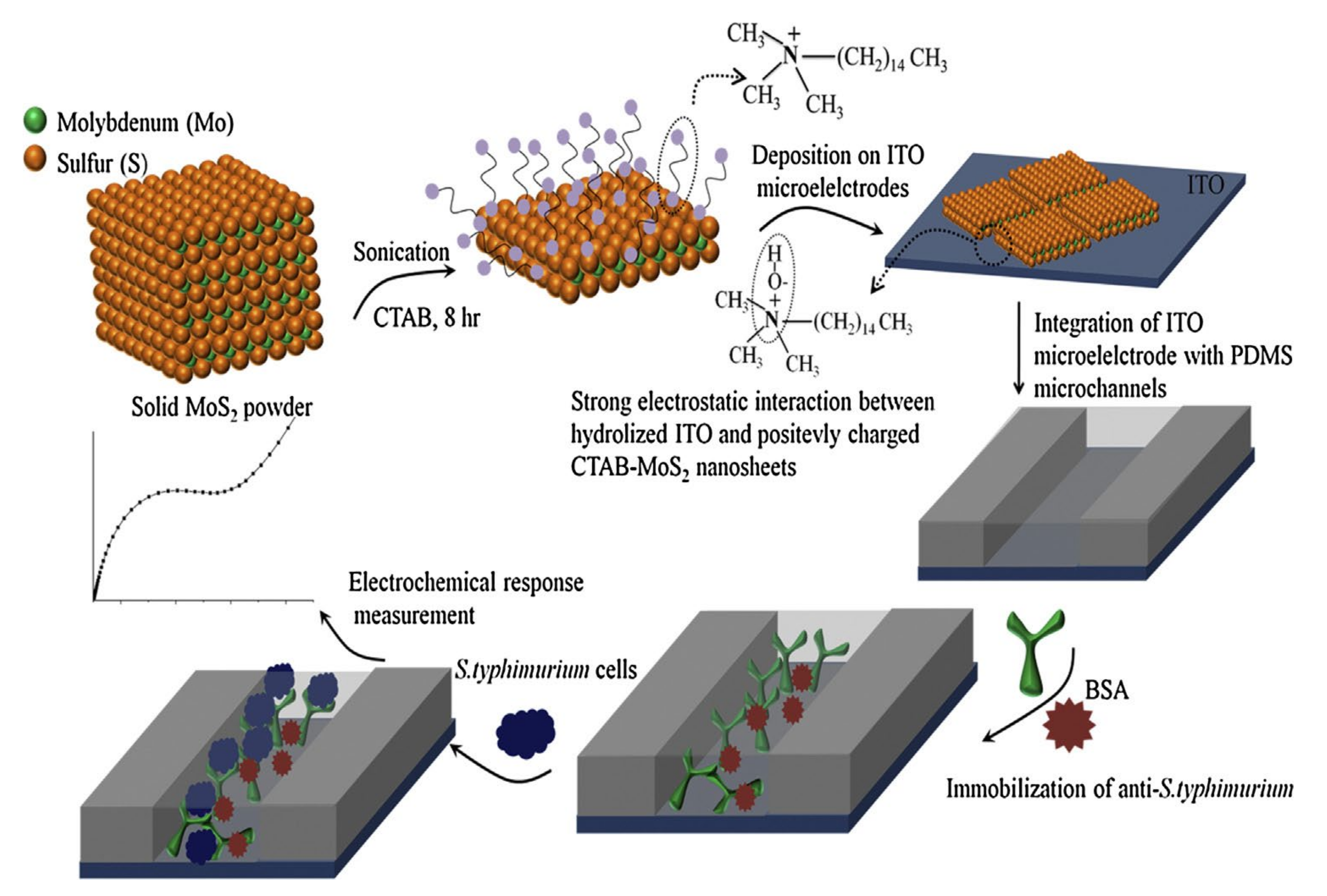
Hexagonal MoS2 is a stratified crystal which has thicknesses equal to the unit cell of the material in a way that each plane of MoS2 is made of molybdenum atoms sandwiched between sulfur atoms and stabilized by van der Waals bonds. Two-dimensional MoS2 is obtained when the material is exfoliated into one or a limited number of layers. Two-dimensional MoS2 shows remarkable electronic, optical, mechanical and chemical characteristics that also make it advantageous for biosensing applications. In addition, when grown into planes with relatively large lateral dimensions, 2D MoS2 is particularly stable in liquid and oxygen containing gaseous media which facilitate their utilization when incorporated into biosensing structures [143]. An electrochemical lab-on-paper genosensor was developed based on carbon ink screen-printed on cellulose paper with a working electrode modified by drop-casted MoS2 nanosheets for detecting Salmonella-specific DNA [144]. The sensitivity of the MoS2-modified electrode was increased more than 10 times due to the enhanced transfer rate of charge carries and unique electron transfer kinetics in MoS2. To provide the selectivity towards Salmonella-DNA, the specific complementary DNA probe was immobilized on MoS2 for on electrode hybridization. The LoD of 20 nM was obtained. Another method for foodborne pathogen detection was suggested based on antibody immobilized onto a microfluidic chip. Exfoliated MoS2 nanosheets in the presence of a surfactant were deposited on ITO electrodes integrated with a microfluidic channel to develop an impedimetric biosensor [145], as shown in Figure 11. The specific antibody directed against Salmonella typhimurium was immobilized on a MoS2/ITO electrode treated with glutaraldehyde. Proper functionalization of 2D nanosheets and optimization of the procedure for antibody molecules association with MoS2 yielded superior electron conduction and resulted in a 1.5 CFU/mL limit of detection.
Metal organic frameworks (MOFs) are a rapidly emerging new class of microporous materials with a wide range of promising applications [146,147]. They basically represent 2D or 3D porous materials assembled using metal cation salts or clusters bridged with polydentate organic ligands with coordination type connections, though 0D and 1D nanostructures are being synthesized also [148]. They have a very high surface area, high pore volume, high porosity and surface functionality and an easily tunable structure. Nanoscale MOFs combine the properties of both MOFs and nanostructures. MOFs are often used to design complex nanocomposite materials through a controlled assembly of MOF nanoparticles, such as NP@MOFs. Two-dimensional metal organic frameworks (MOF) have recently come into focus for biosensing applications [149]. The possibility of tuning their properties in a controllable way and the extremely high surface area is expected to outperform traditional electrochemical sensors. Still, their poor conductivity demands a combination with other highly conductive nanomaterials in the form of nanocomposites. Sensitive impedimetric detection of E. coli in the range 2.1 × 10²–2.1 × 10⁸ CFU/mL with a detection limit of 4 CFU/mL was achieved by combining a (MOF) with a conducting polymer (CP) and PEDOT on modified carbon screen-printed electrodes [150]. Copper (Cu)-MOFs were directly self-assembled and deposited onto a glassy carbon electrode, followed by in situ reduction of AuNPs on the MOF surface and conjugated with a DNA aptamer enabling detection of S. aureus in the range 7–7 × 10⁶ CFU/mL [151]. Graphene and a zirconium based MOF (UiO-67) were combined together with an aptamer loaded AuNP-horseradish conjugate to detect S. typhimurium in spiked milk samples with a detection limit of 5 CFU/mL [152].
Quantum dots (QDs), carbon dots (CDs) and graphene quantum dots (GQDs) are another category of nanomaterials with great potential for application in electrochemical biosensing of foodborne pathogens [27,31]. QDs have been used to modify the structure of polymeric nanodendrons for direct culture-free electrochemical detection of Salmonella in milk with a detection limit of 4 CFU/mL [153]. GQDs combine characteristics both from graphene and carbon dots, offering great versatility for modification with other modifiers and nanomaterials besides low cost, low toxicity, high solubility and good electronic properties. Photoelectrochemical sensing represents an integration of electrochemistry and photochemistry offering high sensitivity, robustness, low cost and simple instrumentation. For instance, GQDs doped with nitrogen were coupled with non-metallic two dimensional hydrated defected tungsten oxide to design a photoelectrochemical aptamer biosensor for E.coli detection in the range 0.1–104 CFU/mL with a low detection limit of 0.05 CFU/mL [154].

4. Conclusion and Perspectives

The present review summarizes the unique properties of 0–3D nanomaterials and their application in the design of electrochemical biosensors for foodborne pathogen detection. Despite the progress and advances in culture based and molecular methods for foodborne pathogen detection, challenges remain for their practical application because they still do not reach the sensitivity, fast response time and low cost needed. Effective foodborne pathogen monitoring that will enable efficient risk assessment and outbreak prediction has to be rapid, ultrasensitive, specific and affordable to be applied in low-resource settings. Electrochemical biosensors offer an exciting opportunity to realize immediate and continuous pathogen detection for on-site risk evaluation. We have highlighted examples showing that electrochemical methods can release results within several hours or even several minutes. Besides, a wide variety of strategies used to improve sensitivity are presented. Some foodborne pathogens, such as Campylobacter, E. coli O157 and L. monocytogenes, have very low infectious doses of a dozen to several hundred cells. Consequently, it is of high importance that low contaminated food items can be identified. Amplification of the detection signal using nanomaterials as electronic conductors, carriers or catalysts enable electrochemical biosensors to exhibit LoDs as low as a single colony forming unit (CFU/mL) or several femto M or even atto M ranges and linear ranges of several orders of magnitude. Although the presented nanomaterials can be used with other detection techniques, such as plasmonic and fluorescent, electrochemical biosensors have the advantage of simple utilization by persons without previous training, versatile detection schema providing a wide range of applications and easy miniaturization [155,156,157]. Moreover, the inexpensive and miniaturized electrochemical devices in handheld formats are excellent candidates for on-site application.
Cyclic voltammetry, differential pulse voltammetry, square wave voltammetry and electrochemical impedance spectroscopy are the methods mostly used for detection of pathogens and their toxins in food samples. At the same time, nanomaterials are generally applied with these methods to enhance the detection signal. Planar gold electrodes are the most commonly used working electrodes. However, nanomaterials with their outstanding properties that arise from their small dimensions and surface reactivity are applied to alleviate the limitations of electrochemical biosensors, such as slow recognition time, low biocompatibility or instability. In addition, some nanomaterials, such as gold nanoparticles or graphene, may tune the electrode properties and offer a variety of surface engineering strategies and functionalization to attach biological entities assuring recognition (e.g., antibodies, aptamers, ssDNA, phages). One of the trends in recent electrochemical biosensors for pathogen detection is to combine several nanomaterials as nanocomposites in electrode design to obtain remarkable synergic effects leading to improved sensing performances. Moreover, specific nanomaterials, such as graphene or metal oxide nanoparticles, have inherent antibacterial activity. Biosensors integrating such nanomaterials are multi-functional, providing not only pathogen identification and quantification but also their elimination.
During the past decade, significant progress has been made in the biosensors field to advance electrochemical devices, taking into account food industry demands. Hence, further improvement is needed to facilitate wide practical applicability of biosensors for detecting foodborne pathogens. Biocompatibility is one high concern. The electrode design, surface modification and functionalization integrating nanomaterials are of great promise to improve the stability and compatibility of electrodes in biological environments for an extended period. Besides, due to the diversity of foodborne pathogens and the possibility of food co-contamination by various microorganisms, multi-electrode detection devices that exhibit different biorecognition elements for simultaneous multiplex detection without mutual interference are also needed for improved detection efficiency. In addition, most electrochemical biochips are designed only for single utilization. We expect that future studies will anticipate environmental friendliness and resource conservation and will include reusability of electrodes in sensor design.
Analytical performances of the majority of presented biosensors refer only to LoD and linear range of detection. However, other parameters, notably accuracy, repeatability, precision, and specificity, should be also investigated and improved to enable sensors general accessibility. Furthermore, the synthesis procedure of integrated nanomaterials has to be optimized to obtain rigorous protocols for mass production and strict quality control of the material without chemical impurities that can alter sensing properties or induce environmental pollution.
Finally, pathogen detection in foods requires sample treatment and transport to the electrode surface, which may cause analyte loss. For this reason, advanced electrochemical biosensors based on nanomaterials that detect pathogens and their toxins in complex matrices without important interferences should be designed. Coupling detection with a microfluidic system for sample handling holds great potential.
In the future, additional excellent nanomaterial-based electrochemical biosensors will emerge, and new design and solutions will be proposed. It is evident that the field of nanomaterials is making tremendous progress and significantly affects biosensor development. Active collaboration between material scientists, microbiologists, electrochemists and device developers in the fields of nanotechnology and food science will result in point-of-need diagnostic devices integrating electrochemical biosensors, microfluidics and nanomaterials. Such devices will offer the food industry food safety analyses and foodstuff screening that can be performed during all phases, from production, packaging, storing and distribution to consumption.

Author Contributions

I.B. and M.R., conceptualization, writing—original draft preparation; F.R., P.V., S.J. and Z.P., writing—original draft preparation; V.R., conceptualization; M.V.N., conceptualization, writing—review and editing; J.V., conceptualization, writing—review and editing, supervision. All authors have read and agreed to the published version of the manuscript.

Funding

This research was supported, in part, by the European Union’s Horizon 2020 research and innovation program under the Marie Skłodowska-Curie grant agreement IPANEMA No 872662 (I.B., M.R, V.R., F.R., S.J. and J.V) and by the Ministry for Education, Science and Technology Development of the Republic of Serbia, under the contract 451-03-9/2021-14/200053 (M.V.N.).

Institutional Review Board Statement

Not applicable.

Informed Consent Statement

Not applicable.

Data Availability Statement

This study did not report any data.

Conflicts of Interest

The authors declare no conflict of interest.

References

  1. Havelaar, A.H.; Kirk, M.D.; Torgerson, P.R.; Gibb, H.J.; Hald, T.; Lake, R.J.; Praet, N.; Bellinger, D.C.; De Silva, N.R.; Gargouri, N. World Health Organization global estimates and regional comparisons of the burden of foodborne disease in 2010. PLoS Med. 2015, 12, e1001923. [Google Scholar] [CrossRef] [Green Version]
  2. Balbinot, S.; Srivastav, A.M.; Vidic, J.; Abdulhalim, I.; Manzano, M. Plasmonic biosensors for food control. Trends Food Sci. Technol. 2021, 111, 128–140. [Google Scholar] [CrossRef]
  3. Vidic, J.; Vizzini, P.; Manzano, M.; Kavanaugh, D.; Ramarao, N.; Zivkovic, M.; Radonic, V.; Knezevic, N.; Giouroudi, I.; Gadjanski, I. Point-of-need DNA testing for detection of foodborne pathogenic bacteria. Sensors 2019, 19, 1100. [Google Scholar] [CrossRef] [PubMed] [Green Version]
  4. Farre, C.; Viezzi, S.; Wright, A.; Robin, P.; Lejal, N.; Manzano, M.; Vidic, J.; Chaix, C. Specific and sensitive detection of Influenza A virus using a biotin-coated nanoparticle enhanced immunomagnetic assay. Anal. Bioanal. Chem. 2020, 1–12. [Google Scholar] [CrossRef]
  5. Vidic, J.; Manzano, M.; Chang, C.-M.; Jaffrezic-Renault, N. Advanced biosensors for detection of pathogens related to livestock and poultry. Vet. Res. 2017, 48, 1–22. [Google Scholar] [CrossRef] [Green Version]
  6. Vizzini, P.; Manzano, M.; Farre, C.; Meylheuc, T.; Chaix, C.; Ramarao, N.; Vidic, J. Highly sensitive detection of Campylobacter spp. In chicken meat using a silica nanoparticle enhanced dot blot DNA biosensor. Biosens. Bioelectron. 2020, 171, 112689. [Google Scholar] [CrossRef] [PubMed]
  7. Vidic, J.; Chaix, C.; Manzano, M.; Heyndrickx, M. Food Sensing: Detection of Bacillus cereus Spores in Dairy Products. Biosensors 2020, 10, 15. [Google Scholar] [CrossRef] [Green Version]
  8. Ramarao, N.; Tran, S.-L.; Marin, M.; Vidic, J. Advanced methods for detection of Bacillus cereus and its pathogenic factors. Sensors 2020, 20, 2667. [Google Scholar] [CrossRef]
  9. Kotsiri, Z.; Vidic, J.; Vantarakis, A. Applications of biosensors for bacteria and virus detection in food and water–A systematic review. J. Environ. Sci. 2022, 111, 367–379. [Google Scholar] [CrossRef]
  10. Vidic, J.; Manzano, M. Electrochemical biosensors for rapid pathogen detection. Curr. Opin. Electrochem. 2021, 29, 100750. [Google Scholar] [CrossRef]
  11. Cesewski, E.; Johnson, B.N. Electrochemical biosensors for pathogen detection. Biosens. Bioelectron. 2020, 159, 112214. [Google Scholar] [CrossRef]
  12. Russo, M.J.; Han, M.; Desroches, P.E.; Manasa, C.S.; Dennaoui, J.; Quigley, A.F.; Kapsa, R.M.; Moulton, S.E.; Guijt, R.M.; Greene, G.W. Antifouling Strategies for Electrochemical Biosensing: Mechanisms and Performance toward Point of Care Based Diagnostic Applications. ACS Sens. 2021, 6, 1482–1507. [Google Scholar] [CrossRef] [PubMed]
  13. Ding, J.; Lei, J.; Ma, X.; Gong, J.; Qin, W. Potentiometric aptasensing of Listeria monocytogenes using protamine as an indicator. Anal. Chem. 2014, 86, 9412–9416. [Google Scholar] [CrossRef]
  14. Manzano, M.; Viezzi, S.; Mazerat, S.; Marks, R.S.; Vidic, J. Rapid and label-free electrochemical DNA biosensor for detecting hepatitis A virus. Biosens. Bioelectron. 2018, 100, 89–95. [Google Scholar] [CrossRef] [PubMed]
  15. Bonnet, R.; Farre, C.; Valera, L.; Vossier, L.; Léon, F.; Dagland, T.; Pouzet, A.; Jaffrézic-Renault, N.; Fareh, J.; Fournier-Wirth, C. Highly labeled methylene blue-ds DNA silica nanoparticles for signal enhancement of immunoassays: Application to the sensitive detection of bacteria in human platelet concentrates. Analyst 2018, 143, 2293–2303. [Google Scholar] [CrossRef] [PubMed]
  16. Miodek, A.; Sauriat-Dorizon, H.; Chevalier, C.; Delmas, B.; Vidic, J.; Korri-Youssoufi, H. Direct electrochemical detection of PB1-F2 protein of influenza A virus in infected cells. Biosens. Bioelectron. 2014, 59, 6–13. [Google Scholar] [CrossRef] [PubMed]
  17. Miodek, A.; Vidic, J.; Sauriat-Dorizon, H.; Richard, C.-A.; Le Goffic, R.; Korri-Youssoufi, H.; Chevalier, C. Electrochemical detection of the oligomerization of PB1-F2 influenza A virus protein in infected cells. Anal. Chem. 2014, 86, 9098–9105. [Google Scholar] [CrossRef]
  18. Kubičárová, T.; Fojta, M.; Vidic, J.; Tomschik, M.; Suznjevic, D.; Paleček, E. Voltammetric and chronopotentiometric measurements with nucleic acid-modified mercury film on a glassy carbon electrode. Electroanal. Int. J. Devoted Fundam. Pract. Asp. Electroanal. 2000, 12, 1390–1396. [Google Scholar] [CrossRef]
  19. Akimov, V.; Alfinito, E.; Pennetta, C.; Reggiani, L.; Minic, J.; Gorojankina, T.; Pajot-Augy, E.; Salesse, R. An impedance network model for the electrical properties of a single-protein nanodevice. In Nonequilibrium Carrier Dynamics in Semiconductors; Springer: Berlin/Heidelberg, Germany, 2006; pp. 229–232. [Google Scholar]
  20. Zhang, R.; Belwal, T.; Li, L.; Lin, X.; Xu, Y.; Luo, Z. Nanomaterial-based biosensors for sensing key foodborne pathogens: Advances from recent decades. Compr. Rev. Food Sci. Food Saf. 2020, 19, 1465–1487. [Google Scholar] [CrossRef]
  21. Quesada-González, D.; Merkoçi, A. Nanomaterial-based devices for point-of-care diagnostic applications. Chem. Soc. Rev. 2018, 47, 4697–4709. [Google Scholar] [CrossRef]
  22. Pokropivny, V.; Skorokhod, V. Classification of nanostructures by dimensionality and concept of surface forms engineering in nanomaterial science. Mater. Sci. Eng. C 2007, 27, 990–993. [Google Scholar] [CrossRef]
  23. Reta, N.; Saint, C.P.; Michelmore, A.; Prieto-Simon, B.; Voelcker, N.H. Nanostructured electrochemical biosensors for label-free detection of water-and food-borne pathogens. ACS Appl. Mater. Interfaces 2018, 10, 6055–6072. [Google Scholar] [CrossRef]
  24. Zhou, X.; Pu, H.; Sun, D.-W. DNA functionalized metal and metal oxide nanoparticles: Principles and recent advances in food safety detection. Crit. Rev. Food Sci. Nutr. 2021, 61, 2277–2296. [Google Scholar] [CrossRef] [PubMed]
  25. Nikolova, M.P.; Chavali, M.S. Metal oxide nanoparticles as biomedical materials. Biomimetics 2020, 5, 27. [Google Scholar] [CrossRef] [PubMed]
  26. Stankic, S.; Suman, S.; Haque, F.; Vidic, J. Pure and multi metal oxide nanoparticles: Synthesis, antibacterial and cytotoxic properties. J. Nanobiotechnol. 2016, 14, 1–20. [Google Scholar] [CrossRef] [PubMed] [Green Version]
  27. Campuzano, S.; Yáñez-Sedeño, P.; Pingarrón, J.M. Carbon dots and graphene quantum dots in electrochemical biosensing. Nanomaterials 2019, 9, 634. [Google Scholar] [CrossRef] [Green Version]
  28. Ji, C.; Zhou, Y.; Leblanc, R.M.; Peng, Z. Recent developments of carbon dots in biosensing: A review. ACS Sens. 2020, 5, 2724–2741. [Google Scholar] [CrossRef]
  29. Vu, Q.K.; Tran, Q.H.; Vu, N.P.; Anh, T.-L.; Le Dang, T.T.; Matteo, T.; Nguyen, T.H.H. A label-free electrochemical biosensor based on screen-printed electrodes modified with gold nanoparticles for quick detection of bacterial pathogens. Mater. Today Commun. 2021, 26, 101726. [Google Scholar] [CrossRef]
  30. Omerović, N.; Djisalov, M.; Živojević, K.; Mladenović, M.; Vunduk, J.; Milenković, I.; Knežević, N.Ž.; Gadjanski, I.; Vidić, J. Antimicrobial nanoparticles and biodegradable polymer composites for active food packaging applications. Compr. Rev. Food Sci. Food Saf. 2021, 20, 2428–2454. [Google Scholar] [CrossRef]
  31. Du, H.; Wang, X.; Yang, Q.; Wu, W. Quantum dot: Lightning invisible foodborne pathogens. Trends Food Sci. Technol. 2021, 110, 1–12. [Google Scholar] [CrossRef]
  32. Muniandy, S.; Teh, S.J.; Thong, K.L.; Thiha, A.; Dinshaw, I.J.; Lai, C.W.; Ibrahim, F.; Leo, B.F. Carbon nanomaterial-based electrochemical biosensors for foodborne bacterial detection. Crit. Rev. Anal. Chem. 2019, 49, 510–533. [Google Scholar] [CrossRef] [PubMed]
  33. Nikolic, M.V.; Vasiljevic, Z.Z.; Auger, S.; Vidic, J. Metal oxide nanoparticles for safe active and intelligent food packaging. Trends Food Sci. Technol. 2021, 116, 655–668. [Google Scholar] [CrossRef]
  34. Li, S.; Ma, L.; Zhou, M.; Li, Y.; Xia, Y.; Fan, X.; Cheng, C.; Luo, H. New opportunities for emerging 2D materials in bioelectronics and biosensors. Curr. Opin. Biomed. Eng. 2020, 13, 32–41. [Google Scholar] [CrossRef]
  35. Nguyen, E.P.; Silva, C.D.C.C.; Merkoçi, A. Recent advancement in biomedical applications on the surface of two-dimensional materials: From biosensing to tissue engineering. Nanoscale 2020, 12, 19043–19067. [Google Scholar] [CrossRef] [PubMed]
  36. Nikolic, M.V.; Milovanovic, V.; Vasiljevic, Z.Z.; Stamenkovic, Z. Semiconductor gas sensors: Materials, technology, design, and application. Sensors 2020, 20, 6694. [Google Scholar] [CrossRef]
  37. Mei, J.; Liao, T.; Kou, L.; Sun, Z. Two-dimensional metal oxide nanomaterials for next-generation rechargeable batteries. Adv. Mater. 2017, 29, 1700176. [Google Scholar] [CrossRef]
  38. Murali, A.; Lokhande, G.; Deo, K.A.; Brokesh, A.; Gaharwar, A.K. Emerging 2D nanomaterials for biomedical applications. Mater. Today 2021, in press, corrected proof. [Google Scholar] [CrossRef]
  39. Jia, Y.; Yi, X.; Li, Z.; Zhang, L.; Yu, B.; Zhang, J.; Wang, X.; Jia, X. Recent advance in biosensing applications based on two-dimensional transition metal oxide nanomaterials. Talanta 2020, 219, 121308. [Google Scholar] [CrossRef]
  40. Shavanova, K.; Bakakina, Y.; Burkova, I.; Shtepliuk, I.; Viter, R.; Ubelis, A.; Beni, V.; Starodub, N.; Yakimova, R.; Khranovskyy, V. Application of 2D non-graphene materials and 2D oxide nanostructures for biosensing technology. Sensors 2016, 16, 223. [Google Scholar] [CrossRef]
  41. Bhati, V.S.; Kumar, M.; Banerjee, R. Gas sensing performance of 2D nanomaterials/metal oxide nanocomposites: A review. J. Mater. Chem. C 2021, 9, 8776–8808. [Google Scholar] [CrossRef]
  42. Xue, L.; Huang, F.; Hao, L.; Cai, G.; Zheng, L.; Li, Y.; Lin, J. A sensitive immunoassay for simultaneous detection of foodborne pathogens using MnO2 nanoflowers-assisted loading and release of quantum dots. Food Chem. 2020, 322, 126719. [Google Scholar] [CrossRef]
  43. Vivaldi, F.M.; Dallinger, A.; Bonini, A.; Poma, N.; Sembranti, L.; Biagini, D.; Salvo, P.; Greco, F.; Di Francesco, F. Three-Dimensional (3D) Laser-Induced Graphene: Structure, Properties, and Application to Chemical Sensing. ACS Appl. Mater. Interfaces 2021, 13, 30245–30260. [Google Scholar] [CrossRef]
  44. Soares, R.R.A.; Hjort, R.G.; Pola, C.C.; Parate, K.; Reis, E.L.; Soares, N.F.F.; McLamore, E.S.; Claussen, J.C.; Gomes, C.L. Laser-induced graphene electrochemical immunosensors for rapid and label-free monitoring of Salmonella enterica in chicken broth. ACS Sens. 2020, 5, 1900–1911. [Google Scholar] [CrossRef]
  45. Zhai, Z.; Leng, B.; Yang, N.; Yang, B.; Liu, L.; Huang, N.; Jiang, X. Rational Construction of 3D-Networked Carbon Nanowalls/Diamond Supporting CuO Architecture for High-Performance Electrochemical Biosensors. Small 2019, 15, 1901527. [Google Scholar] [CrossRef]
  46. Fatema, K.N.; Liu, Y.; Cho, K.Y.; Oh, W.-C. Comparative Study of Electrochemical Biosensors Based on Highly Efficient Mesoporous ZrO2-Ag-G-SiO2 and In2O3-G-SiO2 for Rapid Recognition of E. coli O157: H7. ACS Omega 2020, 5, 22719–22730. [Google Scholar] [CrossRef] [PubMed]
  47. Moro, L.; Turemis, M.; Marini, B.; Ippodrino, R.; Giardi, M.T. Better together: Strategies based on magnetic particles and quantum dots for improved biosensing. Biotechnol. Adv. 2017, 35, 51–63. [Google Scholar] [CrossRef] [PubMed]
  48. Kubičárová, T.; Fojta, M.; Vidic, J.; Havran, L.; Paleček, E. Mercury film electrode as a sensor for the detection of DNA damage. Electroanal. Int. J. Devoted Fundam. Pract. Asp. Electroanal. 2000, 12, 1422–1425. [Google Scholar] [CrossRef]
  49. Kaya, H.O.; Cetin, A.E.; Azimzadeh, M.; Topkaya, S.N. Pathogen detection with electrochemical biosensors: Advantages, challenges and future perspectives. J. Electroanal. Chem. 2021, 882, 114989. [Google Scholar] [CrossRef]
  50. Vidic, J.; Hou, Y. Immobilization of Olfactory Receptors Carried by Nanosomes onto a Gold Sensor Surface. In G Protein-Coupled Receptor Screening Assays; Springer: Berlin/Heidelberg, Germany, 2021; pp. 85–95. [Google Scholar]
  51. Minic, J.; Grosclaude, J.; Aioun, J.; Persuy, M.-A.; Gorojankina, T.; Salesse, R.; Pajot-Augy, E.; Hou, Y.; Helali, S.; Jaffrezic-Renault, N. Immobilization of native membrane-bound rhodopsin on biosensor surfaces. Biochim. Biophys. Acta Gen. Subj. 2005, 1724, 324–332. [Google Scholar] [CrossRef]
  52. Hou, Y.; Jaffrezic-Renault, N.; Martelet, C.; Zhang, A.; Minic-Vidic, J.; Gorojankina, T.; Persuy, M.-A.; Pajot-Augy, E.; Salesse, R.; Akimov, V. A novel detection strategy for odorant molecules based on controlled bioengineering of rat olfactory receptor I7. Biosens. Bioelectron. 2007, 22, 1550–1555. [Google Scholar] [CrossRef] [PubMed]
  53. Cardona, M.; Peter, Y.Y. Fundamentals of Semiconductors; Springer: Berlin/Heidelberg, Germany, 2005. [Google Scholar]
  54. Zare, M.; Namratha, K.; Byrappa, K.; Surendra, D.; Yallappa, S.; Hungund, B. Surfactant assisted solvothermal synthesis of ZnO nanoparticles and study of their antimicrobial and antioxidant properties. J. Mater. Sci. Technol. 2018, 34, 1035–1043. [Google Scholar] [CrossRef]
  55. Sridar, R.; Ramanane, U.U.; Rajasimman, M. ZnO nanoparticles–Synthesis, characterization and its application for phenol removal from synthetic and pharmaceutical industry wastewater. Environ. Nanotechnol. Monit. Manag. 2018, 10, 388–393. [Google Scholar] [CrossRef]
  56. Li, R.; Chen, S.; Lou, Z.; Li, L.; Huang, T.; Song, Y.; Chen, D.; Shen, G. Fabrication of porous SnO2 nanowires gas sensors with enhanced sensitivity. Sens. Actuators B Chem. 2017, 252, 79–85. [Google Scholar] [CrossRef]
  57. Ziarati, A.; Badiei, A.; Luque, R. Black hollow TiO2 nanocubes: Advanced nanoarchitectures for efficient visible light photocatalytic applications. Appl. Catal. B Environ. 2018, 238, 177–183. [Google Scholar] [CrossRef]
  58. Kar, E.; Bose, N.; Dutta, B.; Banerjee, S.; Mukherjee, N.; Mukherjee, S. 2D SnO2 nanosheet/PVDF composite based flexible, self-cleaning piezoelectric energy harvester. Energy Convers. Manag. 2019, 184, 600–608. [Google Scholar] [CrossRef]
  59. Peng, H.; Li, F.; Hua, Z.; Yang, K.; Yin, F.; Yuan, W. Highly sensitive and selective room-temperature nitrogen dioxide sensors based on porous graphene. Sens. Actuators B Chem. 2018, 275, 78–85. [Google Scholar] [CrossRef]
  60. Muniandy, S.; Teh, S.J.; Appaturi, J.N.; Thong, K.L.; Lai, C.W.; Ibrahim, F.; Leo, B.F. A reduced graphene oxide-titanium dioxide nanocomposite based electrochemical aptasensor for rapid and sensitive detection of Salmonella enterica. Bioelectrochemistry 2019, 127, 136–144. [Google Scholar] [CrossRef] [PubMed]
  61. Nadzirah, S.; Azizah, N.; Hashim, U.; Gopinath, S.C.; Kashif, M. Titanium dioxide nanoparticle-based interdigitated electrodes: A novel current to voltage DNA biosensor recognizes E. coli O157: H7. PLoS ONE 2015, 10, e0139766. [Google Scholar] [CrossRef] [PubMed]
  62. Teng, Y.; Zhang, X.; Fu, Y.; Liu, H.; Wang, Z.; Jin, L.; Zhang, W. Optimized ferrocene-functionalized ZnO nanorods for signal amplification in electrochemical immunoassay of Escherichia coli. Biosens. Bioelectron. 2011, 26, 4661–4666. [Google Scholar] [CrossRef] [PubMed]
  63. Purwidyantri, A.; Chen, C.-H.; Chen, L.-Y.; Chen, C.-C.; Luo, J.-D.; Chiou, C.-C.; Tian, Y.-C.; Lin, C.-Y.; Yang, C.-M.; Lai, H.-C. Speckled zno nanograss electrochemical sensor for staphylococcus epidermidis detection. J. Electrochem. Soc. 2017, 164, B205. [Google Scholar] [CrossRef]
  64. Maduraiveeran, G.; Sasidharan, M.; Jin, W. Earth-abundant transition metal and metal oxide nanomaterials: Synthesis and electrochemical applications. Prog. Mater. Sci. 2019, 106, 100574. [Google Scholar] [CrossRef]
  65. Wang, S.; Peng, T.; Meng, Q.; Zhu, X.; Guo, L.; Yao, K.; Wang, Z.; Zheng, P.; Ren, Z.; He, Z. Rapid and ultrasensitive detection of Salmonella typhimurium using a novel impedance biosensor based on SiO2@ MnO2 nanocomposites and interdigitated array microelectrodes. Sens. Actuators B Chem. 2020, 324, 128654. [Google Scholar] [CrossRef]
  66. Patel, M.K.; Singh, J.; Singh, M.K.; Agrawal, V.V.; Ansari, S.; Malhotra, B. Tin oxide quantum dot based DNA sensor for pathogen detection. J. Nanosci. Nanotechnol. 2013, 13, 1671–1678. [Google Scholar] [CrossRef]
  67. Zanet, V.; Vidic, J.; Auger, S.; Vizzini, P.; Lippe, G.; Iacumin, L.; Comi, G.; Manzano, M. Activity evaluation of pure and doped zinc oxide nanoparticles against bacterial pathogens and Saccharomyces cerevisiae. J. Appl. Microbiol. 2019, 127, 1391–1402. [Google Scholar] [CrossRef]
  68. Auger, S.; Henry, C.; Péchaux, C.; Lejal, N.; Zanet, V.; Nikolic, M.V.; Manzano, M.; Vidic, J. Exploring the impact of Mg-doped ZnO nanoparticles on a model soil microorganism Bacillus subtilis. Ecotoxicol. Environ. Saf. 2019, 182, 109421. [Google Scholar] [CrossRef]
  69. Auger, S.; Henry, C.; Péchoux, C.; Suman, S.; Lejal, N.; Bertho, N.; Larcher, T.; Stankic, S.; Vidic, J. Exploring multiple effects of Zn 0.15 Mg 0.85 O nanoparticles on Bacillus subtilis and macrophages. Sci. Rep. 2018, 8, 1–14. [Google Scholar] [CrossRef] [PubMed] [Green Version]
  70. Vidic, J.; Stankic, S.; Haque, F.; Ciric, D.; Le Goffic, R.; Vidy, A.; Jupille, J.; Delmas, B. Selective antibacterial effects of mixed ZnMgO nanoparticles. J. Nanoparticle Res. 2013, 15, 1–10. [Google Scholar] [CrossRef] [PubMed] [Green Version]
  71. Yang, Z.; Wang, Y.; Zhang, D. A novel multifunctional electrochemical platform for simultaneous detection, elimination, and inactivation of pathogenic bacteria based on the Vancomycin-functionalised AgNPs/3D-ZnO nanorod arrays. Biosens. Bioelectron. 2017, 98, 248–253. [Google Scholar] [CrossRef] [PubMed]
  72. Iijima, S.; Ichihashi, T. Single-shell carbon nanotubes of 1-nm diameter. Nature 1993, 363, 603–605. [Google Scholar] [CrossRef]
  73. Pérez-López, B.; Merkoçi, A. Carbon nanotubes and graphene in analytical sciences. Microchim. Acta 2012, 179, 1–16. [Google Scholar] [CrossRef]
  74. Novoselov, K.S.; Geim, A.K.; Morozov, S.V.; Jiang, D.; Zhang, Y.; Dubonos, S.V.; Grigorieva, I.V.; Firsov, A.A. Electric field effect in atomically thin carbon films. Science 2004, 306, 666–669. [Google Scholar] [CrossRef] [PubMed] [Green Version]
  75. McAllister, M.J.; Li, J.-L.; Adamson, D.H.; Schniepp, H.C.; Abdala, A.A.; Liu, J.; Herrera-Alonso, M.; Milius, D.L.; Car, R.; Prud’homme, R.K.; et al. Single Sheet Functionalized Graphene by Oxidation and Thermal Expansion of Graphite. Chem. Mater. 2007, 19, 4396–4404. [Google Scholar] [CrossRef]
  76. Peigney, A.; Laurent, C.; Flahaut, E.; Bacsa, R.R.; Rousset, A. Specific surface area of carbon nanotubes and bundles of carbon nanotubes. Carbon 2001, 39, 507–514. [Google Scholar] [CrossRef] [Green Version]
  77. Evtugyn, G.; Porfireva, A.; Shamagsumova, R.; Hianik, T. Advances in Electrochemical Aptasensors Based on Carbon Nanomaterials. Chemosensors 2020, 8, 96. [Google Scholar] [CrossRef]
  78. Hu, J.; Zhang, Z. Application of Electrochemical Sensors Based on Carbon Nanomaterials for Detection of Flavonoids. Nanomaterials 2020, 10, 2020. [Google Scholar] [CrossRef]
  79. Laghrib, F.; Bakasse, M.; Lahrich, S.; El Mhammedi, M.A. Electrochemical sensors for improved detection of paraquat in food samples: A review. Mater. Sci. Eng. C 2020, 107, 110349. [Google Scholar] [CrossRef]
  80. Le, V.T.; Vasseghian, Y.; Dragoi, E.-N.; Moradi, M.; Mousavi Khaneghah, A. A review on graphene-based electrochemical sensor for mycotoxins detection. Food Chem. Toxicol. 2021, 148, 111931. [Google Scholar] [CrossRef]
  81. Prattis, I.; Hui, E.; Gubeljak, P.; Kaminski Schierle, G.S.; Lombardo, A.; Occhipinti, L.G. Graphene for Biosensing Applications in Point-of-Care Testing. Trends Biotechnol. 2021, 39, 1065–1077. [Google Scholar] [CrossRef]
  82. Smart, A.; Crew, A.; Pemberton, R.; Hughes, G.; Doran, O.; Hart, J.P. Screen-printed carbon based biosensors and their applications in agri-food safety. TrAC Trends Anal. Chem. 2020, 127, 115898. [Google Scholar] [CrossRef]
  83. Arduini, F.; Micheli, L.; Scognamiglio, V.; Mazzaracchio, V.; Moscone, D. Sustainable materials for the design of forefront printed (bio)sensors applied in agrifood sector. TrAC Trends Anal. Chem. 2020, 128, 115909. [Google Scholar] [CrossRef]
  84. Zia, Q.; Alawami, M.; Mokhtar, N.F.K.; Nhari, R.M.H.R.; Hanish, I. Current analytical methods for porcine identification in meat and meat products. Food Chem. 2020, 324, 126664. [Google Scholar] [CrossRef]
  85. Kampeera, J.; Pasakon, P.; Karuwan, C.; Arunrut, N.; Sappat, A.; Sirithammajak, S.; Dechokiattawan, N.; Sumranwanich, T.; Chaivisuthangkura, P.; Ounjai, P.; et al. Point-of-care rapid detection of Vibrio parahaemolyticus in seafood using loop-mediated isothermal amplification and graphene-based screen-printed electrochemical sensor. Biosens. Bioelectron. 2019, 132, 271–278. [Google Scholar] [CrossRef] [PubMed]
  86. Gupta, R.; Raza, N.; Bhardwaj, S.K.; Vikrant, K.; Kim, K.-H.; Bhardwaj, N. Advances in nanomaterial-based electrochemical biosensors for the detection of microbial toxins, pathogenic bacteria in food matrices. J. Hazard. Mater. 2021, 401, 123379. [Google Scholar] [CrossRef] [PubMed]
  87. Pandey, A.; Gurbuz, Y.; Ozguz, V.; Niazi, J.H.; Qureshi, A. Graphene-interfaced electrical biosensor for label-free and sensitive detection of foodborne pathogenic E. coli O157: H7. Biosens. Bioelectron. 2017, 91, 225–231. [Google Scholar] [CrossRef]
  88. Khan, M.S.; Misra, S.K.; Dighe, K.; Wang, Z.; Schwartz-Duval, A.S.; Sar, D.; Pan, D. Electrically-receptive and thermally-responsive paper-based sensor chip for rapid detection of bacterial cells. Biosens. Bioelectron. 2018, 110, 132–140. [Google Scholar] [CrossRef] [PubMed] [Green Version]
  89. Cinti, S.; Arduini, F. Graphene-based screen-printed electrochemical (bio) sensors and their applications: Efforts and criticisms. Biosens. Bioelectron. 2017, 89, 107–122. [Google Scholar] [CrossRef]
  90. Capobianco, J.A.; Armstrong, C.M.; Lee, J.; Gehring, A.G. Detection of pathogenic bacteria in large volume food samples using an enzyme-linked immunoelectrochemical biosensor. Food Control 2021, 119, 107456. [Google Scholar] [CrossRef]
  91. Capobianco, J.A.; Lee, J.; Armstrong, C.M.; Gehring, A.G. Rapid detection of Salmonella enterica serotype Typhimurium in large volume samples using porous electrodes in a flow-through, enzyme-amplified immunoelectrochemical sensor. Anal. Bioanal. Chem. 2019, 411, 5233–5242. [Google Scholar] [CrossRef] [Green Version]
  92. Jaiswal, N.; Pandey, C.M.; Soni, A.; Tiwari, I.; Rosillo-Lopez, M.; Salzmann, C.G.; Malhotra, B.D.; Sumana, G. Electrochemical genosensor based on carboxylated graphene for detection of water-borne pathogen. Sens. Actuators B Chem. 2018, 275, 312–321. [Google Scholar] [CrossRef]
  93. Mathur, A.; Gupta, R.; Kondal, S.; Wadhwa, S.; Pudake, R.N.; Shivani; Kansal, R.; Pundir, C.S.; Narang, J. A new tactics for the detection of S. aureus via paper based geno-interface incorporated with graphene nano dots and zeolites. Int. J. Biol. Macromol. 2018, 112, 364–370. [Google Scholar] [CrossRef]
  94. Xu, S.; Zhang, Y.; Dong, K.; Wen, J.; Zheng, C.; Zhao, S. Electrochemical DNA biosensor based on graphene oxide-chitosan hybrid nanocomposites for detection of Escherichia coli O157: H7. Int. J. Electrochem. Sci. 2017, 12, 3443–3458. [Google Scholar] [CrossRef]
  95. Ma, X.; Jiang, Y.; Jia, F.; Yu, Y.; Chen, J.; Wang, Z. An aptamer-based electrochemical biosensor for the detection of Salmonella. J. Microbiol. Methods 2014, 98, 94–98. [Google Scholar] [CrossRef]
  96. Mutreja, R.; Jariyal, M.; Pathania, P.; Sharma, A.; Sahoo, D.K.; Suri, C.R. Novel surface antigen based impedimetric immunosensor for detection of Salmonella typhimurium in water and juice samples. Biosens. Bioelectron. 2016, 85, 707–713. [Google Scholar] [CrossRef]
  97. Mo, X.; Wu, Z.; Huang, J.; Zhao, G.; Dou, W. A sensitive and regenerative electrochemical immunosensor for quantitative detection of Escherichia coli O157:H7 based on stable polyaniline coated screen-printed carbon electrode and rGO-NR-Au@Pt. Anal. Methods 2019, 11, 1475–1482. [Google Scholar] [CrossRef]
  98. Thakur, B.; Zhou, G.; Chang, J.; Pu, H.; Jin, B.; Sui, X.; Yuan, X.; Yang, C.-H.; Magruder, M.; Chen, J. Rapid detection of single E. coli bacteria using a graphene-based field-effect transistor device. Biosens. Bioelectron. 2018, 110, 16–22. [Google Scholar] [CrossRef] [Green Version]
  99. Pandey, C.M.; Tiwari, I.; Singh, V.N.; Sood, K.; Sumana, G.; Malhotra, B.D. Highly sensitive electrochemical immunosensor based on graphene-wrapped copper oxide-cysteine hierarchical structure for detection of pathogenic bacteria. Sens. Actuators B: Chem. 2017, 238, 1060–1069. [Google Scholar] [CrossRef]
  100. Zhu, F.; Zhao, G.; Dou, W. A non-enzymatic electrochemical immunoassay for quantitative detection of Escherichia coli O157:H7 using Au@ Pt and graphene. Anal. Biochem. 2018, 559, 34–43. [Google Scholar] [CrossRef]
  101. Niu, X.; Zheng, W.; Yin, C.; Weng, W.; Li, G.; Sun, W.; Men, Y. Electrochemical DNA biosensor based on gold nanoparticles and partially reduced graphene oxide modified electrode for the detection of Listeria monocytogenes hly gene sequence. J. Electroanal. Chem. 2017, 806, 116–122. [Google Scholar] [CrossRef]
  102. Ye, Y.; Yan, W.; Liu, Y.; He, S.; Cao, X.; Xu, X.; Zheng, H.; Gunasekaran, S. Electrochemical detection of Salmonella using an invA genosensor on polypyrrole-reduced graphene oxide modified glassy carbon electrode and AuNPs-horseradish peroxidase-streptavidin as nanotag. Anal. Chim. Acta 2019, 1074, 80–88. [Google Scholar] [CrossRef]
  103. Jia, F.; Duan, N.; Wu, S.; Dai, R.; Wang, Z.; Li, X. Impedimetric Salmonella aptasensor using a glassy carbon electrode modified with an electrodeposited composite consisting of reduced graphene oxide and carbon nanotubes. Microchim. Acta 2016, 183, 337–344. [Google Scholar] [CrossRef]
  104. Dinshaw, I.J.; Muniandy, S.; Teh, S.J.; Ibrahim, F.; Leo, B.F.; Thong, K.L. Development of an aptasensor using reduced graphene oxide chitosan complex to detect Salmonella. J. Electroanal. Chem. 2017, 806, 88–96. [Google Scholar] [CrossRef] [Green Version]
  105. You, Z.; Qiu, Q.; Chen, H.; Feng, Y.; Wang, X.; Wang, Y.; Ying, Y. Laser-induced noble metal nanoparticle-graphene composites enabled flexible biosensor for pathogen detection. Biosens. Bioelectron. 2020, 150, 111896. [Google Scholar] [CrossRef]
  106. Choi, H.K.; Lee, J.; Park, M.K.; Oh, J.H. Development of single-walled carbon nanotube-based biosensor for the detection of Staphylococcus aureus. J. Food Qual. 2017, 2017, 5239487. [Google Scholar] [CrossRef] [Green Version]
  107. Bhardwaj, J.; Devarakonda, S.; Kumar, S.; Jang, J. Development of a paper-based electrochemical immunosensor using an antibody-single walled carbon nanotubes bio-conjugate modified electrode for label-free detection of foodborne pathogens. Sens. Actuators B Chem. 2017, 253, 115–123. [Google Scholar] [CrossRef]
  108. Andrade, C.A.; Nascimento, J.M.; Oliveira, I.S.; de Oliveira, C.V.; de Melo, C.P.; Franco, O.L.; Oliveira, M.D. Nanostructured sensor based on carbon nanotubes and clavanin A for bacterial detection. Colloids Surf. B Biointerfaces 2015, 135, 833–839. [Google Scholar] [CrossRef]
  109. Güner, A.; Çevik, E.; Şenel, M.; Alpsoy, L. An electrochemical immunosensor for sensitive detection of Escherichia coli O157: H7 by using chitosan, MWCNT, polypyrrole with gold nanoparticles hybrid sensing platform. Food Chem. 2017, 229, 358–365. [Google Scholar] [CrossRef]
  110. Li, T.; Zhu, F.; Guo, W.; Gu, H.; Zhao, J.; Yan, M.; Liu, S. Selective capture and rapid identification of E. coli O157: H7 by carbon nanotube multilayer biosensors and microfluidic chip-based LAMP. RSC Adv. 2017, 7, 30446–30452. [Google Scholar] [CrossRef] [Green Version]
  111. Saini, K.; Kaushal, A.; Gupta, S.; Kumar, D. Rapid detection of Salmonella enterica in raw milk samples using Stn gene-based biosensor. 3 Biotech 2019, 9, 1–9. [Google Scholar] [CrossRef]
  112. Hasan, M.R.; Pulingam, T.; Appaturi, J.N.; Zifruddin, A.N.; Teh, S.J.; Lim, T.W.; Ibrahim, F.; Leo, B.F.; Thong, K.L. Carbon nanotube-based aptasensor for sensitive electrochemical detection of whole-cell Salmonella. Anal. Biochem. 2018, 554, 34–43. [Google Scholar] [CrossRef]
  113. Ngoensawat, U.; Rijiravanich, P.; Surareungchai, W.; Somasundrum, M. Electrochemical Immunoassay for Salmonella Typhimurium Based on an Immuno-magnetic Redox Label. Electroanalysis 2018, 30, 146–153. [Google Scholar] [CrossRef]
  114. Farooq, U.; Ullah, M.W.; Yang, Q.; Aziz, A.; Xu, J.; Zhou, L.; Wang, S. High-density phage particles immobilization in surface-modified bacterial cellulose for ultra-sensitive and selective electrochemical detection of Staphylococcus aureus. Biosens. Bioelectron. 2020, 157, 112163. [Google Scholar] [CrossRef]
  115. Lu, Y.; Liu, Y.; Zhao, Y.; Li, W.; Qiu, L.; Li, L. A novel and disposable enzyme-labeled amperometric immunosensor based on MWCNT fibers for Listeria monocytogenes detection. J. Nanomater. 2016, 2016, 3895920. [Google Scholar] [CrossRef] [Green Version]
  116. Woznica, N.; Hawelek, L.; Fischer, H.E.; Bobrinetskiy, I.; Burian, A. The atomic scale structure of graphene powder studied by neutron and X-ray diffraction. J. Appl. Crystallogr. 2015, 48, 1429–1436. [Google Scholar] [CrossRef]
  117. Jiang, H.; Yang, J.; Wan, K.; Jiang, D.; Jin, C. Miniaturized Paper-Supported 3D Cell-Based Electrochemical Sensor for Bacterial Lipopolysaccharide Detection. ACS Sens. 2020, 5, 1325–1335. [Google Scholar] [CrossRef]
  118. Kurra, N.; Jiang, Q.; Nayak, P.; Alshareef, H.N. Laser-derived graphene: A three-dimensional printed graphene electrode and its emerging applications. Nano Today 2019, 24, 81–102. [Google Scholar] [CrossRef]
  119. Lin, J.; Peng, Z.; Liu, Y.; Ruiz-Zepeda, F.; Ye, R.; Samuel, E.L.G.; Yacaman, M.J.; Yakobson, B.I.; Tour, J.M. Laser-induced porous graphene films from commercial polymers. Nat. Commun. 2014, 5, 5714. [Google Scholar] [CrossRef]
  120. Lahcen, A.A.; Rauf, S.; Beduk, T.; Durmus, C.; Aljedaibi, A.; Timur, S.; Alshareef, H.N.; Amine, A.; Wolfbeis, O.S.; Salama, K.N. Electrochemical sensors and biosensors using laser-derived graphene: A comprehensive review. Biosensors and Bioelectronics 2020, 168, 112565. [Google Scholar] [CrossRef]
  121. Shirasu, K.; Kitayama, S.; Liu, F.; Yamamoto, G.; Hashida, T. Molecular Dynamics Simulations and Theoretical Model for Engineering Tensile Properties of Single-and Multi-Walled Carbon Nanotubes. Nanomaterials 2021, 11, 795. [Google Scholar] [CrossRef]
  122. García-Aljaro, C.; Cella, L.N.; Shirale, D.J.; Park, M.; Muñoz, F.J.; Yates, M.V.; Mulchandani, A. Carbon nanotubes-based chemiresistive biosensors for detection of microorganisms. Biosens. Bioelectron. 2010, 26, 1437–1441. [Google Scholar] [CrossRef]
  123. Yamada, K.; Kim, C.-T.; Kim, J.-H.; Chung, J.-H.; Lee, H.G.; Jun, S. Single walled carbon nanotube-based junction biosensor for detection of Escherichia coli. PLoS ONE 2014, 9, e105767. [Google Scholar] [CrossRef] [Green Version]
  124. Appaturi, J.N.; Pulingam, T.; Thong, K.L.; Muniandy, S.; Ahmad, N.; Leo, B.F. Rapid and sensitive detection of Salmonella with reduced graphene oxide-carbon nanotube based electrochemical aptasensor. Anal. Biochem. 2020, 589, 113489. [Google Scholar] [CrossRef]
  125. Marin, M.; Nikolic, M.V.; Vidic, J. Rapid point-of-need detection of bacteria and their toxins in food using gold nanoparticles. Compr. Rev. Food Sci. Food Saf. 2021. [Google Scholar] [CrossRef]
  126. Li, Y.; Schluesener, H.J.; Xu, S. Gold nanoparticle-based biosensors. Gold Bull. 2010, 43, 29–41. [Google Scholar] [CrossRef] [Green Version]
  127. Pingarrón, J.M.; Yanez-Sedeno, P.; González-Cortés, A. Gold nanoparticle-based electrochemical biosensors. Electrochim. Acta 2008, 53, 5848–5866. [Google Scholar] [CrossRef]
  128. Raj, P.; Oh, M.H.; Han, K.; Lee, T.Y. Label-Free Electrochemical Biosensor Based on Au@ MoS₂–PANI for Escherichia coli Detection. Chemosensors 2021, 9, 49. [Google Scholar] [CrossRef]
  129. Hassan, A.-R.H.A.-A.; de la Escosura-Muñiz, A.; Merkoçi, A. Highly sensitive and rapid determination of Escherichia coli O157: H7 in minced beef and water using electrocatalytic gold nanoparticle tags. Biosens. Bioelectron. 2015, 67, 511–515. [Google Scholar] [CrossRef]
  130. Afonso, A.S.; Pérez-López, B.; Faria, R.C.; Mattoso, L.H.; Hernández-Herrero, M.; Roig-Sagués, A.X.; Maltez-da Costa, M.; Merkoçi, A. Electrochemical detection of Salmonella using gold nanoparticles. Biosens. Bioelectron. 2013, 40, 121–126. [Google Scholar] [CrossRef]
  131. Eissa, S.; Zourob, M. Ultrasensitive peptide-based multiplexed electrochemical biosensor for the simultaneous detection of Listeria monocytogenes and Staphylococcus aureus. Microchim. Acta 2020, 187, 1–11. [Google Scholar] [CrossRef]
  132. Wang, D.; Chen, Q.; Huo, H.; Bai, S.; Cai, G.; Lai, W.; Lin, J. Efficient separation and quantitative detection of Listeria monocytogenes based on screen-printed interdigitated electrode, urease and magnetic nanoparticles. Food Control 2017, 73, 555–561. [Google Scholar] [CrossRef]
  133. Oliveira, M.D.; Andrade, C.A.; Correia, M.T.; Coelho, L.C.; Singh, P.R.; Zeng, X. Impedimetric biosensor based on self-assembled hybrid cystein-gold nanoparticles and CramoLL lectin for bacterial lipopolysaccharide recognition. J. Colloid Interface Sci. 2011, 362, 194–201. [Google Scholar] [CrossRef] [Green Version]
  134. de Oliveira, T.R.; Martucci, D.H.; Faria, R.C. Simple disposable microfluidic device for Salmonella typhimurium detection by magneto-immunoassay. Sens. Actuators B Chem. 2018, 255, 684–691. [Google Scholar] [CrossRef]
  135. Jiang, X.; Ding, W.; Lv, Z.; Rao, C. Highly sensitive electrochemical immunosensing for Listeria monocytogenes based on 3, 4, 9, 10-perylene tetracarboxylic acid/graphene ribbons as sensing platform and ferrocene/gold nanoparticles as amplifier. Anal. Sci. 2021, 21P113, published online. [Google Scholar] [CrossRef]
  136. Layqah, L.A.; Eissa, S. An electrochemical immunosensor for the corona virus associated with the Middle East respiratory syndrome using an array of gold nanoparticle-modified carbon electrodes. Microchim. Acta 2019, 186, 1–10. [Google Scholar] [CrossRef] [Green Version]
  137. van Doremalen, N.; Bushmaker, T.; Karesh, W.B.; Munster, V.J. Stability of Middle East respiratory syndrome coronavirus in milk. Emerg. Infect. Dis. 2014, 20, 1263. [Google Scholar] [CrossRef]
  138. Imran, M.; Ehrhardt, C.J.; Bertino, M.F.; Shah, M.R.; Yadavalli, V.K. Chitosan Stabilized Silver Nanoparticles for the Electrochemical Detection of Lipopolysaccharide: A Facile Biosensing Approach for Gram-Negative Bacteria. Micromachines 2020, 11, 413. [Google Scholar] [CrossRef] [Green Version]
  139. Kalambate, P.K.; Gadhari, N.S.; Li, X.; Rao, Z.; Navale, S.T.; Shen, Y.; Patil, V.R.; Huang, Y. Recent advances in MXene–based electrochemical sensors and biosensors. TrAC Trends Anal. Chem. 2019, 120, 115643. [Google Scholar] [CrossRef]
  140. Zhang, J.; Li, Y.; Duan, S.; He, F. Highly electrically conductive two-dimensional Ti3C2 Mxenes-based 16S rDNA electrochemical sensor for detecting Mycobacterium tuberculosis. Anal. Chim. Acta 2020, 1123, 9–17. [Google Scholar] [CrossRef]
  141. Sinha, A.; Dhanjai; Tan, B.; Huang, Y.; Zhao, H.; Dang, X.; Chen, J.; Jain, R. MoS2 nanostructures for electrochemical sensing of multidisciplinary targets: A review. TrAC Trends Anal. Chem. 2018, 102, 75–90. [Google Scholar] [CrossRef]
  142. Yu, T.; Breslin, C.B. Review—Two-Dimensional Titanium Carbide MXenes and Their Emerging Applications as Electrochemical Sensors. J. Electrochem. Soc. 2020, 167, 037514. [Google Scholar] [CrossRef]
  143. Kalantar-zadeh, K.; Ou, J.Z. Biosensors based on two-dimensional MoS2. Acs Sens. 2016, 1, 5–16. [Google Scholar] [CrossRef]
  144. Mishra, A.; Fatima, T.; Narang, J.; Shukla, S.K.; Rawal, R.; Mathur, A.; Jain, A.; Khanuja, M. Self-Assembled Two-Dimensional Molybdenum Disulfide Nanosheet Geno-Interface for the Detection of Salmonella. ACS Omega 2019, 4, 14913–14919. [Google Scholar] [CrossRef] [Green Version]
  145. Singh, C.; Ali, M.A.; Kumar, V.; Ahmad, R.; Sumana, G. Functionalized MoS2 nanosheets assembled microfluidic immunosensor for highly sensitive detection of food pathogen. Sens. Actuators B Chem. 2018, 259, 1090–1098. [Google Scholar] [CrossRef]
  146. Dang, S.; Zhu, Q.-L.; Xu, Q. Nanomaterials derived from metal–organic frameworks. Nat. Rev. Mater. 2017, 3, 1–14. [Google Scholar] [CrossRef]
  147. Wang, Q.; Astruc, D. State of the art and prospects in metal–organic framework (MOF)-based and MOF-derived nanocatalysis. Chem. Rev. 2019, 120, 1438–1511. [Google Scholar] [CrossRef]
  148. Meng, L.; Yu, B.; Qin, Y. Templated interfacial synthesis of metal-organic framework (MOF) nano-and micro-structures with precisely controlled shapes and sizes. Commun. Chem. 2021, 4, 1–10. [Google Scholar] [CrossRef]
  149. Varsha, M.; Nageswaran, G. 2D Layered Metal Organic Framework Nanosheets as an Emerging Platform for Electrochemical Sensing. J. Electrochem. Soc. 2020, 167, 136502. [Google Scholar]
  150. Gupta, A.; Sharma, A.L.; Deep, A. Sensitive impedimetric detection of E. coli with metal-organic framework (MIL-53)/polymer (PEDOT) composite modified screen-printed electrodes. J. Environ. Chem. Eng. 2021, 9, 104925. [Google Scholar] [CrossRef]
  151. Sun, Z.; Peng, Y.; Wang, M.; Lin, Y.; Jalalah, M.; Alsareii, S.A.; Harraz, F.A.; Yang, J.; Li, G. Electrochemical Deposition of Cu Metal–Organic Framework Films for the Dual Analysis of Pathogens. Anal. Chem. 2021 93, 8994–9001.
  152. Dai, G.; Li, Z.; Luo, F.; Ai, S.; Chen, B.; Wang, Q. Electrochemical determination of Salmonella typhimurium by using aptamer-loaded gold nanoparticles and a composite prepared from a metal-organic framework (type UiO-67) and graphene. Microchim. Acta 2019, 186, 1–9. [Google Scholar] [CrossRef]
  153. Murasova, P.; Kovarova, A.; Kasparova, J.; Brozkova, I.; Hamiot, A.; Pekarkova, J.; Dupuy, B.; Drbohlavova, J.; Bilkova, Z.; Korecka, L. Direct culture-free electrochemical detection of Salmonella cells in milk based on quantum dots-modified nanostructured dendrons. J. Electroanal. Chem. 2020, 863, 114051. [Google Scholar] [CrossRef]
  154. Jiang, D.; Yang, C.; Fan, Y.; Leung, H.-M.P.; Inthavong, K.; Zhang, Y.; Li, Z.; Yang, M. Ultra-sensitive photoelectrochemical aptamer biosensor for detecting E. coli O157: H7 based on nonmetallic plasmonic two-dimensional hydrated defective tungsten oxide nanosheets coupling with nitrogen-doped graphene quantum dots (dWO3•H2O@ N-GQDs). Biosens. Bioelectron. 2021, 183, 113214. [Google Scholar] [CrossRef] [PubMed]
  155. Gu, C.; Kong, X.; Liu, X.; Gai, P.; Li, F. Enzymatic biofuel-cell-based self-powered biosensor integrated with DNA amplification strategy for ultrasensitive detection of single-nucleotide polymorphism. Anal. Chem. 2019, 91, 8697–8704. [Google Scholar] [CrossRef] [PubMed]
  156. Gao, X.; Li, X.; Sun, X.; Zhang, J.; Zhao, Y.; Liu, X.; Li, F. DNA tetrahedra-cross-linked hydrogel functionalized paper for onsite analysis of dna methyltransferase activity using a personal glucose meter. Anal. Chem. 2020, 92, 4592–4599. [Google Scholar] [CrossRef] [PubMed]
  157. Yin, X.; Hou, T.; Huang, B.; Yang, L.; Li, F. Aptamer recognition-trigged label-free homogeneous electrochemical strategy for an ultrasensitive cancer-derived exosome assay. Chem. Commun. 2019, 55, 13705–13708. [Google Scholar] [CrossRef]
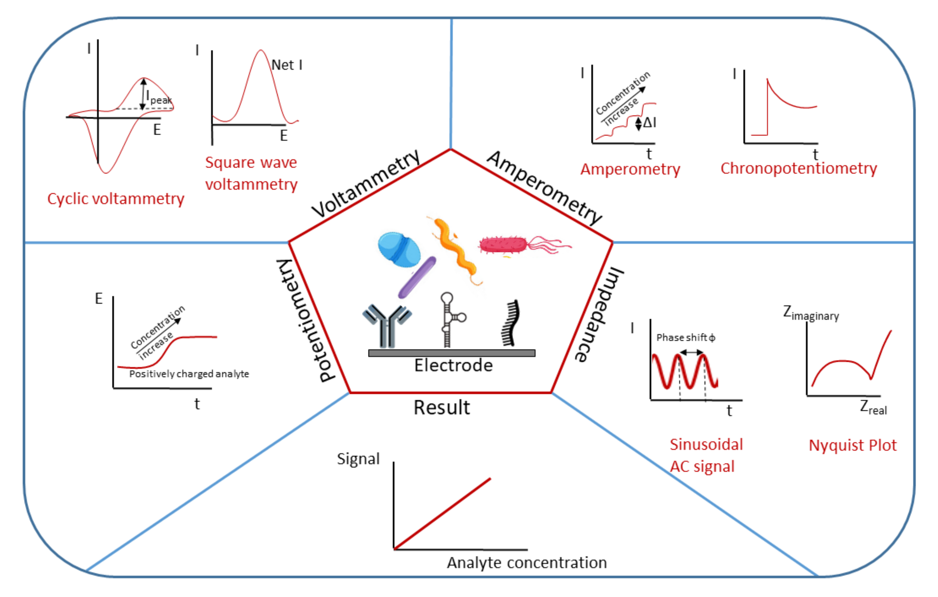
Figure 1. Electrochemical biosensors utilizing different methods (potentiometry, voltammetry, amperometry and electrochemical impedance) for analyte detection and concentration evaluation.
Nanomaterials 11 02700 g001
Figure 2. Illustration of some 0–3D nanostructured material morphologies.
Nanomaterials 11 02700 g002
Figure 3. Schematic presentation of the synthesis process of the ZrO2-Ag-Graphene Oxide -SiO2 nanocomposite. Adapted with permission from [46] Copyright 2020, American Chemical Society.
Nanomaterials 11 02700 g003
Figure 4. Schematic presentation of the fabrication of rGO-TiO2 electrodes and their employing for electrochemical detection of bacteria. Adapted with permission from [60] Copyright 2019, Elsevier.
Nanomaterials 11 02700 g004
Figure 5. Illustration of the detection of S. typhimirium using immunomagnetic separation and impedance monitoring of the release of Mn2+ from the SiO2@MnO2 nanocomposite. Adapted with permission from [65] Copyright 2020, Elsevier.
Nanomaterials 11 02700 g005
Figure 6. Scheme of a graphene chip and the process of PASE activation and antibody immobilization. Adapted with permission from [87] Copyright 2017, Elsevier.
Nanomaterials 11 02700 g006
Figure 7. Scheme of the strategy for the electrochemical detection of the Salmonella invA gene. Adapted with permission from [102] Copyright 2019, Elsevier.
Nanomaterials 11 02700 g007
Figure 8. Scheme of the development of an antibody-SWCNT bioconjugate paper-based electrochemical immunosensor. Adapted with permission from [107] Copyright 2017, Elsevier.
Nanomaterials 11 02700 g008
Figure 9. Schematic representation of the label-free electrochemical biosensor based on Au@MoS2–PANI. Adapted with permission from [128] Copyright 2021, MDPI.
Nanomaterials 11 02700 g009
Figure 10. The microfluidic device connectors and the eight magnets externally attached with double-sided tape over each working electrode, together with a schematic representation of detection. Adapted with permission from [134] Copyright 2018, Elsevier.
Nanomaterials 11 02700 g010
Figure 11. Design of a MoS2 based biosensor for S. typhimurium detection. Adapted with permission from [145] Copyright 2018, Elsevier.
Nanomaterials 11 02700 g011
Publisher’s Note: MDPI stays neutral with regard to jurisdictional claims in published maps and institutional affiliations.