
Enhancement of the Anti-Stokes Fluorescence of Hollow Spherical Carbon Nitride Nanostructures by High Intensity Green Laser


Abstract
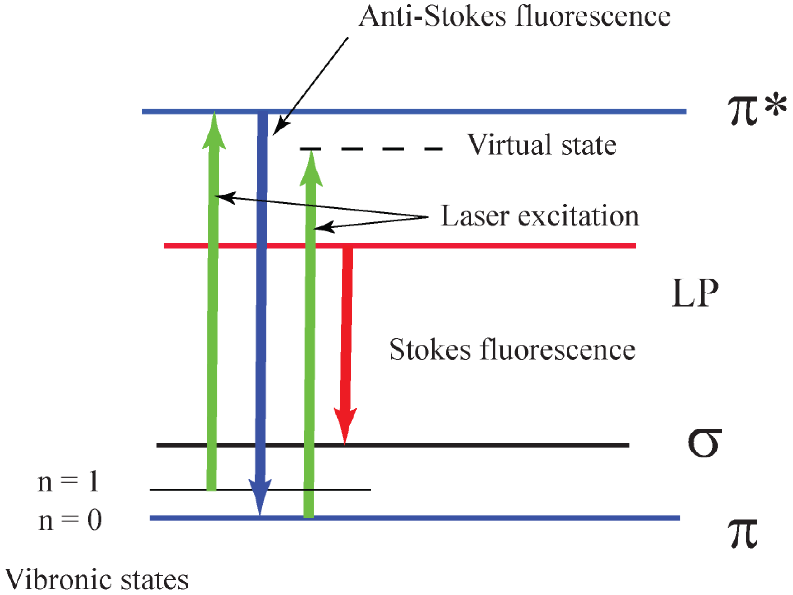
:1. Introduction
2. Materials and Methods
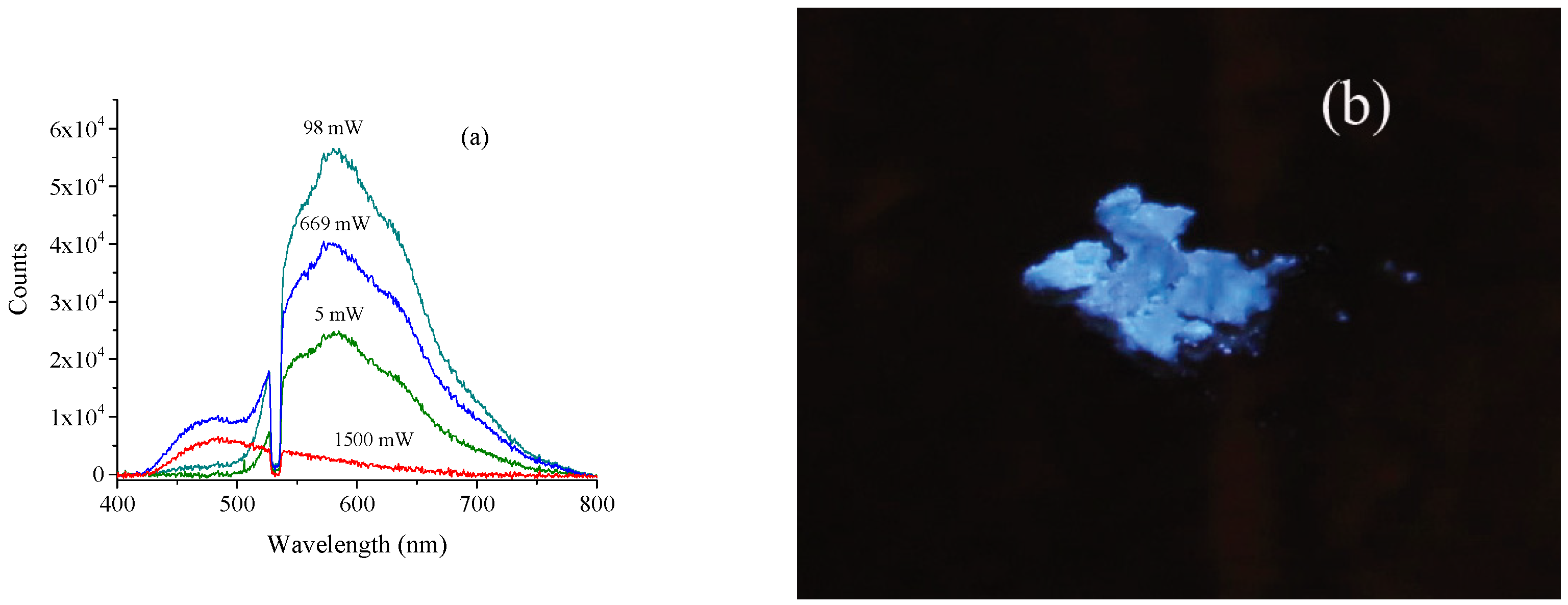
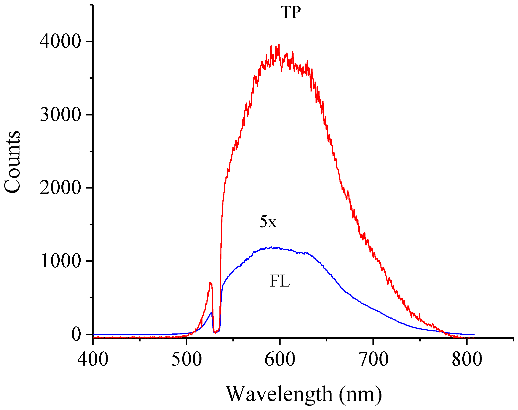
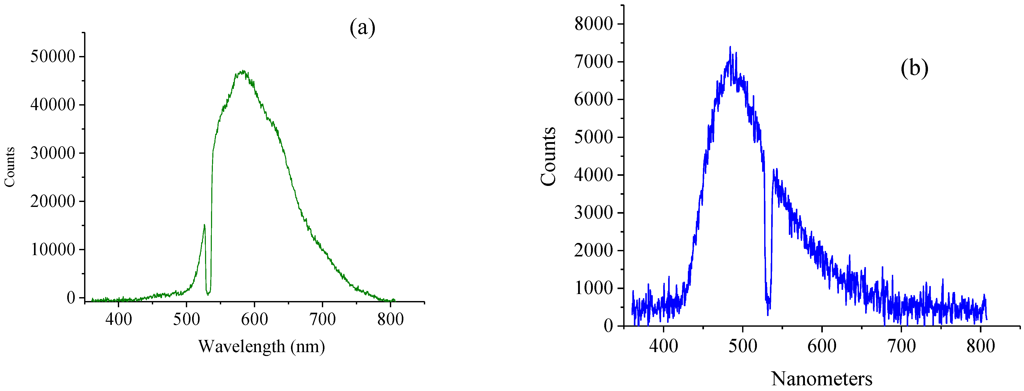
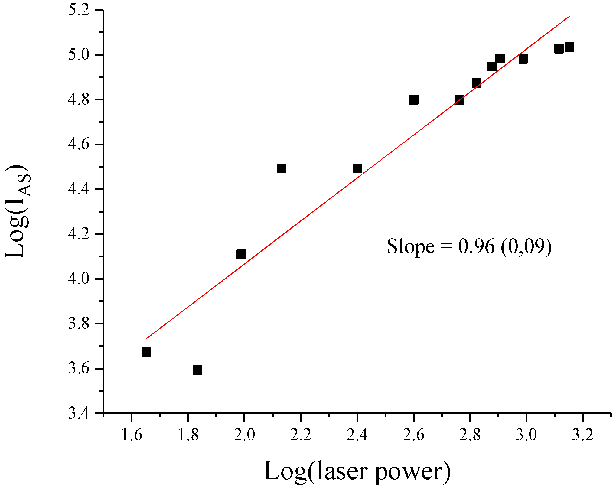
3. Results
4. Discussion
5. Conclusions
Author Contributions
Funding
Acknowledgments
Conflicts of Interest
References
- Wang, F.; Han, Y.; Lim, C.S.; Lu, Y.H.; Wang, J.; Xu, J.; Chen, H.Y.; Zhang, C.; Hong, M.H.; Liu, X.G. Simultaneous phase and size control of upconversion nanocrystals through lanthanide doping. Nature 2010, 463, 1061–1065. [Google Scholar] [CrossRef]
- Thomas, A.; Fischer, A.; Goettmann, F.; Antonietti, M.; Muller, J.O.; Schlogl, R.; Carlsson, J.M. Graphitic carbon nitride materials: Variation of structure and morphology and their use as metal-free catalysts. J. Mater. Chem. 2008, 18, 4893–4908. [Google Scholar] [CrossRef] [Green Version]
- Ai, B.; Duan, X.G.; Sun, H.Q.; Qiu, X.; Wang, S.B. Metal-free graphene-carbon nitride hybrids for photodegradation of organic pollutants in water. Catal. Today 2015, 258, 668–675. [Google Scholar] [CrossRef] [Green Version]
- Zinin, P.V.; Ming, L.C.; Sharma, S.K.; Khabashesku, V.N.; Liu, X.R.; Hong, S.M.; Endo, S.; Acosta, T. Ultraviolet and near-infrared raman spectroscopy of graphitic C3N4 phase. Chem. Phys. Lett. 2009, 472, 69–73. [Google Scholar] [CrossRef]
- Liu, S.; Tian, J.Q.; Wang, L.; Luo, Y.L.; Zhai, J.F.; Sun, X.P. Preparation of photoluminescent carbon nitride dots from ccl4 and 1,2-ethylenediamine: A heat-treatment-based strategy. J. Mater. Chem. 2011, 21, 11726–11729. [Google Scholar] [CrossRef]
- Dong, G.P.; Zhang, Y.H.; Pan, Q.W.; Qiu, J.R. A fantastic graphitic carbon nitride (g-C3N4) material: Electronic structure, photocatalytic and photoelectronic properties. J. Photochem. Photobiol. C-Photochem. Rev. 2014, 20, 33–50. [Google Scholar] [CrossRef]
- Dong, J.; Zhao, Y.L.; Wang, K.Q.; Chen, H.Y.; Liu, L.; Sun, B.L.; Yang, M.F.; Sun, L.P.; Wang, Y.; Yu, X.G.; et al. Fabrication of graphitic carbon nitride quantum dots and their application for simultaneous fluorescence imaging and ph-responsive drug release. ChemistrySelect 2018, 3, 12696–12703. [Google Scholar] [CrossRef]
- Zinin, P.V.; Ryabova, A.V.; Davydov, V.A.; Khabashesku, V.; Boritko, S.; Sharma, S.K.; Pominova, D.V.; Loshenov, V. Anomalous fluorescence of the spherical carbon nitride nanostructures. Chem. Phys. Lett. 2015, 633, 95–98. [Google Scholar] [CrossRef]
- Yuan, B.; Chu, Z.Y.; Li, G.Y.; Jiang, Z.H.; Hu, T.J.; Wang, Q.H.; Wang, C.H. Water-soluble ribbon-like graphitic carbon nitride (g-C3N4): Green synthesis, self-assembly and unique optical properties. J. Mater. Chem. C 2014, 2, 8212–8215. [Google Scholar] [CrossRef]
- Tran, T.T.; Regan, B.; Ekimov, E.A.; Mu, Z.; Zhou, Y.; Gao, W.B.; Narang, P.; Solntsev, A.S.; Toth, M.; Aharonovich, I.; et al. Anti-stokes excitation of solid-state quantum emitters for nanoscale thermometry. Sci. Adv. 2019, 5, eaav9180. [Google Scholar] [CrossRef] [Green Version]
- Djeu, N.; Whitney, W.T. Laser cooling by spontaneous anti-stokes scattering. Phys. Rev. Lett. 1981, 46, 236–239. [Google Scholar] [CrossRef]
- Eliseev, P.G. Anti-stokes luminescence in heavily doped semiconductors as a mechanism of laser cooling. Opto-Electron. Rev. 2008, 16, 199–207. [Google Scholar] [CrossRef]
- Kuzmin, A.N.; Baev, A.; Kachynski, A.V.; Fisher, T.S.; Shakouri, A.; Prasad, P.N. Anti-stokes fluorescence imaging of microscale thermal fields in thin films. J. Appl. Phys. 2011, 110, 5. [Google Scholar] [CrossRef] [Green Version]
- Zhang, X.D.; Wang, H.X.; Wang, H.; Zhang, Q.; Xie, J.F.; Tian, Y.P.; Wang, J.; Xie, Y. Single-layered graphitic-C3N4 quantum dots for two-photon fluorescence imaging of cellular nucleus. Adv. Mater. 2014, 26, 4438. [Google Scholar] [CrossRef]
- Yang, L.Y.; Wu, X.X.; Luo, L.; Liu, Y.; Wang, F. Facile preparation of graphitic-C3N4 quantum dots for application in two-photon imaging. New J. Chem. 2019, 43, 3174–3179. [Google Scholar] [CrossRef]
- Khabashesku, V.N.; Zimmerman, J.L.; Margrave, J.L. Powder synthesis and characterization of amorphous carbon nitride. Chem. Mat. 2000, 12, 3264–3270. [Google Scholar] [CrossRef]
- Zimmerman, J.L.; Williams, R.; Khabashesku, V.N.; Margrave, J.L. Preparation of sphere-shaped nanoscale carbon nitride polymer. Russ. Chem. Bull. 2001, 50, 2020–2027. [Google Scholar] [CrossRef]
- Zimmerman, J.L.; Williams, R.; Khabashesku, V.N.; Margrave, J.L. Synthesis of spherical carbon nitride nanostructures. Nano Lett. 2001, 1, 731–734. [Google Scholar] [CrossRef]
- Misra, A.K.; Sharma, S.K.; Acosta, T.E.; Porter, J.N.; Bates, D.E. Single-pulse standoff raman detection of chemicals from 120 m distance during daytime. Appl. Spectrosc. 2012, 66, 1279–1285. [Google Scholar] [CrossRef]
- Fanchini, G.; Tagliaferro, A.; Ray, S.C. Electronic and vibrational structures of amorphous carbon nitrides. Diam. Relat. Mater. 2003, 12, 208–218. [Google Scholar] [CrossRef]
- Fanchini, G.; Tagliaferro, A.; Conway, N.; Godet, C. Role of lone-pair interactions and local disorder in determining the interdependency of optical constants of a-cn: H thin films. Phys. Rev. B 2002, 66, 195415. [Google Scholar] [CrossRef]
- Gan, Z.X.; Shan, Y.; Chen, J.R.; Gui, Q.F.; Zhang, Q.Z.; Nie, S.P.; Wu, X.L. The origins of the broadband photoluminescence from carbon nitrides and applications to white light emitting. Nano Res. 2016, 9, 1801–1812. [Google Scholar] [CrossRef]
- Auzel, F. Upconversion and anti-stokes processes with f and d ions in solids. Chem. Rev. 2004, 104, 139–173. [Google Scholar] [CrossRef] [PubMed]
- Epstein, R.I.; Buchwald, M.I.; Edwards, B.C.; Gosnell, T.R.; Mungan, C.E. Observation of laser-induced cooling of a solid. Nature 1995, 377, 500–503. [Google Scholar] [CrossRef]
- Wang, F.; Liu, X.G. Recent advances in the chemistry of lanthanide-doped upconversion nanocrystals. Chem. Soc. Rev. 2009, 38, 976–989. [Google Scholar] [CrossRef]
- Cai, P.Q.; Huang, Y.L.; Seo, H.J. Anti-stokes ultraviolet luminescence and exciton detrapping in the two-dimensional perovskite (C6H5C2H4NH3)2PbCl4. J. Phys. Chem. Lett. 2019, 10, 4095–4102. [Google Scholar] [CrossRef]
Publisher’s Note: MDPI stays neutral with regard to jurisdictional claims in published maps and institutional affiliations. |
© 2021 by the authors. Licensee MDPI, Basel, Switzerland. This article is an open access article distributed under the terms and conditions of the Creative Commons Attribution (CC BY) license (https://creativecommons.org/licenses/by/4.0/).
Share and Cite
Zinin, P.V.; Acosta-Maeda, T.E.; Misra, A.K.; Sharma, S.K. Enhancement of the Anti-Stokes Fluorescence of Hollow Spherical Carbon Nitride Nanostructures by High Intensity Green Laser. Nanomaterials 2021, 11, 2529. https://doi.org/10.3390/nano11102529
Zinin PV, Acosta-Maeda TE, Misra AK, Sharma SK. Enhancement of the Anti-Stokes Fluorescence of Hollow Spherical Carbon Nitride Nanostructures by High Intensity Green Laser. Nanomaterials. 2021; 11(10):2529. https://doi.org/10.3390/nano11102529
Chicago/Turabian StyleZinin, Pavel V., Tayro E. Acosta-Maeda, Anupam K. Misra, and Shiv K. Sharma. 2021. "Enhancement of the Anti-Stokes Fluorescence of Hollow Spherical Carbon Nitride Nanostructures by High Intensity Green Laser" Nanomaterials 11, no. 10: 2529. https://doi.org/10.3390/nano11102529
APA StyleZinin, P. V., Acosta-Maeda, T. E., Misra, A. K., & Sharma, S. K. (2021). Enhancement of the Anti-Stokes Fluorescence of Hollow Spherical Carbon Nitride Nanostructures by High Intensity Green Laser. Nanomaterials, 11(10), 2529. https://doi.org/10.3390/nano11102529