Article

Investigation of Physicochemical Properties of the Structurally Modified Nanosized Silicate-Substituted Hydroxyapatite Co-Doped with Eu3+ and Sr2+ Ions

Institute of Low Temperature and Structure Research, Polish Academy of Sciences, Okolna 2, 50-422 Wroclaw, Poland
*
Authors to whom correspondence should be addressed.
Nanomaterials 2021, 11(1), 27; https://doi.org/10.3390/nano11010027
Submission received: 18 November 2020 / Revised: 10 December 2020 / Accepted: 16 December 2020 / Published: 24 December 2020

Abstract

:
In this paper, a series of structurally modified silicate-substituted apatite co-doped with Sr2+ and Eu3+ ions were synthesized by a microwave-assisted hydrothermal method. The concentration of Sr2+ ions was set at 2 mol% and Eu3+ ions were established in the range of 0.5–2 mol% in a molar ratio of calcium ion amount. The XRD (X-ray powder diffraction) technique and infrared (FT-IR) spectroscopy were used to characterize the obtained materials. The Kröger–Vink notation was used to explain the possible charge compensation mechanism. Moreover, the study of the spectroscopic properties (emission, emission excitation and emission kinetics) of the obtained materials as a function of optically active ions and annealing temperature was carried out. The luminescence behavior of Eu3+ ions in the apatite matrix was verified by the Judd–Ofelt (J-O) theory and discussed in detail. The temperature-dependent emission spectra were recorded for the representative materials. Furthermore, the International Commission on Illumination (CIE) chromaticity coordinates and correlated color temperature were determined by the obtained results.

Graphical Abstract

1. Introduction

Nanotechnology has exerted a considerable impact on a vast number of scientific fields in the last decade. Studies on the preparation and characterization of apatite materials have been influenced by this. Nanoapatites are the focus of great research interest due to their biocompatible and nontoxic properties to encourage bone and tissue filling. The structure of the apatite allows for various modifications, providing the opportunity to create a material with intentional and targeted properties. Moreover, two unequal calcium positions can be substituted by ions with +1, +2 or +3 charges, such as Sr2+, Ba2+, K+, Na+, Mn2+, Li+, Mg2+ as well as lanthanide ions, etc. [1,2,3].
There is particular interest in the characterization of nanoapatite materials doped with optically active ions. Consequently, it is possible to successfully replace Ca2+ ion by Eu3+ [1,4,5], Ce3+ [6], Tb3+ [7], Dy3+ [8,9], Nd3+ [9,10], Sm3+ [8,9] ions to obtain materials with characteristic emission in red [1,4], green [11,12], violet [6] as well as blue [6] spectral regions. Recently, the apatite matrix has been the focus of attention and investigated in terms of white light emission. Promising materials are co-doped with Dy3+, Li+ and Eu3+ [13] or La3+, Dy3+ and Sr3+ ions [8]. The vast number of possibilities and favorable results encourage the detailed investigation of a variety of apatite modifications.
Apatite-based structures can tolerate numerous ionic substitutions in order to improve their properties for medical application. Recent research has shown that the combination of silica and strontium co-doping may improve their properties for medical application [14]. In vivo observations as well as in vitro studies have exposed the beneficial effects of using silica-based materials for bone treatment. It has been shown that silica promotes prolyl hydroxylase, stimulates the enzyme involved in collagen synthesis and participates in the proliferation and differentiation of bone mesenchymal stem cells and osteoblasts [15,16]. The synergy of great luminescence properties with bioactivity may result in obtaining a new group of specific materials dedicated to bioimaging and regeneration.
In this paper, we show the physicochemical characterization of silicate-substituted hydroxyapatite co-doped with Sr2+ and Eu3+ ions. Considerable attention has been paid to the luminescence properties, including emission and excitation spectra depending on Eu3+ ion concentration and heat-treating temperature as well as the influence of ambient temperature.

2. Materials and Methods

2.1. Synthesis of the Co-Doped Materials

Synthesizing of silicate-substituted hydroxyapatite co-doped with Eu3+ and Sr2+ ions involved a hydrothermal process. As substrates, the following were used: Ca(NO3)2∙4H2O (99.0–103.0% Alfa Aesar, Haverhill, MA, USA), (NH4)2HPO4 (>99.0% Acros Organics, Schwerte, Germany), Eu2O3 (99.99% Alfa Aesar, Haverhill, MA, USA), Sr(NO3)2 (99.0% min Alfa Aesar, Haverhill, MA, USA) and tetraethyl orthosilicate TEOS (>99% Alfa Aesar, Haverhill, MA, USA). The concentration of strontium ions for all obtained materials was fixed to 2 mol% in a ratio of calcium ion molar content. Moreover, the concentration of optical active Eu3+ ions was set to 0.5; 1.0 and 2.0 mol% in a ratio to the calcium ion molar content. A stoichiometric number of substrates were dissolved separately in deionization water (see Table S1). A stoichiometric amount of Eu2O3 was digested in an excess of HNO3 (65% suprapure Merck KGaA, Darmstadt, Germany) to generate water-soluble Eu(NO3)3·xH2O. Afterwards, all starting substrates were added and mixed into a Teflon vessel. The ammonia solution (NH3∙H2O 25% Avantor, Poland) was used to obtain a pH level of around 10. The hydrothermal process was conducted in a microwave reactor (ERTEC MV 02-02, Wrocław, Poland) for 90 min at elevated temperature (250 °C) and under autogenous pressure (42–45 bar). The achieved materials were centrifuged, cleaned by deionization water several times and dried for 24 h. Then, the obtained materials were heat-treated in the temperature range of 400–600 °C for 3 h, increased in steps of 3.3 °C/min.

2.2. Physical–Chemical Characterization

X-ray powder diffraction studies were carried out using a PANalytical X’Pert Pro X-ray diffractometer (Malvern Panalytical Ltd., Malvern, UK) equipped with Ni-filtered Cu 1 radiation (1 = 1.54060 Å, U = 40 kV, I = 30 mA) in the 2θ range of 10°–70°. XRD patterns were analyzed by Match! software version 3.7.0.124.
The surface morphology and the element mapping were assessed by a FEI Nova NanoSEM 230 scanning electron microscope (SEM, Hillsboro, OR, USA) equipped with EDS spectrometer (EDAX GenesisXM4) and operating at an acceleration voltage in the range 3.0–15.0 kV and spots at 2.5–3.0 were observed. EDX analysis was carried out to confirm the chemical formula.
The Thermo Scientific Nicolet iS50 FT-IR spectrometer (Waltham, MA, USA) equipped with an Automated Beamsplitter exchange system (iS50 ABX containing DLaTGS KBr detector), built-in all-reflective diamond ATR module (iS50 ATR), Thermo Scientific Polaris™, was used to record the Fourier-transformed infrared spectra. As an infrared radiation source, we used the HeNe laser. FT-IR spectra of the powders were recorded in KBr pellets at 295 K temperature in the middle infrared range, from 4000 to 500 cm−1, with a spectral resolution of 2 cm−1.

2.3. Spectroscopy Properties

The emission, excitation emission spectra and luminescence kinetics were recorded by an FLS980 Fluorescence Spectrometer (Edinburgh Instruments, Kirkton Campus, UK) from Edinburgh Instruments equipped with a 450 W Xenon lamp and a Hamamatsu R928P photomultiplier. The hydroxyapatite powders were placed into a quartz tube. The excitation of 300 mm focal length monochromator was in Czerny–Turner configuration. All spectra were corrected during measurement according to the characteristics of the intensity of the excitation source. All spectra were recorded at room temperature. The spectral resolution of the excitation and emission spectra was 0.1 nm. Excitation spectra were recorded, monitoring the maximum of the emission at 618 nm that relates to the 5D07F2 transition, and emission spectra were recorded upon excitation wavelength at 394 nm. Before analysis, the emission spectra were normalized to the 5D07F1 magnetic transition. The luminescence kinetics profiles were recorded according to the 5D07F2 electric dipole transition.
Temperature-dependent emission spectra were recorded using the laser diode (λexc = 375 nm), and as an optical detector, we used the Hamamatsu PMA-12 photonic multichannel analyzer (Hamamatsu Photonics K.K., Hamamatsu City, Japan). The presented emission spectra are the average result of 15 measurements with an exposure time of 500 ms.

3. Results and Discussion

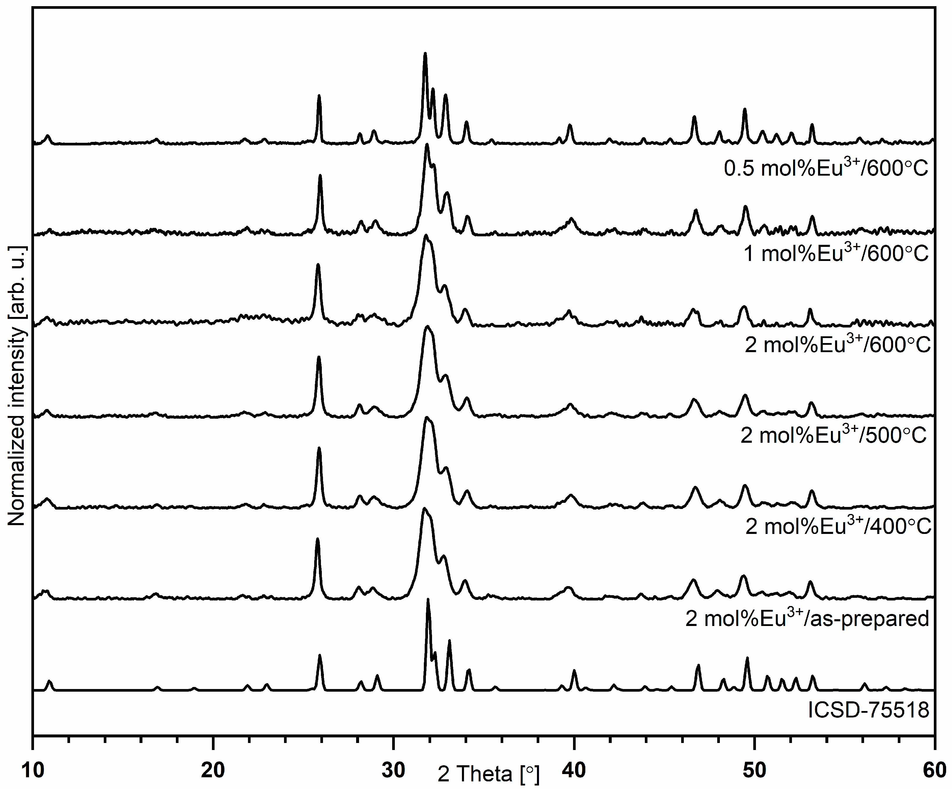
3.1. X-ray Diffraction

The formation of the silicate-substituted hydroxyapatite crystalline nanopowders was investigated by the XRD measurements and is shown in Figure 1 as a function of the concentration of optically active ions as well as heat-treating process. All samples prepared via the hydrothermal method have shown detectable crystallinity for the entire range of sintering temperatures (400–600 °C for 3 h). The presence of the single phase of the final products was confirmed by the reference standard of hexagonal strontium-substituted hydroxyapatite ICSD-75518 [17]. No other phase was detected in the studied powders, indicating that dopant ions were completely dissolved in the silicate-substituted host lattice.
Structural refinement was carried out using the Maud software version 2.93 [18,19] and was based on apatite crystals with a hexagonal structure using better approximations, as well as indexing of the crystallographic information file (CIF). The formation of the hexagonal phase, as well as the successful incorporation of Eu3+ and Sr2+ ions into the apatite lattice, was confirmed by the results. The average grain sizes of silicate-substituted hydroxyapatite nanopowders were in the range of 16 to 56 nm (see Figure S2 and Table S2). The representative SEM image is presented in Figure S1a. The effect of dopant ion substitution is confirmed by EDS measurements (see Figure S1b).
The most intense diffraction peaks corresponding to hydroxyapatite structures are located at 25.9° (002), 31.7° (211), 32.2° (112), 32.9° (300) and 34.0° (202), assigned to the crystallographic planes in brackets. In the obtained materials, Eu3+ and Sr2+ ions replaced Ca2+ ions. In the apatite host lattice, Ca2+ ions are located in two different sites with various chemical and structural environments, Ca(1) and Ca(2) sites with C3 and CS symmetry, respectively. The Ca(1) site is surrounded by nine oxygen atoms coming from PO43− groups, which formed a tricapped trigonal prism with formula CaO9. The Ca(2) site is an irregular polyhedron with formula CaO6OH formed by six oxygen atoms from PO43− and one hydroxyl group [3,20]. The difference between the ionic radii of trivalent europium and divalent strontium ions permit the occupation of two possible crystallographic positions of Ca2+ ions in the apatite host lattice (Ca2+ (CN9) = 1.18 Å, Eu3+ (CN9) = 1.12 Å, Sr2+ (CN9)−1.31 Å (Ca(1) site); Ca2+ (CN7) = 1.06 Å and Eu3+ (CN7) = 1.01 Å, Sr2+ (CN7)−1.21 Å (Ca(2) site)) [5,21].

3.2. Kröger–Vink Notation

The cationic vacancies formed by replacing Ca2+ ions with Eu3+ ions with higher charge could be balanced by SiO44− ions substituted into the PO43− position in the silicate-substituted apatite matrix. The occupation of divalent calcium ion sites by trivalent europium ions could be described according to the Kröger–Vink notation by the charge compensation phenomenon. According to this theory, the total charge in the material should be compensated by the creation of relatively positive or negative charge. The following processes may be observed:
A double negative vacancy on the Ca2+ position (V″Ca) is created by the substitution of divalent calcium ions by trivalent europium ions (Equation (1)):
Eu2O3 + 3Ca*Ca → 2Eu·Ca + 3CaO + V″Ca
The substitution of divalent calcium ions by trivalent rare earth ions could be explained by the creation of interstitial oxygen Oi with double relative negative charge. The mechanism could be described as follows (Equation (2)):
Eu2O3 + 2Ca*Ca → 2Eu∙Ca + 2CaO + Oi
Eu3+ ions first replace into the Ca(1) site and this preference changes with increasing Eu3+ concentration in favor of the Ca(2) site. In case of substitution into the Ca(2) site, where calcium(II) ions are surrounded with one hydroxyl group and six oxygen atoms from PO43−, these hydroxyl groups could participate in the charge compensation mechanism, expressed as (Equation (3)):
Eu2O3 + 2Ca*Ca(2) + 2OH*OH → 2Eu∙Ca(2) + 2OOH + 2CaO + H2O
In the case of the obtained materials, the substitution of the PO43− group by the more negative SiO44− group could create a negative charge on the PO43− position and two positive charge vacancies (VCa) on the hydroxyl group position. The mechanism can be expressed as follows (Equation (4)):
2SiO2 + 2PO4*PO4 + 2OH*OH → 2SiO4′PO4 + 2V∙OH + P2O5 + H2O
In the present study, the charge compensation mechanism could be described as a combination of Equations (1)–(4). Equation (5) combines the creation of negative and positive vacancy, because of Ca2+ substitution by Eu3+ and PO43− substitution by SiO44−, respectively.
2SiO2 + Eu2O3 + 2Ca*Ca + 2PO4*PO4 → 2Eu∙Ca + 2SiO4′PO4 + 2 CaO + P2O5

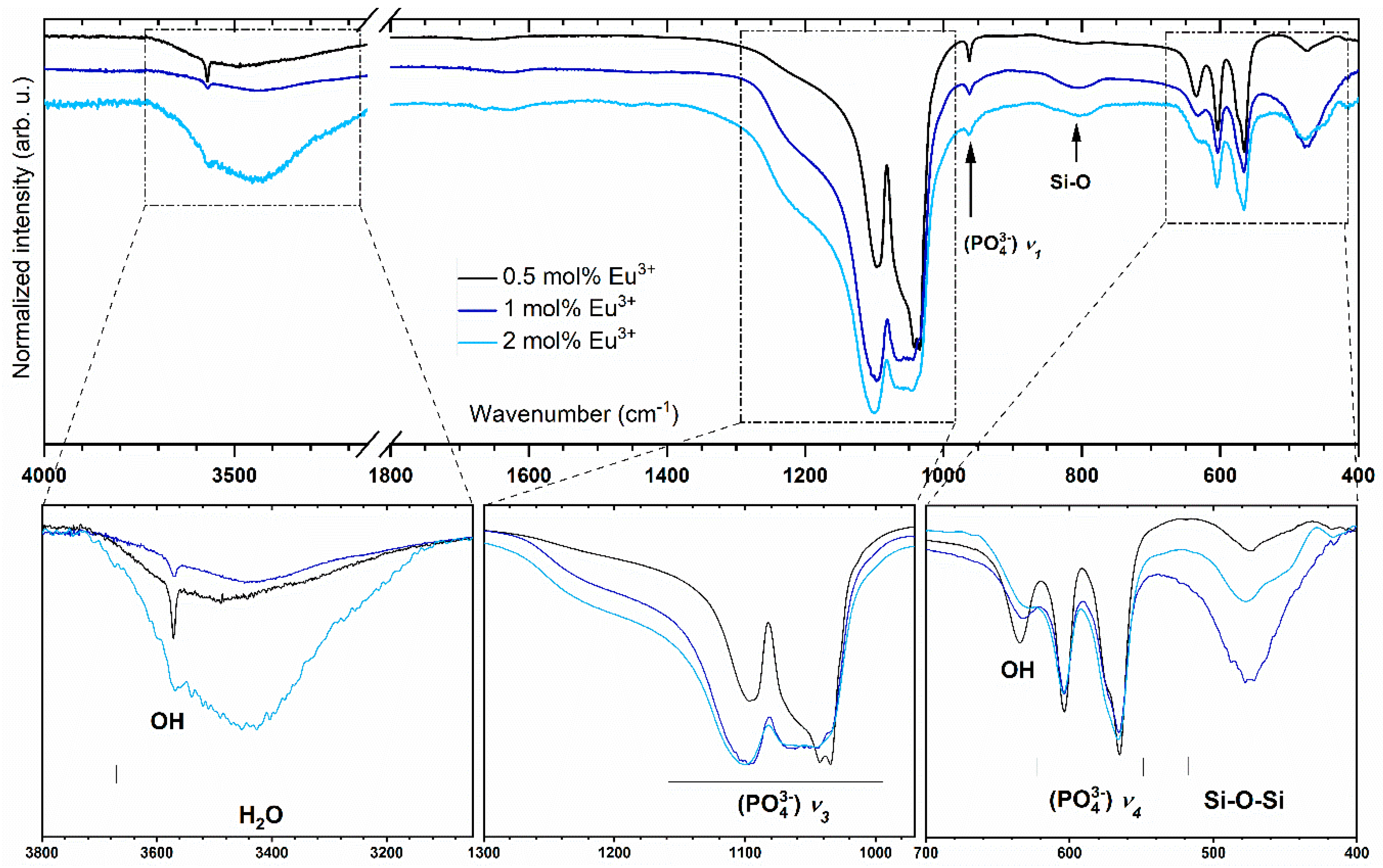
3.3. Infrared Spectra

To confirm the presence of phosphate, silicate and hydroxyl groups, the infrared spectra were measured and are presented in Figure 2. According to previous reports, characteristic peaks are ascribed to the compound of hydroxyapatite [3,22,23]. The triply degenerated antisymmetric stretching vibration ν3(PO43−) of phosphate groups is observed at 1101.1 and 1049.1 cm−1. At 966.4 cm−1, lines are detected which can be described as non-degenerated symmetric stretching bands ν1(PO43−) vibrations. Strong absorption bands associated with the ν4(PO43−) triply degenerated vibrations are located at 566.2 and 604.8 cm−1. Two bands related to the stretching and bending modes of OH groups are observed at 3571.3 and at 604.5 cm−1, respectively. These bands clearly confirm the presence of hydroxyl groups in the crystal structure. The broad peak between 3600 and 3200 cm−1 belongs to H2O vibration. Peaks assigned to (SiO4)4− vibrational modes and Si–O–Si stretch modes are observed at 890 and 478 cm−1, respectively. It should be pointed out that there are similarly located vibrational modes of the (PO4)3− and the silicate groups in the hydroxyapatite matrix, causing some interpretation problems. The Si–O symmetric stretching mode is located at 945 cm−1 and the weak peak corresponding to the P–O symmetric stretching mode is located at 962 cm−1 [23].

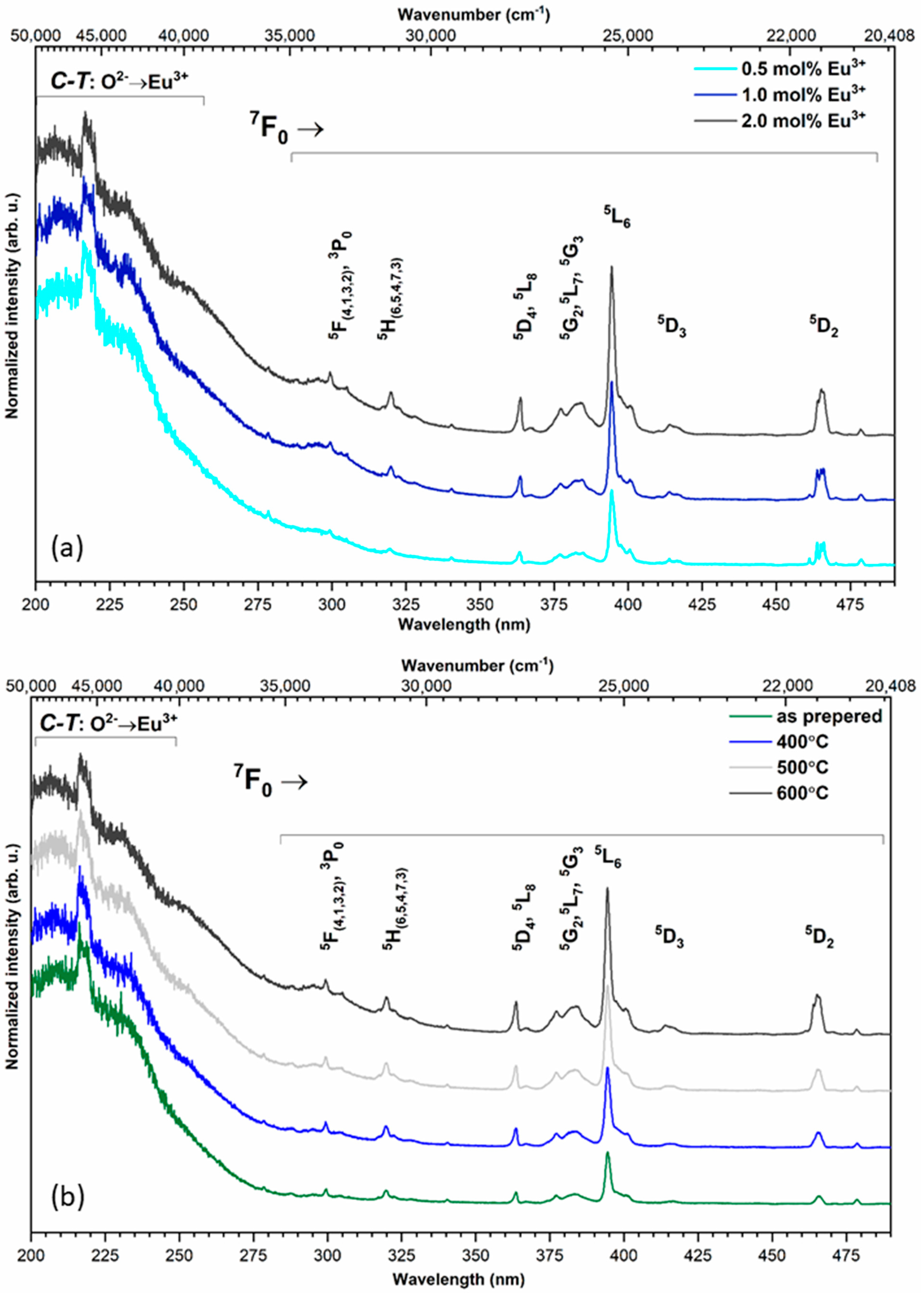
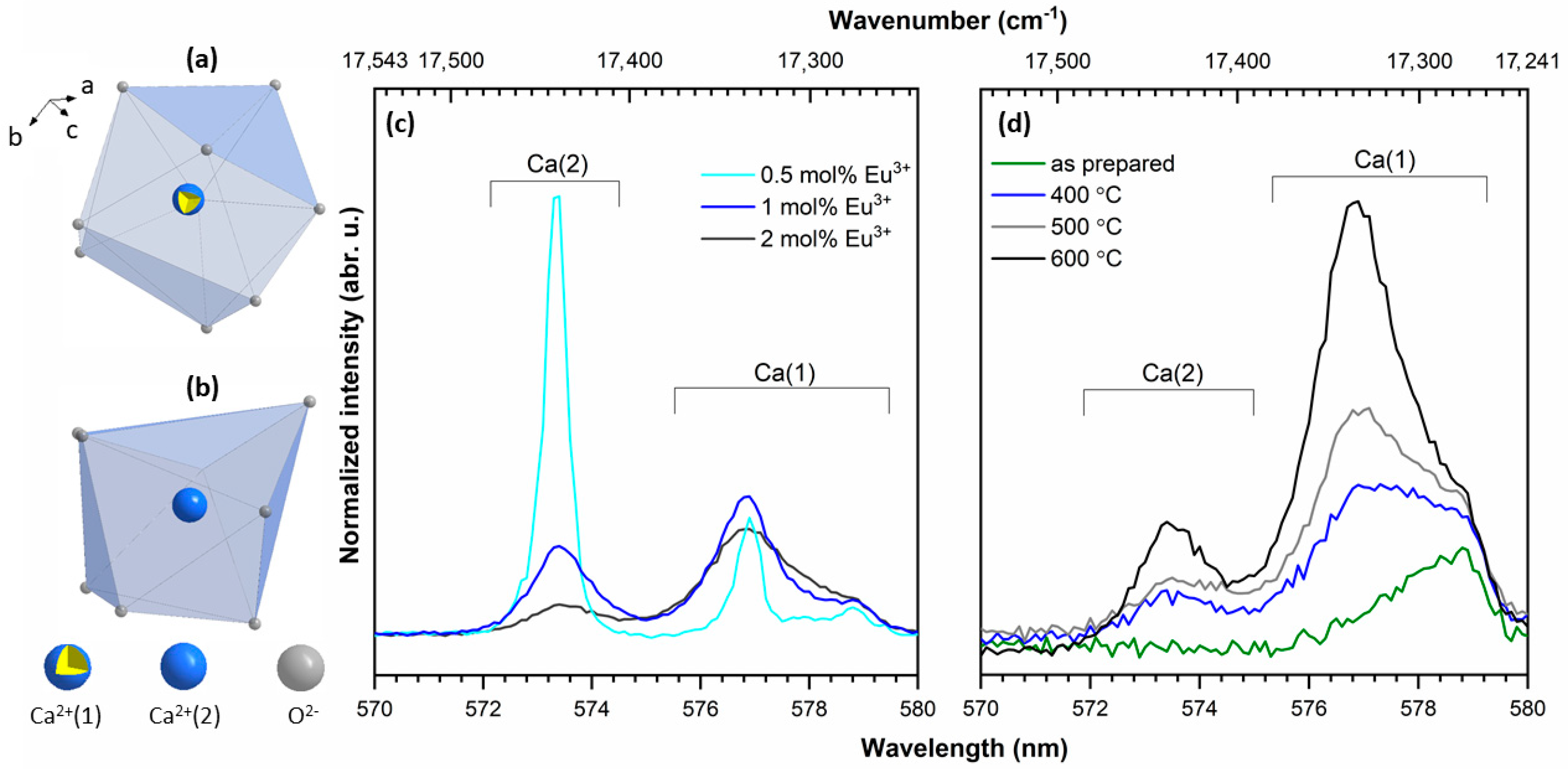
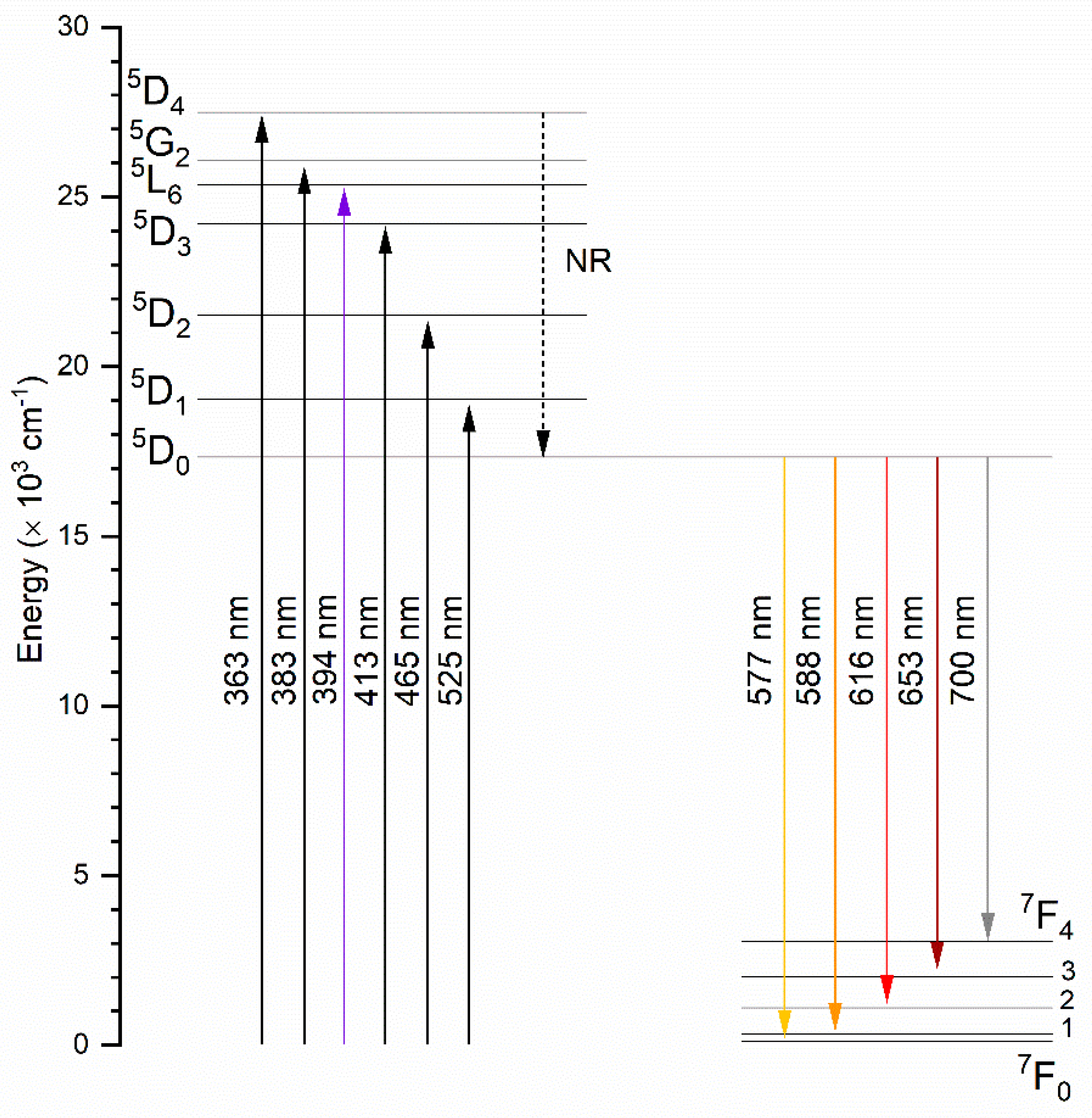
3.4. Spectroscopy Properties

The emission excitation spectra of the silicate-substituted appetites were recorded as a function of the europium ion concentration and heat-treated temperature in Figure 3a,b, respectively. The spectra were recorded at 300 K at an observation wavelength of 616 nm (16,233 cm−1). The presented spectra were normalized to the most intensity bands. In relation to the most intense band of the 5D07F2 transition, the spectra were recorded at an observation wavelength of 618 nm. In the UV range was observed a broad, intense band ascribed to the O2− → Eu3+ charge transfer (CT) transition with a maximum located around 205 nm (48,780 cm−1). Increasing the dopant concentration and heat-treating temperature does not have an influence on the CT maximum position. In the composition of the excitation spectra were recorded sharp, narrow bands, attributed to the 4f–4f transitions of Eu3+ ions, at: 7F05F(4,1,3,2), 3P0 at 299.3 nm (33,411 cm−1), 7F05H(6,5,4,7,3) at 319.8 nm (31,269 cm−1), 7F05D4, 5L8 at 363.7 nm (27,495 cm−1), 7F05G2, 5L7, 5G3 at 383.7 nm (26,062 cm−1), 7F05L6 at 394.4 nm (25,354 cm−1), 7F05D3 at 413.8 nm (24,166 cm−1) and 7F05D2 at 465.1 nm (21,500 cm−1). In lanthanide ions, strongly isolating the 4f orbitals by the external 5s, 5p and 5d shells causes only slight changes in the positions of the electronic transition bands.
Figure 4a represents the emission spectra for the 2 mol% Eu3+-doped sample as a function of sintering temperature. Figure 4b shows the emission spectra as a function of optically active ion concentration for samples sintered at 600 °C. The emission spectra were detected at an excitation wavelength of 394 nm to directly excite the f electrons of Eu3+ ions. All spectra were normalized according to the 5D07F1 magnetic transition. As should have been expected, the emission transitions of the Eu3+ ions are forbidden by selection rules but do not consider the subtle influence of atom vibrations which consequently change the dipole moment, causing the occurrence of forbidden transitions on the spectrum. In the spectra are presented emission lines due to the 5D07FJ transitions for J = 0, 1, 2, 3 and 4, typical of Eu3+ ions, which are ascribed in reference to previous reports [5,24,25,26,27]. The bands located at the listed wavelength were attributed to the following transition: 5D07F0 at 577 nm (17,331 cm−1); 5D07F1 at 588 nm (17,006 cm−1); 5D07F2 at 616 nm (16,233 cm−1); 5D07F3 at 653 nm (15,313 cm−1) and 5D07F0 at 700 nm (14,285 cm−1). In general, the spectroscopic properties can provide information about the local chemical environment of Eu3+ ions. The essential change in emission spectra can be dependent on the quantity of possible crystallographic positions and therefore the amount of potential sites of substitution, as well as the presence of defects, additional phases or impurities [3,27].
The typical emission spectrum of 4f–4f electrons of Eu3+ ions is recorded in the red range of the electromagnetic radiation spectrum. In this range, the 5D07F0,1,2 transitions are observed and the shape of the lines in this region is correlated with the structural properties of the material. Due to these transitions, the trivalent europium ion is called an optical probe. If the Eu3+ ion is located in a centrosymmetric crystal lattice, the 5D07F0 transition is not observed. In the contrary situation, if the Eu3+ ion is placed in a non-centrosymmetric lattice, the 5D07F0 transition is detected on the emission spectra. Moreover, this is possible only in the low-symmetry crystal position as in Cn, Cnv as well as CS symmetry. In the hydroxyapatite matrix, calcium ions are located at two different crystal positions: Ca(1) and Ca(2) with local symmetry at C3 and CS, respectively. Both calcium positions could be occupied by Eu3+ ions. The additional crystallographic position appears as a result of reverse cis and trans symmetry of the Ca(2) site with the same point symmetry. In the environment of the Ca(1) site, nine oxygen atoms from phosphate groups are present. The calcium ion located on the Ca(2) site is in sevenfold coordination with six oxygen atoms from the phosphate group and one from the hydroxyl group [28]. The number and ratio of the intensity of the lines in the range between 570 and 580 nm give information about Eu3+ ion site-occupied preference. The coordination polyhedra of the Ca(1) and Ca(2) cations are presented in Figure 5a,b, respectively.
Emission spectra recorded for the investigated samples in the range of 570 to 580 nm are presented in Figure 5. In the emission spectra of 2 mol% Sr2+ and 0.5 mol% Eu3+ co-doped silicate-substituted hydroxyapatites, the transition attributed to 5D07F0 presents some interesting features due to abnormally strong intensity. This fact can be related to an important perturbation of the symmetry of the dopant induced by the introduction of silicate groups. Moreover, the abnormally strong intensity of the 5D07F0 transition has been reported in apatites such as oxyapatite Ca10(PO4)6O2, fluoroapatite Ca5(PO4)3F, hydroxyapatite Sr10(PO4)6(OH)2, or silicophosphate apatite Sr5(PO4)2SiO4, etc. This intense emission is attributed to the existence of the strong covalence of the Eu3+-O2− bond in the Ca(2) site in the apatite lattice [28,29,30].
In the other cases, the population of the Ca2+ position replaced by the Eu3+ ion could be related to the thermal diffusion process of dopant ions in the apatite structure. It is commonly known that in the case of as-prepared apatite materials, only the emission associated with one type of site with C3 symmetry was observed, whereas with an increase in the calcination temperature, additional 0–0 peaks appeared [1,31,32]. The emission intensity ratio Ca(1)/Ca(2) has been calculated and results are presented in Table 1, showing values from 2.4 for as-prepared samples to 3.1 for sintered samples at 600 °C. Taking into account all results, one can note that in the silicate-substituted strontium-doped hydroxyapatite matrix, the Ca(1) site is more occupied by Eu3+ ions.
To obtain additional insight into the luminescence behavior of Eu3+ ions in silicate-substituted strontium co-doped apatite, the Judd–Ofelt theory was applied [33,34]. The results of the calculation of radiative (Arad), non-radiative (Anrad) and total (Atot) processes, as well as intensity parameters (Ω2, Ω4), quantum efficiency (η) and asymmetry ratio (R), are presented in Table 1. These parameters were calculated based on emission spectra and decay profiles according to equations defined in previous reports [35,36].
A comparison of the Judd–Ofelt intensity parameters (Ω2 and Ω4) is made between the samples with different Eu3+ ion concentrations and sintering temperatures. An increase in the Ω2 value is noted with an increase in the heat-treating temperature. This result indicates the increasingly hypersensitive character of the 5D07F2 transition and the increasing polarization of the Eu3+ ion environment. Consequently, Eu3+ ions in the material heat-treated at the highest temperature are in a more polarizable environment. The influence of silicon group presence is analyzed in comparison with previous work by our group [37]. A decrease in the J-O Ω2 parameter is noted from 6.683 × 10⁻²⁰ cm2 (see [37]) to 4.506×10⁻²⁰ cm2 (see Table 1) for Ca9.7Sr0.2Eu0.1(PO4)6(OH)2 and for Ca9.7Sr0.2Eu0.1(PO4)2(SiO4)4(OH)2, respectively. This observation can be related to the improvement of the Eu3+ cation polyhedral and to the decrease in the covalence character of the Eu3+–O2− bond in silicate-substituted apatite. In the case of the investigated samples, the Ω2 > Ω4 parameter suggests that Eu3+ ions are not located in the local symmetry of centrosymmetric character. The calculated results are in agreement with previous reports regarding apatite systems [1,24,35,37].
The highest quantum efficient (η) is observed for the 1 mol% Eu3+-doped compound with the value of 40%. The quantum efficient is reduced by the increase as well as the decrease of optically active ion concentration to 37% and 34%, respectively. Samples heat-treated at higher temperatures demonstrate higher quantum efficient values, and the η parameter increases by the maximum of 15%.
The luminescence intensity ratio (R) of the electric dipole transition 5D07F2 to the magnetic dipole transition 5D07F1 has been calculated and the results are presented in Table 1. As expected, the sintering process and concentration of optical active ions have a significant influence on the symmetry of the Eu3+ ion environment in the apatite matrix. The R factor increases with an increase in the heat-treating temperature, which indicates lower symmetry around Eu3+ ions and suggests that the covalence of Eu3+-O2− is higher. The R parameter increases for 0.5 mol% Eu3+ to 1.0 mol% Eu3+ from 3.1 to 4.5, respectively. Then, the opposite trend is observed, and the R parameter decreases from 4.5 to 4.1 for 1.0 mol% Eu3+ and 2.0 mol% Eu3+.
Figure 6 presents the energy level diagram of Eu3+ corresponding to the detected excitation and emission spectra. As seen, Eu3+ ions were pumped to upper excited levels. The emission bands from the 5D1, 5D2 and 5D3 levels are not observed at room temperature in the case of the investigated samples, which suggests a fast, non-radiative (NR), multiphonon relaxation from the excited state 5L6 to the 5D0 state.

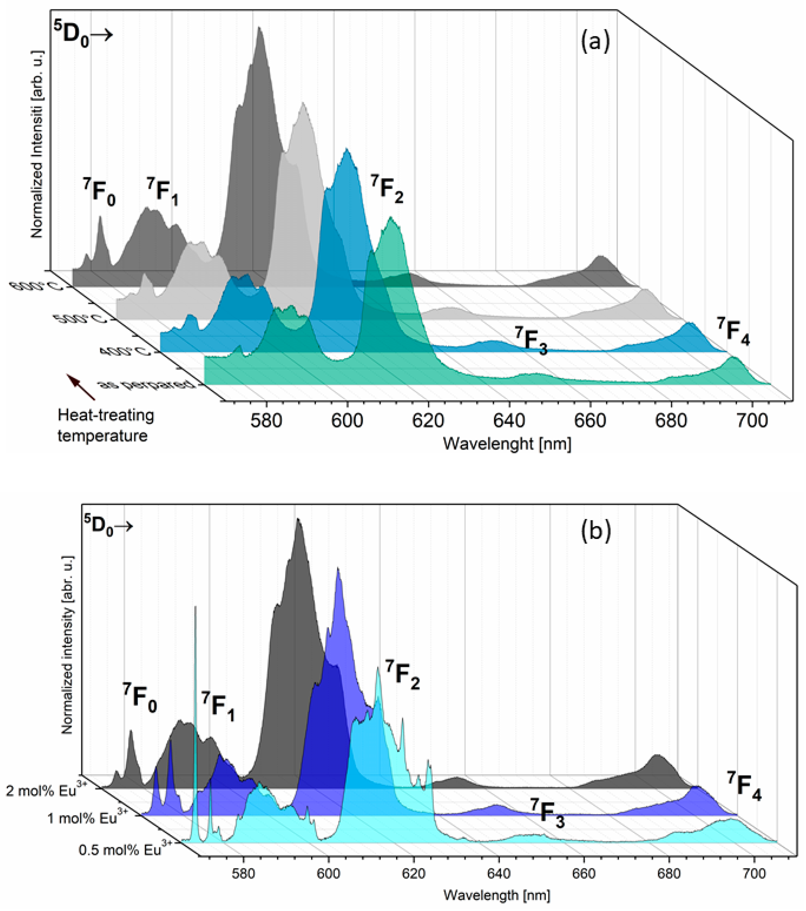
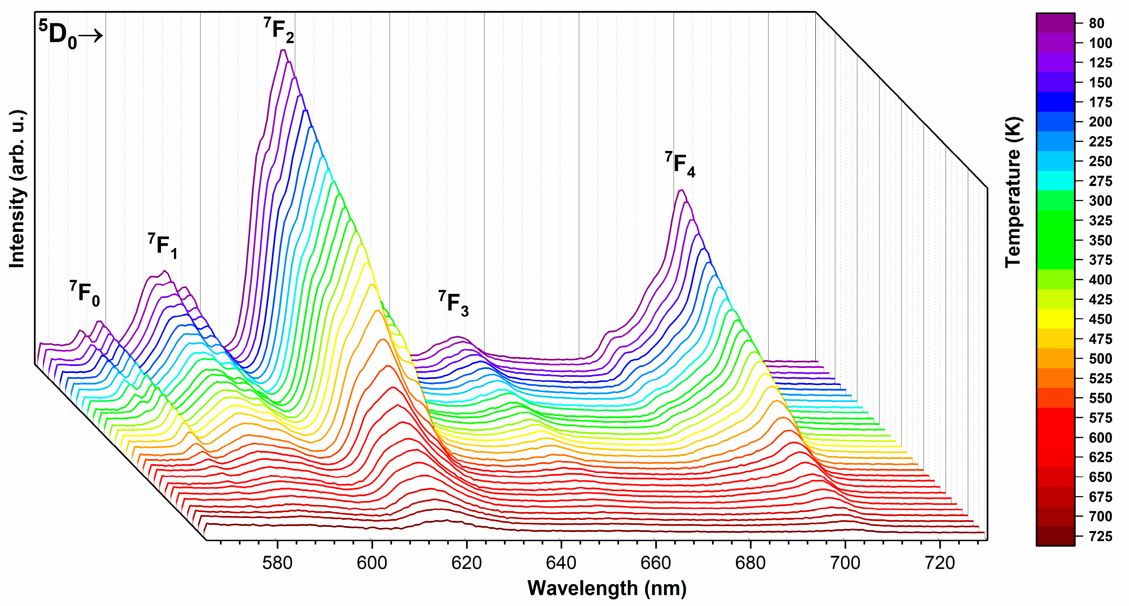
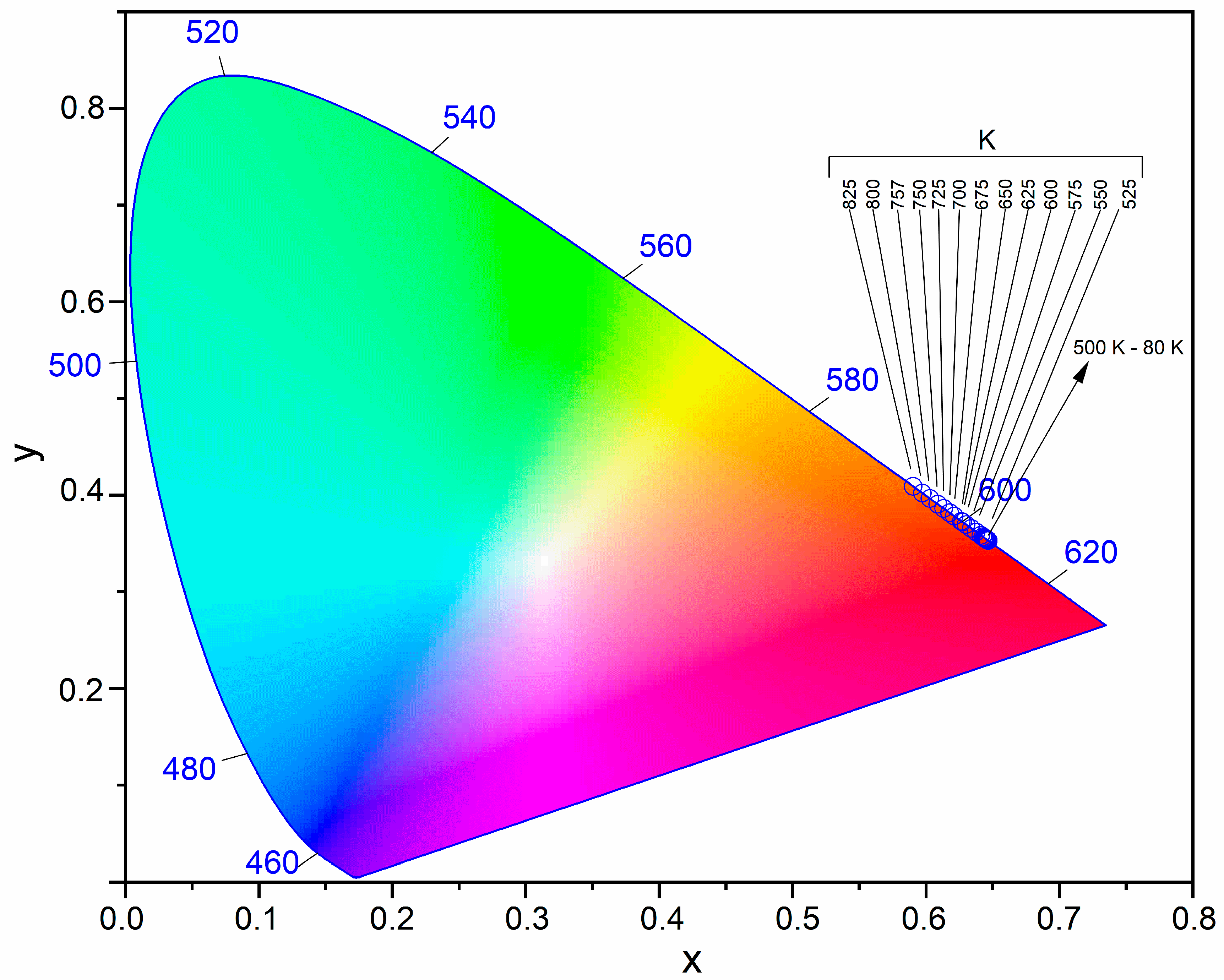
3.5. Temperature-Dependent Emission

To further study the possible application under high temperature, the temperature-dependent emission spectra of the silicate-substituted hydroxyapatite co-doped with 2 mol% Sr2+ and 2 mol% Eu3+ were measured and are presented in Figure 7. Emissions were recorded in the range of 80 to 725 K and at an excitation wavelength of 375 nm into the 5G2 level. It is seen that the emission intensity decreases clearly with an increase in the ambient temperature, but the decrease is not linear in the whole range of measured temperatures. For the most intense line corresponding to the 5D07F2 transition, the linear relationship applies between 80 and 325 K (R2 = 99.4%). Then, between 450 and 800 K, the decrease can be described by the exponential equation (see Figure S3). The line corresponding to the 5D07F0 transition of the Ca(2) calcium site (573 nm) is completely eliminated at 350 K.
In Figure 8 has been shown the Commission Internationale de l’Eclairage (CIE) 1931 chromaticity diagram for the Ca9.6Sr0.2Eu0.2(PO4)2(SiO4)4(OH)2 sample. The CIE color coordinates are listed in Table S3, which are calculated from the temperature-dependent emission spectra [38]. It has been reported that the emission color changed from reddish-orange to orange with the increasing of ambient temperature, but the reddish-orange emission was stable until 750 K. These results show that the Eu3+-activated silicate-substituted apatites have the potential to be color-stable materials capable of operating at a wide range of ambient temperatures.

3.6. Decay Time

The luminescence kinetics corresponding to the 5D07F2 transition were obtained at room temperature. As expected, all the recorded decays presented non-single exponential character. This phenomenon was consistent with the existence of two non-equivalent Eu3+ positions and, because of this fact, the effective emission lifetime was calculated by Equation (6). The recorded decays and calculated luminescence lifetimes (τ) are presented in Figure 9, as a function of Eu3+ concentration (a) and (b) as well as heat-treating temperature ((c) and (d)), respectively.
τ m = 0 t I ( t ) d t 0 I ( t ) d t 0 t m a x t I ( t ) d t 0 t m a x I ( t ) d t

4. Conclusions

In this study, it has been shown for the first time that a series of silicate-substituted hydroxyapatite co-doped with 2 mol% Sr2+ and Eu3+ ions in the range of 0.5–2.0 mol % in a ratio to the entire Ca2+ ion content were successfully synthesized by the hydrothermal method assisted with microwave and heat-treated. The average crystal sizes of the studied materials were in the range of 16–56 nm as calculated by the Rietveld method.
Attention was paid to the structural and spectroscopic properties related to a variable amount of Eu3+ ion concentration. The spectroscopic properties have shown for the sintered samples that Eu3+ ions occupied three independent crystallographic sites: one Ca(1) site with C3 local symmetry and two Ca(2) sites with Cs local symmetry with cis and trans symmetry. The 5D07F2 hypersensitive transition is the most intense for most obtained materials, excluding the sample co-doped with 0.5 mol% Eu3+ ion and sintered at 600 °C, where the most dominant is 5D07F0 transition. Moreover, the charge compensation mechanism in the materials induced by the substitution of Eu3+ and Sr2+ ions into the silicate-substituted hydroxyapatite host lattice was rendered in the Kröger–Vink notation.
The luminescence decay times corresponding to the most intense 5D07F2 transition were recorded. The luminescence kinetics was characterized by a non-exponential decay profile and was in the range of 2.15 ms (0.5 mol% Eu3+) and 19.4 ms (1 mol% Eu3+) to 1.92 ms (2 mol% Eu3+) for the samples sintered at 600 °C. On the other hand, the decay times for the samples doped with 2 mol% Eu3+ as a function of sintered temperature were in the range of 15.2 to 1.92 ms. The typical modes of the vibrations of the silicate-substituted hydroxyapatite ion group were detected in the FT-IR spectra, and these included the OH group vibrations characteristic of the hydroxyapatite matrix. The simplified Judd–Ofelt theory was used for a detailed analysis of the luminescence spectra. The hydroxyapatite containing 1 mol% of Eu3+ ions was evaluated to be the most optically efficient material among all the studied silicate-substituted hydroxyapatites. The International Commission on Illumination (CIE) color coordinates showed that the emission color can be tuned by varying the ambient temperature. The emission color was changed from reddish-orange to orange.

Supplementary Materials

The following are available online at https://www.mdpi.com/2079-4991/11/1/27/s1, Figure S1: The representative SEM image (a) and EDS spectra (b) of the Ca9.6Sr0.2Eu0.2(PO4)2(SiO4)4(OH)2 nanopawders. Figure S2: Representative results of the of the Sr0.2Eu0.2Ca9.6(PO4)2(SiO4)4(OH)2, obtained at 600 °C, Rietveld analysis (red—fitted diffraction; blue—differential pattern; column—reference phase peak position). Figure S3: Temperature-dependent emission intensity of the lines correspond to the listed transitions. Table S1. The amount of substrates used for synthesis of silicate-substituted hydroxyapatite co-doped wth Eu3+ and Sr2+. Table S2: Unit cell parameters (a,c), cell volume (V), grain size as well as refine factor (RW) for the Ca10(PO4)2(SiO4)4(OH)2 co-doped with 2 mol% Sr2+ and x mol/% Eu3+ ions (where x = 0.5–2). Table S3: The comparison of the CIE color coordinates (x,y) of Ca9.6Sr0.2Eu0.2(PO4)2(SiO4)4(OH)2 as a function of ambient temperature.

Author Contributions

R.J.W. conceived and designed the experiments and contributed reagents/materials/analysis tools and participated in funding acquisition in addition to analyzing all data; S.T. designed the experiments in addition to analyzing data. Both authors have read and agreed to the published version of the manuscript.

Funding

The authors would like to acknowledge financial support from the National Science Centre (NCN) within the project “Preparation and characterisation of biocomposites based on nanoapatites for theranostics” (No. UMO-2015/19/B/ST5/01330).

Institutional Review Board Statement

Not applicable.

Informed Consent Statement

Not applicable.

Data Availability Statement

Not applicable.

Acknowledgments

We are grateful to E. Bukowska for the help with XRD measurements and to D. Szymanski for SEM measurements.

Conflicts of Interest

The authors declare no conflict of interest.

References

  1. Zawisza, K.; Strzep, A.; Wiglusz, R.J. Influence of annealing temperature on the spectroscopic properties of hydroxyapatite analogues doped with Eu3+. New J. Chem. 2017, 41, 9990–9999. [Google Scholar] [CrossRef]
  2. Guo, Y.; Moon, B.K.; Choi, B.C.; Jeong, J.H.; Kim, J.H.; Choi, H. Fluorescence properties with red-shift of Eu2+ emission in novel phosphor-silicate apatite Sr3LaNa(PO4)2SiO4 phosphors. Ceram. Int. 2016, 42, 18324–18332. [Google Scholar] [CrossRef]
  3. Zawisza, K.; Wiglusz, R.J. Preferential site occupancy of Eu3+ ions in strontium hydroxyapatite nanocrystalline- Sr10(PO4)6(OH)2 -structural and spectroscopic characterisation. Dalton Trans. 2017, 46, 3265–3275. [Google Scholar] [CrossRef]
  4. Pogosova, M.A.; Eliseev, A.A.; Kazin, P.E.; Azarmi, F. Synthesis, structure, luminescence, and color features of the Eu- and Cu-doped calcium apatite. Dye Pigment. 2017, 141, 209–216. [Google Scholar] [CrossRef]
  5. Sokolnicki, J.; Zych, E. Synthesis and spectroscopic investigations of Sr2Y8(SiO4)6O2:Eu2+,Eu3+ phosphor for white LEDs. J. Lumin. 2015, 158, 65–69. [Google Scholar] [CrossRef]
  6. Xia, Z.; Molokeev, M.S.; Im, W.B.; Unithrattil, S.; Liu, Q. Crystal structure and photoluminescence evolution of La5(SxB1-x)(O13-xNx):Ce3+ solid solution phosphors. J. Phys. Chem. C 2015, 119, 9488–9495. [Google Scholar] [CrossRef]
  7. Sobierajska, P.; Wiglusz, R.J. Influence of the grain sizes on Stokes and anti-Stokes fluorescence in the Yb3+ and Tb3+ ions co-doped nanocrystalline fluorapatite. J. Alloy. Compd. 2019, 785, 808–818. [Google Scholar] [CrossRef]
  8. Liu, N.; Mei, L.; Liao, L.; Fu, J.; Yang, D. High thermal stability apatite phosphors Ca2La8(SiO4)6O2:Dy3+/Sm3+ for white light emission: Synthesis, structure, luminescence properties and energy transfer. Sci. Rep. 2019, 9. [Google Scholar] [CrossRef] [Green Version]
  9. Fleet, M.E.; Liu, X.; Pan, Y. Site preference of rare earth elements in hydroxyapatite [Ca10(PO4)6(OH)2]. J. Solid State Chem. 2000, 149, 391–398. [Google Scholar] [CrossRef]
  10. Chouard, N.; Caurant, D.; Majérus, O.; Dussossoy, J.L.; Loiseau, P.; Grygiel, C.; Peuget, S. External irradiation with heavy ions of neodymium silicate apatite ceramics and glass-ceramics. J. Nucl. Mater. 2019, 516, 11–29. [Google Scholar] [CrossRef]
  11. Zhu, G.; Shi, Y.; Mikami, M.; Shimomura, Y.; Wang, Y. Design, synthesis and characterization of a new apatite phosphor Sr4La2Ca4(PO4)6O2:Ce3+ with long wavelength Ce3+ emission. Opt. Mater. Express 2013, 3, 229. [Google Scholar] [CrossRef]
  12. Xia, Y.; Liu, Y.G.; Huang, Z.; Fang, M.; Molokeev, M.S.; Mei, L. Ca6La4(SiO4)2(PO4)4O2:Eu2+: A novel apatite green-emitting phosphor for near-ultraviolet excited w-LEDs. J. Mater. Chem. C 2016, 4, 4675–4683. [Google Scholar] [CrossRef] [Green Version]
  13. Zhang, W.; Yu, M.; Wu, Z.; Wang, Y.; Zhang, P. White emission enhancement of Ca5(PO4)3Cl:Dy3+ phosphor with Li+/Eu3+ co-doping for white light-emitting diodes. J. Mater. Sci. Mater. Electron. 2018, 29, 8224–8233. [Google Scholar] [CrossRef]
  14. Gu, Z.; Zhang, X.; Li, L.; Wang, Q.; Yu, X.; Feng, T. Acceleration of segmental bone regeneration in a rabbit model by strontium-doped calcium polyphosphate scaffold through stimulating VEGF and bFGF secretion from osteoblasts. Mater. Sci. Eng. C 2013, 33, 274–281. [Google Scholar] [CrossRef] [PubMed]
  15. Wiens, M.; Wang, X.; Schröder, H.C.; Kolb, U.; Schloßmacher, U.; Ushijima, H.; Müller, W.E.G. The role of biosilica in the osteoprotegerin/RANKL ratio in human osteoblast-like cells. Biomaterials 2010, 31, 7716–7725. [Google Scholar] [CrossRef]
  16. Lin, K.; Xia, L.; Li, H.; Jiang, X.; Pan, H.; Xu, Y.; Lu, W.W.; Zhang, Z.; Chang, J. Enhanced osteoporotic bone regeneration by strontium-substituted calcium silicate bioactive ceramics. Biomaterials 2013, 34, 10028–10042. [Google Scholar] [CrossRef]
  17. Kikuchi, M.; Yamazaki, A.; Otsuka, R.; Akao, M.; Aoki, H. Crystal structure of Sr-substituted hydroxyapatite synthesized by hydrothermal method. J. Solid State Chem. 1994, 113, 373–378. [Google Scholar] [CrossRef]
  18. Rietveld, H.M. A profile refinement method for nuclear and magnetic structures. J. Appl. Crystallogr. 1969, 2, 65–71. [Google Scholar] [CrossRef]
  19. Lutterotti, L.; Matthies, S.; Wenk, H.-R. MAUD: A friendly Java program for material analysis using diffraction. IUCr Newsl. CPD 1999, 21, 14–15. [Google Scholar] [CrossRef] [Green Version]
  20. Pham, V.H.; Ngoc Trung, N. Luminescence of europium doped silicon-substituted hydroxyapatite nanobiophosphor via a coprecipitation method. Mater. Lett. 2014, 136, 359–361. [Google Scholar] [CrossRef]
  21. Shannon, R.D. Revised effective ionic radii and systematic studies of interatomie distances in halides and chaleogenides. Acta Cryst. 1976, A32, 751–767. [Google Scholar] [CrossRef]
  22. Balan, E.; Delattre, S.; Roche, D.; Segalen, L.; Morin, G.; Guillaumet, M.; Blanchard, M.; Lazzeri, M.; Brouder, C.; Salje, E.K.H. Line-broadening effects in the powder infrared spectrum of apatite. Phys. Chem. Miner. 2011, 38, 111–122. [Google Scholar] [CrossRef]
  23. Williams, R.L.; Hadley, M.J.; Jiang, P.J.; Rowson, N.A.; Mendes, P.M.; Rappoport, J.Z.; Grover, L.M. Thiol modification of silicon-substituted hydroxyapatite nanocrystals facilitates fluorescent labelling and visualisation of cellular internalisation. J. Mater. Chem. B 2013, 1, 4370–4378. [Google Scholar] [CrossRef] [PubMed] [Green Version]
  24. Targonska, S.; Szyszka, K.; Rewak-Soroczynska, J.; Wiglusz, R.J. A new approach to spectroscopic and structural studies of the nano-sized silicate-substituted hydroxyapatite doped with Eu3+ ions. Dalton Trans. 2019, 48, 8303–8316. [Google Scholar] [CrossRef]
  25. Wiglusz, R.J.; Bednarkiewicz, A.; Strek, W. Synthesis and optical properties of Eu3+ ion doped nanocrystalline hydroxya patites embedded in PMMA matrix. J. Rare Earths 2011, 29, 1111–1116. [Google Scholar] [CrossRef]
  26. Marycz, K.; Sobierajska, P.; Smieszek, A.; Maredziak, M.; Wiglusz, K.; Wiglusz, R.J. Li+ activated nanohydroxyapatite doped with Eu3+ ions enhances proliferative activity and viability of human stem progenitor cells of adipose tissue and olfactory ensheathing cells. Further perspective of nHAP:Li+, Eu3+ application in theranostics. Mater. Sci. Eng. C 2017, 78, 151–162. [Google Scholar] [CrossRef]
  27. Lima, T.A.R.M.; Valerio, M.E.G. X-ray absorption fine structure spectroscopy and photoluminescence study of multifunctional europium (III)-doped hydroxyapatite in the presence of cationic surfactant medium. J. Lumin. 2018, 201, 70–76. [Google Scholar] [CrossRef]
  28. El Ouenzerfi, R.; Kbir-Ariguib, N.; Trabelsi-Ayedi, M.; Piriou, B. Spectroscopic study of Eu3+ in strontium hydroxyapatite Sr10(PO4)6(OH)2. J. Lumin. 1999, 85, 71–77. [Google Scholar] [CrossRef]
  29. Huang, Y.; Gan, J.; Seo, H.J. Luminescence investigation of Eu-activated Sr5(PO4)2SiO4 phosphor by combustion synthesis. J. Am. Ceram. Soc. 2011, 94, 1143–1148. [Google Scholar] [CrossRef]
  30. Yu, R.; Jeong, J.H.; Wang, Y.F. A novel Eu3+- and self-activated vanadate phosphor of Ca4La(VO4)3O with oxyvanadate apatite structure. J. Am. Ceram. Soc. 2017, 100, 5649–5658. [Google Scholar] [CrossRef]
  31. Doat, A.; Fanjul, M.; Pellé, F.; Hollande, E.; Lebugle, A. Europium-doped bioapatite: A new photostable biological probe, internalizable by human cells. Biomaterials 2003, 24, 3365–3371. [Google Scholar] [CrossRef]
  32. Martin, P.; Carlot, G.; Chevarier, A.; Den-Auwer, C.; Panczer, G. Mechanisms involved in thermal diffusion of rare earth elements in apatite. J. Nucl. Mater. 1999, 275, 268–276. [Google Scholar] [CrossRef] [Green Version]
  33. Judd, B.R. Optical absorption intensities of rare-earth ions. Phys. Rev. 1962, 127, 750–761. [Google Scholar] [CrossRef]
  34. Ofelt, G.S. Intensities of crystal spectra of rare-earth ions. J. Chem. Phys. 1962, 37, 511–520. [Google Scholar] [CrossRef]
  35. Kodaira, C.A.; Brito, H.F.; Malta, O.L.; Serra, O.A. Luminescence and energy transfer of the europium (III) tungstate obtained via the Pechini method. J. Lumin. 2003, 101, 11–21. [Google Scholar] [CrossRef]
  36. Hreniak, D.; Strek, W.; Amami, J.; Guyot, Y.; Boulon, G.; Goutaudier, C.; Pazik, R. The size-effect on luminescence properties of BaTiO3:Eu3+ nanocrystallites prepared by the sol-gel method. J. Alloy. Compd. 2004, 380, 348–351. [Google Scholar] [CrossRef]
  37. Sobierajska, P.; Wiglusz, R.J. Influence of Li+ ions on the physicochemical properties of nanocrystalline calcium–strontium hydroxyapatite doped with Eu3+ ions. New J. Chem. 2019, 43, 14908–14916. [Google Scholar] [CrossRef]
  38. Shionoya, S.; Yen, W.; Yamamoto, H. Phosphor Handbook; Phosphor Research Society; CRC Press: Cleveland, OH, USA, 2007. [Google Scholar]
Figure 1. X-ray diffraction pattern of silicate-substituted hydroxyapatite co-doped with 2 mol% Sr2+ and x mol% Eu3+ ions.
Nanomaterials 11 00027 g001
Figure 2. FT-IR spectra of the silicate-substituted hydroxyapatite co-doped with Sr2+ (2.0 mol%) and Eu3+ (0.5, 1.0 and 2.0 mol%) ions.
Nanomaterials 11 00027 g002
Figure 3. Excitation emission spectra of Ca9.8-xSr0.2Eux(PO4)2(SiO4)4(OH)2, where x = 0.5, 1.0, 2.0 mol%, sintered at 600 °C (a) as well as Ca9.6Sr0.2Eu0.2(PO4)2(SiO4)4(OH)2 as a function of sintering temperature (b).
Nanomaterials 11 00027 g003
Figure 4. Emission spectra of Ca9.6Sr0.2Eu0.2(PO4)2(SiO4)4(OH)2 as a function of sintering temperature (a) and Ca9.8-xSr0.2Eux(PO4)2(SiO4)4(OH)2, where x = 0.5, 1.0, 2.0 mol%, sintered at 600 °C (b).
Nanomaterials 11 00027 g004
Figure 5. The projection of the coordination polyhedra of (a) Ca(1) and (b) Ca(2) cations, respectively. Emission spectra of (c) Ca9.8-xSr0.2Eux(PO4)2(SiO4)4(OH)2, where x = 0.5, 1.0, 2.0 mol%, sintered at 600 °C and (d) Ca9.6Sr0.2Eu0.2(PO4)2(SiO4)4(OH)2 as a function of sintering temperature, for the 5D07F0 transition.
Nanomaterials 11 00027 g005
Figure 6. The simplified energy level scheme for Eu3+ ion in silicate-substituted strontium-doped hydroxyapatite.
Nanomaterials 11 00027 g006
Figure 7. Temperature-dependent emission spectra of the Ca9.6Sr0.2Eu0.2(PO4)2(SiO4)4(OH)2, sintered at 600 °C.
Nanomaterials 11 00027 g007
Figure 8. CIE 1931 chromaticity diagram of the Ca9.6Sr0.2Eu0.2(PO4)2(SiO4)4(OH)2 as a function of ambient temperature.
Nanomaterials 11 00027 g008
Figure 9. Calculated average lifetimes (a) as well as luminescence decay profiles (b) of Ca9.8-xSr0.2Eux(PO4)2(SiO4)4(OH)2, sintered at 600 °C, as a function of Eu3+ ion concentration. Luminescence decay profiles (c) and calculated average lifetimes (d) of the Ca9.6Sr0.2Eu0.2(PO4)2(SiO4)4(OH)2 as a function of sintering temperature.
Nanomaterials 11 00027 g009
Table 1. Decay rates of radiative (Arad), non-radiative (Anrad) and total (Atot) processes of 5D07FJ transitions, luminescence lifetimes (τ), intensity parameters (Ω2, Ω4), quantum efficiency (η) and asymmetry ratio (R) for investigated samples.
SampleCa9.8-xSr0.2Eux(PO4)2(SiO4)4(OH)2
600 °C
Ca9.6Sr0.2Eu0.2(PO4)2(SiO4)4(OH)2
x = 0.5 mol% Eu3+x = 1.0 mol% Eu3+x = 2.0 mol% Eu3+as prepared400 °C500 °C600 °C
Ca(1)/Ca(2)0.422.083.062.462.642.783.06
Arad (s−1)159.02207.91192.34147.11158.88168.89192.34
Anrad (s−1)306.85308.56327.17509.69475.04435.80327.17
Atot (s−1)465.87516.48519.51656.80633.92604.69519.51
τ (ms)2.151.941.921.521.581.651.92
Ω2 (10−20 cm2)4.0845.8985.3773.6784.1664.5065.377
Ω4 (10−20 cm2)1.0781.1190.9940.9970.9070.9770.995
h (%)34.1340.2637.0222.4025.0627.9337.02
R2.854.113.752.572.913.143.75
Publisher’s Note: MDPI stays neutral with regard to jurisdictional claims in published maps and institutional affiliations.

