Article

Thickness Optimization of Highly Porous Flame-Aerosol Deposited WO3 Films for NO2 Sensing at ppb

by
Sebastian Abegg
,
David Klein Cerrejon
,
Andreas T. Güntner
and
Sotiris E. Pratsinis
*
Particle Technology Laboratory, ETH Zurich, Sonneggstrasse 3, CH-8006 Zurich, Switzerland
*
Author to whom correspondence should be addressed.
Nanomaterials 2020, 10(6), 1170; https://doi.org/10.3390/nano10061170
Submission received: 6 May 2020 / Revised: 11 June 2020 / Accepted: 12 June 2020 / Published: 16 June 2020
(This article belongs to the Special Issue Nanostructured Sensors)

Abstract

:
Nitrogen dioxide (NO2) is a major air pollutant resulting in respiratory problems, from wheezing, coughing, to even asthma. Low-cost sensors based on WO3 nanoparticles are promising due to their distinct selectivity to detect NO2 at the ppb level. Here, we revealed that controlling the thickness of highly porous (97%) WO3 films between 0.5 and 12.3 μm altered the NO2 sensitivity by more than an order of magnitude. Therefore, films of WO3 nanoparticles (20 nm in diameter by N2 adsorption) with mixed γ- and ε-phase were deposited by single-step flame spray pyrolysis without affecting crystal size, phase composition, and film porosity. That way, sensitivity and selectivity effects were associated unambiguously to thickness, which was not possible yet with other sensor fabrication methods. At the optimum thickness (3.1 μm) and 125 °C, NO2 concentrations were detected down to 3 ppb at 50% relative humidity (RH), and outstanding NO2 selectivity to CO, methanol, ethanol, NH3 (all > 105), H2, CH4, acetone (all > 104), formaldehyde (>103), and H2S (835) was achieved. Such thickness-optimized and porous WO3 films have strong potential for integration into low-power devices for distributed NO2 air quality monitoring.

1. Introduction

Every year, about 16 million Europeans are exposed to NO2 levels above those dictated by air quality guidelines [1]. This is alarming as NO2 (originating primarily from fuel combustion [2]) is one of the most harmful air pollutants [3], triggering several respiratory problems, such as wheezing, bronchitis, and even asthma already at ppb-level exposures [4]. To prevent adverse health effects, authorities have set strict exposure limits of about 100 ppb (1-h mean exposure) and 20 or 53 ppb (annual mean) in the EU [5] or the US [3], respectively. Considering frequently exceeded limits, distributed networks of detectors are urgently needed to recognize emission hotspots, as done already for other pollutants (e.g., trichlorofluoromethane [6]). If compact enough, such detectors could even serve as personal warning systems or support traffic control [7]. Nevertheless, reliable detection of ppb-level NO2 concentrations in the environment in the presence of hundreds of interferents (e.g., CO, CH4, H2S) and humidity is not trivial, requiring highly selective and sensitive but also inexpensive [7] detectors.
Sensors based on reduced graphene oxide [8], carbon nanotubes [9], and transition metal chalcogenides [10], among others, are promising for environmental monitoring due to their room-temperature operation. Nevertheless, they typically lack the required lower limit of detection [8,9,10] and selectivity [8,9] to reliably detect NO2 below 20 ppb in ambient air (Table 1). On the other hand, chemo-resistive semiconductive metal oxides (SMOx) are widely used in gas sensing [11] (e.g., WO3 [12], In2O3 [13], Co3O4 [14], ZnO [15], V2O5 [16]) and are attractive due to their reasonable stability [17], high miniaturization potential, and low production costs [18]. Despite their heating (e.g., 50–200 °C), low power consumption can be achieved (typically few tens of mW) when applied on μ-hotplates [18]. Such SMOx sensors typically consist of films assembled of nanoparticles that are deposited onto micro-electric circuitry [19]. The WO3 is a prominent material for NO2 detection due to its high sensitivity (e.g., down to 10 ppb [12]), optimum performance at low temperatures (typically below 150 °C, Table S1), excellent long-term stability (>1 year [20]), operation in humid conditions (e.g., 80% relative humidity (RH) [21]), and outstanding selectivity over environmental confounders (e.g., SO2, H2S, NH3, and ethanol) [22].
Film morphology optimization is an effective route to improve sensing performance [28]. Consequently, film thickness, porosity, pore size, and available surface area affect analyte’s penetration depth and interaction probability and, thus, sensor sensitivity. In fact, for sputtered WO3 films, the optimum NO2 response has been reported at ~85 nm thickness in the range of 40 to 200 nm [23]. Besides, the NO2 sensitivity of WO3 lamella stacks prepared by precipitation increase with porosity by adding hydrothermally-made SnO2 nanoparticles as spacers [29], though, chemical sensitization by SnO2 might also play a role. In addition, layer-by-layer inkjet printing has been used to obtain different thicknesses of Cu2O and CuO films for NO2 sensing; however, the effect of film thickness on sensor sensitivity and selectivity was not investigated [30]. Changes in film morphology (e.g., thickness, porosity), however, are accompanied typically by process-related changes, for instance, altered crystal size (e.g., during sputtering [23]) or additives [29]. As a result, also the transducer function in the sensing nanoparticles may be altered, making it difficult to associate increasing sensor performance unambiguously to film thickness. Cracks may also form during sensor film preparation (e.g., screen-printing [31] or doctor-blading [32]) that increases with increasing thickness [32] and can affect performance.
Flame spray pyrolysis (FSP) and deposition is an attractive method to generate highly porous films (e.g., 98% [33]) of nanoparticles with controlled film thickness. Nanoparticles are formed by a gas-to-particle conversion in the flame and can be deposited continuously by thermophoresis onto sensor substrates in a single step. Controlling thickness through deposition time is simple, while particle and crystal sizes do not depend on film thickness if particle formation is completed prior to deposition. Furthermore, flame-made sensing films are uniform, crack-free, and of high purity [33]. Such film formation can even be monitored by in situ resistance readout [34]. The FSP has been used already for the fabrication of Cr- [35] or Si-doped [36] WO3 sensors featuring outstanding acetone selectivity for breath analysis (e.g., fat burn monitoring during exercise [37] and dieting [38]) and in nearly-orthogonal sensor arrays [39] for human search and rescue [40]. Besides, wet-phase-deposited sensing films of flame-made Pt-doped SnO2 have shown stable sensor performance when tested for 20 days [41], while flame-deposited Pd-doped SnO2 sensors have featured stable performance for more than three months in a portable device for methanol and ethanol detection [42]. Furthermore, flame-made WOx films (2.6 < x < 2.8) have responded to NO at 280–310 °C [43]. Such films should be accessed easily by NO2 as with CO [33] and provide a high surface area suitable to detect lowest pollutant concentrations, as demonstrated with doped SnO2 for other compounds (e.g., 3 ppb formaldehyde at 90% RH [44] or 5 ppb acetone at 50% RH [45]).
Here, we investigated the effect of porous WO3 film thickness for selectively sensing NO2 at realistic 50% RH. Nanoparticles were produced by FSP with crystal size and composition analyzed by X-ray diffraction and Raman spectroscopy. Highly porous films were obtained by direct deposition onto low-power μ-hotplate substrates. Film thickness was controlled through deposition time and characterized by cross-sectional focused ion beam scanning electron microscopy (FIB-SEM). The sensor performance at different thicknesses was evaluated at 125 °C [46] for sensing 3–100 ppb NO2 among common confounders—CO, H2, CH4, NH3, H2S, formaldehyde, acetone, methanol, and ethanol—at much higher concentrations and benchmarked to state-of-the-art solid-state NO2 detectors. These confounders were selected as they are among the critical ones outdoors [47].

2. Materials and Methods

2.1. Particle and Sensor Film Fabrication

A flame spray pyrolysis (FSP) reactor was used to prepare WO3 nanoparticles [35]. The precursor solution consisted of ammonium metatungstate hydrate (≥85% WO3 basis, Sigma Aldrich, Buchs, Switzerland) dissolved in an equivolumetric solution of ethanol (99.96%, VWR International, Dietikon, Switzerland) and diethylene glycol monobutyl ether (≥98.0%, Sigma Aldrich, Buchs, Switzerland) to obtain final tungsten molarity of 0.2 M [36]. The precursor was fed with 5 mL min−1, through the FSP nozzle, and dispersed by 5 L min−1 oxygen (99.5%, PanGas, Dagmersellen, Switzerland) at a pressure drop of 1.6 bar into a fine spray. Additionally, sheath oxygen was supplied at 5 L min−1 through an annulus surrounding the nozzle. A ring-shaped pilot flame of premixed methane (1.25 L min−1; 99.5%, PanGas, Dagmersellen, Switzerland) and oxygen (3.2 L min−1) ignited and sustained the spray-flame. The as-prepared nanoparticles were collected at 60 cm above the burner on a water-cooled glass-fiber (GF 6 257, Hahnemühle FineArt GmbH, Dassel, Germany) filtered by a vacuum pump and removed with a spatula. The sieved (250 μm mesh) powders were subsequently annealed at 500 °C for 5 h.
For sensor assembly, nanoparticles were flame-deposited for 1 to 18 min directly onto micromachined μ-hotplate sensor substrates (1.9 × 1.7 mm2) featuring a suspended membrane with an integrated heater and interdigitated electrodes (MSGS 5000i [48], Microsens SA, Lausanne, Switzerland) [49]. The substrates were mounted on a water-cooled holder and positioned at 20 cm above the burner. A shadow mask was applied to shield the contact pads. In situ annealing with a particle-free xylene flame (11 mL min−1) at 14.5 cm above the burner was applied to enhance adhesion and cohesion [50]. Subsequent annealing inside an oven at 500 °C for 5 h thermally stabilized the films further.

2.2. Powder and Film Characterization

Particle morphology was obtained by transmission electron microscopy (TEM, HT7700, Hitachi, Tokyo, Japan) at 100 kV. Crystal phases and sizes of the annealed WO3 powder were obtained by X-ray diffraction (XRD, AXS D8 Advance, Bruker, Billerica, MA, USA) operated at 40 kV and 30 mA at 2θ (Cu Kα) = 20 to 60°. The step size and scanning speed were 0.0026° and 0.62° min−1, respectively. Crystal phases were identified with reference structural parameters of monoclinic γ-WO3 (ICSD 80056), triclinic δ-WO3 (80053), and monoclinic ε-WO3 (84163), respectively, using the software Diffrac.eva V3.1 (Bruker, Billerica, MA, USA). Sample displacement was corrected by aligning the pattern to the peaks of crystalline NiO (~325 mesh, Sigma Aldrich, Buchs, Switzerland) that was added as an internal standard [51]. Corresponding crystal sizes and phase compositions were calculated by Rietveld refinement with the software Topas 4.2 (Bruker, Billerica, MA, USA) applied on the main WO3 peaks at 2θ = 20–36°. Additionally, Raman scattering was recorded with a 785 nm laser at 500 mW (operated at 1% intensity) in the range of 300 to 900 cm−1 using an exposure time of 1 min (InVia, Renishaw, Wotton-under-Edge, UK). The specific surface area (SSA) was measured by nitrogen adsorption (TriStar II Plus, Micromeritics, Unterschleißheim, Germany). The Brunauer–Emmett–Teller (BET) equivalent particle diameter was calculated with the density of WO3 (ρs = 7.16 g cm−3).
Film morphology was obtained by FIB-SEM (FEI Helios NanoLab 600i, Thermo Fisher Scientific, Hillsboro, OR, USA) operated at 2 kV. For cross-sectional images, a slice was cut into the sensing film by a focused beam of Ga+ ions at 30 kV. Rough milling to open the cross-section and subsequent polishing were performed at 2.5 and 0.77 nA, respectively. For the determination of film thickness, 40 measurements were taken across one sensing layer of the same sample using the application software of the SEM.
The porosity of flame-deposited WO3 films on Al2O3 substrates (20 × 20 mm2) was assessed by X-ray signal attenuation of the Al2O3 peaks by the WO3 film, as done with similar flame-deposited SnO2 films [33]. Therein, the X-rays penetrate the film with incident intensity I i n and are attenuated by the film’s mass thickness ss and bulk density ρs, resulting in an emerging intensity I e m following the exponential attenuation law [52]:
I e m I i n = e x p ( [ μ ρ s ] ρ s   s s )
The I i n was obtained from the signal of the empty substrate. The mass attenuation coefficient [ μ / ρ s ] for WO3 at 8.04 keV (Cu Kα) was calculated as 13.59 m2/kg using the XCOM Photon Cross Sections Database [53]. Dense film thickness was calculated by correcting the mass thickness s s by the respective X-ray incident angle [33] and compared to the SEM film thickness to obtain the porosity. For the latter, the Al2O3 substrates were split using a wedge to obtain cross-sectional SEM images, and the thickness was evaluated at least 40 points across the cross-section. Finally, the average porosity was calculated from the main Al2O3 peak reflections at 2θ = 25.6, 35.1, 43.3, and 57.5°.

2.3. Gas Sensor Characterization

Up to four sensors were glued (PELCO Carbon Paste, Ted Pella, Redding, CA, USA) onto leadless chip carriers (Chelsea Technology, North Andover, MA, USA). Electrical connections were established by wire bonding (53xxBDA, F&S Bondtec, Braunau, Austria) before installing the chip in a stainless steel sensor chamber [44]. In specific, it consists of a cavity (18.1 × 16.6 × 18 mm3) arranged between a tubular gas inlet and outlet (Figure S1). Sensors were heated by the substrate’s integrated heater with a DC source (HMC 8043, Rohde & Schwarz, Munich, Germany). Sensing tests were carried out at 125 °C [46] and 50% RH (measured at 23 °C) with a total analyte gas mixture flow rate of 300 mL min−1. For that, dry and humidified synthetic air (CnHm and NOx ≤ 0.1 ppm, PanGas, Dagmersellen, Switzerland) were mixed by high-resolution mass flow controllers (EL-FLOW Select, Bronkhorst, Aesch, Switzerland), using a setup described in detail elsewhere [54], and measured just before the chamber inlet using a humidity sensor (SHT2x, Sensirion, Stäfa, Switzerland). That way, RH was maintained with small variation (e.g., 0.3% at 90% RH during 12 h [55]). Analytes were dosed into the dry synthetic airflow from dry calibrated gas bottles (all PanGas, Dagmersellen, Switzerland): NO2, H2S, CH4, NH3, acetone, methanol, ethanol (all 10 ppm in synthetic air); H2 (50 ppm in synthetic air); formaldehyde (10 ppm in N2); CO (500 ppm in synthetic air). Note that the O2 concentration in the gas mixture was reduced from 20 to 18 vol% for 1 ppm formaldehyde in N2; however, this should not affect the sensor performance [56]. All tubing was made out of inert Teflon and heated to 50 °C to avoid water condensation and minimize analyte adsorption.
The ohmic film resistances were continuously measured with a multimeter (Series 2700, Keithley, Cleveland, OH, USA) between the substrate’s interdigitated Pt electrodes. A picoammeter (Series 6487, Keithely, Cleveland, OH, USA) was used to read-out resistances > 100 MOhm. The analyte response for reducing gases was defined as S = Rair/Ranalyte − 1, where Rair and Ranalyte are the film resistances in the absence and presence of analyte [54], respectively. For oxidizing gases, it was defined as S = Ranalyte/Rair − 1. Response and recovery times were defined as the times needed to reach and recover 90% of the resistance change, respectively. Sensor sensitivity was defined as the derivative of the response with respect to the analyte concentration, according to DIN 1319-1:1995-01 5.4.

3. Results and Discussion

3.1. WO3 Nanoparticle Characterization

The filter-collected and annealed powder consisted of agglomerated WO3 nanoparticles, as shown by TEM (Figure 1a). The primary particles were rather spherical and highly crystalline, as indicated by the well-developed lattice fringes (Figure 1a, inset), consistent with the literature [36]. These fringes expanded over entire particles, suggesting monocrystallinity. Particle diameters ranged between 10 and 22 nm, in agreement with the BET equivalent diameter of 20 nm, as calculated from the specific surface area (41.9 m2 g−1) obtained by nitrogen adsorption.
Figure 1b shows the XRD pattern in the relevant range of 2θ = 20–35° for WO3. Between 23 and 25°, three peaks characteristic for monoclinic γ-WO3 (triangles) were visible. Note that also triclinic δ-WO3 (Figure S2, squares) might be present, which featured almost identical peaks as the γ-phase. The γ-peak at 2θ = 24.37° was shifted to 24.14° (magnification in Figure S2). This suggested the presence of ε-WO3 (circles), featuring a peak at 2θ = 24.10°, in agreement with the literature [36]. Besides, the peak at 24.14° had higher intensity than that at 23.15°, characteristic for ε-WO3 (while they should be similar for γ-WO3). The peak at 23.60° might be affected as well due to the overlapping peaks—a characteristic for small crystal sizes. The ε-phase (typically only stable below −40 °C [57]) was obtained by FSP due to its high quenching rates capturing non-equilibrium phases, as shown also for BaCO3 [58], and was completely converted to γ-WO3 after annealing at 700 °C [59]. Neither crystalline impurities nor amorphous humps were identified by XRD (Figure S3). The latter indicated high crystallinity, as expected due to annealing.
Rietveld refinement estimated a phase weight ratio of γ to ε of about 1 to 1 with estimated crystal sizes (dXRD) of 23 and 16 nm, respectively, in agreement with TEM (Figure 1a) and BET. Note that the different phases could not be distinguished by TEM. A spacing of 2.011 Å (Figure 1 inset) was measured between the lattice fringes, which was similar to the (1 3 2) plane in γ-WO3 (2.017 Å) and the (−1 2 2) plane in ε-WO3 (1.994 Å). The crystal sizes compared rather well to reported dXRD of 29 and 21 nm for γ- and ε-WO3, respectively, of flame-made WO3 in the literature [36]. Furthermore, crystal and particle sizes were smaller than the Debye length of WO3 (50 nm at 125 °C [60]), which should be favorable for gas sensing due to completely electron-depleted particles [61] and, especially, because the nanostructures were easily accessible due to the high film porosity.
The Raman spectrum supported the coexistence of γ- and ε-phases in the WO3 films (Figure 1c). The main reflection at 805 cm−1 could be assigned to both γ- [62,63] (triangles) and ε-phases [57] (circles), while peaks at 324 and 715 cm−1 were related only to the γ-phase [62,63]. The shoulders at 350–400 and 600–700 cm−1 suggested the presence of ε-phase peaks at 376, 642, and 688 cm−1 [57]. The fusion of the two peaks into a shoulder at 600–700 cm−1 rather than two distinct peaks (as observed for pure γ-phase) was most likely due to the small ε-phase crystal size (as estimated by XRD), which leads to peak broadening [64]. This shoulder increased with increasing ε-phase content [36].

3.2. Film Characterization

Figure 2a shows a cross-sectional SEM image of a film after 4 min deposition. The film consisted of agglomerated WO3 nanoparticles, similar to the filter-collected ones (Figure 1a), forming a fine network with the large specific surface area—a characteristic for such flame-made films [33]. The resulting open film structure (Figure 2a, inset) provided large pores for rapid NO2 diffusion into the film. In fact, the porosity of the 4-min deposited WO3 film was 96.8 ± 0.1% (Table S2), in good agreement with similarly made Pt-doped SnO2 films [33]. Most importantly, the films were crack-free (e.g., compared to wet-phase deposited ones [32]), exhibited a homogeneous thickness across the entire cross-section, and their morphology was independent of deposition time (Figure S4). Please note that the more compact structures at the cutting edge were caused by melting during FIB.
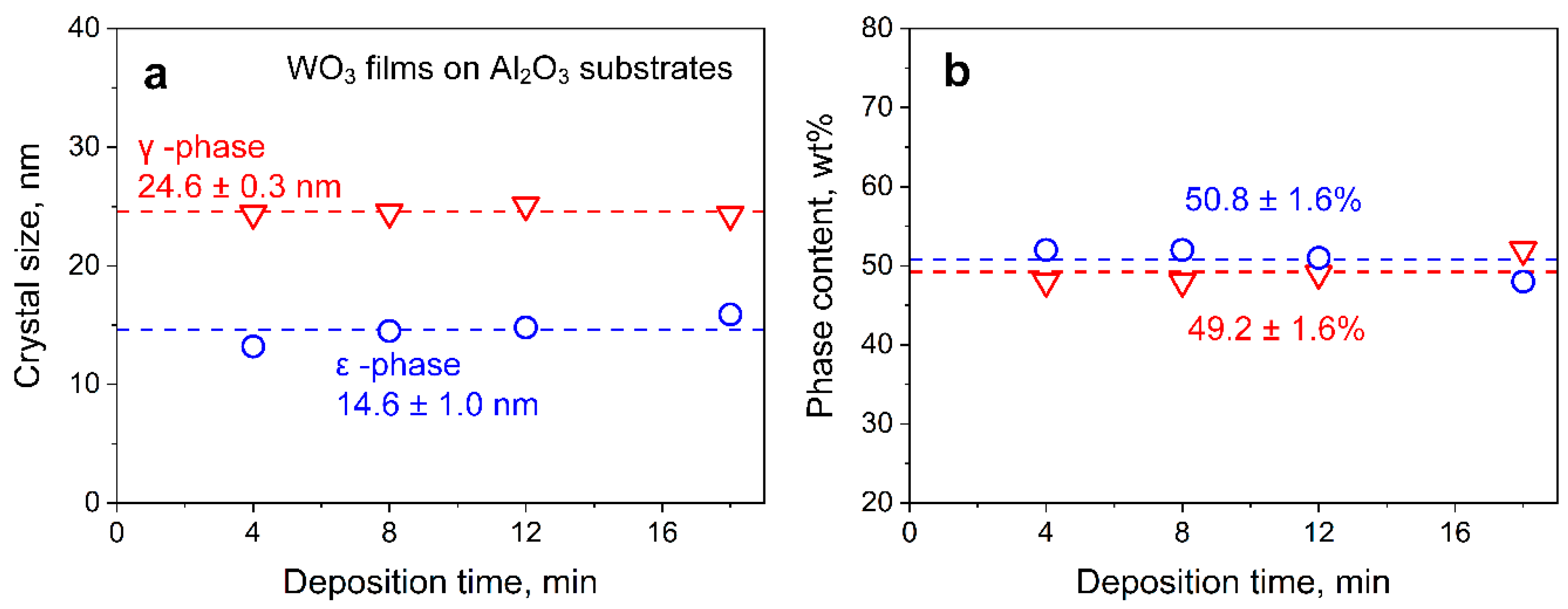
The measured film thickness as a function of deposition time is shown in Figure 2b. Symbols and error bars represented average film thicknesses and standard deviations of, at least, 40 measured thicknesses across the whole cross-section for each deposition time, respectively. Error bars indicated the intra-sample variability. After 1 min of flame deposition, the film exhibited a thickness of about 0.5 μm. The thickness increased rather linearly with deposition time, up to 12.3 μm after 18 min, with a growth rate of 0.69 μm min−1. For thermophoretic deposition, such a linear trend could be obtained when the temperature difference between aerosol and film is constant [33], indicating efficient cooling. Besides, crystal sizes, phase contents (Figure 3), and porosity (Table S2) were not affected by deposition time, indicating that particle growth was completed before deposition [33]. Therefore, changes in sensing performance could be related quite reliably to film thickness.

3.3. Effects of Film Thickness on NO2 Sensing

Figure 4 shows the baseline resistance (triangles) for these flame-made WO3 sensors operated at 125 °C and realistic 50% RH. This sensing temperature was chosen, as identified previously for optimal NO2 sensitivity with hydrothermally prepared films [46]. The resulting power consumption was only 26 mW, making the sensor suitable for integration into battery-driven devices. The baseline resistance decreased steeply from about 109 to 2.6∙107 Ohm when the film thickness increased from 0.5 to 7.7 μm and leveled off thereafter. Such a reduction should be attributed to the additional conduction pathways (i.e., parallel resistances) with increasing film thickness, reducing the overall film resistance, in line with the literature [61]. The sensor baseline resistance drift was only 0.01% h−1 (Figure S7), which could be corrected during long-term operation with readily available algorithms (e.g., zero-calibration protocols [65]). The corresponding sensor responses (circles) when exposed to environmentally relevant 100 ppb NO2 [3] at 50% RH are shown in Figure 4. The NO2 was detected with an average response of 48 for the thinnest 0.5 μm films. Most remarkably, the response more than doubled to 110 and 112 when reaching an optimum thickness between 1.9 and 3.1 μm for 2- and 4-min deposited films, respectively. Error bars were typically <10%, showing good reproducibility of the process. Increasing responses with the thickness could be associated with the increasing number of reaction sites, intensifying chemiresistive interaction with NO2 [66]. For thicker films, however, the NO2 response decreased steeply to 44 at 6 μm thickness and even to 11 at 12.3 μm. Similarly, a decrease in ethanol sensor response with increasing film thickness was observed for flame-deposited SnO2 films [67] and doctor-bladed ones consisting of flame-made ZnO particles [32]. The response difference for the 6 and 7.7 μm thick films was within the error bars and not statistically significant. This response reduction could be attributed to compromised NO2 transport into the film due to increased diffusion resistance with increasing thickness [68]. Furthermore, the NO2 interaction in the upper film layers had less impact on the total resistance change than if the same reaction would occur in the lower layers, where also electrode effects might play a role [69].
The observed optimum (1.9–3.1 μm, Figure 4) for these flame-made and highly porous films occurred at a significantly larger thickness than for sputtered WO3 films (i.e., 85 nm) [23]. For the latter, however, crystal size depended on film thickness due to nucleation and growth during deposition [70] that influenced sensitivity [71]. Optima below 100 nm were also observed for sensing of other gases with compact films, e.g., ion-beam sputtered SnO2 (~7 nm for H2) [72] or atomic layer deposited SnO2 (~10 nm [73] or 2.6 nm [74] for CO). For such compact films, optimum sensitivity was expected if film thickness was in the range of the material’s Debye length (thus fully depleted layers), as shown for SnO2 [74]. This was in contrast to porous films, where the whole film was accessible for the analyte gas, and the transducer function in relation to Debye length was modulated by the particle/crystal size [75] rather than the film thickness. Nevertheless, for porous films obtained from wet methods (e.g., screen-printing), the crack formation was typically observed due to solvent evaporation [76] that could affect sensor results.

3.4. Low-ppb NO2 Sensing

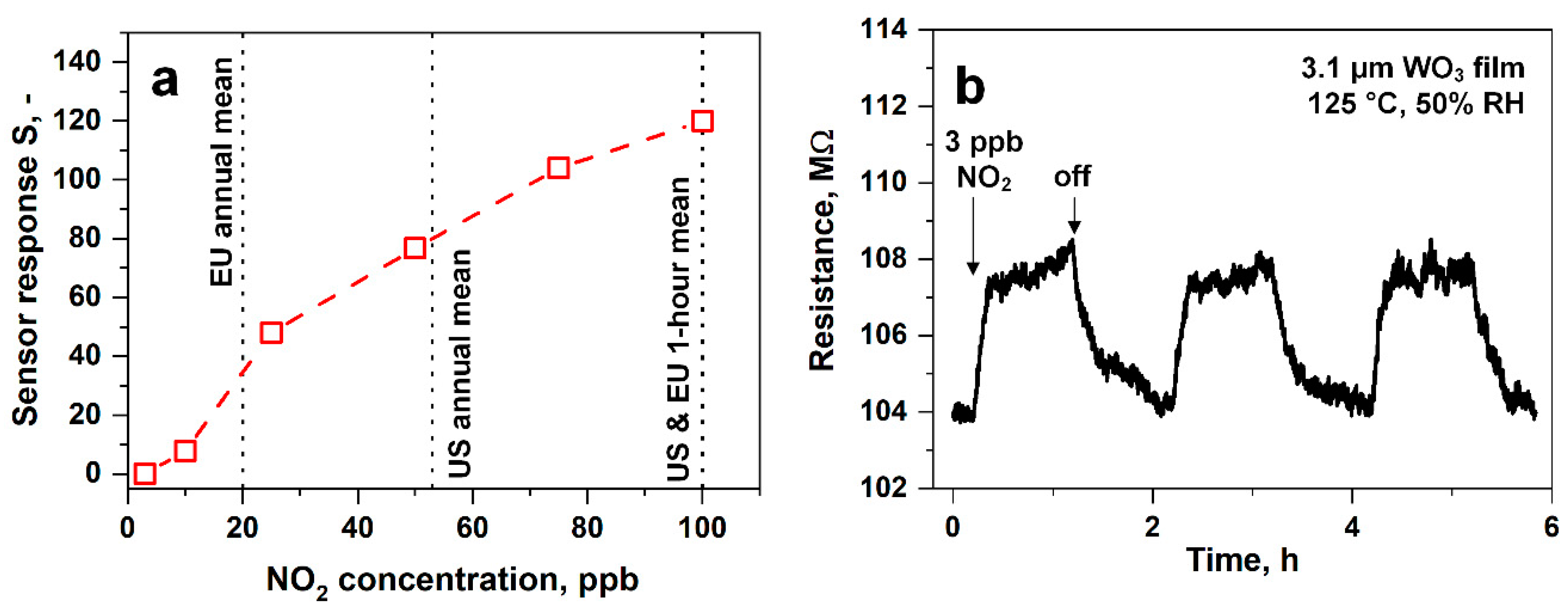
The legal NO2 limits are below 100 ppb [3], defining the required detection range for suitable sensors. Figure 5a shows the responses of the above thickness-optimized sensor (i.e., 3.1 μm thick) to 3–100 ppb NO2 at 50% RH. The sensor response increased continuously with a sensitivity of about 2 ppb−1 up to 25 ppb and thereafter with 1 ppb−1, suggesting the onset of saturation. This was orders of magnitude higher than graphene-based sensors operated at room temperature (~0.1 ppb−1) [77] or WO3 nanowires at 250 °C (~0.002 ppb−1) [78]. Such behavior was expected due to the nonlinear diffusion-reaction theory of such semiconducting metal oxide sensors [79]. Most importantly, hazardous concentrations exceeding the exposure guidelines in the EU and US (vertical dashed lines) could be clearly identified.
Figure 5b shows the change of film resistance when consecutively exposed to low NO2 concentrations of 3 ppb in 50% RH. The film resistance increased from about 104 to 108 MOhm when introducing 3 ppb NO2, as expected for oxidizing gases like NO2 that fill oxygen vacancies located on the surface of WO3 [80], leading to increasing film resistance. Thereafter, the resistance was fully recovered to its baseline. This resulted in a response of 0.04 with a high signal-to-noise ratio of 17. Besides, the same response was obtained upon consecutive exposure to 3 ppb, indicating good reproducibility. While sub-ppb NO2 has already been measured only in dry air [81], the present sensor detected 3 ppb at realistic humidity. This was considerably lower than other metal oxide-based sensors tested in relevant conditions (10–80% RH, Table 1) and commercial electrochemical sensors (lower limit of detection of 10 ppb [82]). Important to note, however, that the NO2 sensitivity of WO3-based sensors decreased with increasing RH, both at low (e.g., 25 °C [83]) and high temperatures (e.g., 300 °C [80]). Therein, H2O competed with NO2 for oxygen vacancies, as observed by in situ diffuse reflectance infrared Fourier transform spectroscopy and film resistance readout [80]. In fact, the NO2 response of WO3 sensors operating at 100 °C and dry air was halved by increasing the RH to 25%, however, influenced much less at higher RH (~30% for every 25% RH increase) [84]. This could be corrected by co-located RH sensors, as shown previously for sensing acetone, ammonia, and isoprene at 30–90% RH [40]. The detection of such low NO2 concentrations with flame-deposited WO3 films was associated with their highly porous and fine film morphology (Figure 2a and Figure S4). In fact, sensor responses at 150 °C increased in sputtered WO3 films after increasing porosity by annealing [85], and highly porous CuBr films showed an order of magnitude higher NH3 responses than denser ones [55]. The corresponding response and recovery times of 7.8 and 43.8 min, respectively, could be further decreased by transient response analysis [86].

3.5. NO2 Selectivity in Realistic Conditions

In environmental monitoring, a key challenge is the selective detection of NO2 in the presence of interfering gases. Therefore, the responses of the above sensor (3.1 μm thickness) were tested for 100 ppb NO2 and its major confounders at much higher concentration (1000 ppb): H2S, formaldehyde, H2, CH4, CO (up to 40 ppm, Figure S8), acetone, methanol, ethanol, and NH3 at 125 °C and 50% RH (Figure 6). Excellent NO2 selectivities were obtained to CO, methanol, ethanol, NH3 (all > 105), H2, CH4, acetone (all > 104), and formaldehyde (>103), indicating negligible cross-interference. Even to H2S, the selectivity was still >800. Please note that WO3 also showed high NO2 selectivity to other confounders, e.g., 150 for NO at 100 °C and 1 ppm [84]. Interestingly, also, the selectivity for most interferents was rather independent of film thickness, while for H2/ethanol and H2S, decreasing and increasing trends, respectively, were observed with the thickness (Table S3). These selectivities are most attractive for environmental NO2 monitoring, even if CO concentrations are present up to 40 ppm in rare situations, such as during traffic [87]. In specific, in the presence of 20 and 40 ppm of CO, the response was 0.02 and 0.07 (Figure S8), respectively, resulting in selectivities > 105 (calculated at the same concentration with NO2). The selectivity could be further increased with filters preceding the sensor, e.g., size-selective membranes [88] or adsorption packed beds (e.g., Al2O3 to retain hydrophilic [89] or Tenax for hydrophobic compounds [49]).
Table 1 shows a selectivity comparison to state-of-the-art NO2 sensors at 10–80% RH and relevant sub-ppm NO2 concentrations. The present flame-made porous WO3 sensor showed superior selectivities over those prepared by sputtering (for NH3 and CO), drop coating (for acetone), and other coating methods (for ethanol), which could be attributed to their more compact film morphology and/or to the presence of ε-phase WO3 in the flame-made films (Figure 1b,c). Furthermore, Fe-doping [12] seems to decrease the selectivity significantly compared to pure WO3 sensors. Only brush-coated In2O3 [24] and SnO2/ZnO (UV excited) sensors [26] feature similar selectivity for most analytes. Finally, carbon-based (rGO, CNT) composites with metal-oxides [8,9,27] operated at room temperature are outperformed, featuring orders of magnitude lower selectivities (e.g., 9 vs. >105 for CO).

4. Conclusions

Highly porous, crack-free, and homogeneous WO3 sensing films with controlled thickness were created by FSP. The films consisted of mixed-phase γ- and ε-WO3, as revealed by XRD and Raman spectroscopy. Their thickness was controlled through the deposition time without affecting the crystal size or phase content of WO3. Such flame-deposited films at an optimal thickness (i.e., 3.1 μm) exhibited an order of magnitude higher NO2 response than the thickest films (i.e., 12.3 μm). This was probably related to a competition of increased amount of reaction sites, hindered analyte penetration, and weakened electrode effects for thicker films. The thickness-optimized WO3 films showed excellent NO2 selectivity over confounding gases. Furthermore, NO2 concentrations were detected down to 3 ppb at 50% RH with a high signal-to-noise ratio (>17), superior to state-of-the-art SMOx-based and commercial NO2 sensors. As a result, such morphology-optimized WO3 films have a high potential for integration into low power devices for distributed and remote NO2 air quality monitoring. In a broader sense, such film morphology optimization could be applied also for other metal-oxide sensors and analytes to improve their sensitivity in environmental monitoring or medical breath analysis [90], where similar challenging sensitivity requirements exist.

Supplementary Materials

The following are available online at https://www.mdpi.com/2079-4991/10/6/1170/s1, Figure S1: Sensor chamber, Figure S2: Phase determination by X-ray diffraction, Figure S3: Extended XRD spectrum of the WO3 powder, Figure S4: Film thickness determination, Figure S5: Flame-deposited WO3 films on Al2O3, Figure S6: WO3 film thickness on Al2O3 as a function of deposition time, Figure S7: Drift, Figure S8: The effect of higher CO concentrations, Table S1: Optimum WO3 temperatures for NO2 sensing, Table S2: Porosity evaluation by X-ray diffraction, Table S3: Film thickness effect on selectivity.

Author Contributions

Conceptualization, S.A., D.K.C., A.T.G., and S.E.P.; methodology, S.A.; validation, S.A. and D.K.C.; formal analysis, S.A. and D.K.C.; investigation, S.A., D.K.C., A.T.G., and S.E.P.; data curation, S.A.; writing—original draft preparation, S.A.; writing—review and editing, A.T.G and S.E.P.; visualization, S.A. and D.K.C.; supervision, A.T.G. and S.E.P.; project administration, S.E.P; funding acquisition, S.E.P. All authors have read and agreed to the published version of the manuscript.

Funding

This research was primarily funded by the Particle Technology Laboratory (ETHZ) and in part by the Swiss National Science Foundation (Project No. 159763 and 175754).

Acknowledgments

The authors thank Ines Weber (ETHZ) for her help with TEM analysis, as well as Zeng Peng from the Scientific Center for Optical and Electron Microscopy (ScopeM) of ETHZ with the FIB-SEM analysis.

Conflicts of Interest

The authors declare no conflict of interest.

References

  1. Beloconi, A.; Vounatsou, P. Bayesian geostatistical modelling of high-resolution NO2 exposure in Europe combining data from monitors, satellites and chemical transport models. Environ. Int. 2020, 138, 105578. [Google Scholar] [CrossRef]
  2. Robinson, E.; Robbins, R.C. Gaseous nitrogen compound pollutants from urban and natural sources. J. Air Pollut. Control Assoc. 1970, 20, 303–306. [Google Scholar] [CrossRef]
  3. U.S. Environmental Protection Agency. Integrated Science Assessment for Oxides of Nitrogen—Health Criteria EPA/600/R-15/068; U.S. Environmental Protection Agency: Washington, DC, USA, 2016.
  4. Shima, M.; Adachi, M. Effect of outdoor and indoor nitrogen dioxide on respiratory symptoms in schoolchildren. Int. J. Epidemiol. 2000, 29, 862–870. [Google Scholar] [CrossRef] [PubMed] [Green Version]
  5. European Parliament and the Council of the European Union. Directive 2008/50/EC. Off. J. Eur. Union 2008, 51, 1–44. [Google Scholar]
  6. Rigby, M.; Park, S.; Saito, T.; Western, L.M.; Redington, A.L.; Fang, X.; Henne, S.; Manning, A.J.; Prinn, R.G.; Dutton, G.S.; et al. Increase in CFC-11 emissions from eastern China based on atmospheric observations. Nature 2019, 569, 546–550. [Google Scholar] [CrossRef]
  7. Popoola, O.A.; Carruthers, D.; Lad, C.; Bright, V.B.; Mead, M.I.; Stettler, M.E.; Saffell, J.R.; Jones, R.L. Use of networks of low cost air quality sensors to quantify air quality in urban settings. Atmos. Environ. 2018, 194, 58–70. [Google Scholar] [CrossRef]
  8. Zhang, H.; Yu, L.; Li, Q.; Du, Y.; Ruan, S. Reduced graphene oxide/α-Fe2O3 hybrid nanocomposites for room temperature NO2 sensing. Sens. Actuators B 2017, 241, 109–115. [Google Scholar] [CrossRef]
  9. Liu, S.; Wang, Z.; Zhang, Y.; Zhang, C.; Zhang, T. High performance room temperature NO2 sensors based on reduced graphene oxide-multiwalled carbon nanotubes-tin oxide nanoparticles hybrids. Sens. Actuators B 2015, 211, 318–324. [Google Scholar] [CrossRef]
  10. Koo, W.T.; Cha, J.H.; Jung, J.W.; Choi, S.J.; Jang, J.S.; Kim, D.H.; Kim, I.D. Few-layered WS2 nanoplates confined in Co, n-doped hollow carbon nanocages: Abundant WS2 edges for highly sensitive gas sensors. Adv. Funct. Mater. 2018, 28, 1802575. [Google Scholar] [CrossRef]
  11. Malik, R.; Tomer, V.K.; Mishra, Y.K.; Lin, L. Functional gas sensing nanomaterials: A panoramic view. Appl. Phys. Rev. 2020, 7, 021301. [Google Scholar] [CrossRef] [Green Version]
  12. Zhang, Z.; Wen, Z.; Ye, Z.; Zhu, L. Ultrasensitive ppb-level NO2 gas sensor based on WO3 hollow nanosphers doped with Fe. Appl. Surf. Sci. 2018, 434, 891–897. [Google Scholar] [CrossRef]
  13. Inyawilert, K.; Channei, D.; Tamaekong, N.; Liewhiran, C.; Wisitsoraat, A.; Tuantranont, A.; Phanichphant, S. Pt-doped In2O3 nanoparticles prepared by flame spray pyrolysis for NO2 sensing. J. Nanopart. Res. 2016, 18. [Google Scholar] [CrossRef]
  14. Akamatsu, T.; Itoh, T.; Izu, N.; Shin, W. NO and NO2 sensing properties of WO3 and Co3O4 based gas sensors. Sensors 2013, 13, 12467–12481. [Google Scholar] [CrossRef] [PubMed] [Green Version]
  15. Casals, O.; Markiewicz, N.; Fabrega, C.; Gràcia, I.; Cané, C.; Wasisto, H.S.; Waag, A.; Prades, J.D. A parts per billion (ppb) sensor for NO2 with microwatt (μW) power requirements based on micro light plates. ACS Sens. 2019, 4, 822–826. [Google Scholar] [CrossRef] [Green Version]
  16. Mane, A.; Moholkar, A. Effect of film thickness on NO2 gas sensing properties of sprayed orthorhombic nanocrystalline V2O5 thin films. Appl. Surf. Sci. 2017, 416, 511–520. [Google Scholar] [CrossRef]
  17. Xia, H.; Wang, Y.; Kong, F.; Wang, S.; Zhu, B.; Guo, X.; Zhang, J.; Wang, Y.; Wu, S. Au-doped WO3-based sensor for NO2 detection at low operating temperature. Sens. Actuators B 2008, 134, 133–139. [Google Scholar] [CrossRef]
  18. Simon, I.; Barsan, N.; Bauer, M.; Weimar, U. Micromachined metal oxide gas sensors: Opportunities to improve sensor performance. Sens. Actuators B 2001, 73, 1–26. [Google Scholar] [CrossRef]
  19. Barsan, N.; Schweizer-Berberich, M.; Göpel, W. Fundamental and practical aspects in the design of nanoscaled SnO2 gas sensors: A status report. Fresenius J. Anal. Chem. 1999, 365, 287–304. [Google Scholar] [CrossRef]
  20. Cantalini, C.; Lozzi, L.; Passacantando, M.; Santucci, S. The comparative effect of two different annealing temperatures and times on the sensitivity and long-term stability of WO3 thin films for detecting NO2. IEEE Sens. J. 2003, 3, 171–179. [Google Scholar] [CrossRef]
  21. Kim, J.-S.; Yoon, J.-W.; Hong, Y.J.; Kang, Y.C.; Abdel-Hady, F.; Wazzan, A.; Lee, J.-H. Highly sensitive and selective detection of ppb-level NO2 using multi-shelled WO3 yolk–shell spheres. Sens. Actuators B 2016, 229, 561–569. [Google Scholar] [CrossRef]
  22. You, L.; He, X.; Wang, D.; Sun, P.; Sun, Y.; Liang, X.; Du, Y.; Lu, G. Ultrasensitive and low operating temperature NO2 gas sensor using nanosheets assembled hierarchical WO3 hollow microspheres. Sens. Actuators B 2012, 173, 426–432. [Google Scholar] [CrossRef]
  23. Prajapati, C.S.; Bhat, N. ppb level detection of NO2 using a WO3 thin film-based sensor: Material optimization, device fabrication and packaging. RSC Adv. 2018, 8, 6590–6599. [Google Scholar] [CrossRef] [Green Version]
  24. Han, D.; Zhai, L.; Gu, F.; Wang, Z. Highly sensitive NO2 gas sensor of ppb-level detection based on In2O3 nanobricks at low temperature. Sens. Actuators B 2018, 262, 655–663. [Google Scholar] [CrossRef]
  25. Wu, T.; Wang, Z.; Tian, M.; Miao, J.; Zhang, H.; Sun, J. UV excitation NO2 gas sensor sensitized by ZnO quantum dots at room temperature. Sens. Actuators B 2018, 259, 526–531. [Google Scholar] [CrossRef]
  26. Lu, G.; Xu, J.; Sun, J.; Yu, Y.; Zhang, Y.; Liu, F. UV-enhanced room temperature NO2 sensor using ZnO nanorods modified with SnO2 nanoparticles. Sens. Actuators B 2012, 162, 82–88. [Google Scholar] [CrossRef]
  27. Wang, Z.; Zhang, T.; Han, T.; Fei, T.; Liu, S.; Lu, G. Oxygen vacancy engineering for enhanced sensing performances: A case of SnO2 nanoparticles-reduced graphene oxide hybrids for ultrasensitive ppb-level room-temperature NO2 sensing. Sens. Actuators B 2018, 266, 812–822. [Google Scholar] [CrossRef]
  28. Korotcenkov, G. The role of morphology and crystallographic structure of metal oxides in response of conductometric-type gas sensors. Mater. Sci. Eng. R Rep. 2008, 61, 1–39. [Google Scholar] [CrossRef]
  29. Kida, T.; Nishiyama, A.; Hua, Z.; Suematsu, K.; Yuasa, M.; Shimanoe, K. WO3 nanolamella gas sensor: Porosity control using SnO2 nanoparticles for enhanced NO2 sensing. Langmuir 2014, 30, 2571–2579. [Google Scholar] [CrossRef]
  30. Gao, H.; Jia, H.; Bierer, B.; Wöllenstein, J.; Lu, Y.; Palzer, S. Scalable gas sensors fabrication to integrate metal oxide nanoparticles with well-defined shape and size. Sens. Actuators B 2017, 249, 639–646. [Google Scholar] [CrossRef]
  31. Ueda, T.; Maeda, T.; Huang, Z.; Higuchi, K.; Izawa, K.; Kamada, K.; Hyodo, T.; Shimizu, Y. Enhancement of methylmercaptan sensing response of WO3 semiconductor gas sensors by gas reactivity and gas diffusivity. Sens. Actuators B 2018, 273, 826–833. [Google Scholar] [CrossRef]
  32. Liewhiran, C.; Phanichphant, S. Influence of thickness on ethanol sensing characteristics of doctor-bladed thick film from flame-made ZnO nanoparticles. Sensors 2007, 7, 185–201. [Google Scholar] [CrossRef] [Green Version]
  33. Mädler, L.; Roessler, A.; Pratsinis, S.E.; Sahm, T.; Gurlo, A.; Barsan, N.; Weimar, U. Direct formation of highly porous gas-sensing films by in situ thermophoretic deposition of flame-made Pt/SnO2 nanoparticles. Sens. Actuators B 2006, 114, 283–295. [Google Scholar] [CrossRef]
  34. Blattmann, C.O.; Güntner, A.T.; Pratsinis, S.E. In situ monitoring the deposition of flame-made chemoresistive gas sensing films. ACS Appl. Mater. Interfaces 2017, 9, 23926–23933. [Google Scholar] [CrossRef] [PubMed]
  35. Wang, L.; Teleki, A.; Pratsinis, S.; Gouma, P. Ferroelectric WO3 nanoparticles for acetone selective detection. Chem. Mater. 2008, 20, 4794–4796. [Google Scholar] [CrossRef]
  36. Righettoni, M.; Tricoli, A.; Pratsinis, S.E. Thermally stable, silica-doped epsilon-WO3 for sensing of acetone in the human breath. Chem. Mater. 2010, 22, 3152–3157. [Google Scholar] [CrossRef]
  37. Güntner, A.T.; Sievi, N.A.; Theodore, S.J.; Gulich, T.; Kohler, M.; Pratsinis, S.E. Noninvasive body fat burn monitoring from exhaled acetone with Si-doped WO3-sensing nanoparticles. Anal. Chem. 2017, 89, 10578–10584. [Google Scholar] [CrossRef]
  38. Güntner, A.T.; Kompalla, J.F.; Landis, H.; Theodore, S.; Geidl, B.; Sievi, N.; Kohler, M.; Pratsinis, S.E.; Gerber, P. Guiding ketogenic diet with breath acetone sensors. Sensors 2018, 18, 3655. [Google Scholar] [CrossRef] [Green Version]
  39. Pineau, N.J.; Kompalla, J.F.; Güntner, A.T.; Pratsinis, S.E. Orthogonal gas sensor arrays by chemoresistive material design. Microchim. Acta 2018, 185, 563. [Google Scholar] [CrossRef] [Green Version]
  40. Güntner, A.T.; Pineau, N.J.; Mochalski, P.; Wiesenhofer, H.; Agapiou, A.; Mayhew, C.A.; Pratsinis, S.E. Sniffing entrapped humans with sensor arrays. Anal. Chem. 2018, 90, 4940–4945. [Google Scholar] [CrossRef] [Green Version]
  41. Mädler, L.; Sahm, T.; Gurlo, A.; Grunwaldt, J.-D.; Barsan, N.; Weimar, U.; Pratsinis, S.E. Sensing low concentrations of CO using flame-spray-made Pt/SnO2 nanoparticles. J. Nanopart. Res. 2006, 8, 783–796. [Google Scholar] [CrossRef] [Green Version]
  42. Abegg, S.; Magro, L.; van den Broek, J.; Pratsinis, S.E.; Güntner, A.T. A pocket-sized device enables detection of methanol adulteration in alcoholic beverages. Nat. Food 2020, accepted. [Google Scholar] [CrossRef]
  43. Huelser, T.; Lorke, A.; Ifeacho, P.; Wiggers, H.; Schulz, C. Core and grain boundary sensitivity of tungsten-oxide sensor devices by molecular beam assisted particle deposition. J. Appl. Phys. 2007, 102, 124305. [Google Scholar] [CrossRef]
  44. Güntner, A.T.; Koren, V.; Chikkadi, K.; Righettoni, M.; Pratsinis, S.E. E-nose sensing of low-ppb formaldehyde in gas mixtures at high relative humidity for breath screening of lung cancer? ACS Sens. 2016, 1, 528–535. [Google Scholar] [CrossRef]
  45. Pineau, N.J.; Keller, S.D.; Güntner, A.T.; Pratsinis, S.E. Palladium embedded in SnO2 enhances the sensitivity of flame-made chemoresistive gas sensors. Microchim. Acta 2020, 187, 96. [Google Scholar] [CrossRef]
  46. You, L.; Sun, Y.; Ma, J.; Guan, Y.; Sun, J.; Du, Y.; Lu, G. Highly sensitive NO2 sensor based on square-like tungsten oxide prepared with hydrothermal treatment. Sens. Actuators B 2011, 157, 401–407. [Google Scholar] [CrossRef]
  47. Koppmann, R. (Ed.) Volatile Organic Compounds in the Atmosphere; Blackwell Publishing Ltd: Oxford, UK, 2007. [Google Scholar]
  48. Datasheet MSGS 5000i. Available online: http://microsens.ch/products/pdf/MSGS_5000i_Datasheet.pdf (accessed on 19 May 2020).
  49. van den Broek, J.; Abegg, S.; Pratsinis, S.E.; Güntner, A.T. Highly selective detection of methanol over ethanol by a handheld gas sensor. Nat. Commun. 2019, 10, 4220. [Google Scholar] [CrossRef]
  50. Tricoli, A.; Graf, M.; Mayer, F.; Kühne, S.; Hierlemann, A.; Pratsinis, S.E. Micropatterning layers by flame aerosol deposition-annealing. Adv. Mater. 2008, 20, 3005–3010. [Google Scholar] [CrossRef]
  51. Güntner, A.T.; Pineau, N.J.; Chie, D.; Krumeich, F.; Pratsinis, S.E. Selective sensing of isoprene by Ti-doped ZnO for breath diagnostics. J. Mater. Chem. B 2016, 4, 5358–5366. [Google Scholar] [CrossRef]
  52. Hubbell, J. Photon mass attenuation and energy-absorption coefficients. Int. J. Appl. Radiat. Isot. 1982, 33, 1269–1290. [Google Scholar] [CrossRef]
  53. Berger, M.; Hubbell, J.; Seltzer, S.; Chang, J.; Coursey, J.; Sukumar, R.; Zucker, D.; Olsen, K. XCOM: Photon cross Sections Database, NIST Standard Reference Database 8 (XGAM). Available online: http://physics.nist.gov/PhysRefData/Xcom/Text/XCOM.html (accessed on 30 April 2019).
  54. Güntner, A.T.; Righettoni, M.; Pratsinis, S.E. Selective sensing of NH3 by Si-doped alpha-MoO3 for breath analysis. Sens. Actuators B 2016, 223, 266–273. [Google Scholar] [CrossRef]
  55. Güntner, A.T.; Wied, M.; Pineau, N.J.; Pratsinis, S.E. Rapid and selective NH3 sensing by porous CuBr. Adv. Sci. 2020, 7, 1903390. [Google Scholar] [CrossRef] [Green Version]
  56. Sari, W.P.; Leigh, S.; Covington, J. Tungsten oxide based sensor for oxygen detection. Proceedings 2018, 2, 952. [Google Scholar] [CrossRef] [Green Version]
  57. Arai, M.; Hayashi, S.; Yamamoto, K.; Kim, S.S. Raman studies of phase-transitions in gas-evaporated WO3 microcrystals. Solid State Commun. 1990, 75, 613–616. [Google Scholar] [CrossRef]
  58. Strobel, R.; Maciejewski, M.; Pratsinis, S.E.; Baiker, A. Unprecedented formation of metastable monoclinic BaCO3 nanoparticles. Thermochim. Acta 2006, 445, 23–26. [Google Scholar] [CrossRef]
  59. Righettoni, M.; Pratsinis, S.E. Annealing dynamics of WO3 by in situ XRD. Mater. Res. Bull. 2014, 59, 199–204. [Google Scholar] [CrossRef]
  60. Labidi, A.; Lambert-Mauriat, C.; Jacolin, C.; Bendahan, M.; Maaref, M.; Aguir, K. DC and AC characterizations of WO3 sensors under ethanol vapors. Sens. Actuators B 2006, 119, 374–379. [Google Scholar] [CrossRef]
  61. Barsan, N.; Weimar, U. Conduction model of metal oxide gas sensors. J. Electroceram. 2001, 7, 143–167. [Google Scholar] [CrossRef]
  62. Daniel, M.F.; Desbat, B.; Lassegues, J.C.; Gerand, B.; Figlarz, M. Infrared and raman-study of WO3 tungsten trioxides and WO3, xH2O tungsten trioxide hydrates. J. Solid State Chem. 1987, 67, 235–247. [Google Scholar] [CrossRef]
  63. Salje, E. Lattice dynamics of WO3. Acta Crystallogr. A 1975, 31, 360–363. [Google Scholar] [CrossRef]
  64. Viera, G.; Huet, S.; Boufendi, L. Crystal size and temperature measurements in nanostructured silicon using Raman spectroscopy. J. Appl. Phys. 2001, 90, 4175–4183. [Google Scholar] [CrossRef]
  65. Sun, L.; Westerdahl, D.; Ning, Z. Development and evaluation of a novel and cost-effective approach for low-cost NO2 sensor drift correction. Sensors 2017, 17, 1916. [Google Scholar] [CrossRef] [PubMed]
  66. Tricoli, A.; Righettoni, M.; Teleki, A. Semiconductor gas sensors: Dry synthesis and application. Angew. Chem. Int. Edit. 2010, 49, 7632–7659. [Google Scholar] [CrossRef] [PubMed]
  67. Tricoli, A.; Pratsinis, S.E. Dispersed nanoelectrode devices. Nat. Nanotechnol. 2010, 5, 54–60. [Google Scholar] [CrossRef] [PubMed]
  68. Matsunaga, N.; Sakai, G.; Shimanoe, K.; Yamazoe, N. Diffusion equation-based study of thin film semiconductor gas sensor-response transient. Sens. Actuators B 2002, 83, 216–221. [Google Scholar] [CrossRef]
  69. Penza, M.; Martucci, C.; Cassano, G. NOx gas sensing characteristics of WO3 thin films activated by noble metals (Pd, Pt, Au) layers. Sens. Actuators B 1998, 50, 52–59. [Google Scholar] [CrossRef]
  70. Tosun, B.S.; Feist, R.K.; Gunawan, A.; Mkhoyan, K.A.; Campbell, S.A.; Aydil, E.S. Sputter deposition of semicrystalline tin dioxide films. Thin Solid Films 2012, 520, 2554–2561. [Google Scholar] [CrossRef]
  71. Xu, C.; Tamaki, J.; Miura, N.; Yamazoe, N. Grain size effects on gas sensitivity of porous SnO2-based elements. Sens. Actuators B 1991, 3, 147–155. [Google Scholar] [CrossRef]
  72. Suzuki, T.; Yamazaki, T.; Yoshioka, H.; Hikichi, K. Thickness dependence of H2 gas sensor in amorphous SnOx films prepared by ion-beam sputtering. J. Mater. Sci. 1988, 23, 145–149. [Google Scholar] [CrossRef]
  73. Rosental, A.; Tarre, A.; Gerst, A.; Sundqvist, J.; Hårsta, A.; Aidla, A.; Aarik, J.; Sammelselg, V.; Uustare, T. Gas sensing properties of epitaxial SnO2 thin films prepared by atomic layer deposition. Sens. Actuators B 2003, 93, 552–555. [Google Scholar] [CrossRef]
  74. Du, X.; George, S. Thickness dependence of sensor response for CO gas sensing by tin oxide films grown using atomic layer deposition. Sens. Actuators B 2008, 135, 152–160. [Google Scholar] [CrossRef]
  75. Tamaki, J.; Zhang, Z.; Fujimori, K.; Akiyama, M.; Harada, T.; Miura, N.; Yamazoe, N. Grain-size effects in tungsten oxide-based sensor for nitrogen oxides. J. Electrochem. Soc. 1994, 141, 2207–2210. [Google Scholar] [CrossRef]
  76. Kozuka, H.; Takenaka, S.; Tokita, H.; Hirano, T.; Higashi, Y.; Hamatani, T. Stress and cracks in gel-derived ceramic coatings and thick film formation. J. Sol Gel. Sci. Techn. 2003, 26, 681–686. [Google Scholar] [CrossRef]
  77. Wu, J.; Feng, S.; Wei, X.; Shen, J.; Lu, W.; Shi, H.; Tao, K.; Lu, S.; Sun, T.; Yu, L.; et al. Facile synthesis of 3D graphene flowers for ultrasensitive and highly reversible gas sensing. Adv. Funct. Mater. 2016, 26, 7462–7469. [Google Scholar] [CrossRef]
  78. Hoa, N.D.; El-Safty, S.A. Gas nanosensor design packages based on tungsten oxide: Mesocages, hollow spheres, and nanowires. Nanotechnology 2011, 22, 485503. [Google Scholar] [CrossRef] [PubMed]
  79. Gardner, J.W. A non-linear diffusion-reaction model of electrical conduction in semiconductor gas sensors. Sens. Actuators B 1990, 1, 166–170. [Google Scholar] [CrossRef]
  80. Staerz, A.; Berthold, C.; Russ, T.; Wicker, S.; Weimar, U.; Barsan, N. The oxidizing effect of humidity on WO3 based sensors. Sens. Actuators B 2016, 237, 54–58. [Google Scholar] [CrossRef]
  81. Xiao, B.; Song, S.; Wang, P.; Zhao, Q.; Chuai, M.; Zhang, M. Promoting effects of Ag on In2O3 nanospheres of sub-ppb NO2 detection. Sens. Actuators B 2017, 241, 489–497. [Google Scholar] [CrossRef]
  82. NO2-A43F Nitrogen Dioxide Sensor 4-Electrode (Alphasense). Available online: http://www.alphasense.com/WEB1213/wp-content/uploads/2019/09/NO2-A43F.pdf (accessed on 16 December 2019).
  83. Bai, S.; Ma, Y.; Shu, X.; Sun, J.; Feng, Y.; Luo, R.; Li, D.; Chen, A. Doping metal elements of WO3 for enhancement of NO2-sensing performance at room temperature. Ind. Eng. Chem. Res. 2017, 56, 2616–2623. [Google Scholar] [CrossRef]
  84. Yang, L.; Marikutsa, A.; Rumyantseva, M.; Konstantinova, E.; Khmelevsky, N.; Gaskov, A. Quasi similar routes of NO2 and NO sensing by nanocrystalline WO3: Evidence by in situ DRIFT spectroscopy. Sensors 2019, 19, 3405. [Google Scholar] [CrossRef] [PubMed] [Green Version]
  85. Zeng, J.; Hu, M.; Wang, W.; Chen, H.; Qin, Y. NO2-sensing properties of porous WO3 gas sensor based on anodized sputtered tungsten thin film. Sens. Actuators B 2012, 161, 447–452. [Google Scholar] [CrossRef]
  86. Lampe, U.; Gerblinger, J.; Meixner, H. Comparison of transient response of exhaust-gas sensors based on thin films of selected metal oxides. Sens. Actuators B 1992, 7, 787–791. [Google Scholar] [CrossRef]
  87. Bevan, M.A.; Proctor, C.J.; Baker-Rogers, J.; Warren, N.D. Exposure to carbon monoxide, respirable suspended particulates and volatile organic compounds while commuting by bicycle. Environ. Sci. Technol. 1991, 25, 788–791. [Google Scholar] [CrossRef]
  88. Güntner, A.T.; Abegg, S.; Wegner, K.; Pratsinis, S.E. Zeolite membranes for highly selective formaldehyde sensors. Sens. Actuators B 2018, 257, 916–923. [Google Scholar] [CrossRef]
  89. van den Broek, J.; Güntner, A.T.; Pratsinis, S.E. Highly selective and rapid breath isoprene sensing enabled by activated alumina filter. ACS Sens. 2018, 3, 677–683. [Google Scholar] [CrossRef] [PubMed] [Green Version]
  90. Güntner, A.T.; Abegg, S.; Königstein, K.; Gerber, P.A.; Schmidt-Trucksäss, A.; Pratsinis, S.E. Breath sensors for health monitoring. ACS Sens. 2019, 4, 268–280. [Google Scholar] [CrossRef] [PubMed]
Figure 1. Material characterization. (a) TEM image with marked primary particle sizes in nm with inset at higher magnification, showing the lattice orientation and spacing in Å. (b) XRD pattern and (c) Raman spectrum of the filter-collected and annealed WO3 particles. Reference peak positions of monoclinic γ-WO3 (triangles, ICSD 80056) and ε-WO3 (circles, 84163).
Nanomaterials 10 01170 g001
Figure 2. Film characterization. Cross-sectional SEM images of (a) porous 4-min flame-deposited film (on microsensor substrates) after cutting a square with a focused ion beam. Higher magnification is provided as inset. (b) The thickness of the deposited films as a function of deposition time. Symbols indicate average thicknesses, and error bars (some hidden behind the symbols) the standard deviations of, at least, 40 measured thicknesses per film. Dashed line represents a linear fit with indicated deposition rate.
Nanomaterials 10 01170 g002
Figure 3. Effect of deposition time on WO3 composition. (a) Crystal sizes and (b) phase content for γ- (triangles) and ε-WO3 (circles) films on Al2O3 substrates (Figure S5) with comparable crystal size and phase composition to filter-collected WO3 powders. These films were prepared at identical deposition times to those on microsensor substrates that were too small and had too little mass for XRD. The films on Al2O3 were thicker and grew faster (Figure S6) than those on microsensor substrates due to the masking of the latter. Even for these on Al2O3, no reliable XRD evaluation could be obtained for 1 and 2 min deposition due to their weak WO3 signal.
Nanomaterials 10 01170 g003
Figure 4. Effect of film thickness on sensor response. Baseline resistance (triangles) for flame-deposited WO3 films at 125 °C and 50% relative humidity (RH) (at 23 °C) and corresponding sensor responses to 100 ppb NO2 (circles). Symbols indicate the averages, and vertical error bars the corresponding variabilities of three identically produced sensors. Horizontal error bars represent the thickness variability of a sensing film (of 40 measurements).
Nanomaterials 10 01170 g004
Figure 5. Low ppb NO2 detection. (a) Sensor responses of the thickness-optimized WO3 film (3.1 µm) in the relevant concentration range from 3 to 100 ppb NO2 measured at 50% RH (at 23 °C) and 125 °C. Threshold exposure limits from the EU [5] and the US [3] are indicated by dashed lines. (b) Film resistance upon consecutive exposure to 3 ppb NO2.
Nanomaterials 10 01170 g005
Figure 6. Selectivity. Sensor responses of the thickness-optimized 3.1 µm WO3 film to 100 ppb of NO2 and 1 ppm H2S, formaldehyde, hydrogen, methanol, CO, acetone, methanol, ethanol, and ammonia at 50% RH (at 23 °C). Please note that no response was detectable for CO, methanol, and ammonia.
Nanomaterials 10 01170 g006
Table 1. Literature comparison. Selectivity, relative humidity (RH), sensing temperature (Tsensor), and lower limit of detection (LOD) of state-of-the-art solid-state NO2 sensors tested at humid conditions. The LOD represents the lowest concentration measured in the respective study. Responses were linearly interpolated to the same concentrations. Non-conventional abbreviations: RT: room temperature; MeOH: Methanol; EtOH: Ethanol; Ace: Acetone; FA: Formaldehyde.
MaterialRH (%) (TRH (°C))LOD (ppb)Tsensor (°C)NO2 Selectivity
H2NH3CH4MeOHEtOHAceCOH2SFA
WO345 (n/a)16150 510 600 [23]
WO380 (25)50200 >105 3000>104 [21]
WO340 (25)c4075 >105 7130 6240 [22]
Fe:WO345 (n/a)1012018530 20185 [12]
In2O325 (n/a)10050 105 >105>105>105 >105[24]
SnO2/ZnO10 (20)5040 50004300104>1044700 [25]
SnO2/ZnO30 (20)200RT>105 >104 >1056600 [26]
rGO-SnO225 (n/a)50RT 50 100 110[27]
rGO-Fe2O325 (RT)100RT121016 9 [8]
rGO-CNT-SnO225 (n/a)1000RT 38 77 [9]
WO350 (23)3125>104>105>104>105>105>104>105835>103This work