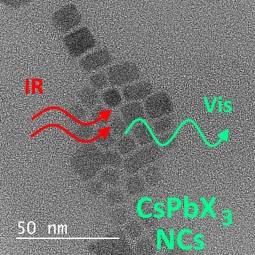
The Two-Photon Absorption Cross-Section Studies of CsPbX3 (X = I, Br, Cl) Nanocrystals

 , ,
, ,  and
and 
Abstract

1. Introduction
2. Materials and Methods
3. Results and Discussion
4. Conclusions
Supplementary Materials
Author Contributions
Funding
Conflicts of Interest
References
- Malinkiewicz, O.; Yella, A.; Lee, Y.H.; Espallargas, G.M.; Graetzel, M.; Nazeeruddin, M.K.; Bolink, H.J. Perovskite solar cells employing organic charge-transport layers. Nat. Photonics 2014, 8, 128–132. [Google Scholar] [CrossRef]
- A decade of perovskite photovoltaics. Nat. Energy 2019, 4, 1. [CrossRef]
- Green, M.A.; Ho-Baillie, A.; Snaith, H.J. The emergence of perovskite solar cells. Nat. Photonics 2014, 8, 506–514. [Google Scholar] [CrossRef]
- Tan, Z.K.; Moghaddam, R.S.; Lai, M.L.; Docampo, P.; Higler, R.; Deschler, F.; Price, M.; Sadhanala, A.; Pazos, L.M.; Credgington, D.; et al. Bright light-emitting diodes based on organometal halide perovskite. Nat. Nanotechnol. 2014, 9, 687–692. [Google Scholar] [CrossRef]
- Deschler, F.; Price, M.; Pathak, S.; Klintberg, L.E.; Jarausch, D.D.; Higler, R.; Huttner, S.; Leijtens, T.; Stranks, S.D.; Snaith, H.J.; et al. High photoluminescence efficiency and optically pumped lasing in solution-processed mixed halide perovskite semiconductors. J. Phys. Chem. Lett. 2014, 5, 1421–1426. [Google Scholar] [CrossRef]
- Bai, S.; Yuan, Z.C.; Gao, F. Colloidal metal halide perovskite nanocrystals: Synthesis, characterization, and applications. J. Mater. Chem. C 2016, 4, 3898–3904. [Google Scholar] [CrossRef]
- Protesescu, L.; Yakunin, S.; Bodnarchuk, M.I.; Krieg, F.; Caputo, R.; Hendon, C.H.; Yang, R.X.; Walsh, A.; Kovalenko, M.V. Nanocrystals of cesium lead halide perovskites (CsPbX3, X = Cl, Br, and I): Novel optoelectronic materials showing bright emission with wide color gamut. Nano Lett. 2015, 15, 3692–3696. [Google Scholar] [CrossRef]
- Nedelcu, G.; Protesescu, L.; Yakunin, S.; Bodnarchuk, M.I.; Grotevent, M.J.; Kovalenko, M.V. Fast anion-exchange in highly luminescent nanocrystals of cesium lead halide perovskites (CsPbX3, X = Cl, Br, I). Nano Lett. 2015, 15, 5635–5640. [Google Scholar] [CrossRef]
- Akkerman, Q.A.; D’Innocenzo, V.; Accornero, S.; Scarpellini, A.; Petrozza, A.; Prato, M.; Manna, L. Tuning the optical properties of cesium lead halide perovskite nanocrystals by anion exchange reactions. J. Am. Chem. Soc. 2015, 137, 10276–10281. [Google Scholar] [CrossRef]
- Kim, Y.; Yassitepe, E.; Voznyy, O.; Comin, R.; Walters, G.; Gong, X.W.; Kanjanaboos, P.; Nogueira, A.F.; Sargent, E.H. Efficient luminescence from perovskite quantum dot solids. ACS Appl. Mater. Int. 2015, 7, 25007–25013. [Google Scholar] [CrossRef]
- Liu, Z.; Hu, Z.; Zhang, Z.; Du, J.; Yang, J.; Tang, X.; Liu, W.; Leng, Y. Two-photon pumped amplified spontaneous emission and lasing from formamidinium lead bromine nanocrystals. ACS Photonics 2019, 6, 3150–3158. [Google Scholar] [CrossRef]
- Wang, Y.; Li, X.M.; Song, J.Z.; Xiao, L.; Zeng, H.B.; Sun, H.D. All-inorganic colloidal perovskite quantum dots: A new class of lasing materials with favorable characteristics. Adv. Mater. 2015, 27, 7101. [Google Scholar] [CrossRef]
- Pan, J.; Sarmah, S.P.; Murali, B.; Dursun, I.; Peng, W.; Parida, M.R.; Liu, J.; Sinatra, L.; Alyami, N.; Zhao, C.; et al. Air-stable surface-passivated perovskite quantum dots for ultra-robust, single- and two-photon-induced amplified spontaneous emission. J. Phys. Chem. Lett. 2015, 6, 5027–5033. [Google Scholar] [CrossRef]
- Xu, Y.Q.; Chen, Q.; Zhang, C.F.; Wang, R.; Wu, H.; Zhang, X.Y.; Xing, G.C.; Yu, W.W.; Wang, X.Y.; Zhang, Y.; et al. Two-photon-pumped perovskite semiconductor nanocrystal lasers. J. Am. Chem. Soc. 2016, 138, 3761–3768. [Google Scholar] [CrossRef]
- Zhang, C.; Zhang, F.; Zhu, T.; Cheng, A.; Xu, J.; Zhang, Q.; Mohney, S.E.; Henderson, R.H.; Wang, Y.A. Two-photon-pumped lasing from colloidal nanocrystal quantum dots. Opt. Lett. 2008, 33, 2437–2439. [Google Scholar] [CrossRef]
- Wang, Y.; Li, X.M.; Zhao, X.; Xiao, L.; Zeng, H.B.; Sun, H.D. Nonlinear absorption and low-threshold multiphoton pumped stimulated emission from all-inorganic perovskite nanocrystals. Nano Lett. 2016, 16, 448–453. [Google Scholar] [CrossRef]
- Chen, J.; Žídek, K.; Chábera, P.; Liu, D.; Cheng, P.; Nuuttila, L.; Al-Marri, M.J.; Lehtivuori, H.; Messing, M.E.; Han, K.; et al. Size- and wavelength-dependent two-photon absorption cross-section of CsPbBr3 perovskite quantum dots. J. Phys. Chem. Lett. 2017, 8, 2316–2321. [Google Scholar] [CrossRef]
- Li, J.; Zhang, S.; Dong, H.; Yuan, X.; Jiang, X.; Wang, J.; Zhang, L. Two-photon absorption and emission in CsPb(Br/I)3 cesium lead halide perovskite quantum dots. CrystEngComm 2016, 18, 7945–7949. [Google Scholar] [CrossRef]
- Pramanik, A.; Gates, K.; Gao, Y.; Begum, S.; Chandra Ray, P. Several orders-of-magnitude enhancement of multiphoton absorption property for CsPbX3 perovskite quantum dots by manipulating halide stoichiometry. J. Phys. Chem. C 2019, 123, 5150–5156. [Google Scholar] [CrossRef]
- Wawrzynczyk, D.; Szeremeta, J.; Samoc, M.; Nyk, M. Third-order nonlinear optical properties of infrared emitting PbS and PbSe quantum dots. J. Phys. Chem. C 2016, 120, 21939–21945. [Google Scholar] [CrossRef]
- Wawrzynczyk, D.; Szeremeta, J.; Samoc, M.; Nyk, M. Optical nonlinearities of colloidal InP@ZnS core-shell quantum dots probed by Z-scan and two-photon excited emission. APL Mater. 2015, 3, 116108. [Google Scholar] [CrossRef]
- Nyk, M.; Szeremeta, J.; Wawrzynczyk, D.; Samoc, M. Enhancement of two-photon absorption cross section in CdSe quantum rods. J. Phys. Chem. C 2014, 118, 17914–17921. [Google Scholar] [CrossRef]
- Szeremeta, J.; Nyk, M.; Wawrzynczyk, D.; Samoc, M. Wavelength dependence of nonlinear optical properties of colloidal CdS quantum dots. Nanoscale 2013, 5, 2388–2393. [Google Scholar] [CrossRef]
- Nyk, M.; Wawrzynczyk, D.; Szeremeta, J.; Samoc, M. Spectrally resolved size-dependent third-order nonlinear optical properties of colloidal CdSe quantum dots. Appl. Phys. Lett. 2012, 100, 041102. [Google Scholar] [CrossRef]
- Samoc, M.; Samoc, A.; Luther-Davies, B.; Bao, Z.N.; Yu, L.P. Femtosecond Z-scan and degenerate four-wave mixing measurements of real and imaginary parts of the third-order nonlinearity of soluble conjugated polymers. J. Opt. Soc. Am. B 1998, 15, 817–825. [Google Scholar] [CrossRef]
- Samoc, M.; Matczyszyn, K.; Nyk, M.; Olesiak-Banska, J.; Wawrzynczyk, D.; Hanczyc, P.; Szeremeta, J.; Wielgus, M.; Gordel, M.; Mazur, L.; et al. Nonlinear absorption and nonlinear refraction: Maximizing the merit factors. Proc. SPIE 2012, 8258. [Google Scholar] [CrossRef]
- Schwich, T.; Cifuentes, M.P.; Gugger, P.A.; Samoc, M.; Humphrey, M.G. Electronic, molecular weight, molecular volume, and financial cost-scaling and comparison of two-photon absorption efficiency in disparate molecules (organometallic complexes for nonlinear optics. 48.)—A response to “Comment on ‘Organometallic complexes for nonlinear optics. 45. dispersion of the third-order nonlinear optical properties of triphenylamine-cored alkynylruthenium dendrimers.’ increasing the nonlinear response by two orders of magnitude”. Adv. Mater. 2011, 23, 1433–1435. [Google Scholar]
- Voura, E.B.; Jaiswal, J.K.; Mattoussi, H.; Simon, S.M. Tracking metastatic tumor cell extravasation with quantum dot nanocrystals and fluorescence emission-scanning microscopy. Nat. Med. 2004, 10, 993–998. [Google Scholar] [CrossRef]
- Chen, W.; Joly, A.G.; McCready, D.E. Upconversion luminescence from CdSe nanoparticles. J. Chem. Phys. 2005, 122, 224708. [Google Scholar] [CrossRef]
- Sheik-Bahae, M.; Said, A.A.; Wei, T.H.; Hagan, D.J.; Van Stryland, E.W. Sensitive measurement of optical nonlinearities using a single beam. IEEE J. Quantum Electron. 1990, 26, 760–769. [Google Scholar] [CrossRef]
- Szeremeta, J.; Kolkowski, R.; Nyk, M.; Samoc, M. Wavelength dependence of the complex third-order nonlinear optical susceptibility of poly(3-hexylthiophene) studied by femtosecond Z-Scan in solution and thin film. J. Phys. Chem. C 2013, 117, 26197–26203. [Google Scholar] [CrossRef]
- Makarov, N.S.; Drobizhev, M.; Rebane, A. Two-photon absorption standards in the 550–1600 nm excitation wavelength range. Opt. Express 2008, 16, 4029–4047. [Google Scholar] [CrossRef] [PubMed]
- Persson, K. Materials DATA on CsPbBr3/CsPbCl3/CsPbI3 (SG:221) 2014 from Materials Project Database. Available online: https://materialsproject.org/ (accessed on 12 February 2020).
- Zhang, X.J.; Wang, H.C.; Tang, A.C.; Lin, S.Y.; Tong, H.C.; Chen, C.Y.; Lee, Y.C.; Tsai, T.L.; Liu, R.S. Robust and stable narrow-band green emitter: An option for advanced wide-color-gamut backlight display. Chem. Mater. 2016, 28, 8493–8497. [Google Scholar] [CrossRef]
- Babin, V.; Fabeni, P.; Nikl, M.; Nitsch, K.; Pazzi, G.P.; Zazubovich, S. Luminescent CsPbI3 and Cs4PbI6 aggregates in annealed CsI:Pb crystals. Phys. Status. Solidi B 2001, 226, 419–428. [Google Scholar] [CrossRef]
- Sharma, S.; Weiden, N.; Weiss, A. Phase diagrams of quasibinary systems of the type: ABX3—A′BX3; ABX3 —AB′X3, and ABX3—ABX′3; X = Halogen. Z. Phys. Chem. 1992, 175, 63–80. [Google Scholar] [CrossRef]
- Lou, S.; Xuan, T.; Wang, J. Stability: A desiderated problem for the lead halide perovskites. Opt. Mater. X 2019, 1, 100023. [Google Scholar] [CrossRef]
- Liu, M.N.; Matuhina, A.; Zhang, H.C.; Vivo, P. Advances in the stability of halide perovskite nanocrystals. Materials 2019, 12, 3733. [Google Scholar] [CrossRef]
- Boote, B.W.; Andaraarachchi, H.P.; Rosales, B.A.; Blome-Fernández, R.; Zhu, F.; Reichert, M.D.; Santra, K.; Li, J.; Petrich, J.W.; Vela, J.; et al. Unveiling the photo- and thermal-stability of cesium lead halide perovskite nanocrystals. Chem. Phys. Chem. 2019, 20, 2647–2656. [Google Scholar] [CrossRef]
- Chen, J.; Liu, D.; Al-Marri, M.J.; Nuuttila, L.; Lehtivuori, H.; Zheng, K. Photo-stability of CsPbBr3 perovskite quantum dots for optoelectronic application. Sci. China Mater. 2016, 59, 719. [Google Scholar] [CrossRef]
- Zhang, C.; Fernando, J.F.S.; Firestein, K.L.; Treifeldt, J.E.v.; Siriwardena, D.; Fang, X.; Golberg, D. Thermal stability of CsPbBr3 perovskite as revealed by in situ transmission electron microscopy. APL Mater. 2019, 7. [Google Scholar] [CrossRef]
- Albota, M.; Beljonne, D.; Bredas, J.L.; Ehrlich, J.E.; Fu, J.Y.; Heikal, A.A.; Hess, S.E.; Kogej, T.; Levin, M.D.; Marder, S.R.; et al. Design of organic molecules with large two-photon absorption cross sections. Science 1998, 281, 1653–1656. [Google Scholar] [CrossRef]
- Sutton, R.J.; Filip, M.R.; Haghighirad, A.A.; Sakai, N.; Wenger, B.; Giustino, F.; Snaith, H.J. Cubic or orthorhombic? Revealing the crystal structure of metastable black-phase CsPbI3 by theory and experiment. ACS Energy Lett. 2018, 3, 1787–1794. [Google Scholar] [CrossRef]
- Rodová, M.; Brožek, J.; Knížek, K.; Nitsch, K. Phase transitions in ternary caesium lead bromide. J. Therm. Anal. Calorim. 2003, 71, 667–673. [Google Scholar] [CrossRef]
- López, C.A.; Abia, C.; Alvarez-Galván, M.C.; Hong, B.-K.; Martínez-Huerta, M.V.; Serrano-Sánchez, F.; Carrascoso, F.; Castellanos-Gómez, A.; Fernández-Díaz, M.T.; Alonso, J.A. Crystal structure features of CsPbBr3 perovskite prepared by mechanochemical synthesis. ACS Omega 2020, 5, 5931–5938. [Google Scholar] [CrossRef] [PubMed]
- Kim, M.K.; Jo, V.; Ok, K.M. New variant of highly symmetric layered perovskite with coordinated NO3− ligand: Hydrothermal synthesis, structure, and characterization of Cs2PbCl2(NO3)2. Inorg. Chem. 2009, 48, 7368–7372. [Google Scholar] [CrossRef] [PubMed]
- Balu, M.; Padilha, L.A.; Hagan, D.J.; Van Stryland, E.W.; Yao, S.; Belfield, K.; Zheng, S.J.; Barlow, S.; Marder, S. Broadband Z-scan characterization using a high-spectral-irradiance, high-quality supercontinuum. J. Opt. Soc. Am. B 2008, 25, 159–165. [Google Scholar] [CrossRef]


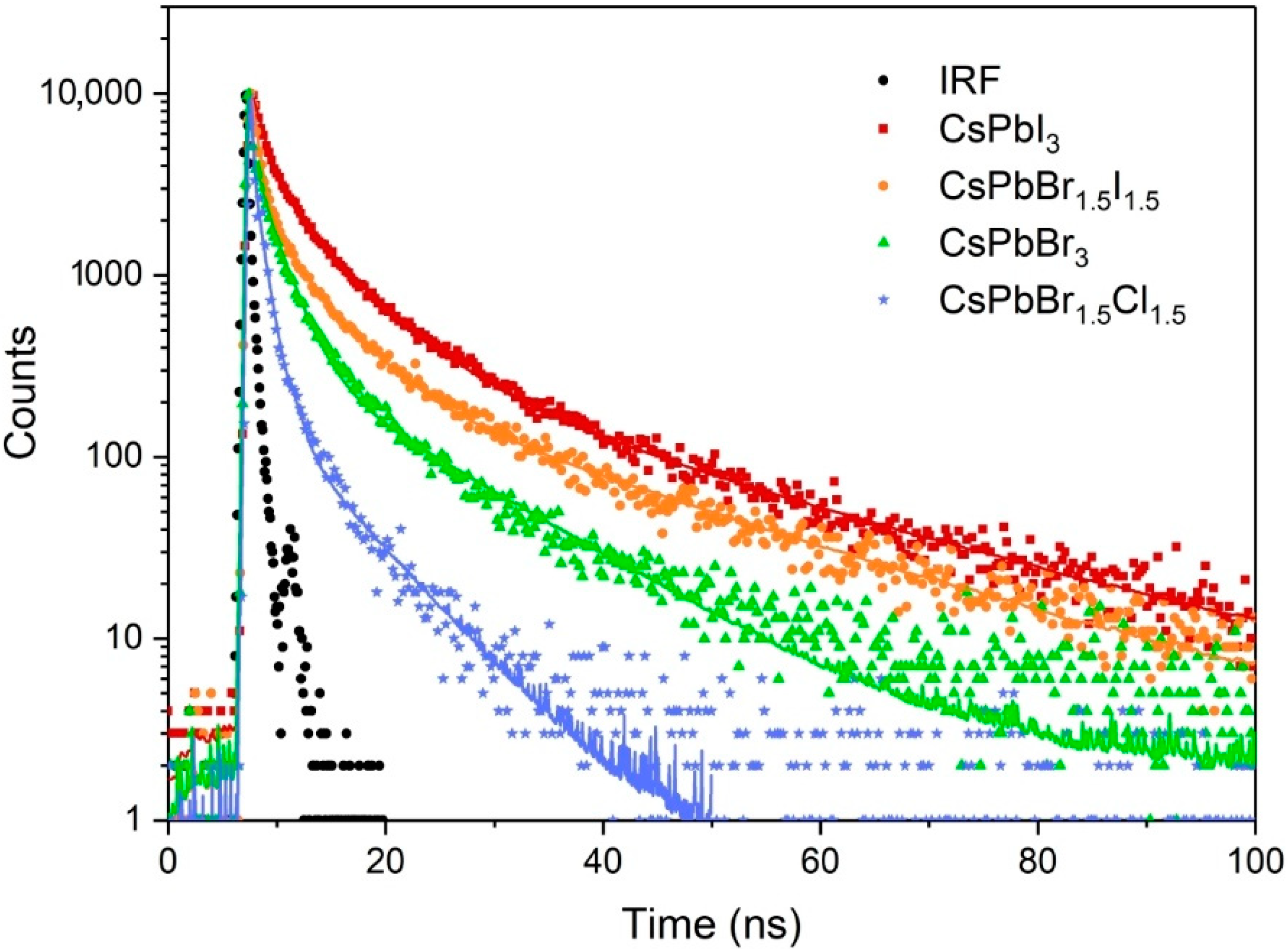
| Perovskite Formula | Diameter from DLS (nm) | Max. Emission λ (nm) | Quantum Yield (%) | Emission Lifetime (ns) | ||
|---|---|---|---|---|---|---|
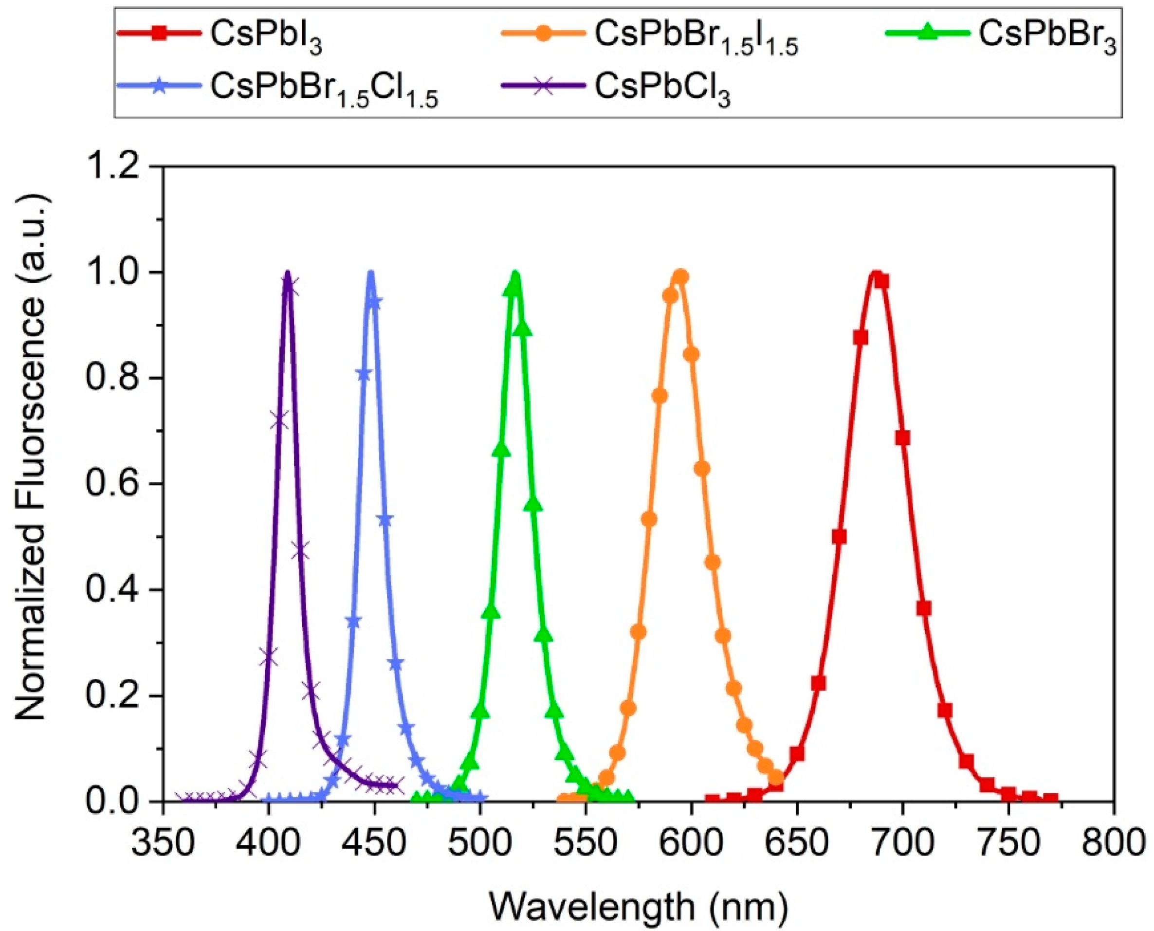
| CsPbI3 | 16.3 ± 2.8 | 687 | 7.3 | 0.96 ± 0.01 | 5.65 ± 0.04 | 23.6 ± 0.2 |
| CsPbBr1.5I1.5 | 19.8 ± 3.3 | 593 | 6.2 | 0.55 ± 0.01 | 4.55 ± 0.04 | 22.1 ± 0.15 |
| CsPbBr3 | 19.6 ± 3.0 | 516 | 8.1 | 0.03 ± 0.01 | 2.03 ± 0.01 | 12.2 ± 0.08 |
| CsPbBr1.5Cl1.5 | 16.1 ± 3.1 | 448 | 4.0 | 0.01 ± 0.01 | 1.03 ± 0.01 | 6.58 ± 0.08 |
| CsPbCl3 | 21.7 ± 2.6 | 409 | 0.7 | NA | NA | NA |
| Perovskite Formula | ||||||
|---|---|---|---|---|---|---|
| CsPbI3 | 54 ± 7 | NA | 720.8 | 0.075 ± 0.012 | 5.7 × 10⁵ | 5.5 ± 0.6 |
| CsPbBr1.5I1.5 | 10 ± 2 | 114 ± 32 | 610.0 | 0.016 ± 0.002 | 8.3 × 10⁵ | 1.4 ± 0.2 |
| CsPbBr3 | 97 ± 9 | 67 ± 16 | 579.8 | 0.17 ± 0.02 | 8.4 × 10⁵ | 14.1 ± 1.4 |
| CsPbBr1.5Cl1.5 | 170 ± 9 | 23 ± 4 | 512.6 | 0.33 ± 0.02 | 7.9 × 10⁵ | 26.5 ± 1.4 |
| CsPbCl3 | 10 ± 1 | NA | 446.5 | 0.022 ± 0.002 | 8.8 × 10⁵ | 2.0 ± 0.2 |
| Perovskite Formula | ||||
|---|---|---|---|---|
| CsPbI3 | 29 ± 6 | 1.9 ± 0.8 | 1.0 ± 0.05 | 5.3 |
| CsPbBr1.5I1.5 | 92 ± 16 | 3.6 ± 0.9 | 2.1 ± 0.06 | 65.7 |
| CsPbBr3 | 347 ± 42 | 13.5 ± 1.6 | 7.8 ± 0.1 | 24.6 |
| CsPbBr1.5Cl1.5 | 71 ± 38 | 2.6 ± 0.4 | 1.5 ± 0.08 | 2.7 |
| CsPbCl3 | 120 ± 23 | 3.3 ± 0.5 | 1.9 ± 0.05 | 60 |
© 2020 by the authors. Licensee MDPI, Basel, Switzerland. This article is an open access article distributed under the terms and conditions of the Creative Commons Attribution (CC BY) license (http://creativecommons.org/licenses/by/4.0/).
Share and Cite
Szeremeta, J.; Antoniak, M.A.; Wawrzyńczyk, D.; Nyk, M.; Samoć, M. The Two-Photon Absorption Cross-Section Studies of CsPbX3 (X = I, Br, Cl) Nanocrystals. Nanomaterials 2020, 10, 1054. https://doi.org/10.3390/nano10061054
Szeremeta J, Antoniak MA, Wawrzyńczyk D, Nyk M, Samoć M. The Two-Photon Absorption Cross-Section Studies of CsPbX3 (X = I, Br, Cl) Nanocrystals. Nanomaterials. 2020; 10(6):1054. https://doi.org/10.3390/nano10061054
Chicago/Turabian StyleSzeremeta, Janusz, Magda A. Antoniak, Dominika Wawrzyńczyk, Marcin Nyk, and Marek Samoć. 2020. "The Two-Photon Absorption Cross-Section Studies of CsPbX3 (X = I, Br, Cl) Nanocrystals" Nanomaterials 10, no. 6: 1054. https://doi.org/10.3390/nano10061054
APA StyleSzeremeta, J., Antoniak, M. A., Wawrzyńczyk, D., Nyk, M., & Samoć, M. (2020). The Two-Photon Absorption Cross-Section Studies of CsPbX3 (X = I, Br, Cl) Nanocrystals. Nanomaterials, 10(6), 1054. https://doi.org/10.3390/nano10061054