Multimodal/Multifunctional Nanomaterials in (Bio)electrochemistry: Now and in the Coming Decade


Abstract
1. Introduction
2. Multifunctional Metal-Based Nanomaterials
2.1. Metal Nanoparticles (MNPs)
2.1.1. Metal-Based Nanozymes
2.1.2. Multifunctional MNPs in Electrochemical Affinity Bioplatforms
2.2. Multifunctional MNPs Involving Ordered Nanostructures
2.3. Multifunctional Nanomaterials Involving Quantum Dots (QDs)
2.4. Two-Dimensional (2D) Transition Metal Multifunctional Nanomaterials
3. Multifunctional Carbon Nanomaterials
3.1. Magnetic Carbon Nanomaterials
3.2. Carbon Nanozymes
3.3. Multifunctional Biomedical Applications
3.4. Multifunctional Carbon Nanomaterials for Signal Amplification
4. Multifunctional Silica Nanomaterials
5. Overview and Look to the Future
Author Contributions
Funding
Acknowledgments
Conflicts of Interest
References
- Yoon, J.; Shin, M.; Lee, T.; Choi, J.-W. Highly sensitive biosensors based on biomolecules and functional nanomaterials depending on the types of nanomaterials: A perspective review. Materials 2020, 13, 299. [Google Scholar] [CrossRef]
- Wongkaew, N.; Simsek, M.; Griesche, C.; Baeumner, A.J. Functional nanomaterials and nanostructures enhancing electrochemical biosensors and lab-on-a-chip performances: Recent progress, applications, and future perspective. Chem. Rev. 2019, 119, 120–194. [Google Scholar] [CrossRef] [PubMed]
- Cho, I.-H.; Kim, D.H.; Park, S. Electrochemical biosensors: Perspective on functional nanomaterials for on-site analysis. Biomater. Res. 2020, 24, 6. [Google Scholar] [CrossRef]
- Bezinge, L.; Suea-Ngam, A.; deMello, A.J.; Shih, C.-J. Nanomaterials for molecular signal amplification in electrochemical nucleic acid biosensing: Recent advances and future prospects for point-of-care diagnostics. Mol. Syst. Des. Eng. 2020, 5, 49. [Google Scholar] [CrossRef]
- Simón de Dios, A.; Díaz-García, M.E. Multifunctional nanoparticles: Analytical prospects. Anal. Chim. Acta 2010, 666, 1. [Google Scholar] [CrossRef] [PubMed]
- Dimcheva, N.; Horozova, E.; Ivanov, Y.; Godjevrgova, T. Self-assembly of acetylcholinesterase on gold nanoparticles electrodeposited on graphite. Cent. Eur. J. Chem. 2013, 11, 1740–1748. [Google Scholar] [CrossRef]
- Niu, Y.; Liu, J.; Chen, W.; Yin, C.; Weng, W.; Li, X.; Wang, X.; Li, G.; Sun, W.A. Direct electron transfer biosensor based on a horseradish peroxidase and gold nanotriangle modified electrode and electrocatalysis. Anal. Methods 2018, 10, 5297. [Google Scholar] [CrossRef]
- Tang, H.; Cai, D.; Ren, T.; Xiong, P.; Liu, Y.; Gu, H.; Shi, G. Fabrication of a low background signal glucose biosensor with 3D network materials as the electrocatalyst. Anal. Biochem. 2019, 567, 63–71. [Google Scholar] [CrossRef]
- Guzsvány, V.; Anojčić, J.; Vajdle, O.; Radulović, E.; Madarász, D.; Kónya, Z.; Kalcher, K. Amperometric determination of glucose in white grape and in tablets as ingredient by screen-printed electrode modified with glucose oxidase and composite of platinum and multiwalled carbon nanotubes. Food Anal. Methods 2019, 12, 570–580. [Google Scholar] [CrossRef]
- Jiang, Y.; Zhang, X.; Pei, L.; Yue, S.; Ma, L.; Zhou, L.; Huang, Z.; He, Y.; Gao, J. Silver nanoparticles modified two-dimensional transition metal carbides as nanocarriers to fabricate acetylcholinesterase-based electrochemical biosensor. Chem. Eng. J. 2018, 339, 547–556. [Google Scholar] [CrossRef]
- Chang, H.; Zhang, H.; Lv, J.; Zhang, B.; Wei, W.; Guo, J. Pt NPs and DNAzyme functionalized polymer nanospheres as triple signal amplification strategy for highly sensitive electrochemical immunosensor of tumour marker. Biosens. Bioelectron. 2016, 86, 156–163. [Google Scholar] [CrossRef] [PubMed]
- Dong, L.; Yin, L.; Tian, G.; Wang, Y.; Pei, H.; Wu, Q.; Cheng, W.; Ding, S.; Xia, Q. An enzyme-free ultrasensitive electrochemical immunosensor for calprotectin detection based on PtNi nanoparticles functionalized 2D Cu metal organic framework nanosheets. Sens. Actuators B-Chem. 2020, 308, 127687. [Google Scholar] [CrossRef]
- Ehzari, H.; Amiri, M.; Safari, M. Enzyme-free sandwich-type electrochemical immunosensor for highly sensitive prostate specific antigen based on conjugation of quantum dots and antibody on surface of modified glassy carbon electrode with core-shell magnetic metal-organic frameworks. Talanta 2020, 210, 120641. [Google Scholar] [CrossRef] [PubMed]
- Feng, J.; Li, Y.; Li, M.; Li, F.; Han, J.; Dong, Y.; Chen, Z.; Wang, P.; Liu, H.; Wei, Q. A novel sandwich-type electrochemical immunosensor for PSA detection based on PtCu bimetallic hybrid (2D/2D) rGO/g-C3N4. Biosens. Bioelectron. 2017, 91, 441–448. [Google Scholar] [CrossRef]
- Hartati, Y.W.; Letelay, L.K.; Gaffar, S.; Wyantuti, S.; Bahti, H.H. Cerium oxide-monoclonal antibody bioconjugate for electrochemical immunosensing of HER2 as a breast cancer biomarker. Sens. Biosens. Res. 2000, 27, 100316. [Google Scholar] [CrossRef]
- Lah, Z.M.A.N.H.; Ahmad, S.A.A.; Zaini, M.S.; Kamarudin, M.A. An electrochemical sandwich immunosensor for the detection of HER2 using antibody-conjugated PbS quantum dot as a label. J. Pharm. Biomed. Anal. 2019, 174, 608–617. [Google Scholar] [CrossRef]
- Li, F.; Li, Y.; Feng, J.; Dong, Y.; Wang, P.; Chen, L.; Chen, Z.; Liu, H.; Wei, Q. Ultrasensitive amperometric immunosensor for PSA detection based on Cu2O@CeO2-Au nanocomposites as integrated triple signal amplification strategy. Biosens. Bioelectron. 2017, 87, 630–637. [Google Scholar] [CrossRef]
- Li, Y.; Zhang, Y.; Li, F.; Feng, J.; Li, M.; Chen, L.; Dong, Y. Ultrasensitive electrochemical immunosensor for quantitative detection of SCCA using Co3O4@CeO2-Au@Pt nanocomposite as enzyme-mimetic labels. Biosens. Bioelectron. 2017, 92, 33–39. [Google Scholar] [CrossRef]
- Liu, Y.; He, G.; Liu, H.; Yin, H.; Gao, F.; Chen, J.; Zhang, S.; Yang, B. Electrochemical immunosensor based on AuBP@Pt nanostructure and AuPd-PDA nanozyme for ultrasensitive detection of APOE4. RSC Adv. 2020, 10, 7912. [Google Scholar] [CrossRef]
- Martín-Yerga, D.; González-García, M.B.; Costa-García, A. Electrochemical immunosensor for anti-tissue transglutaminase antibodies based on the in situ detection of quantum dots. Talanta 2014, 130, 598–602. [Google Scholar]
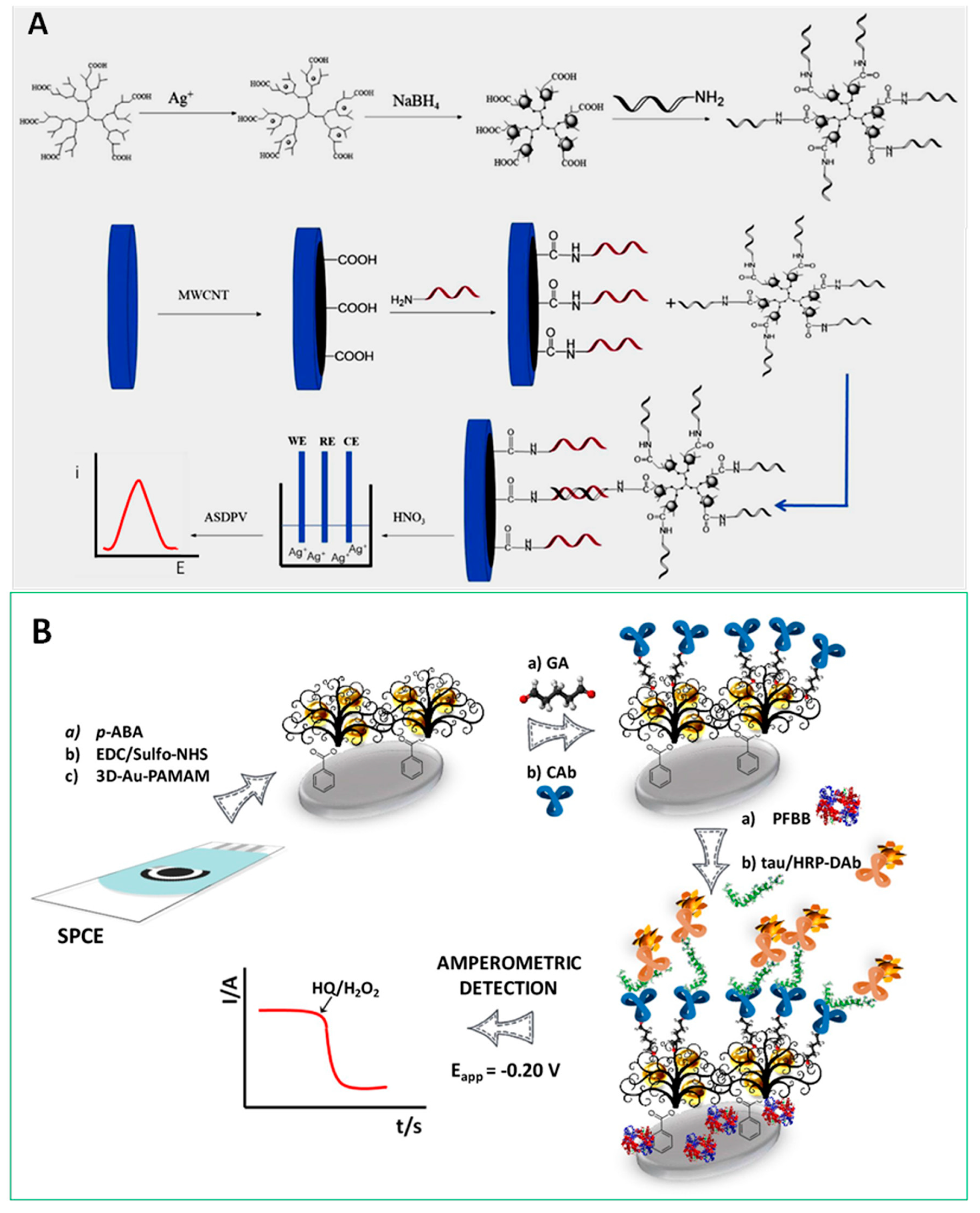
- Razzino, C.A.; Serafín, V.; Gamella, M.; Pedrero, M.; Montero-Calle, A.; Barderas, R.; Calero, M.; Lobo, A.O.; Yáñez-Sedeño, P.; Campuzano, S.; et al. An electrochemical immunosensor using gold nanoparticles-PAMAM-nanostructured screen-printed carbon electrodes for tau protein determination in plasma and brain tissues from Alzheimer patients. Biosens. Bioelectron. 2020, 163, 112238. [Google Scholar] [CrossRef] [PubMed]
- Serafín, V.; Razzino, C.A.; Gamella, M.; Pedrero, M.; Povedano, E.; Montero-Calle, A.; Barderas, R.; Calero, M.; Lobo, A.O.; Yáñez-Sedeño, P.; et al. Disposable immunoplatforms for the simultaneous determination of biomarkers for neurodegenerative disorders using poly(amidoamine) dendrimer/gold nanoparticle nanocomposite. Anal. Bioanal. Chem. 2020. [Google Scholar] [CrossRef] [PubMed]
- Shen, Y.; Shen, G.; Zhang, Y. Voltammetric immunoassay for α-fetoprotein by using a gold nanoparticle/dendrimer conjugate and a ferrocene derived ionic liquid. Microchim. Acta 2018, 185, 346. [Google Scholar] [CrossRef] [PubMed]
- Su, S.; Sun, Q.; Wan, L.; Gu, X.; Zhu, D.; Zhou, Y.; Chao, J.; Wang, L. Ultrasensitive analysis of carcinoembryonic antigen based on MoS2-based electrochemical immunosensor with triple signal amplification. Biosens. Bioelectron. 2019, 140, 111353. [Google Scholar] [CrossRef]
- Yu, S.; Zou, G.; Wei, Q. Ultrasensitive electrochemical immunosensor for quantitative detection of tumor specific growth factor by using Ag@CeO2 nanocomposite as labels. Talanta 2016, 156–157, 11–17. [Google Scholar] [CrossRef]
- Das, R.; Dhiman, A.; Kapil, A.; Bansal, V.; Sharma, T.K. Aptamer-mediated colorimetric and electrochemical detection of Pseudomonas aeruginosa utilizing peroxidase-mimic activity of gold NanoZyme. Anal. Bioanal. Chem. 2019, 411, 1229–1238. [Google Scholar] [CrossRef]
- Fu, X.-M.; Liu, Z.-J.; Cai, S.-X.; Zhao, Y.-P.; Wu, D.-Z.; Li, C.-Y.; Chen, J.-H. Electrochemical aptasensor for the detection of vascular endothelial growth factor (VEGF) based on DNA-templated Ag/Pt bimetallic nanoclusters. Chin. Chem. Lett. 2016, 27, 920–926. [Google Scholar] [CrossRef]
- Liu, H.; Xu, S.; He, Z.; Deng, A.; Zhu, J.J. Supersandwich cytosensor for selective and ultrasensitive detection of cancer cells using aptamer-DNS concatamer-Quantum Dots probes. Anal. Chem. 2013, 85, 3385–3392. [Google Scholar]
- Ou, D.; Sun, D.; Lin, X.; Liang, Z.; Zhong, Y.; Chen, Z. A dual-aptamer-based biosensor for specific detection of breast cancer biomarker HER2 via flower-like nanozymes and DNA nanostructures. J. Mater. Chem. B 2019, 7, 3661–3669. [Google Scholar] [CrossRef]
- Wang, Y.; Ning, G.; Bi, H.; Wu, Y.; Liu, G.; Zhao, Y. A novel ratiometric electrochemical assay for ochratoxin A coupling Au nanoparticles decorated MoS2 nanosheets with aptamer. Electrochim. Acta 2018, 285, 120–127. [Google Scholar] [CrossRef]
- Zhao, R.-N.; Feng, Z.; Zhao, Y.-N.; Jia, L.-P.; Ma, R.-N.; Zhang, W.; Shang, L.; Xue, Q.-W.; Wang, H.-S. A sensitive electrochemical aptasensor for Mucin 1 detection based on catalytic hairpin assembly coupled with PtPdNPs peroxidase-like activity. Talanta 2019, 200, 503–510. [Google Scholar] [CrossRef] [PubMed]
- Jin, X.; Zhou, L.; Zhu, B.; Jiang, X.; Zhu, N. Silver-dendrimer nanocomposites as oligonucleotide labels for electrochemical stripping detection of DNA hybridization. Biosens. Bioelectron. 2018, 107, 237–243. [Google Scholar] [CrossRef] [PubMed]
- Liu, L.; Chang, Y.; Xia, N.; Peng, P.; Zhang, L.; Jiang, M.; Zhang, J.; Liu, L. Simple, sensitive and label-free electrochemical detection of microRNAs based on the in situ formation of silver nanoparticles aggregates for signal amplification. Biosens. Bioelectron. 2017, 94, 235–242. [Google Scholar] [CrossRef] [PubMed]
- Liu, L.; Xing, Y.; Zhang, H.; Liu, R.; Liu, H.; Xia, N. Amplified voltammetric detection of glycoproteins using 4-mercaptophenylboronic acid/biotin-modified multifunctional gold nanoparticles as labels. Int. J. Nanomed. 2014, 9, 2619–2626. [Google Scholar]
- Liu, L.; Xia, N.; Liu, H.; Kang, X.; Liu, X.; Xue, C.; He, X. Highly sensitive and label-free electrochemical detection of microRNAs based on triple signal amplification of multifunctional gold nanoparticles, enzymes and redox-cycling reaction. Biosens. Bioelectron. 2014, 53, 399–405. [Google Scholar] [CrossRef] [PubMed]
- Ma, X.; Qian, K.; Ejeromedoghene, O.; Kandawa-Schulz, M.; Wang, Y. Electrochemical detection of microRNA based on SA-PPy/AuNPs nanocomposite with the signal amplification through catalytic hairpin assembly reaction and the spontaneous catalytic reaction of Fe3+/Cu2+. Electrochim. Acta 2020, 362, 137168. [Google Scholar] [CrossRef]
- Park, S.Y.; Kim, J.; Yim, G.; Jang, H.; Lee, Y.; Kim, S.M.; Park, C.; Lee, M.-H.; Lee, T. Fabrication of electrochemical biosensor composed of multi-functional DNA/rhodium nanoplate heterolayer for thyroxine detection in clinical sample. Colloids Surf. B Biointerfaces 2020, 195, 111240. [Google Scholar] [CrossRef]
- Tian, L.; Qian, K.; Qi, J.; Liu, Q.; Yao, C.; Song, W.; Wang, Y. Gold nanoparticles superlattices assembly for electrochemical biosensor detection of microRNA-21. Biosens. Bioelectron. 2018, 99, 564–570. [Google Scholar] [CrossRef]
- Yan, T.; Zhu, L.; Ju, H.; Lei, J. DNA-Walker-Induced Allosteric Switch for Tandem Signal Amplification with Palladium Nanoparticles/Metal-Organic Framework Tags in Electrochemical Biosensing. Anal. Chem. 2018, 90, 14493–14499. [Google Scholar] [CrossRef]
- Zhang, W.; Dai, Z.; Liu, X.; Yang, J. High-Performance Electrochemical Sensing of Circulating Tumor DNA in Peripheral Blood Based on Poly-Xanthurenic Acid Functionalized MoS2 Nanosheets. Biosens. Bioelectron. 2018, 105, 116–120. [Google Scholar] [CrossRef]
- Dimecheva, N. Nanostructures of noble metals as functional materials in biosensors. Curr. Opin. Electrochem. 2020, 19, 35–41. [Google Scholar] [CrossRef]
- Du, D.; Wang, M.; Cai, J.; Qin, Y.; Zhang, A. One-step synthesis of multiwalled carbon nanotubes-gold nanocomposites for fabricating amperometric acetylcholinesterase biosensor. Sens. Actuators B-Chem. 2010, 143, 524–529. [Google Scholar] [CrossRef]
- Zhang, P.; Sun, T.; Rong, S.; Zeng, D.; Yu, H.; Zhang, Z.; Chang, D.; Pan, H. A sensitive amperometric AChE-biosensor for organophosphate pesticides detection based on conjugated polymer and Ag-rGO-NH2 nanocomposite. Bioelectrochemistry 2019, 127, 163–170. [Google Scholar] [CrossRef] [PubMed]
- Wang, C.; Liu, C.; Luo, J.; Tian, Y.; Zhou, N. Direct electrochemical detection of kanamycin based on peroxidase-like activity of gold nanoparticles. Anal. Chim. Acta 2016, 936, 75–82. [Google Scholar] [CrossRef]
- Stasyuk, N.; Gayda, G.; Zakalskiy, A.; Zakalska, O.; Serkiz, R.; Gonchar, M. Amperometric biosensors based on oxidases and PtRu nanoparticles as artificial peroxidase. Food Chem. 2019, 285, 213–220. [Google Scholar] [CrossRef]
- Kim, H.Y.; Song, J.; Park, K.S.; Park, H.G. Simple and label-free strategy for terminal transferase assay using a personal glucose meter. Chem. Commun. 2020, 56, 8912–8915. [Google Scholar] [CrossRef]
- Chen, J.; Yu, C.; Zhao, Y.; Niu, Y.; Zhang, L.; Yu, Y.; Wu, J.; He, J. A novel non-invasive detection method for the FGFR3 gene mutation in maternal plasma for a fetal achondroplasia diagnosis based on signal amplification by hemin-MOFs/PtNPs. Biosens. Bioelectron. 2017, 91, 892–899. [Google Scholar] [CrossRef]
- He, L.; Duan, F.; Song, Y.; Guo, C.; Zhao, H.; Tian, J.-Y.; Zhang, Z.; Liu, C.-S.; Zhang, X.; Wang, P.; et al. 2D zirconium-based metal-organic framework nanosheets for highly sensitive detection of mucin 1: Consistency between electrochemical and surface plasmon resonance methods. 2D Mater. 2017, 4, 025098. [Google Scholar] [CrossRef]
- Li, Y.; Xie, M.; Zhang, X.; Liu, Q.; Lin, D.; Xu, C.; Xie, F. Co-MOF nanosheet array: A high-performance electrochemical sensor for non-enzymatic glucose detection. Sens. Actuators. B Chem. 2019, 278, 126–132. [Google Scholar] [CrossRef]
- Fatima, B.; Hussain, D.; Bashir, S.; Hussain, H.T.; Aslam, R.; Nawaz, R.; Rashid, H.N.; Bashir, N.; Majeed, S.; Ashiq, M.N.; et al. Catalase immobilized antimonene quantum dots used as an electrochemical biosensor for quantitative determination of H2O2 from CA-125 diagnosed ovarian cancer samples. Mater. Sci. Eng. C 2020, 117, 111296. [Google Scholar] [CrossRef]
- Freitas, M.; Neves, M.M.P.S.; Nouws, H.P.A.; Delerue-Matos, C. Quantum dots as nanolabels for breast cancer biomarker HER2-ECD analysis in human serum. Talanta 2020, 208, 120430. [Google Scholar] [CrossRef]
- Li, C.-C.; Hu, J.; Lu, M.; Zhang, C.-Y. Quantum dot-based electrochemical biosensor for stripping voltammetric detection of telomerase at the single-cell level. Biosens. Bioelectron. 2018, 122, 51–57. [Google Scholar] [CrossRef] [PubMed]
- Yang, B.; Zhang, S.; Fang, X.; Kong, J. Double signal amplification strategy for ultrasensitive electrochemical biosensor based on nuclease and quantum dot-DNA nanocomposites in the detection of breast cancer 1 gene mutation. Biosens. Bioelectron. 2019, 142, 111544. [Google Scholar] [CrossRef] [PubMed]
- Rezaei, H.; Motovali-bashi, M.; Radfar, S. An enzyme-free electrochemical biosensor for simultaneous detection of two hemophilia A biomarkers: Combining target recycling with quantum dots-encapsulated metal-organic frameworks for signal amplification. Anal. Chim. Acta 2019, 1092, 66–74. [Google Scholar] [CrossRef] [PubMed]
- Shuai, H.-L.; Huang, K.-J.; Chen, Y.-X.; Fang, L.-X.; Jia, M.-P. Au nanoparticles/hollow molybdenum disulfide microcubes based biosensor for microRNA-21 detection coupled with duplex-specific nuclease and enzyme signal amplification. Biosens. Bioelectron. 2019, 89, 989–997. [Google Scholar] [CrossRef]
- Hong, G.; Chen, R.; Xu, L.; Lu, X.; Yang, Z.; Zhou, G.; Li, L.; Chen, W.; Peng, H. One-pot ultrasonic synthesis of multifunctional Au nanoparticle ferrocene-WS2 nanosheet composite for the construction of an electrochemical biosensing platform. Anal. Chim. Acta 2020, 1099, 52–59. [Google Scholar] [CrossRef]
- Yao, P.; Yu, S.; Shen, H.; Yang, J.; Min, L.; Yang, Z.; Zhu, X. A TiO2–SnS2 nanocomposite as a novel matrix for the development of an enzymatic electrochemical glucose biosensor. New J. Chem. 2019, 43, 16748. [Google Scholar] [CrossRef]
- Kumar, S.; Lei, Y.; Alshareef, N.H.; Quevedo-Lopez, M.A.; Salama, K.N. Biofunctionalized two-dimensional Ti3C2 MXenes for ultrasensitive detection of cancer biomarker. Biosens. Bioelectron. 2018, 121, 243–249. [Google Scholar] [CrossRef]
- Liu, L.; Wei, Y.; Jiao, S.; Zhu, S.; Liu, X. A novel label-free strategy for the ultrasensitive miRNA-182 detection based on MoS2/Ti3C2 nanohybrids. Biosens. Bioelectron. 2019, 137, 45–51. [Google Scholar] [CrossRef]
- Golchin, K.; Golchin, J.; Ghaderi, S.; Alidadiani, N.; Eslamkhah, S.; Eslamkhah, M.; Davaran, S.; Akbarzadeh, A. Gold nanoparticles applications: From artificial enzyme till drug delivery. Artif. Cells Nanomed. Biotechnol. 2018, 46, 250–254. [Google Scholar] [CrossRef]
- Rick, J.; Tsai, M.-C.; Hwang, B.J. Biosensors incorporating Bimetallic Nanoparticles. Nanomaterials 2015, 6, 5. [Google Scholar] [CrossRef] [PubMed]
- Gao, L.; Zhuang, J.; Nie, L.; Zhang, J.; Zhang, Y.; Gu, N.; Wang, T.; Feng, J.; Yang, D.; Perrett, S.; et al. Intrinsic peroxidase-like activity of ferromagnetic nanoparticles. Nat. Nanotechnol. 2007, 2, 577–583. [Google Scholar] [CrossRef] [PubMed]
- Yang, Y.; Mao, Z.; Huang, W.; Liu, L.; Li, J.; Li, J.; Wu, Q. Redox enzyme-mimicking activities of CeO2 nanostructures: Intrinsic influence of exposed facets. Sci. Rep. 2016, 6, 35344. [Google Scholar] [CrossRef] [PubMed]
- Verma, N.; Kumar, N. Synthesis and biomedical applications of copper oxide nanoparticles: An expanding horizon. ACS Biomater. Sci. Eng. 2019, 5, 1170–1188. [Google Scholar] [CrossRef]
- Yang, T.; Fruergaard, A.S.; Winther, A.K.; Zelikin, A.N.; Chandrawati, R. Zinc oxide particles catalytically generate nitric oxide from endogenous and exogenous products. Small 2020, 16, 1906744. [Google Scholar] [CrossRef]
- Mu, J.; Wang, Y.; Zhao, M.; Zhang, L. Intrinsic peroxidase-like activity and catalase-like activity of Co3O4 nanoparticles. Chem. Commun. 2012, 48, 2540–2542. [Google Scholar] [CrossRef]
- Yue, Y.; Wei, H.; Guo, J.; Yang, Y. Ceria-based peroxidase-mimicking nanozyme with enhanced activity: A coordination chemistry strategy. Colloids Surf. A Physicochem. Eng. Asp. 2020, 125715. [Google Scholar] [CrossRef]
- Singh, S. Cerium oxide based nanozymes: Redox phenomenon at biointerfaces. Biointerphases 2016, 11, 04B202. [Google Scholar] [CrossRef]
- Zhao, F.; Sun, T.; Geng, F.; Chen, P.; Gao, Y. Metal-Organic Frameworks-Based Electrochemical Sensors and Biosensors. Int. J. Electrochem. Sci. 2019, 14, 5287–5304. [Google Scholar] [CrossRef]
- Ulhakim, M.T.; Rezki, M.; Dewi, K.K.; Abrori, S.A.; Harimurti, S.; Septiani, N.L.W.; Kurnia, K.A.; Setyaningsih, W.; Darmawan, N.; Yuliarto, B. Review—Recent trend on two-dimensional metal-organic frameworks for electrochemical biosensor application. J. Electrochem. Soc. 2020, 167, 136509. [Google Scholar] [CrossRef]
- Sánchez, A.; Villalonga, A.; Martínez-García, G.; Parrado, C.; Villalonga, R. Dendrimers as soft nanomaterials for electrochemical immunosensors. Nanomaterials 2019, 9, 1745. [Google Scholar] [CrossRef]
- Kokkinos, C.; Economou, A. Emerging trends in biosensing using stripping voltammetric detection of metal-containing nanolabels—A review. Anal. Chim. Acta 2017, 961, 12–32. [Google Scholar] [CrossRef] [PubMed]
- Bolotsky, A.; Butler, D.; Dong, C.; Gerace, K.; Glavin, N.R.; Muratore, C.; Robinson, J.A.; Ebrahim, A. Two-dimensional materials in biosensing and healthcare: From in vitro diagnostics to optogenetics and beyond. ACS Nano 2019, 13, 9781–9810. [Google Scholar] [CrossRef] [PubMed]
- Wen, W.; Song, Y.; Yan, X.; Zhu, C.; Du, D.; Wang, S.; Asiri, A.M.; Lin, Y. Recent advances in emerging 2D nanomaterials for biosensing and bioimaging applications. Mater. Today 2018, 21, 2. [Google Scholar] [CrossRef]
- Kalambate, P.K.; Gadhari, N.S.; Li, X.; Rao, Z.; Navale, S.T.; Shen, Y.; Patil, V.R.; Huang, Y. Recent advances in MXene-based electrochemical sensors and biosensors. TrAC Trends Anal. Chem. 2019, 120, 115643. [Google Scholar] [CrossRef]
- Lorencova, L.; Bertok, T.; Filip, J.; Jerigova, M.; Velic, D.; Kasak, P.; Mahmoud, K.A.; Tkac, J. Highly stable Ti3C2Tx (MXene)/Pt nanoparticles-modified glassy carbon electrode for H2O2 and small molecules sensing applications. Sens. Actuators B 2018, 263, 360–368. [Google Scholar] [CrossRef]
- Villalonga, R.; Villalonga, M.L.; Díez, P.; Pingarrón, J.M. Decorating carbon nanotubes with polyethylene glycol-coated magnetic nanoparticles for implementing highly sensitive enzyme biosensors. J. Mater. Chem. 2011, 21, 12858–12864. [Google Scholar] [CrossRef]
- Khoshsafar, H.; Bagheri, H.; Rezaei, M.; Shirzadmehr, A.; Hajian, A.; Sepehri, Z. Magnetic carbon paste electrode modified with a high performance composite based on molecularly imprinted carbon nanotubes for sensitive determination of levofloxacin. J. Electrochem. Soc. 2016, 163, B422–B427. [Google Scholar] [CrossRef]
- Sánchez-Tirado, E.; González-Cortés, A.; Yáñez-Sedeño, P.; Pingarrón, J.M. Magnetic multiwalled carbon nanotubes as nanocarrier tags for sensitive determination of fetuin in saliva. Biosens. Bioelectron. 2018, 113, 88–94. [Google Scholar] [CrossRef]
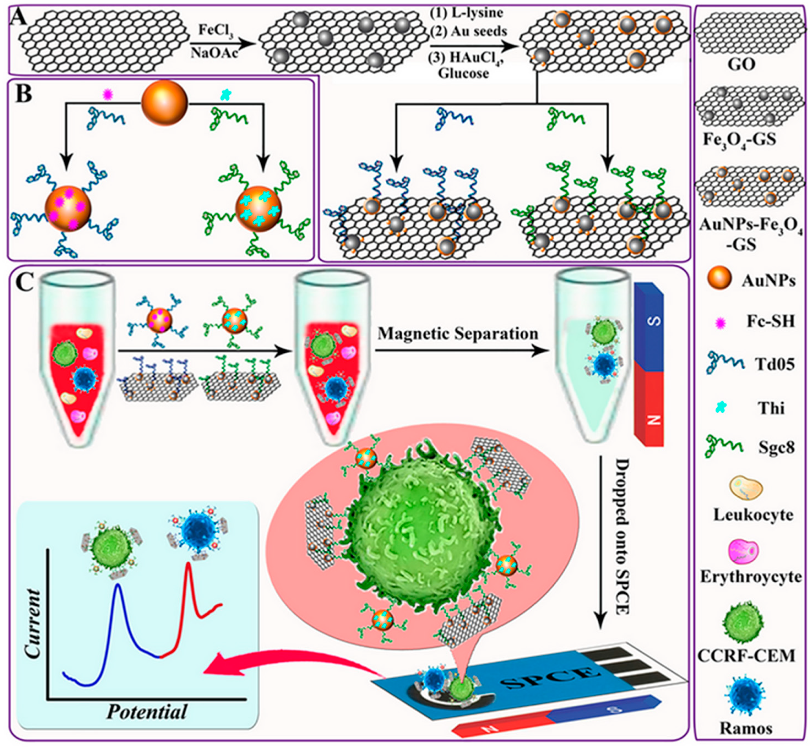
- Dou, B.; Xu, L.; Jiang, B.; Yuan, R.; Xiang, Y. Aptamer-functionalized and gold nanoparticle array-decorated magnetic graphene nanosheets enable multiplexed and sensitive electrochemical detection of rare circulating tumor cells in whole blood. Anal. Chem. 2019, 91, 10792–10799. [Google Scholar] [CrossRef]
- Li, F.; Han, J.; Jiang, L.; Wang, Y.; Li, Y.; Dong, Y.; Wei, Q. An ultrasensitive sandwich-type electrochemical immunosensor based on signal amplification strategy of gold nanoparticles functionalized magnetic multi-walled carbon nanotubes loaded with lead ions. Biosens. Bioelectron. 2015, 68, 626–632. [Google Scholar] [CrossRef] [PubMed]
- Lin, C.-W.; Wei, K.-C.; Liao, S.-S.; Huang, C.-Y.; Sun, C.-L.; Wu, P.J.; Lu, Y.J.; Yang, H.W.; Ma, C.C.M. A reusable magnetic graphene oxide-modified biosensor for vascular endothelial growth factor detection in cancer diagnosis. Biosens. Bioelectron. 2015, 67, 431–437. [Google Scholar] [CrossRef] [PubMed]
- Li, F.; Li, Y.; Dong, Y.; Jiang, L.; Wang, P.; Liu, Q.; Liu, H.; Wei, Q. An ultrasensitive label-free electrochemical immunosensor based on signal amplification strategy of multifunctional magnetic graphene loaded with cadmium ions. Sci. Rep. 2016, 6, 21281. [Google Scholar] [CrossRef] [PubMed]
- Khoshfetrat, S.M.; Mehrgardi, M.A. Amplified detection of leukemia cancer cells using an aptamer-conjugated gold-coated magnetic nanoparticles on a nitrogen-doped graphene modified electrode. Bioelectrochem. 2017, 114, 24–32. [Google Scholar] [CrossRef] [PubMed]
- He, Z.; Wei, J.; Gan, C.; Liu, W.; Liu, Y. A rolling circle amplification signal-enhanced immunosensor for ultrasensitive microcystin-LR detection based on a magnetic graphene functionalized electrode. RSC Adv. 2017, 7, 39906. [Google Scholar] [CrossRef]
- Li, Y.; Zhang, Y.; Li, F.; Li, M.; Chen, L.; Dong, Y.; Wei, Q. Sandwich-type amperometric immunosensor using functionalized magnetic graphene loaded gold and silver core-shell nanocomposites for the detection of carcinoembryonic antigen. J. Electroanal. Chem. 2017, 795, 1–9. [Google Scholar]
- Sun, B.; Gou, Y.; Ma, Y.; Zheng, X.; Bai, R.; Attia, A.; Abdelmoaty, A.; Hu, F. Investigate electrochemical immunosensor of cortisol based on gold nanoparticles/magnetic functionalized reduced graphene oxide. Biosens. Bioelectron. 2017, 88, 55–62. [Google Scholar] [CrossRef]
- Jahanbani, S.; Benvidi, A. A novel electrochemical DNA biosensor based on a modified magnetic bar carbon paste electrode with Fe3O4NPs-reduced graphene oxide/PANHS nanocomposite. Mater. Sci. Eng. C 2016, 68, 1–8. [Google Scholar] [CrossRef]
- Di Tocco, A.; Robledo, S.N.; Osunac, Y.; Sandoval-Cortez, J.; Granero, A.M.; Vettorazzi, N.R.; Martínez, J.L.; Segura, E.P.; Ilinác, A.; Zona, A.; et al. Development of an electrochemical biosensor for the determination of triglycerides in serum samples based on a lipase/magnetite-chitosan/copper oxide nanoparticles/multiwalled carbon nanotubes/pectin composite. Talanta 2018, 190, 30–37. [Google Scholar] [CrossRef]
- Azadbakht, A.; Derikvandi, Z. Aptamer-based sensor for diclofenac quantification using carbon nanotubes and graphene oxide decorated with magnetic nanomaterials. J. Iran. Chem. Soc. 2018, 15, 595–606. [Google Scholar] [CrossRef]
- Waifalkar, P.P.; Chougaleb, A.D.; Kolluc, P.; Patila, P.S.; Patile, P.B. Thin film magnetic nanoparticle decorated graphene based electrochemical nanobiosensor for H2O2 sensing using HRP. Colloids Surf. B Biointerfaces 2018, 167, 425–431. [Google Scholar] [CrossRef] [PubMed]
- Rodríguez, B.A.G.; Pérez-Caro, M.; Alencar, R.S.; Souza Filho, A.G.; Aguiar, A. Graphene nanoribbons and iron oxide nanoparticles composite as a potential candidate in DNA sensing applications. J. Appl. Phys. 2020, 127, 044901. [Google Scholar] [CrossRef]
- Neravathu, D.; Paloly, A.R.; Sajan, P.; Satheesh, M.; Bushiri, M.J. Hybrid nanomaterial of ZnFe2O4/α-Fe2O3 implanted graphene for electrochemical glucose sensing application. Diam. Relat. Mater. 2020, 106, 107852. [Google Scholar] [CrossRef]
- Ehzari, H.; Samimi, M.; Safari, M.; Gholivand, M.B. Label-free electrochemical immunosensor for sensitive HER2 biomarker detection using the core-shell magnetic metal-organic frameworks. J. Electroanal. Chem. 2020, 877, 114722. [Google Scholar] [CrossRef]
- Shahnavaz, Z.; Hamid, S.B.A. Fabrication of a novel metal chromite—carbon nanotube composite for the highly efficient electrocatalytic reduction of hydrogen peroxide. Appl. Surf. Sci. 2017, 407, 379–385. [Google Scholar] [CrossRef]
- Gallay, P.; Eguílaz, M.; Rivas, G. Designing electrochemical interfaces based on nanohybrids of avidin functionalized-carbon nanotubes and ruthenium nanoparticles as peroxidase-like nanozyme with supramolecular recognition properties for site-specific anchoring of biotinylated residues. Biosens. Bioelectron. 2020, 148, 111764. [Google Scholar] [CrossRef]
- Liu, J.-X.; Ding, S.-N. Non-enzymatic amperometric determination of cellular hydrogen peroxide using dendrimer-encapsulated Pt nanoclusters/carbon nanotubes hybrid composites modified glassy carbon electrode. Sens. Actuators B-Chem. 2017, 251, 200–207. [Google Scholar] [CrossRef]
- Wang, H.; Li, S.; Si, Y.M.; Sun, Z.Z.; Li, S.Y.; Lin, Y.H. Recyclable enzyme mimic of cubic Fe3O4 nanoparticles loaded on graphene oxide dispersed carbon nanotubes with enhanced peroxidase-like catalysis and electrocatalysis. J. Mater. Chem. B 2014, 2, 4442–4448. [Google Scholar] [CrossRef]
- Bai, J.; Sun, C.; Jiang, X. Carbon dots-decorated multiwalled carbon nanotubes nanocomposites as a high-performance electrochemical sensor for detection of H2O2 in living cells. Anal. Bioanal. Chem. 2016, 408, 4705–4714. [Google Scholar] [CrossRef]
- Zhang, Y.; Gao, G.; Liu, H.; Fu, H.; Fan, J.; Wang, K.; Chen, Y.; Li, B.; Zhang, C.; Zhi, X.; et al. Identification of volatile biomarkers of gastric cancer cells and ultrasensitive electrochemical detection based on sensing interface of Au-Ag alloy coated MWCNTs. Theranostics 2014, 4, 154–162. [Google Scholar] [CrossRef]
- Savas, S.; Altintas, Z. Graphene quantum dots as nanozymes for electrochemical sensing of Yersinia enterocolitica in milk and human serum. Materials 2019, 12, 2189. [Google Scholar] [CrossRef] [PubMed]
- Serafín, V.; Valverde, A.; Martínez-García, G.; Martínez-Periñán, E.; Comba, F.; Garranzo-Asensio, M.; Barderas, R.; Yáñez-Sedeño, P.; Campuzano, S.; Pingarrón, J.M.; et al. Graphene quantum dots-functionalized multi-walled carbon nanotubes as nanocarriers in electrochemical immunosensing. Determination of IL-13 receptor α2 in colorectal cells and tumor tissues with different metastatic potential. Sens. Actuators B. 2019, 284, 711–722. [Google Scholar]
- Yang, Y.; Liu, Q.; Liu, Y.; Chui, J.; Liu, H.; Wang, P.; Li, Y.; Chen, L.; Zhao, Z.; Dong, Y. A novel label-free electrochemical immunosensor based on functionalized nitrogen-doped graphene quantum dots for carcinoembryonic antigen detection. Biosens. Bioelectron. 2017, 90, 31–38. [Google Scholar] [CrossRef]
- Zhang, Y.; Wu, C.; Zhou, X.; Wu, X.; Yang, Y.; Wu, H.; Guo, S.; Zhang, J. Graphene quantum dots/gold electrode and its application in living cell H2O2 detection. Nanoscale 2013, 5, 1816–1819. [Google Scholar] [CrossRef]
- Wang, Y.; Zhao, H.; Song, H.; Dong, J.; Xu, J. Monodispersed gold nanoparticles entrapped in ordered mesoporous carbon/silica nanocomposites as xanthine oxidase mimic for electrochemical sensing of xanthine. Microchim. Acta 2020, 187, 543. [Google Scholar] [CrossRef]
- Belkhalfa, H.; Teodorescu, F.; Quéniat, G.; Coffinier, Y.; Dokhan, N.; Sam, S.; Abderrahmani, A.; Boukherroub, R.; Szunerits, S. Insulin impregnated reduced graphene oxide/Ni(OH)2 thin films for electrochemical insulin release and glucose sensing. Sens. Actuators B-Chem. 2016, 237, 693–701. [Google Scholar] [CrossRef]
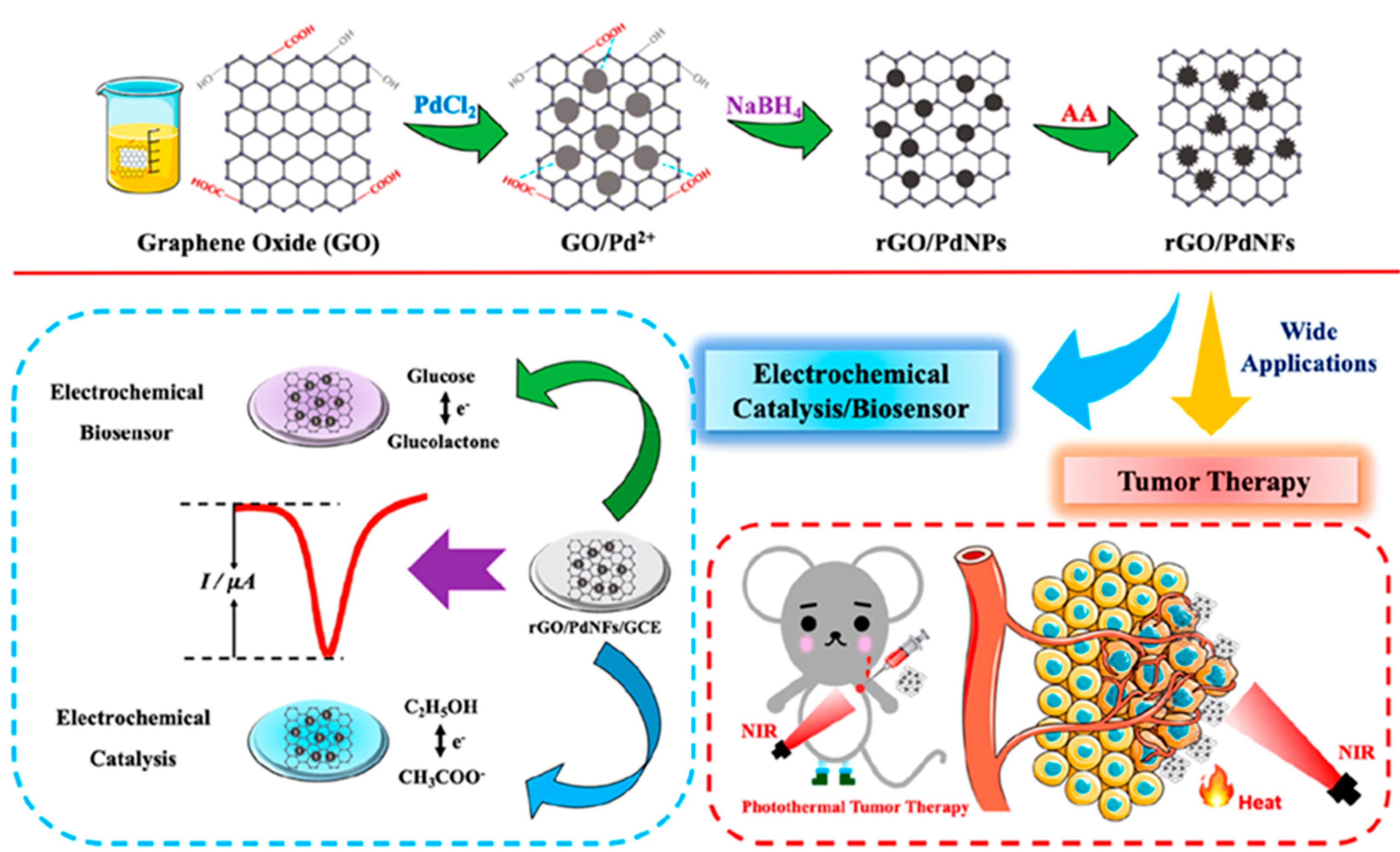
- He, Y.; Cao, W.; Cong, C.; Zhang, X.; Luo, L.; Li, L.; Cui, H.; Gao, D. Rationally designed multifunctional carbon−palladium nanohybrids for wide applications: From electrochemical catalysis/nonenzymatic sensor to photothermal tumor therapy. ACS Sustain. Chem. Eng. 2019, 7, 3584–3592. [Google Scholar] [CrossRef]
- He, W.; Liu, R.; Zhou, P.; Liu, Q.; Cui, T. Flexible micro-sensors with self-assembled graphene on a polyolefin substrate for dopamine detection. Biosens. Bioelectron. 2020, 167, 112473. [Google Scholar] [CrossRef]
- Moonla, C.; Goud, K.Y.; Teymourian, H.; Tangkuaram, T.; Ingrande, J.; Suresh, P.; Wang, J. An integrated microcatheter-based dual-analyte sensor system for simultaneous, real-time measurement of propofol and fentanyl. Talanta 2020, 218, 121205. [Google Scholar] [CrossRef]
- Ni, S.; Shen, Z.; Zhang, P.; Liu, G. Enhanced performance of an electrochemical aptasensor for real-time detection of vascular endothelial growth factor (VEGF) by nanofabrication and ratiometric measurement. Anal. Chim. Acta 2020, 1121, 74–82. [Google Scholar]
- Jiang, Y.; Xiao, X.; Li, C.; Luo, Y.; Chen, S.; Shi, G.; Han, K.; Gu, H. Facile ratiometric electrochemical sensor for in vivo/online repetitive measurements of cerebral ascorbic acid in brain microdiaysate. Anal. Chem. 2020, 92, 3981–3989. [Google Scholar] [CrossRef] [PubMed]
- Zhang, D.; Ma, J.; Meng, X.; Xu, Z.; Zhang, J.; Fang, Y.; Guo, Y. Electrochemical aptamer-based microsensor for real-time monitoring of adenosine in vivo. Anal. Chim. Acta 2019, 1076, 55–63. [Google Scholar] [CrossRef] [PubMed]
- Wang, L.; Song, Y.; Zhang, Y.; Xu, S.; Xu, H.; Wang, M.; Wang, Y.; Cai, X. A microelectrode array electrodeposited with reduced graphene oxide and Pt nanoparticles for norepinephrine and electrophysiological recordings. J. Micromech. Microeng. 2017, 27, 115001. [Google Scholar] [CrossRef]
- Rezaei, B.; Jamei, H.R.; Ensafi, A.A. An ultrasensitive and selective electrochemical aptasensor based on rGO-MWCNTs/Chitosan/carbon quantum dot for the detection of lysozyme. Biosens. Bioelectron. 2018, 115, 37–44. [Google Scholar] [CrossRef] [PubMed]
- Sánchez-Tirado, E.; Arellano, L.M.; González-Cortés, A.; Yáñez-Sedeño, P.; Langa, F.; Pingarrón, J.M. Viologen-functionalized single-walled carbon nanotubes as carrier nanotags for electrochemical immunosensing. Application to TGF-β1 cytokine. Biosens. Bioelectron. 2017, 98, 240–247. [Google Scholar] [CrossRef] [PubMed]
- He, B.S.; Yan, S.S. Electrochemical aptasensor based on aptamer complimentary strand conjugate and thionine for sensitive detection of tetracycline with multiwalled carbon nanotubes and gold nanoparticles amplification. Anal. Methods 2018, 10, 783–790. [Google Scholar] [CrossRef]
- Luo, Y.; Wang, Y.; Yan, H.; Wu, Y.; Zhu, C.; Du, D.; Lin, Y. SWCNTs@GQDs composites as nanocarriers for enzyme-free dual signal amplification electrochemical immunoassay of cancer biomarker. Anal. Chim. Acta 2018, 1042, 44–51. [Google Scholar] [CrossRef]
- You, H.; Mu, Z.; Zhao, M.; Zhou, J.; Chen, Y.; Bai, L. Voltammetric aptasensor for sulfadimethoxine using a nanohybrid composed of multifunctional fullerene, reduced graphene oxide and Pt@Au nanoparticles, and based on direct electron transfer to the active site of glucose oxidase. Microchim. Acta 2019, 186, 1. [Google Scholar] [CrossRef]
- Zheng, Z.; Wang, M.; Shi, X.; Wang, C. Palladium nanoparticles/graphitic carbon nitride nanosheets-carbon nanotubes as a catalytic amplification platform for the selective determination of 17α-ethinylestradiol in feedstuffs. Sci. Rep. 2019, 9, 14162. [Google Scholar] [CrossRef]
- Rakhi, R.B.; Nayak, P.; Xia, C.; Alshareef, H.N. Novel amperometric glucose biosensor based on MXene nanocomposite. Sci. Rep. 2016, 6, 36422. [Google Scholar] [CrossRef]
- Medetalibeyoglu, H.; Beytur, M.; Akyıldırım, O.; Atar, N.; Yola, M.L. Validated electrochemical immunosensor for ultra-sensitive procalcitonin detection: Carbon electrode modified with gold nanoparticles functionalized sulfur doped MXene as sensor platform and carboxylated graphitic carbon nitride as signal amplification. Sens. Actuators B-Chem. 2020, 319, 128195. [Google Scholar] [CrossRef]
- Serafín, V.; Valverde, A.; Garranzo-Asensio, M.; Barderas, R.; Campuzano, S.; Yáñez-Sedeño, P.; Pingarrón, J.M. Simultaneous amperometric immunosensing of the metastasis-related biomarkers IL-13Rα2 and CDH-17 by using grafted screen-printed electrodes and a composite prepared from quantum dots and carbon nanotubes for signal amplification. Microchim. Acta 2019, 186, 411. [Google Scholar] [CrossRef] [PubMed]
- Mu, Z.; Ma, L.; Wang, J.; Zhou, J.; Yuan, Y.; Bai, L. A target-induced amperometic aptasensor for sensitive zearalenone detection by CS@AB-MWCNTs nanocomposite as enhancers. Food Chem. 2020, 340, 128128. [Google Scholar] [CrossRef] [PubMed]
- Garg, B.; Bisht, T. Carbon nanodots as peroxidase nanozymes for biosensing. Molecules 2016, 21, 1653. [Google Scholar] [CrossRef] [PubMed]
- Huang, Y.; Ren, J.; Qu, X. Nanozymes: Classification, catalytic mechanisms, activity regulation, and applications. Chem. Rev. 2019, 119, 4357–4412. [Google Scholar] [CrossRef]
- Mahmudunnabi, R.G.; Farhana, F.Z.; Kashaninejad, N.; Firoz, S.H.; Shim, Y.-B.; Shiddiky, M.J.A. Nanozyme-based electrochemical biosensors for disease biomarker detection. Analyst 2020, 145, 4398–4420. [Google Scholar] [CrossRef]
- Liang, M.; Yan, X. Nanozymes: From new concepts, mechanisms, and standards to applications. Acc. Chem. Res. 2019, 52, 2190–2200. [Google Scholar] [CrossRef]
- Sun, H.; Zhou, Y.; Ren, J.; Qu, X. Carbon Nanozymes: Enzymatic Properties, Catalytic Mechanism, and Applications. Angew. Chem. Int. Ed. 2018, 57, 9224–9237. [Google Scholar] [CrossRef]
- Cui, R.; Han, Z.; Zhu, J.J. Helical carbon nanotubes: Intrinsic peroxidase catalytic activity and its application for biocatalysis and biosensing. Chem. Eur. J. 2011, 17, 9377–9384. [Google Scholar] [CrossRef]
- Wang, H.; Jiang, H.; Wang, S.; Shi, W.B.; He, J.C.; Liu, H.; Huang, Y.M. Fe3O4-MWCNT magnetic nanocomposites as efficient peroxidase mimic catalysts in a Fenton-like reaction for water purification without pH limitation. RSC Adv. 2014, 4, 45809–45815. [Google Scholar] [CrossRef]
- Ye, H.; Mohar, J.; Wang, Q.; Catalano, M.; Kim, M.J.; Xia, X. Peroxidase-like properties of ruthenium nanoframes. Sci. Bull. 2016, 61, 1739–1745. [Google Scholar] [CrossRef]
- Nekoueian, K.; Amiri, M.; Sillanpää, M.; Marken, F.; Boukherroub, R.; Szunerits, S. Carbon-based quantum particles: An electroanalytical and biomedical perspective. Chem. Soc. Rev. 2019, 48, 4281–4316. [Google Scholar] [CrossRef]
- Hu, Y.; Gao, X.J.; Zhu, Y.; Muhammad, F.; Tan, S.; Cao, W.; Lin, S.; Jin, Z.; Gao, X.; Wei, H. Nitrogen-doped carbon nanomaterials as highly active and specific peroxidase mimics. Chem. Mater. 2018, 30, 6431–6439. [Google Scholar] [CrossRef]
- Fan, K.; Xi, J.; Fan, L.; Wang, P.; Zhu, C.; Tang, Y.; Xu, X.; Liang, M.; Jiang, B.; Yan, X.; et al. In vivo guiding nitrogen-doped carbon nanozyme for tumor catalytic therapy. Nat. Commun. 2018, 9, 1440–1450. [Google Scholar] [CrossRef]
- Raphey, V.R.; Henna, T.K.; Nivitha, K.P.; Mufeedha, P.; Sabu, C.; Pramod, K. Advanced biomedical applications of carbon nanotube. Mat. Sci. Eng. C 2019, 100, 616–630. [Google Scholar] [CrossRef]
- Raghav, S.; Painuli, R.; Kumar, D. Multifunctional nanomaterials for multifaceted applications in biomedical arena. Int. J. Pharmacol. 2017, 13, 890–906. [Google Scholar] [CrossRef]
- Saleemi, M.A.; Kong, Y.L.; Yong, P.V.C.; Wong, E.H. An overview of recent development in therapeutic drug carrier system using carbon nanotubes. J. Drug Deliv. Sci. Technol. 2020, 59, 101855. [Google Scholar] [CrossRef]
- Zhou, Y.; Chen, C.; Zhao, J.; Fei, J.; Ding, Y.; Cai, Y. Reversible switched detection of dihydroxybenzenes using a temperature-sensitive electrochemical sensing film. Electrochim. Acta 2016, 192, 158. [Google Scholar] [CrossRef]
- Wu, X.; Bai, X.; Ma, Y.; Wei, J.; Peng, J.; Shi, K.; Yao, H. Construction of multiple switchable sensors and logic gates based on carboxylated multi-walled carbon nanotubes/poly(N,N-diethylacrylamide). Sensors 2018, 18, 3358. [Google Scholar] [CrossRef]
- Ma, Y.; Hou, S.; Xue, S.; Wu, X.; Yao, H.; Shi, K.; Ma, W.; Zuo, L.; Yao, H. The fabrication of multiple stimuli-responsive film electrode and logic gates of rutin. J. Electrochem. Soc. 2020, 167, 067526. [Google Scholar] [CrossRef]
- Shervedani, R.K.; Foroushani, M.S.; Kefayat, A.; Torabi, M.; Rahsepar, F.R. Construction and characterization of a theranostic system based on graphene/manganese chelate. Biosens. Bioelectron. 2018, 117, 794–801. [Google Scholar] [CrossRef]
- Foroushani, M.S.; Shervedani, R.K.; Kefayat, A.; Torabi, M.; Ghahremani, F.; Yaghoobi, F. Folate-graphene chelate manganese nanoparticles as a theranostic system for colon cancer MR imaging and drug delivery: In-vivo examinations. J. Drug Deliv. Sci. Technol. 2019, 54, 101223. [Google Scholar] [CrossRef]
- Lee, D.; Kim, Y.H.; Park, S. Enzyme electrode platform using methyl viologen electrochemically immobilized on carbon materials. J. Electrochem. Soc. 2016, 163, G93–G98. [Google Scholar] [CrossRef]
- Chan, M.-H.; Liu, R.-S.; Hsiao, M. Graphitic carbon nitride-based nanocomposites and their biological applications: A review. Nanoscale 2019, 11, 14993–15003. [Google Scholar] [CrossRef]
- Kresge, C.; Leonowicz, M.; Roth, W.J.; Vartuli, J.; Beck, J. Ordered mesoporous molecular sieves synthesized by a liquid-crystal template mechanism. Nature 1992, 359, 710–712. [Google Scholar] [CrossRef]
- Narayan, R.; Nayak, U.; Raichur, A.; Garg, S. Mesoporous silica nanoparticles: A comprehensive review on synthesis and recent advances. Pharmaceutics 2018, 10, 118. [Google Scholar] [CrossRef]
- Hasanzadeh, M.; Shadjou, N.; de la Guardia, M.; Eskandani, M.; Sheikhzadeh, P. Mesoporous silica-based materials for use in biosensors. TrAC Trends Anal. Chem. 2012, 33, 117–129. [Google Scholar] [CrossRef]
- Sancenón, F.; Pascual, L.; Oroval, M.; Aznar, E.; Martínez-Máñez, R. Gated silica mesoporous materials in sensing applications. Chem. Open 2015, 4, 418–437. [Google Scholar] [CrossRef]
- Kordasht, H.K.; Pazhuhi, M.; Pashazadeh-Panahi, P.; Hasanzadeh, M.; Shadjou, N. Multifunctional aptasensors based on mesoporous silica nanoparticles as an efficient platform for bioanalytical applications: Recent advances. TrAC Trends Anal. Chem. 2020, 124, 115778. [Google Scholar] [CrossRef]
- Feyen, M.; Weidenthaler, C.; Schüth, F.; Lu, A.-H. Regioselectively controlled synthesis of colloidal mushroom nanostructures and their hollow derivatives. J. Am. Chem. Soc. 2010, 132, 6791–6799. [Google Scholar] [CrossRef]
- Tang, J.; Tang, D.; Niessner, R.; Knopp, D.; Chen, G. Hierarchical dendritic gold microstructure-based aptasensor for ultrasensitive electrochemical detection of thrombin using functionalized mesoporous silica nanospheres as signal tags. Anal. Chim. Acta 2012, 720, 1–8. [Google Scholar] [CrossRef]
- Huang, L.; Yang, X.; Qi, C.; Niu, X.; Zhao, C.; Zhao, X.; Shangguan, D.; Yang, Y. A label-free electrochemical biosensor based on a DNA aptamer against codeine. Anal. Chim. Acta 2013, 787, 203–210. [Google Scholar] [CrossRef]
- Su, M.; Liu, Y.; Zhang, Y.; Wang, Z.; Li, Y.; He, P. Robust and underwater superoleophobic coating with excellent corrosion and biofouling resistance in harsh environments. Appl. Surf. Sci. 2018, 436, 152–161. [Google Scholar] [CrossRef]
- Shekari, Z.; Zare, H.R.; Falahati, A. Developing an impedimetric aptasensor for selective label–free detection of CEA as a cancer biomarker based on gold nanoparticles loaded in functionalized mesoporous silica films. J. Electrochem. Soc. 2017, 164, B739–B745. [Google Scholar] [CrossRef]
- Wu, S.H.; Hung, Y.; Mou, C.Y. Mesoporous silica nanoparticles as nanocarriers. Chem. Commun. 2011, 47, 9972–9985. [Google Scholar] [CrossRef]
- Castillo, R.R.; Baeza, A.; Vallet-Regí, M. Recent applications of the combination of mesoporous silica nanoparticles with nucleic acids: Development of bioresponsive devices, carriers and sensors. Biomater. Sci. 2017, 5, 353–377. [Google Scholar] [CrossRef]
- Walcarius, A. Silica-based electrochemical sensors and biosensors: Recent trends. Curr. Op. Electrochem. 2018, 10, 88–97. [Google Scholar] [CrossRef]
- Jiménez-Falcao, S.; Villalonga, A.; Arévalo-Villena, M.; Briones-Pérez, A.; Martínez-Máñez, R.; Martínez-Ruiz, P.; Villalonga, R. Enzyme-controlled mesoporous nanosensor for the detection of living Saccharomyces cerevisiae. Sens. Actuators B-Chem. 2020, 303, 127197. [Google Scholar] [CrossRef]
- Slowing, I.I.; Trewyn, B.G.; Giri, S.; Lin, V.S.-Y. Mesoporous silica nanoparticles for drug delivery and biosensing applications. Adv. Funct. Mater. 2007, 17, 1225–1236. [Google Scholar] [CrossRef]
- Xing, Y. Synthesis and electrochemical characterization of uniformly dispersed high loading Pt nanoparticles on sonochemically-treated carbon nanotubes. J. Phys. Chem. B 2004, 108, 19255–19259. [Google Scholar] [CrossRef]
- Yang, M.; Li, H.; Javadi, A.; Gong, S. Multifunctional mesoporous silica nanoparticles as labels for the preparation of ultrasensitive electrochemical immunosensors. Biomaterials 2010, 31, 3281–3286. [Google Scholar] [CrossRef] [PubMed]
- Miyahara, M.; Vinu, A.; Ariga, K. Adsorption myoglobin over mesoporous silica molecular sieves: Pore size effect and pore-filling model. Mater. Sci. Eng. C 2007, 27, 232–236. [Google Scholar] [CrossRef]
- Shekari, Z.; Zare, H.R.; Falahati, A. An ultrasensitive aptasensor for hemin and hemoglobin based on signal amplification via electrocatalytic oxygen reduction. Anal. Biochem. 2017, 518, 102–109. [Google Scholar] [CrossRef] [PubMed]
- Muthamizh, S.; Ribes, À.; Anusuyajanakiraman, M.; Narayanan, V.; Soto, J.; Martínez-Máñez, R.; Aznar, E. Implementation of oligonucleotide-gated supports for the electrochemical detection of Ochratoxin, A. Supramol. Chem. 2017, 29, 776–783. [Google Scholar] [CrossRef]
- Eguílaz, M.; Villalonga, R.; Rivas, G. Electrochemical biointerfaces based on carbon nanotubes-mesoporous silica hybrid material: Bioelectrocatalysis of hemoglobin and biosensing applications. Biosens. Bioelectron. 2018, 111, 144–151. [Google Scholar] [CrossRef]
- Trewyn, B.G.; Giri, S.; Slowing, I.I.; Lin, V.S.-Y. Mesoporous silica nanoparticle based controlled release, drug delivery, and biosensor systems. Chem. Commun. 2007, 3236–3245. [Google Scholar] [CrossRef]
- Hudson, S.P.; Padera, R.F.; Langer, R.; Kohane, D.S. The biocompatibility of mesoporous silicates. Biomaterials 2008, 29, 4045–4055. [Google Scholar] [CrossRef]
- Øye, G.; Sjöblom, J.; Stöcker, M. Synthesis, characterization and potential applications of new materials in the mesoporous range. Adv. Colloid Interface Sci. 2001, 89, 439–466. [Google Scholar] [CrossRef]
- Hashkavayi, A.B.; Raoof, J.B.; Azimi, R.; Ojani, R. Label-free and sensitive aptasensor based on dendritic gold nanostructures on functionalized SBA-15 for determination of chloramphenicol. Anal. Bioanal. Chem. 2016, 408, 2557–2565. [Google Scholar] [CrossRef]
- Kleitz, F.; Liu, D.; Anilkumar, G.M.; Park, I.-S.; Solovyov, L.A.; Shmakov, A.N.; Ryoo, R. Large cage face-centered-cubic Fm3m mesoporous silica: Synthesis and structure. J. Phys. Chem. B 2003, 107, 14296–14300. [Google Scholar] [CrossRef]
- Nandiyanto, A.B.D.; Kim, S.-G.; Iskandar, F.; Okuyama, K. Synthesis of spherical mesoporous silica nanoparticles with nanometer-size controllable pores and outer diameters. Microporous Mesoporous Mater. 2009, 120, 447–453. [Google Scholar] [CrossRef]
- Tozuka, Y.; Wongmekiat, A.; Kimura, K.; Moribe, K.; Yamamura, S.; Yamamoto, K. Effect of pore size of FSM-16 on the entrapment of flurbiprofen in mesoporous structures. Chem. Pharm. Bull. 2005, 53, 974–977. [Google Scholar] [CrossRef] [PubMed]
- Kordasht, H.K.; Moosavy, M.-H.; Hasanzadeh, M.; Soleymani, J.; Mokhtarzadeh, A. Determination of Aflatoxin M1 using aptamer based biosensor on the surface of dendritic fibrous nano-silica functionalized by amine groups. Anal. Methods 2019, 11, 3910–3919. [Google Scholar] [CrossRef]
- Saadaoui, M.; Fernández, I.; Sánchez, A.; Díez, P.; Campuzano, S.; Raouafi, N.; Pingarrón, J.M.; Villalonga, R. Mesoporous silica thin film mechanized with a DNAzyme-based molecular switch for electrochemical biosensing. Electrochem. Commun. 2015, 58, 57–61. [Google Scholar] [CrossRef]
- Saadaoui, M.; Fernández, I.; Luna, G.; Díez, P.; Campuzano, S.; Raouafi, N.; Sánchez, A.; Pingarrón, J.M.; Villalonga, R. Label-free electrochemical genosensor based on mesoporous silica thin film. Anal. Bioanal. Chem. 2016, 408, 7321–7327. [Google Scholar] [CrossRef] [PubMed]
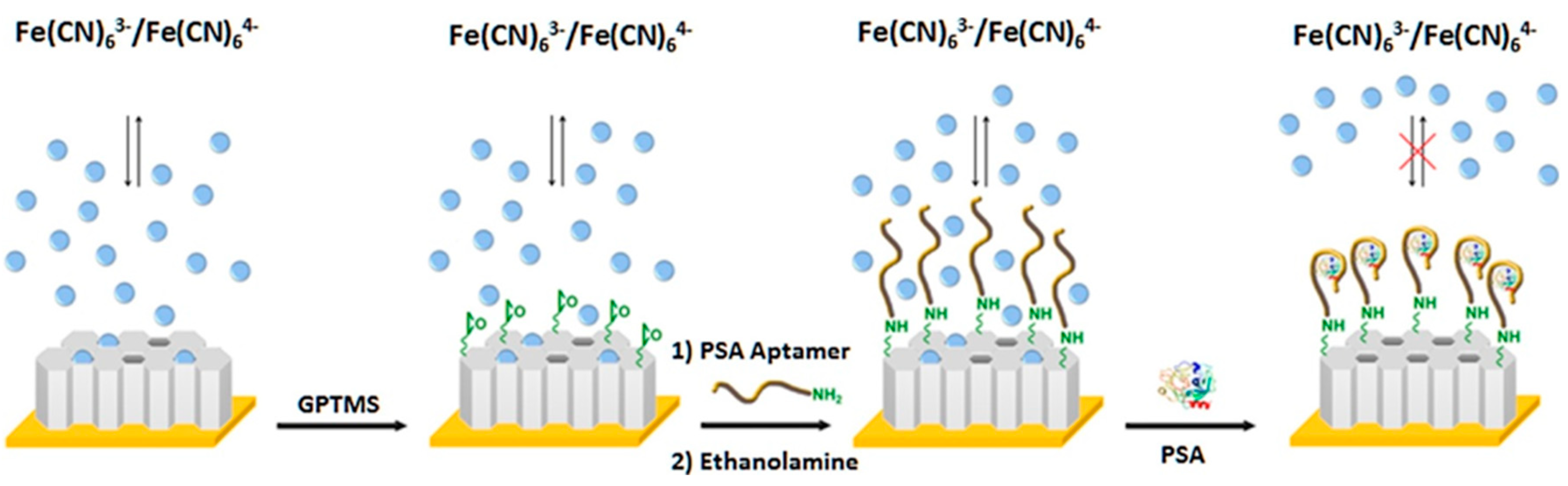
- Argoubi, W.; Sánchez, A.; Parrado, C.; Raouafi, N.; Villalonga, R. Label-free electrochemical aptasensing platform based on mesoporous silica thin film for the detection of prostate specific antigen. Sens. Actuators B Chem. 2018, 255, 309–315. [Google Scholar] [CrossRef]
- Roushani, M.; Ghanbari, K. An electrochemical aptasensor for streptomycin based on covalent attachment of the aptamer onto a mesoporous silica thin film-coated gold electrode. Microchim. Acta 2019, 186, 115. [Google Scholar] [CrossRef]
- Zhang, J.; Chai, Y.; Yuan, R.; Yuan, Y.; Bai, L.; Xie, S. A highly sensitive electrochemical aptasensor for thrombin detection using functionalized mesoporous silica@multiwalled carbon nanotubes as signal tags and DNAzyme signal amplification. Analyst 2013, 138, 6938–6945. [Google Scholar] [CrossRef]
- Zhou, Q.; Tang, D. Graphene oxide-gated mesoporous silica nanocontainers using aptamers for arsenite detection with glucometer readout. J. Mater. Chem. B 2018, 6, 6585–6591. [Google Scholar] [CrossRef]
- Zhou, S.; Wang, Y.; Zhu, J.J. Simultaneous detection of tumor cell apoptosis regulators Bcl-2 and Bax through a dual-signal-marked electrochemical immunosensor. ACS Appl. Mater. Interfaces 2016, 8, 7674–7682. [Google Scholar] [CrossRef]
- Hashkavayi, A.B.; Raoof, J.B. Design an aptasensor based on structure-switching aptamer on dendritic gold nanostructures/Fe3O4@SiO2/DABCO modified screen printed electrode for highly selective detection of epirubicin. Biosens. Bioelectron. 2017, 91, 650–657. [Google Scholar] [CrossRef]
- Bagheri, E.; Ansari, L.; Sameiyan, E.; Abnous, K.; Taghdisi, S.M.; Ramezani, M.; Alibolandi, M. Sensors design based on hybrid gold-silica nanostructures. Biosens. Bioelectron. 2020, 153, 112054. [Google Scholar] [CrossRef] [PubMed]
- Feng, T.; Qiao, X.; Wang, H.; Sun, Z.; Hong, C. A sandwich-type electrochemical immunosensor for carcinoembryonic antigen based on signal amplification strategy of optimized ferrocene functionalized Fe3O4@SiO2 as labels. Biosens. Bioelectron. 2016, 79, 48–54. [Google Scholar] [CrossRef] [PubMed]
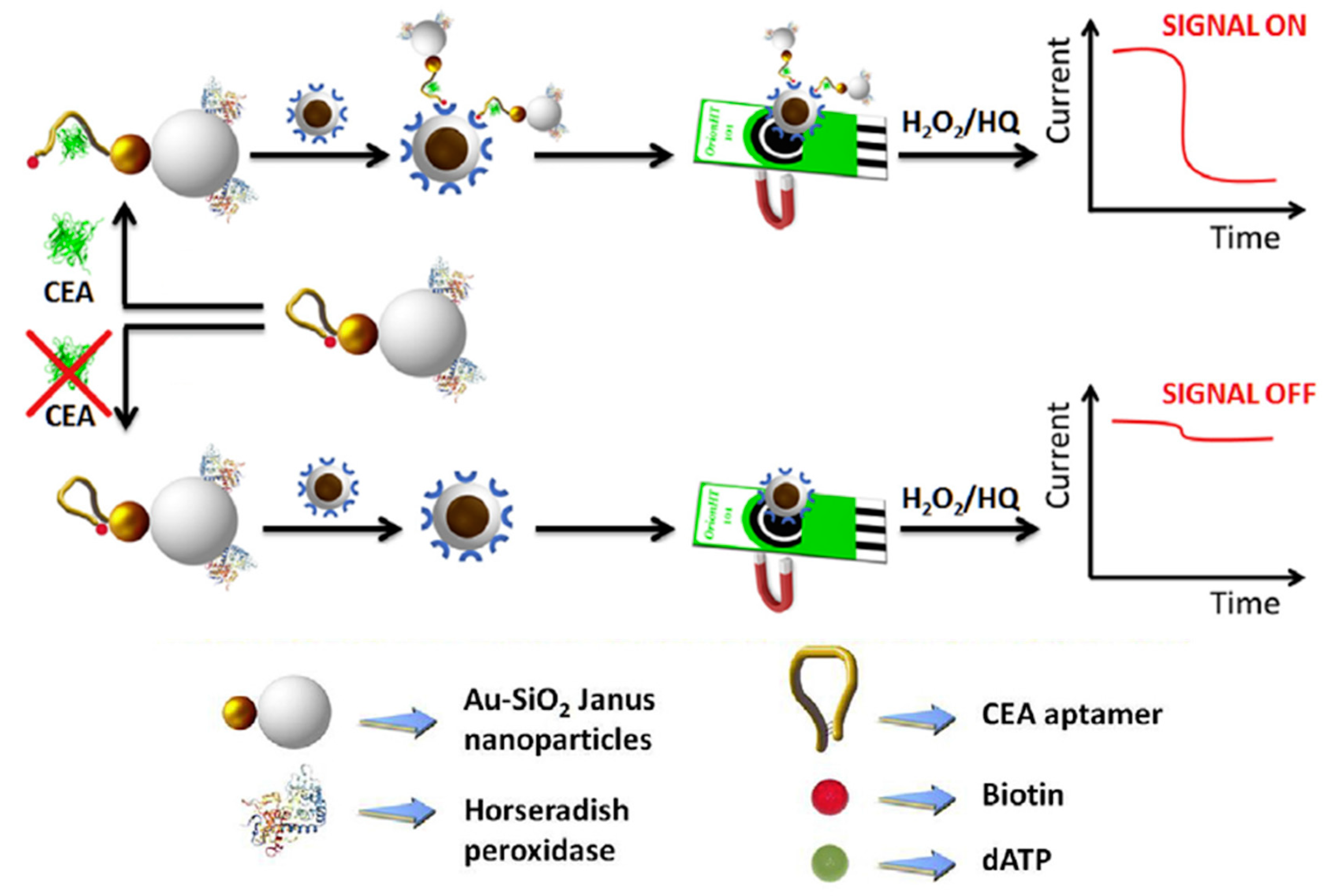
- Paniagua, G.; Villalonga, A.; Eguílaz, M.; Vegas, B.; Parrado, C.; Rivas, G.; Díez, P.; Villalonga, R. Amperometric aptasensor for carcinoembryonic antigen based on the use of bifunctionalized Janus nanoparticles as biorecognition signaling element. Anal. Chim. Acta 2019, 1061, 84–91. [Google Scholar] [CrossRef]
- Sánchez, A.; Díez, P.; Martínez-Ruíz, P.; Villalonga, R.; Pingarrón, J.M. Janus Au-mesoporous silica nanoparticles as electrochemical biorecognition-signaling system. Electrochem. Commun. 2013, 30, 51–54. [Google Scholar] [CrossRef]
- Boujakhrout, A.; Sánchez, E.; Díez, P.; Sánchez, A.; Martínez-Ruiz, P.; Parrado, C.; Pingarrón, J.M.; Villalonga, R. Single-walled carbon nanotubes/Au–mesoporous silica Janus nanoparticles as building blocks for the preparation of a bienzyme biosensor. ChemElectroChem 2015, 2, 1735–1741. [Google Scholar] [CrossRef]
- Jiménez-Falcao, S.; Parra-Nieto, J.; Pérez-Cuadrado, H.; Martínez-Máñez, R.; Martínez-Ruiz, P.; Villalonga, R. Avidin-gated mesoporous silica nanoparticles for signal amplification in electrochemical. Electrochem. Commun. 2019, 108, 106556. [Google Scholar] [CrossRef]
- Fu, L.; Zhuang, J.; Lai, W.; Que, X.; Lu, M.; Tang, D. Portable and quantitative monitoring of heavy metal ions using DNAzyme-capped mesoporous silica nanoparticles with a glucometer readout. J. Mater. Chem. B 2013, 1, 6123–6128. [Google Scholar] [CrossRef]
- Wang, Y.; Lu, M.; Zhu, J.; Tian, S. Wrapping DNA-gated mesoporous silica nanoparticles for quantitative monitoring of telomerase activity with glucometer readout. J. Mater. Chem. B 2014, 2, 5847–5853. [Google Scholar] [CrossRef]
- Liang, X.; Wang, L.; Wang, D.; Zeng, L.; Fang, Z. Portable and quantitative monitoring of mercury ions using DNA-gated mesoporous silica nanoparticles using a glucometer readout. Chem. Commun. 2016, 52, 2192. [Google Scholar] [CrossRef]
- Wang, L.; Zhu, F.; Chen, M.; Xiong, Y.; Zhu, Y.; Xie, S.; Liu, Q.; Yang, H.; Chen, X. Development of a “dual gates” locked, target-triggered nanodevice for point-of-care testing with a glucometer readout. ACS Sens. 2019, 4, 968–976. [Google Scholar] [CrossRef] [PubMed]
- Liu, B.; Zhang, B.; Cui, Y.; Chen, H.; Gao, Z.; Tang, D. Multifunctional goldesilica nanostructures for ultrasensitive electrochemical immunoassay of streptomycin residues. ACS Appl. Mater. Interfaces 2011, 3, 4668–4676. [Google Scholar] [CrossRef] [PubMed]
- You, M.; Yang, S.; Tang, W.; Zhang, F.; He, P. Molecularly imprinted polymers-based electrochemical DNA biosensor for the determination of BRCA-1 amplified by SiO2@Ag. Biosens. Bioelectron. 2018, 112, 72–78. [Google Scholar] [CrossRef] [PubMed]
- Zhao, Y.; Zheng, Y.; Kong, R.; Xia, L.; Qu, F. Ultrasensitive electrochemical immunosensor based on horseradish peroxidase (HRP)-loaded silica-poly(acrylic acid) brushes for protein biomarker detection. Biosens. Bioelectron. 2016, 75, 383–388. [Google Scholar] [CrossRef]
- Wang, J.; Guo, J.; Zhang, J.; Zhang, W.; Zhang, Y. RNA aptamer-based electrochemical aptasensor for C-reactive protein detection using functionalized silica microspheres as immunoprobes. Biosens. Bioelectron. 2017, 95, 100–105. [Google Scholar] [CrossRef]
- Fernández, I.; Sánchez, A.; Díez, P.; Martínez-Ruiz, P.; Di Pierro, P.; Porta, R.; Villalonga, R.; Pingarrón, J.M. Nanochannel-based electrochemical assay for transglutaminase activity. Chem. Commun. 2014, 87, 13356–13358. [Google Scholar]
- Aznar, E.; Oroval, M.; Pascual, L.; Murguia, J.R.; Martínez-Mánez, R.; Sancenón, F. Gated materials for on-command release of guest molecules. Chem. Rev. 2016, 116, 561–718. [Google Scholar] [CrossRef]
- Parvanian, S.; Mostafavi, S.M.; Aghashiri, M. Multifunctional nanoparticle developments in cancer diagnosis and treatment. Sens. Biosens. Res. 2017, 13, 81–87. [Google Scholar] [CrossRef]
- Mazzola, L. Commercializing nanotechnology. Nat. Biotechnol. 2003, 21, 1137–1143. [Google Scholar] [CrossRef]


| Electrode | Nanomaterial/Role | Biosensing Approach/Format | Detection Technique | Target Analyte | LR/LOD | Sample | Ref. |
|---|---|---|---|---|---|---|---|
| -Metal nanoparticles (MNPs)- | |||||||
| CILE | CILE modified with AuNT and HRP and coated with nafion/Direct electron transfer of HRP | Enzyme-based | CV | TCA and NaNO2 | TCA: 1.0–250 mM/0.33 mM NaNO2: 1.6–66.0 mM/0.53 mM | Facial peel solution | [7] |
| GCE/AuNPs | Self-assembly of GS-IL/AuNRs through thiolated sol–gel matrix Positively charged GS-IL was used for GOx immobilization GS-IL and Au NRs acted as high electroactive catalyst. | Enzyme-based | Amperometry −0.2 V (H2O2) | Glucose | 1–764 μM/0.38 µM | Serum and brain microdialysate | [8] |
| SPCE | PtNPs-MWCNTs-GOx/Electrode modifier to enzyme immobilization and electrocatalytic | Enzyme-based | Amperometry −0.5 V (H2O2) | Glucose | 65.8–260.6 μg mL−1/35.0 μg mL−1 | White grapes and glucose tablets | [9] |
| GCE | Electroactive polymer nanospheres synthesized from polymerization of ferrocenedicarboxylic acid/nanocarriers of Ab2, PtNPs, and hemin/G-quadruplex bioelectrocatalytic complex | Antibody based/Sandwich | DPV | AFP | 0.1 pg mL−1–100 ng mL−1/0.086 pg mL−1 | Human serum | [11] |
| GCE | GCE modified with Au@Thi/GO loaded with cAb PtCu@rGO/g-C3N4 conjugated with DAb | Antibody based/Sandwich | Amperometry −0.4 V | PSA | 50 fg mL−1–40 ng mL−1/16.6 fg mL−1 | Human serum | [14] |
| SPCE-AuNPs | PEGylated nanoCeO2 conjugated to thiolated-cAb | Antibody based/Direct | CV ([Fe(CN)6]3−/4−) | HER2 | 1–500 pg mL−1/34.9 pg mL−1 | Serum from breast cancer patient | [15] |
| GCE | Cu2O@CeO2-AuNP/nanocarrier of cAb and electrocatalytic activity reduction of H2O2 | Antibody based/Direct | Amperometry −0.4 V | PSA | 0.1 pg mL−1–100 ng mL−1/0.03 pg mL−1 | Human serum | [17] |
| GCE-AuNP | Co3O4@CeO2-Au@Pt/nanocarrier of DAb and enzyme-mimetic label | Antibody based/Sandwich | Amperometry −0.2 V | SCCA | 100 fg mL−1–80 ng mL−1/33 fg mL−1 | Spiked human serum | [18] |
| GCE | AuBP@Pt/Electrode modifier AuPd-PDA nanozyme/DAb nanocarrier and electrocatalytic activity reduction of H2O2 | Antibody based/Sandwich | Amperometry −0.25 V | APOE4 | 0.05–2000 ng mL−1/15.4 pg mL−1 | Commercial goat serum | [19] |
| SPCE | SPCEs modified with a 3D network PAMAM-AuNPs nanocomposite/to immobilize cAb | Antibody based/Sandwich | Amperometry −0.2 V (H2O2 + HQ) | tau protein | 6–5000 pg mL−1/1.7 pg mL−1 | Raw plasma and brain tissue from healthy and post mortem diagnosed AD patients | [21] |
| dSPCE | 3D-Au-PAMAM on p-ABA-dSPCE for the immobilization of cAb | Antibody based/Sandwich | Amperometry −0.2 V (H2O2 + HQ) | tau and TDP-43 proteins | Tau: 0.008–5.0 ng mL−1/2.3 pg mL−1 TDP-43: 0.043–15.0 ng mL−1/12.8 pg mL−1 | Plasma and brain tissue extracts from healthy and NDD patients | [22] |
| Fc-IL-CHO/AuNP-PAMAM-based platform was used to immobilize the cAb | Antibody based/Direct | DPV (Fc) | α-fetoprotein | 0.05–30 ng mL−1/0.02 ng mL−1 | Human serum samples | [23] | |
| GCE | rGO-TEPA as electrode modifier and Ag@CeO2 nanocomposite as labels | Antibody based/Sandwich | Amperometry −0.4 V (H2O2) | TGSF | 0.500–100 pg mL−1/0.2 pg mL−1 | Spiked human serum | [25] |
| GCE | Poly-L-lysine/Electrode modifier DNA-Ag/Pt NCs/peroxidase-mimicking activity | Aptamer based/Sandwich | Amperometry −0.1 V (H2O2 + TMB) | VEGF | 6–20 pM/4.6 pM (175 pg mL−1) | Human serum | [27] |
| GCE | TDNs–aptamer1 as recognition probe Mn3O4-Pd@Pt-aptamer2-HRP as nanozymes and nanoprobe | Aptamer based/Sandwich | DPV (H2O2 + HQ) | HER2 | 0.1–100.0 ng mL−1/0.08 ng mL−1 | Human serum | [29] |
| AuE | Signal amplification by CHA reaction PtPdNPs/HRP as mimic nanozyme | Aptamer based/Displacement | Amperometry −0.45 V (H2O2 + TMB) | Mucin 1 | 100 fg mL−1–1 ng mL−1/16 fg mL−1 | Human serum | [31] |
| GCE | MWCNT/Electrode modifier for ssDNA immobilization Ag-DNCs-labeled DNA probe | DNA-based/Direct | Ag+ by DPASV | DNA hybridization | 10–300 pM/0.78 pM | - | [32] |
| GCE | AuNPs/Electrode modifier for immobilization of S-DNA probe MPBA-AgNPs as label | DNA-based/Direct | LSV | miRNA-21 | 0.1–50 fM/20 aM | Serum | [33] |
| Gold | MPBA-biotin-AuNPs and Strept-AP as labels | Aptamer-based/Direct | DPV (pAPP) | rHuEPO | 0.02–2 pM/8 fM | - | [34] |
| GCE | SA-PPy/AuNPs/Electrode modifier for hairpin immobilization Signal amplification by CHA reaction and Cu2+/Fe3+ catalytic reaction | Aptamer-based/Displacement | SWV | miRNA | 1 fM–1 nM/0.34 fM | Spiked serum | [36] |
| Au micro-gap electrode | pRhNPs/Electrode modifier and T4 DNA aptamer 3-way junction-Ag+ immobilization | Aptamer-based/Direct | EIS ([Fe(CN)6]3−/4−) | T4 | up to 11.41 pM/10.33 pM | Human serum | [37] |
| GCE | Self-assemble AuNPs superlattice through pPPy for sDNA immobilization/Electrode modifier TB as tracing tag | DNA-based/Direct | DPV | miRNA-21 | 100 aM–1 nM/78 aM | Human serum | [38] |
| GCE | poly(FBThF) and Ag-rGO-NH2 nanocomposite/AChE immobilization and catalytic activity | AChE | Amperometry (ATCl) | Malathion trichlorfon | Malathion: 0.099–9.9 μg L−1/0.032 µg mL−1 Trichlorfon: 0.0206–2.06 μg L−1/0.001 μg L−1 | Food | [43] |
| Bare gold | AuNPs functionalized with aptamer/mimetic HRP activity | Nanozyme/target-induced replacement of the aptamer | DPV (H2O2, thi) | kanamycin | 0.1–60 nM/0.06 nM | Honey samples | [44] |
| GE | PtRu nanoparticles/Electrode modifier and for AO and AMO immobilization | Nanozyme-based | Amperometry −0.32 V (H2O2) | Ethanol Methylamine | Ethanol: 25–200 μM Methylamine: 20–600 μM | Red wine | [45] |
| - | CeO2 NPs/GOx mimicking activity | Nanozyme/TdT induced the aggregation of CeO2 | PGM signal | Glucose | up to 100 U mL−1/0.7 U mL−1 | Human blood | [46] |
| -Multifunctional MNPs involving ordered nanostructures- | |||||||
| GCE/AuNP@MWCNTs | bimetallic Cu-TCPP(Fe) nanosheets to immobilize PtNi nanospheres/nanocarriers of detection antibody and electrocatalytic H2O2 reduction | Antibody based/Sandwich | Amperometry (H2O2) | CALP | 200 fg mL−1–50 ng mL−1/137 fg mL−1 | Healthy human serum | [12] |
| ITO | In the presence of the target, DNA-walker-induced conformation switch to immobilize Pd/PCN-224/catalysis of NaBH4 oxidation | DNA-based/Direct | LSV (NaBH4) | DNA | 100 fM–100 nM/33.6 fM | Human serum | [39] |
| GCE | rGO-TEPA/AuNPs/Strep/immobilization of thiol-DNA probe hemin-MOFs/PtNPs/signal amplification | DNA-based/Direct | Amperometry (H2O2) | FGFR3 mutation gene | 0.1 fM–1 nM/0.033 fM | Serum from patients in different gestation periods | [47] |
| Bare gold | 2D-Zr-MOF/Electrode modifier ad for aptamer immobilization | Apasensor-based/Direct | EIS ([Fe(CN)6]4−/3−) | Mucin 1 | 0.001–0.5 ng·mL−1/0.12 pg mL−1 | Human serum | [48] |
| Co-MOF nanosheet array on nickel foam/glucose oxidation electrocatalysis | Nanozyme/Direct | Amperometry | Glucose | 0.001–3 mM/1.3 nM | Human blood serum and fruit juice | [49] | |
| -Multifunctional nanomaterials involving quantum dots (QDs)- | |||||||
| GCE | Modification with iron magnetic nanoparticles with a core shell MOFs to immobilize the cAb and NiCd-QDs conjugated to DAb as electroactive non-enzymatic probe | Antibody based/Sandwich | DPV | PSA | 1 pg mL−1–100 ng mL−1/0.45 pg mL−1 | Human serum | [13] |
| GCE | PbS-QDs conjugated to DAb as a label | Antibody based/Sandwich | stripping of Pb(II) by SWASV | HER2 | 1–100 ng mL−1/0.28 ng mL−1 | Spiked serum samples | [16] |
| SPCE array | CdSe/ZnS-QDs conjugated with anti-h-IgG as label | Antibody based/Sandwich | DPASV | Anti-tTG IgG | Up to 40 U mL−1/2.2 U mL−1 | [20] | |
| GCE | MWCNTs@PDA@AuNPs/Electrode modifier for immobilization of Con A Aptamer-DNA concatamer-QDs/recognizing probes | Aptamer-based/Sandwich | ASV | Cancer cells | 1.0 × 10²–1.0 × 10⁶ cells mL−1/50 cells mL−1 | Model cancer cells | [28] |
| GCE | Cat@AMQDs/Electrode modifier | Enzyme-based | Amperometry −0.1 V (H2O2) | CA125 | 50–300 µM/4.4 μM | Ovarian cancer serum | [50] |
| SPCE | core/shell CdSe@ZnS-QDs-Strep/signal tag | Antibody based/Sandwich | DPASV | HER2-ECD | 10–150 ng mL−1/2.1 ng mL−1 | spiked human serum samples | [51] |
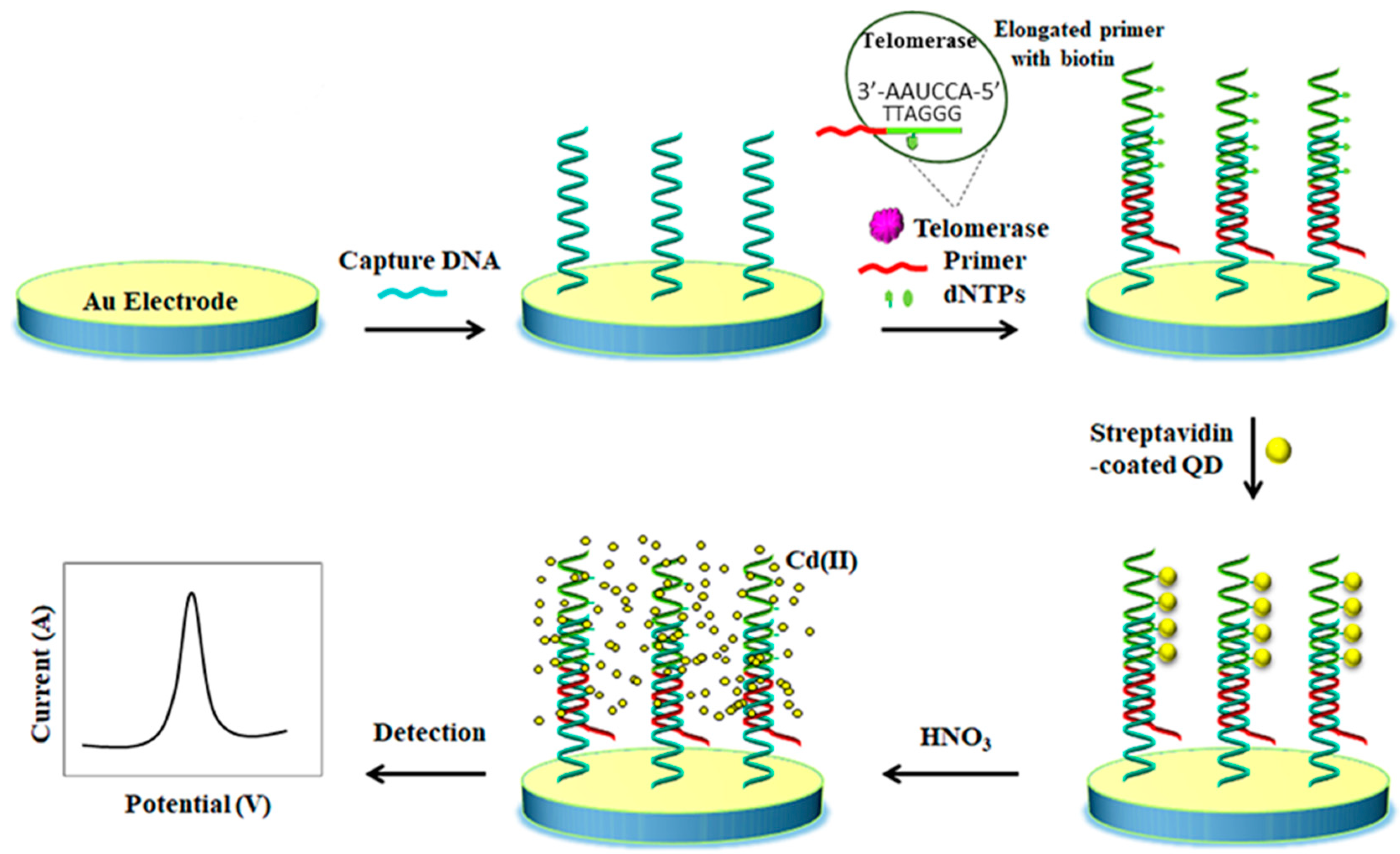
| Bare gold | streptavidin-CdQD | DNA-based | SWASV | Telomerase activity | 1 to 105 cells | HeLa cells, HEK293T cells and MRC-5 cells | [52] |
| Bare gold | 3-CdTeQD-DNA nanocomposite hybridizes with the cleaved DNA probe on the electrode after DNA probe-mRNA interaction and DSN | DNA-based | DPASV | mRNA of BRCA1 | 5 aM–5 fM/1.2 aM | Healthy human serum samples | [53] |
| GCE | AuNP/Electrode modifier for immobilization of two specific harpins PbS@ZIF-8-S1 and CdS@ZIF-8-S2/signaling tag | DNA-based/CHA for signal application | DPASV | Hemophilia A biomarkers: miR-1246 and miR-4521 | miR-1246: 0.05 pM–1.0 mM miR-4521: 0.05 pM–1.0 mM | Human serum | [54] |
| -Two-dimensional (2D) transition metals- | |||||||
| GCE | Ag@Ti3C2Tx nanocomposites as nanocarriers of AChE | Enzyme-based | DPV | Malathion | 10−14–10−8 M/3.27 × 10−15 M | Tap water | [10] |
| GCE | MoS2-AuNPs as electrode modifier and nanoprobe for immobilizing the cAb and HRP-DAb respect. | Antibody based/Sandwich | DPV (H2O2 + o-PD) | CEA | 10 fg mL−1–1 ng mL−1/1.2 fg mL−1 | Spiked samples | [24] |
| SPE | MoS2-AuNPs-β-CD as electrode modifier and for immobilizing the MB-aptamer probe | Aptamer-based/Displacement | DPV (Fc-COOH) | OTA | 0.1–50 nM/0.06 nM | - | [30] |
| CPE | PXA film functionalized MoS2 nanosheets/Electrode modifier for DNA immobilization and signal tag | DNA-based/Displacement | EIS | Circulating tumor DNA | 1.0 × 10⁻¹⁶–1.0 × 10⁻¹⁰ mol L−1/1.8 × 10–17 mol L−1 | PIK3CA gene in peripheral blood of patients with gastric carcinoma | [40] |
| GCE | AuNPs-MoS2 microcubes/Electrode modifier for biotin-cDNA DNS cleaves duplexes DNA-mRNA and mRNA is released | DNA-based/strep-AP | DPV FcM (redox mediator) TCEP (reducing reagent) and AAP (enzyme substrate) | miRNA-21 | 0.1 fM–0.1 pM/0.086 fM | Human serum samples breast from cancer patients | [55] |
| GCE | Av-MBs/Electrode modifier for cAb immobilization AuNP-FMC-WS2 nanocomposite/for DAb immobilization and signal tag | Antibody based/Sandwich | DPV | CA72-4 | 2–50 U L−1/0.6 U L−1 | Human serum | [56] |
| GCE | TiO2–SnS2 nanocomposite/Electrode modifier for GOx immobilization | Enzyme-based | Amperometry −0.45 V | Glucose | 0.008–1.13 and 1.13–5.53 mM/1.8 µM | Human serum samples | [57] |
| GCE | Ti3C2-MXene functionalized with APTES/Electrode modifier for cAb immobilization | Antibody based/Direct | CV ([Ru(NH3)6]3+) | CEA | 1.0 × 10⁻⁴–2000 ng mL−1/1.8 × 10−5 ng mL−1 | Spiked human sera sample | [58] |
| GCE | MoS2/Ti3C2 nanohybrids and AuNPs/Electrode modifier for probe RNA immobilization | DNA-based/Direct | DPV | miRNA-182 | 1 fM–0.1/0.43 fM | Serum samples | [59] |
| Electrode | Nanomaterial/Role | (Bio)sensing Approach/Format | Detection Technique | Target Analyte | LR/LOD | Sample | Ref. |
|---|---|---|---|---|---|---|---|
| -Magnetic carbon nanomaterials- | |||||||
| CPE | m-CNTs@MIP/selective detection | Sensor | Voltammetry | Levofloxacin | 0.003–0.440 μM/0.8 nM. | Spiked serum, urine | [78] |
| SPCE | m-MWCNTs/nanocarrier tag for Ab2 | Sandwich-type immunosensor | Amperometry (H2O2 + HQ) | Fetuin | 20–2000 pg mL−1/16 pg mL−1 | Saliva | [79] |
| SPCE | AuNPs-Fe3O4-GS/electrode modifier | Aptasensor | SWV | CTCs | 5–500 cells mL−1/3–4 cells mL−1 | Whole blood | [80] |
| GCE | Pb2+@AuNPs-MWCNTs-Fe3O4/label for Ab2 | Sandwich-type immunosensor | Amperometry (H2O2) | AFP | 10 fg/mL–100 ng mL−1/3.33 fg mL−1 | Spiked serum | [81] |
| AuE | Fe3O4/GO electrode modifier for avastin Ab1 immobilization | Label-free immunosensor | DPV [Fe(CN)6]3−/4− | VEGF | 31.25–2000 pg mL−1 | Plasma | [82] |
| GCE | Au@Ag/GS-Fe3O4/Cd2+/electrode modifier for immobilization of anti-IgG | Label-free immunosensor | Amperometry | IgG | 5 fg mL−1–50 ng mL−1/2 fg mL−1 | Serum | [83] |
| SPCE | Fe3O4@AuNPs/NGr | Aptamer | DPV | Leukemia cancer cells | 10–106 cell mL−1 | Plasma | [84] |
| ITO | Fe3O4@PDA/rGO/electrode modifier for Ab1 immobilization | Sandwich-type immunosensor | CV | MC-LR | 0.01–50 mg L−1/0.007 mg L−1 | Water | [85] |
| GCE | GS-Fe3O4/Au@Ag/Ni2+/label for Ab2 loading | Sandwich-type immunosensor | Amperometry | CEA | 0.1 pg/mL–100 ng/mL/0.0697 pg/mL | Serum | [86] |
| GCE | Fe3O4@AuNPs/rGO/electrode modifier for cortisol immobilization | Immunosensor/Direct competitive | DPV (H2O2/HRP) | Cortisol | 0.1–1000 ng mL−1/0.05 ng mL−1 | Serum | [87] |
| CPE | Fe3O4/rGO-PANHS/electrode modifier for BRCA1 5382 insC ssDNA immobilization | Label-free DNA biosensor | EIS | BRCA1 5382 insC mutation. | 1.0 × 10⁻¹⁸–1.0 × 10⁻⁸ mol L−1/2.8 × 10−19 mol L−1 | Spiked genome samples | [88] |
| GCE | CNP-L/CuONP/MWCNTs/Pe | Enzymatic biosensor | Amperometry | Triglycerides | 0.001–0.05 g L−1/0.0032 g L−1 (triolein) | Serum | [89] |
| GCE | Fe3O4/CNTs/GO for aptamer immobilization | Label-free aptasensorr | DPV [Fe(CN)6]3−/4− | Diclofenac | 100–1300 pM/ 33 pM | Ampoules | [90] |
| PtE | HRP/Fe3O4/Chit/rGO/electrode modifier | Enzymatic biosensor | CV | H2O2 | up to 100 μM | - | [91] |
| SPCE | C@GNRs/electrode modifier for immobilization of ssDNA | DNA hybridization biosensor | CV Fe(CN)63− | ssDNA | - | - | [92] |
| GCE | ZnFe2O4/α-Fe2O3/Gr/electrode modifier | Non-enzymatic | Amperometric | Glucose | 1–10 mM | - | [93] |
| GCE | Fe3O4@TMU-21/MWCNTs/electrode modifier | Label-free immunosensor | Amperometric (H2O2) | HER2 | 1.0 pg mL−1–100 ng mL−1/0.3 pg mL−1 | Serum | [94] |
| -Carbon nanozymes- | |||||||
| GCE | ZnCr2O4/MWCNTs/electrode modifier | Enzyme-free sensor | Amperometry | H2O2 | 50 μM–34.8 mM/<0.11 μM | Lens cleaning solution | [95] |
| GCE | RuNPs/MWCNTs-Av/HRP mimic nanozyme | Modified electrode | Amperometry | H2O2 | 5.0 × 10⁻⁷ M–1.75 × 10⁻³ M/65 nM | - | [96] |
| GCE | Pt-DEN/CNTs/electrode modifier, HRP mimic nanozyme | Enzyme-free sensor | Amperometry | H2O2 | 3–400 μM/0.8 μM | H2O2 released from living cells | [97] |
| GCE | Fe3O4/CNTs/GO/electrode modifier, HRP mimic nanozyme | Enzyme-free sensor and enzyme biosensor (GOx) | CV (H2O2) Amperometry (glucose) | H2O2, glucose | 0.01–0.50 mM (H2O2) 0.050–5.0 mM (glucose) | - | [98] |
| GCE | CDs/MWCNTs/electrode modifier, HRP mimic nanozyme | Enzyme-free sensor | Amperometry | H2O2 | 3.5 × 10⁻⁶–3.0 × 10⁻⁴ M/0.25 μM | H2O2 released from living cells, serum | [99] |
| GCE | Au-Ag/MWCNTs/electrode modifier | Enzyme-free sensor | CV | Gastric cancer cells-volatile biomarkers | up to 0.0025% (v/v)/0.3 ppb (3-octanone), up to 0.055% (v/v)/0.5 ppb (butanone) | MGC-803, GES-1 cells | [100] |
| AuE | GQDs/electrode modifier, Ab1 immobilization, HRP mimic nanozyme | Label-free immunosensor | Amperometry | Yersinia enterecolitica | 6.23 × 10²–6.23 × 10⁸ cfu mL−1/5 cfu mL−1 (milk), 30 cfu mL−1 (serum) | Milk, serum | [101] |
| SPCE | GDQs/MWCNTs/Ab2 and HRP nanocarrier, HRP mimic nanozyme | Sandwich-type immunosensor | Amperometry (H2O2, HQ) | IL-13Rα2 | 2.7–100 ng mL−1/0.8 ng mL−1 | Raw cellular lysates | [102] |
| GCE | PtPd/N-GQDs@Au/electrode modifier/catalytic activity towards H2O2 | Label-free immunosensor | Amperometry (H2O2) | CEA | 5 fg mL−1–50 ng mL−1/2 fg/mL | Spiked serum | [103] |
| AuE | GQDs/HRP mimic nanozyme | Enzyme-free sensor | Amperometry | H2O2 | - | Human breast cancer MCF-7 cells | [104] |
| GCE | Au/OMCS/electrode modifier/xanthine oxidase mimics | Enzyme-free sensor | DPV | Xanthine | 0.10–20 μM/0.006 μM | Spiked urine | [105] |
| -Multifunctional biomedical applications- | |||||||
| AuE | rGO/insulin/Ni(OH)2/insulin releasing, glucose detection | Enzyme-free sensor | Amperometry | Glucose | 5 μM–10 mM/~ 5 μM | - | [106] |
| GCE | rGO/PdNFs/electrode modifier | Enzyme-free sensor | Amperometry | Glucose | 10–90 nM/8 nM | - | [107] |
| Au/PO | Gr/electrode modifier | Flexible microsensor | DPV | Dopamine | 0.3 μM to 56.8 μM/0.11 μM | - | [108] |
| CPE | PVC/rGO/AuNPs/CNT/electrode modifier | Dual microcatheter | SWV | Propofol, fentanyl | 25–125 μM (PPF), 10–50 nM (FTN) | Whole blood | [109] |
| GCE | rGO/MB-AuNPs/electrode modifier, immobilization of aptamer-Fc | Ratiometric aptasensor | SWV | VEGF | 2–500 pg mL−1/0.1 pg mL−1 | Serum | [110] |
| GCE | rGO/MB/electrode modifier | Ratiometric sensor | DPV | Cerebral ascorbic acid | 0.5 μM–1000 μM/ 10 nM | Brain micro-dialysate | [111] |
| needle-type electrode | rGO/AuNCs/immobilization of ssDNA | Micro-aptasensor (hybridization) | DPV (MB) | Adenosine | 0.1 nM–1 mM/~0.1 nM | In vivo (rat body) | [112] |
| micro-electrode array | rGOPtNPs/electrode modifier | Microsensor | DPV | Norepinephrine | -/0.08 μM | Brain silice secretion | [113] |
| GCE | rGO-MWCNTs /Chit/CQDs/aptamer immobilization | Label-free aptasensor | DPV, EIS [Fe(CN)6]3−/4− | Lysozyme | 20 fmol L−1–10 nmol L−1/3.7 fmol L−1 (DPV); 10 fmol L−1–100 nmol L−1/1.9 fmol L−1 (EIS) | Spiked serum, urine | [114] |
| -Multifunctional carbon nanomaterials for signal amplification- | |||||||
| SPCE | V-Phe-SWCNT (-HRP)/carrier tag for immobilization of Ab2 | Sandwich-type immunosensor | Amperometry | TGF-β1 | 2.5–1000 pg mL−1/0.95 pg mL−1 | Saliva | [115] |
| AuE | COOH-MWCNTs/AuNPs/immobilization of thionine and aptamer | Aptasensor | DPV | Tetracycline | 0.1 nM–1 μM/0.06 nM | - | [116] |
| GCE | AuNPs-rGO/electrode modifier and Ab1 immobilization; SWCNTs-GQDs/carrier tag for Ab2 immobilization | Sandwich-type immunosensor | SWV (H2O2) | CEA | 50–650 pg mL−1/5.3 pg mL−1 | Spiked serum | [117] |
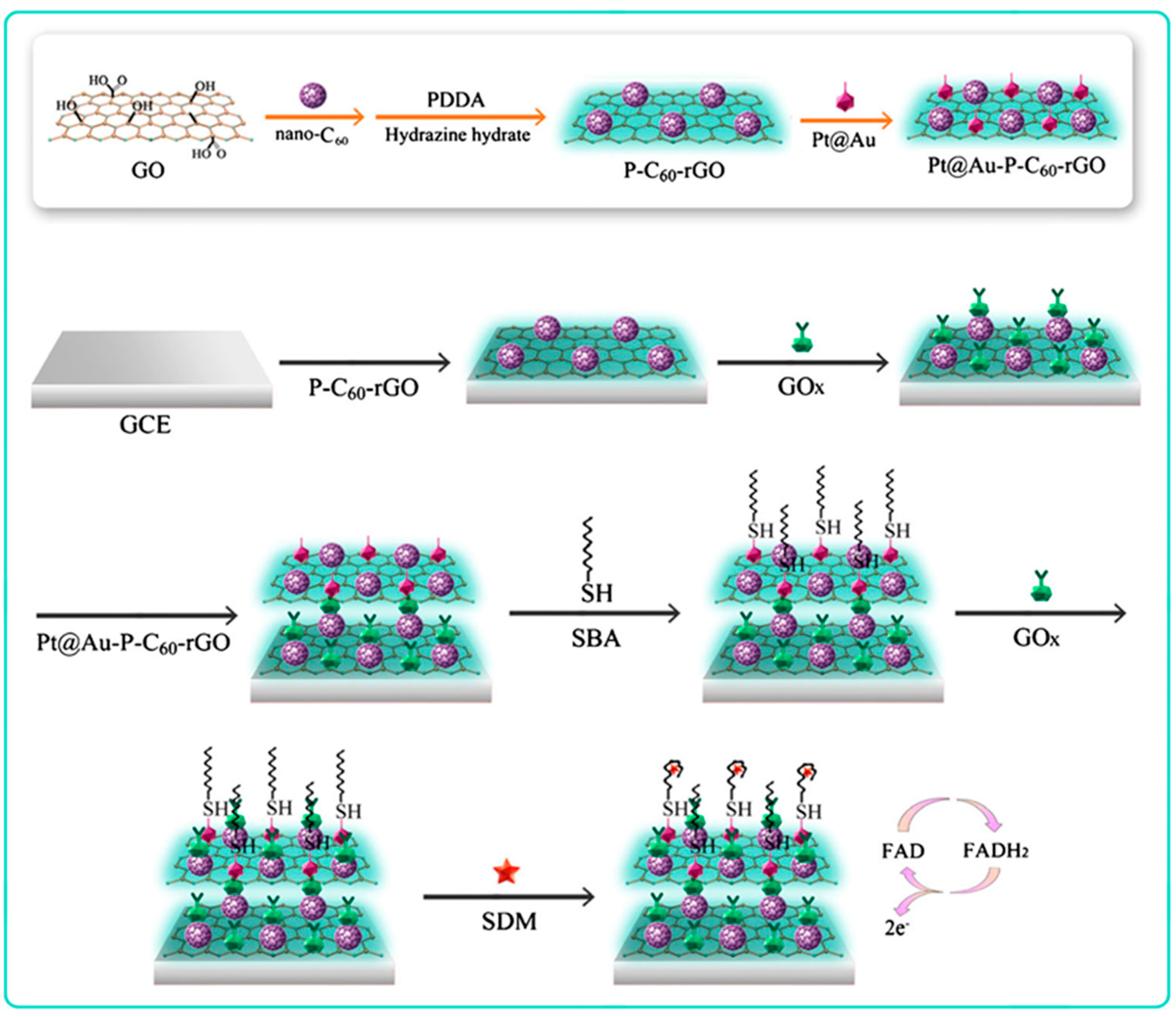
| GCE | Pt@Au-P-C60-rGO electrode modifier for immobilization of SH-aptamer and GOx | Label-free aptasensor | CV | Sulfadime-thoxime | 10−5–50 ng mL−1/8.68 fg mL−1 | Spiked milk | [118] |
| GCE | Pd/g-C3N4-CNTs/electrode modifier | Sensor | DPV | EE2 | 2.0 × 10⁻⁶ –1.5 × 10⁻⁴ M/5.0 × 10−7 M | Chicken and pig foodstuffs | [119] |
| GCE | GOx/Au/MXene/Nafion | Enzyme biosensor | Amperometry | glucose | 0.1–18 mM/5.9 μM | - | [120] |
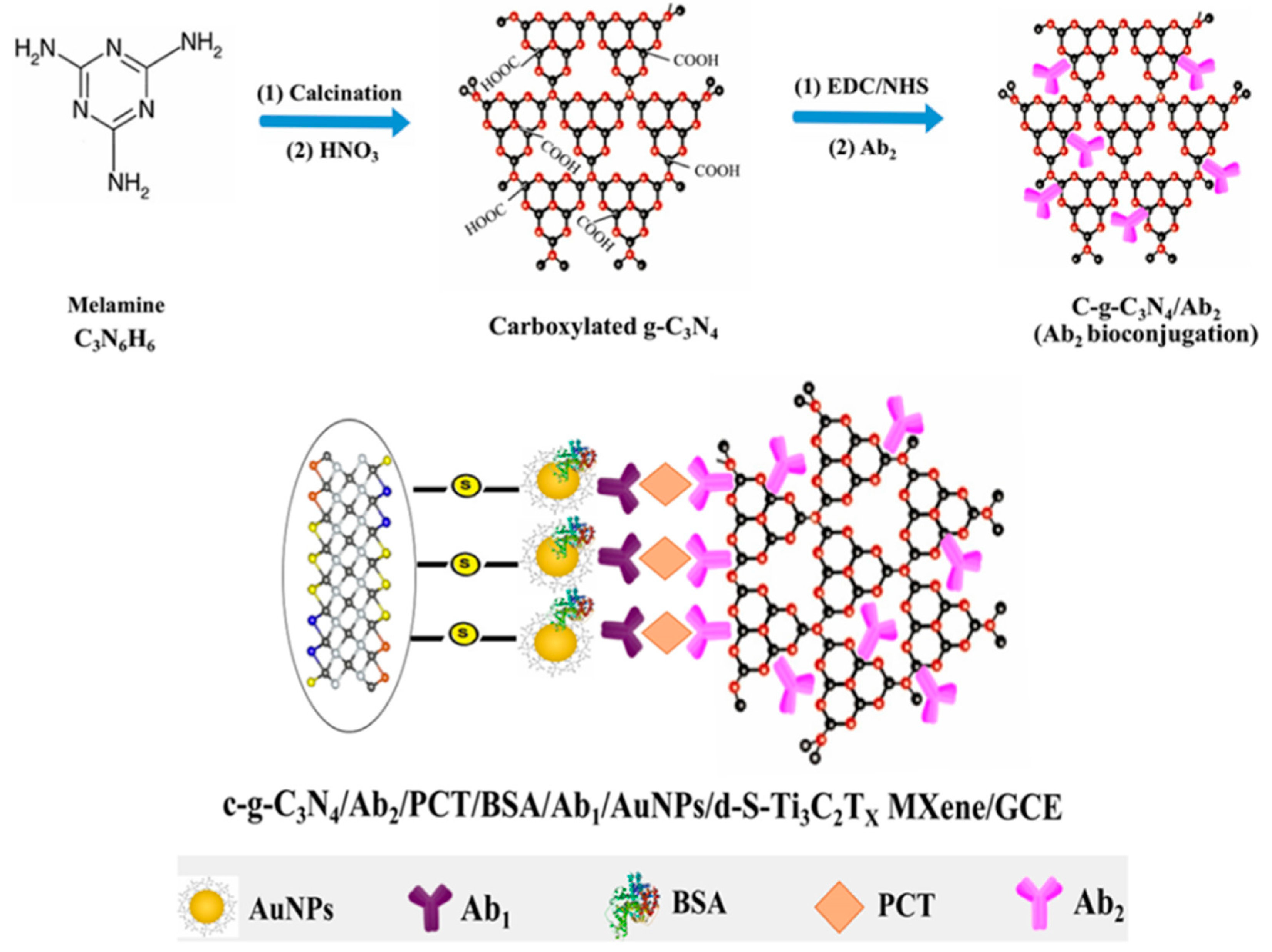
| AuNPs/ GCE | c-g-C3N4/carrier tag for Ab2 immobilizantion | Sandwich-type immunosensor | DPV | Procalcitonin | 0.01–1.0 pg mL−1/2.0 fg mL−1 | Plasma | [121] |
| dSPCE | GQDs/MWCNTs/HRP, carrier tag for Ab2 immobilization | Dual sandwich-type immunosensor | Amperometry (H2O2, HQ) | IL-13Rα2, CDH-17 | 4.92–100 ng mL−1/1.4 ng mL−1 (IL-13sRα2); 0.11–10 ng mL−1/0.03 ng mL−1 (CDH-17) | Lysates from breast and colorectal cancer cells | [122] |
| GCE | Chit@AB-MWCNTs/AuNPs/electrode modifier for immobilization of aptamer and specific binding to GO-ZEN Apt. | Aptasensor | DPV ([Fe(CN)6]3−/4−) | ZEN | 10 fg mL−1–1 ng mL−1/ 3.64 fg mL−1 | Corn oil and corn flour | [123] |
| Type | Names | Characteristic Features | Ref. | |
|---|---|---|---|---|
| MSNs | Mobil Crystalline Materials (MCM) | MCM-48, MCM-41, and MCM-50 | MCM-41: honeycomb-like structure with a pore diameter of 2.5–6 nm and an easily functionalized surface due to the presence of silanol groups. MCM-48 and MCM-50: cubic and lamella-like arrangement, respectively. MCM-41 and MCM-48: can be functionalized with amino groups | [154,162,163,164,165] |
| Santa Barbara Amorphous (SBA) | SBA-11, SBA-12, and SBA 15, SBA-16 | Pores of 4.6–30 nm and thicker silica walls. SBA-15: high surface to area ratio, porosity, uniform pore size distributions, with 30 nm average pore diameter and thermal stability. | [166,167,168,169] | |
| Korea advanced institute of science and technology (KIT) | KIT-5, KIT-6 | KIT-6: composed of two interwoven mesoporous networks similar to that found in MCM-48 silica, but possessing much larger pore diameters of 5–12 nm. | [147,170] | |
| Center for research chemistry and catalysis (COK) | COK-12 | Large-pore ordered mesoporous silica (OMS), analogous to the SBA-15, but synthesized in a more environmentally friendly way and exhibiting a shorter plate-like structure | [171] | |
| Folded sheets of mesoporous (FSM) | FSM-16 | Narrowly distributed pore size of of 2.8 nm in diameter | [172] | |
| Fibrous silica nanospheres | KCC-1 | Particles with extremely large surface area, highly stable, which tolerate high temperature, can be kept at room temperature for months without any changes in their properties and modified with various agents to change the surface properties | [173] | |
| Mesoporous silica film (MSF) | MSF | Mesoporous that have hexagonal arrays with parallel cylindrical channels | [174,175,176,177] | |
| Electrode | Nanomaterial/Role | Biosensing Approach/Format | Detection Technique | Target Analyte | LR/LOD | Sample | Ref. |
|---|---|---|---|---|---|---|---|
| -MSNs- | |||||||
| Disk AuE | Janus type Au-MSNs modified with Strep and HRP on the Au and MS faces, respectively/Signaling tags | Affinity-based/Direct | CV and EIE ([Fe(CN)6]4-/3-) | - | - | - | [185] |
| GCE coated with SWCNTs | Janus type Au-MSNs modified with GOx and HRP on the Au and MS faces, respectively/Electrode modifier | Enzyme-based | Amperometry (H2O2+ HQ) | Glucose | 490 nM–600 μM/360 nM | Commercial soft drinks | [186] |
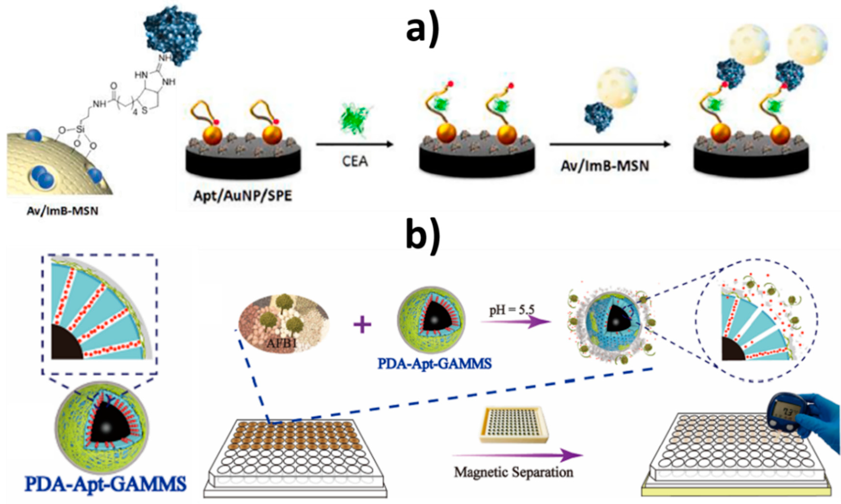
| SPCE | Janus type Au-MSNs modified with biotin thiol-modified anti-CEA DNA hairpin aptamer and HRP on the Au and mesoporous silica faces/Signaling tags and avidin-modified Fe3O4@SiO2 NPs/Solid support | Aptamer-based/Direct | Amperometry (H2O2 + HQ) | CEA | 1–5000 ng mL−1/1.2 pM | Spiked human blood plasma | [183] |
| - | MB-loaded aptamer-gated MSNs (MCM-41)/Electrode modifiers | Aptamer-based/Direct | DPV (MB) | OTA | Up to 50 nM/0.003 nM | Doped wheat samples | [164] |
| AuNPs-SPCE | MB-loaded MSNs and capped with an avidin/imminobiotin stimulus-responsive gate-like ensemble/Nanocarriers of signaling elements | Aptamer-based/Direct | DPV (MB) | CEA | 1.0 pg mL−1–160 ng mL−1/280 fg mL−1 | Human serum samples (5-fold diluted) | [187] |
| - | Glucose-loaded DNAzyme-capped MSNs | Affinity-based/Direct | PGM | Pb2+ | 1.0 pM–0.7 nM/1 pM | Spiked drinking water | [188] |
| - | Glucose-loaded wrapping DNA-capped MSNs | Telomerase + dNTPs-assisted extension | PGM | Telomerase | 100–5000 HeLa cells mL−1/80 HeLa cells mL−1 | HeLa extracts | [189] |
| - | Glucose-loaded DNA-gated MSNs | DNA-based/Direct | PGM | Hg2+ | 0.1–80 nM/0.1 nM | Spiked tap water and lake water | [190] |
| - | Aptamer-capped MSNs with glucose loaded on GO nanosheets attached to the aptamer through π-stacking interactions | Aptamer-based/Displacement | PGM | AsO3- | 0.01–100 ng mL−1/2.3 pg mL−1 | River, lake, seawater and tap water | [179] |
| - | “Dual gates” aminated magnetic mesoporous silica nanocomposites loaded with glucose and bearing PDA-aptamer two-tier shells | Aptamer-based/Direct | PGM | AFB1 | 0.03–8 ng mL−1/0.02 ng mL−1 | Pearl rice, maize and wheat | [191] |
| GCE | Au-MSNs/Electrode modifier | Aptamer-based/Direct | DPV ([Fe(CN)6]4−/3−) | Codein | 10 pM–100 nM/3 pM | - | [156] |
| Graphite SPE | AuNPs/SBA-15@DABCO/Electrode modifier | Aptamer-based/Direct | DPV (hemin) | CAP | 0.03–0.15 μM and 0.15–7.0 μM/4.0 nM | Spiked human blood serum | [169] |
| SPCE | AuNPs/Fe3O4@SiO2/DABCO/Electrode modifier | Aptamer-based/Direct | LSV (epirubicin) | Epirubicin | 0.07 μM–1.0 μM and 1.0 μM–21.0 μM/0.04 μM | Spiked human blood serum | [152] |
| GCE | AMSNs (MCM-41)/Electrode modifier | Aptamer-based/Direct | DPV (electrocatalytic reduction of oxygen by hemin) | Hemin and Hb | Hemin and Hb: 1.0 × 10⁻¹⁹–1.0 × 10⁻⁶ M/Hemin: 7.5 × 10⁻²⁰ M, Hb: 6.5 × 10⁻²⁰ M | Blood (Hb) | [163] |
| GCE | AuNPs incorporated in AMSNs (MCM-41)/Electrode modifier | Aptamer-based/Direct | EIS ([Fe(CN)6]4−/3−) | CEA | 1.0 × 10⁻³−100.0 ng mL−1 9.8 × 10⁻⁴ ng mL−1 | Human serum | [154] |
| GCE | GQDs-CS/KCC-1-NH2-Tb/Electrode modifier | Aptamer-based/Direct | DPV (TB) | AFB1 | 0.1 μM–1 fM/LQ = 10 fM | Spiked milk samples | [173] |
| GCE | MWCNTs-MSNs (MCM41)-Hb/Electrode modifier | Enzymatic-based (Hb DET) | Amperometry | NO2-, TCA | NO2−: 1.0 × 10⁻⁷–1.25 × 10⁻⁴ M/16 nM TCA: 5.0 × 10⁻⁵–2.7 × 10⁻² M/3 μM | Spiked tap water | [165] |
| GCE | PAMAM–rGO/electrode modifier and mSiO2@MWCNT/nanocarriers of Thi, platinum nanoparticles (PtNPs), and hemin/G-quadruplex bioelectrocatalytic complex | Aptamer-based/Sandwich | DPV (H2O2) | Thrombin | 0.0001–80 nM/50 fM | Spiked human serum | [178] |
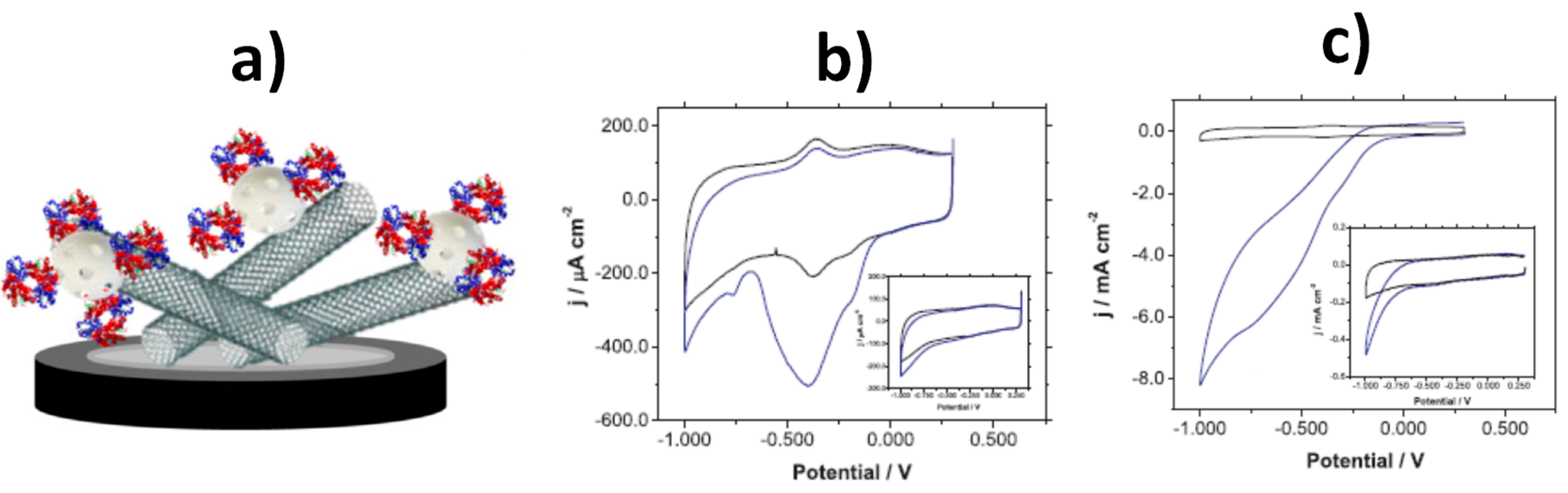
| GCE modified with HDGMs | Au-MSNs modified with Thi/Signaling tags | Aptamer-based/Sandwich | DPV (Thi) | Thrombin | 0.03 pM–0.018 μM/15 fM | Spiked fetal calf serum | [151] |
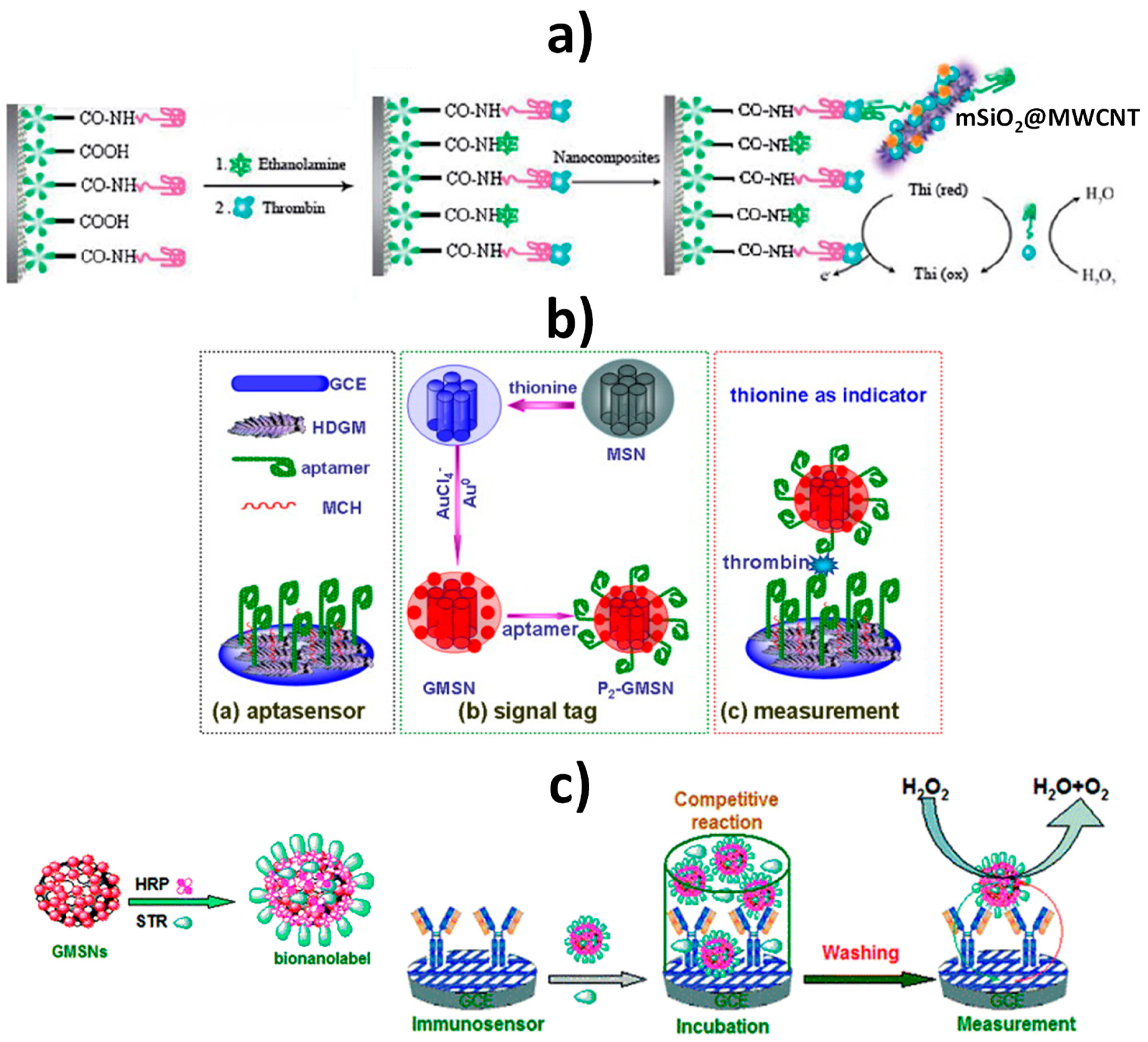
| GCE | TRSiNs doped with Thi/electrode modifier and GMSNs/nanocarriers of STR-BSA and HRP | Antibody-based/Competitive | DPV (H2O2+Thi) | STR | 0.05–50 ng mL−1 5 pg mL−1 | Spiked samples (honey, milk, kidney, and muscle) | [192] |
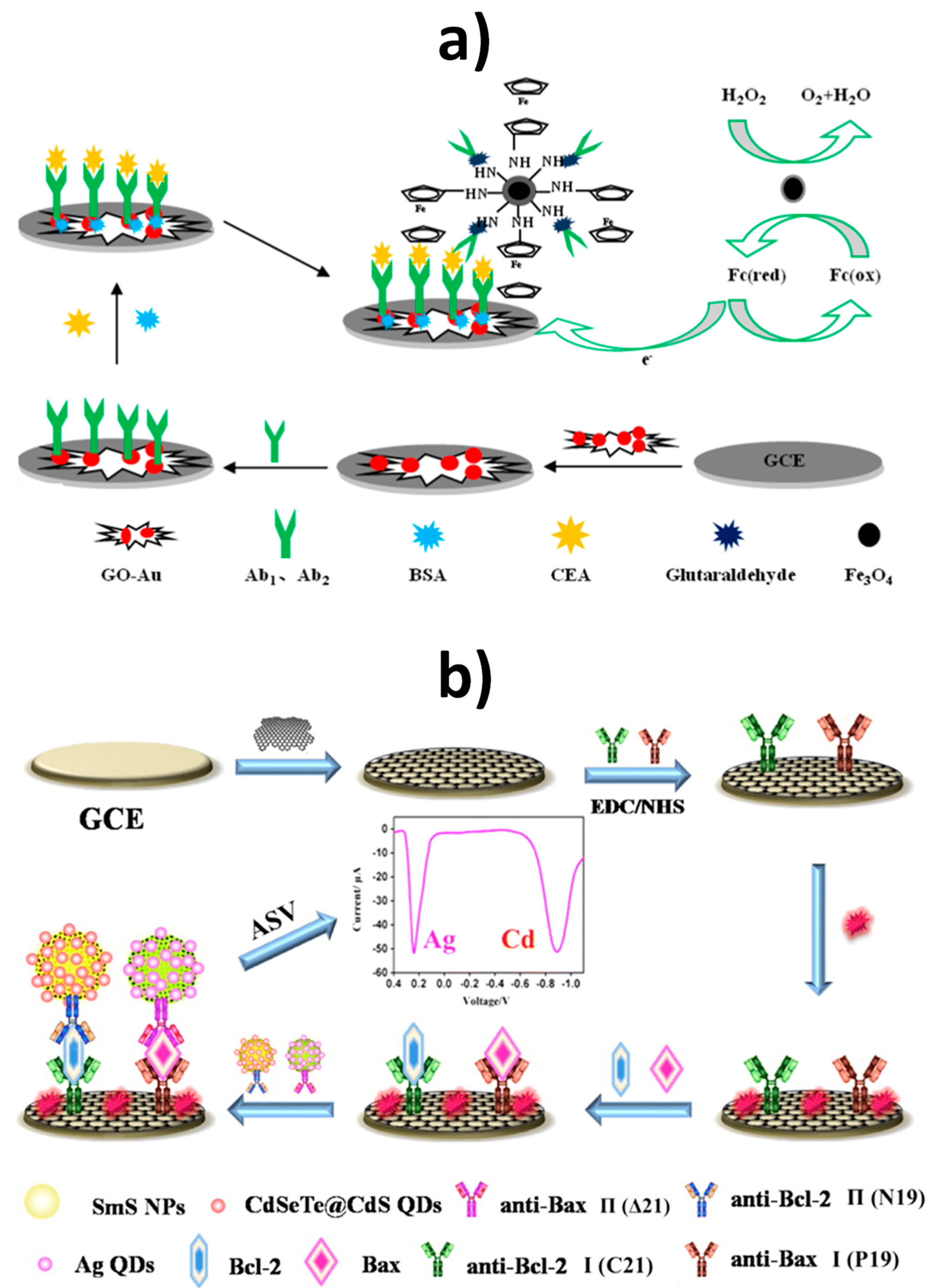
| GO–AuNPs-modified GCE | Fe3O4@SiO2–NH2/Nanocarriers of Fc-COOH and DAb | Antibody-based/Sandwich | DPV (H2O2+Fc) | CEA | 0.001 ng mL−1–80 ng mL−1/0.0002 ng mL−1 | Spiked human serum samples | [182] |
| RGO-GCE | Mesoporous SiO2 decorated with DAb and CdSeTe@CdS quantum dots (QDs) or Ag nanoclusters (NCs)/nanocarriers of signaling elements | Antibody-based/Sandwich | ASV (Ag, Cd) | Bcl-2 and Bax | 1–250 ng mL−1/∼0.5 fmol | Nilotinib-treated chronic myeloid leukemia K562 cells | [180] |
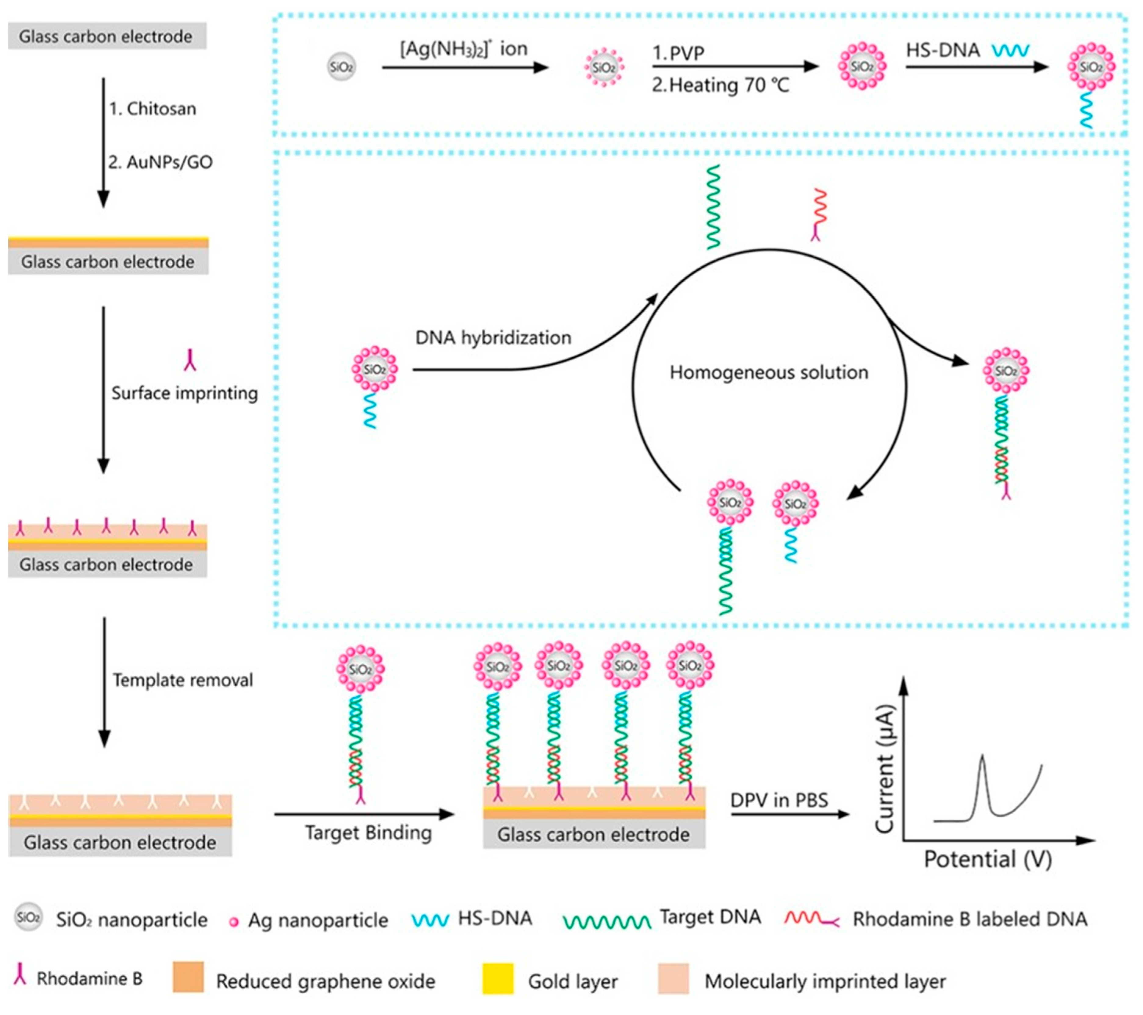
| AuNPs-GO modified GCE covered with MIPs for RhB | DNA-modified SiO2@AgNPs/Tracing tag | DNA-based/Sandwich | DPV (RhB) | BRCA-1 | 10 fM–100 nM/2.53 fM | Spiked human serum | [193] |
| -SiO2-SPAABs- | |||||||
| GO-GCE | SiO2-SPAABs decorated with HRP and DAb/Nanocarriers | Antibody-based/Sandwich | DPV (H2O2 + ODP) | human IgG (HIgG) | 100 pg mL−1–100 μg mL−1 | Spiked serum samples | [194] |
| -Si MSs- | |||||||
| AuNPs-GCE | Au-Si MSs modified with signal molecules (Zn2+) and antibodies (Ab)/Signaling tags | Mixed aptamer and antibody-based/Sandwich | SWV (Zn2+) | CRP | 0.005–125 ng mL−1/0.0017 ng mL−1 | Real serum samples | [195] |
| -MSF- | |||||||
| Disk AuE | NH2-MSF/Electrode modifier | Biocatalytic-based | CV/EIS ([Fe(CN)6]4−/3−) | TGase activity (in the presence of N-benzyloxycarbonyl-L-glutaminylglycine) and indirectly of Pb2+ due to TGase activity irreversible inhibition | TGase activity: 0.4–185 μU mL−1 Pb2+: 4.0–500 μM | - | [196] |
| Disk AuE | DNAzyme-gated MSF/Electrode modifier | DNAzyme-based | SWV ([Fe(CN)6]4−/3−) | Cu2+, AA | Cu2+: 310 ppb–4.5 ppm/250 ppb AA: 1.0–120 μM/700 nM | Certified water samples (Cu2+) and Redoxon® pills (AA) | [174] |
| Disk AuE | DNA-gated MSF/Electrode modifier | DNA-based biosensing approach | SWV ([Fe(CN)6]4−/3−) | E. coli 16S rRNA | Synthetic target DNA, a copy of partial region of the E. coli 16S rRNA gene (position 432–461 according to the 5′→3′ nucleotide sequence): 5–700 nM/ 2.5 nM | Raw E. coli lysate | [175] |
| Disk AuE | Aptamer-gated MSF/Electrode modifier | Aptamer-based biosensing approach | DPV ([Fe(CN)6]4−/3−) | PSA | 1 to 300 ng mL−1/280 pg mL−1 | Spiked artificial urine samples and blood serum from a healthy individual | [176] |
| AuE | AgNPs-MSF/Electrode modifier | Aptamer-based biosensing approach | DPV ([Fe(CN)6]4−/3−) | STR | 1 fg mL−1–6.2 ng mL−1/0.33 fg mL−1 | Spiked milk and blood serum samples | [177] |
Publisher’s Note: MDPI stays neutral with regard to jurisdictional claims in published maps and institutional affiliations. |
© 2020 by the authors. Licensee MDPI, Basel, Switzerland. This article is an open access article distributed under the terms and conditions of the Creative Commons Attribution (CC BY) license (http://creativecommons.org/licenses/by/4.0/).
Share and Cite
Yáñez-Sedeño, P.; González-Cortés, A.; Campuzano, S.; Pingarrón, J.M. Multimodal/Multifunctional Nanomaterials in (Bio)electrochemistry: Now and in the Coming Decade. Nanomaterials 2020, 10, 2556. https://doi.org/10.3390/nano10122556
Yáñez-Sedeño P, González-Cortés A, Campuzano S, Pingarrón JM. Multimodal/Multifunctional Nanomaterials in (Bio)electrochemistry: Now and in the Coming Decade. Nanomaterials. 2020; 10(12):2556. https://doi.org/10.3390/nano10122556
Chicago/Turabian StyleYáñez-Sedeño, Paloma, Araceli González-Cortés, Susana Campuzano, and José Manuel Pingarrón. 2020. "Multimodal/Multifunctional Nanomaterials in (Bio)electrochemistry: Now and in the Coming Decade" Nanomaterials 10, no. 12: 2556. https://doi.org/10.3390/nano10122556
APA StyleYáñez-Sedeño, P., González-Cortés, A., Campuzano, S., & Pingarrón, J. M. (2020). Multimodal/Multifunctional Nanomaterials in (Bio)electrochemistry: Now and in the Coming Decade. Nanomaterials, 10(12), 2556. https://doi.org/10.3390/nano10122556