Article

Temperature Sensing in the Short-Wave Infrared Spectral Region Using Core-Shell NaGdF4:Yb3+, Ho3+, Er3+@NaYF4 Nanothermometers

Prokhorov General Physics Institute of the Russian Academy of Sciences, Moscow 119991, Russia
*
Author to whom correspondence should be addressed.
Nanomaterials 2020, 10(10), 1992; https://doi.org/10.3390/nano10101992
Submission received: 18 August 2020 / Revised: 13 September 2020 / Accepted: 28 September 2020 / Published: 9 October 2020

Abstract

:
The short-wave infrared region (SWIR) is promising for deep-tissue visualization and temperature sensing due to higher penetration depth and reduced scattering of radiation. However, the strong quenching of luminescence in biological media and low thermal sensitivity of nanothermometers in this region are major drawbacks that limit their practical application. Nanoparticles doped with rare-earth ions are widely used as thermal sensors operating in the SWIR region through the luminescence intensity ratio (LIR) approach. In this study, the effect of the shell on the sensitivity of temperature determination using NaGdF4 nanoparticles doped with rare-earth ions (REI) Yb3+, Ho3+, and Er3+ coated with an inert NaYF4 shell was investigated. We found that coating the nanoparticles with a shell significantly increases the intensity of luminescence in the SWIR range, prevents water from quenching luminescence, and decreases the temperature of laser-induced heating. Thermometry in the SWIR spectral region was demonstrated using synthesized nanoparticles in dry powder and in water. The core-shell nanoparticles obtained had intense luminescence and made it possible to determine temperatures in the range of 20–40 °C. The relative thermal sensitivity of core-shell NPs was 0.68% °C−1 in water and 4.2% °C−1 in dry powder.

1. Introduction

The development of contactless thermal sensors has attracted much attention in the last decade [1,2,3,4]. Conventional thermocouples and thermometers are unsuitable for remote temperature control [5]. Precise contactless monitoring and temperature control at the microscopic level are highly important for controlling various biological functions and their changes during thermal therapies as well as visualization of thermal changes in vivo [6,7,8,9,10,11]. Contactless luminescent nanothermometers are promising for biomedical applications. Various luminescent materials with temperature-dependent optical features have been studied as optical thermometers. The most popular are quantum dots [12,13], organic molecules [14,15,16], polymers [17,18,19], DNA or protein-conjugated systems [20], and lanthanide-doped nanophosphors [21,22].
The depth of light penetration limits the use of optical thermometry methods for biological tissues [23,24]. Obtaining diagnostic information from deep layers of biological tissues such as the brain or the skin is complicated due to their heterogeneity and scattering [25,26]. Recent studies have shown that the low scattering in the short-wave infrared region (SWIR, 1100–2000 nm, the so-called second and third biological tissue transparency window) significantly increases the depth and sensitivity of fluorescence imaging, and provides excellent contrast and high signal-to-noise ratio for the obtained luminescent images [27,28,29].
However, the practical application of SWIR imaging and thermometry is still limited by a small number of SWIR phosphors. Most organic SWIR dyes and many types of nanomaterials developed for lasers and the telecommunications industry are unsuitable because of their low quantum efficiency (<0.1%), poor solubility in water, and potential toxicity. The organic dyes known to be fluorescent in the SWIR region, such as IR-26, IR-1048, and IR-1061, are highly hydrophobic, have an extremely low quantum yield (less than 0.05%), and contain toxic metals [30,31]. In the last decade, much attention has been paid to several classes of dyes and nanomaterials: cyanine dyes, lanthanide complexes, platinum complexes, certain types of quantum dots, small gold nanoparticles, and single-walled carbon nanotubes [32]. Inorganic nanoparticles containing rare-earth ions (REIs) may also be of interest. The advantages of rare-earth ions as luminescent labels include narrow-band radiation, a large spectral distance between excitation and emission wavelengths (which is characteristic for the up- and downconversion), long luminescence lifetime, high photostability and low toxicity of materials, minimal autofluorescence of biological tissues, and the greatest depth of sounding when excited in the near-infrared region (NIR) [33]. In comparison with other phosphors, rare-earth ions can be excited through multiple electron states and, due to internal conversion, can produce luminescence bands in a wide range of visible, NIR, and SWIR spectra [34].
Obtaining SWIR-based luminescent markers with high quantum efficiency under infrared excitation is an urgent task today. This study was devoted to the synthesis of NaGdF4 nanoparticles doped with rare-earth ions Yb3+, Ho3+, and Er3+ covered with an inert NaYF4 shell, and to the study of the effect of the shell on the characteristics of luminescence and thermal sensitivity of the synthesized nanoparticles at various pump power densities.
The luminescence intensity ratio approach, which tracks temperature-induced changes in luminescence intensity at two different wavelengths, is widely used for thermal sensing [35,36]. This self-referential technique allows the dependency on measurement conditions to be reduced and high sensing accuracy to be achieved.
Two main approaches can be used for temperature sensing: the luminescence intensity ratio from a single type of lanthanide ions or from two or more types doped into a single host matrix. To quantitatively assess the thermometric capability of a nanothermometer, the sensor sensitivity is usually used, defined as the rate of change of the temperature-sensitive parameter with temperature. The first single-ion approach is based on the temperature-dependent emission from two thermally coupled levels, and is therefore limited by the energy gap between them. In the second approach, the temperature-induced emission changes from the luminescence intensity ratio of two or more different lanthanide ions doped into a single host matrix are analyzed, wherein only one emission is influenced by temperature changes depending on the surrounding medium.
The majority of lanthanide-based nanothermometers in the visible (VIS) or NIR region operate via the upconversion luminescence intensity ratio approach [37]. To overcome the limitations attributed to low light penetration depth in this region, developing downconversion nanoparticles with the excitation and emission wavelength matching one of the three biological windows has been suggested [38,39,40]. In those regions, the tissue becomes partly transparent, allowing for higher penetration depth, better spatial resolution, and reduced side effects [41]. One study demonstrated the fundamental possibility of contactless thermometry in the SWIR spectral range using nanoparticles doped with rare-earth ions [42]. The same group also optimized the concentration of doped impurities of triply doped NaYF4: Yb3+, Ho3+, Er3+ core-only nanoparticles (NPs) operating in the over-1000 nm region to obtain high sensitivity to temperature changes [43]. The maximum sensitivity was 2.15% °C−1 in water and 2.17% °C−1 in cyclohexane.
The synthesis of core-shell LaF3 NPs for bioimaging in SWIR was reported recently [44]. The relative thermal sensitivity obtained ranged from 0.1% °C−1 (core only) to 0.41% °C−1 and 0.36% °C−1 (core-shell), but was lower compared to their upconversion counterparts [45,46]. The relative sensitivity values of 1.1% °C−1 were obtained for NaGdF4: Er3+, Ho3+, Yb3+, Nd3+ core-multi-shell NPs operating in the NIR [47]. However, core-multi-shell materials are usually complex (multilayer structure) and their synthesis is a multistep process. In another study, the use of the Yb3+/Er3+ 1010/810 and 1010/660 nm emission band ratios of core-shell β-NaYF4:Yb3+−Er3+@SiO2 nanorods for temperature-sensing purposes was presented and a sensitivity of 1.64% °C−1 was obtained for 1010/810 nm luminescence intensity ratios [48].
Several groups have studied the dependence of temperature determination sensitivity on structural properties of NPs such as composition, phonon energy of the host material [49], NP size [50], and the dopant concentration [51], as well as on the influence of the surrounding medium [38,52]. In water, the overall emission intensity is lower due to an increase in water-induced non-radiative losses. These mainly affect the sensitizer Yb3+ [53,54] and therefore suppress the activator emission. It has previously been shown that the quenching of luminescence in the VIS and NIR regions could be prevented by using core-shell or complex core-multi-shell structures [55,56,57]. In this study, we examined the influence of NaYF4 inert shell on the luminescence intensity in SWIR and the thermal sensing sensitivity of Yb3+, Ho3+, Er3+ tri-doped NaGdF4 NPs. We also investigated the effect of the pump power density on laser-induced heating and thermal sensitivity. High pump power densities were shown to cause intense heating, which negatively affected the thermometric properties of the studied nanoparticles. At low pump power densities, the luminescence intensity was low and strongly quenched in water, which negatively affected the accuracy of temperature determination. Coating nanoparticles with an inert shell can significantly improve luminescence characteristics, prevent quenching, and obtain high thermal sensitivity.

2. Materials and Methods

The NaGdF4 NPs doped with Yb3+, Ho3+, and Er3+ ions was synthesized via the solvothermal technique in oleic acid [58]. The concentration ratio for ions was 20:3:0.5, previously described as optimal for thermosensing in SWIR [43]. Rare-earth element acetates (99.99% purity, Lanhit, Moscow, Russia), sodium hydroxide (NaOH) and ammonium fluoride (NH4F) with chemical purity (Lanhit, Moscow, Russia), oleic acid (Sigma Aldrich, St. Louis, MO, USA), and 1-octadecene (Sigma Aldrich, St. Louis, MO, USA) were used as precursors. Ytterbium, erbium, holmium, and gadolinium acetates; oleic acid; and 1-octadecene were placed in a 250 mL three-necked flask under reflux. The reaction mixture was heated to 140 °C under argon with vigorous stirring until the acetates were completely dissolved and transformed into rare-earth oleates. Water and acetic acid were then removed under vacuum and the reaction mixture was cooled down to room temperature. The appropriate solutions of NaOH and NH4F in methanol were added in stoichiometric amounts with respect to rare-earth acetates. The reaction mixture was then heated up to 50–60 °C and held for one hour under vacuum to remove the methanol and cause the nucleation of cubic phase α-NaGdF4:Yb3+, Ho3+, Er3+ NPs. After the removal of the methanol, the reaction mixture was heated up to a predetermined temperature of 295 °C. The temperature was maintained for 1.5–2 h to transition from cubic to the hexagonal phase β-NaGdF4:Yb3+, Ho3+, Er3+ and then cooled to 25 °C.
The synthesis of NaYF4 shell involved the same steps. Yttrium acetate, oleic acid, and 1-octadecene were added to the three-necked flask under reflux. The reaction mixture was then heated up to 140 °C in argon with vigorous stirring. Water and acetic acid were removed under vacuum and the reaction mixture was cooled down to room temperature. After heating to 50–60 °C and the removal of methanol, the reaction mixture was heated to 295 °C and held for 1.5–2 h for NaYF4 formation, then cooled to 25 °C. The NPs were collected by centrifugation (8500 rpm, 5 min). The resulting NPs were washed three times with chloroform and ethanol. The determination of the phase composition of the synthesized samples was performed via the X-ray powder diffraction (XRD) technique (D8 Bruker® Advance diffractometer with Cu-Kα radiation, Powder 2.0 software from Laboratory of Inorganic Crystallochemistry, MSU by Oleynikov Peter, Moscow, Russia). The particle size and morphology were analyzed using transmission electron microscopy (TEM, Libra 200 FE microscope, Carl Zeiss AG, Oberkochen, Germany) with ImageJ software (version 1.53a, open source Java image processing program, https://imagej.net).
For the temperature-dependent luminescence spectra measurements, the samples were spread thinly over a horizontal aluminum plate and excited using a semiconductor laser with a wavelength of 980 nm, which was operated in a continuous wave regime. A dry sample was also suspended in water to study its influence on SWIR luminescence. A thin layer of the colloid (1 mm) was poured into an open cuvette. The laser beam was focused at a spot 1 cm in diameter with pump power density in the 0.4–4.0 W/cm2 range. The luminescence spectra in the 1000–1700 nm region were measured using an optical fiber spectrometer DWARF-Star (StellarNet, Tampa, Fl, USA). A hard-coated edge pass filter FELH-1050 (Thorlabs, Newton, N.J., USA) was used to filter the signal from the laser excitation. The luminescence was captured with a fiber with a core diameter of 200 μm. Temperature was controlled using thermometric infrared camera JADE MWIR SC7300M (CEDIP, Croissy-Beaubourg, France) synchronized with the spectrometer for the simultaneous measurement of the luminescence spectrum and the sample temperature.
The temperature-dependent emission spectra of the NaGdF4:Yb3+, Ho3+, Er3+ and NaGdF4:Yb3+, Ho3+, Er3+@NaYF4 NPs were analyzed. To quantify the thermal sensitivity, the ratio between the integral intensities of Ho3+ in the 1125–1200 nm range (IHo) and Er3+ in the 1450–1650 nm range (IEr) emission peaks (luminescence intensity ratio, LIR) were used:
LIR = IHo/IEr.
The relative thermal sensitivity of the NaGdF4:Yb3+, Ho3+, Er3+ and NaGdF4:Yb3+, Ho3+, Er3+@NaYF4 NPs was determined by the ratio [59]:
S = 1/LIR × dLIR/dT.

3. Results and Discussion

As a result, the NaGdF4:Yb3+, Ho3+, Er3+, and NaGdF4:Yb3+, Ho3+, Er3+@NaYF4 NPs (Yb3+:Ho3+:Er3+ = 20:3:0.5 mol %) were synthesized. The notations “core” and “core-shell” are used hereafter, respectively. According to XRD data from JCPDS #27-0699, the synthesized samples were a pure hexagonal phase. The X-ray powder patterns of the obtained samples are presented in Figure 1.
The deviation of the X-ray peak positions was associated with the difference in the radii of the gadolinium, ytterbium, erbium, holmium, and yttrium cations [60], which resulted in the unit cell parameters changing from a = 6.020 Å, c = 3.601 Å for JCPDS #27-0699 to a = 6.028(2) Å, c = 3.569 Å for the core and a = 6.006(1) Å, c = 3.549(1) Å for the core-shell NPs. The NaGdF4:Yb3+, Ho3+, Er3+@NaYF4 sample consisted of particles with obvious core-shell architecture with semi-spherical morphology and an average diameter of 23 nm (Figure 1b). The thickness of the shell was about 3 nm.
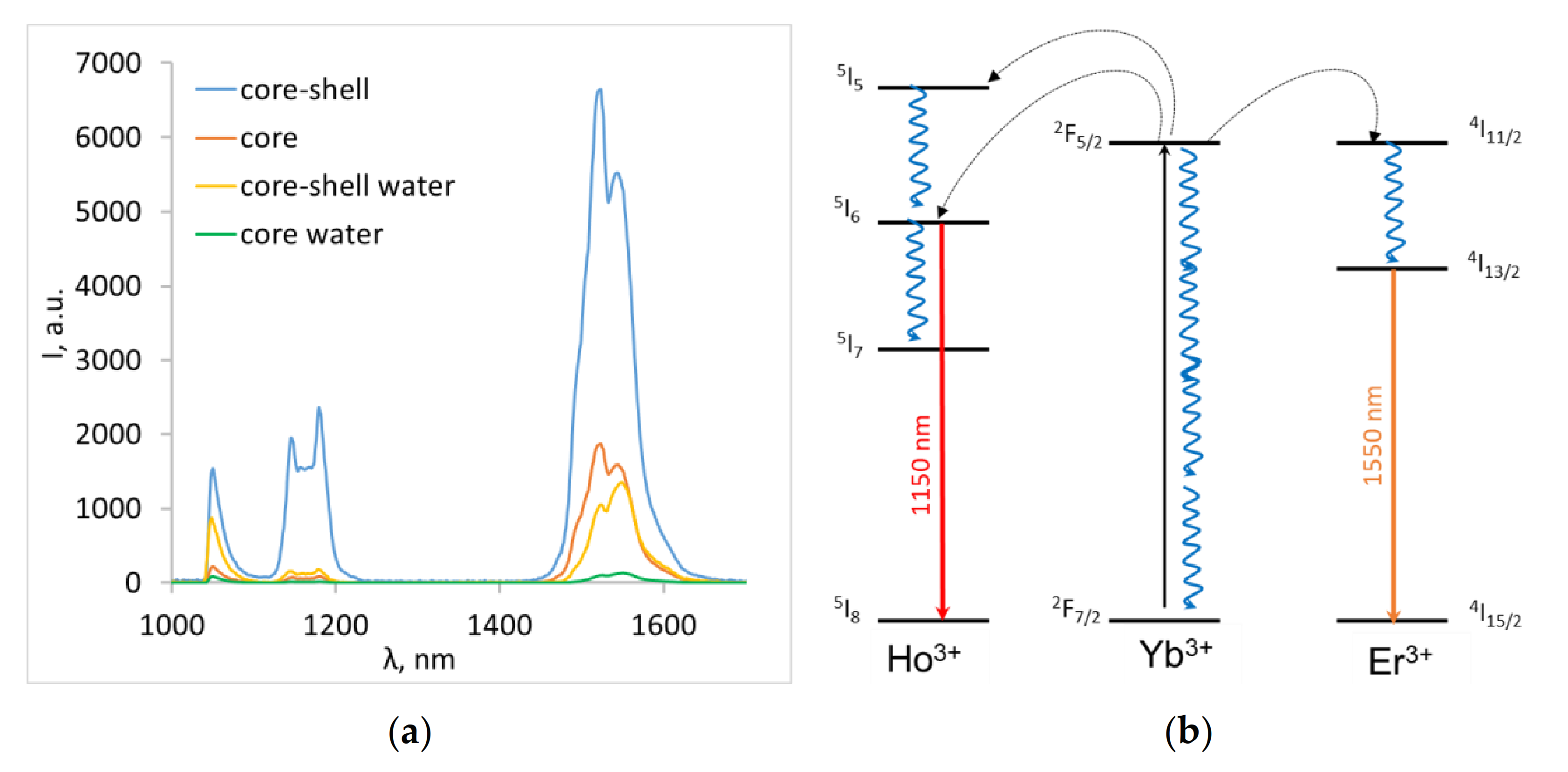
The luminescence spectra of the obtained core and core-shell nanoparticles in powder and in water are shown in Figure 2.
All samples emitted the characteristic Ho3+ and Er3+ luminescence at 1150 nm (5I65I8) and 1550 nm (4I13/24I15/2) with no evident change in the band position. The emission intensity was highly dependent on the interaction with the surrounding medium and decreased in water. The decrease was more pronounced for the core nanoparticles: the integral luminescence intensity in 1050–1600 nm region decreased 5-fold for core-shell NPs compared to 14-fold for core-only NPs. This phenomenon is well known and could be explained by the quenching of surface-ion luminescence by high vibrational energy ligands in the surrounding medium or surface defects [61]. The non-radiative losses mainly affected the sensitizer (Yb3+) and therefore suppressed the Ho3+ and Er3+ emission.
A study of laser-induced heating of core and core-shell particles was conducted by varying laser power density between 0.4 and 4.0 W/cm2 (Figure 3).
We noted that the core NPs heated up much more, especially at high pump power densities. After 10 s of irradiation with 2 W/cm2 pump power density, the temperature reached 38 °C for core-shell and 64 °C for core NPs. The differences were less significant at lower pump power densities, and the heating temperature was about 29 °C at 0.4 W/cm2 (the lowest power density used) and 32.5 °C at 1 W/cm2 both for the core and the core-shell NPs. In this case, heating to an equilibrium temperature occurred in less than 10 s. For further spectroscopic studies of the dependence of the luminescence spectra on the power density during laser-induced heating, the temperature in the time range of 10–30 s (when the temperature was already stable) was averaged.
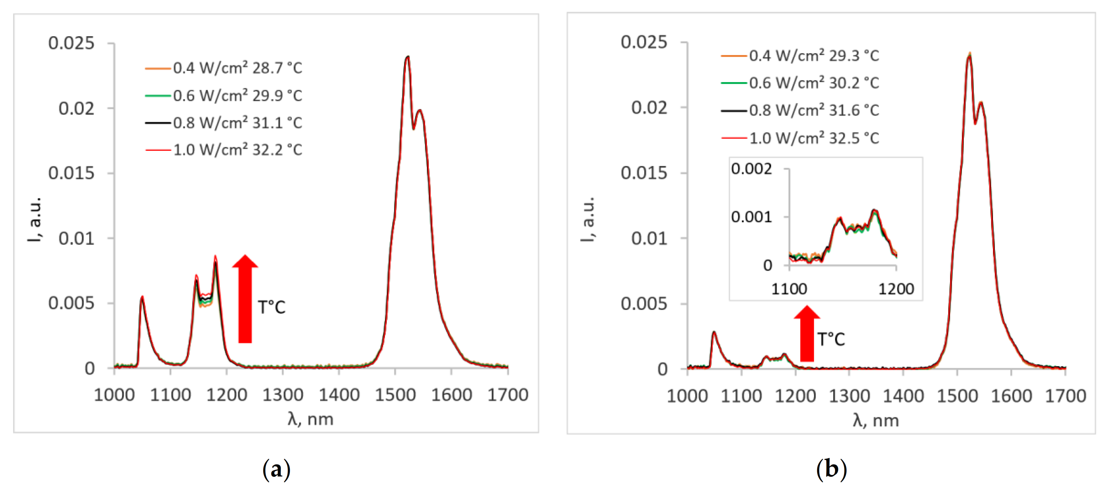
The influences of pump power density and, consequently, the temperature on the emission spectra of NPs in powder are shown in Figure 4a.
It was evident that for the dry sample, the Ho3+ emission rose with the increase in temperature for core-shell nanoparticles. This dependence could be explained by the non-resonant nature of the Yb3+ → Ho3+ transition. In this case, the energy transfer is facilitated by the phonon-assisted processes, where the energy mismatch is compensated by simultaneous emission or absorption of one or more phonons by the host lattice [62]. Because the phonon-assisted energy transfer is temperature-dependent [63], the Ho3+ emission tends to increase with the increased temperature. The Yb3+ → Er3+ energy transfer is resonant, so the temperature change does not affect the Er3+ emission. It was also noted that the temperature-dependent changes in the luminescence spectrum of the core nanoparticles (Figure 4b) were negligible and difficult to detect due to the low intensity of the characteristic Ho3+ peak at a wavelength of 1150 nm.
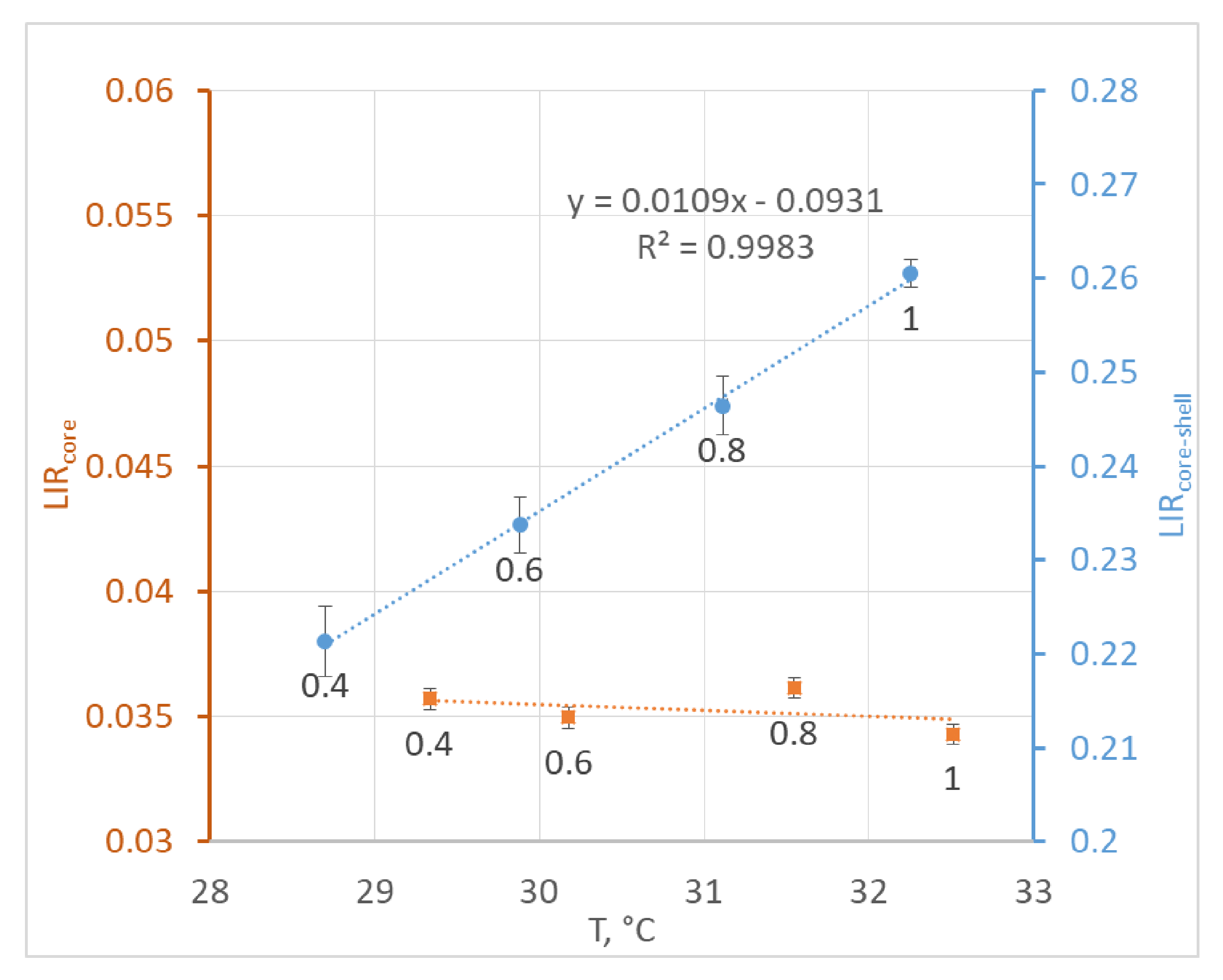
The LIR values, which were determined from the temperature-dependent emission spectra, are presented in Figure 5.
The observed dependency of LIR on temperature for the core-shell NPs was almost linear, whereas for the core NPs, the observed changes in LIR were within the measurement error. The relative thermal sensitivity for core-shell nanoparticles in powder was determined to be 4.2% °C−1.
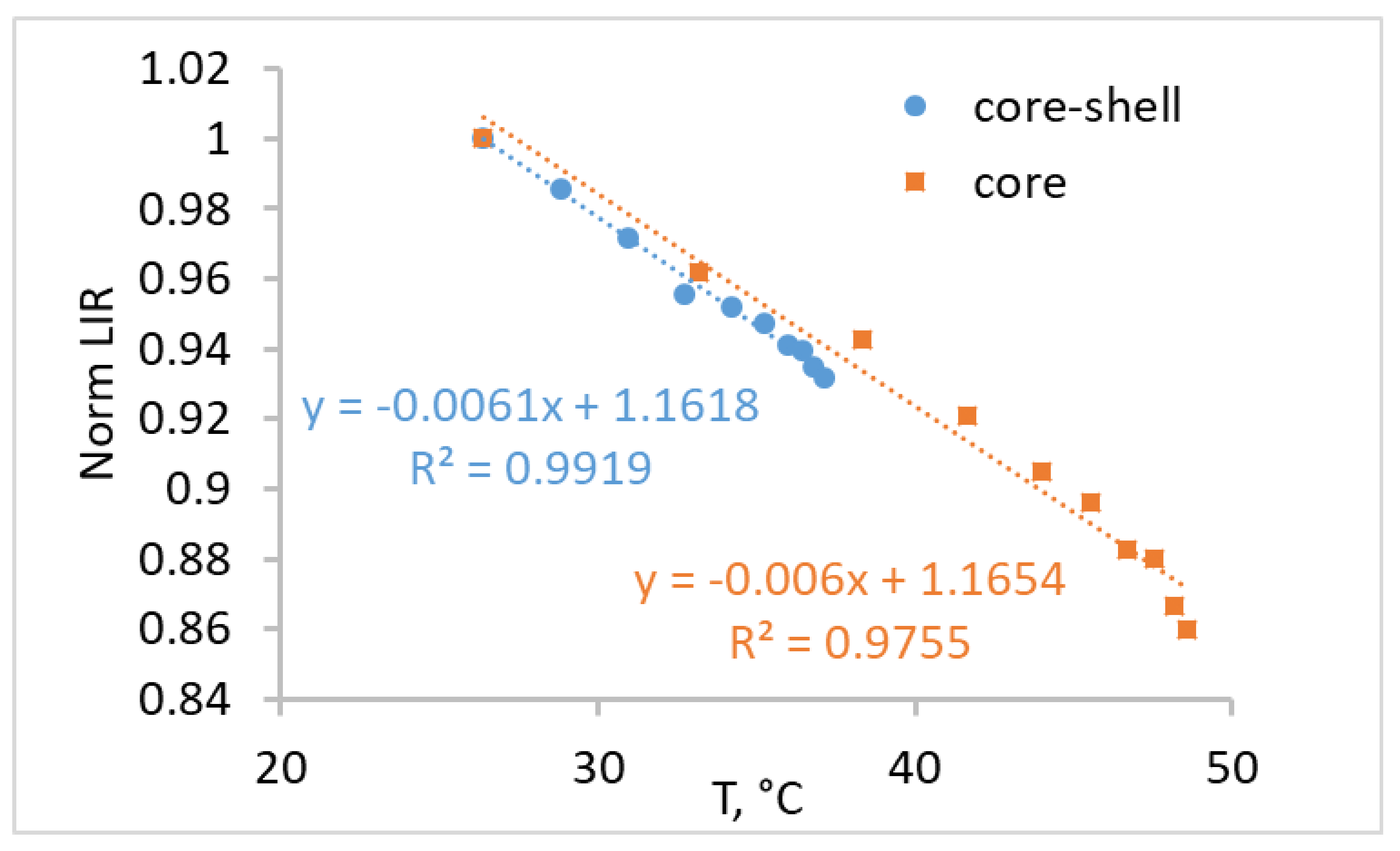
Laser-induced heating of synthesized core and core-shell nanoparticles in water was also studied. As with dry powder, the heating temperature of the core NPs was higher than for core-shell NPs, amounting to 48 °C for the core and 37 °C for the core-shell NPs at 1 W/cm2. The dependence of LIR on the heating temperature for the core and core-shell NPs in water is presented in Figure 6.
The dependence of LIR on temperature is significantly different in water than in dry powder. Another slope of dependence was attributed to the interaction with the environment [38,39]. The O‒H vibrational stretching modes of water affected the relaxation processes of the 5I65I7, 5I55I6 transitions of Ho3+, and the 4I11/24I13/2 transition of Er3+ (Figure 2b). This led to an almost constant intensity of Ho3+ emission attributed to the 5I65I8 transition. At the same time, the Er3+ emission was raised by enhanced 4I11/24I13/2 vibrational relaxation, which led to an increase in the population emitting the 4I13/2 level. The influence of vibrational relaxation increased with the temperature.
Coating particles with an inert shell had no significant effect on the character and slope of the obtained dependence of LIR on the temperature (Figure 6). This was attributed to Yb3+ ions interacting more strongly with vibrational modes of water than the Er3+ and Ho3+ ions [61]. It is well known that Yb–Yb energy migration is efficient and can travel long distances until it is quenched by the surface [64]. Using an inert shell is therefore an effective strategy to prevent quenching and enhance brightness. The relative thermal sensitivity in the 26–36 °C region for the core and core-shell nanoparticles in water was determined to be 0.67 and 0.68% °C−1, respectively.
The detected luminescence signal was significantly lower for uncoated particles than for coated particles. Additionally, a significant decrease in the luminescent signal intensity for uncoated particles was observed in biological media, both due to the quenching of the Yb3+ donor and the quenching of the luminescence of Er3+ and Ho3+. This resulted in the need for high pump power density to obtain a sufficient signal intensity. High pump power can induce the heating of the particles, which could reduce the sensitivity and accuracy of temperature determination and also lead to damage to biological samples. Coating nanoparticles with an inert shell could significantly increase the signal intensity and reduce the measurement error. At the same time, the positive effect of the shell occurs mainly in the reduction of Yb3+ quenching and not in the higher sensitivity of thermometry by the luminescence of Er3+ and Ho3+.

4. Conclusions

Tri-doped Yb3+, Ho3+, Er3+ core and core-shell β-NaGdF4 NPs were successfully synthesized in this study.
The influence of NaYF4 inert shell on the intensity of luminescence in SWIR and the thermal sensing sensitivity of Yb3+, Ho3+, Er3+ tri-doped NaGdF4 NPs was investigated. We found that in the case of core NPs, higher pump power density is needed for Ho3+ excitation and the detection of temperature-dependent changes in the Ho3+ luminescence spectrum. We also demonstrated that an increase in pump power density leads to laser-induced heating, which is stronger in the case of core NPs and could negatively affect the thermometric properties of the NPs and lead to degradation of samples. Coating nanoparticles with an inert shell can significantly improve the characteristics of luminescence, prevent quenching, and obtain high thermal sensitivity. The relative thermal sensitivity for core-shell nanoparticles in dry powder was determined to be 4.2% °C−1.
Coating the NPs with an inert shell was also shown to prevent the quenching of SWIR luminescence by water. The decrease in integral luminescence intensity in the 1050–1600 nm region in water was 5-fold for the core-shell NPs and 14-fold for the core-only NPs. The coating of the particles with an inert shell had no significant effect on the character and slope of the obtained dependence of LIR on the temperature. We attribute this to Yb3+ ions interacting more strongly with vibrational modes of water than Er3+ and Ho3+ ions. The relative thermal sensitivity in the 26–36 °C region in water for the core and core-shell nanoparticles was determined to be 0.67 and 0.68% °C−1, respectively.
Thus, core-shell nanothermometers demonstrated a high intensity of the luminescent signal in SWIR and high thermal sensitivity in both water (0.68% °C−1) and dry powder (4.2% °C−1).

Author Contributions

Conceptualization, V.L. and P.F.; methodology, I.R., A.R., and S.K.; software, V.P.; formal analysis, A.R. and S.K.; investigation, V.P., D.P., I.R., and O.U.; resources, V.L., O.U., and P.F.; writing—original draft preparation, D.P.; writing—review and editing, I.R., A.R., and S.K.; project administration, D.P.; funding acquisition, D.P. All authors have read and agreed to the published version of the manuscript.

Funding

This research was funded by the Russian Science Foundation, grant number 17-72-20186.

Conflicts of Interest

The authors declare no conflict of interest.

References

  1. Brites, C.D.S.; Martínez, E.D.; Urbano, R.R.; Rettori, C.; Carlos, L.D. Self-calibrated double luminescent thermometers through upconverting nanoparticles. Front. Chem. 2019, 7. [Google Scholar] [CrossRef] [PubMed] [Green Version]
  2. Brites, C.D.S.; Balabhadra, S.; Carlos, L.D. Lanthanide-Based Thermometers: At the Cutting-Edge of Luminescence Thermometry. Adv. Opt. Mater. 2019, 7, 1801239. [Google Scholar] [CrossRef] [Green Version]
  3. Brites, C.D.S.; Kuznetsov, S.V.; Konyushkin, V.A.; Nakladov, A.N.; Fedorov, P.P.; Carlos, L.D. Simultaneous Measurement of the Emission Quantum Yield and Local Temperature: The Illustrative Example of SrF2:Yb3+/Er3+ Single Crystals. Eur. J. Inorg. Chem. 2020. [Google Scholar] [CrossRef]
  4. Liang, L.; Liu, X. Nanocrystals feel the heat. Nat. Photonics 2018, 12, 124–125. [Google Scholar] [CrossRef]
  5. Kolesnikov, I.E.; Kalinichev, A.A.; Kurochkin, M.A.; Mamonova, D.V.; Kolesnikov, E.Y.; Kurochkin, A.V.; Lähderanta, E.; Mikhailov, M.D. Y2O3:Nd3+ nanocrystals as ratiometric luminescence thermal sensors operating in the optical windows of biological tissues. J. Lumin. 2018, 204, 506–512. [Google Scholar] [CrossRef]
  6. Lal, S.; Clare, S.E.; Halas, N.J. Nanoshell-enabled photothermal cancer therapy: Impending clinical impact. Acc. Chem. Res. 2008, 41, 1842–1851. [Google Scholar] [CrossRef] [PubMed]
  7. Lindquist, S. The heat-shock response. Ann. Rev. Biochem. 1986, 55, 1151–1191. [Google Scholar] [CrossRef]
  8. Begasse, M.L.; Leaver, M.; Vazquez, F.; Grill, S.W.; Hyman, A.A. Temperature dependence of cell division timing accounts for a shift in the thermal limits of c. elegans and C.briggsae. Cell Rep. 2015, 10, 647–653. [Google Scholar] [CrossRef] [Green Version]
  9. Jaque, D.; Martínez Maestro, L.; Del Rosal, B.; Haro-Gonzalez, P.; Benayas, A.; Plaza, J.L.; Martín Rodríguez, E.; Solé, J.G. Nanoparticles for photothermal therapies. Nanoscale 2014, 6, 9494–9530. [Google Scholar] [CrossRef]
  10. Ai, X.; Mu, J.; Xing, B. Recent advances of light-mediated theranostics. Theranostics 2016, 6, 2439–2457. [Google Scholar] [CrossRef]
  11. Hemmer, E.; Acosta-Mora, P.; Méndez-Ramos, J.; Fischer, S. Optical nanoprobes for biomedical applications: Shining a light on upconverting and near-infrared emitting nanoparticles for imaging, thermal sensing, and photodynamic therapy. J. Mater. Chem. B 2017, 5, 4365–4392. [Google Scholar] [CrossRef] [PubMed]
  12. Liu, H.; Fan, Y.; Wang, J.; Song, Z.; Shi, H.; Han, R.; Sha, Y.; Jiang, Y. Intracellular Temperature Sensing: An Ultra-bright Luminescent Nanothermometer with Non-sensitivity to pH and Ionic Strength. Sci. Rep. 2015, 5. [Google Scholar] [CrossRef] [PubMed] [Green Version]
  13. Maestro, L.M.; Rodríguez, E.M.; Rodríguez, F.S.; De La Cruz, M.C.I.; Juarranz, A.; Naccache, R.; Vetrone, F.; Jaque, D.; Capobianco, J.A.; Solé, J.G. CdSe quantum dots for two-photon fluorescence thermal imaging. Nano Lett. 2010, 10, 5109–5115. [Google Scholar] [CrossRef] [PubMed]
  14. De La Rosa, V.R.; Woisel, P.; Hoogenboom, R. Supramolecular control over thermoresponsive polymers. Mater. Today 2016, 19, 44–55. [Google Scholar] [CrossRef]
  15. Yang, G.; Liu, X.; Feng, J.; Li, S.; Li, Y. Organic Dye Thermometry. In Thermometry at the Nanoscale: Techniques and Selected Applications; Carlos, L.D., Palacio, F., Eds.; The Royal Society of Chemistry: London, UK, 2016; Volume 38, pp. 167–189. [Google Scholar]
  16. Paviolo, C.; Clayton, A.H.A.; Mcarthur, S.L.; Stoddart, P.R. Temperature measurement in the microscopic regime: A comparison between fluorescence lifetime- and intensity-based methods. J. Microsc. 2013, 250, 179–188. [Google Scholar] [CrossRef]
  17. Uchiyama, S.; Kawai, N.; De Silva, A.P.; Iwai, K. Fluorescent Polymeric and Logic Gate with Temperature and pH as Inputs. J. Am. Chem. Soc. 2004, 126, 3032–3033. [Google Scholar] [CrossRef]
  18. Okabe, K.; Inada, N.; Gota, C.; Harada, Y.; Funatsu, T.; Uchiyama, S. Intracellular temperature mapping with a fluorescent polymeric thermometer and fluorescence lifetime imaging microscopy. Nat. Commun. 2012, 3. [Google Scholar] [CrossRef] [Green Version]
  19. Zarrintaj, P.; Jouyandeh, M.; Ganjali, M.R.; Hadavand, B.S.; Mozafari, M.; Sheiko, S.S.; Vatankhah-Varnoosfaderani, M.; Gutiérrez, T.J.; Saeb, M.R. Thermo-sensitive polymers in medicine: A review. Eur. Polym. J. 2019, 117, 402–423. [Google Scholar] [CrossRef]
  20. Donner, J.S.; Thompson, S.A.; Kreuzer, M.P.; Baffou, G.; Quidant, R. Mapping intracellular temperature using green fluorescent protein. Nano Lett. 2012, 12, 2107–2111. [Google Scholar] [CrossRef] [Green Version]
  21. Zhu, X.; Feng, W.; Chang, J.; Tan, Y.W.; Li, J.; Chen, M.; Sun, Y.; Li, F. Temperature-feedback upconversion nanocomposite for accurate photothermal therapy at facile temperature. Nat. Commun. 2016, 7. [Google Scholar] [CrossRef]
  22. Vetrone, F.; Naccache, R.; Zamarrón, A.; De La Fuente, A.J.; Sanz-Rodríguez, F.; Maestro, L.M.; Rodriguez, E.M.; Jaque, D.; Sole, J.G.; Capobianco, J.A. Temperature sensing using fluorescent nanothermometers. ACS Nano 2010, 4, 3254–3258. [Google Scholar] [CrossRef] [PubMed]
  23. Weissleder, R. A clearer vision for in vivo imaging: Progress continues in the development of smaller, more penetrable probes for biological imaging. Nat. Biotechnol. 2001, 19, 316–317. [Google Scholar] [CrossRef] [PubMed]
  24. Jacques, S.L. Optical properties of biological tissues: A review. Phys. Med. Biol. 2013, 58, 5007–5008. [Google Scholar] [CrossRef]
  25. Wang, M.; Wu, C.; Sinefeld, D.; Li, B.; Xia, F.; Xu, C. Comparing the effective attenuation lengths for long wavelength in vivo imaging of the mouse brain. Biomed. Opt. Express 2018, 9, 3534–3543. [Google Scholar] [CrossRef] [PubMed]
  26. Mustafa, F.H.; Jaafar, M.S. Comparison of wavelength-dependent penetration depths of lasers in different types of skin in photodynamic therapy. Indian J. Phys. 2013, 87, 203–209. [Google Scholar] [CrossRef]
  27. Hong, G.; Diao, S.; Chang, J.; Antaris, A.L.; Chen, C.; Zhang, B.; Zhao, S.; Atochin, D.N.; Huang, P.L.; Andreasson, K.I.; et al. Through-skull fluorescence imaging of the brain in a new near-infrared window. Nat. Photonics 2014, 8, 723–730. [Google Scholar] [CrossRef]
  28. Zhan, Q.; He, S.; Qian, J.; Cheng, H.; Cai, F. Optimization of optical excitation of upconversion nanoparticles for rapid microscopy and deeper tissue imaging with higher quantum yield. Theranostics 2013, 3, 306–316. [Google Scholar] [CrossRef] [Green Version]
  29. Hemmer, E.; Benayas, A.; Légaré, F.; Vetrone, F. Exploiting the biological windows: Current perspectives on fluorescent bioprobes emitting above 1000 nm. Nanoscale Horiz. 2016, 1, 168–184. [Google Scholar] [CrossRef]
  30. Semonin, O.E.; Johnson, J.C.; Luther, J.M.; Midgett, A.G.; Nozik, A.J.; Beard, M.C. Absolute photoluminescence quantum yields of IR-26 Dye, PbS, and PbSe quantum dots. J. Phys. Chem. Lett. 2010, 1, 2445–2450. [Google Scholar] [CrossRef]
  31. Hatami, S.; Würth, C.; Kaiser, M.; Leubner, S.; Gabriel, S.; Bahrig, L.; Lesnyak, V.; Pauli, J.; Gaponik, N.; Eychmüller, A.; et al. Absolute photoluminescence quantum yields of IR26 and IR-emissive Cd1-xHgxTe and PbS quantum dots-method- and material-inherent challenges. Nanoscale 2015, 7, 133–143. [Google Scholar] [CrossRef] [Green Version]
  32. Thimsen, E.; Sadtler, B.; Berezin, M.Y. Shortwave-infrared (SWIR) emitters for biological imaging: A review of challenges and opportunities. Nanophotonics 2017, 6, 1043–1054. [Google Scholar] [CrossRef]
  33. Escudero, A.; Carrillo-Carrión, C.; Zyuzin, M.V.; Parak, W.J. Luminescent Rare-earth-based Nanoparticles: A Summarized Overview of their Synthesis, Functionalization, and Applications. Top. Curr. Chem. 2016, 374, 48. [Google Scholar] [CrossRef] [PubMed] [Green Version]
  34. Ma, D.; Xu, X.; Hu, M.; Wang, J.; Zhang, Z.; Yang, J.; Meng, L. Rare-Earth-Based Nanoparticles with Simultaneously Enhanced Near-Infrared (NIR)-Visible (Vis) and NIR-NIR Dual-Conversion Luminescence for Multimodal Imaging. Chem. Asian J. 2016, 11, 1050–1058. [Google Scholar] [CrossRef] [PubMed]
  35. Zhou, H.; Sharma, M.; Berezin, O.; Zuckerman, D.; Berezin, M.Y. Nanothermometry: From Microscopy to Thermal Treatments. ChemPhysChem 2016, 17, 27–36. [Google Scholar] [CrossRef]
  36. Wade, S.A.; Collins, S.F.; Baxter, G.W. Fluorescence intensity ratio technique for optical fiber point temperature sensing. J. Appl. Phys. 2003, 94, 4743–4756. [Google Scholar] [CrossRef]
  37. Quintanilla, M.; Benayas, A.; Naccache, R.; Vetrone, F. Luminescent Nanothermometry with Lanthanide-doped Nanoparticles. In Thermometry at the Nanoscale: Techniques and Selected Applications; Carlos, L.D., Palacio, F., Eds.; The Royal Society of Chemistry: London, UK, 2016; Volume 38, pp. 124–166. [Google Scholar]
  38. Kamimura, M.; Matsumoto, T.; Suyari, S.; Umezawa, M.; Soga, K. Ratiometric near-infrared fluorescence nanothermometry in the OTN-NIR (NIR II/III) biological window based on rare-earth doped β-NaYF 4 nanoparticles. J. Mater. Chem. B 2017, 5, 1917–1925. [Google Scholar] [CrossRef]
  39. Gouveia-Neto, A.S.; Silva, J.F.; Vermelho, M.V.D.; Gomes, A.S.L.; Jacinto, C. Generation of multiwavelength light in the region of the biological windows in Tm3+-doped fiber excited at 1.064 μm. Appl. Phys. Lett. 2016, 109. [Google Scholar] [CrossRef]
  40. Smith, A.M.; Mancini, M.C.; Nie, S. Bioimaging: Second window for in vivo imaging. Nat. Nanotechnol. 2009, 4, 710–711. [Google Scholar] [CrossRef] [Green Version]
  41. Naczynski, D.J.; Tan, M.C.; Zevon, M.; Wall, B.; Kohl, J.; Kulesa, A.; Chen, S.; Roth, C.M.; Riman, R.E.; Moghe, P.V. Rare-earth-doped biological composites as in vivo shortwave infrared reporters. Nat. Commun. 2013, 4. [Google Scholar] [CrossRef]
  42. Sekiyama, S.; Umezawa, M.; Kuraoka, S.; Ube, T.; Kamimura, M.; Soga, K. Temperature Sensing of Deep Abdominal Region in Mice by Using Over-1000 nm Near-Infrared Luminescence of Rare-Earth-Doped NaYF4 Nanothermometer. Sci. Rep. 2018, 8, 1–12. [Google Scholar] [CrossRef]
  43. Wortmann, L.; Suyari, S.; Ube, T.; Kamimura, M.; Soga, K. Tuning the thermal sensitivity of β-NaYF4:Yb3+, Ho3+, Er3+ nanothermometers for optimal temperature sensing in OTN-NIR (NIR II/III) biological window. J. Lumin. 2018, 198, 236–242. [Google Scholar] [CrossRef]
  44. Ximendes, E.C.; Rocha, U.; Sales, T.O.; Fernández, N.; Sanz-Rodríguez, F.; Martín, I.R.; Jacinto, C.; Jaque, D. In Vivo Subcutaneous Thermal Video Recording by Supersensitive Infrared Nanothermometers. Adv. Funct. Mater. 2017, 27. [Google Scholar] [CrossRef]
  45. Savchuk, O.A.; Haro-González, P.; Carvajal, J.J.; Jaque, D.; Massons, J.; Aguiló, M.; Díaz, F. Er:Yb:NaY2F5O up-converting nanoparticles for sub-tissue fluorescence lifetime thermal sensing. Nanoscale 2014, 6, 9727–9733. [Google Scholar] [CrossRef] [PubMed]
  46. Savchuk, O.; Marti, J.J.C.; Cascales, C.; Haro-Gonzalez, P.; Sanz-Rodríguez, F.; Aguilo, M.; Diaz, F. Bifunctional Tm3+,Yb3+:GdVO4@SiO2 core-shell nanoparticles in HeLa cells: Upconversion luminescence nanothermometry in the first biological window and biolabelling in the visible. Nanomaterials 2020, 10, 993. [Google Scholar] [CrossRef] [PubMed]
  47. Cortelletti, P.; Skripka, A.; Facciotti, C.; Pedroni, M.; Caputo, G.; Pinna, N.; Quintanilla, M.; Benayas, A.; Vetrone, F.; Speghini, A. Tuning the sensitivity of lanthanide-activated NIR nanothermometers in the biological windows. Nanoscale 2018, 10, 2568–2576. [Google Scholar] [CrossRef] [PubMed]
  48. Runowski, M.; Stopikowska, N.; Szeremeta, D.; Goderski, S.; Skwierczyńska, M.; Lis, S. Upconverting Lanthanide Fluoride Core@Shell Nanorods for Luminescent Thermometry in the First and Second Biological Windows: β-NaYF4:Yb3+ − Er3+ @SiO2 Temperature Sensor. ACS Appl. Mater. Interfaces 2019, 11, 13389–13396. [Google Scholar] [CrossRef]
  49. Suo, H.; Guo, C.; Zheng, J.; Zhou, B.; Ma, C.; Zhao, X.; Li, T.; Guo, P.; Goldys, E.M. Sensitivity Modulation of Upconverting Thermometry through Engineering Phonon Energy of a Matrix. ACS Appl. Mater. Interfaces 2016, 8, 30312–30319. [Google Scholar] [CrossRef]
  50. Dong, B.; Hua, R.N.; Cao, B.S.; Li, Z.P.; He, Y.Y.; Zhang, Z.Y.; Wolfbeis, O.S. Size dependence of the upconverted luminescence of NaYF4: Er, Yb microspheres for use in ratiometric thermometry. Phys. Chem. Chem. Phys. 2014, 16, 20009–20012. [Google Scholar] [CrossRef]
  51. Kumar, V.; Som, S.; Dutta, S.; Das, S.; Swart, H.C. Influence of Ho3+ doping on the temperature sensing behavior of Er3+-Yb3+ doped La2CaZnO5 phosphor. RSC Adv. 2016, 6, 84914–84925. [Google Scholar] [CrossRef]
  52. Skripka, A.; Benayas, A.; Marin, R.; Canton, P.; Hemmer, E.; Vetrone, F. Double rare-earth nanothermometer in aqueous media: Opening the third optical transparency window to temperature sensing. Nanoscale 2017, 9, 3079–3085. [Google Scholar] [CrossRef] [Green Version]
  53. Prorok, K.; Pawlyta, M.; Strek, W.; Bednarkiewicz, A. Energy Migration Up-conversion of Tb3+ in Yb3+ and Nd3+ Codoped Active-Core/Active-Shell Colloidal Nanoparticles. Chem. Mater. 2016, 28, 2295–2300. [Google Scholar] [CrossRef]
  54. Wang, C.; Xia, H.; Feng, Z.; Zhang, Z.; Jiang, D.; Zhang, J.; He, S.; Tang, Q.; Sheng, Q.; Gu, X.; et al. Infrared spectral properties for α-NaYF4 single crystal of various Er3+ doping concentrations. Opt. Laser Technol. 2016, 82, 157–162. [Google Scholar] [CrossRef]
  55. Chen, G.; Shen, J.; Ohulchanskyy, T.Y.; Patel, N.J.; Kutikov, A.; Li, Z.; Song, J.; Pandey, R.K.; Agren, H.; Prasad, P.N.; et al. (α-NaYbF4:Tm3+)/CaF2 core/shell nanoparticles with efficient near-infrared to near-infrared upconversion for high-contrast deep tissue bioimaging. ACS Nano 2012, 6, 8280–8287. [Google Scholar] [CrossRef] [PubMed] [Green Version]
  56. Chen, B.; Wang, F. Combating Concentration Quenching in Upconversion Nanoparticles. Acc. Chem. Res. 2019. [Google Scholar] [CrossRef] [PubMed]
  57. Shen, B.; Cheng, S.; Gu, Y.; Ni, D.; Gao, Y.; Su, Q.; Feng, W.; Li, F. Revisiting the optimized doping ratio in core/shell nanostructured upconversion particles. Nanoscale 2017, 9, 1964–1971. [Google Scholar] [CrossRef] [PubMed]
  58. Liu, J.; Chen, G.; Hao, S.; Yang, C. Sub-6 nm monodisperse hexagonal core/shell NaGdF4 nanocrystals with enhanced upconversion photoluminescence. Nanoscale 2017, 9, 91–98. [Google Scholar] [CrossRef] [PubMed]
  59. Rai, V.K.; Rai, S.B. A comparative study of FIR and FL based temperature sensing schemes: An example of Pr3+. Appl. Phys. B Lasers Opt. 2007, 87, 323–325. [Google Scholar] [CrossRef]
  60. Shannon, R.D. Revised effective ionic radii and systematic studies of interaction distance in halides and chalcogenides. Acta Crystallogr. A 1976, 32, 751–767. [Google Scholar] [CrossRef]
  61. Arppe, R.; Hyppänen, I.; Perälä, N.; Peltomaa, R.; Kaiser, M.; Würth, C.; Christ, S.; Resch-Genger, U.; Schäferling, M.; Soukka, T. Quenching of the upconversion luminescence of NaYF4:Yb3+,Er3+ and NaYF4:Yb3+,Tm3+ nanophosphors by water: The role of the sensitizer Yb3+ in non-radiative relaxation. Nanoscale 2015, 7, 11746–11757. [Google Scholar] [CrossRef] [Green Version]
  62. Nadort, A.; Zhao, J.; Goldys, E.M. Lanthanide upconversion luminescence at the nanoscale: Fundamentals and optical properties. Nanoscale 2016, 8, 13099–13130. [Google Scholar] [CrossRef] [Green Version]
  63. Miyakawa, T.; Dexter, D.L. Phonon sidebands, multiphonon relaxation of excited states, and phonon-assisted energy transfer between ions in solids. Phys. Rev. B 1970, 1, 2961–2969. [Google Scholar] [CrossRef]
  64. Chen, X.; Peng, D.; Ju, Q.; Wang, F. Photon upconversion in core-shell nanoparticles. Chem. Soc. Rev. 2015, 44, 1318–1330. [Google Scholar] [CrossRef] [PubMed]
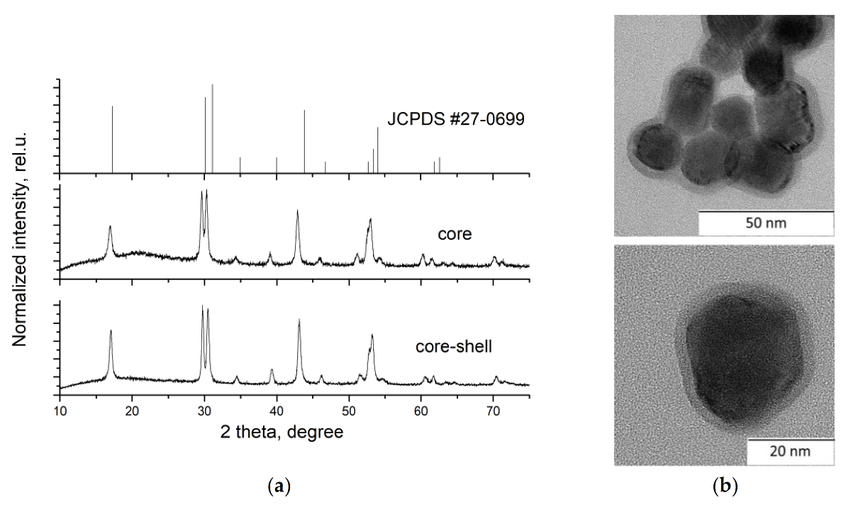
Figure 1. (a) XRD diagrams of JCPDS #27-0699 and NaGdF4:Yb3+, Ho3+, Er3+ core and core-shell nanoparticles and (b) TEM images of NaGdF4:Yb3+, Ho3+, Er3+@NaYF4 core-shell nanoparticles.
Nanomaterials 10 01992 g001
Figure 2. (a) The luminescence spectra of NaGdF4:Yb3+, Ho3+, Er3+ core and core-shell nanoparticles measured at 30 °C in powder and in aqueous solution and (b) simplified energy level diagram of the Yb3+-, Ho3+-, and Er3+-emitting centers.
Nanomaterials 10 01992 g002
Figure 3. The influence of pump power density on heating temperature of (a) core NaGdF4:Yb3+, Ho3+, Er3+ NPs and (b) core-shell NaGdF4:Yb3+, Ho3+, Er3+ NPs.
Nanomaterials 10 01992 g003
Figure 4. The influence of pump power density and, consequently, the temperature on the emission spectra of nanoparticles in powder: (a) core-shell and (b) core.
Nanomaterials 10 01992 g004
Figure 5. The dependence of luminescence intensity ratio (LIR) on the temperature for the core and core-shell nanoparticles in powder. The pump power density in W/cm2 is shown as data labels.
Nanomaterials 10 01992 g005
Figure 6. The dependence of luminescence intensity ratio (LIR) normalized to the value at 29 °C on the heating temperature for the core and core-shell nanoparticles in water.
Nanomaterials 10 01992 g006