Article

Polyherbal-Mediated Synthesis of Copper Nanoparticles Using Hygrophila auriculata and Leucas aspera: Cytotoxicity, Antioxidant Effect, and Antibacterial Potential Against Healthcare-Associated Pathogens

by
Gayathri Vijayakumar
1,
Amrutha Raja
1,
Swathi Ganesan
1,
Teja Sri Senthil
1,
Jainitha Kandasamy
1,
Prathiksha Senthil Kumaran
2 and
Senthil Kumaran Rangarajulu
3,*
1
Department of Biotechnology, Hindustan Institute of Technology & Science, Chennai 603103, India
2
Department of Biotechnology, Anna University, Chennai 600025, India
3
Department of Biological Engineering, Konkuk University, Seoul 05029, Republic of Korea
*
Author to whom correspondence should be addressed.
J. Funct. Biomater. 2026, 17(4), 169; https://doi.org/10.3390/jfb17040169
Submission received: 17 February 2026 / Revised: 22 March 2026 / Accepted: 23 March 2026 / Published: 1 April 2026

Abstract

An eco-friendly green synthesis approach was employed to produce copper nanoparticles (CuNPs) using a polyherbal extract derived from two medicinally important plant species, Hygrophila auriculata (Schumach.) Heine and Leucas aspera (Willd.) Link. The plant extracts were initially subjected to phytochemical screening to identify bioactive constituents potentially involved in nanoparticle synthesis. The synthesized CuNPs were characterized using UV-visible spectroscopy, Fourier-transform infrared spectroscopy (FTIR), gas chromatography–mass spectrometry (GC-MS), field-emission scanning electron microscopy coupled with energy-dispersive X-ray analysis (FESEM-EDAX), X-ray diffraction (XRD), and thin-layer chromatography (TLC). UV-visible spectroscopy revealed a characteristic absorption peak at 233.6 nm. FTIR analysis indicated the presence of functional groups associated with nanoparticle reduction and stabilization, whereas FESEM imaging showed predominantly spherical particles with sizes ranging 63–68 nm. Elemental composition was confirmed using EDAX analysis. XRD analysis demonstrated polycrystalline nature of the CuNPs, with an average crystallite size of 11.5 nm. GC-MS analysis and phytochemical screening further confirmed the presence of bioactive compounds, whereas TLC analysis revealed differences in mobility between the plant extract and synthesized CuNPs. Antibacterial activity of the synthesized CuNPs was evaluated using the agar well diffusion method against clinically relevant bacterial strains, including those of Staphylococcus aureus, Pseudomonas aeruginosa, Escherichia coli, and Streptococcus pyogenes. The polyherbal-derived CuNPs produced larger inhibition zones than the individual plant extracts, particularly against multidrug-resistant pathogens such as P. aeruginosa and S. aureus. Additionally, the nanoparticles exhibited concentration-dependent antioxidant activity in the 2,2-diphenyl-1-picrylhydrazyl assay at concentrations ranging 10–50 mg/mL, with radical scavenging activity increasing from 29.9% to 76.5% and a corresponding decrease in absorbance from 0.698 to 0.234 (p < 0.05). Cytotoxic evaluation in HepG2 cells after 48 h of exposure demonstrated dose-dependent morphological changes and reduced cell viability. These findings suggest that polyherbal-derived CuNPs possess antibacterial, antioxidant, and cytotoxic properties with potential relevance for biomedical applications.

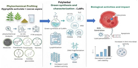
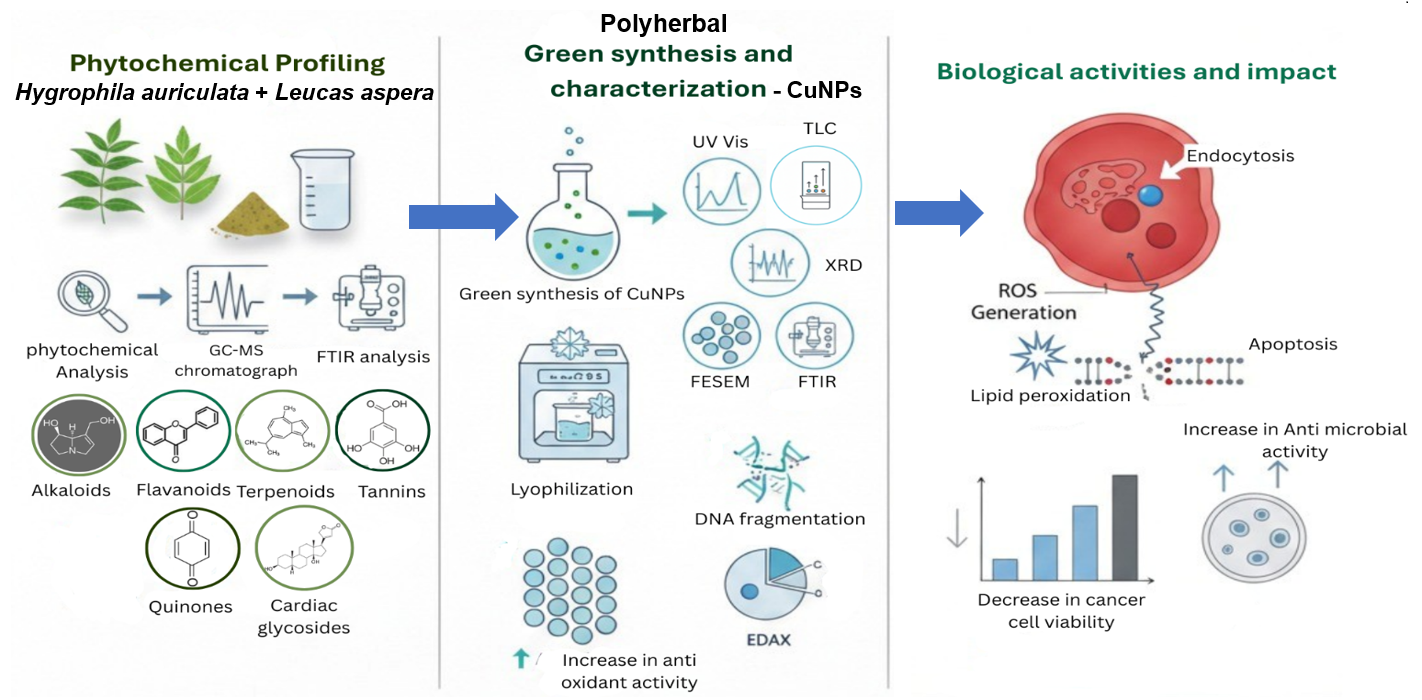
Graphical Abstract

1. Introduction

Interest in eco-friendly and sustainable approaches for the synthesis of metal nanoparticles using plant-based systems rich in phytochemical constituents has increased in recent years [1,2]. Plant-mediated green synthesis has emerged as a promising alternative to conventional chemical and physical methods because plant extracts contain diverse secondary metabolites, such as flavonoids, phenolics, terpenoids, alkaloids, tannins, and glycosides, which can function as natural reducing and stabilizing agents [3]. These phytochemicals facilitate nanoparticle formation and may enhance biocompatibility and biological activity, rendering plant-derived nanoparticles particularly attractive for biomedical applications [4,5]. Various plant parts, including leaves, roots, stems, flowers, and seeds, have been successfully used for nanoparticle synthesis owing to their rich and variable phytochemical composition [6].
Medicinal plants have long been used in traditional medical systems worldwide for treating diverse ailments. Previous studies have shown that the selection of plant species and plant parts is crucial for determining nanoparticle size, morphology, stability, and biological efficacy [7,8,9]. In this context, Hygrophila auriculata and Leucas aspera were selected for the present study because of their well-documented medicinal properties and high phytochemical content, which may contribute to the synthesis and stabilization of copper nanoparticles (CuNPs) while potentially imparting additional biological functionality.
H. auriculata (family Acanthaceae) is widely used in Ayurvedic and Siddha medicine for treating inflammatory disorders, urinary diseases, liver ailments, wounds, and microbial infections; it is known for its antioxidant, antimicrobial, anti-inflammatory, and hepatoprotective activities [10,11]. Previous phytochemical investigations have reported the presence of flavonoids, alkaloids, tannins, and terpenoids in H. auriculata, highlighting its biochemical richness [12,13,14]. Similarly, L. aspera (family Lamiaceae) is an aromatic medicinal herb traditionally used to treat inflammation, wounds, cough, and viral infections [15]. Previous studies have shown that it contains flavonoids, phenolics, terpenoids, tannins, and essential oils and exhibits notable antibacterial, antioxidant, and anti-inflammatory properties [16,17].
CuNPs have attracted considerable attention owing to their unique physicochemical properties, including high surface area, enhanced reactivity, and relatively low cost compared with noble metal nanoparticles. These characteristics have enabled their application in fields such as electronics, textiles, agriculture, energy, and biomedicine because of their electrical, thermal, catalytic, and antimicrobial properties [18,19,20,21,22]. However, most current studies focus on the synthesis of CuNPs using extracts from a single plant species [23,24,25], whereas polyherbal green synthesis approaches remain relatively unexplored. Although H. auriculata and L. aspera contain similar classes of phytochemicals based on preliminary screening, variations in the specific composition and relative abundance of these compounds may exist between the two species. Therefore, a combined extract was investigated to provide a broadened phytochemical environment for CuNP synthesis without presuming an inherent synergistic effect. Additionally, although antimicrobial and cytotoxic activities of plant-mediated nanoparticles are frequently reported, detailed biological validation and mechanistic investigations, such as DNA fragmentation analysis to support anticancer potential, are not always thoroughly conducted [23].
In the present study, CuNPs were synthesized using a polyherbal extract of H. auriculata and L. aspera via a green synthesis approach. The synthesized CuNPs were characterized using UV-visible spectroscopy, Fourier-transform infrared spectroscopy (FTIR), gas chromatography–mass spectrometry (GC-MS), field-emission scanning electron microscopy coupled with energy-dispersive X-ray analysis (FESEM-EDAX), X-ray diffraction (XRD), and thin-layer chromatography (TLC). Phytochemical screening was performed to identify the biomolecules involved in reduction and capping. Biological potential of the polyherbal CuNPs was further evaluated using antibacterial activity assays, 2,2-diphenyl-1-picrylhydrazyl (DPPH) antioxidant assay, cytotoxicity assessment using the MTT assay in HepG2 cells, and DNA fragmentation analysis, thereby providing a comprehensive evaluation of their potential biomedical relevance.

2. Materials and Methods

2.1. Biosynthesis of CuNPs

All chemicals used in this study were of analytical grade. Fresh plant materials of H. auriculata and L. aspera were collected from different locations in Chennai, Tamil Nadu, India. The plant samples were thoroughly washed with distilled water, shade-dried at room temperature, and ground into a fine powder. The powdered materials were stored separately in airtight containers at room temperature until further use.
For preparation of the polyherbal extract, 5 g of each dried plant powder was added separately to 50 mL of distilled water and heated at 70–80 °C for 20 min using a heating mantle (Krishnaram Scientific, Chennai, India). The extracts were cooled to room temperature and filtered through Whatman No. 1 filter paper. Equal volumes (25 mL each) of H. auriculata and L. aspera extracts were mixed in a 1:1 ratio to obtain the polyherbal extract.
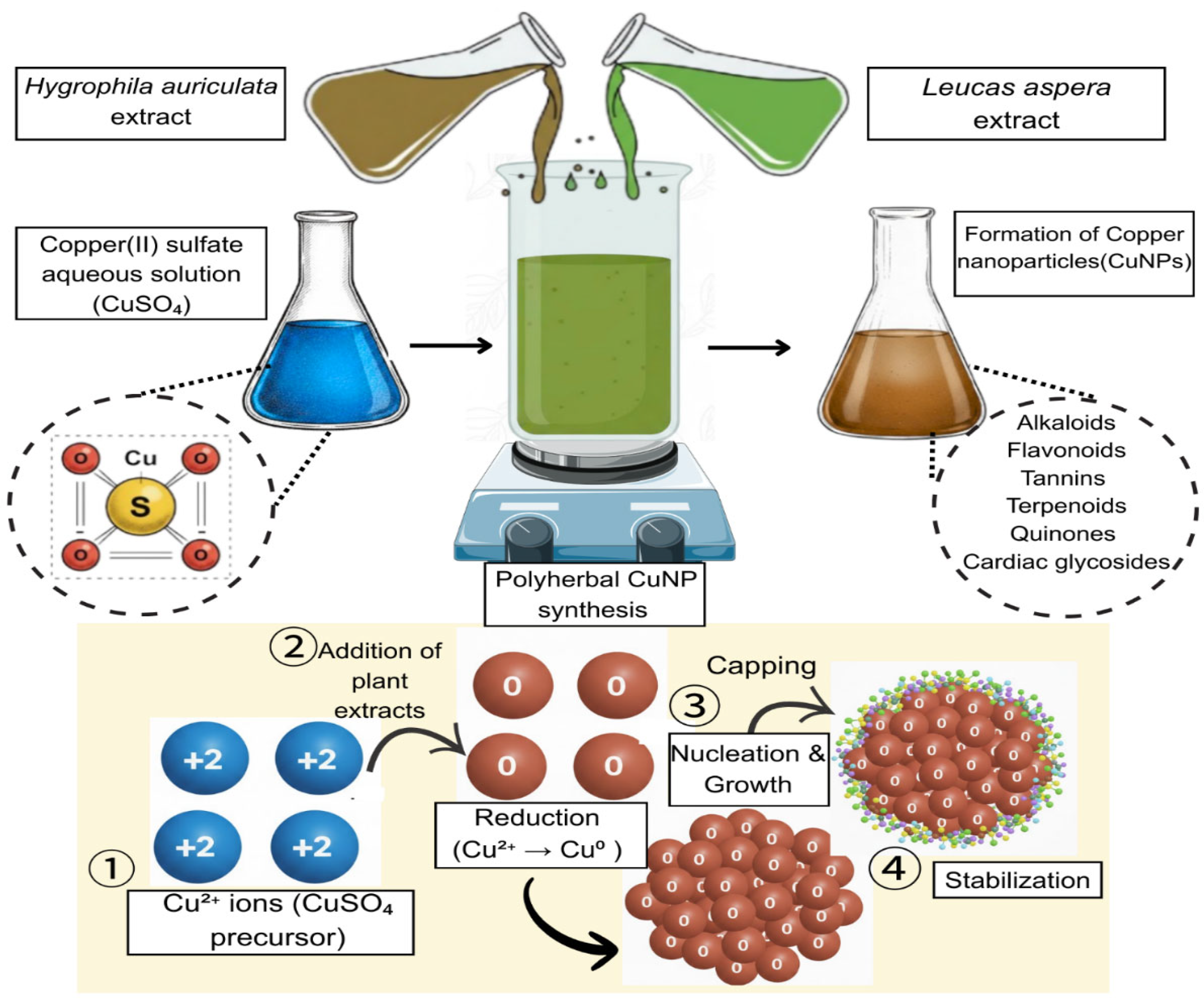
Separately, a 0.5 M aqueous solution of copper sulfate (50 mL) was prepared and added dropwise to the polyherbal extract in a 1:1 ratio under continuous stirring using a magnetic stirrer (Krishnaram Scientific, Chennai, India) (Figure 1). The reaction mixture was incubated at 37 °C for 24 h. CuNP formation was indicated by a visible color change in the reaction mixture. The synthesized nanoparticles were subsequently lyophilized for further characterization and evaluation of their application potential [26,27,28,29].

2.2. Phytochemical Screening of Plant Extracts

Preliminary phytochemical screening of each plant extract was performed separately using standard qualitative assays for alkaloids, saponins, and flavonoids (Shinoda test), tannins and terpenoids (Salkowski test), and quinones and cardiac glycosides (Keller–Killiani test) according to previously reported protocols [30,31,32].

2.3. Characterization of Synthesized CuNPs

Polyherbal green-synthesized CuNPs were initially analyzed using UV-visible spectroscopy with a double-beam spectrophotometer (Systronics, Ahmedabad, India). Absorbance spectra were recorded in the wavelength range of 200–800 nm to monitor nanoparticle formation [33].
Surface morphology and particle size of the synthesized CuNPs were examined using FESEM. FESEM Images were obtained using an Apreo 2 S HiVac FESEM system (Thermo Fisher Scientific, Waltham, MA, USA) operated at an accelerating voltage of 15 kV. Signals were detected using an Everhart–Thornley detector (TESCAN SEA Pte. Ltd, The Gateway East Tower, Singapore) at a magnification of 100,000×. Elemental composition of the nanoparticles was determined by energy-dispersive X-ray spectroscopy (EDS) using an Oxford Instruments EDS system (Oxford Instruments, Bristol, UK) coupled to the FESEM instrument [34].
FTIR was used to identify functional groups associated with the surface of the synthesized CuNPs. FTIR spectra were recorded using a Bruker Fourier-transform infrared spectrometer (Bruker Optics, Ettlingen, Germany) equipped with an attenuated total reflectance accessory employing a ZnSe crystal and an RT-DLATGS detector. Spectra were collected at a resolution of 2 cm−1 in the range of 400–4000 cm−1 [35].
GC-MS analysis was conducted to identify phytochemical constituents present in the polyherbal extract using an Agilent 7890A gas chromatograph coupled with a mass spectrometer (Agilent Technologies, Santa Clara, CA, USA). The analysis was performed in full-scan mode over a mass range of 1–455 m/z, with a total run time of 47.95 min [36,37].
XRD analysis was performed to determine crystalline structure, phase composition, and average crystallite size of the synthesized CuNPs. Diffraction patterns were recorded using a PANalytical Empyrean X-ray diffractometer (Malvern PANalytical, Almelo, The Netherlands) equipped with a PIXcel3D detector and Cu Kα radiation (λ = 1.5406 Å) over a 2θ range of 10–80°. Average crystallite size was estimated using the Scherrer equation [34].

2.4. Antibacterial Activity of Plant Extracts and CuNPs

Antibacterial activity of the synthesized CuNPs was evaluated using the standard disc diffusion method. Four pathogenic bacterial strains were used: Escherichia coli (ATCC 25922), Pseudomonas aeruginosa (ATCC 27853), Staphylococcus aureus (ATCC 25923), and Streptococcus pyogenes (ATCC 19615). The bacterial strains were obtained from the Department of Microbiology, Vellore Institute of Technology (VIT), Vellore, India.
Bacterial suspension (100 µL) was uniformly spread onto Mueller–Hinton agar plates. Three wells (approximately 6 mm in diameter) were punched into the agar and, respectively, filled with 100 µL of sterilized L. aspera extract, H. auriculata extract (100 mg/mL each), and copper nanoparticles (100 µg/mL). Prior to use, plant extracts and CuNP suspensions were filtered through Whatman No. 1 filter paper to remove debris and sterilized using a 0.22 µm membrane filter (Membrane Hitec: Chennai, India).
Positive and negative controls were included using discs impregnated with ceftazidime (30 µg) and distilled water, respectively. The plates were incubated at 37 °C for 24 h; diameters of the zones of inhibition were measured in millimeters. All experiments were performed in triplicate [26].

2.5. In Vitro Antioxidant Assay of CuNPs

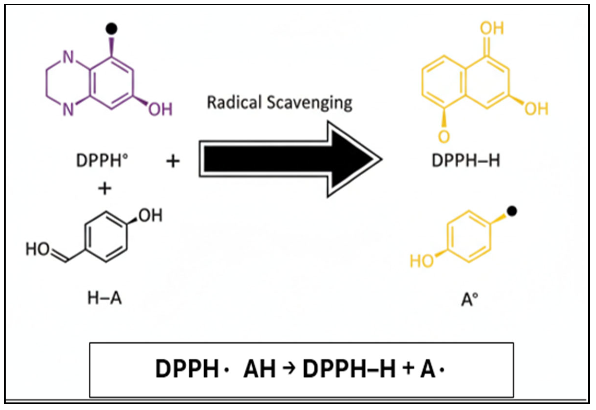
Activity was evaluated using the DPPH free radical scavenging assay. DPPH is a stable nitrogen-centered free radical commonly used to assess antioxidant capacity. The reduction in DPPH• to its scavenged form (DPPH-H) occurs via hydrogen atom donation from antioxidant compounds (Figure 2).
A 0.1 mM DPPH solution was prepared by dissolving 4 mg of DPPH in 100 mL of ethanol. Different concentrations of the sample extracts (20–200 µg/mL) were prepared using dimethyl sulfoxide (DMSO) and adjusted to a final volume of 40 µL. Subsequently, 2.96 mL of the DPPH solution was added; the reaction mixture was incubated in the dark at room temperature for 20 min. After incubation, absorbance was measured at 517 nm using a UV-visible spectrophotometer (Hitachi, Tokyo, Japan). The control consisted of 3 mL of DPPH solution without the sample.
All antioxidant experiments were performed in triplicate (n = 3). The results are expressed as mean ± standard deviation (SD). Statistical analysis was conducted using one-way analysis of variance (ANOVA) followed by Tukey’s post hoc test to determine significant differences between treated samples and the control.
Radical scavenging activity (%) was calculated using the following equation [38,39]:
Radical   scavenging   activity   ( % )   =     A b s o r b a n c e   c o n t r o l     A b s o r b a n c e   s a m p l e A b s o r b a n c e   c o n t r o l     × 100

2.6. Cytotoxicity Assay of CuNPs

Cytotoxicity of the polyherbal green-synthesized CuNPs was evaluated using the HepG2 human liver cancer cell line obtained from the National Centre for Cell Sciences (NCCS), Pune, India. The MTT assay was used to determine cell viability.
HepG2 cells were cultured in Dulbecco’s Modified Eagle’s Medium (DMEM) supplemented with 10% fetal bovine serum and maintained at 37 °C in a humidified incubator containing 5% carbon dioxide (CO2). Cells were harvested using trypsin-EDTA and seeded into 96-well plates at a density of approximately 1 × 10⁴ cells per well. After incubation for 24 h to allow cell attachment, the medium was replaced with serum-free DMEM containing CuNPs at different concentrations; untreated cells served as the control.
Following 24 h of treatment, 100 µL of MTT solution (0.5 mg/mL) was added to each well and incubated for 4 h at 37 °C. The resulting formazan crystals were dissolved in 100 µL of DMSO; absorbance was measured at 570 nm using a microplate reader (EPS Biosolutions, Chennai, India), with 630 nm as the reference wavelength. All experiments were performed in triplicate (n = 3). The results are expressed as mean values [40,41].
Cell viability (%) was calculated using the following formula:
Cell   viability   ( % )   =   S a m p l e   O D C o n t r o l   O D     × 100

2.7. Analysis of DNA Fragmentation

DNA fragmentation analysis was performed to evaluate apoptosis in HepG2 cells following treatment with polyherbal green-synthesized CuNPs. The assay was conducted according to previously reported methods, with minor modifications.
HepG2 cells were treated with CuNPs at half maximal inhibitory concentration (IC50) determined by the MTT assay and incubated for 24 h at 37 °C under humidified conditions containing 5% CO2 in serum-free medium. After treatment, cells were harvested by trypsinization, washed with phosphate-buffered saline, and subjected to genomic DNA isolation using a TrueScreen DNA Isolation Kit (Aura Biotechnologies Private Limited, Chennai, India) according to the manufacturer’s instructions.
The cell pellet was resuspended in lysis buffer, added lysozyme and proteinase K, and incubated at 65 °C to ensure complete cell lysis and protein digestion. Genomic DNA was purified using a spin column–based method involving sequential washing steps and subsequently eluted in elution buffer.
The isolated DNA samples were analyzed by agarose gel electrophoresis using a 1% agarose gel prepared in 1 × TAE buffer containing ethidium bromide. A DNA ladder was loaded into the first well as a molecular size marker. Subsequent wells contained DNA from HepG2 cells treated with concentrations below the IC50, at the IC50, and untreated cells (negative control). Electrophoresis was performed under appropriate voltage conditions; DNA bands were visualized using a UV transilluminator (EPS Biosolutions, Chennai, India) [42].
Densitometric analysis of agarose gel images was performed using ImageJ (version 1.8.0) to quantify DNA fragmentation. Pixel densities corresponding to intact genomic DNA bands and fragmented DNA smears were measured after background subtraction; the percentage of fragmentation was calculated using the following formula [43]:
Fragmentation   %   =   P i x e l   d e n s i t y   o f   F r a g m e n t e d   S m e a r P i x e l   d e n s i t y   o f   i n t a c t   b a n d + P i x e l   d e n s i t y   o f   F r a g m e n t e d   S m e a r   × 100

2.8. TLC

TLC was performed to compare phytochemical profiles of the polyherbal extract and synthesized CuNPs. The plant extract and nanoparticle suspension were applied onto aluminum-backed TLC plates.
The mobile phase consisted of chloroform and methanol in a 1:2 ratio. After chromatographic development, the separated compounds were visualized using iodine vapor. The plates were subsequently examined under UV illumination to observe the migration of phytochemical constituents [44,45].

2.9. Statistical Analysis

All results are presented as mean ± SD. Statistical analysis was performed using one-way ANOVA followed by Tukey’s post hoc test. Differences were considered statistically significant at p < 0.05.

3. Results and Discussion

3.1. Phytochemical Screening

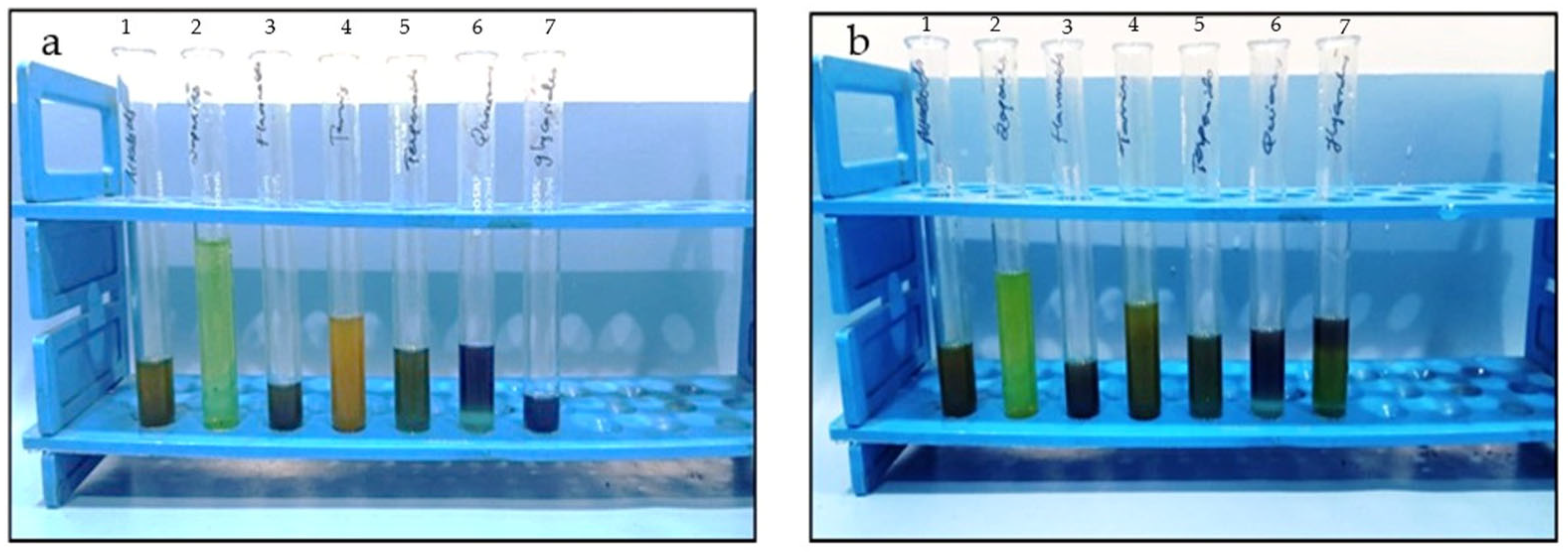
Phytochemical screening of L. aspera and H. auriculata leaf extracts revealed the presence of alkaloids, flavonoids, tannins, terpenoids, quinones, and cardiac glycosides; saponins were absent in both extracts (Table 1; Figure 3a,b). Strong positive reactions (+++) were observed for flavonoids, tannins, alkaloids, and quinones; terpenoids and cardiac glycosides exhibited moderate presence (++). Qualitative phytochemical profiles of the two plant extracts were largely comparable, with only minor variations in relative intensity of the detected compounds.
The phytochemical composition observed in L. aspera and H. auriculata is consistent with the findings of previous reports describing their rich secondary metabolite profiles [46,47]. The strong presence of flavonoids suggests their potential involvement as reducing and capping agents during the green synthesis of CuNPs. Flavonoids donate electrons and stabilize nanoparticle surfaces via hydroxyl and carbonyl functional groups.
Additionally, the detection of tannins and quinones supports antioxidant and antimicrobial potential of the plant extracts. These compounds are known to contribute to redox activity and microbial inhibition, which may enhance biological performance of the synthesized nanoparticles. The presence of cardiac glycosides and terpenoids further highlights medicinal relevance of the selected plants. Overall, the phytochemical richness of L. aspera and H. auriculata supports their suitability for polyherbal-mediated nanoparticle synthesis, with potential biological functionality.

3.2. UV-Visible Spectroscopy

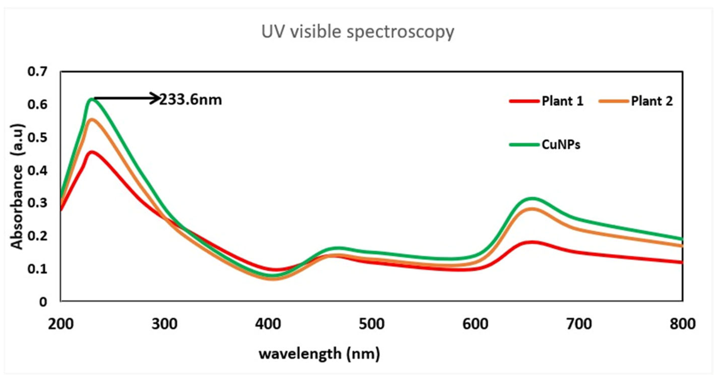
UV-visible spectroscopy performed in the range of 200–800 nm revealed a characteristic absorption peak at 233.6 nm (Figure 4), indicating CuNP formation. The distilled water control exhibited no significant absorption within the measured range. CuNPs synthesized using the polyherbal extract exhibited higher absorbance than those synthesized using the individual plant extracts, suggesting effective nanoparticle formation and stabilization by bioactive phytochemicals.
The absorption peak at 233.6 nm may be attributed to electronic transitions associated with copper-oxygen species such as Cu2O or CuO, as well as contributions from phytochemicals present in the plant extract, rather than with the classical surface plasmon resonance (SPR) of metallic CuNPs, which typically occurs at approximately 550–600 nm. Absence of a distinct SPR peak in the visible region may be associated with small particle size, partial oxidation, or surface capping by organic molecules. Compared with the individual plant extracts, the synthesized nanoparticles exhibited higher absorbance intensity, further supporting the formation of CuNPs stabilized by polyherbal phytochemicals. Similar observations have been reported in previous studies on the plant-mediated synthesis of CuNPs, where phytochemicals are found to play a critical role in reduction and stabilization [26,48,49,50].

3.3. FTIR

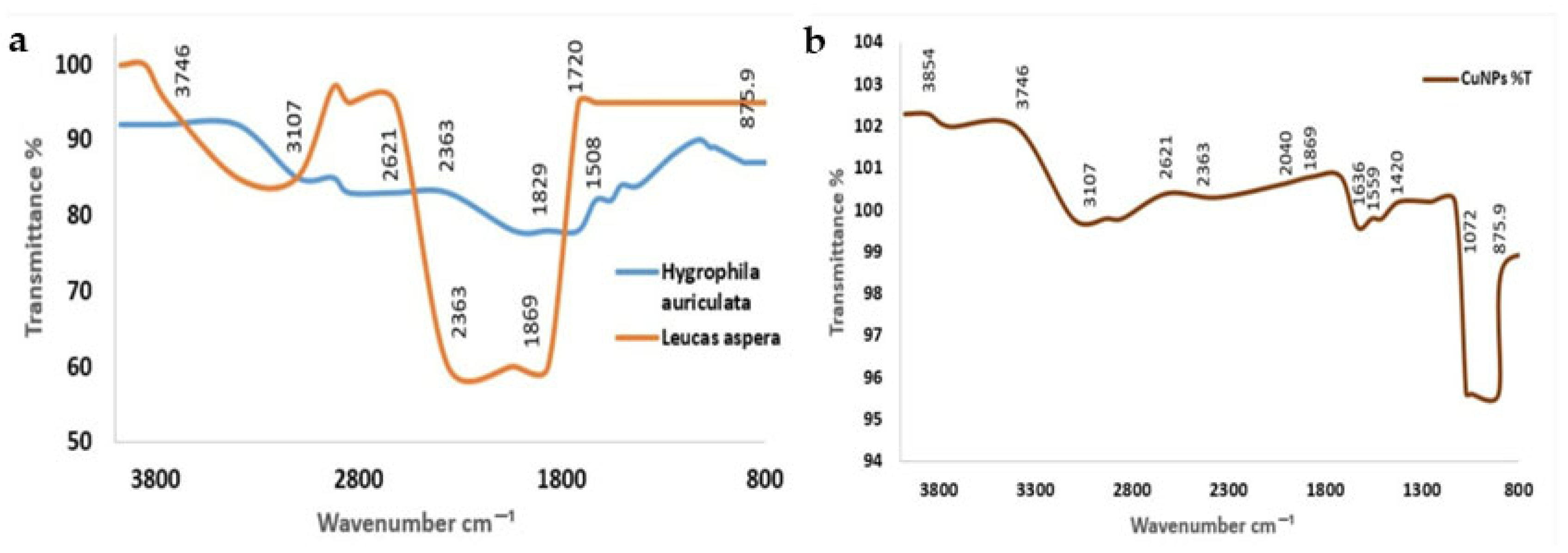
FTIR spectral analysis was performed to identify functional groups associated with the polyherbal-mediated CuNPs based on their characteristic absorption bands (Figure 5). The observed peaks and their corresponding functional groups are summarized in Table 2. The FTIR spectrum displayed bands corresponding to hydroxyl (–OH), carbonyl (C=O), amine (N–H), alkene (C=C), and C–O functional groups. These findings suggest the presence of phenolic and alcoholic compounds that may participate in nanoparticle reduction and stabilization.
The detection of carbonyl- and amine-related bands may indicate the involvement of phytoconstituents such as flavonoids and other bioactive compounds in capping [51,52]. Overall, the FTIR results indicate presence of multiple functional groups within the polyherbal extract that can interact with the nanoparticle surface. These functional moieties may contribute to the reduction of copper ions and stabilization of nanoparticles by surface binding.

3.4. GC-MS Analysis

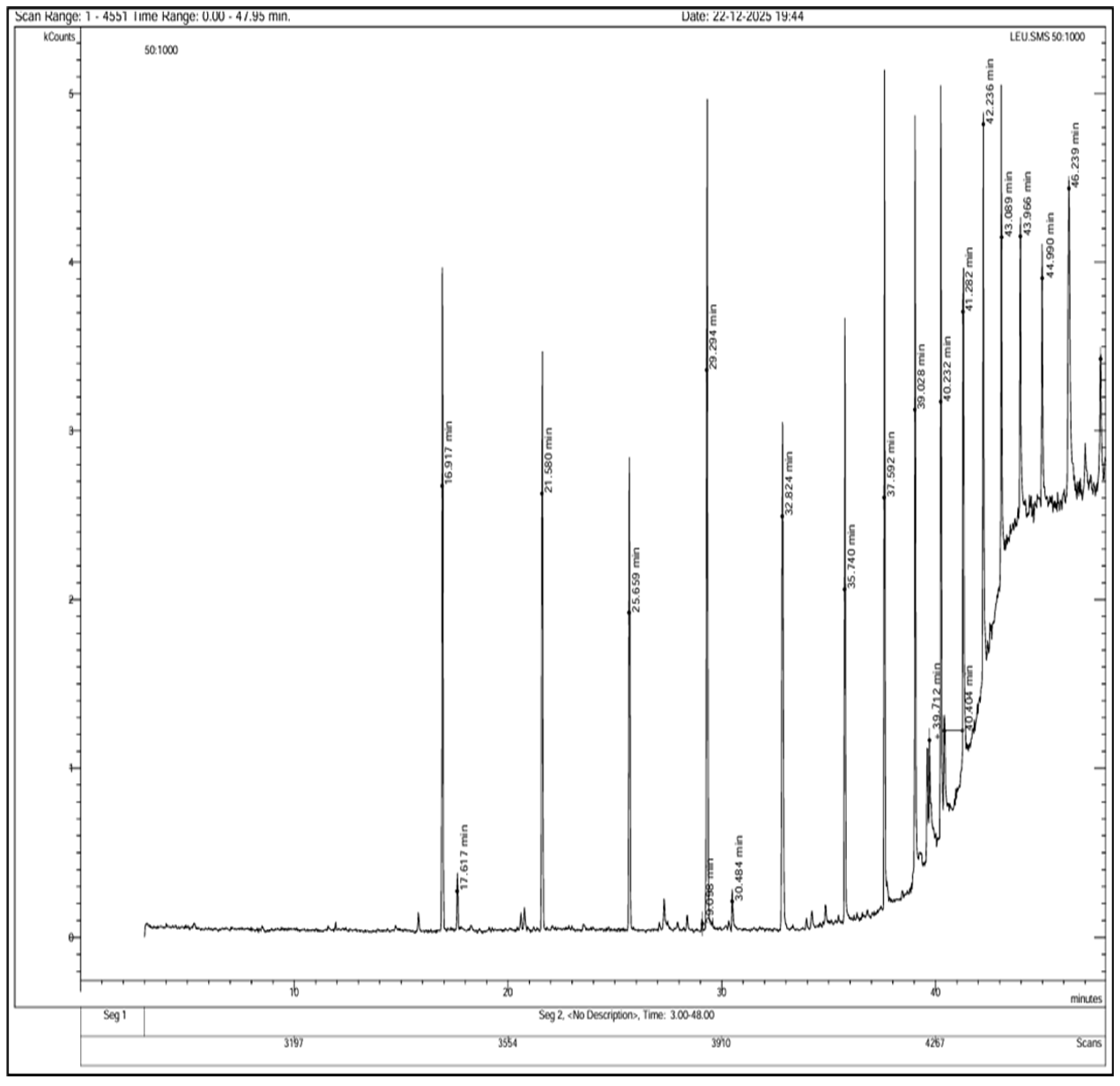
GC-MS analysis of the polyherbal extract revealed a complex phytochemical profile, with multiple peaks observed between retention times of 16.9 and 47.9 min in the chromatogram (Figure 6). Tentatively identified compounds included aromatic compounds, phenolic compounds, organic acids, phosphonic acid derivatives, amide-containing compounds, and sulfur-containing compounds (Table 3). Major constituents included benzene and naphthalene derivatives, acetic acid esters, ethylphosphonic acid derivatives, thiophenecarboxylic acid, propenamide derivatives, and phenolic compounds such as 2,4-dihydroxypropiophenone, indicating the presence of diverse bioactive secondary metabolites.
The diverse phytochemical constituents identified by GC-MS are likely to play a critical role in the green synthesis of CuNPs. Phenolic compounds and organic acids can act as reducing agents via electron donation and are well known for their antioxidant properties [53]. The presence of thiophene derivatives may contribute to antibacterial activity of the polyherbal-mediated CuNPs; such compounds disrupt bacterial cell membranes and interfere with metabolic pathways [54]. Additionally, naphthalene derivatives and steroidal oximes detected in the extract are associated with antimicrobial and anticancer activities, particularly those against HepG2 cells [55,56].
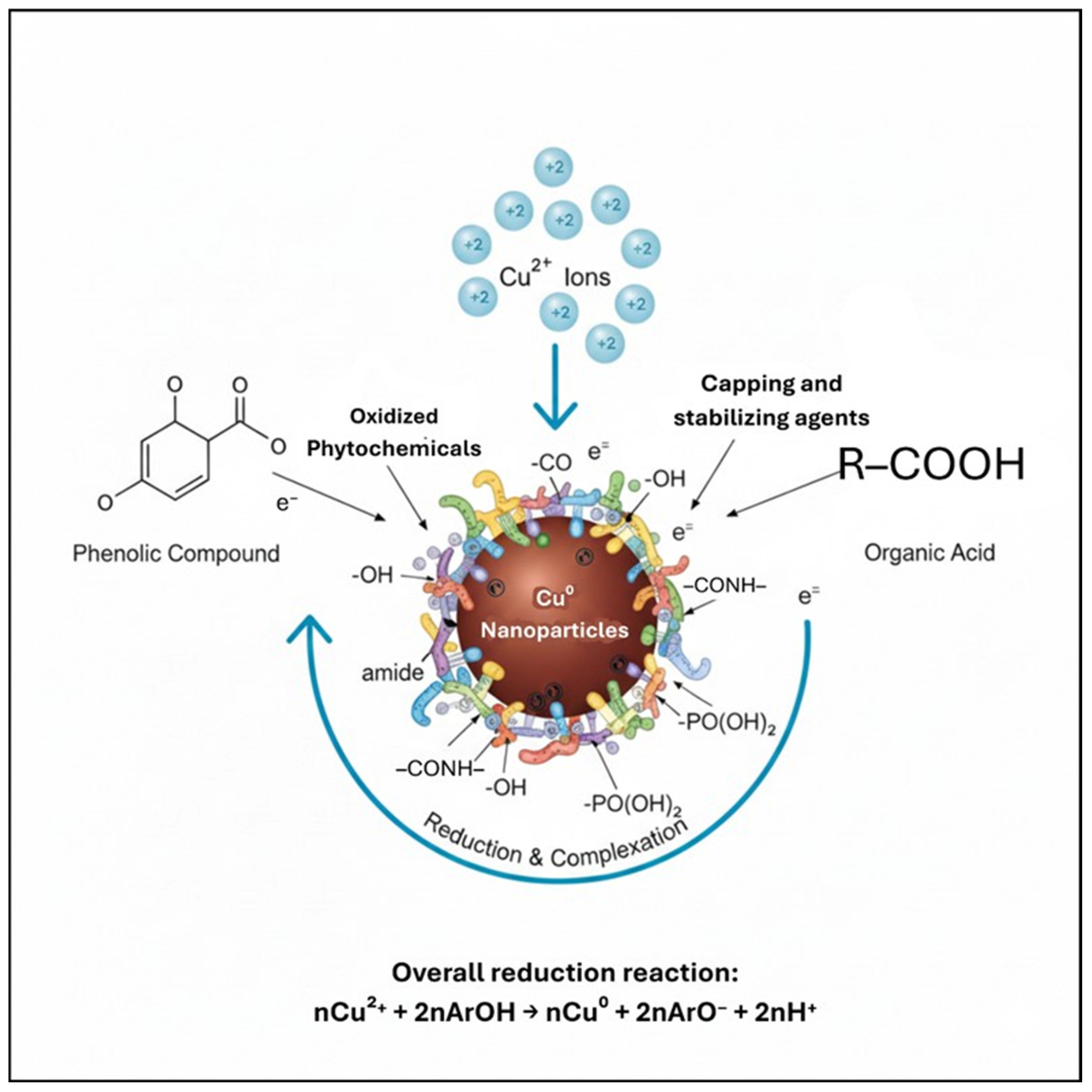
Functional groups such as hydroxyl, carbonyl, amide, and phosphonic moieties identified by GC-MS can interact with the nanoparticle surface, acting as capping and stabilizing agents (Figure 7) [57,58,59]. These findings complemented the FTIR results, which showed characteristic absorption bands corresponding to O–H, C=O, N–H, and C–O functional groups. Collectively, GC-MS and FTIR analyses confirm the involvement of plant-derived biomolecules in the reduction, capping, and stabilization of CuNPs. Diverse phytochemical composition of the polyherbal extract may contribute to nanoparticle stability and biological activity.

3.5. FESEM-EDAX

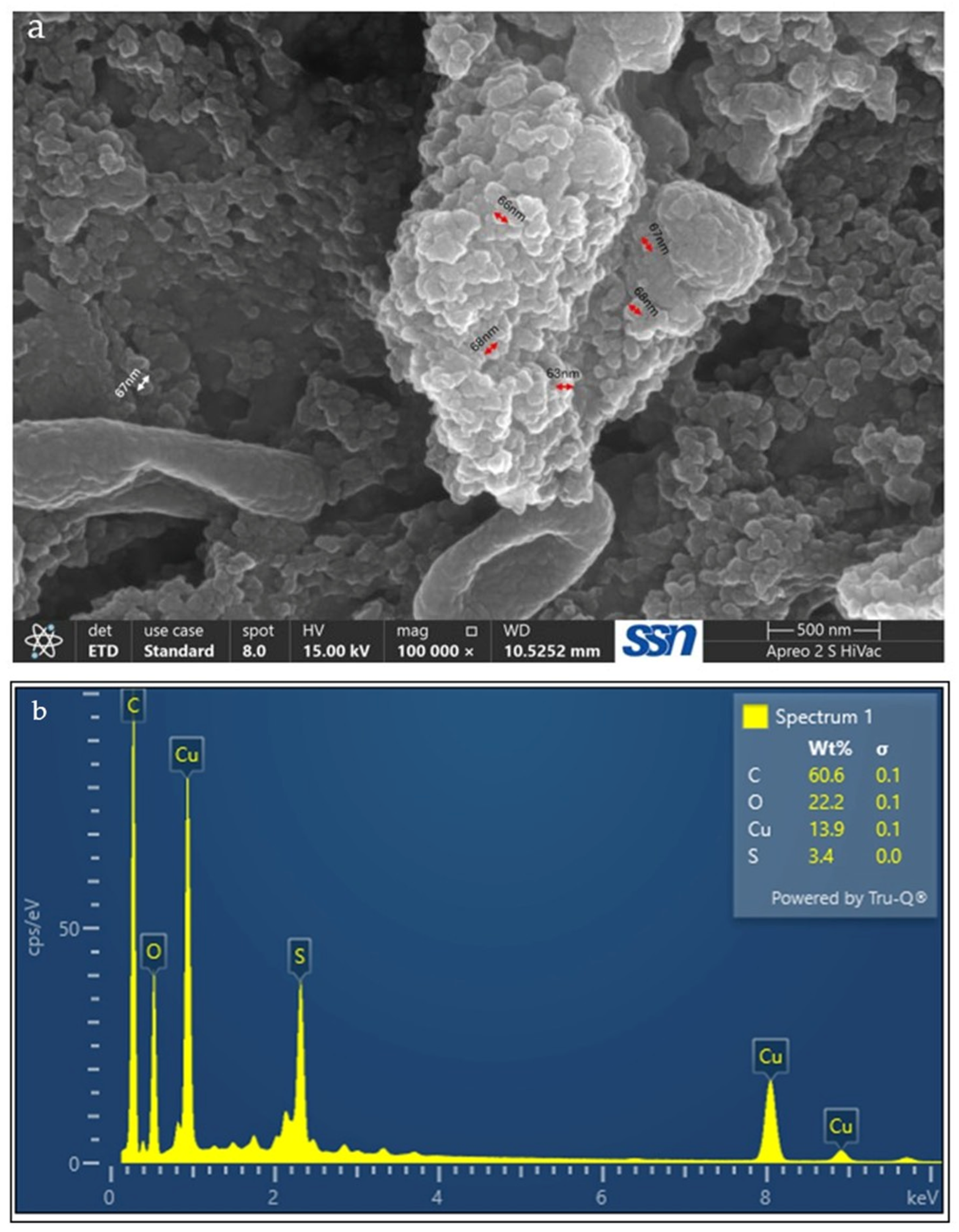
FESEM analysis was performed to examine morphology and particle size of the polyherbal-synthesized CuNPs (Figure 8a). FESEM images revealed that the nanoparticles exhibited predominantly spherical to quasi-spherical morphology with relatively good dispersion. Average particle size was calculated as 66.5 ± 1.87 nm using ImageJ. The observed surface morphology indicates the formation of crystalline nanoscale particles.
EDAX analysis (Figure 8b) confirmed the presence of copper as a major elemental component, along with oxygen, carbon, and sulfur. The detection of these elements suggests the formation of copper nanoparticles with phytochemical residues associated with the nanoparticle surface.
The spherical to quasi-spherical morphology observed in the FESEM images is consistent with the findings of previous reports on plant-mediated green synthesis of CuNPs, where phytochemicals are found to act as reducing and capping agents that influence particle growth [60,61]. The relatively uniform morphology observed in the present study may be associated with the presence of multiple phytoconstituents in the polyherbal extract, which may interact with the nanoparticle surface during nucleation and growth.
Compared with monoherbal systems, the presence of diverse biomolecules in polyherbal extracts may enhance surface passivation, potentially contributing to improved morphological homogeneity. The average particle size obtained in this study falls within the nanoscale range reported for CuNPs synthesized using leaf extracts of Hyptis suaveolens, Fortunella margarita, and Eucalyptus globulus, which typically range from 50 to 75 nm [62,63,64]. The relatively narrow size distribution suggests controlled nanoparticle growth, possibly due to cooperative capping effects from multiple phytochemical classes that limit aggregation.
EDAX results further support the green synthesis mechanism. The presence of carbon and oxygen alongside copper is commonly observed in plant-mediated CuNP synthesis and may be attributed to phytochemicals acting as capping and stabilizing agents [33]. The detection of sulfur may indicate the involvement of sulfur-containing heterocyclic compounds that can contribute to nanoparticle stability and biological activity.

3.6. XRD Analysis

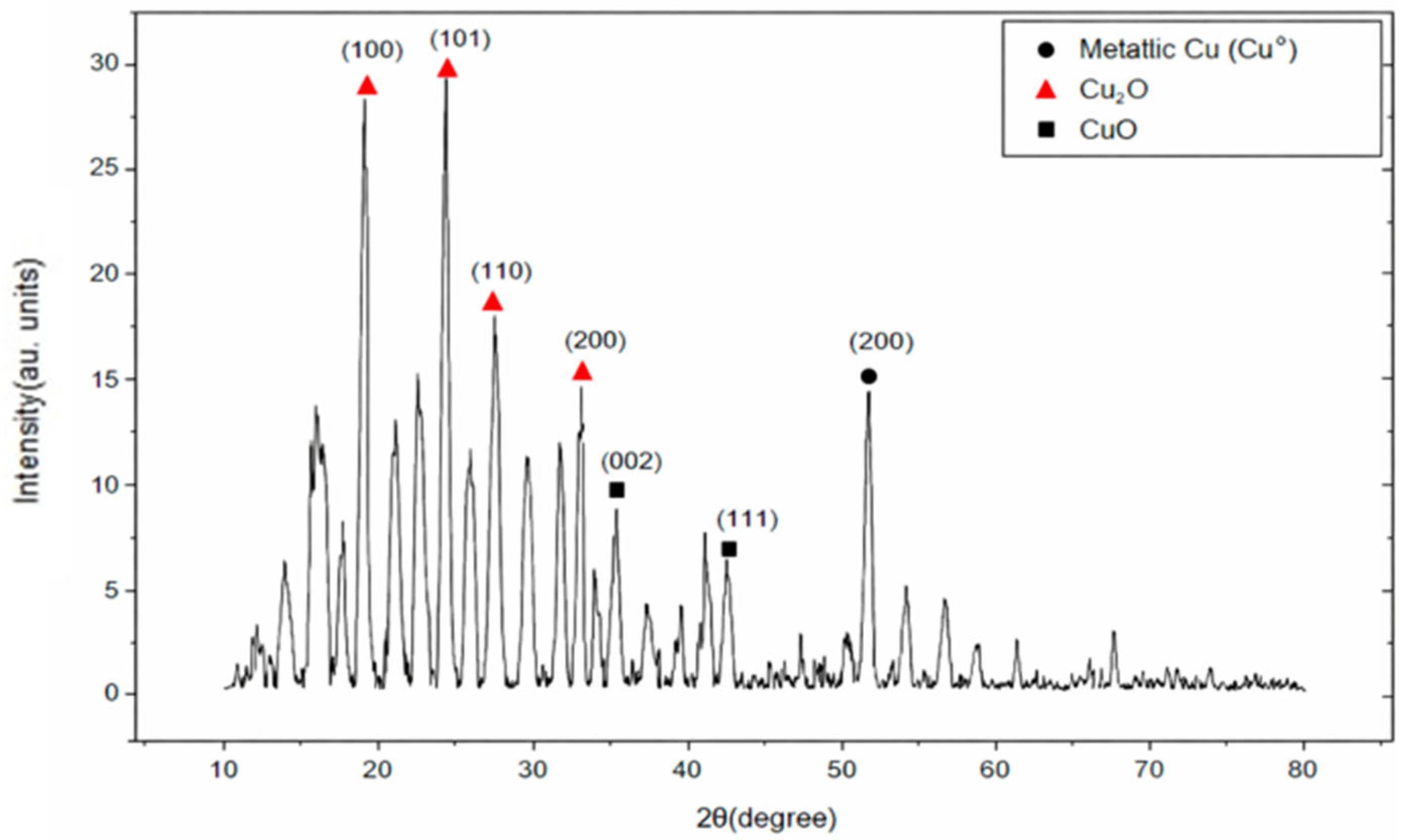
XRD analysis was performed to examine crystalline structure and phase composition of the polyherbal-synthesized CuNPs. The corresponding diffraction pattern is shown in Figure 9. The XRD pattern confirmed crystalline nature of the synthesized material and revealed the presence of multiple copper-based phases.
Crystallite size was calculated using the Debye–Scherrer equation:
D = Kλ/(β cos θ)
where K = 0.9, λ = 0.15406 nm (Cu Kα radiation), β represents the full width at half maximum (FWHM), and θ denotes the Bragg angle (Table 4). Crystallite sizes calculated from the major diffraction peaks were 11.4, 11.4, 10.7, 13.5, 10.0, 12.1, and 10.1 nm, resulting in an average crystallite size of approximately 11.5 nm.
XRD analysis confirmed the presence of metallic copper, along with Cu2O and a minor CuO phase. Although the reduction in Cu2+ ions to Cu0 was substantial, it was not complete. The Cu2O phase may result from the partial reduction in Cu2+ to Cu+, whereas CuO and Cu2O phases may also form owing to the surface oxidation of CuNP during drying or storage.
The diffraction peaks were indexed using standard JCPDS reference cards: Cu (04-0836), Cu2O (05-0667), and CuO (48-1548), confirming mixed-phase nature of the synthesized nanoparticles. Most prominent peaks corresponded to Cu2O and Cu, whereas CuO was present as a minor phase. Weak unindexed reflections were likely associated with nanoscale effects, minor surface oxidation, or instrumental background; no additional crystalline impurity phases were detected within the instrumental detection limits.
Multiphase crystalline structure observed in the XRD pattern is consistent with the findings of previous reports on green-synthesized CuNPs, where metallic copper and oxide phases are found to coexist [65,66,67]. Average crystallite size in the nanoscale range suggests controlled crystal growth under the given synthesis conditions. The relatively small crystallite size may be influenced by phytochemicals present in the extract, which may act as stabilizing agents and limit excessive crystal growth. However, additional comparative studies are required to determine specific influence of the polyherbal system on structural regulation [68,69].

3.7. TLC

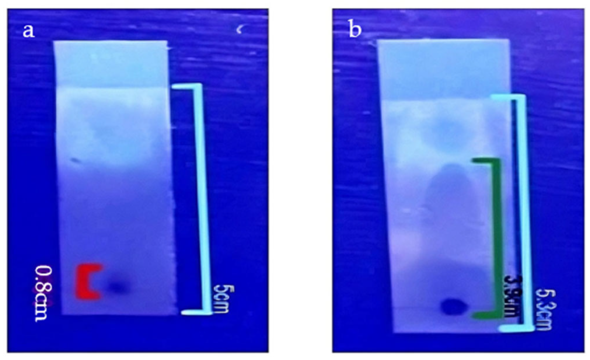
TLC was performed to compare the mobility of phytochemicals present in the plant extract and synthesized CuNPs (Figure 10). Both the plant extract and CuNP suspension were prepared in distilled water; 5 µL of each sample was spotted onto aluminum-backed silica gel TLC plates. The plates were developed using an appropriate solvent system; retention factor (Rf) was calculated using the following equation [70]:
R f = Distance   travelled   by   the   substance Distance   travelled   by   the   solvent   front
The calculated Rf values reflected relative mobility of the analyzed components. The plant extract exhibited an elevated Rf value (0.74), whereas the nanoparticle suspension exhibited minimal migration, with a Rf value of 0.16. Limited mobility of the nanoparticle sample may be attributed to enhanced interactions with the stationary phase and reduced diffusion owing to the large size and high polarity of nanoparticle-associated phytochemicals.
Similar trends have been reported in previous TLC analyses of plant-mediated nanoparticle systems, where free phytochemical constituents in plant extracts are found to display higher mobility than nanoparticle-associated compounds [71]. These observations suggest that phytochemicals conjugated to the CuNPs may enhance adsorption to the stationary phase and reduce chromatographic mobility.

3.8. Antibacterial Activity

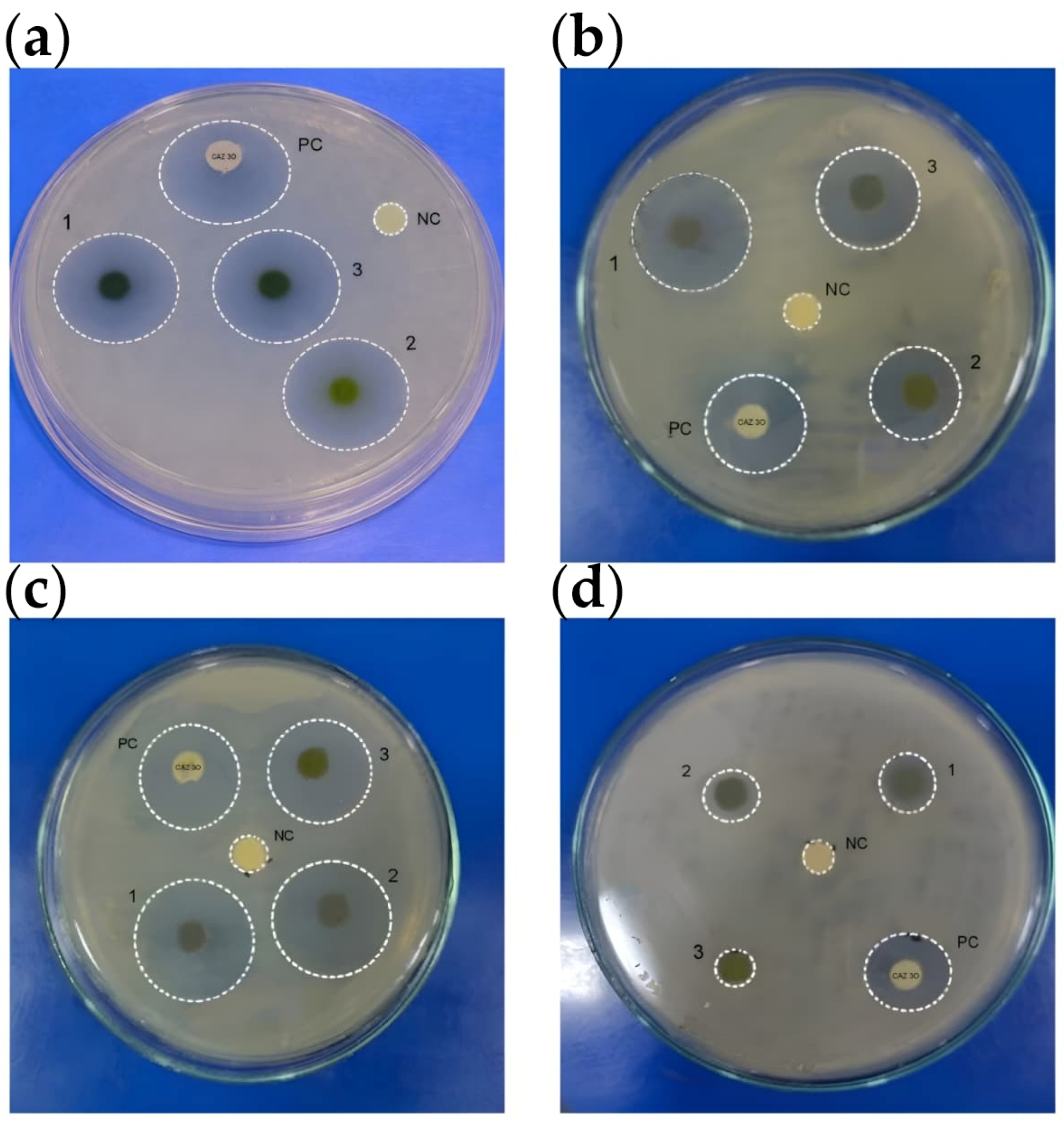
Antibacterial activity of the synthesized samples was evaluated using the zone of inhibition method (Figure 11). The corresponding inhibition diameters are summarized in Table 5. Among the tested samples, the polyherbal-mediated CuNPs exhibited the highest antibacterial activity against all tested bacterial strains. The largest zones of inhibition were observed against S. aureus (20 mm) and E. coli (28 mm), indicating strong antibacterial activity.
Enhanced antibacterial activity of the synthesized CuNPs is consistent with the findings of previous reports demonstrating the effectiveness of CuNPs against Gram-positive and Gram-negative bacteria [72,73]. Considering that the synthesized material exhibits a multiphase composition comprising Cu2O, CuO, and metallic Cu, the observed antibacterial activity may arise from the combined contributions of these phases rather than from metallic copper alone.
Compared with monoherbal green synthesis systems, the polyherbal approach may provide a broader spectrum of bioactive phytochemicals. Antibacterial activity of the plant extracts themselves may be attributed to phytochemical constituents such as phenolics, flavonoids, alkaloids, and terpenoids, which disrupt bacterial cell membranes and interfere with essential cellular processes [46,47,74].
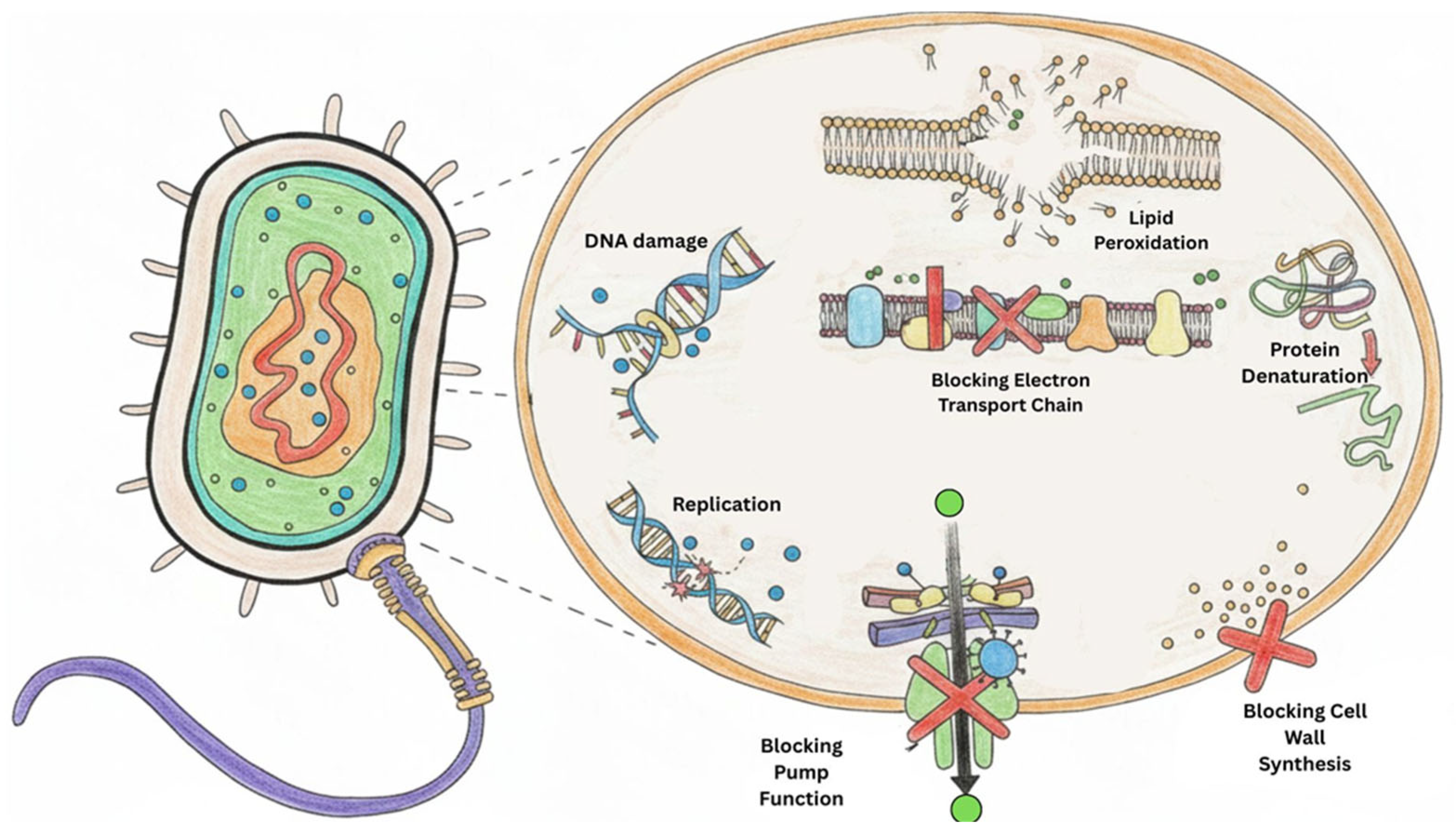
The antibacterial efficacy of CuNPs may also be associated with their nanoscale dimensions, which facilitate close interaction with bacterial cells. CuNPs adhere to bacterial cell walls, disrupt membrane integrity, and generate reactive oxygen species, leading to oxidative stress, lipid peroxidation, protein denaturation, and ultimately, bacterial cell death (Figure 12) [75]. However, the precise antibacterial mechanism in the present study requires further investigation. Surface-bound phytochemicals originating from the polyherbal extract may additionally influence nanoparticle-cell interactions and contribute to the observed antibacterial activity.

3.9. In Vitro Antioxidant Assay

Antioxidant activity of the polyherbal CuNPs was evaluated using the DPPH radical scavenging assay at concentrations of 10, 20, 30, 40, and 50 mg/mL. The nanoparticles exhibited a concentration-dependent increase in DPPH radical scavenging activity (Table 6). Correspondingly, optical density (OD) values decreased progressively with increasing nanoparticle concentration (Table 7), indicating enhanced scavenging of DPPH radicals.
Statistical analysis revealed that the absorbance observed at higher concentrations (30–50 mg/mL) was significantly reduced (p < 0.05) compared with that in the control. The increased antioxidant activity at elevated nanoparticle concentrations may be attributed to surface-bound phytochemicals, particularly phenolic and flavonoid compounds, which participate in hydrogen atom donation and electron transfer reactions. Similar concentration-dependent DPPH radical scavenging activity has been reported for other green-synthesized CuNPs, including those prepared using Passiflora flower extract [76]. Additionally, CuNPs synthesized using Moringa oleifera leaf extracts have demonstrated comparable concentration-dependent antioxidant activity [45]. These findings suggest that the synthesized nanoparticles possess measurable antioxidant potential.

3.10. Cytotoxicity Assay

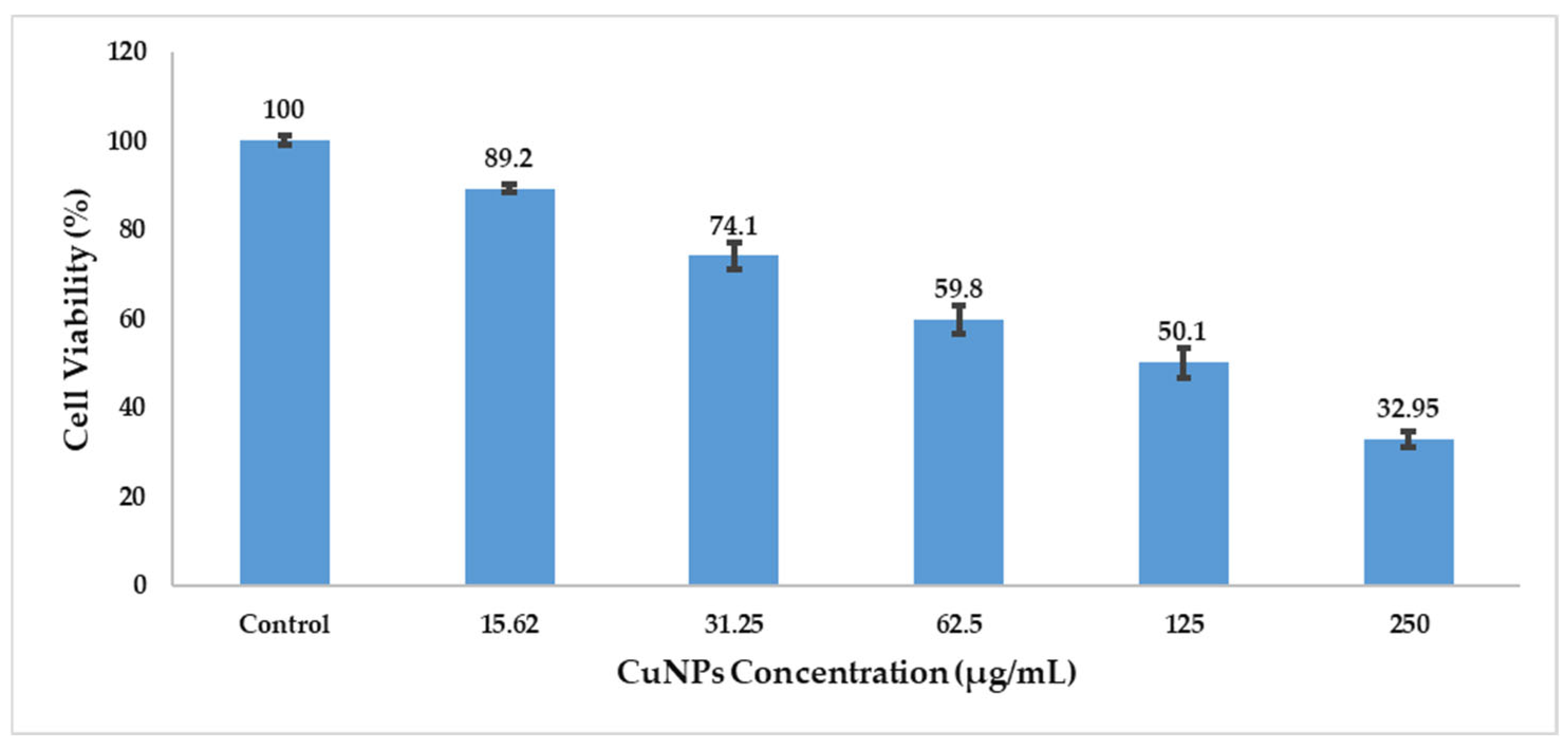
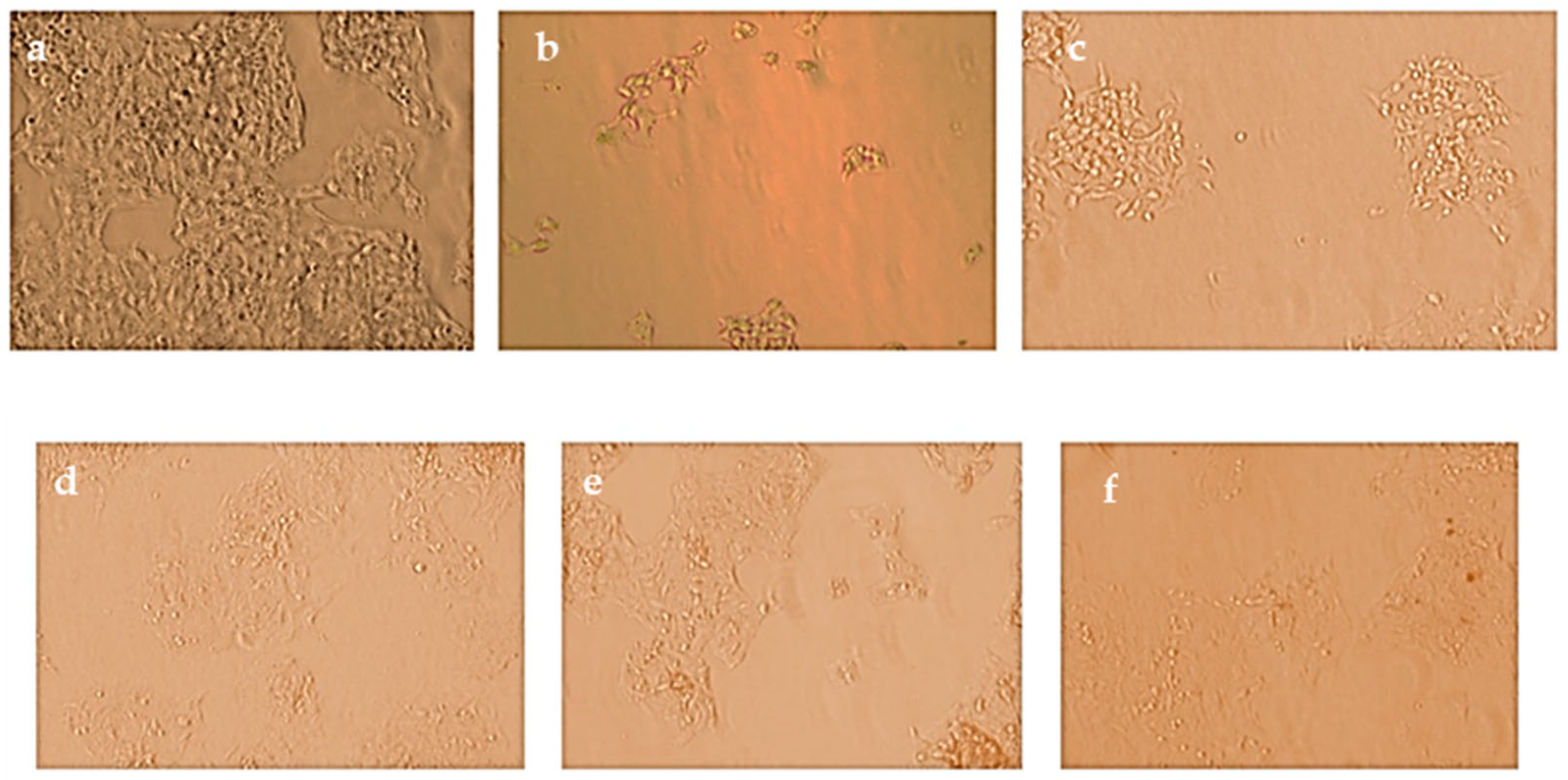
Cytotoxic potential of the polyherbal CuNPs was evaluated against HepG2 cells using the MTT assay. A concentration-dependent reduction in cell viability was observed after 48 h of treatment. Cell viability decreased from 89.2 ± 0.96% at 15.62 µg/mL to 32.95 ± 1.70% at 250 µg/mL compared with that of the untreated control cells (100%) (Table 8). Approximately 50% cell viability (IC50) was observed at a concentration of 125 µg/mL, indicating moderate cytotoxic activity against HepG2 cells (Figure 10).
Statistical analysis revealed that CuNPs significantly reduced cell viability at concentrations ≥ 62.5 µg/mL (p < 0.05), whereas higher concentrations (125 and 250 µg/mL) produced highly significant cytotoxic effects (p < 0.01). The reduction in cell viability correlates with decreased mitochondrial metabolic activity, as indicated by reduced formazan formation (Figure 13 and Figure 14).
The dose-dependent cytotoxicity observed in HepG2 cells suggests that the polyherbal CuNPs possess potential anticancer activity. The observed reduction in cell viability may be associated with mitochondrial dysfunction and oxidative stress, which are key mechanisms underlying CuNP-induced cytotoxicity [77,78]. Compared with monoherbal nanoparticle systems, polyherbal-mediated CuNPs may exhibit altered biological activity owing to the presence of multiple phytochemicals that interact with copper ions during nanoparticle formation. These interactions may influence nanoparticle surface chemistry and contribute to the observed cytotoxic effects.

3.11. DNA Fragmentation Analysis

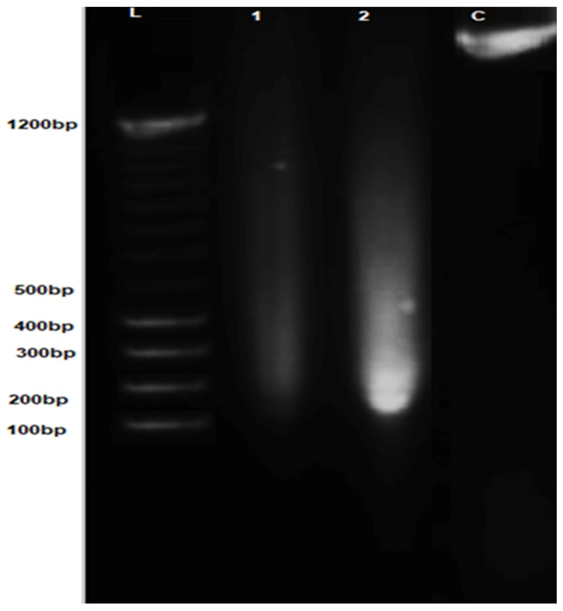
DNA fragmentation analysis was performed to investigate the mode of cell death induced by polyherbal CuNPs in HepG2 cells. Treated cells exhibited a pronounced fragmented DNA smear rather than a single intact genomic DNA band, indicating substantial DNA damage compared with untreated cells (Figure 15).
Densitometric analysis of the agarose gel images using ImageJ revealed that approximately 90–95% of the total DNA was present in fragmented form in cells treated at the IC50, as calculated using the fragmentation percentage formula [42,43].
DNA fragmentation is widely recognized as a biochemical hallmark of apoptosis, reflecting the endonuclease-mediated cleavage of chromosomal DNA [79]. The high degree of DNA fragmentation observed in IC50-treated HepG2 cells suggests that polyherbal CuNPs induce apoptotic DNA damage. These results are consistent with the dose-dependent reduction in cell viability observed in the MTT assay and indicate that cytotoxic effects of the nanoparticles may be primarily associated with apoptosis rather than with necrotic cell death.

4. Conclusions

An eco-friendly and sustainable polyherbal green synthesis of CuNPs was successfully achieved using H. auriculata and L. aspera. Preliminary phytochemical screening confirmed the presence of bioactive constituents in the individual plant extracts. Subsequent characterization using UV–Vis spectroscopy, FTIR, XRD, FESEM-EDAX, and GC-MS confirmed that the formation of CuNPs was reduced and stabilized by phytoconstituents present in the polyherbal extract. The nanoparticles exhibited predominantly spherical morphology, with an average crystallite size of approximately 11.5 nm. TLC revealed distinct Rf values for the plant extract (0.74) and CuNPs (0.16), indicating altered interactions with the stationary phase following nanoparticle formation.
The synthesized CuNPs exhibited antibacterial activity against E. coli, P. aeruginosa, S. pyogenes, and Staphylococcus spp. compared with the plant extract alone. FTIR and GC-MS analyses indicated the presence of bioactive phytoconstituents, including phenolics and esters, which may contribute to the observed antibacterial effects. Additionally, the CuNPs demonstrated concentration-dependent antioxidant activity in the DPPH assay and induced dose-dependent cytotoxic effects in HepG2 cells, as evaluated by the MTT assay.
Although in vitro cytotoxic effects were observed, the molecular mechanisms underlying nanoparticle interactions with cancer-related targets remain unresolved. Future studies integrating computational cancer biology approaches, such as molecular docking, molecular dynamics simulations, and in silico pathway analysis, may provide further insight into nanoparticle–protein interactions and potential structure–activity relationships. Such investigations, combined with experimental validation, will strengthen mechanistic understanding.
Overall, the findings suggest that polyherbal green-synthesized CuNPs are promising multifunctional nanomaterials with potential applications in sustainable nanomedicine and strategies aimed at addressing healthcare-associated infections.

Author Contributions

Conceptualization, G.V. and S.K.R.; methodology, A.R.; software, S.G.; validation, A.R. and S.G.; formal analysis, G.V.; investigation, T.S.S.; resources, J.K.; data curation, T.S.S. and J.K.; writing—original draft preparation, G.V.; writing—review and editing, S.K.R. and P.S.K.; visualization, P.S.K.; supervision, S.K.R.; project administration, S.K.R. and P.S.K.; funding acquisition, S.K.R. All authors have read and agreed to the published version of the manuscript.

Funding

This research received no external funding.

Institutional Review Board Statement

Not applicable.

Informed Consent Statement

Not applicable.

Data Availability Statement

The original contributions presented in the study are included in the article; further inquiries can be directed to the corresponding author.

Conflicts of Interest

The authors declare no conflicts of interest.

Abbreviations

The following abbreviations are used in this manuscript:
βPeak broadening (in radians)
CO2Carbon dioxide
CuNPsCopper nanoparticles
DMEMDulbecco’s Modified Eagle’s Medium
DMSODimethyl sulfoxide
DPPH2,2-Diphenyl-1-picrylhydrazyl
EDAX/EDSEnergy dispersive X-ray analysis/Energy dispersive X-ray spectroscopy
E. coliEscherichia coli
FESEMField emission scanning electron microscopy
FTIRFourier transform infrared spectroscopy
FWHMFull width at half maximum
GC–MSGas chromatography–mass spectrometry
hklMiller indices
IC50Half maximal inhibitory concentration
SPRSurface plasmon resonance

References

  1. Villagrán, Z.; Anaya-Esparza, L.M.; Velázquez-Carriles, C.A.; Silva-Jara, J.M.; Ruvalcaba-Gómez, J.M.; Aurora-Vigo, E.F.; Rodríguez-Lafitte, E.; Rodríguez-Barajas, N.; Balderas-León, I.; Martínez-Esquivias, F. Plant-based extracts as reducing, capping, and stabilizing agents for the green synthesis of inorganic nanoparticles. Resources 2024, 13, 70. [Google Scholar] [CrossRef]
  2. Lithi, I.J.; Nakib, K.I.A.; Chowdhury, A.M.S.; Hossain, M.S. A review on the green synthesis of metal (Ag, Cu, and Au) and metal oxide (ZnO, MgO, Co3O4, and TiO2) nanoparticles using plant extracts for developing antimicrobial properties. Nanoscale Adv. 2025, 7, 2446–2473. [Google Scholar] [PubMed]
  3. Mittal, A.K.; Chisti, Y.; Banerjee, U.C. Synthesis of metallic nanoparticles using plant extracts. Biotechnol. Adv. 2013, 31, 346–356. [Google Scholar] [CrossRef] [PubMed]
  4. Thatyana, M.; Dube, N.P.; Kemboi, D.; Manicum, A.-L.E.; Mokgalaka-Fleischmann, N.S.; Tembu, J.V. Advances in Phytonanotechnology: A Plant-mediated green synthesis of metal nanoparticles using Phyllanthus plant extracts and their antimicrobial and anticancer applications. Nanomaterials 2023, 13, 2616. [Google Scholar] [CrossRef]
  5. Kumar, S.; Tripathy, S.K.; Kaushik, N. A Review on green synthesis of copper nanoparticles using plant extracts: Methods, characterization, and applications. Pharm. Nanotechnol. 2025; in press.
  6. Saifuddin, N.N.; Matussin, S.N.; Fariduddin, Q.; Khan, M.M. Potentials of roots, stems, leaves, flowers, fruits, and seeds extract for the synthesis of silver nanoparticles. Bioprocess Biosyst. Eng. 2024, 47, 1119–1137. [Google Scholar] [CrossRef]
  7. Velgosová, O.; Dolinská, S.; Podolská, H.; Mačák, L.; Čižmárová, E. Impact of plant extract phytochemicals on the synthesis of silver nanoparticles. Materials 2024, 17, 2252. [Google Scholar] [CrossRef] [PubMed]
  8. Vanlalveni, C.; Lallianrawna, S.; Biswas, A.; Selvaraj, M.; Changmai, B.; Rokhum, S.L. Green synthesis of silver nanoparticles using plant extracts and their antimicrobial activities: A Review of Recent Literature. RSC Adv. 2021, 11, 2804–2837. [Google Scholar] [CrossRef] [PubMed]
  9. Singh, H.; Desimone, M.F.; Pandya, S.; Jasani, S.; George, N.; Adnan, M.; Aldarhami, A.; Bazaid, A.S.; Alderhami, A.S. Revisiting the green synthesis of nanoparticles: Uncovering influences of plant extracts as reducing agents for enhanced synthesis efficiency and its biomedical applications. Int. J. Nanomed. 2023, 18, 4727–4750. [Google Scholar] [CrossRef]
  10. Sikdar, D.; Mondal, A.; Ghosh, R.; Jana, S. Unveiling the Aerial Mysteries of Hygrophila auriculata: Ethnomedicinal insights, phytochemical profiles, and pharmacological potentials. Int. J. Res. Sci. Innov. 2025, 12, 1322–1329. [Google Scholar] [CrossRef]
  11. Sethiya, N.K.; Ahmed, N.M.; Shekh, R.M.; Kumar, V.; Singh, P.K.; Kumar, V. Ethnomedicinal, phytochemical and pharmacological updates on Hygrophila auriculata (Schum.) Heine: An overview. J. Integr. Med. 2018, 16, 299–311. [Google Scholar]
  12. Venkatesan, A.; Kichenamourty, G.; Seetharaman, S. Phytochemical and therapeutic potential of Hygrophila auriculata: A comprehensive review. J. Nat. Rem. 2025, 25, 2317–2325. [Google Scholar] [CrossRef]
  13. Malpotra, M.; Garg, M.; Singh, N.; Dey Sadhu, S.; Chopra, R.; Sharma, B. An overview of bioactive components and phytopharmaceutical potentials of Hygrophila auriculata—A herbaceous medicinal plant. Phytomed. Plus 2025, 5, 100737. [Google Scholar] [CrossRef]
  14. Das, A.; Venkatesh, K.G.; Das, K.; Kumar, S.; Rout, S.; Kumar, S. Phytochemical and Cytotoxicity Analysis of Flowers of Hygrophila auriculata (Schumach.) Heine. In Plants and Secondary Metabolites; Ambika Prasad Research Foundation: Odisha, India, 2025; Volume 5, pp. 18–24. [Google Scholar]
  15. Soujanya, K.; Kumari, B.A.; Jyothsna, E. Leucas aspera: A wild traditional green leafy vegetable with immense pharmacological properties. Int. J. Adv. Biochem. Res. 2024, 8, 504–508. [Google Scholar] [CrossRef]
  16. Hariharasudhan, A.B.; Shanmadi, J.P.; Devaakshayah Shreenithee, R.A. Phytochemical constituents, extraction and pharmacological activity of Leucas aspera. World J. Biol. Pharm. Health Sci. 2024, 19, 238–243. [Google Scholar] [CrossRef]
  17. Vasudha, K.; Archana, D.; Mutyalamma, B.; Kishori, B. Phytochemical screening, antimicrobial and antioxidant activities of root and leaf extracts of Leucas aspera. Asian J. Pharm. Clin. Res. 2019, 12, 141–147. [Google Scholar] [CrossRef]
  18. Harishchandra, B.D.; Pappuswamy, M.; Antony, P.U.; Shama, G.; Pragatheesh, A.; Arumugam, V.A.; Periyaswamy, T.; Sundaram, R. Copper nanoparticles: A review on synthesis, characterization and applications. Asia Pac. J. Cancer Biol. 2020, 5, 201–210. [Google Scholar] [CrossRef]
  19. Aggarwal, K.; Brahma, A.; Nisha; Sharma, Y.; Rajput, P.; Sachdeva, B.; Singh, A.; Chandra, R.; Singh, S. Recent progress in copper nanomaterials: Catalysis, energy, biomedicine, and environmental applications. Mater. Adv. 2025, 6, 9296–9339. [Google Scholar] [CrossRef]
  20. Ivanova, I.A.; Daskalova, D.S.; Yordanova, L.P.; Pavlova, E.L. Copper and copper nanoparticles applications and their role against infections: A minireview. Processes 2024, 12, 352. [Google Scholar] [CrossRef]
  21. Gabal, E.; Ramadan, M.M.; Asran, A.; Alghuthaymi, M.A.; Abd-Elsalam, K.A. Copper Nanostructures Applications in Plant Protection. In Nanobiotechnology Applications in Plant Protection; Abd-Elsalam, K.A., Prasad, R., Eds.; Springer: Cham, Switzerland, 2018; pp. 63–86. [Google Scholar]
  22. Hemashree, S.; Pruthvi, M.L.; Mahesh, M.K. Green synthesis of copper nanoparticles using leaf extract of Phyllanthus niruri Lin. and study of its antimicrobial activity. Int. J. Res. Appl. Sci. Eng. Technol. 2023, 11, 459–467. [Google Scholar] [CrossRef]
  23. Alahdal, F.A.M.; Qashqoosh, M.T.A.; Manea, Y.K.; Mohammed, R.K.A.; Naqvi, S. Green synthesis and characterization of copper nanoparticles using Phragmanthera austroarabica extract and their biological/environmental applications. Sustain. Mater. Technol. 2023, 35, e00540. [Google Scholar] [CrossRef]
  24. Joshi, S.; Mithawala, I.; Savaliya, Y.; Patil, R. Synthesis and characterization of copper nanoparticles using plant extract of Mentha piperita leaves, Anethum graveolens L., and Calotropis procera. Curr. Nanomed. 2024, 14, 65–72. [Google Scholar] [CrossRef]
  25. Makwana, S.J.; Upadhyay, D.L.; Bhattacharya, I.P. Green synthesis of copper nano-particles using leaf extract of Indian medicinal plants and their phytochemical analysis. Int. J. Sci. Soc. Sci. Res. 2024, 1, 62–69. [Google Scholar]
  26. Vijayakumar, G.; Kesavan, H.; Kannan, A.; Arulanandam, D.; Kim, J.H.; Kim, K.J.; Song, H.J.; Kim, H.J.; Rangarajulu, S.K. Phytosynthesis of copper nanoparticles using extracts of spices and their antibacterial properties. Processes 2021, 9, 1341. [Google Scholar] [CrossRef]
  27. Trenkenschuh, E.; Friess, W. Freeze-drying of nanoparticles: How to overcome colloidal instability by formulation and process optimization. Eur. J. Pharm. Biopharm. 2021, 165, 345–360. [Google Scholar] [CrossRef]
  28. Abdelwahed, W.; Degobert, G.; Stainmesse, S.; Fessi, H. Freeze-drying of nanoparticles: Formulation, process and storage considerations. Adv. Drug Deliv. Rev. 2006, 58, 1688–1713. [Google Scholar] [CrossRef]
  29. Jinu, U.; Gomathi, M.; Saiqa, I.; Geetha, N.; Benelli, G.; Venkatachalam, P. Green engineered biomolecule-capped silver and copper nanohybrids using Prosopis cineraria leaf extract: Enhanced antibacterial activity against microbial pathogens of public health relevance and cytotoxicity on human breast cancer cells (MCF-7). Microb. Pathog. 2017, 105, 86–95. [Google Scholar] [CrossRef] [PubMed]
  30. Doss, A. Preliminary phytochemical screening of some Indian medicinal plants. Anc. Sci. Life 2009, 29, 12–16. [Google Scholar]
  31. Odeja, O.O.; Ogwuche, C.E.; Elemike, E.E.; Obi, G. Phytochemical screening, antioxidant and antimicrobial activities of Acalypha ciliata plant. Clin. Phytosci. 2016, 2, 12. [Google Scholar] [CrossRef][Green Version]
  32. Lydia, E.; John, S.; Mohammed, R.; Sivapriya, T. Investigation on the phytochemicals present in the fruit peel of Carica papaya and evaluation of its antioxidant and antimicrobial property. Res. J. Pharmacogn. Phytochem. 2016, 8, 217–222. [Google Scholar] [CrossRef]
  33. Peddi, P.; PTSRK, P.R.; Rani, N.U.; Tulasi, S.L. Green synthesis, characterization, antioxidant, antibacterial, and photocatalytic activity of Suaeda maritima (L.) Dumort aqueous extract-mediated copper oxide nanoparticles. J. Genet. Eng. Biotechnol. 2021, 19, 131. [Google Scholar] [CrossRef]
  34. Chakraborty, N.; Banerjee, J.; Chakraborty, P.; Banerjee, A.; Chanda, S.; Ray, K.; Acharya, K.; Sarkar, J. Green synthesis of copper/copper oxide nanoparticles and their applications: A review. GREEN Chem. Lett. Rev. 2022, 151, 187–215. [Google Scholar] [CrossRef]
  35. Santos, N.M.; Gomes, A.S.; Cavalcante, D.G.S.M.; Santos, L.F.; Teixeira, S.R.; Cabrera, F.C.; Job, A.E. Green synthesis of colloidal gold nanoparticles using latex from Hevea brasiliensis and evaluation of their in vitro cytotoxicity and genotoxicity. IET Nanobiotechnol. 2019, 13, 307–315. [Google Scholar] [CrossRef] [PubMed]
  36. Idu, M.; Aihiokhai, M.O.; Imoni, C.A.; Akokigho, C.E.; Olali, N.C. Gas chromatography-mass spectrometry (GC-MS) analysis and phytochemical screening of polyherbal aqueous leaves extract (PALE). J. Chem. Appl. Microbiol. Res. 2021, 14, 10–18. [Google Scholar]
  37. Pawar, M.K.; Dwivedi, J.A.; Dwivedi, U.K. Structural elucidation of phytoconstituents (phenols, long chain fatty acids, flavonoid, terpenoids, coumarin) found in Chromolaena odorata through gas chromatography: Mass spectroscopy (GC-MS) technique. Indian J. Pharm. Educ. Res. 2025, 59, 308. [Google Scholar] [CrossRef]
  38. Brand-Williams, W.; Cuvelier, M.E.; Berset, C. Use of a free radical method to evaluate antioxidant activity. LWT-Food Sci. Technol. 1995, 28, 25–30. [Google Scholar] [CrossRef]
  39. Guntur, S.R.; Sampath Kumar, N.; Hegde, M.M.; Dirisala, V.R. In vitro studies of the antimicrobial and free-radical scavenging potentials of silver nanoparticles biosynthesized from the extract of Desmostachya bipinnata. Anal. Chem. Insights 2018, 13, 1177390118782877. [Google Scholar] [CrossRef]
  40. Ghasemi, M.; Turnbull, T.; Sebastian, S.; Kempson, I. The MTT Assay: Utility, limitations, pitfalls, and interpretation in bulk and single-cell analysis. Int. J. Mol. Sci. 2021, 22, 12827. [Google Scholar] [CrossRef]
  41. Rajagopal, G.; Nivetha, A.; Sundar, M.; Panneerselvam, T.; Murugesan, S.; Parasuraman, P.; Kumar, S.; Ilango, S.; Kunjiappan, S. Mixed phytochemicals mediated synthesis of copper nanoparticles for anticancer and larvicidal applications. Heliyon 2021, 7, e07360. [Google Scholar] [CrossRef]
  42. Kasibhatla, S.; Amarante-Mendes, G.P.; Finucane, D.; Brunner, T.; Bossy-Wetzel, E.; Green, D.R. Analysis of DNA fragmentation using agarose gel electrophoresis. Cold Spring Harb. Protoc. 2006, 2006, pdb-prot4429. [Google Scholar] [CrossRef]
  43. Schneider, C.A.; Rasband, W.S.; Eliceiri, K.W. NIH Image to ImageJ: 25 years of image analysis. Nat. Methods 2012, 9, 671–675. [Google Scholar] [CrossRef]
  44. Kowalska, T.; Sajewicz, M. Thin-layer chromatography (TLC) in the screening of botanicals–its versatile potential and selected applications. Molecules 2022, 27, 6607. [Google Scholar] [CrossRef]
  45. Thomas, A.; Kanakdhar, A.; Shirsat, A.; Deshkar, S.; Kothapalli, L. A high performance thin layer chromatographic method using a design of experiment approach for estimation of phytochemicals in extracts of Moringa oleifera leaves. Turk. J. Pharm. Sci. 2020, 17, 148–158. [Google Scholar] [CrossRef] [PubMed]
  46. Sungar, S.; Toragall, M.M.; Mujeeb, M.A.; Puranik, S.I.; Akbar, A.A.; Aladkatti, R.H.; Ghagane, S.C. Phytochemical analysis, antimicrobial and anti-inflammatory efficacy of Leucas aspera leaf extracts. Arch. Razi Inst. 2024, 79, 761–768. [Google Scholar]
  47. Gnanasri, M.; Bharathiraja, N.; Mangaleshwari, R.; Nagalingam, M.; Ramkumar, A. Antioxidant and antimicrobial activities along with physico-chemical studies of medicinal plant Hygrophila auriculata. Biosci. Res. 2023, 5, 1–14. [Google Scholar]
  48. Renuga, D.; Jeyasundari, J.; Shakthi Athithan, A.S.; Brightson Arul Jacob, Y. Synthesis and characterization of copper oxide nanoparticles using Brassica oleracea var. italic extract for its antifungal application. Mater. Res. Express 2020, 7, 045007. [Google Scholar] [CrossRef]
  49. Rehana, D.; Mahendiran, D.; Kumar, R.S.; Rahiman, A.K. Evaluation of antioxidant and anticancer activity of copper oxide nanoparticles synthesized using medicinally important plant extracts. Biomed. Pharmacother. 2017, 89, 1067–1077. [Google Scholar] [CrossRef]
  50. Nouren, S.; Bibi, I.; Kausar, A.; Sultan, M.; Nawaz Bhatti, H.; Safa, Y.; Sadaf, S.; Alwadai, N.; Iqbal, M. Green synthesis of CuO nanoparticles using Jasmin sambac extract: Conditions optimization and photocatalytic degradation of methylene blue dye. J. King Saud Univ. Sci. 2024, 36, 103089. [Google Scholar]
  51. Pasieczna-Patkowska, S.; Cichy, M.; Flieger, J. Application of fourier transform infrared (FTIR) spectroscopy in characterization of green synthesized nanoparticles. Molecules 2025, 30, 684. [Google Scholar] [CrossRef]
  52. Abdullah, V.S.; Amin, K.Y.M.; Amin, H.I.M. Plant-assisted aynthesis, phytochemical profiling, and bioactivity evaluation of copper nanoparticles derived from Tordylium trachycarpum (Apiaceae). Biomolecules 2025, 15, 1693. [Google Scholar]
  53. Marinova, E.M.; Yanishlieva, N.V. Antioxidant activity and mechanism of action of some phenolic acids at ambient and high temperatures. Food Chem. 2003, 81, 189–197. [Google Scholar] [CrossRef]
  54. Mabkhot, Y.N.; Kaal, N.A.; Alterary, S.; Al-Showiman, S.S.; Farghaly, T.A.; Mubarak, M.S. Antimicrobial activity of thiophene derivatives derived from ethyl (E)-5-(3-(dimethylamino)acryloyl)-4-methyl-2-(phenylamino)thiophene-3-carboxylate. Chem. Cent. J. 2017, 11, 75. [Google Scholar]
  55. Budhiraja, A.; Kadian, K.; Kaur, M.; Aggarwal, V.; Garg, A.; Sapra, S.; Nepali, K.; Suri, O.P.; Dhar, K.L. Synthesis and biological evaluation of naphthalene, furan and pyrrole based chalcones as cytotoxic and antimicrobial agents. Med. Chem. Res. 2012, 21, 2133–2140. [Google Scholar]
  56. Gomes, A.R.; Pires, A.S.; Abrantes, A.M.; Gonçalves, A.C.; Costa, S.C.; Varela, C.L.; Silva, E.T.; Botelho, M.F.; Roleira, F.M.F. Design, synthesis, and antitumor activity evaluation of steroidal oximes. Bioorg. Med. Chem. 2021, 46, 116360. [Google Scholar]
  57. Chaikin, Y.; Bendikov, T.A.; Cohen, H.; Vaskevich, A.; Rubinstein, I. Phosphonate-stabilized silver nanoparticles: One-step synthesis and monolayer assembly. J. Mater. Chem. C 2013, 1, 3573–3583. [Google Scholar] [CrossRef]
  58. Zhang, F.; Zhou, Y.; Chen, Y.; Shi, Z.; Tang, Y.; Lu, T. Facile controlled preparation of phosphonic acid-functionalized gold nanoparticles. J. Colloid Interface Sci. 2010, 351, 421–426. [Google Scholar] [CrossRef]
  59. Quiñones, R.; Shoup, D.; Behnke, G.; Peck, C.; Agarwal, S.; Gupta, R.K.; Fagan, J.W.; Mueller, K.T.; Iuliucci, R.J.; Wang, Q. Study of perfluorophosphonic acid surface modifications on zinc oxide nanoparticles. Materials 2017, 10, 1363. [Google Scholar] [CrossRef]
  60. Amaliyah, S.; Pangesti, D.P.; Masruri, M.; Sabarudin, A.; Sumitro, S.B. Green synthesis and characterization of copper nanoparticles using Piper retrofractum Vahl. extract as bioreductor and capping agent. Heliyon 2020, 6, e04636. [Google Scholar] [CrossRef]
  61. Nkosi, N.C.; Basson, A.K.; Ntombela, Z.G.; Dlamini, N.G.; Pullabhotla, R.V.S.R. A Review on bioflocculant-synthesized copper nanoparticles: Characterization and application in wastewater treatment. Bioengineering 2024, 11, 1007. [Google Scholar] [CrossRef]
  62. Shubhashree, K.R.; Roopa, R.; Gangula, A.K.; Nagananda, G.S.; Badiya, P.K.; Ramamurthy, S.S.; Aramwit, P.; Reddy, N. Green synthesis of copper nanoparticles using aqueous extracts from Hyptis suaveolens (L.). Mater. Chem. Phys. 2022, 280, 125795. [Google Scholar] [CrossRef]
  63. Amjad, R.; Mubeen, B.; Ali, S.S.; Imam, S.S.; Alshehri, S.; Ghoneim, M.M.; Alzarea, S.I.; Rasool, R.; Ullah, I.; Nadeem, M.S.; et al. Green synthesis and characterization of copper nanoparticles using Fortunella margarita leaves. Polymers 2021, 13, 4364. [Google Scholar] [CrossRef] [PubMed]
  64. Anwaar, S.; Altaf, F.; Anwar, T.; Qureshi, H.; Siddiqi, E.H.; Soufan, W.; Zaman, W. Biogenic synthesis of copper oxide nanoparticles using Eucalyptus globulus leaf extract and its impact on germination and phytochemical composition of Lactuca sativa. Sci. Rep. 2024, 14, 30203. [Google Scholar] [CrossRef]
  65. Mohamed, E.A. Green synthesis of copper & copper oxide nanoparticles using the extract of seedless dates. Heliyon 2020, 6, e03123. [Google Scholar] [CrossRef]
  66. Sampaio, S.; Viana, J.C. Optimization of the green synthesis of Cu/Cu2O particles for maximum yield production and reduced oxidation for electronic applications. Mater. Sci. Eng. B 2021, 263, 114807. [Google Scholar]
  67. Murthy, H.C.A.; Abebe, B.; Prakash, C.H.; Shantaveerayya, K.A. Review on green synthesis of Cu and CuO nanomaterials for multifunctional applications. Mater. Sci. Res. India 2018, 15, 279–295. [Google Scholar] [CrossRef]
  68. Hassan, E.; Gahlan, A.A.; Gouda, G.A. Biosynthesis approach of copper nanoparticles, physicochemical characterization, cefixime wastewater treatment, and antibacterial activities. BMC Chem. 2023, 17, 71. [Google Scholar] [CrossRef]
  69. Priya, M.; Venkatesan, R.; Deepa, S.; Sana, S.S.; Arumugam, S.; Karami, A.M.; Vetcher, A.A.; Kim, S.C. Green synthesis, characterization, antibacterial, and antifungal activity of copper oxide nanoparticles derived from Morinda citrifolia leaf extract. Sci. Rep. 2023, 13, 18838. [Google Scholar] [CrossRef]
  70. Premnath, S.M.; Zubair, M. Chromatography. In StatPearls; StatPearls Publishing: Treasure Island, FL, USA, 2025. [Google Scholar]
  71. Urbain, A.; Simões-Pires, C.A. Thin-layer chromatography of plants, with chemical and biological detection methods. In Encyclopedia of Analytical Chemistry: Applications, Theory and Instrumentation; Meyers, R.A., Ed.; John Wiley & Sons: Chichester, UK, 2020; pp. 1–29. [Google Scholar]
  72. Dhaif-Allah, M.A.H.; Qaid, A.; AL-Gunaid, M.Q.A.; Abdulrab, W.A.; Al-Asbahi, M.G.; Al-Ghabri, W.; Shashikala, B.S.; Al-Horaibi, S.A.; Al-Ostoot, F.H. Ecofriendly synthesis and antibacterial assessment of silver and copper nanoparticles using Solanum incanum L. extracts. Discov. Chem. 2025, 2, 293. [Google Scholar] [CrossRef]
  73. Tahir, A.; Quispe, C.; Herrera-Bravo, J.; Iqbal, H.; ul Haq, Z.; Anum, F.; Javed, Z.; Sehar, A.; Sharifi-Rad, J. Green synthesis, characterization and antibacterial, antifungal, larvicidal and anti-termite activities of copper nanoparticles derived from Grewia asiatica L. Bull. Natl. Res. Cent. 2022, 46, 88. [Google Scholar] [CrossRef]
  74. Pellosi, D.S.; Paiva, G.S.M.; Vital, V.G.; Mendes, A.L.; Santos, N.G.; Kuriki, F.K.; Lira, K.D.L.; Oliveira, G.C.M.; Gomes, Y.R.; Lobo, F.G.; et al. Harnessing copper nanoparticles for antimicrobial applications: Advances and challenges. Antibiotics 2025, 14, 1170. [Google Scholar] [CrossRef] [PubMed]
  75. Salah, I.; Parkin, I.P.; Allan, E. Copper as an antimicrobial agent: Recent advances. RSC Adv. 2021, 11, 18179–18186. [Google Scholar] [CrossRef] [PubMed]
  76. Sahib, O.H.; Mohammed, L.Z.; Mahdi, M.A. Biosynthesis of copper nanoparticles using Passiflora flower extract and study their antimicrobial, antioxidant and anticancer activity. Asian Pac. J. Cancer Prev. 2025, 26, 2953–2963. [Google Scholar] [CrossRef] [PubMed]
  77. Huang, X.; Wan, F.; Ma, L.; Phan, J.B.; Lim, R.X.; Li, C.; Chen, J.; Deng, J.; Li, Y.; Chen, W.; et al. Investigation of copper-cysteamine nanoparticles as a new photosensitizer for anti-hepatocellular carcinoma. Cancer Biol. Ther. 2019, 20, 812–825. [Google Scholar] [CrossRef] [PubMed]
  78. Siddiqui, M.A.; Alhadlaq, H.A.; Ahmad, J.; Al-Khedhairy, A.A.; Musarrat, J.; Ahamed, M. Copper oxide nanoparticles induced mitochondria mediated apoptosis in human hepatocarcinoma cells. PLoS ONE 2013, 8, e69534. [Google Scholar] [CrossRef] [PubMed]
  79. Elmore, S. Apoptosis: A review of programmed cell death. Toxicol. Pathol. 2007, 35, 495–516. [Google Scholar] [CrossRef] [PubMed]
Figure 1. Polyherbal green synthesis of copper-based nanoparticles (CuNPs) using Hygrophila auriculata and Leucas aspera.
Jfb 17 00169 g001
Figure 2. Schematic representation of the 2,2-diphenyl-1-picrylhydrazyl (DPPH) radical scavenging mechanism, illustrating reduction in the DPPH radical (DPPH•) to its non-radical form (DPPH–H) via hydrogen atom or electron donation by antioxidant molecules.
Jfb 17 00169 g002
Figure 3. Qualitative phytochemical profiles of: (a) L. aspera and (b) H. auriculata leaf extracts. The figure represents qualitative detection of major secondary metabolites, including alkaloids, flavonoids, tannins, terpenoids, quinones, and cardiac glycosides, based on standard phytochemical screening tests.
Jfb 17 00169 g003
Figure 4. UV-visible absorption spectra of H. auriculata leaf extract (plant 1), L. aspera leaf extract (plant 2), and polyherbal-mediated CuNPs recorded in the range of 200–800 nm. The CuNPs exhibited a characteristic absorption peak at 233.6 nm, confirming nanoparticle formation, whereas the plant extracts exhibited UV-region absorption attributed to phytochemical constituents.
Jfb 17 00169 g004
Figure 5. Fourier-transform infrared spectroscopy (FTIR) spectra showing peak positions and corresponding functional groups involved in the polyherbal-mediated synthesis of CuNPs. (a) FTIR spectra of the polyherbal H. auriculata and L. aspera extracts, indicating major functional groups responsible for reduction and stabilization. (b) FTIR spectrum of synthesized CuNPs, confirming the involvement of phytochemicals in nanoparticle formation and capping.
Jfb 17 00169 g005
Figure 6. Gas chromatography–mass spectrometry (GC-MS) chromatogram of the polyherbal leaf extract showing the distribution of phytochemical constituents. Multiple peaks were observed between retention times of 16.9 and 47.9 min, corresponding to aromatic compounds, phenolics, organic acids, phosphonic acid derivatives, amide-containing compounds, and sulfur-containing compounds.
Jfb 17 00169 g006
Figure 7. Schematic representation of the polyherbal-mediated green synthesis of copper nanoparticles (CuNPs), illustrating the role of phytochemicals identified by GC–MS in the reduction of Cu2+ ions and the subsequent capping and stabilization of CuNPs. Phenolic compounds, organic acids, and other functional groups (e.g., hydroxyl, carbonyl, amide, and phosphonic moieties) act as reducing, complexing, and stabilizing agents, consistent with the functional group analysis obtained from FTIR spectroscopy.
Jfb 17 00169 g007
Figure 8. Field-emission scanning electron microscopy (FESEM) and energy-dispersive X-ray (EDAX) analyses of polyherbal-synthesized CuNPs. (a) FESEM micrograph showing predominantly spherical to quasi-spherical morphology with good dispersion and an average particle size of 66.5 ± 1.87 nm. (b) EDAX spectrum confirming copper as a major elemental component, along with oxygen, carbon, and sulfur, indicating the presence of phytochemical residues on the nanoparticle surface.
Jfb 17 00169 g008
Figure 9. X-ray diffraction (XRD) pattern of the synthesized CuNPs. The indexed diffraction peaks correspond to Cu2O (), metallic Cu (●), and CuO (■) phases. Peak positions were matched with standard JCPDS cards: Cu (04-0836), Cu2O (05-0667), and CuO (48-1548), confirming mixed-phase composition of the sample.
Jfb 17 00169 g009
Figure 10. Thin-layer chromatography (TLC) analysis showing the retention factor (Rf) values of (a) polyherbal-synthesized CuNPs and (b) polyherbal plant extract. Rf values were 0.16 for the CuNPs and 0.74 for the plant extract; Rf values were calculated based on distances traveled by the solvent front and respective substances.
Jfb 17 00169 g010
Figure 11. Antibacterial activity of polyherbal-synthesized copper nanoparticles (CuNPs) against selected pathogenic bacteria, evaluated using the agar well diffusion method and expressed as zone of inhibition (mm). The experiments were carried out using plant extracts at 100 mg/mL and CuNPs at 100 µg/mL. (a) Staphylococcus aureus, (b) Pseudomonas aeruginosa, (c) Escherichia coli, and (d) Streptococcus pyogenes. The treatments of CuNPs and plant extracts were represented in numbers, where No. 1 corresponds to polyherbal-CuNPs, No. 2 to Leucas aspera extract and No. 3 to Hygrophila auriculata extract. NC represents the negative control (sterile water), while PC represents the positive control (CAZ-30 - ceftazidime, 30 µg/mL). The zones of inhibition observed around each well indicate the antibacterial effect against the respective bacterial strains, with CuNPs showing a more pronounced activity compared to the individual plant extracts.
Jfb 17 00169 g011
Figure 12. Proposed antibacterial mechanism of action of polyherbal-synthesized CuNPs. CuNPs interact with bacterial cell surfaces, disrupt cell wall and membrane integrity, generate reactive oxygen species, and interfere with essential cellular processes, including DNA replication, electron transport, protein function, and lipid metabolism, ultimately leading to bacterial cell death.
Jfb 17 00169 g012
Figure 13. Morphological changes in HepG2 cells following treatment with polyherbal-synthesized CuNPs observed under a light microscope. (a) Untreated control cells exhibiting normal morphology and high confluency. (bf) Cells treated with decreasing CuNP concentrations: (b) 250 µg/mL; (c) 125 µg/mL; (d) 62.5 µg/mL; (e) 31.25 µg/mL; and (f) 15.62 µg/mL. The size measurement of scale bar line is 500 µm (af).
Jfb 17 00169 g013
Figure 14. Effect of polyherbal-synthesized CuNPs on cell viability following 48 h exposure. Cells were incubated at 37 °C in a humidified atmosphere containing 5% carbon dioxide (CO2); cell viability (%) was determined at different CuNP concentrations using untreated cells as the control. The values are presented as mean ± SD of three independent experiments.
Jfb 17 00169 g014
Figure 15. DNA fragmentation analysis of HepG2 cells treated with polyherbal-synthesized CuNPs. Lane L: DNA molecular weight marker; lane 1: HepG2 cells treated with a reduced concentration of CuNPs; lane 2: HepG2 cells treated at the IC50 concentration; lane C: negative control (crude DNA).
Jfb 17 00169 g015
Table 1. Phytochemical screening results of L. aspera and H. auriculata leaf extracts.
PhytochemicalsL. asperaH. auriculata
Alkaloids++++++
Saponins
Flavonoids++++++
Tannins++++++
Terpenoids++++
Quinones++++++
Cardiac Glycosides++++
− indicates compound absence; ++ indicates moderate presence; +++ indicates high presence.
Table 2. FTIR peak positions and corresponding functional groups of polyherbal-mediated CuNPs.
Wavenumber (cm−1)Corresponding Functional GroupVibration Type
3854-OHStretching
3746-OHStretching
3107=C-HStretching
2621S-H/-COOHStretching
2363C≡C/CO2Asymmetric stretching
2040C≡C/C≡NStretching
1869C=OStretching
1636C=OAmide I/flavonoids
1559N-HBending (amide II)
1420C-H/-COO-Bending
1072C-OStretching
875.9MetalMetal- oxygen vibration
Table 3. GC–MS–based tentative identification of phytochemical constituents present in the polyherbal leaf extract, showing retention time (RT), compound name, common name, peak area, relative abundance (%), and library match score (R.Match).
RT (min)Compound NameCommon NameAreaAmount (%)R.Match
17.617Benzene, 1-(1-methylethyl)-Cumene (Isopropylbenzene)13780.721880
30.484Acetic acid, 1,4-dihydroxy-9-octadecyl esterPhenolic fatty acid ester8480.444715
32.824Ethylphosphonic acid, bis(trimethylsilyl) esterPhosphonic acid derivative14,0307.345826
39.6002-Thiophenecarboxylic acid, 3-methylThiophene derivative19901.042590
39.712Propanamide, 2-(3-thienyl) aminoAmide-thiophene derivative41022.147560
46.2391,4-Dimethyl-2-(2,6-diethyl) naphthaleneAlkyl-substituted naphthalene13,2046.912555
47.01116-Hydroxyimino-5-androsten-3-oneSteroidal oxime derivative14730.771598
Table 4. XRD peak positions (2θ), relative intensities, corresponding Miller indices (hkl), full width at half maximum (FWHM), and crystallite sizes of the synthesized-CuNPs.
2θ (°)Intensity (a.u)hklCrystallite Size D (nm)FWHM
18.8028.7273204(100)/Cu2O11.40.70113
24.1029.407143(101)/Cu2O11.40.69649
27.1517.2268219(110)/Cu2O10.70.74070
32.7414.3441695(200)/Cu2O13.50.54619
35.198.59931039(002)/CuO10.00.79426
42.335.85144986(111) Cu12.10.53811
51.5714.3505492(200) Cu10.10.73174
Table 5. Antibacterial activity of individual plant extracts (L. aspera and H. auriculata), the polyherbal extract, and the polyherbal-synthesized CuNPs against selected pathogenic bacteria at a concentration of 100 mg/mL (extracts) and 100 µg/mL (nanoparticles), expressed as zone of inhibition (mm). The values are presented as mean ± standard deviation (SD) (n = 3). Ceftazidime (30 µg/mL) was used as the positive control. ND indicates not determined. Statistical significance was considered at p < 0.05.
OrganismPositive Control (CAZ)Negative ControlPlant 1Plant 2Polyherbal CuNPs
S. pyogenes18 ± 1014 ± 113 ± 120 ± 1
E. coli30 ± 1025 ± 126 ± 128 ± 1
P. aeruginosa15 ± 1010 ± 111 ± 113 ± 1
S. aureus19 ± 1018 ± 119 ± 120 ± 1
Table 6. DPPH radical scavenging activity (%) of polyherbal-synthesized CuNPs at different concentrations.
ConcentrationPercentage of Activity
10 mg/mL29.9%
20 mg/mL41.8%
30 mg/mL54.1%
40 mg/mL60.5%
50 mg/mL76.5%
Table 7. DPPH radical scavenging assay of polyherbal-synthesized CuNPs: optical density (OD) values at different concentrations, expressed as the mean ± SD of three independent trials.
ConcentrationTrial-1
OD
Trial-2
OD
Trial-3
OD
Mean ± SD
OD
10 mg/mL0.6980.6970.6980.698 ± 0.001
20 mg/mL0.5790.5790.5780.579 ± 0.001
30 mg/mL0.4570.4570.4570.457 ± 0.001
40 mg/mL0.3930.3930.3920.393 ± 0.001
50 mg/mL0.2340.2330.2340.234 ± 0.001
Control0.9960.9970.9960.996 ± 0.001
Table 8. Effect of polyherbal-synthesized CuNPs on the viability of HepG2 cells, expressed as mean OD ± SD and corresponding cell viability (%) at different concentrations.
Concentration (µg/mL)Trial-1 (OD)Trial-2 (OD)Trial-3 (OD)Mean OD ± SDCell Viability (%)
Control0.0910.0930.0950.093 ± 0.002100.0 ± 1.08 a
15.620.0830.0840.0820.083 ± 0.00189.2 ± 0.96 b
31.250.0720.0690.0660.069 ± 0.00374.1 ± 3.06 c
62.50.0530.0550.0590.056 ± 0.00359.8 ± 3.25 d
1250.0430.0490.0480.047 ± 0.00350.1 ± 3.31 e
2500.0300.0290.0330.031 ± 0.00232.95 ± 1.70 f
a Untreated control cells; b 250 µg/mL; c 125 µg/mL; d 62.5 µg/mL; e 31.25 µg/mL; f 15.62 µg/mL.
Disclaimer/Publisher’s Note: The statements, opinions and data contained in all publications are solely those of the individual author(s) and contributor(s) and not of MDPI and/or the editor(s). MDPI and/or the editor(s) disclaim responsibility for any injury to people or property resulting from any ideas, methods, instructions or products referred to in the content.